metal content and substrate specificity - Brandeis ScholarWorks
62
Evolutional and functional diversification of HD-GYP phosphodiesterases; metal content and substrate specificity Master’s Thesis Presented to The Faculty of the Graduate School of Arts and Sciences Brandeis University Department of Biochemistry Maria-Eirini Pandelia, Advisor In Partial Fulfillment of the Requirements for Master of Science in Biochemistry and Biophysics by Sining Sun May 2018
-
Upload
khangminh22 -
Category
Documents
-
view
2 -
download
0
Transcript of metal content and substrate specificity - Brandeis ScholarWorks

Evolutional and functional diversification of HD-GYP phosphodiesterases; metal content and substrate specificity
Master’s Thesis
Presented to
The Faculty of the Graduate School of Arts and Sciences
Brandeis University
Department of Biochemistry
Maria-Eirini Pandelia, Advisor
In Partial Fulfillment
of the Requirements for
Master of Science
in
Biochemistry and Biophysics
by
Sining Sun
May 2018

Copyright by
Sining Sun
© 2018
iii
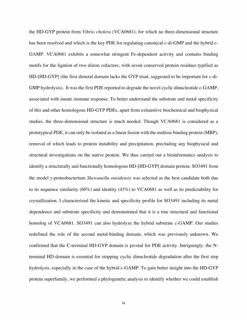
Abstract
Evolutional and functional diversification of HD-GYP phosphodiesterases; metal content and substrate specificity
A thesis presented to the Department of Biochemistry
Graduate School of Arts and Sciences
Brandeis University
Waltham, Massachusetts
By Sining Sun
Cyclic dinucleotides (CDNs) act as intracellular messengers and modulate many cellular
activities including innate immune activation, virulence factor production and biofilm formation
in bacterial pathogens. The cellular levels of CDNs are fine-tuned by cyclases (involved in their
synthesis) and phosphodiesterases (PDEs) (involved in their degradation). HD-GYP proteins
represent a relative novel addition to the PDE functional superfamily, and belong to a large
superfamily of proteins annotated as HD-domain, designated by the tandem histidine-aspartate as
a core structural motif. In this study, we aimed to determine the metal cofactor specificity of HD-
GYP PDEs and catalytic proficiencies regarding different substrates and their abilities to perform
one-step (to 5’-pNpN) or two-step hydrolysis (to two molecules of NMP). We initially focused on
iv
the HD-GYP protein from Vibrio cholera (VCA0681), for which no three-dimensional structure
has been resolved and which is the key PDE for regulating canonical c-di-GMP and the hybrid c-
GAMP. VCA0681 exhibits a somewhat stringent Fe-dependent activity and contains binding
motifs for the ligation of two diiron cofactors, with seven conserved protein residues typified as
HD-[HD-GYP] (the first dimetal domain lacks the GYP triad, suggested to be important for c-di-
GMP hydrolysis). It was the first PDE reported to degrade the novel cyclic dinucleotide c-GAMP,
associated with innate immune response. To better understand the substrate and metal specificity
of this and other homologous HD-GYP PDEs, apart from exhaustive biochemical and biophysical
studies, the three-dimensional structure is much needed. Though VCA0681 is considered as a
prototypical PDE, it can only be isolated as a linear fusion with the maltose binding protein (MBP),
removal of which leads to protein instability and precipitation, precluding any biophysical and
structural investigations on the native protein. We thus carried out a bioinformatics analysis to
identify a structurally and functionally homologous HD-[HD-GYP] domain protein. SO3491 from
the model g-proteobacterium Shewanella oneidensis was selected as the best candidate both due
to its sequence similarity (60%) and identity (43%) to VCA0681 as well as its predictability for
crystallization. I characterized the kinetic and specificity profile for SO3491 including its metal
dependence and substrate specificity and demonstrated that it is a true structural and functional
homolog of VCA0681. SO3491 can also hydrolyze the hybrid substrate c-GAMP. Our studies
redefined the role of the second metal-binding domain, which was previously unknown. We
confirmed that the C-terminal HD-GYP domain is pivotal for PDE activity. Intriguingly, the N-
terminal HD-domain is essential for stopping cyclic dinucleotide degradation after the first step
hydrolysis, especially in the case of the hybrid c-GAMP. To gain better insight into the HD-GYP
protein superfamily, we performed a phylogenetic analysis to identify whether we could establish
v
a correlation between metal content, substrate specificity and mode of action for these HD-GYPs,
with the ultimate aim to delineate their multiple occurrences and specific roles within the cell. This
analysis reveals a biochemical and structural diversion of these proteins and provides a good
predictive map for assigning metal-dependence, level of activity and reaction outcome in different
HD-GYPs, allowing for constructing an evolutional diversification profile.
vi
Table of Contents
Abstract .................................................................................................................... iii
List of Figures ........................................................................................................ viii
List of Tables ............................................................................................................ x
1. Introduction ........................................................................................................... 1
1.1 Cyclic dinucleotides (CDNs) ........................................................................... 1
1.2 HD-GYP phosphodiesterases (PDEs) .............................................................. 3
1.3 The Vibrio cholera VCA0681 and Shewanela oneidensis SO3491 ................ 5
1.4 Goal of the thesis ............................................................................................. 7
2. Experimental Procedures ...................................................................................... 7
2.1 Material and methods ...................................................................................... 7
2.2 Plasmid construction, cloning and mutagenesis .............................................. 7
2.3 Protein expression and purification ................................................................. 9
2.4 Cleavage of the SUMO tag by the Ulp1 protease .......................................... 10
2.5 Cleavage of the MBP-tag with the S219V TEV protease ............................. 11
2.6 Ferrozine assay .............................................................................................. 12
2.7 High Performace liquid chromatography (HPLC) ........................................ 12
2.8 Multiple turnover assays ................................................................................ 13
2.9 Determination of steady-state of SO3491 with c-di-GMP and c-GAMP ...... 14

vii
3. Results ................................................................................................................. 18
3.1 Wildtype VCA0681 and SO3491 .................................................................. 19
Redox State Differentiation ................................................................................................... 24
Metal Cofactor Specificity ..................................................................................................... 27
Substrate Specificity .............................................................................................................. 29
Steady-State kinetic parameters for c-di-GMP and c-GAMP ............................................... 31
O2-sensitivity of the ‘active’ FeIIFeII form of the wt MBP-SO3491 ........................................ 32
3.2 Individual HD-domains ................................................................................. 34
3.3 Bioinformatic and phylogenetic analysis ....................................................... 40
4. Discussion ........................................................................................................... 44
5. Future Directions and Experiments .................................................................... 48
Bibliography ........................................................................................................... 49
viii
List of Figures
Figure 1.1. Structure of cyclic dinucleotides. ................................................................................. 2
Figure 1.2. Scheme of c-di-GMP hydrolysis by HD-GYP phosphodiesterases. ............................ 3
Figure 1.3. Crystal structures of HD-GYP protein active sites. ...................................................... 4
Figure 1.4. A homology model of VCA0681 featuring both HD dinuclear centers.. ..................... 6
Figure 3.1 Mössbauer spectra of wt MBP-VCA0681 and wt MBP-SO3491 ............................... 20
Figure 3.2. Mössbauer spectra of the MBP-SO3491 reduced with sodium ascorbate and sodium
dithionite ................................................................................................................................ 22
Figure 3.3. EPR spectra of the different forms of SO3491 and MBP-VCA0681. ........................ 23
Figure 3.4. Activity assays with ascorbate and dithionite reduced MBP-SO3491 ....................... 25
Figure 3.5. Activity assays with wt MBP-SO3491 and wt MBP-VCA0681. ............................... 27
Figure 3.6. Activity assays with Mn-loaded wild-type MBP-SO3491 ......................................... 28
Figure 3.7. Activity assays of wt MBP-SO3491 with different substrates ................................... 30
Figure 3.8. Steady-state kinetic parameters of SO3491 ................................................................ 31
Figure 3.9. Stop-flow experiment set up ....................................................................................... 33
Figure 3.10. Stop-flow UV-VIS spectra ....................................................................................... 34
Figure 3.11. Thermoshift denaturation curves of the different SO3491 constructs. ..................... 36
ix
Figure 3.12. HPLC chromatograms of the activity assays with wt, D75A MBP-VCA0681 and wt,
D69A MBP-SO3491 ............................................................................................................. 37
Figure 3.13. HPLC chromatograms of the activity assays with D75A MBP-VCA0681 and c-
GAMP .................................................................................................................................... 38
Figure 3.14. UV-Visible spectra of the different wt and variant SO3491 constructs in their
aerobically isolated state ........................................................................................................ 39
Figure 3.15. Maximum-likelihood rooted phylogenetic tree ........................................................ 41
Figure 3.16. Unrooted phylogenetic tree ...................................................................................... 41
Figure 3.17. Sequence alignment of PA4781 and tri-nuclear HD-GYP Proteins ......................... 43
x
List of Tables
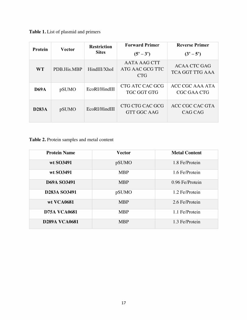
Table 1. List of plasmid and primers ............................................................................................ 17
Table 2. Protein samples and metal content .................................................................................. 17
Table 3. Kinetic parameters of cyclic dinucleotide specific phosphodiesterases belonging to the
HD-GYP and the EAL subtype. ............................................................................................ 32
1
Chapter 1
Introduction
Cyclic dinucleotides (CDNs)
Cyclic dinucleotides (CDNs) act as intracellular messengers and modulate many cellular
processes, including innate immune activation, responses to small molecules (i.e. oxygen, nitric
oxide), virulence factor production, cell motility, and biofilm formation in bacterial pathogens (1-
3). Among all CDNs, c-di-GMP is the most intensively studied and best-understood member of
the cyclic dinucleotide family of second messengers. It is an important bacterial signaling molecule
involved in virulence response and biofilm formation. The biofilm formation is linked with several
chronic bacterial infections, such as bloodstream infections and urinary tract infections (4, 5).
Some recent studies have revealed that biofilm formation plays an important role in wound
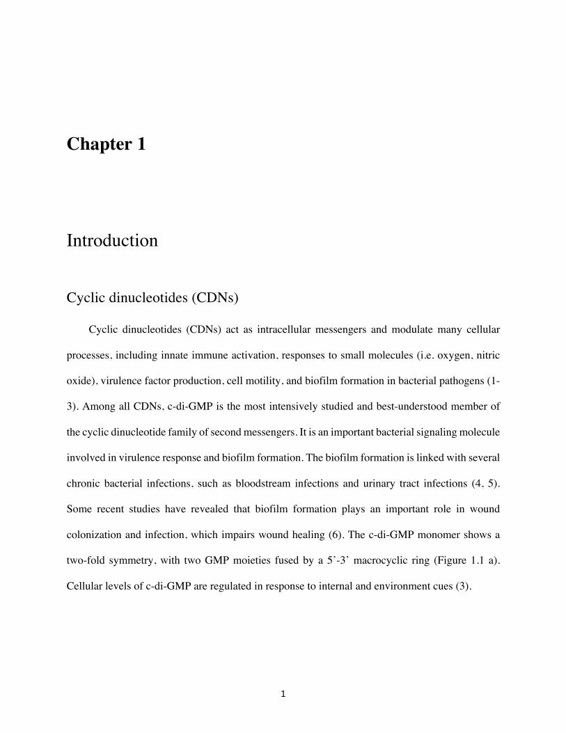
colonization and infection, which impairs wound healing (6). The c-di-GMP monomer shows a
two-fold symmetry, with two GMP moieties fused by a 5’-3’ macrocyclic ring (Figure 1.1 a).
Cellular levels of c-di-GMP are regulated in response to internal and environment cues (3).
2
Recent studies revealed the importance of a novel hybrid cyclic dinucleotide, cyclic-GAMP.
Bacterial c-GAMP exhibits 3’-3’ linkage and mammalian c-GAMP shows a 2’-3’ linkage (Figure
1.1 c & b respectively). 3’3’-GAMP is synthesized by the novel dinucleotide cyclase DncV in
Vibrio cholera. This bacterial c-GAMP was shown to be responsible for efficient intestinal
colonization and chemotaxis regulation in the bacterium V. cholera (7). 2’3’-cGAMP was found
as a product of cGAMP synthase, cGAS, in mammals (8). This mammalian c-GAMP acts as
second messenger to activate the innate immune responses (9, 10).
Figure 1.1. Structure of cyclic dinucleotides. a) 3’5’-c-di-GMP b) 2’3’-c-GAMP c) 3’3’-c-GAMP
The intracellular levels of CDNs are tuned by the concerted (and somewhat opposing)
action of two types of enzymes: a) diguanylate synthases (CDN synthesis from two NTP molecules)
and b) phosphodiesterases (CDN hydrolytic degradation to either the linear 5’phosphoguanylyl-
(3’-5’)-guanosine (5’-pGpG) or GMP)). HD-GYP proteins represent a relatively novel addition to
the PDE functional superfamily, and belong to a large superfamily of proteins annotated as HD-
domain proteins. These HD-domain proteins contain the characteristic tandem histidine-aspartate
structural motif. Here we are interested in the representative HD-GYP proteins that have two
tandem HD binding motifs.
3
HD-GYP phosphodiesterases (PDEs)
HD-GYP consists of a large subfamily of proteins, which belongs to the larger HD-domain
superfamily of metal-dependent hydrolases and oxygenases. HD-GYP proteins can come in a
variety of types and flavors with respect to the domains they are composed of. Some HD-GYP
proteins contain binuclear metal-binding site, while some can bind three metal cofactors at the
active site. Some HD-GYP proteins contain an extra domain that was predicted as an activator of
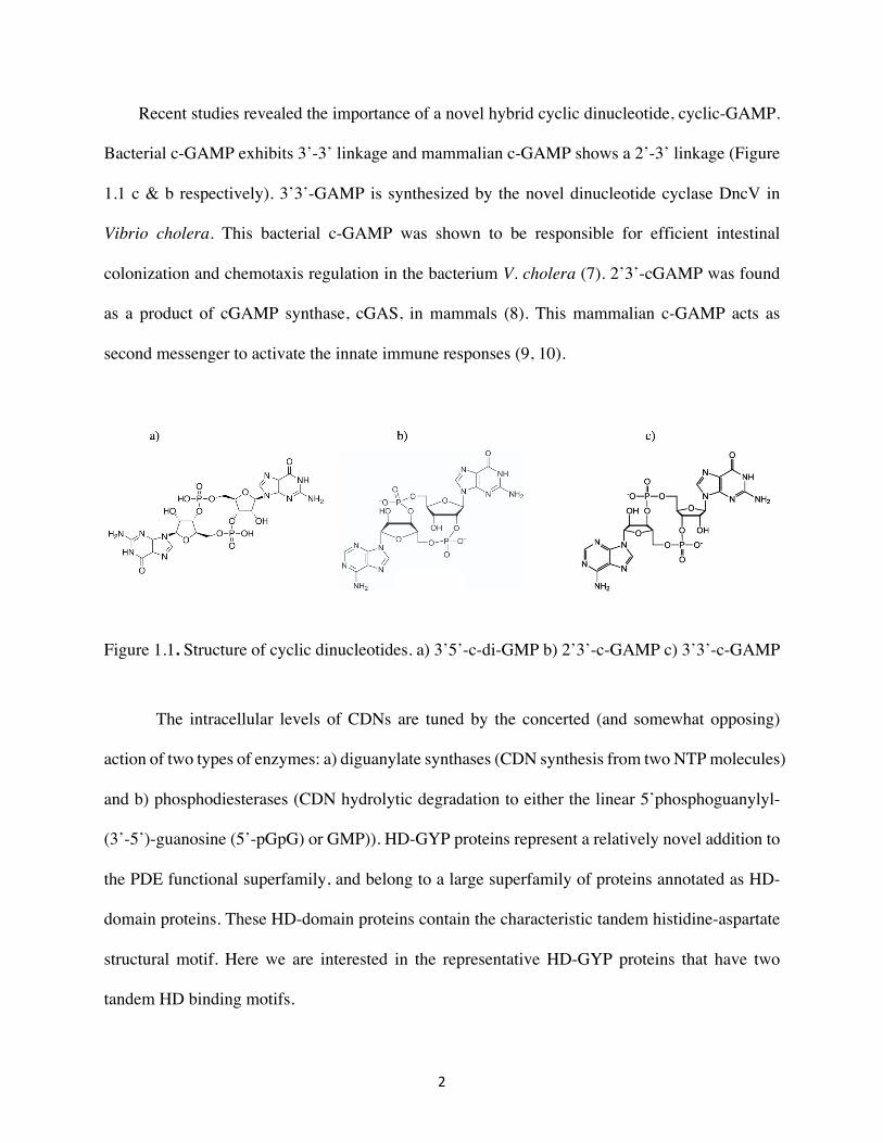
the PDE activity. HD-GYP proteins break down c-di-GMP to GMP via the linearized product 5’-
pGpG, even though in some cases 5’-pGpG was reported as the only detected product (Figure 1.2)
(11, 12)
Figure 1.2. Scheme of c-di-GMP hydrolysis by HD-GYP phosphodiesterases.
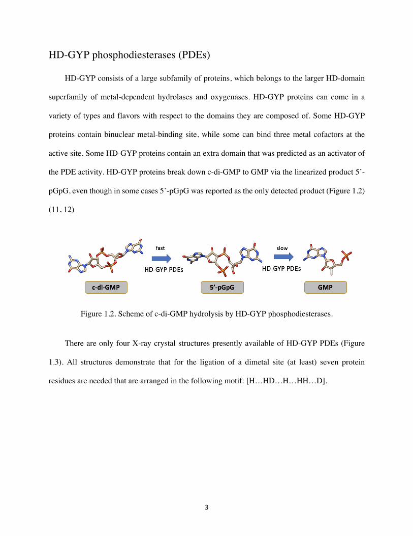
There are only four X-ray crystal structures presently available of HD-GYP PDEs (Figure
1.3). All structures demonstrate that for the ligation of a dimetal site (at least) seven protein
residues are needed that are arranged in the following motif: [H…HD…H…HH…D].
4
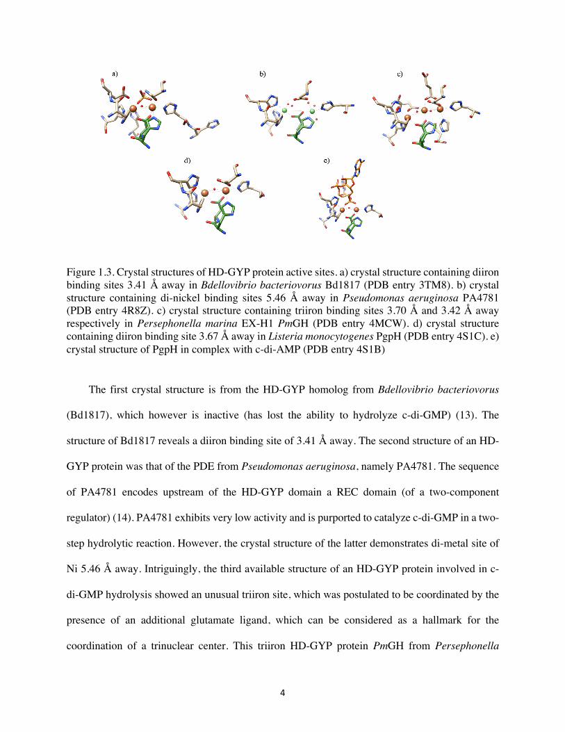
Figure 1.3. Crystal structures of HD-GYP protein active sites. a) crystal structure containing diiron binding sites 3.41 Å away in Bdellovibrio bacteriovorus Bd1817 (PDB entry 3TM8). b) crystal structure containing di-nickel binding sites 5.46 Å away in Pseudomonas aeruginosa PA4781 (PDB entry 4R8Z). c) crystal structure containing triiron binding sites 3.70 Å and 3.42 Å away respectively in Persephonella marina EX-H1 PmGH (PDB entry 4MCW). d) crystal structure containing diiron binding site 3.67 Å away in Listeria monocytogenes PgpH (PDB entry 4S1C). e) crystal structure of PgpH in complex with c-di-AMP (PDB entry 4S1B)
The first crystal structure is from the HD-GYP homolog from Bdellovibrio bacteriovorus
(Bd1817), which however is inactive (has lost the ability to hydrolyze c-di-GMP) (13). The
structure of Bd1817 reveals a diiron binding site of 3.41 Å away. The second structure of an HD-
GYP protein was that of the PDE from Pseudomonas aeruginosa, namely PA4781. The sequence
of PA4781 encodes upstream of the HD-GYP domain a REC domain (of a two-component
regulator) (14). PA4781 exhibits very low activity and is purported to catalyze c-di-GMP in a two-
step hydrolytic reaction. However, the crystal structure of the latter demonstrates di-metal site of
Ni 5.46 Å away. Intriguingly, the third available structure of an HD-GYP protein involved in c-
di-GMP hydrolysis showed an unusual triiron site, which was postulated to be coordinated by the
presence of an additional glutamate ligand, which can be considered as a hallmark for the
coordination of a trinuclear center. This triiron HD-GYP protein PmGH from Persephonella
5
marina EX-H1 showed complete hydrolysis of the c-di-GMP to GMP. The last crystal structure
of an HD-GYP protein is in complex with substrate c-di-AMP. This HD-GYP protein PgpH from
Listeria monocytogenes hydrolyzes c-di-AMP into 5’-pApA (33).
The Vibrio cholera VCA0681 and Shewanela oneidensis SO3491
Vibrio cholera is an aquatic organism that can transiently colonize human small intestine,
which can cause fatal diarrhea to the host. In marine habitats, V. cholera is often found attached to
other organisms in microbial communities called biofilms (15). Understanding the mechanisms
used by V. cholera to control biofilm formation has been an ongoing research. Previous work with
infant mouse model has shown that c-di-GMP inhibits the ability of V. cholera to effectively
colonize the intestine (16).
V. cholera encodes nine HD-GYP proteins that were predicted to be enzymatically active and
four of them were found to be active in vivo (17). VCA0681 has two domains containing two
concerted HD-domain dimetal sites coordinated by seven ligand residues, however only the C-
terminal domain is followed by the GYP sequence triad (considered important for CDN hydrolysis)
(Figure 1.4). Therefore, the VCA0681 is better described as containing a double HD-[HD-GYP]
bimetallic active site. It is considered the most active HD-GYP phosphodiesterase in V. cholera to
degrade c-di-GMP into the linearized product 5’-pGpG (18). Expression of VCA0681 significantly
increased motility of V. cholerae through 0.3% agar medium and decreased biofilm formation in
rich medium on glass (19). It is also the first reported phosphodiesterase that can break down the
novel hybrid cyclic dinucleotide, c-GAMP (2).
6
VCA0681 is very challenging to study biochemically, because it is hardly soluble unless
expressed as a fusion with the maltose-binding protein (MBP) (20). In addition, after proteolytic
cleavage of the solubility tag (MBP), the half-life stability of the protein is of the order of an hour,
precluding any crystallization attempts. We generated a sequence similarity network and
phylogenetic analysis to get a better insight into the HD-GYP superfamily proteins. VCA0681’s
homolog, SO3491, is from the model g-proteobacterium Shewanella oneidensis. We selected this
protein on the basis of the sequence similarity network. SO3491 is 60% sequence similarity and
43% identity with VCA0681. It was also highly predicted for crystallization. Based on the
bioinformatics analysis, SO3491 was chosen as a candidate that would be better suited for further
biochemical and structural characterization.
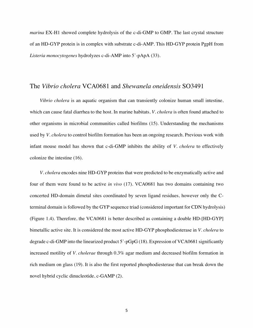
Figure 1.4. A) A homology model of VCA0681 featuring both HD dinuclear centers. The color of each center correlates with the colored text. The GYP-triad is colored in green. B) Schematic construct of VCA0681 regarding two HD domains. C) Sequence alignment of the two individual HD domains in VCA0681, where HD1 represents HD75 (residues 1 – 233) and HD2 represents HD289 (residues 234 – 431). The aligned six residues at the binding sites are boxed in green. The sequence similarity and identity between these two HD domains are 18%, respectively.
7
Goal of the thesis
In my thesis project, the main goal is to understand the chemical nature of the transition
metal content and the two dimetal domains in VCA0681 and its homolog SO3491, and dictate
substrate specificity and reaction outcome (one- vs two-step hydrolysis). As described, we focused
on the VCA0681 homolog SO3491, because it was better suited for us to characterize it both
biochemically and structurally. To better understand the roles of individual HD domain, knockouts
of each HD domain mutants were constructed and assessed for activities. Extensive bioinformatics
and phylogenetic analysis were carried out, in which we collected HD-GYP proteins with scant
biochemical information. The overall aim is to be able to gain insight into the chemical diversity
and evolutionary diversification of the HD-GYP protein superfamily, with respect to the chemical
nature of the cofactor, their respective specific activities and their reaction outcome. The
invaluable information gained can serve as a functional map predictor that will allow portraying
the differences in these proteins and allowing for concluding for their individual roles in the cell.
7
Chapter 2
Experimental Procedures
2.1 Material and methods
All chemicals unless specified were obtained by Fisher Scientific (NH) and were of high purity
grade. c-di-GMP, 5’-pGpG and c-GAMP were initially purchased from Axxora, LLC
(Farmingdale, NY), and GMP was from Acros Organics.In all subsequent experiments, the cyclic
dinucleotides were enzymatically synthesized employing the WspR* and DncV cyclases, as
previously described (21).
2.2 Plasmid construction, cloning and mutagenesis
pSUMO-SO3491
The gene encoding for the HD-GYP phosphodiesterase protein from Shewanella oneidensis was
inserted into the pSUMO expression vector (LifeSensors, Inc) with kanamycin resistance to
express the protein of interest as a linear chimera with the SUMO protein tag. The vector was

8
kindly gifted by Dr. Squire J. Booker (The Pennsylvania State University). This vector encodes
for an N-terminal His6-tag, the small ubiquitin-like modifier protein (SUMO), and a Ulp1
recognition site for the subsequent cleavage of the SUMO-tag from the protein of interest.
MBP-SO3491
The SO3491 encoding gene was inserted with PCR-based cloning using the primers listed in Table
1 via the HindIII and XhoI restriction sites in the PDB.His.MBP (Berkeley Structural Genomics
Center) expression vector that has kanamycin resistance. This vector encodes for an N-terminal
His6-tag, the maltose binding protein (MBP), and TEV recognition site prior to the SO3491 gene,
to allow for the subsequent cleavage of the MBP-tag.
D69A and D283A pSUMO-SO3491
Single-point amino acid substitutions were generated by back-to-back PCR (Q-5, New England
Biolabs, MA) using the primers listed in Table 1.
D69A MBP-SO3491
The D69A SO3491 encoding gene was inserted with PCR-based cloning using the primers listed
in Table 1 via the HindIII and XhoI restriction sites in the PDB.His.MBP (Berkeley Structural
Genomics Center) expression vector that has kanamycin resistance.
WT, D75A and D28A variants of VCA0681
The plasmids expressing for the wild-type and single-point variants of VCA0681 as fusion proteins
with an MBP-tag were kindly gifted by Dr. Donald M. Kurtz (Department of Chemistry,
University of Texas at San Antonio) (9).
9
2.3 Protein expression and purification
All plasmids were transformed in T7 Escherichia (E.) coli express competent cells (New England
Biolabs, MA). Transformed cells were grown in M9 minimal media (M9) to direct specific metal
incorporation with either 200 µM Mn2+ or 250 µM Fe2+ with 50 mg/L kanamycin at 37 °C with
shaking (220 rpm) until OD600 reached a value between 0.6-0.8. Protein expression was then
induced with the addition of 0.25 mM IPTG. Cell cultures were incubated at 18 °C with shaking
(220 rpm) for 16-20 hours. Cultures were centrifuged at 8,000 x g for 20 minutes; cell pellets were
flash frozen in liquid N2 and stored at -80 °C. Cell pellets were resuspended in the lysis buffer (50
mM HEPES, 300 mM NaCl and 10 mM imidazole, pH 7.5). The suspension was lysed using a
QSonica sonicator (Newton, CT) (~ 30 min per 100 mL suspension) with the addition of 250 µM
ammonium iron sulfate and 0.045 mg/mL PMSF. Another 0.045 mg/mL PMSF was added after
sonication. Lysed cells were centrifuged at 22,000 x g for 30 minutes. The supernatant was loaded
onto a Ni2+-NTA immobilized affinity chromatography column (~100 mL resin per 500 mL lysate)
equilibrated with the lysis buffer. The column was washed first with lysis buffer and then with
buffer A (50 mM HEPES, 20 mM imidazole and 200 mM NaCl, pH 7.5). The protein was eluted
by washing with buffer B (50 mM HEPES, 150 mM NaCl and 300 mM imidazole, pH 7.5).
Fractions containing the protein were pooled and concentrated at 3,500 x g using a 30 K Amicon
Ultra-15 Centrifugal Filter Unit (Tullagreen, Carrigtwohill Co. Cork, Ireland). The concentrated
protein was then re-buffered in the storage buffer (50 mM HEPES, 300 mM NaCl and 10%, pH
7.5) using the same centrifugal device to remove imidazole. Protein for crystallography was further
purified by size exclusion chromatography using a HiLoadTM 16/600 SuperdexTM 200 pg column
equilibrated with the storage buffer. Fractions containing the purified protein were pooled and
concentrated at 3,500 x g using a 30 K Amicon Centrifugal Filter. The protein was flash frozen in
10
liquid N2 and stored at -80 °C. Protein purity was estimated by SDS-PAGE with Coomassie
staining, and protein concentration was determined by using a molar absorption coefficient of
48,820 M-1•cm-1 at 280 nm (https://web.expasy.org/protparam/). The iron content was determined
by the ferrozine assay.
2.4 Cleavage of the SUMO tag by the Ulp1 protease
The ubiquitin-like protease 1 (Ulp1), recognizes the three-dimensional structure of the SUMO
domain and cleaves after di-glycine at the C-terminus. For the protease cleavage reactions, Ulp1
(~1 µg per mg of pSUMO-SO_3491) was incubated with SUMO-SO_3491 for 16 to 20 hours on
ice. The reaction mixture was then loaded onto a Ni2+-NTA immobilized affinity chromatography
column (~100 mL resin per 500 mL lysate) equilibrated with the lysis buffer. The cleaved proteins
were eluted by washing the column with the lysis buffer. The SUMO-tag, Ulp1 and uncleaved
pSUMO-SO_3491 (which all contain an N-terminal His6-tag) were then eluted with buffer B.
Fractions containing the untagged (native) SO_3491 protein were pooled and concentrated at 3,500
x g using a 30K Amicon Centrifugal Filter. The concentrated protein was re-buffered in the storage
buffer and concentrated to the desired final concentration. Protein for crystallography was further
purified by size exclusion chromatography using a Sephadex S-200 column equilibrated with 50
mM HEPES Buffer (pH 7.5) containing 300 mM NaCl, 10 mM imidazole and 10% glycerol.
Fractions containing the protein were pooled and concentrated at 3,500 x g using a 30 K Amicon
Centrifugal Filter. The protein was flash frozen in liquid N2 and stored at -80 °C. Protein purity
was estimated by SDS-PAGE with Coomassie staining, and protein concentration was determined
11
by using a molar absorption coefficient of 45,540 M-1•cm-1 at 280 nm. The iron content was
determined by the ferrozine assay.
2.5 Cleavage of the MBP-tag with the S219V TEV protease
The tobacco etch virus (TEV) protease (27 kDa) recognizes the ENLYFGQ sequence and cleaves
between Q and G. The cleavage reactions were initiated by addition of TEV (~ 1 µg per mg of
MBP- SO_3491) and they were carried out for 4 hours on ice. A second addition of the same
amount of TEV is performed and the reactions if further incubated for another 12 to 16 hours on
ice (total ratio of TEV to MBP-SO_3491 was 1:25. The reaction mixture was then loaded on to a
MBP-trap HP prepacked column (GE Healthcare) equilibrated with the storage buffer. The
overnight reaction mixture was loaded onto the column with a flow rate of 0.5 mL/min. Fractions
containing the untagged (native) SO_3491 were collected by washing the column with the storage
buffer and concentrated using a 30 K Amicon Centrifugal Filter. TEV and uncleaved MBP-
SO_3491 (both of which contain an N-terminal His6-tag) were eluted by washing the column with
elution buffer containing 50 mM HEPES, 150 mM NaCl, 10mM Maltose and 10% glycerol (pH
7.5). Protein purity was estimated by SDS-PAGE with Coomassie staining. Due to the low binding
affinity of the MBP-tag of the fusion protein, the first purification could not provide pure cleaved
SO_3491. The collected fractions were further purified by size exclusion chromatography using a
Sephadex S-200 column equilibrated with the storage buffer. Fractions containing the cleaved
protein were pooled and concentrated at 3,500 x g using a 30K Amicon Centrifugal Filter. Protein
purity was estimated by SDS-PAGE with Coomassie staining again. The concentrated protein
mixture was then purified a second time with MBP-trap HP prepacked column (GE Healthcare).
Protein purity was again estimated by SDS-PAGE with Coomassie staining.
12
2.6 Ferrozine assay
The Fe content was determined by ferrozine assay following the procedures of Bollinger (Bollinger,
1993).35
The protein was diluted to 150 µM with water to a final volume of 83.3 µL. After the addition of
16.7 µL of 50% TCA, the sample was spun at 17,000 x g for 2 min. The volume of the supernatant
was carefully measured and the supernatant was mixed with 20 µL 75 mM ascorbic acid, 20 µL
10 mM ferrozine, 120 µL saturated NH4OAc and water to make a 660 µL solution. To make a
control sample, 500µL water was used without any protein. The Fe concentration was estimated
on the basis of the absorbance at 562 nm, corresponding to the Fe2+-ferrozine chromophore.
2.7 High Performace liquid chromatography (HPLC)
Denatured reaction samples were centrifuged at 21,130 x g for 2 minutes and the supernatant were
filtered with 0.22 µm nylon Spin-X Centrifuge Tube Filter (Corning Incorporated, Corning, NY).
The filtered samples were then transferred into HPLC tubes analyzed on a 6125B Agilent
spectrometer equipped with a 1260 Infinity Liquid Chromatography system and a 1260 Infinity
Photodiode Array Detector WR. Samples were injected on an Agilent reverse-phase C18-A Polaris
column (particle size 5 µm, 150 x 4.6 mm) and educts and products were separated using a gradient
method utilizing a water-based mobile phase (10 mM KH2PO4 and 10 mM TBAH, pH 6, Solvent
A) and an organic-based mobile phase (methanol with 10 mM TBAH, Solvent B). Substrates and
products were eluted from the column with a gradient of 95% solvent A and 5% solvent B to 50%
13
solvent A and 50% solvent B at a flow rate of 1.5 mL/min for 25 min. Comparison of integrated
peak intensities to that of internal standards (substrates and products) of known concentration
enabled quantification of the analyses. Nucleotides were detected at a wavelength of 254 nm.
2.8 Multiple turnover assays
End-point reactions to test for activity of the wild-type and variant HD-GYP domain proteins with
substrates and substrate analogues contained final concentrations of 1 µM (di-iron concentration)
wildtype and knockout-construct SO_3491 and VCA0681 for c-di-GMP reactions and 5 µM (di-
iron concentration) for c-GAMP reactions,100 µM MgCl2, 200 µM sodium dithionite and 50 µM
substrate in 100 mM HEPES buffer (pH 8) containing 150 mM NaCl and 10% glycerol. Reactions
examining the substrate specificity of the HD-GYP proteins were carried out with 5 µM (di-iron
concentration) SO_3491, 100 µM MgCl2, 200 µM sodium dithionite and 50 µM of substrates (c-
diAMP, 5’pGpG, 5’pApG and 3’5’cGMP) in 100 mM HEPES buffer (pH 8) containing 150 mM
NaCl and 10% glycerol. All reaction components (O2-free) were mixed in an anoxic chamber
(CoyLab). Reaction buffer, MgCl2, enzyme and sodium dithionite (50-fold excess with respect to
cofactor concentration) were incubated for 20 minutes before the addition of substrate to allow full
reduction of the metal cofactor to the diferrous form (as confirmed in 57Fe-labeled samples
examined by Moessbauer spectroscopy, which confirm that the cofactor is in the Fe2II/II form). All
reactions were initiated with the addition of substrate or substrate analogs and terminated at the
described time-points by quenching at 95 °C for 10 min. The efficiency of heat quenching for rapid
inactivating the enzymes at the desired time-points, were confirmed by comparison of quenching
of the reactions in 2 M acetic acid.
14
2.9 Determination of steady-state parameters of SO3491 with c-di-GMP
and c-GAMP
Reactions to test for activity of the native SO_3491 with c-di-GMP contained final concentration
of 0.4 µM (Fe2II/II concentration) wild-type untagged SO_3491 and varying concentrations of c-di-
GMP (5 µM, 10 µM, 20 µM, 60 µM, 120 µM and 240 µM). The reaction mixture additionally
contained 100 µM MgCl2, 200 µM sodium dithionite and 50 µM substrate in 100 mM HEPES
buffer (pH 8) containing 150 mM NaCl and 10% glycerol. All reaction components (O2-free) were
mixed in an anoxic chamber. Reactions were terminated at different time points by denaturing at
95 °C for 10 min. Initial rates of reactions were obtained by fitting concentrations of c-di-GMP
over time with a linear equation. The initial rates for different c-di-GMP concentrations were then
plotted and fitted with an Michaelis-Menten equation to determine to kcat and KM according to the
equation:
v = #$%&∙[*+,-+./0]234[*+,-+./0]
(Eq. 1)
2.10 UV-VIS spectroscopy
UV-visible spectra were recorded on a Cary 60 spectrometer (Varian, Walnut Creek, CA) using
the associated WinUV software package.

15
2.11 Thermofluor assays
The apparent melting temperature values for the HD-GYP proteins in this study were determined
by performing fluorescence thermal shift assays on a StepOne Plus Real Time PCR System
(Company, city). Experiments were carried out in MicroAmp Fast Optical 96-Well plates, in 25
µL reactions including 10x SYPRO fluorescent dye, 10 𝜇M protein in a buffer containing 50
mM HEPES, pH 8.0, and 200 mM NaCl. Reactions were heated from 25 to 95 °C, and
fluorescence intensity (570 nm) was recorded at 0.3 °C temperature increments.
2.12 Mössbauer Spectroscopy
All samples were prepared in storage buffer under oxygen-free conditions in an anaerobic
glovebox (Coylab). The samples were reacted with an excess amount of sodium dithionite (10-20
equivalents) for 20-30 minutes at 22 °C prior to freezing in liquid N2. Mössbauer spectra were
recorded on WEB Research (Edina, MN) instruments that have been described previously (15).
The spectrometer used to acquire the weak-field spectra is equipped with a Janis SVT-400
variable-temperature cryostat. The external magnetic field was applied parallel to the γ beam. All
isomer shifts are quoted relative to the centroid of the spectrum of α -iron metal at room
temperature. Mössbauer spectra were simulated using the WMOSS spectral analysis software
(www.wmoss.org, WEB Research, Edina, MN).
16
2.13 Electron paramagnetic resonance (EPR) spectroscopy
All samples were prepared in storage buffer under oxygen-free conditions in an anaerobic
glovebox (Coy). The samples were reacted with ascorbate sodium salt for 30 minutes at 22 °C
prior freezing in liquid N2. EPR spectra were acquired on a Bruker E500 Elexsys continuous wave
(CW) X-Band spectrometer (operating at approx. 9.38 GHz) equipped with a rectangular resonator
(TE102) and a continuous-flow cryostat (Oxford 910) with a temperature controller (Oxford ITC
503). The spectra were recorded at variable temperatures between 10-40 K at a microwave power
of 0.2 mW, using a modulation amplitude of 1 mT, a microwave frequency of 9.38 GHz.
17
Table 1. List of plasmid and primers
Protein Vector Restriction Sites
Forward Primer
(5’ – 3’)
Reverse Primer
(3’ – 5’)
WT PDB.His.MBP HindIII/XhoI AATA AAG CTT
ATG AAC GCG TTC CTG
ACAA CTC GAG TCA GGT TTG AAA
D69A pSUMO EcoRI/HindIII CTG ATC CAC GCG TGC GGT GTG
ACC CGC AAA ATA CGC GAA CTG
D283A pSUMO EcoRI/HindIII CTG CTG CAC GCG GTT GGC AAG
ACC CGC CAC GTA CAG CAG
Table 2. Protein samples and metal content
Protein Name Vector Metal Content
wt SO3491 pSUMO 1.8 Fe/Protein
wt SO3491 MBP 1.6 Fe/Protein
D69A SO3491 MBP 0.96 Fe/Protein
D283A SO3491 pSUMO 1.2 Fe/Protein
wt VCA0681 MBP 2.6 Fe/Protein
D75A VCA0681 MBP 1.1 Fe/Protein
D289A VCA0681 MBP 1.3 Fe/Protein
18
Chapter 3
Results
The non-heme carboxylate-bridged diiron cofactor in HD-domain proteins can attain three
overall redox states, differing in the level of reduction, namely FeIIIFeIII (diferric), FeIIFeIII (mixed-
valent) and FeIIFeII (diferrous). These three redox states have been demonstrated for HD-domain
oxygenases (i.e. myo-inositol oxygenase- MIOX, and the organophosphonate degrading PhnZ),
for which the mixed-valent (MV) form of the cofactor can be accumulated to ~60-70%. This is in
contrast to canonical diiron oxygenases and oxidases of the ferritin-like superfamily, which do not
detectably accumulate the MV form. This discrepancy can be rationalized by the fact that ferritin-
like non-heme dinuclear sites activate their substrates using O2 from the diferrous manifold,
whereas HD-domain oxygenases catalyze their four-electron oxidative conversions from the MV
manifold. Diiron hydrolases are not that common, but it has been proposed that the hydrolysis
reaction occurs at the FeIIFeII level of the cofactor. It is presently not well understood whether HD-
domain hydrolytic enzymes can accumulate the FeIIFeIII form to significant yields and whether the
MV form is potent towards hydrolysis.
VCA0681 has an unconventional arrangement, harboring two diiron sites, and has been
shown to be strictly Fe-dependent (no other metal ions could stimulate activity, with the exception
19
perhaps of Mn2+, which allowed a detectable but otherwise very sluggish activity to be considered
relevant/physiological) (12). Only the C-terminal diiron site is an active PDE domain (on the basis
of knock-out variants of VCA0681) (12), whereas the role/function of the other bimetallic site is
presently unknown.
3.1 Wildtype VCA0681 and SO3491
We proceeded to study the active-sites of wt MBP-VCA0681 and wt MBP-SO3491 by
Mössbauer and EPR spectroscopies, so as to 1) characterize the individual forms of the enzymes
we would carry out activity assays with; 2) to ensure that the Fe in the sample is assembled
exclusively in bimetallic sites and does not exists in monomeric species (which would not support
activity); 3) and lastly quantify the extent of accumulation of the FeIIFeIII and compare it to that
stabilized in HD-domain oxygenases (i.e. PhnZ) (22).
20
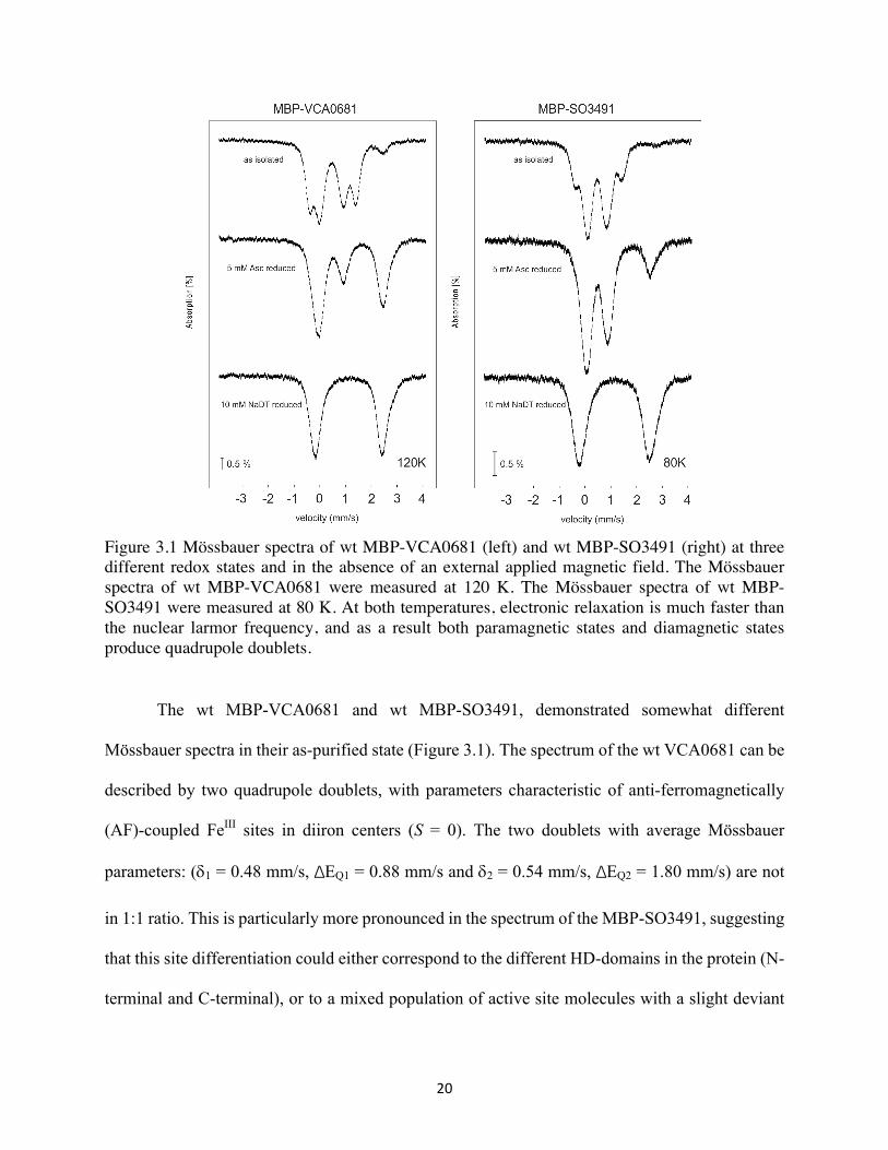
Figure 3.1 Mössbauer spectra of wt MBP-VCA0681 (left) and wt MBP-SO3491 (right) at three different redox states and in the absence of an external applied magnetic field. The Mössbauer spectra of wt MBP-VCA0681 were measured at 120 K. The Mössbauer spectra of wt MBP-SO3491 were measured at 80 K. At both temperatures, electronic relaxation is much faster than the nuclear larmor frequency, and as a result both paramagnetic states and diamagnetic states produce quadrupole doublets.
The wt MBP-VCA0681 and wt MBP-SO3491, demonstrated somewhat different
Mössbauer spectra in their as-purified state (Figure 3.1). The spectrum of the wt VCA0681 can be
described by two quadrupole doublets, with parameters characteristic of anti-ferromagnetically
(AF)-coupled FeIII sites in diiron centers (S = 0). The two doublets with average Mössbauer
parameters: (d1 = 0.48 mm/s, ΔEQ1 = 0.88 mm/s and d2 = 0.54 mm/s, ΔEQ2 = 1.80 mm/s) are not
in 1:1 ratio. This is particularly more pronounced in the spectrum of the MBP-SO3491, suggesting
that this site differentiation could either correspond to the different HD-domains in the protein (N-
terminal and C-terminal), or to a mixed population of active site molecules with a slight deviant
21
ligation or the presence of a small-ligand bound to the active site. The two last hypotheses, are
perhaps more plausible when compared to the Mössbauer data of another, as-purified HD-GYP
that harbors only one diiron site (e.g. VCA0931, results not shown). Different levels of reduction
were achieved by reducing either with sodium ascorbate (weak reducing agent) or sodium
dithionite (strong reducing agent). Addition of excess of sodium ascorbate (5 mM) under O2-free
conditions produced also different extent of reduction both for VCA0681 and SO3491 (Figure 3.1).
Quantification of the respective redox states yields for the VCA0681: 10% FeIIIFeIII (all-ferric),
45% FeIIFeIII (MV), and 45% of FeIIFeII (all-ferrous), while for the SO3491 the distribution is
somewhat different with: 65% FeIIIFeIII (all-ferric), 16% FeIIFeIII (MV), and 19% of FeIIFeII.(all-
ferrous), respectively. This is in contrast to oxygenases, for which under these mild reducing
conditions almost no diferrous form accumulates (< 4 %) and the majority of the sample is in the
MV form (S = 1/2, ~70%) and the rest in a diferric form (S = 0, ~30%) (23). This result
demonstrates that the two redox couples, are now ‘closer’ to each other and that there is an upshift
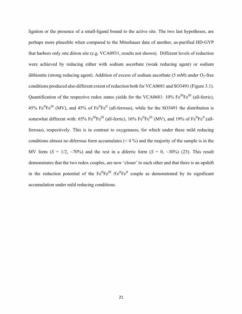
in the reduction potential of the FeIIFeIII /FeIIFeII couple as demonstrated by its significant
accumulation under mild reducing conditions.
22
Figure 3.2. Mössbauer spectra of the MBP-SO3491 reduced with 5 mM sodium ascorbate (left) and 10 mM sodium dithionite (right). Doublets colored in varying colors correspond to the relative amount of the iron in each redox state. Left: sodium ascorbate reduced MBP-SO3491 gave 16% MV form, 19% diferrous form and the rest in the diferric form. The green shade corresponds the FeII amount complexed in the FeIIFeIII state, the blue doublet to the Fe complexed in the FeIIFeII state, the red shade to the FeIIIFeIII form, and the orange shade to the FeIII and is part of the FeIIFeIII state. Right: sodium dithionite reduced MBP-SO3491 gave 100% diferrous form. The spectrum was fitted with two quadrupole doublets corresponding to two FeIIFeII species. The spectra were recorded at 80 K and in the absence of an applied magnetic field.
These two combined observations are in contrast to those observed for HD-domain
oxygenases, for which the yield of the FeIIFeIII form is the same irrespective of the excess sodium
ascorbate level. It has to be noted, that we performed reduction with sodium ascorbate under the
exact same conditions with all three proteins (MBP-VCA0681, MBP-SO3491, and the PhnZ
oxygenase). The rationale for using a limiting amount of reductant was to be able to accumulate
FeIIFeIII and not as much (if at all) FeIIFeII, so that we could carry out activity assays to demonstrate
whether the mixed-valent form is active. On the other hand, reduction of both wt MBP-VCA0681
and MBP-SO3491 with excess of sodium dithionite under O2-free conditions, leads to 100%
reduction of the diiron sites to the diferrous state (Figure 3.2).
23
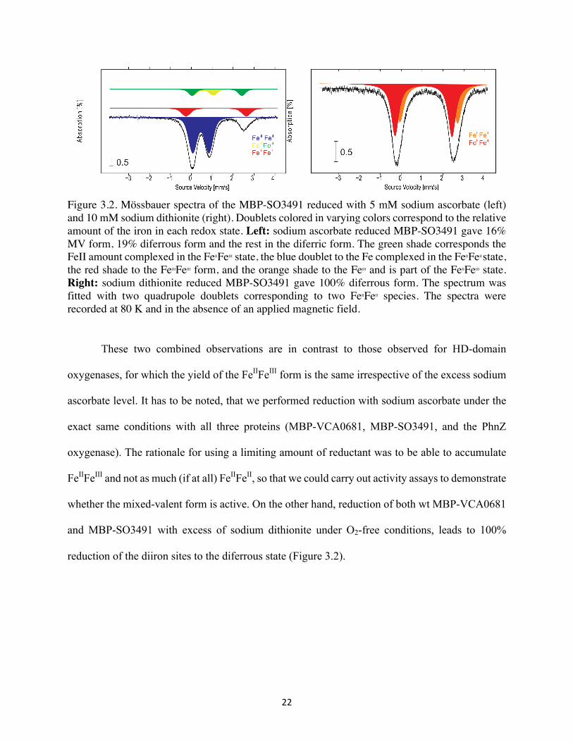
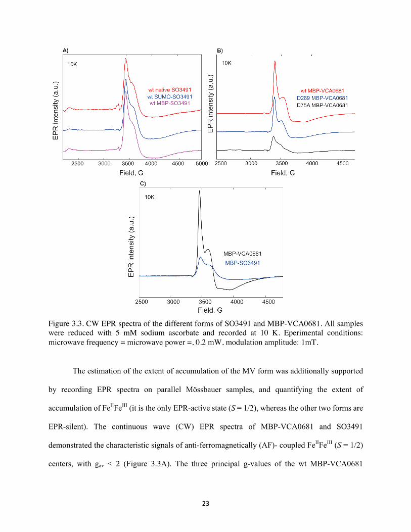
Figure 3.3. CW EPR spectra of the different forms of SO3491 and MBP-VCA0681. All samples were reduced with 5 mM sodium ascorbate and recorded at 10 K. Eperimental conditions: microwave frequency = microwave power =, 0.2 mW, modulation amplitude: 1mT.
The estimation of the extent of accumulation of the MV form was additionally supported
by recording EPR spectra on parallel Mössbauer samples, and quantifying the extent of
accumulation of FeIIFeIII (it is the only EPR-active state (S = 1/2), whereas the other two forms are
EPR-silent). The continuous wave (CW) EPR spectra of MBP-VCA0681 and SO3491
demonstrated the characteristic signals of anti-ferromagnetically (AF)- coupled FeIIFeIII (S = 1/2)
centers, with gav < 2 (Figure 3.3A). The three principal g-values of the wt MBP-VCA0681
24
spectrum are 1.94, 1.82 and 1.7 and those of MBP-SO3491 are 1.93, 1.8, 1.65, respectively. The
slight shifts in the g-values and the broadness of the spectra are representative of slight differences
in the local active-site protein environment and/or structural heterogeneity of their active centers.
The wt SUMO-SO3491, MBP-SO3491 and native (“untagged”) SO3491 all demonstrate the same
features in their EPR spectra (Figure 3.3B). We conclude that the tag does not alter the properties
or the local environment surrounding the two bimetallic sites. The intensity of SUMO-SO3491
were significantly smaller than that of MBP-VCA0681, which is consistent with the Mössbauer
quantification. MBP-VCA0681 accumulated more FeIIFeIII than MBP-SO349 when reduced under
same conditions (Figure 3.3C).
Redox State Differentiation
After establishing the distribution of the respective diiron redox states in the samples, we
proceeded to examine whether the MV form is also active towards c-di-GMP hydrolysis. We thus
performed activity assays under conditions of the ‘same’ cofactor concentration, on the basis of
both the EPR and Mössbauer quantifications. Our activity assays were performed with sodium
ascorbate reduced and sodium dithionite reduced proteins and with c-di-GMP as a substrate in a
time-dependent manner. Both protein samples showed accumulation of the linearized product 5’-
pGpG over time (Figure 3.4A, B). The peak areas of c-di-GMP and 5’-pGpG were obtain after
integration of the HPLC elution profile (Figure 3.4C, D). On the basis of the EPR and Mössbauer
quantifications, there is ~1 µM Fe FeIIFeIII and ~1.2 µM FeIIFeII present in the sodium ascorbate
reduced sample. If both forms (MV and diferrous) of MBP-SO3491 are active, the effect on
product formation would be additive. However, the yield of product (5’-pGpG) by the ascorbate
reduced MBP-SO3491 is comparable to that obtained in the case of the dithionite reduced enzyme
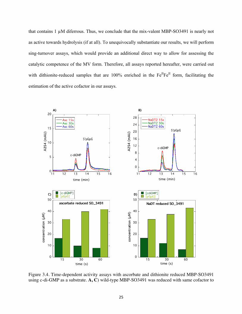
25
that contains 1 µM diferrous. Thus, we conclude that the mix-valent MBP-SO3491 is nearly not
as active towards hydrolysis (if at all). To unequivocally substantiate our results, we will perform
sing-turnover assays, which would provide an additional direct way to allow for assessing the
catalytic competence of the MV form. Therefore, all assays reported hereafter, were carried out
with dithionite-reduced samples that are 100% enriched in the FeIIFeII form, facilitating the
estimation of the active cofactor in our assays.
Figure 3.4. Time-dependent activity assays with ascorbate and dithionite reduced MBP-SO3491 using c-di-GMP as a substrate. A, C) wild-type MBP-SO3491 was reduced with same cofactor to
26
ascorbate ratio for as the EPR sample of 20 minutes under O2-free conditions. B, D) wild-type MBP-SO3491 was reduced with 200 equivalent cofactor concentrations of NaDT for 20 minutes under anaerobic condition. The reactions were heat-quenched at 15 seconds, 30 seconds and 1 minute inside the glovebox.
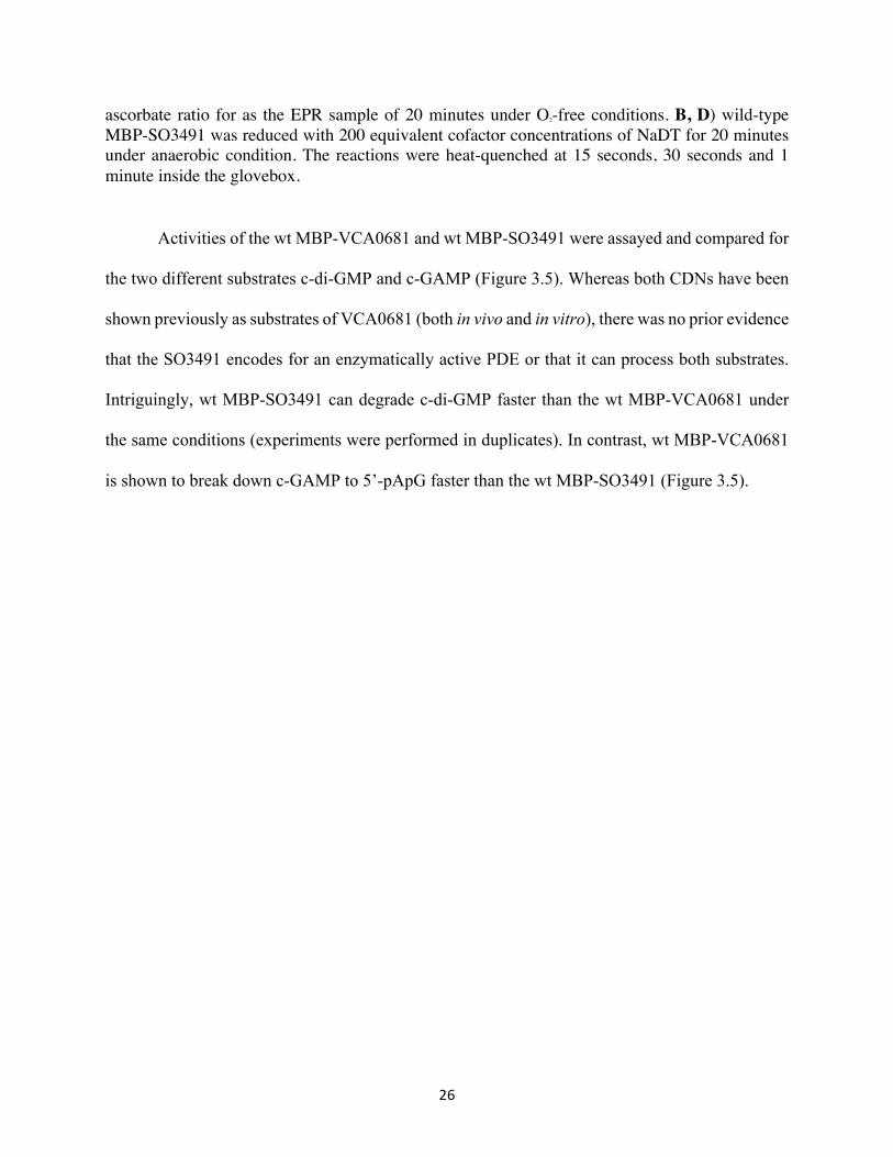
Activities of the wt MBP-VCA0681 and wt MBP-SO3491 were assayed and compared for
the two different substrates c-di-GMP and c-GAMP (Figure 3.5). Whereas both CDNs have been
shown previously as substrates of VCA0681 (both in vivo and in vitro), there was no prior evidence
that the SO3491 encodes for an enzymatically active PDE or that it can process both substrates.
Intriguingly, wt MBP-SO3491 can degrade c-di-GMP faster than the wt MBP-VCA0681 under
the same conditions (experiments were performed in duplicates). In contrast, wt MBP-VCA0681
is shown to break down c-GAMP to 5’-pApG faster than the wt MBP-SO3491 (Figure 3.5).
27
Figure 3.5. Activity assays with NaDT reduced wt MBP-SO3491 and wt MBP-VCA0681 using c-di-GMP and c-GAMP as substrates. A, C) 1 µM (FeIIFeII) wt MBP-SO3491 and wt MBP-VCA0681 were reacted with 50 µM c-di-GMP for 4 minutes, 8 minutes, 1 hour and 2 hours. Dark green bars represent c-di-GMP and light green bars represent 5’-pGpG. B, D) 5 µM (FeIIFeII) wt MBP-SO3491 and wt MBP-VCA0681 were reacted with 50 µM c-GAMP for 10 minutes, 1 hour and 2 hours, respectively. Orange bars represent c-GAMP and yellow bars represent 5’-pApG. All assays were carried out at room temperature under O2-free conditions in an anaerobic glovebox.
Metal Cofactor Specificity
Manganese was another potential metal ion that could yield detectable yet very slow PDE
activity (12). Here we expressed and purified MBP-SO3491 with controlled manganese

28
supplementation in order to examine the PDE activity of the Mn-loaded MBP-SO3491. The
manganese content of Mn-MBP-SO3491 could not yet precisely been determined (the ICP-AES
results are pending). All experiments with the Mn-MBP-SO3491 activities were carried out
assuming 2 Mn/protein on the basis of the analogous Fe content with Fe-MBP-SO3491. To avoid
any contaminating activity due to the residual Fe co-purified with the Mn-MBP-SO3491 proteins
(0.45 Fe/protein), we performed the assays under aerobic conditions, with samples that were prior
incubated with 1 equivalent amount (with respect to cofactor concentration) of FeKCN6 for 20
minutes before addition of the substrate. FeKCN6 will fully oxidize any residual reduced forms of
the cofactor to the diferric form, which is inactive. Figure 3.6 shows the activities of MBP-SO3491
with different metal cofactors and their abilities to use c-di-GMP and c-GAMP as substrates in a
time-dependent manner. Although a lot slower than the Fe-loaded MBP-SO3491, Mn-loaded
MBP-SO3491 can also degrade c-di-GMP and c-GAMP into 5’-pGpG and 5’-pApG.
Figure 3.6. Activity assays with Mn-loaded wild-type MBP-SO3491 with c-di-GMP and c-GAMP as substrates. A) 1 µM (dimanganese) wt MBP-SO3491 were reacted with 50 µM c-di-GMP for 4 minutes, 8 minutes, 1 hour and 2 hours. Dark green bars represent c-di-GMP and light green bars represent the 5’-pGpG product. B) 5 µM (dimanganese) wt MBP-SO3491 were reacted with 50 µM c-GAMP for 10 minutes, 1 hour and 2 hours. Orange bars represent c-GAMP and yellow bars
29
represent the 5’-pApG product. These assays were performed at room temperature, under air and in the presence of FeKCN6.
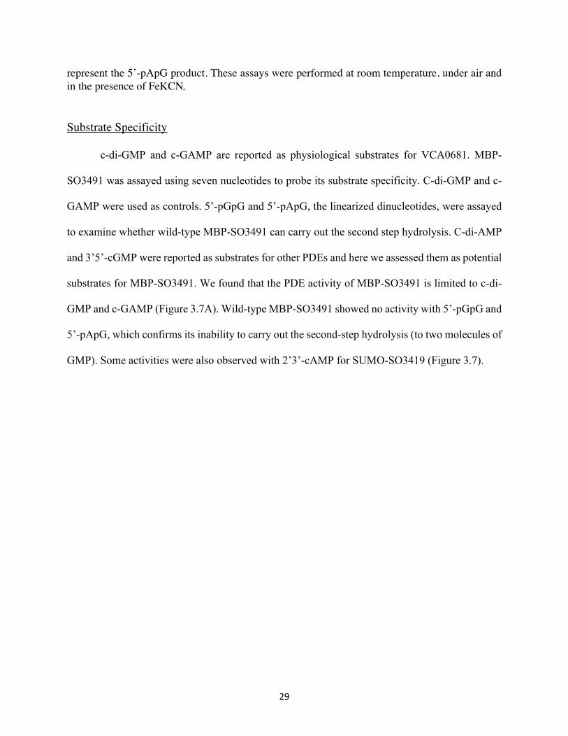
Substrate Specificity
c-di-GMP and c-GAMP are reported as physiological substrates for VCA0681. MBP-
SO3491 was assayed using seven nucleotides to probe its substrate specificity. C-di-GMP and c-
GAMP were used as controls. 5’-pGpG and 5’-pApG, the linearized dinucleotides, were assayed
to examine whether wild-type MBP-SO3491 can carry out the second step hydrolysis. C-di-AMP
and 3’5’-cGMP were reported as substrates for other PDEs and here we assessed them as potential
substrates for MBP-SO3491. We found that the PDE activity of MBP-SO3491 is limited to c-di-
GMP and c-GAMP (Figure 3.7A). Wild-type MBP-SO3491 showed no activity with 5’-pGpG and
5’-pApG, which confirms its inability to carry out the second-step hydrolysis (to two molecules of
GMP). Some activities were also observed with 2’3’-cAMP for SUMO-SO3419 (Figure 3.7).

30
Figure 3.7. A) Activity assays of wt MBP-SO3491 with different substrates (the c-di-AMP result not shown here). All activities were performed with 5 µM metal cofactor and 50 µM substrate analogs. The reactions were heat quenched after 1 hour. B) Activity assay of the wt SUMO-SO3491 (5 µM metal cofactor) with 50 µM 2’3’-cAMP. The reaction was heat-quenched after 1 hour.
31
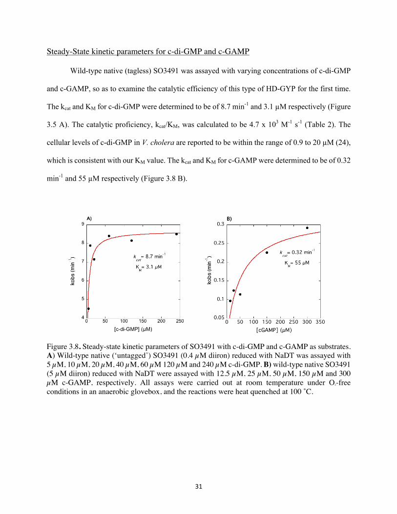
Steady-State kinetic parameters for c-di-GMP and c-GAMP
Wild-type native (tagless) SO3491 was assayed with varying concentrations of c-di-GMP
and c-GAMP, so as to examine the catalytic efficiency of this type of HD-GYP for the first time.
The kcat and KM for c-di-GMP were determined to be of 8.7 min-1 and 3.1 µM respectively (Figure
3.5 A). The catalytic proficiency, kcat/KM, was calculated to be 4.7 x 10³ M-1 s-1 (Table 2). The
cellular levels of c-di-GMP in V. cholera are reported to be within the range of 0.9 to 20 µM (24),
which is consistent with our KM value. The kcat and KM for c-GAMP were determined to be of 0.32
min-1 and 55 µM respectively (Figure 3.8 B).
Figure 3.8. Steady-state kinetic parameters of SO3491 with c-di-GMP and c-GAMP as substrates. A) Wild-type native (‘untagged’) SO3491 (0.4 µM diiron) reduced with NaDT was assayed with 5 µM, 10 µM, 20 µM, 40 µM, 60 µM 120 µM and 240 µM c-di-GMP. B) wild-type native SO3491 (5 µM diiron) reduced with NaDT were assayed with 12.5 µM, 25 µM, 50 µM, 150 µM and 300 µM c-GAMP, respectively. All assays were carried out at room temperature under O2-free conditions in an anaerobic glovebox, and the reactions were heat quenched at 100 ˚C.
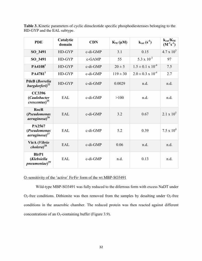
32
Table 3. Kinetic parameters of cyclic dinucleotide specific phosphodiesterases belonging to the HD-GYP and the EAL subtype.
PDE Catalytic domain CDN KM (µM) kcat (s-1) kcat/KM
(M-1s-1)
SO_3491 HD-GYP c-di-GMP 3.1 0.15 4.7 x 10³
SO_3491 HD-GYP c-GAMP 55 5.3 x 10⁻³ 97
PA41081 HD-GYP c-di-GMP 20 ± 5 1.5 ± 0.1 x 10⁻⁴ 7.5
PA47811 HD-GYP c-di-GMP 119 ± 30 2.0 ± 0.3 x 10⁻⁴ 2.7
PdeB (Borrelia burgdorferi)22 HD-GYP c-di-GMP 0.0029 n.d. n.d.
CC3396 (Caulobacter crescentus)25
EAL c-di-GMP >100 n.d. n.d.
RocR (Pseudomonas aeruginosa)26
EAL c-di-GMP 3.2 0.67 2.1 x 10⁵
PA2567 (Pseudomonas aeruginosa)27
EAL c-di-GMP 5.2 0.39 7.5 x 10⁴
VieA (Vibrio cholera)28 EAL c-di-GMP 0.06 n.d. n.d.
BlrP1 (Klebsiella
pneumoniae)29 EAL c-di-GMP n.d. 0.13 n.d.
O2-sensitivity of the ‘active’ FeIIFeII form of the wt MBP-SO3491
Wild-type MBP-SO3491 was fully reduced to the diferrous form with excess NaDT under
O2-free conditions. Dithionite was then removed from the samples by desalting under O2-free
conditions in the anaerobic chamber. The reduced protein was then reacted against different
concentrations of an O2-containing buffer (Figure 3.9).
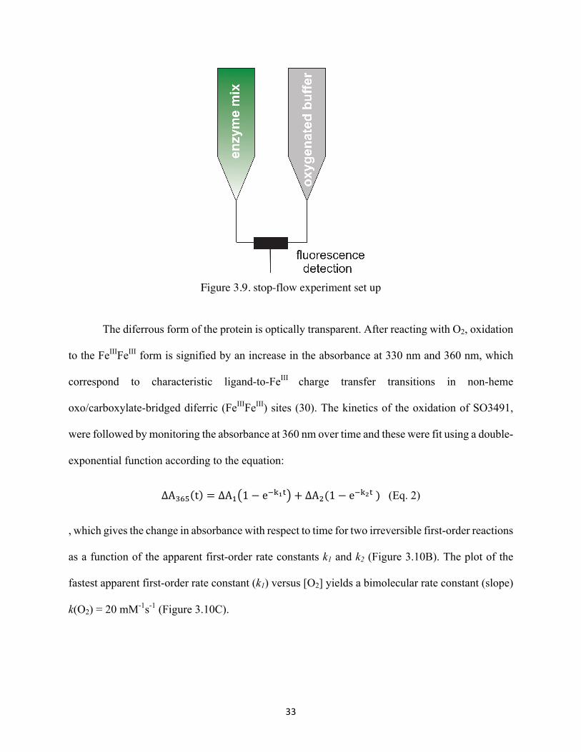
33
Figure 3.9. stop-flow experiment set up
The diferrous form of the protein is optically transparent. After reacting with O2, oxidation
to the FeIIIFeIII form is signified by an increase in the absorbance at 330 nm and 360 nm, which
correspond to characteristic ligand-to-FeIII charge transfer transitions in non-heme
oxo/carboxylate-bridged diferric (FeIIIFeIII) sites (30). The kinetics of the oxidation of SO3491,
were followed by monitoring the absorbance at 360 nm over time and these were fit using a double-
exponential function according to the equation:
ΔA89: t = ΔA< 1 − e+@AB + ΔAD(1 − e+@FB) (Eq. 2)
, which gives the change in absorbance with respect to time for two irreversible first-order reactions
as a function of the apparent first-order rate constants k1 and k2 (Figure 3.10B). The plot of the
fastest apparent first-order rate constant (k1) versus [O2] yields a bimolecular rate constant (slope)
k(O2) = 20 mM-1s-1 (Figure 3.10C).
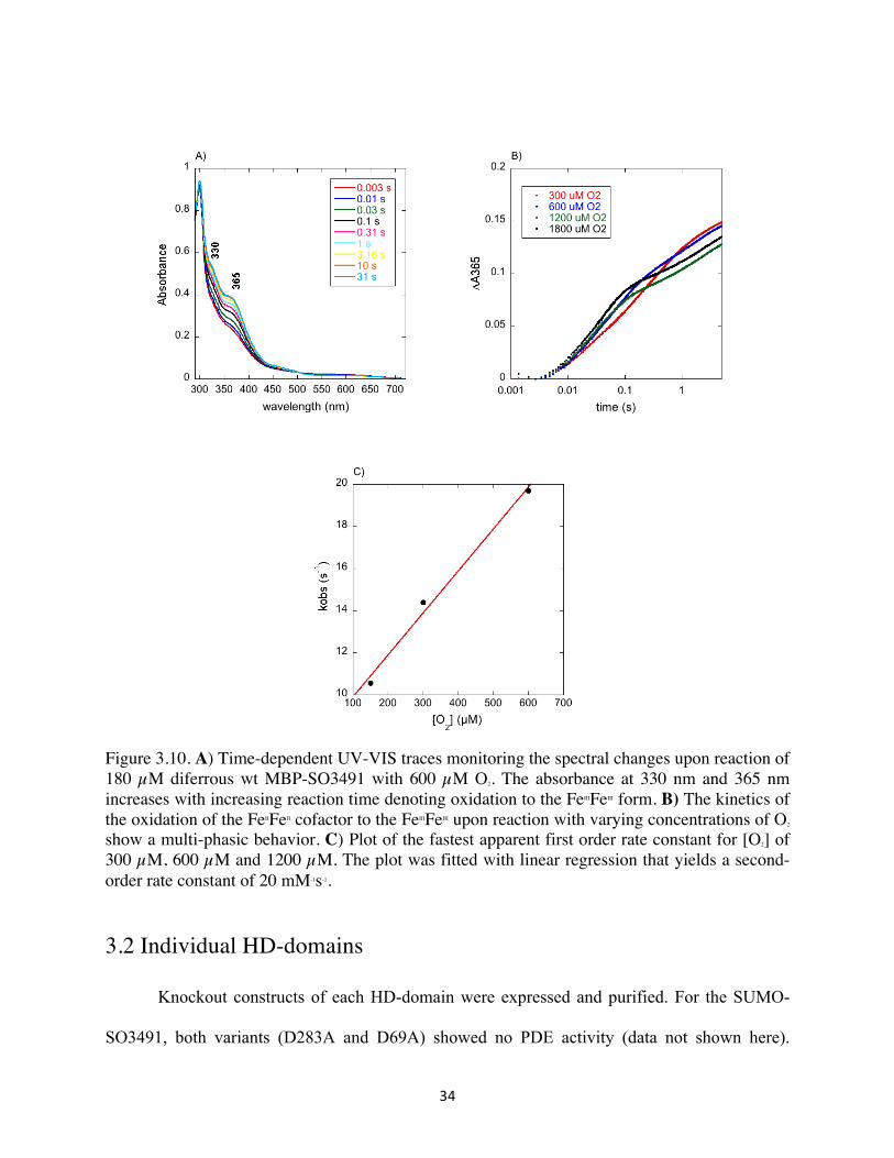
34
Figure 3.10. A) Time-dependent UV-VIS traces monitoring the spectral changes upon reaction of 180 µM diferrous wt MBP-SO3491 with 600 µM O2. The absorbance at 330 nm and 365 nm increases with increasing reaction time denoting oxidation to the FeIIIFeIII form. B) The kinetics of the oxidation of the FeIIFeII cofactor to the FeIIIFeIII upon reaction with varying concentrations of O2 show a multi-phasic behavior. C) Plot of the fastest apparent first order rate constant for [O2] of 300 µM, 600 µM and 1200 µM. The plot was fitted with linear regression that yields a second-order rate constant of 20 mM-1s-1.
3.2 Individual HD-domains
Knockout constructs of each HD-domain were expressed and purified. For the SUMO-
SO3491, both variants (D283A and D69A) showed no PDE activity (data not shown here).
35
However, based on previous results on D75A MBP-VCA0681 (ours and ref.12), we expected that
the analogous variant D69A (SUMO)-SO3491 should retain PDE activity (or be as active as the
wt). To confirm that the aminoacid substitution was not destabilizing the structure of the protein,
we replaced the N-terminal SUMO tag (12 kDa) with an MBP tag (42 kDa, similar to VCA0681).
The protein stability was examined by a thermoshift assay.
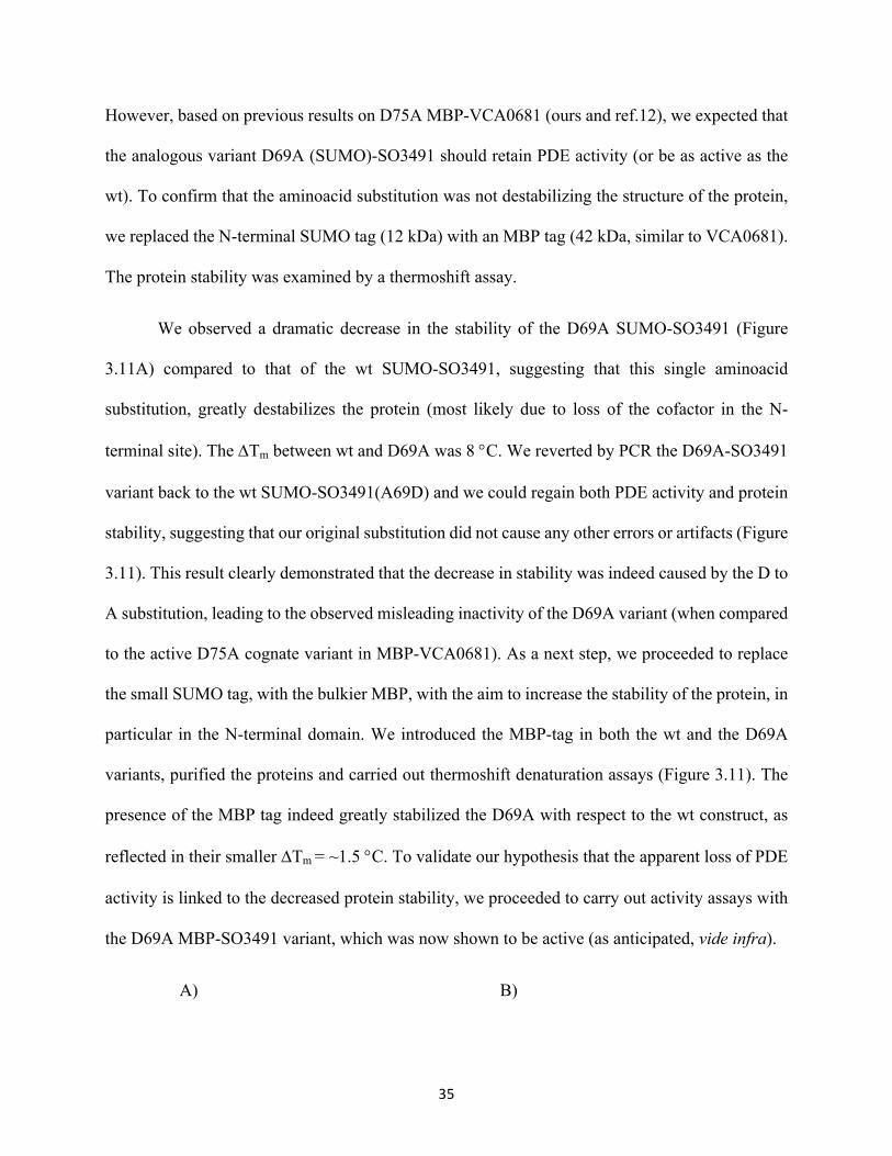
We observed a dramatic decrease in the stability of the D69A SUMO-SO3491 (Figure
3.11A) compared to that of the wt SUMO-SO3491, suggesting that this single aminoacid
substitution, greatly destabilizes the protein (most likely due to loss of the cofactor in the N-
terminal site). The DTm between wt and D69A was 8 °C. We reverted by PCR the D69A-SO3491
variant back to the wt SUMO-SO3491(A69D) and we could regain both PDE activity and protein
stability, suggesting that our original substitution did not cause any other errors or artifacts (Figure
3.11). This result clearly demonstrated that the decrease in stability was indeed caused by the D to
A substitution, leading to the observed misleading inactivity of the D69A variant (when compared
to the active D75A cognate variant in MBP-VCA0681). As a next step, we proceeded to replace
the small SUMO tag, with the bulkier MBP, with the aim to increase the stability of the protein, in
particular in the N-terminal domain. We introduced the MBP-tag in both the wt and the D69A
variants, purified the proteins and carried out thermoshift denaturation assays (Figure 3.11). The
presence of the MBP tag indeed greatly stabilized the D69A with respect to the wt construct, as
reflected in their smaller DTm = ~1.5 °C. To validate our hypothesis that the apparent loss of PDE
activity is linked to the decreased protein stability, we proceeded to carry out activity assays with
the D69A MBP-SO3491 variant, which was now shown to be active (as anticipated, vide infra).
A) B)
36
Figure 3.11. Thermoshift denaturation curves of the different SO3491 constructs. A) Fluorescence intensities of the wt SUMO-SO3491, D69A-to-A69D SUMO-SO3491 (wt-like) and D69A SUMO-SO3491. B) Fluorescence intensities of the wt SUMO-SO3491, D69A SUMO-SO3491, wt MBP-SO3491 and D69A MBP- SO3491 over a range of temperatures (30 - 95 °C).
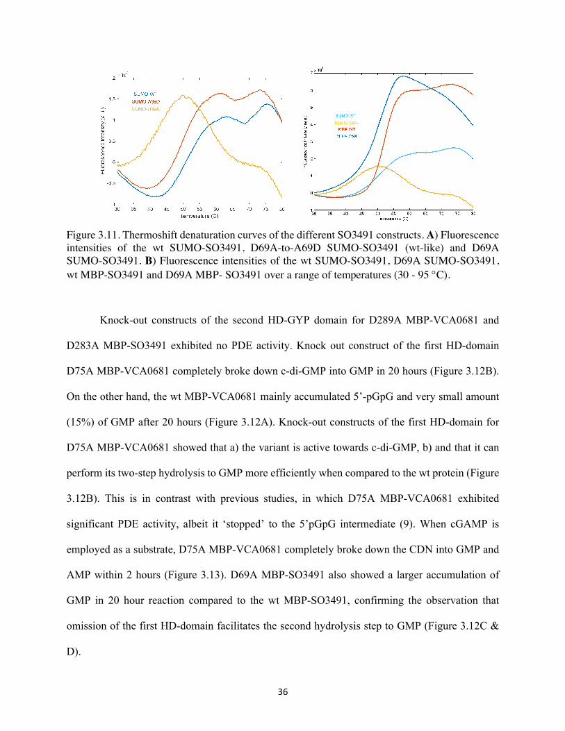
Knock-out constructs of the second HD-GYP domain for D289A MBP-VCA0681 and
D283A MBP-SO3491 exhibited no PDE activity. Knock out construct of the first HD-domain
D75A MBP-VCA0681 completely broke down c-di-GMP into GMP in 20 hours (Figure 3.12B).
On the other hand, the wt MBP-VCA0681 mainly accumulated 5’-pGpG and very small amount
(15%) of GMP after 20 hours (Figure 3.12A). Knock-out constructs of the first HD-domain for
D75A MBP-VCA0681 showed that a) the variant is active towards c-di-GMP, b) and that it can
perform its two-step hydrolysis to GMP more efficiently when compared to the wt protein (Figure
3.12B). This is in contrast with previous studies, in which D75A MBP-VCA0681 exhibited
significant PDE activity, albeit it ‘stopped’ to the 5’pGpG intermediate (9). When cGAMP is
employed as a substrate, D75A MBP-VCA0681 completely broke down the CDN into GMP and
AMP within 2 hours (Figure 3.13). D69A MBP-SO3491 also showed a larger accumulation of
GMP in 20 hour reaction compared to the wt MBP-SO3491, confirming the observation that
omission of the first HD-domain facilitates the second hydrolysis step to GMP (Figure 3.12C &
D).

37
Figure 3.12. HPLC chromatograms of the activity assays carried out with the dithionite-reduced wt, D75A MBP-VCA0681 and wt, D69A MBP-SO3491 and c-di-GMP as the substrate. All reactions were carried out for 1 hour and 20 hours, respectively. GMP elutes at circa 6 minutes, 5’-pGpG at 14 minutes and c-di-GMP at 13 minutes. A, C) 1 µM diiron wt MBP-VCA0681 and MBP-SO3491 reduced with NaDT were reacted with 50 µM c-di-GMP. B, D) 5 µM diiron D75A MBP-VCA0681 and D69A MBP-SO3491 were reacted with 50 µM c-di-MP. All reactions were carried out under O2-free conditions in an anaerobic glovebox and terminated by heat quenching at 100 °C.

38
Figure 3.13. HPLC chromatograms of the activity assays carried out with the dithionite-reduced D75A MBP-VCA0681 and with c-GAMP as the substrate for 10 minutes, 40 minutes and 1 hour, respectively. GMP and AMP eluted at circa 3.5 minutes and 5.2 minutes respectively. cGAMP and 5’-pApG eluted at circa 11.6 minutes and 12.2 minutes. All reactions were carried out under O2-free conditions in an anaerobic glovebox and terminated by heat quenching at 100 °C.
The different constructs of SO3491 were further characterized by optical spectroscopy
(Figure 3.14). All proteins were in a final concentration of 100 µM except for D69A MBP-3491
(50 µM). The UV/VIS spectrum of the Fe-loaded MBP-SO3491 showed absorbances at ~ 330,
365 nm, which are characteristic of non-heme oxo/carboxylate-bridged diferric (FeIIIFeIII) sites.
The optical spectra of the wt Mn-loaded SUMO- and MBP-SO3491, as expected, lack those
absorbances (because dimanganous sites in a similar ligation environment are optically
transparent). The SO3491 variants were examined to determine whether knocking out of one the
two HD-domains would have a detectable effect in the spectra (i.e. whether the two sites would
have slightly different optical characteristics that would allow their discrimination). The
absorption intensity for the knockout variants (D69A and D283A) MBP-SO3491 are in-line with
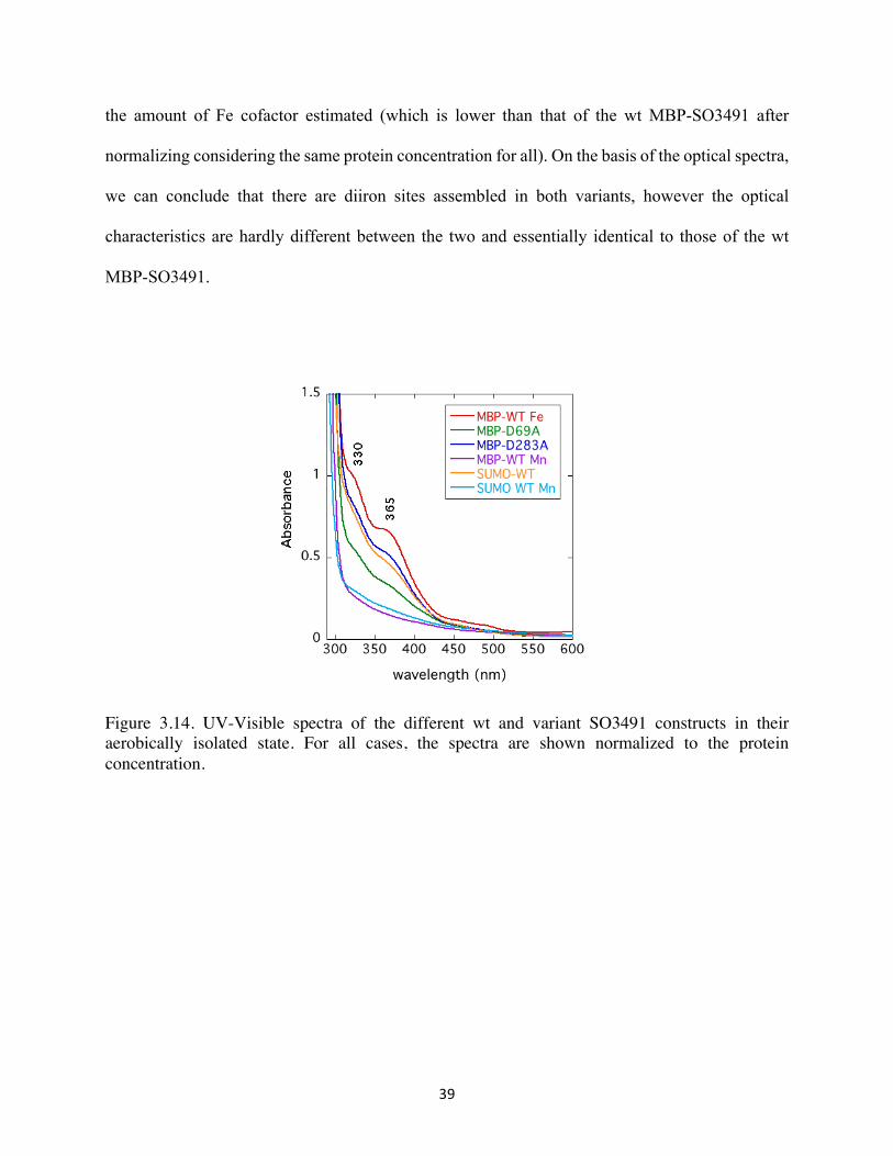
39
the amount of Fe cofactor estimated (which is lower than that of the wt MBP-SO3491 after
normalizing considering the same protein concentration for all). On the basis of the optical spectra,
we can conclude that there are diiron sites assembled in both variants, however the optical
characteristics are hardly different between the two and essentially identical to those of the wt
MBP-SO3491.
Figure 3.14. UV-Visible spectra of the different wt and variant SO3491 constructs in their aerobically isolated state. For all cases, the spectra are shown normalized to the protein concentration.
40
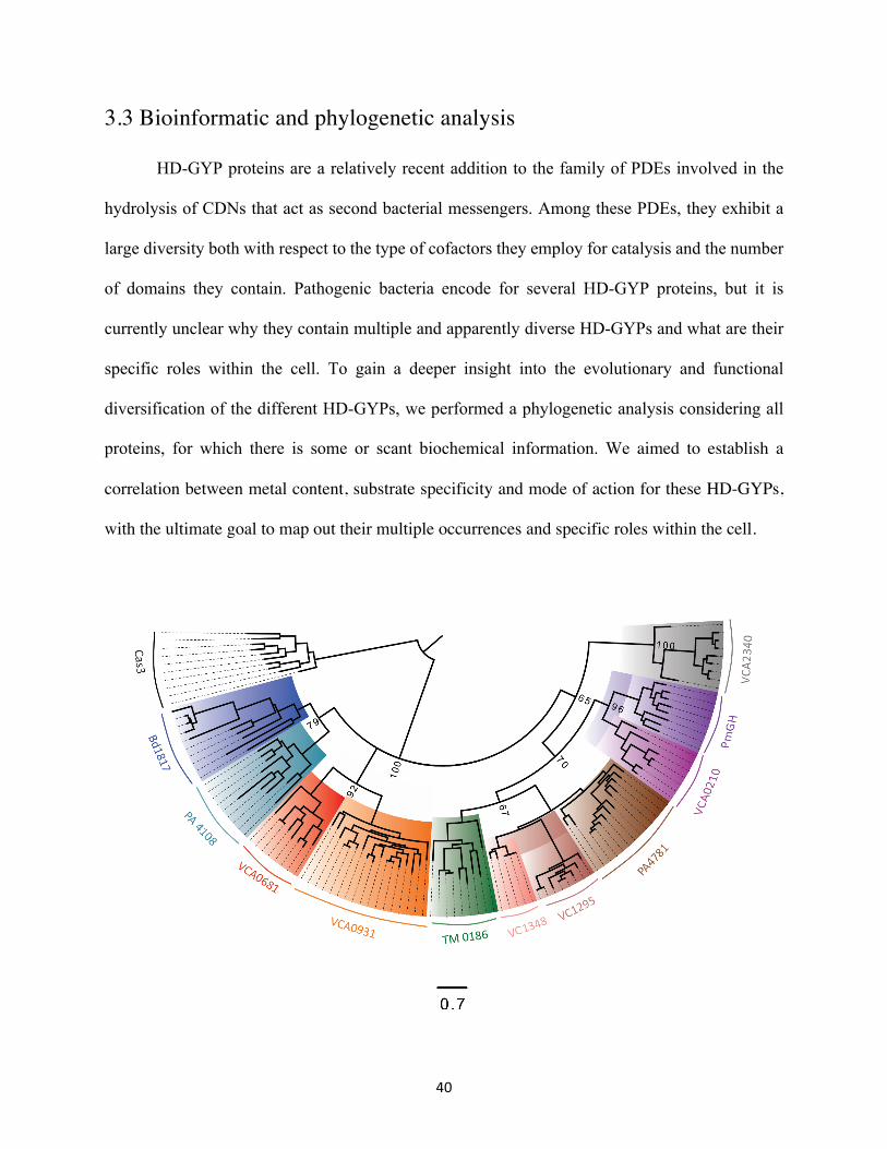
3.3 Bioinformatic and phylogenetic analysis
HD-GYP proteins are a relatively recent addition to the family of PDEs involved in the
hydrolysis of CDNs that act as second bacterial messengers. Among these PDEs, they exhibit a
large diversity both with respect to the type of cofactors they employ for catalysis and the number
of domains they contain. Pathogenic bacteria encode for several HD-GYP proteins, but it is
currently unclear why they contain multiple and apparently diverse HD-GYPs and what are their
specific roles within the cell. To gain a deeper insight into the evolutionary and functional
diversification of the different HD-GYPs, we performed a phylogenetic analysis considering all
proteins, for which there is some or scant biochemical information. We aimed to establish a
correlation between metal content, substrate specificity and mode of action for these HD-GYPs,
with the ultimate goal to map out their multiple occurrences and specific roles within the cell.

41
Figure 3.15. Maximum-likelihood rooted phylogenetic tree generated with 103 sequences and including 10 sequences of the Cas3 PDE subfamily as an outgroup (31). The tree was computed with the RaxML Blackbox software assuming the LG model, a gamma model for the rate of heterogeneity, invariant sites, and empirical base frequencies. The bootstrap values showing the confidence of the nodes have been included for the major branches. The scale bar represents the number of substitutions per site. Sequences were aligned using bothMAFFT and MUSCLE methods in the EMBL-EBI program. Proteins segregated in the different sub-branches are shaded with different colors.
Figure 3.16. Unrooted phylogenetic tree generated with 103 sequences and including 10 sequences of the Cas3 PDE subfamily as an outgroup (31). The tree was computed with the RaxML Blackbox software assuming the LG model, a gamma model for the rate of heterogeneity, invariant sites, and empirical base frequencies. The metal cofactor and plausible function of proteins are also listed.

42
We generated a phylogenetic tree with the CRISPR HD-domain PDE, Cas3, as an outgroup.
The phylogeny braches out into three major clades (Figure of 3.15 & 3.16). One clade with a
common ancestor includes our protein of interest, VCA0681 and SO3491. The common ground
of these HD-GYP proteins is that they accumulate the linearized product 5’-pGpG. The second
cluster includes HD-GYP proteins that have lost PDE function, and these are PA4108 and Bd1817
(1, 13). The structure of Bd1817 from Bdellovibrio bacteriovorus shows an asparagine residue
coordinated in the active site, and which is purported to disrupt the ligand environment at the active
site and lead to loss of PDE function (Figure 1.3) (13). The last and largest clade includes the
putative tri-nuclear HD-GYP phosphodiesterases, which are typified by an extra glutamate residue
important for the ligation of the third metal ion, and suggested to degrade c-di-GMP completely
into GMP. This glutamate was predicted to coordinate the third metal cofactor in the trimetal
binding sites and performs the two-step hydrolysis without detecting 5’-pNpN as an intermediate
product (32). For these putative trinuclear PDEs, which include VCA0210, TM0186, PA4781,
VCA1348 and VC2340. TM0186, VC1295 and VC2340, there is some biochemical evidence that
they can hydrolyze c-di-GMP to GMP (12). VCA0210 and PA4781 contain an N-terminal
phosphoreciever (REC) domain that may activate or inhibit PDE activity through an allosteric
mechanism involving its phosphorylation (19).
Although PA4781 proteins are grouped within the putative tri-nuclear HD-GYP containing
proteins, it was found to exhibit a very sluggish PDE, which appears to be too slow to be
functionally relevant (1, 14). We generated a sequence alignment with the PA4781 and PmGH
proteins (Figure 3.17). Some of the PA4781s have an extra proline instead of the extra glutamate
in the active site, which may result in the loss of PDE activity and ability to coordinate the third
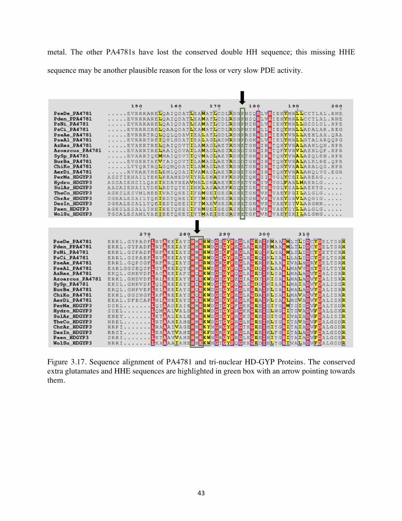
43
metal. The other PA4781s have lost the conserved double HH sequence; this missing HHE
sequence may be another plausible reason for the loss or very slow PDE activity.
Figure 3.17. Sequence alignment of PA4781 and tri-nuclear HD-GYP Proteins. The conserved extra glutamates and HHE sequences are highlighted in green box with an arrow pointing towards them.
44
Chapter 4
Discussion
We have biochemically and structurally characterized the VCA0681 homolog, SO3491
from the model g-proteobacterium Shewanella oneidensis. We established that SO3491 is a true
functional homolog of VCA0681 from Vibrio cholera. SO3491 showed similar metal dependence
and comparable activities with VCA0681. The PDE activity of VCA0681 is strongly Fe-dependent,
but can also be sustained by Mn (albeit less efficiently). The Fe-loaded VCA0681 is highly active
with c-di-GMP and is the first reported enzyme that can degrade the novel cyclic dinucleotide c-
GAMP. SO3491 is also as active as VCA0681 with both c-di-GMP and c-GAMP. SO3491 purified
with different metal cofactors (Fe and Mn, respectively) allowed us to examine the dependence of
its catalytic activity on the chemical nature of the cofactor and its ability to hydrolyze different
substrates. Mn-loaded SO3491 demonstrated hydrolysis of c-di-GMP to 5’-pGpG, although very
slow compared to the Fe-loaded SO3491. Phosphodiesterase activities of VCA0681 and SO3491
are limited to c-di-GMP and c-GAMP substrates, and the product of their reactions is the one-step
hydrolyzed 5’-pGpG and 5’-pApG linearized dinucleotides, respectively.
VCA0681 and SO3491 harbor two diiron sites, among which one has the HD-GYP motif
and the other one contains only the HD motif. The diiron binding sites of VCA0681 and SO3491
45
can attain three redox sates, the diferric, mixed-valent and the diferrous forms. The FeIIFeIII was
accumulated to a certain extent by reduction with small excess amount of sodium ascorbate. The
FeIIFeII was attained in homogeneity (100%) by reduction with excess amount of sodium dithionite.
However, quantification on the basis of the Mössbauer and EPR spectra, FeIIFeIII accumulates only
in small percentage (16%) in SO3491 and FeIIFeII accumulation (19%) cannot be avoided in the
ascorbate reduced protein. Our results indicate that FeIIFeIII of SO3491 either does not contribute
detectably to the activity or is much slower than the FeIIFeII in hydrolyzing c-di-GMP. The catalytic
form FeIIFeII is quite sensitive against O2 oxidative inactivation and can react with O2 with a second-
order rate constant of 20 mM-1s-1.
Steady-state kinetic assays were carried out to determine the catalytic efficiency of SO3491.
Wt native (‘untagged’) SO3491 hydrolyzes c-di-GMP with kcat and KM of 0.15 s-1 and 3.1 µM
respectively. The cellular levels of c-di-GMP in V. cholerae are within the range of 0.9 to 20 µM
(24). SO3491 is enzymatically quite proficient when compared on the basis of its catalytic
efficiency to other HD-GYP PDEs (Table 2), to hydrolyze c-di-GMP and thus switch the bacterial
behavior to non-pathogenic one. SO3491 hydrolyzes c-GAMP with a kcat of 5.3 x 10⁻³ s-1 and KM
of 55 µM, which is much slower than c-di-GMP. Currently there is no other report for the catalytic
proficiency of these enzymes with c-GAMP as a substrate, not the cellular levels of c-GAMP have
been to our knowledge estimated.
Variants of VCA0681 and SO3491, in which one of the HD dimetal sites was knocked out,
were assayed to explore each site’s unique role. We found that the C-terminal HD domain (HD289
and HD283, in VCA0681 and SO3491, respectively) is essential for PDE activity. The absence of
this HD-domain completely abolishes the PDE activities of both VCA0681 and SO3491. On the
other hand, knocking out the N-terminal HD domain does not eliminate PDE activity. In contrast,
46
it allows for a two-step hydrolysis to take place, most likely through a structural rearrangement or
flexibility imposed on the C-terminal ‘active’ HD-GYP domain, which is now competent
breakdown the cyclic di-nucleotide completely to the individual nucleotides. While currently the
exact functional purpose of the N-terminal HD domain is not precisely known, we propose that it
serves mainly a structural role and that it can divert the reaction outcome via allosteric or
conformational changes imposed on the C-terminal domain. Overall it appears to serve a ‘stop’
function, so that the reaction can be halted to the 5’pGpG product, which may be relevant for the
cell, especially because 5’pGpG has been proposed to serve a similar function to c-di-GMP (i.e. a
second bacterial messaging molecule).
The bioinformatic and phylogenetic analysis informed us the biochemical and structural
diversion of HD-GYP proteins, for which only a few representatives have been characterized. HD-
GYP proteins appear to have segregated into three main groups on the basis of their metal cofactors,
their respective specific activities and their reaction outcome. One phylogenetic cluster of HD-
GYP proteins contains representatives containing putative tri-nuclear metal binding sites and
proposed to afford a two-step hydrolysis that can break down cyclic dinucleotides completely into
the individual nucleotides without obvious accumulation of the linearized intermediates. Another
group of HD-GYP phosphodiesterases, including our proteins of interest VCA0681 and SO3491,
on the other hand accumulate mainly the linearized 5’-pNpN products after first-step hydrolysis,
which may be related to their different roles within the cell and their expression under different
environmental metal ion concentrations. The last clade of the phylogenetic tree contains HD-GYP
proteins that are enzymatically inactive. These proteins can cooperate with different metals at their
active sites, suggesting that the metal binding sites may play a structural role more than a catalytic
role (14). These have been shown to bind c-di-GMP, which would suggest that their likely evolved
47
function is to act as CDN sensors rather than CDN degraders. Overall our phylogenetic analysis
suggests that there are three main classes of HD-GYP proteins, which are further branching out to
smaller clades that demonstrate slightly different activities, metal ion content and substrate
specificities. This divergence is a most likely outcome of an evolutionary diversification of HD-
GYPs as a response to different environmental cues. We believe that our analysis can serve as a
functional map predictor that allows portraying the somewhat different roles of these proteins in
the cell, and opens up the stage for our better understanding for the regulation of important second
bacterial messengers, such as c-di-GMP.

48
Chapter 5
Future Directions and Experiments
We hypothesized that the mutation of the N-terminal HD domain strongly destabilizes the
protein. This hypothesis need to be verified by thermoshift denaturation assays to obtain the
melting curves of the D283A MBP-SO3491. The site differentiation on the Mössbauer spectra
could either correspond to the different HD-domains of the protein or to a mixed population of
active site molecules with a slight deviant ligation or the presence of a small ligand bound to the
active site. In order to clarify the reason caused this differentiation, Mössbauer spectra on the
MBP-D69A should be obtained, where only one HD-domain is presented. It is also important to
study the kinetic parameters of D69A MBP-SO3491, wt MBP-VCA0681 and D75A MBP-
VCA068, which can give more knowledge on the catalytic proficiency of distinct HD-domain
phosphodiesterases.
49
Bibliography
1. Stelitano, V. et al. C-di-GMP hydrolysis by Pseudomonas aeruginosa HD-GYP phosphodiesterases: analysis of the reaction mechanism and novel roles for pGpG. PLoS One 8, e74920 (2013).
2. Gao, J., Tao, J., Liang, W., Zhao, M., Du, X., Cui, S., . . . Jiang, Z. (2015). Identification and characterization of phosphodiecasterases that specifically degrade 3'3'-cyclic GMP-AMP. Cell Res, 25(5), 539-550. doi:10.1038/cr.2015.40
3. Jenal, U., Reinders, A., & Lori, C. (2017). Cyclic di-GMP: second messenger extraordinaire. Nat Rev Microbiol, 15(5), 271-284. doi:10.1038/nrmicro.2016.190
4. Rodney M. Donlan; Biofilm Formation: A Clinically Relevant Microbiological Process, Clinical Infectious Diseases, Volume 33, Issue 8, 15 October 2001, Pages 1387–1392, https://doi.org/10.1086/322972
5. Imamura, Y., Chandra, J., Mukherjee, P. K., Lattif, A. A., Szczotka-Flynn, L. B., Pearlman, E., . . . Ghannoum, M. A. (2008). Fusarium and Candida albicans biofilms on soft contact lenses: model development, influence of lens type, and susceptibility to lens care solutions. Antimicrob Agents Chemother, 52(1), 171-182. doi:10.1128/AAC.00387-07
6. Davis, S., Ricotti. C., Cazzaniga. A., Welsh. E., Eaglstein W., Mertz. P. (2007) Microscopic and physiologic evidence for biofim-associated wound colonization in vivo Wound Repair and Regeneration. 16 (1): 23-9. Doi: 10.1111/j.1524-475x.2007.00303.x
7. Davies BW, Bogard RW, Young TS, Mekalanos JJ. Coordinated regulation of accessory genetic elements produces cyclic di-nucleotides for V. cholerae virulence. Cell 2012; 149:358-370.
8. Sun L, Wu J, Du F, Chen X, Chen ZJ. Cyclic GMP-AMP synthase is a cytosolic DNA sensor that activates the type I interferon pathway. Science 2013; 339:786-791
9. Wu J, Sun L, Chen X, et al. Cyclic GMP-AMP is an endogenous second messenger in innate immune signaling by cytosolic DNA. Science 2013; 339:826-830.
10. Gao P, Ascano M, Wu Y, et al. Cyclic [G(2ʹ,5ʹ)pA(3ʹ,5ʹ)p] is the metazoan second messenger produced by DNA-activated cyclic GMP-AMP synthase. Cell 2013; 153:1094-1107.
11. Plate, L., and Marletta, M. A. (2012) Nitric oxide modulates bacterial biofilm formation through a multicomponent cyclic-di-GMP signaling network. Mol. Cell 46, 449−460.
12. Miner, K. D., Klose, K. E., and Kurtz, D. M., Jr. (2013) An HDGYP cyclic di-guanosine monophosphate phosphodiesterase with a non-heme diiron-carboxylate active site. Biochemistry 52, 5329−5331.
50
13. Lovering, A. L., Capeness, M. J., Lambert, C., Hobley, L., & Sockett, R. E. (2011). The structure of an unconventional HD-GYP protein from Bdellovibrio reveals the roles of conserved residues in this class of cyclic-di-GMP phosphodiesterases. MBio, 2(5). doi:10.1128/mBio.00163-11
14. Rinaldo, S., Paiardini, A., Stelitano, V., Brunotti, P., Cervoni, L., Fernicola, S., . . . Giardina, G. (2015). Structural basis of functional diversification of the HD-GYP domain revealed by the Pseudomonas aeruginosa PA4781 protein, which displays an unselective bimetallic binding site. J Bacteriol, 197(8), 1525-1535. doi:10.1128/JB.02606-14
15. Huq, A., E. B. Small, P. A. West, M. I. Huq, R. Rahman, and R. R. Colwell. 1983. Ecological relationships between Vibrio cholerae and planktonic crustacean copepods. Appl. Environ. Microbiol. 45:275–283.
16. Tischler AD, Camilli A: Cyclic diguanylate regulates Vibrio cholera virulence gene expression. Infect Immun 2005, 280(39):33324–33330.
17. Robert W McKee, A. K., Benjamin Mudrak, Courtney Whitaker, Rita Tamayo. (2014). <A systematic analysis of the in vitro and in vivo functions of the HD-GYP domain proteins of Vibrio cholerae.pdf>. BMC microbiology, 14(272), 1471-2180. doi:10.1186/s12866-014-0272-9
18. Hammer, B. K., & Bassler, B. L. (2009). Distinct sensory pathways in Vibrio cholerae El Tor and classical biotypes modulate cyclic dimeric GMP levels to control biofilm formation. J Bacteriol, 191(1), 169-177. doi:10.1128/JB.01307-08
19. McKee. R., Kariisa. A., Mudrak. B., Whitaker. C., Tamaya. R. A systematic analysis of the in vitro and in vivo functions of the HD-GYP domain proteins of Vibrio cholerae. BMC Microbiology 2014 14: 272
20. Miner, K. D., & Kurtz, D. M., Jr. (2016). Active Site Metal Occupancy and Cyclic Di-GMP Phosphodiesterase Activity of Thermotoga maritima HD-GYP. Biochemistry, 55(6), 970-979. doi:10.1021/acs.biochem.5b01227
21. Fang, P., & Guo, M. (2015). The Nature's Clever Trick for Making Cyclic Dinucleotide. Structure, 23(5), 801-802. doi:10.1016/j.str.2015.04.011
22. Sultan SZ, Pitzer JE, Boquoi T, Hobbs G, Miller MR et al. (2011) Analysis of the HD-GYP domain cyclic dimeric GMP phosphodiesterase reveals a role in motility and the enzootic life cycleof Borrelia burgdorferi. Infect Immun 79: 3273-3283. doi:10.1128/IAI. 05153-11. PubMed: 21670168.
23. Worsdorfer, B., Lingaraju, M., Yennawar, N. H., Boal, A. K., Krebs, C., Bollinger, J. M., Jr., & Pandelia, M. E. (2013). Organophosphonate-degrading PhnZ reveals an emerging family of HD domain mixed-valent diiron oxygenases. Proc Natl Acad Sci U S A, 110(47), 18874-18879. doi:10.1073/pnas.1315927110
24. Koestler, B. J., & Waters, C. M. (2013). Exploring environmental control of cyclic di-GMP signaling in Vibrio cholerae by using the ex vivo lysate cyclic di-GMP assay (TELCA). Appl Environ Microbiol, 79(17), 5233-5241. doi:10.1128/AEM.01596-13
25. Christen M, Christen B, Folcher M, Schauerte A, Jenal U (2005) Identification and characterization of a cyclic di-GMP-specific phosphodiesterase and its allosteric control by GTP. J Biol Chem 280: 30829-30837. doi:10.1074/jbc.M504429200. PubMed: 15994307.
51
26. Rao F, Yang Y, Qi Y, Liang ZX (2008) Catalytic mechanism of cyclic di- GMP-specific phosphodiesterase: a study of the EAL domaincontaining RocR from Pseudomonas aeruginosa. J Bacteriol 190: 3622-3631. doi:10.1128/JB.00165-08. PubMed: 18344366.
27. Rao F, Qi Y, Chong HS, Kotaka M, Li B et al. (2009) The functional role of a conserved loop in EAL domain-based cyclic di-GMP-specific phosphodiesterase. J Bacteriol 191: 4722-4731. doi:10.1128/JB. 00327-09. PubMed: 19376848.
28. Tamayo R, Tischler AD, Camilli A (2005) The EAL domain protein VieA is a cyclic diguanylate phosphodiesterase. J Biol Chem 280: 33324-33330. doi:10.1074/jbc.M506500200. PubMed: 16081414.
29. Barends TR, Hartmann E, Griese JJ, Beitlich T, Kirienko NV et al. (2009) Structure and mechanism of a bacterial light-regulated cyclic nucleotide phosphodiesterase. Nature 459: 1015-1018. doi:10.1038/ nature07966. PubMed: 19536266.
30. Kurtz, D. M., Jr. (1990) Oxo- and hydroxo-bridged diiron complexes: A chemical perspective on a biological unit. Chem. Rev. 90, 585−606.
31. A. Stamatakis: "RAxML Version 8: A tool for Phylogenetic Analysis and Post-Analysis of Large Phylogenies". In Bioinformatics, 2014,
32. Bellini, D., Caly, D. L., McCarthy, Y., Bumann, M., An, S. Q., Dow, J. M., . . . Walsh, M. A. (2014). Crystal structure of an HD-GYP domain cyclic-di-GMP phosphodiesterase reveals an enzyme with a novel trinuclear catalytic iron centre. Mol Microbiol, 91(1), 26-38. doi:10.1111/mmi.12447
33. Huynh, T. N., Luo, S., Pensinger, D., Sauer, J. D., Tong, L., & Woodward, J. J. (2015). An HD-domain phosphodiesterase mediates cooperative hydrolysis of c-di-AMP to affect bacterial growth and virulence. Proc Natl Acad Sci U S A, 112(7), E747-756. doi:10.1073/pnas.1416485112
34. Aracind, L., Koonin, Eugene V. (1998). The HD domain defines a new superfamily of metal-dependent phosphohydrolases. Elsevier Science, 23(12), 469-472
35. Bollinger JM, Jr. (1993) On the chemical mechanism of assembly of the tyrosyl radical-dinuclear iron cluster cofactor of E. Coli ribonucleotide reductase. Ph. D. (Massachusetts Institute of Technology).