
31762101545844.pdf - ScholarWorks
131
Biology of the eremophilanes produced by Drechslera gigantea by Gregory James Bunkers A thesis submitted in partial fulfillment of the requirements for the degree of Doctor of Philosophy in Plant Pathology Montana State University © Copyright by Gregory James Bunkers (1989) Abstract: Drechslera gigantea. the causative agent of zonate eyespot disease on grasses, produces at least twelve bioactive molecules known as eremophilanes. Their structures have been elucidated using conventional spectroscopy and x-ray crystallography. A study to examine the biological aspects of these eremophilanes was undertaken. A procedure, using high performance liquid chromatography to quantify eremophilane levels in culture filtrates of D. gigantea was developed. This procedure was used to study eremophilane production by D. gigantea under different cultural conditions. Leaf material from quackgrass (Agropyron repens), a host of the fungus, stimulated toxin production. Amendments such as L-leucine or the sterol biosynthesis inhibitor chloro-choline chloride (CCC) also exhibited stimulatory activity. The structure-activity relationships of the eremophilanes were investigated using three different bioassays. No distinct functional groups or structural characteristic could be correlated to activity. However, in all three bioassays, the eremophilanes with the higher oxidation states were generally less active. To determine the mode of action, the effects of eremophilanes on the physiology of the plant were studied. Eremophilane bioactivities mimic the activity of known phytohormones. Comparative studies indicated that these activities seem not to be associated with induction of known phytohormones but are inherent properties of the eremophilane molecules. The eremophilanes were shown to inhibit protein synthesis both in vitro and in vivo. The proposed mode of action of the eremophilanes is inhibition of protein synthesis. Finally, the fate of the eremophilanes in planta was investigated. [14C]-petasol was applied to detached oat leaves (Avena sativa cv. Park) and the radiolabel was traced. The [14C]-petasol was converted to a compound which exhibited different chromatographic properties than petasol and was not bioactive. NMR analysis indicated that the petasol moiety was present in the conversion product. Hydrolysis of the conversion product led to the recovery of petasol. Amino acid analysis indicated that amino acids were also present in the hydrolysate. Results indicate that the plant is capable of modifying this eremophilane to form a petasol-amino acid conjugate.
-
Upload
khangminh22 -
Category
Documents
-
view
1 -
download
0
Transcript of 31762101545844.pdf - ScholarWorks
Biology of the eremophilanes produced by Drechslera giganteaby Gregory James Bunkers
A thesis submitted in partial fulfillment of the requirements for the degree of Doctor of Philosophy inPlant PathologyMontana State University© Copyright by Gregory James Bunkers (1989)
Abstract:Drechslera gigantea. the causative agent of zonate eyespot disease on grasses, produces at least twelvebioactive molecules known as eremophilanes. Their structures have been elucidated using conventionalspectroscopy and x-ray crystallography. A study to examine the biological aspects of theseeremophilanes was undertaken.
A procedure, using high performance liquid chromatography to quantify eremophilane levels in culturefiltrates of D. gigantea was developed. This procedure was used to study eremophilane production byD. gigantea under different cultural conditions. Leaf material from quackgrass (Agropyron repens), ahost of the fungus, stimulated toxin production. Amendments such as L-leucine or the sterolbiosynthesis inhibitor chloro-choline chloride (CCC) also exhibited stimulatory activity.
The structure-activity relationships of the eremophilanes were investigated using three differentbioassays. No distinct functional groups or structural characteristic could be correlated to activity.However, in all three bioassays, the eremophilanes with the higher oxidation states were generally lessactive.
To determine the mode of action, the effects of eremophilanes on the physiology of the plant werestudied. Eremophilane bioactivities mimic the activity of known phytohormones. Comparative studiesindicated that these activities seem not to be associated with induction of known phytohormones butare inherent properties of the eremophilane molecules. The eremophilanes were shown to inhibitprotein synthesis both in vitro and in vivo. The proposed mode of action of the eremophilanes isinhibition of protein synthesis.
Finally, the fate of the eremophilanes in planta was investigated. [14C]-petasol was applied to detachedoat leaves (Avena sativa cv. Park) and the radiolabel was traced. The [14C]-petasol was converted to acompound which exhibited different chromatographic properties than petasol and was not bioactive.NMR analysis indicated that the petasol moiety was present in the conversion product. Hydrolysis ofthe conversion product led to the recovery of petasol. Amino acid analysis indicated that amino acidswere also present in the hydrolysate. Results indicate that the plant is capable of modifying thiseremophilane to form a petasol-amino acid conjugate.
BIOLOGY OF THE EREMOPHILANES PRODUCED BY DRECHSLERA GIGANTEA
byGregory James Bunkers
A thesis submitted in partial fulfillment of the requirements for the degree
ofDoctor of Philosophy
inPlant Pathology
MONTANA STATE UNIVERSITY Bozeman, Montana
May 1989

6 ^ ^ii
APPROVAL
of a thesis submitted by
Gregory James Bunkers
This thesis has been read by each member of the thesis committee and has been found to be satisfactory regarding content, English usage, format, citations, bibliographic style, and consistency, and is ready for submission to the College of Graduate Studies.
Date Chairperson, Graduate Committee
Approved for the Major Department
HjTjDate Head, Major Department
Approved for the College of Graduate Studies
Graduat DeanDate

iii
STATEMENT OF PERMISSION TO USE
In presenting this thesis in partial fulfillment of the requirements for a doctoral degree at Montana State University, I agree that the Library shall make it available to borrowers under rules of the Library. I further agree that copying of this thesis is allowable only for scholarly purposes, consistent with "fair use" as prescribed in the U.S. Copyright Law. Requests for extensive copying or reproduction of this thesis should be referred to University Microfilms International, 300 North Zeeb Road, Ann Arbor, Michigan 48106, to whom I have granted "the exclusive right to reproduce and distribute Copies of the dissertation in and from microfilm and the right to reproduce and distribute by abstract in any format."
SignatureDate
V
ACKNOWLEDGEMENTS
I would like to thank my major professor. Dr. Gary A. Strobel for financial support and for his advice and guidance during my Ph.D. program. Thanks to Dr. Doug Kenfield for his valuable advice and guidance. Doug gave of himself unselfishly, both as a scientist and as a friend.
I thank Drs. Joe Sears, Fumio Sugawara and David Teplow for running various analyses and for their valuable advice.
I would like to thank my parents, Yvo and Stella, and my family for their emotional support. To Eric Gallandt, Willie and Patty Inskeep, and all who shared forays into the wilderness and helped maintain sanity, thank you.Also, thanks to the "Wild Bunch", Sej, Michael and Autumn Kenfield whose drawings brightened my desk.
Finally, I would like to thank Leslie Harrison for her friendship and support during this stressful period of my life, her ideas and encouragement were invaluable.
vi
TABLE OF CONTENTS
Page
STATEMENT OF PERMISSION TO USE. .........................iiiVITA...... ivACKNOWLEDGEMENTS............... V
TABLE OF CONTENTS........... . ........................... viLIST OF TABLES.............. ............................ ixLIST OF FIGURES.......... ............................... xiABSTRACT. .................. .xiii
CHAPTERI. INTRODUCTION............. ................. ....... III. PRODUCTION OF EREMOPHILANES BY DRECHSLERA
GIGANTEA IN LIQUID CULTURE........................ 11Introduction.......... 11Materials and Methods.......... 12
Fungus............................. 12Liquid Culture......... 12Determination of Petasol Production. ........ 13Plant Extraction...... ............ . ..... 13
Results and Discussion. .......................... 15Defining Culture Conditions............... 15Plant Products and Petasol Production........ 17Effect of Sterol Biosynthesis Inhibitorson Petasol Production. ............. 19
Petasol Production Over Time..... ........22Labeling Petasol with [14C].......... 23
Summary......... 25
vii
III. STRUCTURE-ACTIVITY RELATIONSHIPS OF THEEREMOPHILANES........... 26Introduction............................. ....... 26Materials and Methods............................ 27
Eremophilanes.............. 27Leaf Puncture Wound Droplet Overlay Assay.... 29Root-Induction Assay.................... 29Brine Shrimp Assay........................... 3 0
Results and Discussion........................... 30Leaf Assay.............. .30Rooting Assay................................ 33Brine Shrimp Assay........................... 34
Summary.................. 35IV. PHYSIOLOGICAL INVESTIGATIONS: MODE OF ACTION....37
Introduction......................... 37Materials and Methods............................ 40Plant Materials........................... ...40Chlorophyll Retention and Quantification
of the Green Island Effect................. 40Preparation of Crude Membrane Fraction....... 41Binding Assay...................... 41Determination of Sink Activity............... 42CO2 Fixation Experiments......................43Chloroplast Isolation and the Hill Reaction...44Proteolytic Enzyme Assay..................... 45Extraction of Soluble Protein........ .......45SDS-Polyacrylamide Electrophoresis.. .......46In Vitro Protein Translation................. 47
Results and Discussion........................... 49Chlorophyll Retention in Detached Leavesof Monocots............................ .... 49
Light and Heat Effects on Green IslandFormation.................................. 54
Binding Site Studies................. 57Effect of Eremophilanes on Chloroplasts....... 60Effect of Eremophilanes on Proteolytic
Enzymes.................... 63Protein Synthesis and the Eremophilanes......66Summary.......................................... 73
V. METABOLISM OF EREMOPHILANES IN PLANTA............ 76
TABLE OF CONTENTS (continued)
Page
Introduction............ .........................76Materials and Methods .......................... 77
viii
Plant Material............................... 77[14C] -Labeled Eremophilanes................... 77Translocation of Eremophilanes............... 78Leaf Treatment and Extraction........ 79Purification of Conversion Product........ 79Chlorophyll Retention......................... 81Mass Spectrometry............................ 81Nuclear Magnetic Resonance Spectrometry...... 81Amino Acid Analysis..... 82
Results and Discussion........................ 82Translocation of Eremophilanes in the- Leaf....82Analysis of Radioactive Metabolites....;.......83 .Purification of Conversion Product........... 85Structural Analysis of Conversion Product.... 87Conversion of Other Eremophilanes............ 92Conversion of Petasol in Dicots...............94Biological Activity of Conversion Product..... 95
Summary........................... 96VI. CONCLUSION....................................... 98
REFERENCES CITED....................................... 105APPENDICES............................................. 112
Appendix A - Recipe for Modified M-ID Medium.... 113Appendix B - Isolation of Plant Fractions....... 115
TABLE OF CONTENTS (continued)
Page

LIST OF TABLES
Table PageI. Petasol nroduction bv D. cricrantea in licruid culture. 202 . Effects of eremophilanes, using the leaf
puncture wound droplet overlay assay, on some species of monocots and dicots at 15-20 nmol. 32
3 . Stimulation of rhizogenesis in mung bean hypocotyls. 33
4. Brine shrimp assay. 345. Sink activity of the eremophilanes. 516. Induction of chlorophyll synthesis in etiolated
cucumber. 537. Delay of senescence in oat leaves floating on
solutions. 548. Effect of light on green island development
in oat leaves. 559. Effect of preheating leaf on green island
formation. 5610. Binding of [14C]-petasol to a crude membrane
fraction from oat leaves. 5811. Carbon dioxide fixation in leaves treated
with eremophilanes. 6212. Effect of eremophilanes on the Hill Reaction
in chloroplasts isolated from spinach and oat leave. 62
13. Ficin and protease Type VII protease activity in the presence of eremophilanes. .64

X
LIST OF TABLES (continued)
Table Page14. Green island formation of Pondera wheat leaves. 7015. Translocation of exogenously applied
eremophilane in detached oat leaves. 8316. 1H-NMR assignments for petasol and conversion -
product. 8817. Chromatogram report from amino acid analysis
of conversion product hydrolysate. 9118. Recipe for modified M-ID medium. 114

LIST OF FIGURES
1. ' The carbon skeletons of eremophilanes andisoprene. 7
2. Influence of amendments to the medium on petasolproduction and growth by D. gigantea. 16
Figure Page
3. Effect of different plant fractions on petasolproduction and growth by D. gigantea. 19
4. Effect of sterol biosynthesis inhibitors onpetasol production and growth by D. gigantea. 21
5. Petasol production and growth by D. giganteaover time. 22
6. Specific radioactivity of [1AC] -petasol purified from D. gigantea cultures grows in M-ID brothwith radiolabeled compounds added. 24
7. Molecular structures of the eremophilanes 288. Chlorophyll retention in detached oat leaves
treated with eremophilanes and kinetin. 509. Chlorophyll retention in oat leaves away from
the site of eremophilane application. . 5210. Elution profile of radioactivity off a Sephadex
G-25 column (0.9 X 30). 5811. Chlorophyll content in oat leaves treated with
eremophilanes. 6012. Proteolytic enzyme activity of protein extracts
from oat leaves treated with eremophilanes and kinetin. 65
13. Chlorophyll content in oat leaves treated ,with petasol and protein synthesis inhibitors. 67
xiiLIST OF FIGURES (continued)
Figure14. In vitro incorporation of [14C] -leucine into TCA precipitable protein.15. SDS-polyacrylamide gel of proteins from the
reaction mixtures and an autoradiograph of this gel.
16. In vivo incorporation of [14C]-leucine into TCA precipitable protein.
17. SDS-polyacrylamide gel of protein extracts from eremophilane/ [14C] -leucine treated oat leaves and an autoradiograph of this gel.
18. Autoradiograph of a TLC plate of radiolabeled petasol and crude extract from leaf treated with radiolabeled petasol.
19. Purification schedule for purifying CP from green island tissue of detached oat leaves.
20. FAB-MS spectra of the conversion product.21. 1H-NMR spectra of the conversion product in
d6-DMSO.22. 1H-NMR spectra of petasol in CDCl3.23. Autoradiograph of TLC plate showing the products
from hydrolysis of CP.24. Autoradiograph of TLC plate showing crude leaf
extracts from leaves treated with radiolabeled petasol, 11,12-epoxypetasol, and gigantenone.
25.1 Autoradiograph of TLC plate of radiolabeled petasol, conversion product, and crude leaf extract from [14C]-petasol treated mung bean leaves.
26. Chlorophyll content in detached oat leaves treated with petasol and CP.Isolation schedule for fractionation of plant constituents.
69
70
72
73
84
8688
8889
91
93
95
96
Page
27.116

xiii
ABSTRACT
Drechslera giaantea. the causative agent of zonate eyespot disease on grasses, produces at least twelve bioactive molecules known as eremophilanes. Their structures have been elucidated using conventional spectroscopy and x-ray crystallography. A study to examine the biological aspects of these eremophilanes was undertaken.
A procedure, using high performance liquid chromatography to quantify eremophilane levels in culture filtrates of cricrantea was developed. This procedure was used to study eremophilane production by Djl criqantea under different cultural conditions. Leaf material from quackgrass (Aaronvron repens), a host of the fungus, stimulated toxin production. Amendments such as L-Ieucine or the sterol biosynthesis inhibitor chloro-choline chloride (CCC) also exhibited stimulatory activity.
The structure-activity relationships of the eremophilanes were investigated using three different bioassays. No distinct functional groups or structural characteristic could be correlated to activity. However, in all three bioassays, the eremophilanes with the higher oxidation states were generally less active.
To determine the mode of action, the effects of eremophilanes on the physiology of the plant were studied. Eremophilane bioactivities mimic the activity of known phytohormones. Comparative studies indicated that these activities seem not to be associated with induction of known phytohormones but are inherent properties of the eremophilane molecules. The eremophilanes were shown to inhibit protein synthesis both in vitro and in vivo. The proposed mode of action of the eremophilanes is inhibition of protein synthesis.
Finally, the fate of the eremophilanes in planta was investigated. [14C)-petasol was applied to detached oat leaves (Avena sativa cv. Park) and the radiolabel was traced. The [14C]-petasol was converted to a compound which exhibited different chromatographic properties than petasol and was not bioactive. NMR analysis indicated that the petasol moiety was present in the conversion product. Hydrolysis of the conversion product led to the recovery of petasol. Amino acid analysis indicated that amino acids were also present in the hydrolysate. Results indicate that the plant is capable of modifying this eremophilane to form a petasol-amino acid conjugate.
I
CHAPTER I
INTRODUCTION
Higher plants and their pathogenic fungi have evolved together throughout millennia, with their dynamic biochemical interchanges constantly adjusting. Included in the biochemical interchange are fungal-produced phytotoxins. The concept that plant pathogens produce pathogenic toxins originated about a century ago (62). Gaumann (29) concluded that microorganisms responsible for disease act by virtue of the toxins they produce, however, that phytopathogens induce disease through toxigenic action has not been rigorously proved. Arguments for their participation in pathogenesis center on two main points: I) some toxins are host specific and loss of ability to produce the toxin in culture is accompanied by loss of pathogenicity; 2) the symptomatic effects of the toxin are so distinctly similar to all or part of the disease syndrome that there can be little doubt that the toxin is involved. The other side of the argument has been stated quite succinctly by Day (16) who commented that pathogenicity based on toxin production may not be a major mechanism, but rather an evolutionary fluke.
An understanding of the relevance of toxins in disease expression by plant pathogenic microorganisms has been slow
2to develop. This has largely been due to the lack of understanding of the chemistry involved, or of the necessity for chemical purity. Ultimately the goal should be to define plant pathogenesis at the molecular level; studies using chemically characterized toxins provide a powerful tool and useful starting point for reaching this objective.
Pathogen produced toxins have also found application in many other areas of plant pathology including use as models for studies of disease physiology (58), screening for resistance among populations of plants (10), and perhaps the greatest use is as experimental tools in selecting for disease-resistant cells or tissue in vitro (30).
The practical significance of pathologically important toxins is being further recognized as more applications become apparent. The chemical specificity of some toxins in particular make them extremely good metabolic probes for investigating cellular and enzymatic functions. Fusicoccin, a toxin produced by Fusicoccum amygdali, is a prime example of a toxin that is proving exceedingly useful for studies on plant metabolism. Basically, it stimulates membrane bound ATPases which leads to a proton electrochemical gradient established across the membrane (42). This primary event leads to a number of effects on different physiological processes including proton extrusion, cell enlargement, increased respiration, dark CO2 fixation and ABA antagonism (42) .
3Some toxins have been proposed for use in taxonomy.
Tentoxin, produced by Alternaria tenuis, has been used in taxonomic applications involving higher plants, utilizing its property to inhibit the chloroplast coupling factor (CF1) only from selective plants. (9) „ This selective inhibition was used to determine species ancestry in cases where the putative parents vary in their reaction to tentoxin.
High molecular weight polysaccharides produced by Xanthomonas and Pseudomonas spp., which act as wilt inducing toxins, have found commercial application. In particular "xanthan" gum, produced by X_*_ camoestris pv. campestris. is used in a wide variety of commercial applications (23). Pseudomonas spp., as well as several fungi, produce similar exopolysaccharides which have been characterized as proteinaceous and polymerized DNA heteropolysaccharides (73). It is likely that because of this rich diversity, more commercial use will be.made of these substances and more organisms will be surveyed for their production and properties.
There is much work being done to develop weed pathogens as weed control agents. Ideally, the pathogen will establish itself and reach epiphytotic levels thus reducing the weed population to subeconomic levels. Skeleton weed (Chondrilla iuncea) has been successfully controlled in Australia by the introduction of the rust fungus Puccinia
4chondrillina from the region where the weed originated (6). Attempts to duplicate this success are now being made throughout the world (6, and refs, therein).Mycoherbicides, plant pathogenic fungi developed to control weeds, are available commercially in the United States. DeVine, a formulation of Phvtoohthora oalmivora used for control of strangler vine CMorrenia pdprata), and Collego, a formulation of Colletotrichum aloeosporiodes f. sp. aeschvnomene used for control of northern jointvetch CAeschvnomene viroinica), are being used for weed control in the southeastern United States (66).
Use of live pathogens as biocontrol agents is not without risk. The pathogen may actually prove more virulent on important crops in the surrounding area than to the desired target weed, risking dire results. Containment of the pathogen to the targeted area would be difficult because of the somewhat unpredictable manner of such dispersal mechanisms as wind, water, and animal vectors (3). Although the "escape" of these control agents is a potential, the more realistic concern is actually the inability of a chosen pathogen to propagate disease in a target area. Many obstacles must be overcome including inoculation, genetic diversity of the host, and unpredictable environmental factors such as temperature and humidity. These difficulties in using live phytopathogens as control agents have led to increased interest in phytotoxins produced by
5weed pathogens.
Potential applications of toxins produced by weed: - I
pathogens have only recently been realized. If weed pathogens, which produce host-specific toxins, can be identified it could lead to weed control with natural or synthetic compounds having a narrow spectrum of activity and minimal impact on the environment. A desire for selective, environmentally low impact herbicides has generated increased research activity in phytotoxins produced by weed pathogens as potential chemicals for the herbicide industry (22). At the very least, natural compounds may suggest structural types and arrangements of functional groups that could serve as starting points for the development of improved herbicides.
Our laboratory has focused on weed pathogens with hope of finding both novel and more selective chemical control agents. This led to the study of Drechslera gigantea, the causal agent of zonate eyespot disease on numerous grasses (21). This leaf disease is a serious problem in many commonly cultivated turfgrasses and also occurs in weeds such as crabgrass (Digitaria spp.), quackgrass (Agrgpyron repens), and bermudagrass ICvnodon dactylgn). Drechsler (21) pointed out that during the summer of 1922 it appeared in the vicinity of Washington, D.C., as probably the single most destructive parasitic fungus affecting the Gramineae family. He also suggested that in nature there were no
6physiological varieties or races paralleling generic divisions in the Gramineae. Dale (14), however, did indicate that several lines of bermudagrass have been introduced that show some degree of resistance to the disease.
Symptoms of zonate eyespot disease first appear as brown flecks, often not exceeding 0.05 mm in width and 0.2 mm in length (13). These flecks eventually enlarge and fade to a greyish oval lesion surrounded by reddish brown or dark green borders thus suggesting the name "eyespot". When these lesions become numerous and coalesce causing the leaf to dry somewhat, conidiophores appear around the periphery of the lesions as well as from the bleached area in the center of the lesion. In nature, Drechsler (21) found a peculiar type of secondary development which led to production of copious numbers of spores. During times of heavy dews or prolonged periods of water persisting on infected foliage many of the lesions could be found to be surrounded by a water soaked zone supporting growth of superficial mycelia. With the onset of drier conditions growth of the superficial mycelium ceases and abundant sporulation occurs. Rather meager speculation in culture led Drechsler to speculate on the importance of this secondary development on sporulation.
When grown in liquid culture, Di. qjqantea produces several bioactive terpenoids (28,37). Terpenes constitute
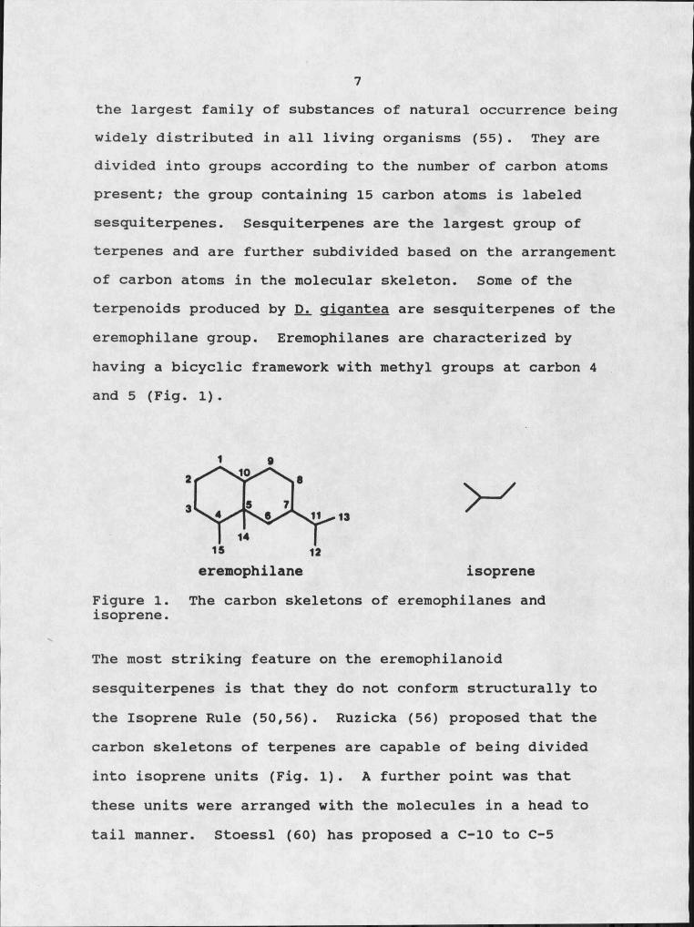
7the largest family of substances of natural occurrence being widely distributed in all living organisms (55). They are divided into groups according to the number of carbon atoms present; the group containing 15 carbon atoms is labeled sesquiterpenes. Sesquiterpenes are the largest group of terpenes and are further subdivided based on the arrangement of carbon atoms in the molecular skeleton. Some of the terpenoids produced by Di. aiaantea are sesquiterpenes of the eremophilane group. Eremophilanes are characterized by having a bicyclic framework with methyl groups at carbon 4 and 5 (Fig. I).
Figure I. isoprene.
eremophilane isopreneThe carbon skeletons of eremophilanes and
The most striking feature on the eremophilanoid sesquiterpenes is that they do not conform structurally to the Isoprene Rule (50,56). Ruzicka (56) proposed that the carbon skeletons of terpenes are capable of being divided into isoprene units (Fig. I). A further point was that these units were arranged with the molecules in a head to tail manner. Stoessl (60) has proposed a C-IO to C-5
8migration to explain this structural anomaly.
Over two hundred compounds belonging to the eremophilane group have been isolated from diverse plants and even animal sources (50). Numerous eremophilanes occur in fungi as well. These include phomenone (Phoma exigua), phaseolinone CMacrophomina phaseolina), sporogen AO-I CAspergillus pryzae), PR-toxin CPenicillium roaueforti), and bipolaroxin CBipolaris cynodontis) (19,54,65,71). Phomenone and phaseolinone cause necrosis on dicots. Sporogen AO-I has been reported to be a sporulation agent involved in the physiology of sporulation of Ai. pryzae. PR-toxin is a potent mycotoxin produced by Pi. roaueforti when cultured on corn kernels. Bipolaroxin was the first eremophilane that showed potential as a herbicide. Bipolaroxin shows some host selectivity, causing lesions on bermudagrass at 38 iM while a concentration of 0.7 mM is required to produce detectable lesions on wild oats, sugarcane, and corn (64).
Organic extracts of Di. oioantea culture filtrates contain at least 12 eremophilanes, many of which have novel structures (28,37). Purification and structure elucidation of these eremophilanes was carried out in our lab and the lab of Dr. Jon Clardy at Cornell University, Ithaca, New York and has been discussed previously (28).
Interest in these compounds began with the observation that when eremophilanes were tested for phytotoxicity on dicots a necrotic lesion developed as had been previously

9reported for phomenone and phaseolinone. . However, when tested on monocots several of these, compounds evoked localized areas of chlorophyll retention or green islands.In addition some of the eremophilanes simulate phytohormonelike activity in their ability to stimulate rhizogenesis in mung bean hypocotyls (37). In tissue culture, these compounds promote rooting in calIi of sunflower and enhanced shoot growth was noted in explants of asparagus (37). Dr. Doug Kenfield at Montana State University initiated the preliminary work on the biological activity of these compounds. This thesis reports a continuation of those investigations.
The objective of this study was to examine four different aspects of the biology of Di gioantea and its eremophilanes. First, the production of eremophilanes by D. oioantea in liquid culture was studied. It was of interest to learn what culture conditions best suited production and if the host plant produced products which enhanced production of eremophilanes. Secondly, structure-activity relationships of the eremophilanes were examined. The structural diversities and variable activities of the eremophilanes were very amenable to this type of study. Thirdly, effects of the eremophilanes on the physiology of the plant were studied. Perhaps specific metabolic sites of interactions between the eremophilanes and targets within the plant could be determined. The final aspect of this

10study focused on the fate of eremophilanes in the plant tissues to determine what happens to the eremophilanes after they enter the plant.
11
CHAPTER II
PRODUCTION OF EREMOPHILANES BY DRECHSLERA GIGANTEAIN LIQUID CULTURE
Introduction
The first requirement for any study involving a natural product is to have a pure compound in enough quantity with which to work. The research depends on reliable methods for isolation and purification of the compound of interest. An important consideration in approaching the problem of producing a toxin is the nature of the subsequent experiments. For example, to study the fate of eremophilanes in plants, radiolabeled toxins would be needed. In this case, radiolabeled precursors would be added to the culture media to obtain labeled toxins. The. use of this final product is a factor in determining quantities of toxin needed and the required state of purity.
Experiments designed to explore the effects of various cultural conditions on fermentation processes have long been popular with microbiologists. The choice of culture conditions can be one of the most important aspects in the production of toxins. Unfortunately the understanding of the basic metabolic processes that occur within the cell, especially those associated with the formation of secondary
12products, is limited. Therefore, attempts to optimize toxin yields must, for the most part, be conducted empirically.
This chapter deals with the aspects of production of the eremophilanes by Di. qiqantea when grown in liquid culture.. Levels of the eremophilane petasol in the culture filtrates were monitored using high performance liquid chromatography (HPLC). Petasol was chosen because it was produced in the largest quantities (28), is biologically active and an HPLC protocol for monitoring petasol was easily developed due to its long retention time (Rt) .
Materials and Methods
FungusThe fungal isolate of Di. qiqantea used in these studies
was kindly supplied by Dr. E. S. Luttrell of the University of Georgia, Athens. The fungus was maintained on potato, dextrose agar containing 18% V-8 juice (v/v).
Liquid Culture. The basic liquid medium for growing Di. qiqantea in
culture was a modified M-ID medium (24) (see Appendix A for recipe). Amendments were added to this basic medium to study, their ability to increase production of eremophilanes. All amendments were added prior to autoclaving with the exception of the radiolabeled compounds, which were filter sterilized through a 0.2 ^m filter. The fungus was cultured
13in 125 ml Erlenmeyer flasks containing 25 ml of medium. The fungus was grown at room temperature in all experiments.
Determination of Petasol ProductionCultures were filtered through Whatman #1 filter paper
to remove the mycelial mat. The mycelial mat was dried at IOO0C for 24 hours before weighing. Culture filtrates were evaporated to dryness under reduced pressure at 35°C, using a Perkin-Elmer rotary evaporator. The residue was resuspended in methanol:water (1:1)(v/v) to obtain a 10 fold concentration. The quantity of petasol in the concentrated filtrate was determined using HPLC. HPLC was performed with a Waters MG00OA solvent delivery system fitted with a Waters Model UKG injection valve. This was coupled to a Waters Model 440 UV detector with an Omniscribe D5000 chart recorder. Analytical reverse phase HPLC was preformed with a Lichrosorb RP-18 (5 /im, 4 X 250 mm, E. Merck) column using an acetonitrile:water (1:1) mobile phase with a flow rate of 0.5 ml/min. The column effluent was monitored at 254 nm. With this system, petasol had a retention time (tR) of 10.5 min. Petasol was. quantitated by calculating either peak area or peak height and using a standard curve to determine petasol quantities.
Plant ExtractionQuackgrass (Aaronvron repens) was grown under
controlled environmental conditions of 8 hours of darkness
14at 28°C and 16 hours of light at 32°C/day. Leaf tissue (10 g) was quick frozen using dry ice, then homogenized with 50 ml of methanol:chloroform:water (M:C:W)(12:5:3)(v/v/v) in a Sorvall Omni-Mixer for 30 seconds at full power. The plant homogenate was filtered through several layers of cheese cloth to remove debris. The filtrate was extracted with chloroform by the addition of 0.25 volumes of chloroform and 0.35 volumes of water. The aqueous phase was further fractionated using Dowex I and Dowex 50 (see Appendix B for isolation schedule). Anion exchange chromatography was performed using a column of Dowex I (10 X 50 mm, formate form) washed with I M sodium acetate and equilibrated with . distilled water. After loading the sample, the column was washed with 3 volumes of distilled water. The effluent was collected and saved for further fractionation. The Dowex I column was eluted with 6N formic acid. The collected eluate was refered to as the "organic acid fraction". The effluent was carried through cation exchange chromatography on a column of Dowex 50 (10 X 50 mm, H+ form). The column was washed with 6 N HCl and equilibrated with distilled water. After loading the sample the column was washed with 3 volumes of distilled water. The effluent was collected and labeled "neutral sugars" fraction. The Dowex 50 column was eluted with 4 N ammonium hydroxide and the eluate was refered to as the "amino acids" fraction. Each fraction was evaporated to dryness and the fractions from the aqueous
15phase were resuspended in I ml of 50 mM MES, pH 6.8, buffer. The "organic phase" fraction was resuspended in I ml of M:C:W (12:5:3). The fractions were tested for their ability to stimulate petasol production by including them in the media. Aliquots (2 00 /zl) of each fraction were added to the appropriate flasks of modified M-ID media prior to autoclaving.
Results and Discussion
Defining Culture ConditionsThe first decision to be made in defining culture
conditions is the choice between solid or liquid media. Because earlier work (37) was based on purification of the eremophilanes from liquid media it was the medium of choice.
The liquid culture can be grown as a still culture or agitated. The principal argument for agitation is that growth will usually occur at a faster rate, probably because it allows for more rapid diffusion, through the culture medium, of oxygen and media constituents. Agitation of liquid cultures of JX. oioantea did not significantly increase the amount of petasol produced when compared to the control (Fig. 2).
Generally, the biogenesis of toxins follows already established synthetic routes of secondary metabolism (31). The isoprenoid allylpyrophosphate chains which ultimately cyclize to form eremophilanes are derived from mevalonic
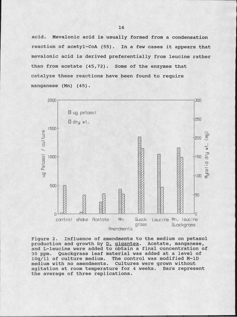
16acid. Mevalonic acid is usually formed from a condensation reaction of acetyl-CoA (55). In a few cases it appears that mevalonic acid is derived preferentially from leucine rather than from acetate (45,72). Some of the enzymes that catalyze these reactions have been found to require manganese (Mn) (45).
§ ug petosoI
0 dry wt.
o 1000
Quack Leucine Mn, leucinecontrol shake Acetategrass Quackgrass
Amendments
Figure 2. Influence of amendments to the medium on petasol production and growth by JD2, gigantea. Acetate, manganese, and L-Ieucine were added to obtain a final concentration of 30 ppm. Quackgrass leaf material was added at a level of 10g/ll of culture medium. The control was modified M-ID medium with no amendments. Cultures were grown without agitation at room temperature for 4 weeks. Bars represent the average of three replications.
Mycelia dry wt.
(mg)
17The precursors of mevalonic,acid, acetate and leucine,
were tested for their ability to increase petasol production. At a concentration of 30 ppm, L-Ieucine significantly increased toxin production while acetate showed no significant effect (Fig. 2). These data indicate that Di. cfioantea preferentially uses L-Ieucine as a precursor to mevalonic acid and subsequent terpenoid synthesis. The reported manganese requirement of the catabolic enzymes led to the inclusion of manganese in the form of MnCl2 in the liquid media. At a concentration of 30 ppm, Mn significantly increased petasol production above control levels but, did not stimulate production to the extent of leucine (Fig. 2).
Plant Products and Petasol ProductionOne aspect of toxin production that has been virtually
unexplored is the influence of host metabolites on toxin production. Matern et al. (44) characterized a plant glycolipid that activated toxin production in Helminthosporium sacchari. This report followed an earlier report in which serinol was identified as an "activator" of toxin production by Hi. sacchari (51) .
Leaf material from quackgrass, a host of Di. qiqantea. was added to the culture media prior to autoclaving. This led to a significant increase in petasol production along with an increase in fungal growth as seen in the dry weight
18of the mycelial mat (Fig. 2). When plant material was added to the culture medium along with Mn and leucine, two other compounds that slightly stimulated production, no additive or synergistic effect could be detected (Fig. 2) suggesting that there is an upper limit to toxin production.
To further investigate the nature of the stimilatory ability of quackgrass, a crude extraction and fractionation of quackgrass leaf tissue was carried out. The "organic phase" fraction, containing the organic solvent soluble plant compounds, significantly stimulated petasol production and was statistically equivalent in stimulation to the crude plant homogenate (Fig. 3). The "amino acid" fraction was also found to increase petasol production. The effect of the "amino acid" fraction was significantly greater than the "organic phase" fraction but was not statistically different than the crude plant homogenate. The stimulatory effect of the "amino acid" fraction may be solely due to an increased concentration of leucine in the media, however, no further studies were done in this area. The fact that the host plant tissue does have an effect on toxin production indicates an interaction at the molecular level between the host and pathogen in regulation of toxin production.

19
i ug petasol 0 dry wt.
b; 2500
m 1500
control Plant Organic Organic Amino Neutral Homogenate Phase fields Acids Sugars
Plant FractionFigure 3. Effect of different plant fractions on petasol production and growth by D _ gigantea. Aliquots (200 1) from the plant fractions were added to the medium prior to autoclaving. Cultures were grown without agitation at room temperature for 4 weeks. Bars represent the average value of three replicates.
Effect of Sterol Biosynthesis Inhibitors on Petasol Production
Another strategy in trying to increase production of a desired molecule is to manipulate pathways in an attempt to divert or funnel more of the available carbon into the molecule of interest. Eremophilanes split off from the mevalonic acid pathway at the point of farnesyl pyrophosphate. The farnesyl pyrophosphate pool is drawn off
Myce
lio dr
y wt.
(mg)
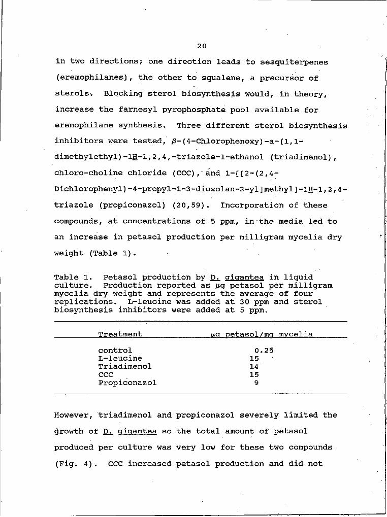
20in two directions; one direction leads to sesquiterpenes (eremophilanes), the other to squalene> a precursor of sterols. Blocking sterol biosynthesis would, in theory, increase the farnesyl pyrophosphate pool available for eremophilane synthesis. Three different sterol biosynthesis inhibitors were tested, /8-(4-Chlorophenoxy) -a-(1,1- dimethylethyl)-1H-1,2,4,-triazole-l-ethanol (triadimenol), chloro-choline chloride (CCC), and l-[[2-(2,4- Dichlorophenyl)-4-propyl-l-3-dioxolan-2-yl]methyl]-1H-1,2,4- triazole (propiconazol) (20,59). Incorporation of these compounds, at concentrations of 5 ppm, in the media led to an increase in petasol production per milligram mycelia dry weight (Table I).
Table I. Petasol production by 1%. oicrantea in liquid culture. Production reported as ng petasol per milligram mycelia dry weight and represents the average of four replications. L-Ieucine was added at 30 ppm and sterol biosynthesis inhibitors were added at 5 ppm.
Treatment uq oetasol/mg mvceliacontrol 0.25L-Ieucine 15Triadimenol 14CCC 15Propiconazol 9
However, triadimenol and propiconazol severely limited the growth of Djl qigantea so the total amount of petasol produced per culture was very low for these two compounds (Fig. 4). CCC increased petasol production and did not
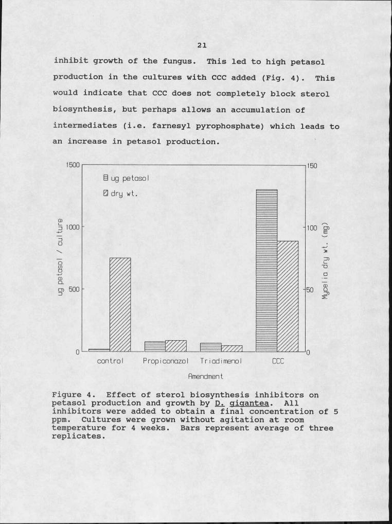
21inhibit growth of the fungus. This led to high petasol production in the cultures with CCC added (Fig. 4). This would indicate that CCC does not completely block sterol biosynthesis, but perhaps allows an accumulation of intermediates (i.e. farnesyl pyrophosphate) which leads to an increase in petasol production.
B ug petasol 0 dry wt.
o 1000
control Propiconazol TriadimenolAmendment
Figure 4. Effect of sterol biosynthesis inhibitors on petasol production and growth by gigantea. Allinhibitors were added to obtain a final concentration of 5 ppm. Cultures were grown without agitation at room temperature for 4 weeks. Bars represent average of three replicates.
Myce
lio dr
y wt.
(mg)
22Petasol Production Over Time
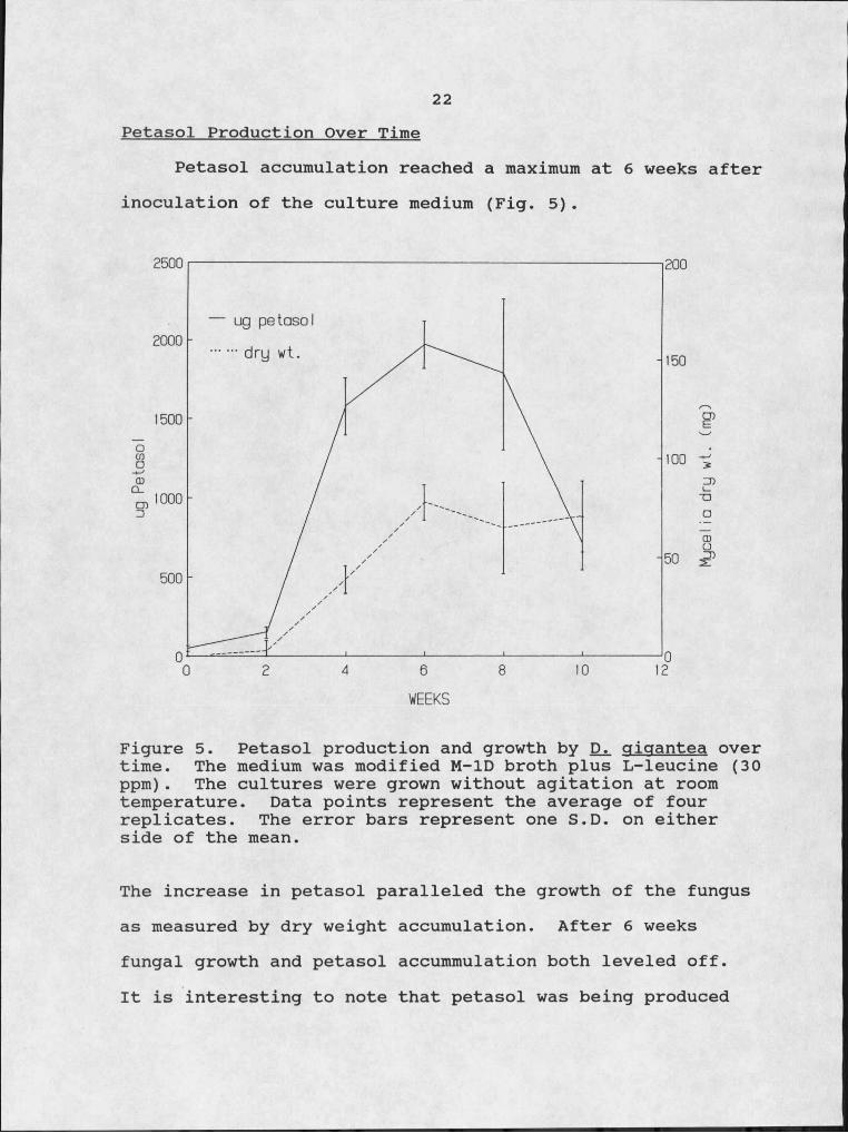
Petasol accumulation reached a maximum at 6 weeks after inoculation of the culture medium (Fig. 5).
— ug petasol
.. dry wt.
WEEKS
Figure 5. Petasol production and growth by gigantea over time. The medium was modified M-ID broth plus L-Ieucine (30 ppm). The cultures were grown without agitation at room temperature. Data points represent the average of four replicates. The error bars represent one S.D. on either side of the mean.
The increase in petasol paralleled the growth of the fungus as measured by dry weight accumulation. After 6 weeks fungal growth and petasol accummulation both leveled off.It is interesting to note that petasol was being produced

23during the growth phase of the fungus. This would suggest that the eremophilanes may play a role in the normal physiology of the fungus and are not just waste products of the stationary growth phase.
After 8 weeks there was a.decrease in the amount of petasol in the culture filtrate. This decrease indicates that petasol is degraded; either by fungal produced products, compounds in the media, or by natural instability.
Labeling Petasol with T14Cl[14C]-labeled L-Ieucine, sodium acetate,
mevalonolactone and glucose were each added separately to the media at the time of inoculation. The fungus incorporated [14C] into petasol with the highest specific activity when [14C]-sodium acetate was added to the culture medium (Fig. 6). [14C]-Mevalonolactone yielded a higherspecific activity in petasol then either [14C]-glucose or [14C]-leucine. The low specific activity of petasol obtained from the [14C]-leucine culture was very surprising since earlier data indicated that L-Ieucine was perhaps a precursor to the eremophilanes. The conversion of leucine into mevalonic acid goes through a /3-methylglutaconyI-CoA intermediate formed by cleavage of the leucine carboxyl carbon. The commercial radiolabeled leucine was labeled at the carboxyl carbon, so the [14C] would be lost in the formation of mevalonic acid, accounting for the low specific radioactivity.
24COIOX
gCX
UC
ZD
>
Q
§T l
OCDCL
CO
MC-Leucine MC-GIucose MC-MevaIonolactateAmendments
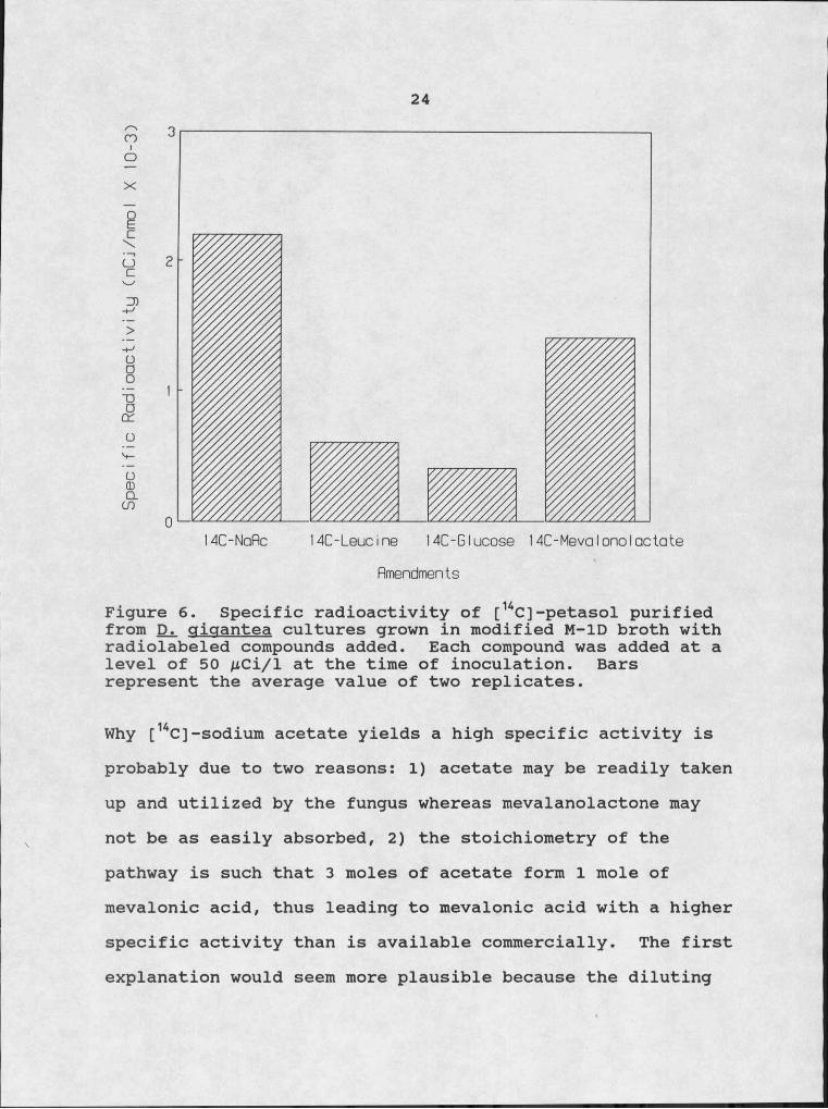
Figure 6. Specific radioactivity of [KC]-petasol purified from Djl qjqantea cultures grown in modified M-ID broth with radiolabeled compounds added. Each compound was added at a level of 50 AtCi/1 at the time of inoculation. Bars represent the average value of two replicates.
Why [14C]-sodium acetate yields a high specific activity is probably due to two reasons: I) acetate may be readily taken up and utilized by the fungus whereas mevalanolactone may not be as easily absorbed, 2) the stoichiometry of the pathway is such that 3 moles of acetate form I mole of mevalonic acid, thus leading to mevalonic acid with a higher specific activity than is available commercially. The first explanation would seem more plausible because the diluting
25effect of naturally produced acetate from non-radioactive sources would greatly decrease the chances of two or three radiolabeled acetate molecules being condensed to form mevalonic acid.
Summary
Eremophilane production by JDi. oicrantea was stimulated by compounds found in the host plant. These compounds may be simple amino acids and/or more complex organic solvent soluble compounds. Plant products also increased the growth of the fungus. L-Ieucine and CCC, a sterol biosynthesis inhibitor, also stimulated petasol production. Di. qicrantea produced eremophilanes during its growth phase and during later stages of stationary growth petasol was degraded.
One obvious question that could be asked about eremophilane production by JDi. crioantea is, are eremophilanes produced during a natural infection? Several attempts at infecting different hosts were not successful and naturally infected material could not be obtained, so this question cannot be answered at this time.
26
CHAPTER III;
STRUCTURE-ACTIVITY RELATIONSHIPS OF THE EREMOPHILANES.
Introduction
Until recently structure-activity correlations with fungal toxins have been more the exception than the rule.The information provided by studies on structure-activity relationships may help in determining mode of action. Functional groups required for activity may indicate a particular type of action. Certain structural features may be found to be required for binding or inserting into an active site.
Investigations on other eremophilanes, specifically, phomenone and PR-toxin have been carried out. Phomenone, a known phytotoxic metabolite produced by certain phytopathogenic Phoma species (54), is also produced by Drechslera cricrantea. PR-toxin, which has the eremophi lane ring system (71), is a mycotoxin produced by Penicillium rocrueforti and is strongly toxic to mice and rats (70) .
The biological activity of PR-toxin was attributed to the presence of the aldehyde group at C-13 (47). Capasso et al. (11), looking at both phomenone and PR-toxin, found that the aldehyde function did not account for the overall
27toxicity of the PR-toxin. Their data indicated that structural modifications of the eremophilane ring, arising from oxidation or reduction reactions, had variable effects on toxicity, whereas de-epoxidation abolished biological activity. They concluded that the epoxy ring of eremophilanes played a role in the biological activity.
The structural diversity of the eremophilanes produced by Djl cficrantea is very amenable to structure-activity relationship studies. Derivatives of phomenone and petasol, 13-aldophomenone and 13-aldopetasol respectively, were included in the study to obtain further characterization of the structure-activity relationships. The biological activities of the eremophilanes were compared using three different bioassays: I) effect on detached leaves of 11 plant species using the leaf puncture wound-droplet overlay assay, 2) effect on rhizogenesis in mung bean (Phaseplus aurepus) hypocotyls and 3) mortality of brine shrimp CArtemia salina).
Materials and Methods
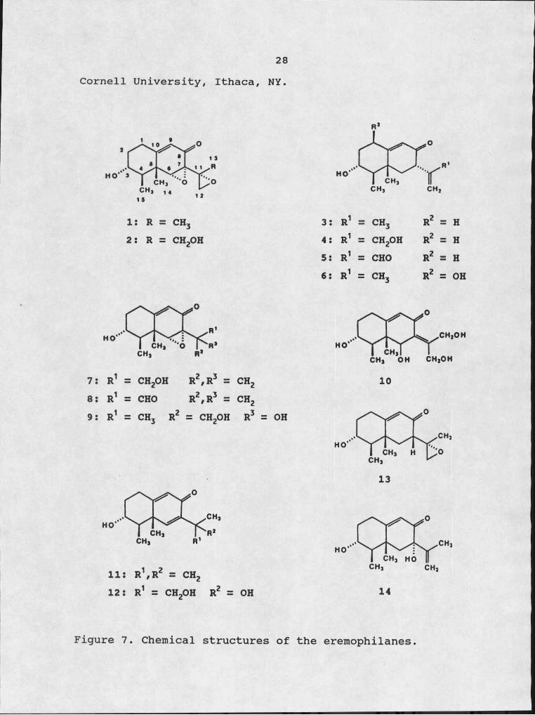
EremophilanesThe structures of all the eremophilanes used in this
study are listed in Figure 7. Petasol(S), gigantenone(l), and 11,12-epoxypetasol(13) were purified from culture filtrates of Di cficrantea as previously described (28) . 1-Hydroxypetasol(6) was kindly provided by Dr. Yali Hallock,
28Cornell University, Ithaca, NY.
I: R = CH3 2: R = CH2OH
3: R1 = CH3 R2 = H4: R1 = CH2OH R2 = H5S R1 = CHO R2 = H6: R1 = CH3 R2 = OH
7: R1 ZZ CH2OH R2zR3 = CH28: R1 =Z CHO R2zR3 = CH29: R1 CH3 R2 = CH2OH R3 OH
11: R1zR2 = CH212: R1 = CH2OH R2 = OH
CH1OH
CH1 OH CH1OH
CH1 H
14
Figure 7. Chemical structures of the eremophilanes

/29
The remaining eremophilanes and their derivatives were gifts from Dr. Fumio Sugawara, RIKEN Institute, Japan. The isolation, purification and derivatization of these compounds has been previously described (28).
Leaf Puncture Wound Droplet Overlay AssayPlants were grown under controlled environmental
conditions of 8 hours of darkness at 2 S0C and 16 hours of light at 32°C/day. The leaves of dicots were detached at the base of the petiole and the apical 6 cm of the monocot leaves were excised for testing. The leaf puncture wound- droplet overlay assay consists of a puncture being made in the detached leaf with a 22 gauge needle, the wound is then overlayed with I to 10 ^l of the solution to be tested (64). Methanol (10%) was used to solubilize the eremophilanes and as a control solution. The leaves were incubated in a petri dish containing moistened filter paper. After 72 hours the leaves were checked for symptom development. The nature and extent to which the leaf tissue was affected was measured and recorded.
Root-Induction AssayTen day old mung bean seedlings were harvested and the
cotyledons were removed. The stems were trimmed leaving 3 cm of hypocotyl below the point of cotyledon attachment plus the shoot which included two primary leaves and the apical bud. Five cuttings were placed in a test tube
30containing 3ml of test solution at a concentration of I X IO"5 M. The tubes were placed under constant light (18 /LtE/m2/sec) at 25°C for 24 hours. The cuttings were then placed in individual test tubes containing 5ml of distilled water and incubated under constant light. After incubation, the roots on the hypocotyl were counted.
Brine Shrimp AssayBrine shrimp eggs were hatched in artificial seawater
(Instant Ocean™, Aquarium Systems) in an Erlenmeyer flask (125 ml) , shaking for 24 hours at 25°C. Aliquots containing 15 to 35 shrimp larvae were transfered to wells of a 96 well micro-titer plate. Eremophilanes solubilized in 10% methanol (v/v) were added to the wells to a total volume of 50 jLtl and a final concentration of I X 10"4 M. Equal volumes of 10% methanol was added to control wells. After 48 hours at 25°C the larvae mortality was recorded.Mortality was determined by counting the number of dead larvae, then adding 100 of 37% formaldehyde to kill the remaining larvae and counting total larvae.
Results and Discussion
Leaf AssayGigantenone (I) is known to cause green islands on
monocots and necrosis on dicots, with the exception of cucumber (Cucumis satiya), in the leaf puncture wound-

31droplet overlay assay (37). As can be seen from Table 2, I gave typical green islands on all five monocots (a-e) tested and on cucumber, while only necrotic or chlorotic lesions were observed on the other dicots (f-k). 2, 3, 4, 7, and 14all followed this same pattern of green islands on monocots and necrosis or chlorosis on dicots. 3 contains no epoxy rings in its structure, while I has two epoxy rings. Both 3 and I are highly active, yet 13 which has intermediate epoxidation shows greatly reduced activity on both monocots and dicots. 14 and 4, which are hydroxylated versions of 3, were less active and affected fewer plant species than 3.The addition of an aldehyde group at C-13 of 3 and 7 led to the formation of 5 and 8, respectively. This oxidation led to decreased activities for both 5 and 8 when compared to their parent compounds. The highly oxidized eremophilanes 9, 10, and 12 showed little green island forming activity, causing necrosis or having no reaction on most of the plants tested. In addition, 10 and 12 show some selectively, affecting only a few of the plant species tested. This selectivity may have potential in herbicide developement studies.

Table 2. Effects of eremophilanes, using the leaf puncture wound droplet overlay assay, on some species of monocots (a—e) and dicots (f—Jc) at 15—20 nmol. All compounds with the exception of 5 and 8 have been isolated from culture filtrates of D. criaantea. Observations were made after 72 hours.
Plants 3 I 7 2 14 4Compounds 6 5 8 11 13 9 10 12
a. Zea mays 'W64A' g+++ g+++ g+++ 9+++ 9++ 9+++ g++ 9++ 9+++ 9+ 9+ 9+ - - - -b. Avena sativa 'Park' g+++ g+++ 9++ 9++ 9+ 9+ 9+ 9+ 9+ - - - -c. Aoroovron reoens g+++ g+++ g+++ g+++ 9++ * - n* n* -- n** n* - -d. Cvnodon dactvlon g+++ 9++ 9+ g++ 9+ 9+ 9+ -- 9+ 9+ 9+ 9+ 9+ 9+e. Sorohum haleoense g+++ g+++ g++ 9++ g+++ 9+ 9+ g++ 9+ 9+ 9+ - n** n*f . Lvcooersicon esculentum n*** n** n*** n*** n* n* n* n** n* C** n*** - ~ - -o. Euohdrbia heteroohvlia c*** c* Q * * C** - ~ n** c*** c*** C** c* - -h. Cucumis sativa 9++ g+++ 9++ 9+ n** n* n* n*i. Helianthus annus n** — 9++ n** 9+ — n* n* - - - - n* - - " -i. Glvcine max 'Hobbit' n*** c* n** n* n* n** n** n** n* n* -- . n*k. Phaseolus aureous n*** n*** n*** . n** n* n** - - n* n** n* n* - -g=Green island; n=Necrosis; C=Chlorosis; (--)=No response Lesion size: +++,>15mm2; ++,5-15mm2; +<5mm2
***,>10mm dia.; **,5-10mm dia.; *,<5mm dia.
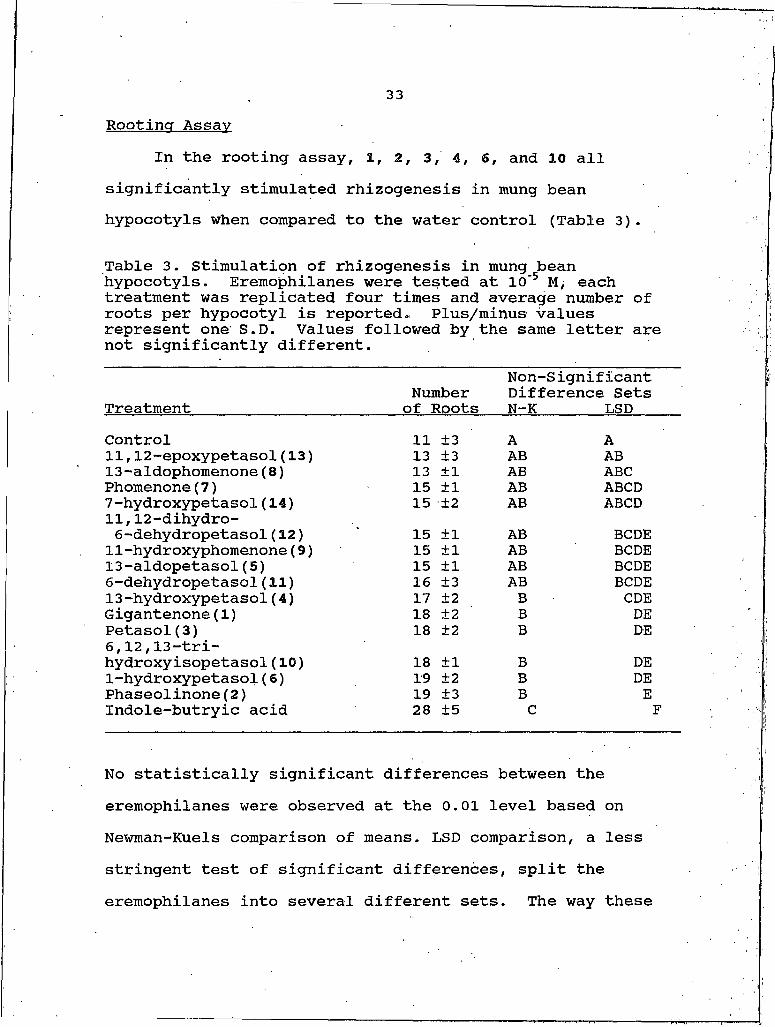
33Rootincr Assay
In the rooting assay, I, 2, 3, 4, 6, and 10 all significantly stimulated rhizogenesis in mung bean hypocotyls when compared to the water control (Table 3).
Table 3. Stimulation of rhizogenesis in mung bean hypocotyls. Eremophilanes were tested at IO'5 M, each treatment was replicated four times and average number of roots per hypocotyl is reported. Plus/minus values represent one S.D. Values followed by the same letter are not significantly different.
TreatmentNumber of Roots
Non-Significant Difference Sets N-K LSD
Control 11 ±3 A A11,12-epoxypetasol(13) 13 ±3 AB AB13-aldophomenone(8) 13 ±1 AB ABCPhomenone(7) 15 ±1 AB ABCD7-hydroxypetasoI(14) 15 ±2 AB ABCD11,12-dihydro- 6-dehydropetasol(12) 15 ±1 AB BCDEII-hydroxyphomenone(9) 15 ±1 AB BCDE13-aldopetasol(5) 15 ±1 AB BCDE6-dehydropetasol(11) 16 ±3 AB BCDE13-hydroxypetasol(4) 17 ±2 B CDEGigantenone(I) 18 ±2 B DEPetasol(S) 18 ±2 B DE6,12,13-tri- hydroxy isopetasol (10) 18 ±1 B DE1-hydroxypetasol(6) 19 ±2 B DEPhaseolinone(2) 19 ±3 B EIndole-butryic acid 28 ±5 C F
No statistically significant differences between the eremophilanes were observed at the 0.01 level based on Newman-Kuels comparison of means. LSD comparison, a less stringent test of significant differences, split the eremophilanes into several different sets. The way these
34sets were grouped indicates that this activity may be associated with degree of oxidation about the ring system. Two exceptions to this generality are 10 and 13. 10 ishighly oxidized with its three hydroxyl groups yet was very effective in stimulating rhizogenesis. 13 has a lower oxidation state than either I or 3, which were very active, but it has little stimulatory activity.
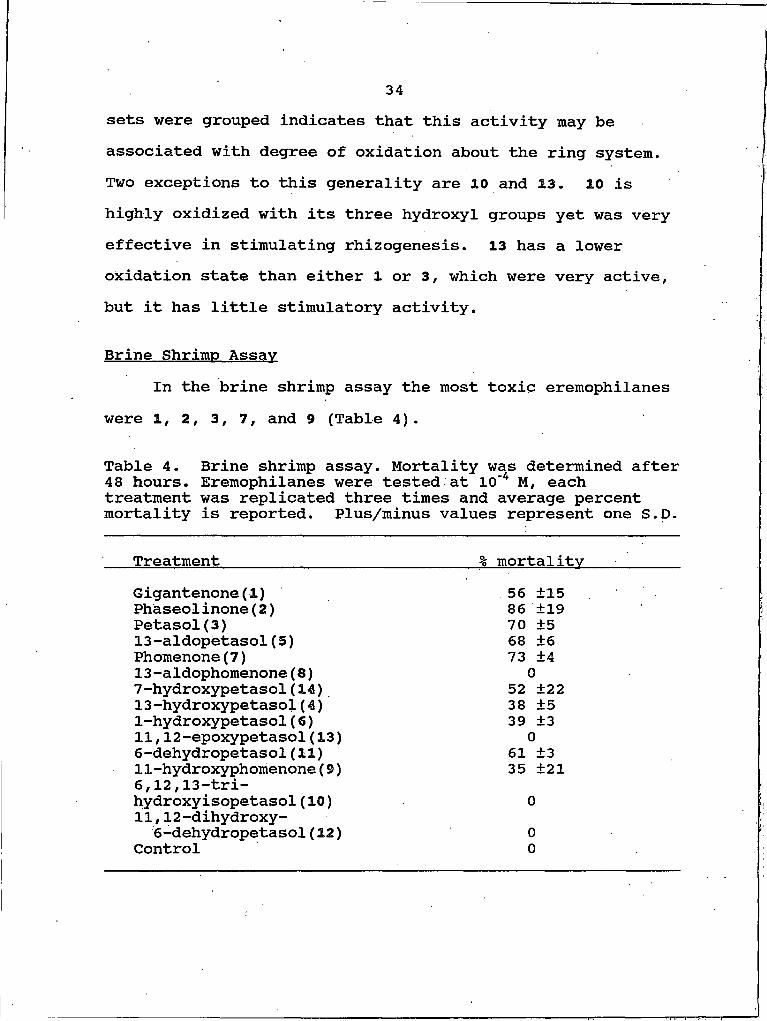
Brine Shrimp AssayIn the brine shrimp assay the most toxic eremophilanes
were I, 2, 3, 7, and 9 (Table 4).
Table 4. Brine shrimp assay. Mortality was determined after 48 hours. Eremophilanes were tested at 10‘4 M, each treatment was replicated three times and average percent mortality is reported. Plus/minus values represent one S.D.
Treatment % mortalityGigantenone(I) 56 ±15Phaseolinone(2) 86 ±19Petasol(S) 70 ±513-aldopetasol(5) 68 ±6Phomenone(7) 73 ±413-aldophomenone(8) 07-hydroxypetasol(14) 52 ±2213-hydroxypetasol(4) 38 ±51-hydroxypetaspl(6) 39 ±311,12-epoxypetasol(13) 06-dehydropetasol(11) 61 ±311-hydroxyphomenone(9) 35 ±216,12,13-trihydroxy isopetasol (10) 011,12-dihydroxy-
6-dehydropetasol(12) 0Control 0

35Again, with a few exceptions, the trend indicates that higher oxidation levels are less toxic (i.e. less biologically active). The most striking observation was the difference between 7 and its aldehyde derivative 8. The conversion of the C-13 hydroxyl to an aldehyde completely abolished toxicity.
Summary
Moule et al. (47) reported that some biological properties of PR-toxin are dependent on the presence of the aldehyde function at C-13. The results from the brine shrimp assay indicated that the C-13 aldehyde function is not neccesary and may even abolish activity. Capasso et al. (11) presented evidence that the C-13 aldehyde group is not necessary; this supports my findings. Additionally, they suggested that the epoxy function was critical for biological activity. However, their evidence was based on two modifications of phomenone at different sites on the ring system, one eliminated the epoxide, the second was addition of a methoxy to C-9. This makes it difficult to assign the loss of activity to the de-epoxidation or to the methoxy addition. The data reported here on the biological activities of the eremophilanes show epoxidation is not necessary. 3 was very active in every assay while 13, which is an epoxidated analog of 3, was much less active in thesame assays.

36I, 2, 3, and I most consistently showed high biological
activities in the three assays used. There is no evident functional group that is present in these molecules and lacking in the less active ones that would account for activity. The results do suggest that the degree of oxidation of the eremophilane could play a role in the biological activity, though no clear trend is evident and exceptions do exist.
Molecular modeling, using computer generated models, could be used to study the electronic configurations of these molecules. This information would reveal electronXdense areas, patches of hydrophobicity and hydrophilicity on the outer surface of the molecule, and other molecular characteristics that may be related to activity.
37
CHAPTER IV
PHYSIOLOGICAL INVESTIGATIONS: MODE OF ACTION
Introduction
Studies on the mode of action of phytotoxins historically have been accorded great promise for yielding details of the chemical mechanisms at the level of plant cell metabolism (42). Intuition suggests plant metabolic regulation will be altered at the molecular site of action in plant-toxin interactions. The objective, of course, is to learn where or how the phytotoxin interacts with the host cell.
A number of sites of toxin action have been suggested (46). However, studies on the exact description at the molecular level of toxin-target interactions have been complicated by several factors. Some of the difficulties limiting precise conclusions include : I) requirement for chemically pure toxins, 2) structurally characterizing the toxin, 3) multiple effects due to multiple targets of the toxin. The first arises directly from the intent of such studies; a molecular explanation of toxin action, not an explanation of the effects of contaminating compounds of either fungal or media origin. Secondly, accurate

38structural information can facilitate subsequent studies designed to relate the structural characteristics of the toxic compound to its molecular role. Structural analogies between the toxin and intermediary metabolites in the host plant constitute a basis for focusing attention of specific metabolic sequences or physiological parameters. The multiple and diverse effects often observed for many toxins ' complicate the notion that there is a single site of action. The multiple effects may well be a cascade of events triggered by action at a single site.
With the eremophilanes the first two difficulties have already been overcome. However, their multiple effects at the physiological level such as green island formation on monocots, necrosis on dicots, stimulation of rhizogensis, and pleiotropic effects in tissue culture (37), make the problem more complex.
Understandably, the focus of research with phytotoxins is on plant species or tissues in which the producing fungus or the phytotoxin itself plays a role in nature. However, some notable advances in understanding the molecular basis for toxin action have occurred with studies on nonhost species. Fusicoccin offers an illustration of the potential role of nonhost species in investigating the mode of action of a toxin (42). The choice of plant species or plant tissue is based on several considerations including: I) sensitivity to phytotoxin, 2) reliable bioassay, 3) ease of
39handling, 4) availability, and 5) genetic variation in plant tissue. With these considerations in mind Avena sativa cv. Park was chosen with which to work. Park oat consistently yields green islands when treated with petasol and gigantenone but is relatively insensitive to 11,12- epoxypetasol. The width and flatness of the oat leaf facilited the use of the puncture wound droplet overlay assay and a ready source of a genetically homogenous population was available.
The molecular site of action is determined, in the final analysis, from.results of bioassays. The number of assays required is dependent on the ability of the assays to probe molecular events. When the molecular action of the phytotoxin is unknown, several assays are run in a 'shotgun' fashion to look at possible metabolic effects. To make the approach somewhat methodical, I relied on the use of visual symptoms as a molecular site predictor, focusing on the green island phenomenon.
In this chapter the bioassays, and their results, that were used to investigate the molecular action of eremophilanes are discussed. Based on these results a probable mode of action is presented.
40Materials and Methods
Plant MaterialsAvena sativa cv. Park seeds were kindly provided by Dr.
Tom Blake, Montana State University, Bozeman, MT. Plants were grown in vermiculite under environmentally controlled conditions of 18 hours light, 6 hours dark/day with a constant temperature of 26°C.
Chlorophyll Retention and Quantification of the Green Island Effect
The leaves of 10 day old oat seedlings were excised 5 cm from the tip and a puncture was made in the center of the detached leaf with a 22 gauge needle. The puncture wound was overlayed with 10-25 nmole eremophilane (or other test solutions) solubilized in 5-10% methanol. Control leaves were treated with 10% methanol. The leaves were incubated in a moist chamber in the dark at 24-26°C. As measured from the site of application, I cm sections were cut from the leaves as follows: base = 0.5 cm to 1.5 cm basipetal to puncture; middle = 0.5 cm basipetal to 0.5 acropetal to puncture; and tip = 0.5 cm to 1.5 cm acropetal to punctures. Chlorophyll was extracted without maceration following the procedures of Hiscox and Israelstam (32). Individual leaf segments were placed in test tubes containing 3 ml dimethylsulfoxide (DMSO). The tubes were then placed in a water bath at 65°C for 30 min. The leaf segments were
41removed and dried at IOO0C for 24 hours. The absorbance of the resulting DMSO sample was read at 665 nm using a Beckman DU-50 spectrophotometer. Chlorophyll content was recorded as the absorbance at 665 nm divided by the dry weight in grams of the leaf tissue.
Chlorophyll Content = __A665__dry wt. (g)
Preparation of Crude Membrane FractionCrude membrane preparations were obtained from oat leaf
tissue following the methods of Strobel (61). Leaf tissue (5 g) from 10 day old seedlings was cut into pieces approximately I cm long. These pieces were ground in 30 ml of 50 mM Tris-HCl, pH 7.2, for 2 min. in a Sorvall Omni- Mixer at top speed. The homogenized material was passed through several layers of cheesecloth to remove debris. The filtrate was centrifuged at SOOxg for 10 min., the pellet was discarded and the supernatant centrifuged at 48,OOOxg for 20 min. The resulting pellet, containing the crude membrane preparation, was resuspended in 10 mM Tris-HCl buffer, pH 7.2.
Binding AssayBinding activity was investigated using two different
techniques. Aliquots (I ml) of the resuspended membrane preparation were incubated with either [14C] -petasol or a cold' petasol: [14C]-petasol (10:1) mixture for 30 min. at
42room temperature. Following incubation the aliquots were centrifuged at 48,OOOxg for 20 min. The supernatant was saved and the resulting pellet was resuspended in I ml of 10 mM Tris-HCl buffer, pH 7.2. Radioactivity of aliquots from both the supernatant and the resuspended pellet were determined using a Packard Tri-Carb Liquid Scintillation counter. Each sample was solubulized in 10 ml Aquasol and counted for 50 min. The counting time varied slightly depending on the sample, this was done to ensure enough counts were made («10,000 minimum) to obtain a statistically reliable estimate of dpm (4). Cpm was converted to dpm using a quench correction curve.
In the second test for binding activity, [14C]-petasol was incubated with an aliquot of the resuspended membrane preparation for 30 min. The sample was then loaded onto a Sephadex G-25 column (0.9 X 30 cm) equilbrated with 10 mM Tris-HCL, pH 7.2, the column was eluted with the equilibration buffer. Fractions (2 ml) were collected and radioactivity of the fractions was determined as described above.
Determination of Sink ActivityLeaves from 10 day old oat seedlings were trimmed 5 cm
from the tip. Two punctures were made in each leaf, one being I cm from the tip and the other being made I cm from the base. Approximately 50,000 dpm of [14Cj-Iabeled amino
43acid or sugar were applied to the acropetal puncture, 5 jug of petasol in 5 jul of 10% methanol was applied to the basipetal puncture. The leaves were placed in a moist chamber and incubated for 4 days either in the dark or under intermittent light conditions (6 /iE/m2/sec, 12 hr/day) .After incubation the leaves were cut in half and placed in scintillation vials. The leaf halves were digested by adding 0.5 ml perchloric acid:hydrogen peroxide (1:2) and placing the vials at IOO0C for 30 min. according to Bhuller et al. (5). Radioactivity of the digests was determined by adding 10 ml Aquasol and counting vials for 50 min. in a scintillation counter.
CO2 Fixation ExperimentsThe apical 5 cm of leaves from 10 day old oat seedlings
were excised, placed in a moist chamber, treated with 20 nmole of the test solutions (see puncture wound overlay assay above), and incubated for 3 days. Fresh leaf pieces were obtained and used as a control whereas leaves treated with 10% methanol and incubated were used as incubated controls. The leaves were exposed to 20 juCi of [14CJ-CO2 released from [14C] -sodium bicarbonate having a specific- activity of 8.4 juCi/mmole. The reaction chamber was incubated under cool white fluorescent light (18 juE/m2/sec) or in the dark. A I cm segment was taken from each leaf for [14C] analysis. A segment from 0.5 cm basipetal and 0.5 cm

44acropetal to the puncture was excised. Each leaf segment was digested and radioactivity determined as described above.
Chloroolast Isolation and the Hill ReactionChloroplasts were isolated from two sources, leaves
from 10 day old oat seedlings and from mature spinach leaves pruchased at a local grocery store, according to Leegood and Walker (41) . Leaf tissue (10 g) was ground in 50 ml of a 500 mM sucrose, 100 mM potassium phosphate, pH 7.4 buffer for 10-15 seconds in a Sorvall Omni-Mixer at 50% power. The grindate was filtered through several layers of cheesecloth to remove debris. The filtrate was centrifuged at IOOOxg for 10 min., the supernatant was discarded and the pellet containing the chloroplasts was resuspended in 2 ml HEPES- sorbitol buffer containing, 330 mM sorbitol, 2 mM EDTA, I mM magnesium chloride, I mM manganese chloride and 50 mM HEPES, pH 7.6. The chlorophyll content of this suspension was determined by diluting a 10 p.1 aliquot of the suspension to I ml with 95% ethanol and measuring the absorbance at 642 nm. The absorbance was used in the following calculation:
chlorophylla+b = X 1000 X 100 (dillution factor)39.4
The Hill reaction was followed by monitoring the reduction of 2,6-dichlorophenol-indolephenol (DCPIP). The reaction mixture consisted of a 25 /Ltg chlorophyll equivalent
45of chloroplast suspension, 10 /itg of test compound, 200 //I of 0.2 inM DCPIP and HEPES-sorbitol buffer to a total volume of I ml. The reaction was run for ten minutes under constant light. Dark controls were run for comparison. The absorbance was read at 580 nm. Activity was expressed as percent change in A580 of treatment relative to the control.
Proteolytic Enzyme AssayProteolytic enzyme activity was determined using the
assay of Peterson and Huffaker (49), with modification. The assay solution contained : 0.2 ml protein preparation (100 units), 0.4 ml 0.2 M K5PO4, pH 6.0, 0.1 ml eremophilane solution, 0.3 ml Azocasien (10 mg/ml). The assay solution was incubated 2 hours at 3 O0C. Two ml of cold 12% perchloric acid (PCA) were added to stop the reaction and precipitate the protein. The tubes were spun in a swinging bucket rotor at I,500xg for 10 min and 0.5 ml of the supernatant was diluted to 1.5 ml with distilled water. The absorbance of the diluted solution was read at 340 nm. Activity was calculated as follows:
I unit = (A340/mg of enzyme/min) X 3 (dilution factor)
Extraction of Soluble ProteinLeaves from 10 day old oat seedlings were treated with
assay solutions and incubated as described above. After incubation, leaf segments were excised, weighed and quick
46frozen with dry ice. The frozen leaf segments were pulverized with a mortar and pestle and 5 times the weight in volume of extraction buffer was added. Extraction buffer contained 250 mM sucrose, 100 mM ammonium sulfate, 20 mM magnesium acetate, 5 mM /3-mercaptoethanol, and 50 mM MES, pH 6.5. The ground tissue was thoroughly mixed with extraction buffer then filtered to remove debris. The soluble protein fraction was kept at approximately 40C at all times.Protein was determined by the method of Bradford (7). For this analysis Bio-Rad Protein Dye reagent was used with bovine serum albumin (BSA) as the standard. Protease activity of the leaf extracts was determined as described above with slight modifications. The assay mixture consisted of 80 mM potassium phosphate, pH 6.0, 3 mg/ml Azocasien and soluble protein preparation to a final volume of I ml.
SDS-Polvacrvlamide Gel ElectrophoresisThe discontinuous SDS buffer system described by
Laemmli (40) was followed with some modifications. Gel slab dimensions were 0.75 X 160 X 180 mm. The separating gel contained 10% acrylamide, 0-26% bisacrylamide, 0.1% SDS, 375 mM Tris-HGl, pH 8.8, 0.033% ammonium persulfate, 0.033%(v/v) TEMED. The stacking gel contained 4.0% acrylamide, 0.1% bisacrylamide, 0.1% SDS, 125 mM Tris-HCl, pH 6.8,0.008% ammonium persulfate, 0.005% TEMED. Samples,
47containing 20 to 40 ng of protein, were mixed 1:1 volume ratio with sample buffer containing 125 mM Tris-HCl, pH 6.8, 4.0% SDS, 10.0% y3-mercaptoethanol, 0.01% bromophenol blue and 40% sucrose. Samples were heated for two minutes at IOO0C, 25 to 75 JLil aliquots were applied to the gels. Electrode buffer containing 19 mM glycine, 25 mM Tris-HCL,. pH 8.5 and 0.1% SDS was used in both electrode chambers. Electrophoresis was conducted at 40 mA with variable voltage until the bromophenol blue tracking dye reached the bottom of the gel. Duration of electrophoresis was about 2.5 to 3 hours.
Protein was stained with Coomassie Blue (CB) following the procedures of Howard and Traut (33). Gels were soaked 4-10 hours in a 50% methanol, 10% acetic acid and 0.25% CB solution. The gels were then destained with several rinses of a 50% methanol and 10% acetic acid solution. After sufficient destaining, 5 ml of glycerol was added to 200 /Li! of the destaining solution and the gels were soaked for I hour, then dried using a Hoeffer Model SE 540 gel drier.
In vitro Protein TranslationThe wheat germ extract in vitro translation system of
Promega™ was used for all in vitro studies. Translation analysis was performed by incubating I jug of Brome Mosaic virus (BMV) RNA up to 120 minutes in a 50 jul reaction.[14C]-Leucine incorporation was determined by alkali
48resistant, trichloroacetic acid (TCA) precipitable counts and SDS-polyacrylamide gel electrophoresis. The reaction mixture consisted of 25 fil wheat germ extract, 4 /il I mM amino acid mixture (minus leucine), 2 fil RNA (BMV RNA at 0.5 Hg/111) , 2.5 Ail [14C]-leucine (250,000 DPM total) f 5 ^l I M potassium acetate, 11.5 jil of test solution (eremophilanes or other controls to a final concentration of I X 10'3 M) , for a total volume of 50 ill. The reaction mixture was incubated at 25°C for 120 minutes. Aliquots (2 ill) were removed at 30 minute intervals and TCA precipitable counts determined. The following protocol was used for TCA precipitation: I) to the 2 ill aliquot add 1.0 ml of IN sodium hydroxide, 1.5% hydrogen peroxide and incubate for ten minutes at 37°C, 2) add 40 ml of ice cold 25% TCA and incubate on ice for 30 minutes, 3) collect the precipitates on Whatman GF/C filters with suction and wash the filter with 10 ml ice cold 8% TCA, 4) wash the filters with a few milliters of cold acetone and pull the vacuum until the filters are dry. The filters were analyzed by standard scintillation counting techniques as described above.At the end of the 120 minute incubation period 10 /il aliquots were removed for SDS-polyacrylamide gel electrophoresis.
49Results and Discussion
Chlorophyll Retention in Detached Leaves of MonocotsIt is well known that cytokinins delay senescence when
applied to detached leaves of plants (26). It is also known that infections caused by rusts and powdery mildews result in the development of green islands, or areas of delayed senescence, around the infection site (18). These symptoms resemble a cytokinin effect and can be simulated by cytokinin treatments. Kiraly et al. (39) were first to demonstrate increased cytokinin activity in rust-infected tissue. DeKhuijzen and Staples (17) confirmed these investigations and concluded that the cytokinin increase in infected leaves is of host origin.
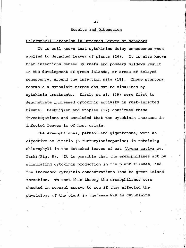
The eremophilanes, petasol and gigantenone, were as effective as kinetin (6-furfurylaminopurine) in retaining chlorophyll in the detached leaves of oat (Avena sativa cv. Park)(Fig. 8). It is possible that the eremophilanes act by stimulating cytokinin production in the plant tissues, and the increased cytokinin concentrations lead to green island formation. To test this theory the eremophilanes were checked in several assays to see if they affected the physiology of the plant in the same way as cytokinins.
50
* Control o Kinetin O Pe tosoI A G igon ter □ 1 1 , 12-epoxypetosol
0.A--
TIME (hours)
Figure 8. Chlorophyll retention in detached oat leaves treated with eremophilanes and kinetin. Compounds (20 nmol) were applied to puncture wounds made in the center of the leaf. The leaves were in incubated for three days in the dark before measuring chlorophyll. Chlorophyll content is reported as absorbance at 665nm divided by dry weight of the leaf in grams. Data points represent the average of three replicates.
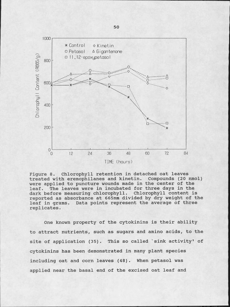
One known property of the cytokinins is their ability to attract nutrients, such as sugars and amino acids, to the site of application (35). This so called 'sink activity1 of cytokinins has been demonstrated in many plant species including oat and corn leaves (48) . When petasol was applied near the basal end of the excised oat leaf and

51either a sugar or an amino acid was applied near the tip of the leaf there was no directed transport of the radiolabeled
I ; Icompounds!toward the site of eremophilane application (Table 5). Gigantenone was tested, with similar results. Reversal of the sites of application again showed no directed transport. This indicates that polarity of movement is not involved.
Table 5. Sink activity of the eremophilanes. The values represent the average of three replications.
Leaf Section Treatment % of Total DPMtop 14Otrp 70
bottom 10% MeOH 30top 14Otrp 85bottom 5 jLtg Petasol 15top 14Oglucose 86
bottom 10% MeOH 14top 14C-glucose 88
bottom 5 ng Petasol 12
Pozar and Kiraly (53) demonstrated induced senescence in tissues away from the site of application of cytokinins. Enhanced senescence, away from the site of application, did not occur when eremophilanes were applied to detached oat leaves. Data presented in Figure 9 shows that leaves treated with petasol had normal chlorophyll loss when compared to the control. The acropetal portion of some leaves was excised from the site of petasol application over
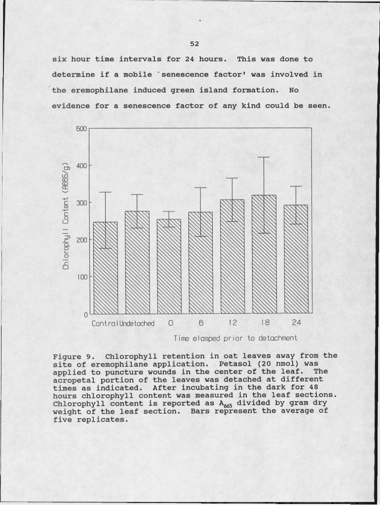
52six hour time intervals for 24 hours. This was done to determine if a mobile 'senescence factor' was involved in the eremophilane induced green island formation. No evidence for a senescence factor of any kind could be seen.
I
c§
I6
Contro I Undetached 0 6 12 18 24Time e Iasped prior to detachment
Figure 9. Chlorophyll retention in oat leaves away from the site of eremophilane application. Petasol (20 nmol) was applied to puncture wounds in the center of the leaf. The acropetal portion of the leaves was detached at different times as indicated. After incubating in the dark for 48 hours chlorophyll content was measured in the leaf sections. Chlorophyll content is reported as A665 divided by gram dry weight of the leaf section. Bars represent the average of five replicates.
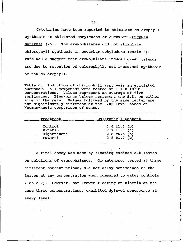
53Cytokinins have been reported to stimulate chlorophyll
synthesis in etiolated cotyledons of cucumber (Cucumis satiyus) (25) . The eremophilanes did not stimulate chlorophyll synthesis in cucumber cotyledons (Table 6).This would suggest that eremophilane induced green islands are due to retention of chlorophyll, not increased synthesis of new chlorophyll.
Table 6. Induction of chlorophyll synthesis in etiolated cucumber. All compounds were tested at 1.1 X IO'3 M concentrations. Values represent an average of five replicates. Plus/minus values represent one S.D. on either side of the mean. Values followed by the same letter are not significantly different at the 0.05 level based on Newman-Keuls comparison of means.
Treatment Chloronhvll ContentControl 3.6 ±1.2 (b)Kinetin 7.7 ±1.3 (a)Gigantenone 2.8 ±0.5 (b)Petasol 2.5 ±1.1 (b)
A final assay was made by floating excised oat leaves on solutions of eremophilanes. Gigantenone, tested at three different concentrations, did not delay senescence of the leaves at any concentration when compared to water controls (Table 7). However, oat leaves floating on kinetin at the same three concentrations, exhibited delayed senescence at every level.
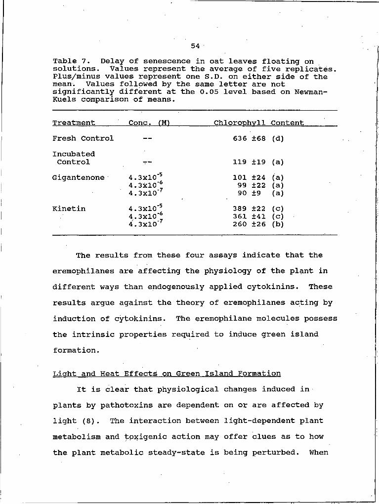
54
Table 7. Delay of senescence in oat leaves floating on solutions. Values represent the average of five replicates. Plus/minus values represent one S.D. on either side of the mean. Values followed by the same letter are not significantly different at the 0.05 level based on Newman- Kuels comparison of means.
Treatment Cone. fM) Chloronhvll ContentFresh Control — 636 ±68 (d)IncubatedControl — — 119 ±19 (a)
Gigantenone 4 . 3X 10'5 4 . 3x 10 6 4 . 3X 10"7
1019990
±24±22±9
(a)(a)(a)
Kinetin 4 . 3X 10'54 . 3X 10 ' *
389361
±22±41
(c)(c)
4 . 3X 10'7 260 ±26 (b)
The results from these four assays indicate that the eremophilanes are affecting the physiology of the plant in different ways than endogenously applied cytokinins. These results argue against the theory of eremophilanes acting by induction of cytokinins. The eremophilane molecules possess the intrinsic properties required to induce green island formation.
Light and Heat Effects on Green Island FormationIt is clear that physiological changes induced in
plants by pathotoxins are dependent on or are affected by light (8). The interaction between light-dependent plant metabolism and tpxigenic action may offer clues as to how the plant metabolic steady-state is being perturbed. When
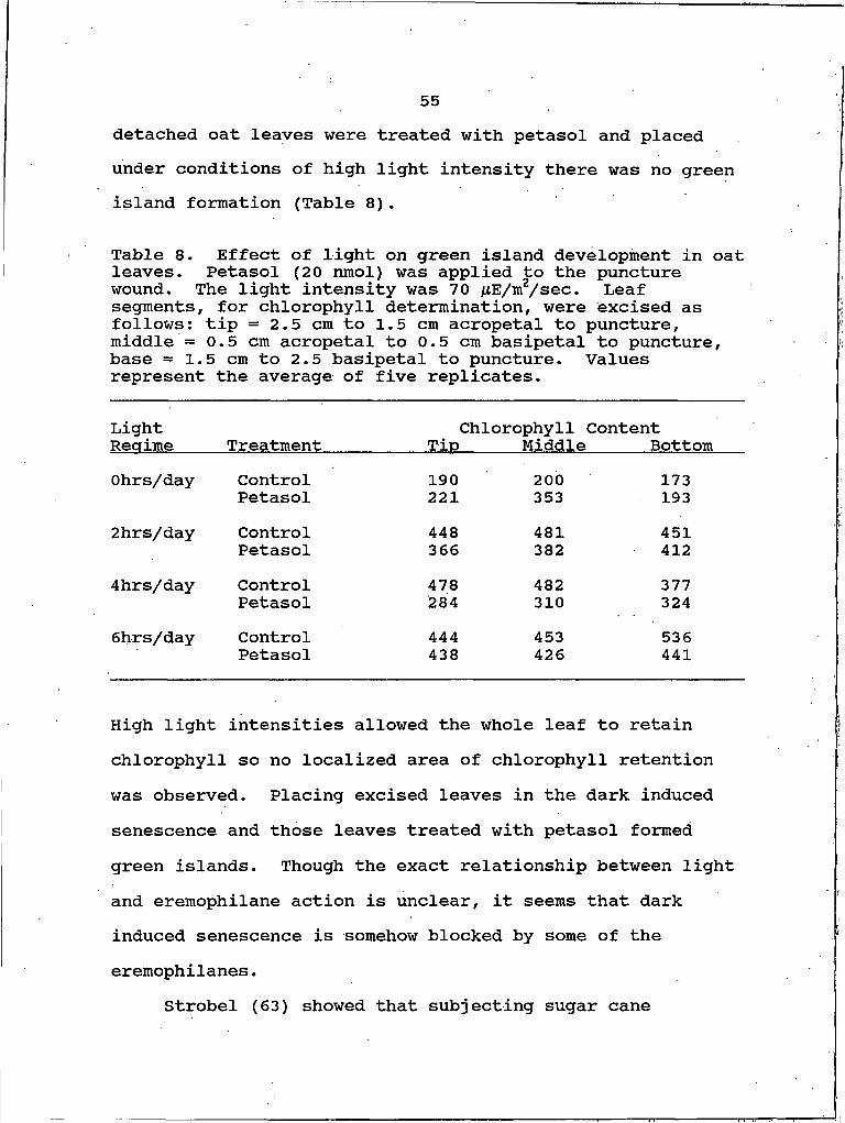
55detached oat leaves were treated with petasol and placed under conditions of high light intensity there was no green island formation (Table 8).
Table 8. Effect of light on green island development in oat leaves. Petasol (20 nmol) was applied to the puncture wound. The light intensity was 70 /LtE/m2/sec. Leaf segments, for chlorophyll determination, were excised as follows: tip = 2.5 cm to 1.5 cm acropetal to puncture, middle = 0.5 cm acropetal to 0.5 cm basipetal to puncture, base = 1.5 cm to 2.5 basipetal to puncture. Values represent the average of five replicates.
LightReaime Treatment
Chlorophyll Content Tin Middle Bottom
Ohrs/day Control 190 200 173Petasol 221 353 1932hrs/day Control 448 481 451
Petasol 366 382 4124hrs/day Control 478 482 377
Petasol 284 310 3246hrs/day Control 444 453 536
Petasol 438 426 441
High light intensities allowed the whole leaf to retain chlorophyll so no localized area of chlorophyll retention was observed. Placing excised leaves in the dark induced senescence and those leaves treated with petasol formed green islands. Though the exact relationship between light and eremophilane action is unclear, it seems that dark induced senescence is somehow blocked by some of the eremophiIanes.
Strobel (63) showed that subjecting sugar cane
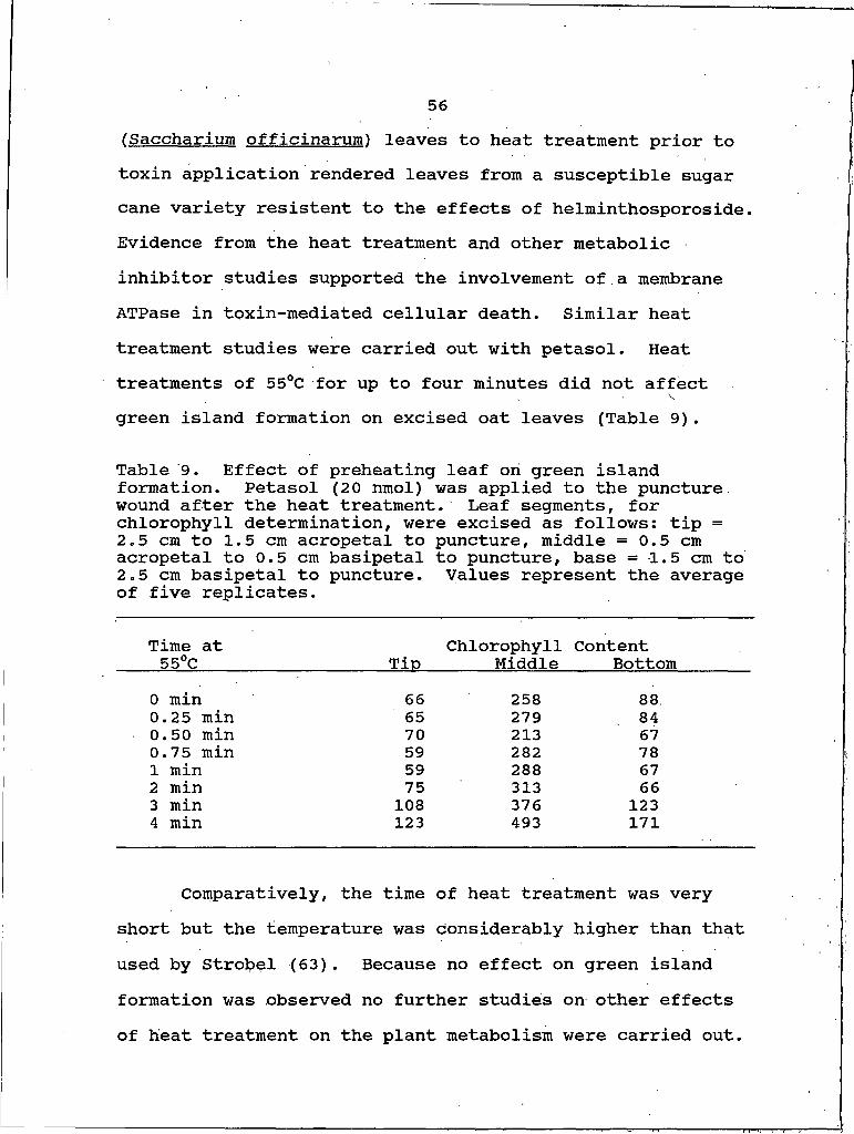
56fSaccharium officinarum) leaves to heat treatment prior to toxin application rendered leaves from a susceptible sugar cane variety resistant to the effects of helminthosporoside. Evidence from the heat treatment and other metabolic inhibitor studies supported the involvement of.a membrane ATPase in toxin-mediated cellular death. Similar heat treatment studies were carried out with petasol. Heat treatments of 55°C for up to four minutes did not affect green island formation on excised oat leaves (Table 9).
Table 9. Effect of preheating leaf on green island formation. Petasol (20 nmol) was applied to the puncture, wound after the heat treatment. Leaf segments, for chlorophyll determination, were excised as follows: tip =2.5 cm to 1.5 cm acropetal to puncture, middle = 0.5 cm acropetal to 0.5 cm basipetal to puncture, base = 1.5 cm to2.5 cm basipetal to puncture. Values represent the average of five replicates.
Time at 55°C Tin
ChlorophyllMiddle
ContentBottom
0 min 66 258 88.0.25 min 65 279 . 840.50 min 70 213 670.75 min 59 282 78I min 59 288 672 min 75 313 663 min 108 376 1234 min 123 493 171
Comparatively, the time of heat treatment was very short but the temperature was considerably higher than that used by Strobel (63). Because no effect on green island formation was observed no further studies on other effects of heat treatment on the plant metabolism were carried out.
57Binding Site Studies
There is extensive literature purporting binding sites on the piasinalemma as the primary site of toxin action (1,2,36,57). The effects of plant hormones, such as auxins and cytokinins, are also thought to be mediated through binding or receptor sites, either on the plasmalemma or at other sites in the cell (21,34). The possibility of the eremophilanes acting via a plasmalemma binding site was investigated.
[14C]-Petasol was incubated with an aliquot of a crude membrane preparation from oats. After incubation the mixture was chromatographed on a column of Sephadex G-25. Figure 10 shows a typical elution pattern of radioactivity, after chromatography. The large peak represents unbound [14C]-petasol that Was retained by the column. There was no peak of radioactivity at or near the void volume which would presumably represent bound petasol.
In a second experiment, [14C]-petasol and a mixture of [ 14C]-petasol:'cold• petasol (1:10) were each incubated with separate aliquots of the crude membrane preparation from oats. After incubation the mixtures were centrifuged at high speed to pellet the membranes and the radioactivity in the supernatant and pellet was determined. Table 10 shows that very little radioactivity was found in the pellet of either preparation. Because interference of binding by the
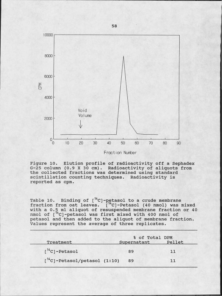
58IOOOO
Void Vo I ume
Q ______________ I______________ I______________ I______________ I______________ I______________ I______________ I______________ I______________
0 10 20 30 40 50 60 70 80 90
Frac tion Number
Figure 10. Elution profile of radioactivity off a Sephadex G-25 column (0.9 X 30 cm). Radioactivity of aliquots from the collected fractions was determined using standard scintillation counting techniques. Radioactivity is reported as cpm.
Table 10. Binding of [uC]-petasol to a crude membrane fraction from oat leaves. [14C]-Petasol (40 nmol) was mixed with a 0.5 ml aliquot of resuspended membrane fraction or 40 nmol of [14C]-petasol was first mixed with 400 nmol of petasol and then added to the aliquot of membrane fraction. Values represent the average of three replicates.
% of Total DPMTreatment Supernatant Pellet[14C] -Petasol 89 11[ 14C] -Petasol/petasol (1:10) 89 11
59cold' petasol was not observed, the small amount of
radioactivity in the pellet was probably due to nonspecific binding.
Competitive binding studies, using petasol and 11,12- epoxypetasol, were done in vivo with detached oat leaves. Petasol is a strong inducer of green island formation on detached oat leaves, while 11,12-epoxypetasol is a much weaker inducer of green islands. Application of equimolar J amounts of 11,12-epoxypetasol and petasol to puncture wounds led to green island formation typical of petasol application as measured by chlorophyll retention (Fig. 11). However, when 11,12-epoxypetasol was applied first and several hours time was allowed to lapse before addition of petasol, very small green islands were formed. On control leaves, petasol was able to cause green islands even after delaying application for several hours. By increasing the quantity of petasol applied to the leaf the dampening effect of11.12- epoxypetasol is overcome. The data suggests that11.12- epoxypetasol and petasol compete for the same binding site. 11,12-epoxypetasol binding would lead to small or no green islands whereas the binding of petasol leads to formation of large green islands. The binding site does not seem to be on the plasmalemma but rather on the cell wall or in the cytosol. No further studies were done to localize the binding site.
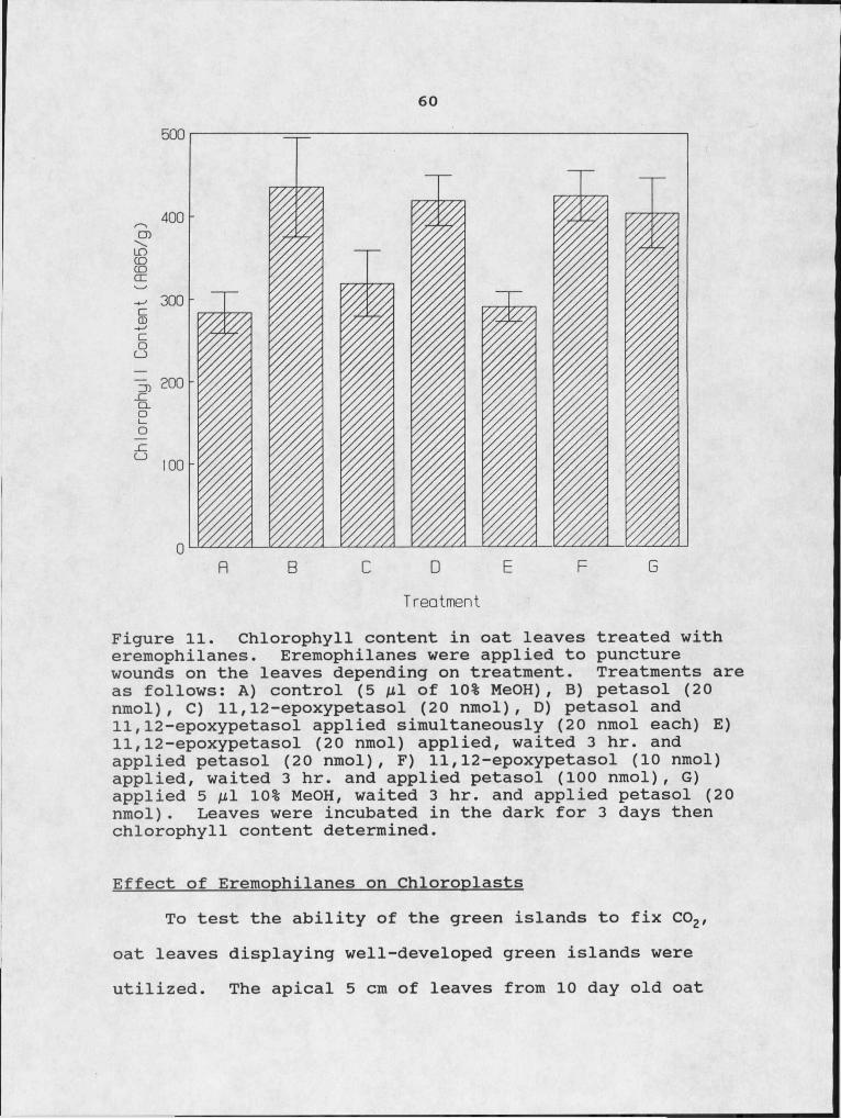
60
Treatment
Figure 11. Chlorophyll content in oat leaves treated with eremophilanes. Eremophilanes were applied to puncture wounds on the leaves depending on treatment. Treatments are as follows: A) control (5 /il of 10% MeOH) , B) petasol (20 nmol), C) 11,12-epoxypetasol (20 nmol), D) petasol and11.12- epoxypetasol applied simultaneously (20 nmol each) E)11.12- epoxypetasol (20 nmol) applied, waited 3 hr. and applied petasol (20 nmol), F) 11,12-epoxypetasol (10 nmol) applied, waited 3 hr. and applied petasol (100 nmol), G) applied 5 /xl 10% MeOH, waited 3 hr. and applied petasol (20 nmol). Leaves were incubated in the dark for 3 days then chlorophyll content determined.
Effect of Eremophilanes on ChloroolastsTo test the ability of the green islands to fix CO2,
oat leaves displaying well-developed green islands were utilized. The apical 5 cm of leaves from 10 day old oat
61seedlings were excised and placed in a moist chamber. Puncture wounds were made and either petasol, gigantenone or11,12-epoxypetasol was applied. The leaves were incubated for four days under intermittent light (6juE/m2/sec, 12 hour/day) to allow green islands to form. Incubated and freshly excised leaves were.placed in a small airtight chamber containing 0.05 mmole [14Cj-CO2 (8.4 Ci/mole) . The chambers were placed either under constant light or in the dark. Sections I cm on either side of the puncture wound were excised and digested. After digestion, 10 ml of Aquasol was added to the samples which were then counted in a scintillation counter. CO2 fixation values (pmoles C02/hr) were then calculated based upon the specific radioactivity of [14C] -CO2. As shown by Table 11, green islands induced by petasol and gigantenone were about.half as effective in CO2 fixation as fresh control leaves underconstant, light conditions. In the absence of light, green
1.islands induced by petasol were able to fix CO2 as efficiently as fresh control leaves. However, gigantenone induced green islands were not very effective in fixing CO2 undep these conditions. Leaves treated with 11,12- epoxypetasol and incubated control leaves did not effectively fix CO2 under light conditions or in the dark.
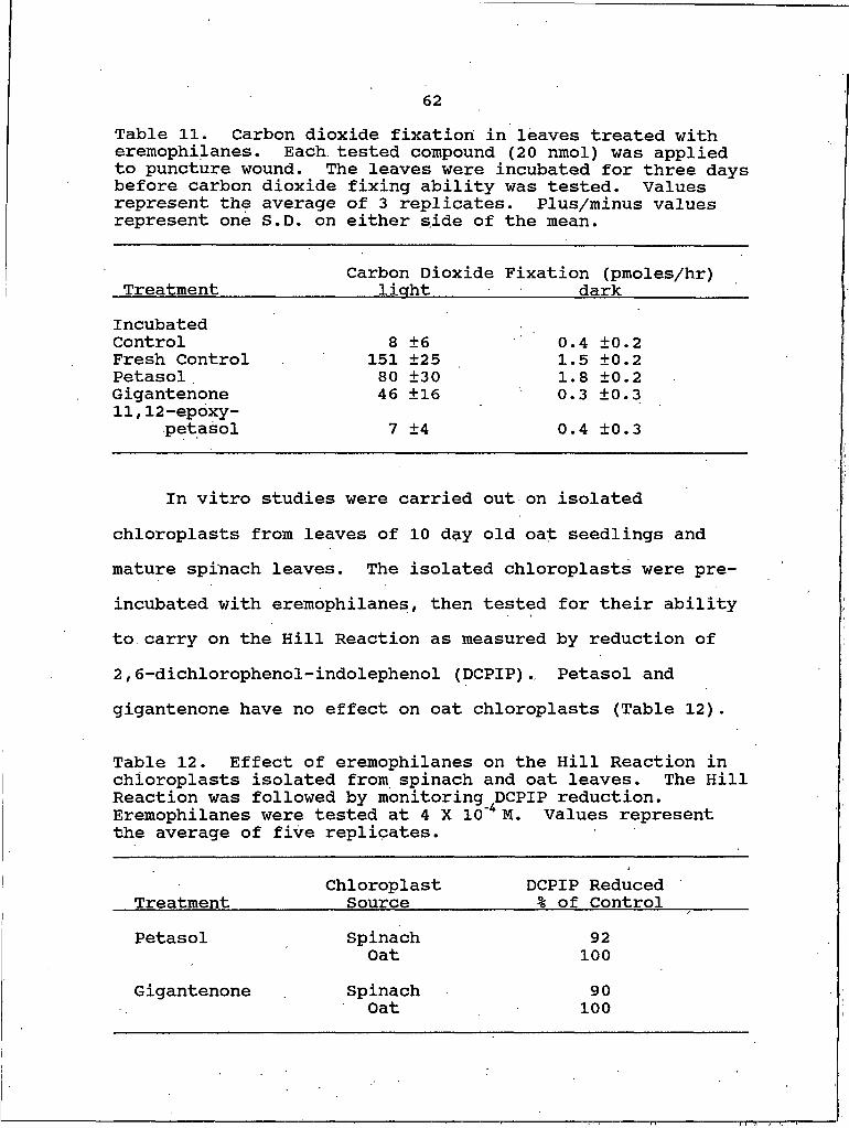
62
Table 11. Carbon dioxide fixation in leaves treated with eremophilanes. Each tested compound (20 nmol) was applied to puncture wound. The leaves were incubated for three days before carbon dioxide fixing ability was tested. Values represent the average of 3 replicates. Plus/minus values represent one S.D. on either side of the mean.
Carbon Dioxide Fixation (pmoles/hr)Treatment_______________light_____ ■ _____ darkIncubatedControl 8 ±6 0.4 ±0.2Fresh Control 151 ±25 1.5 ±0.2Petasol . 80 ±30 1.8 ±0.2Gigantenone 46 ±16 0.3 ±0.311,12-epoxy-
petasol 7 ±4 0.4 ±0.3
In vitro studies were carried out on isolated chloroplasts from leaves of 10 day old oat seedlings and mature spinach leaves. The isolated chloroplasts were preincubated with eremophilanes, then tested for their ability to carry on the Hill Reaction as measured by reduction of 2,6-dichlorophenol-indolephenol (DCPIP). Petasol and gigantenone have no effect on oat chloroplasts (Table 12).
Table 12. Effect of eremophilanes on the Hill Reaction in chloroplasts isolated from spinach and oat leaves. The Hill Reaction was followed by monitoring DCPIP reduction. Eremophilanes were tested at 4 X IO"4 M. Values represent the average of five replicates.
TreatmentChloroplast
SourceDCPIP Reduced % of Control
Petasol Spinach 92Oat 100
Gigantenone Spinach 90Oat 100
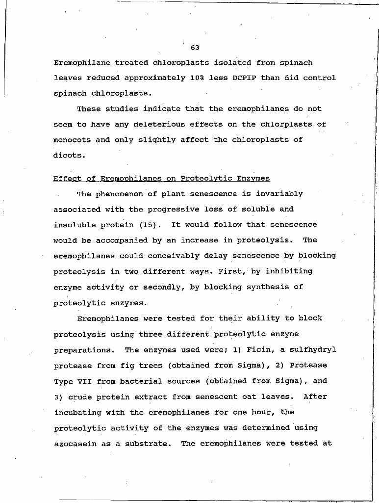
63Eremophilane treated chloroplasts isolated from spinach leaves reduced approximately 10% less DCPIP than did control spinach chloroplasts.
These studies indicate that the eremophilanes do not seem to have any deleterious effects on the chlorplasts of monocots and only slightly affect the chloroplasts of dicots.
Effect of Eremophilanes on Proteolytic EnzvmesThe phenomenon of plant senescence is invariably
associated with the progressive loss of soluble and insoluble protein (15). It would follow that senescence would be accompanied by an increase in proteolysis. The eremophilanes could conceivably delay senescence by blocking proteolysis in two different ways. First, by inhibiting enzyme activity or secondly, by blocking synthesis of proteolytic enzymes.
Eremophilanes were tested for their ability to block proteolysis using three different proteolytic enzyme preparations. The enzymes used were; I) Ficin, a sulfhydryl protease from fig trees (obtained from Sigma), 2) Protease Type VII from bacterial sources (obtained from Sigma), and 3) crude protein extract from senescent oat leaves. After incubating with the eremophilanes for one hour, the proteolytic activity of the enzymes was determined using azocasein as a substrate. The eremophilanes were tested at

64three different concentrations. No inhibition of the proteolytic activity of Ficin or Protease Type VII was observed when treated with the petasol, gigantenone or11,12-epoxypetasol at any of the three concentrations tested (Table 13).
Table 13. Ficin and Protease Type VII protease activity in the presence of eremophilanes. Values represent the average of three replicates.
Treatment Cone.fM) % Activity Ficin
of Control Protease
Control - 100 100Petasol io"4 100 100
IO"6 100 100IO'8 100 100
Gigantenone 10-6 100 10010 J 100 100IO"8 100 100
11,12-epoxy- 10I 100 100petasol 10-8 100 100
10 8 100 100
The eremophilanes, petasol and gigantenone, were also ineffective in inhibiting the proteolytic activity in the crude protein preparation from senescent oat leaves at 4 X IO"4 M levels. The eremophilanes do not seem to have any direct effect on proteolytic activity in vitro.
To test the effect of eremophilane treatment on in vivo proteolytic enzyme levels, protein was extracted from leaves treated with eremophilanes and proteolytic enzyme activity
65was measured. The apical 5 cm of leaves from 10 day old oat seedlings were excised and petasol, gigantenone and 11,12- epoxypetasol were applied to puncture wounds. After incubation, segments extending I cm on each side of the puncture wound were excised and total soluble protein was extracted. The levels of proteolytic activity in the petasol and gigantenone treated leaves was significantly less than that of the incubated control leaves (Fig. 12).
LOOXCE
O■rSZD
>
S
£CJ
SCL
Fresh Incubated K in e tin Petasol Gigantenone 11. 12-epoxy- Control Control petasol
Treatment
Figure 12. Proteolytic enzyme activity of protein extracts from oat leaves treated with eremophilanes and kinetin. The puncture wound droplet overlay technique was used to apply 20 nmol of each compound solubilized in 10% MeOH.Proteolytic activity is reported as enzyme units, see Materials and Methods for calculation of an enzyme unit.Bars represent the average of three replicates.
66Leaves treated with 11,12-epoxypetasol exhibited very small green islands and had levels of proteolytic activity comparable to the levels of incubated control leaves.Protein extracts from fresh control leaves showed the lowest amount of proteolytic activity.
These data suggest that some of the eremophilanes can , control the level of proteolytic activity in vivo. Whether petasol and gigantenone control proteolytic activity levels by inhibiting zymogen conversion or by inhibiting de novo synthesis is not known.
Protein Synthesis and the EremophilanesThe protein synthesis inhibitor cycloheximide was used
to investigate the possibility that new proteins, synthesized de novo, may be required for eremophilane induced green island formation. When cycloheximide was applied along with petasol to puncture wounds on detached oat leaves green islands were formed (Fig. 13). This would indicate that new protein synthesis is not required for green island formation. However, cycloheximide itself was capable of delaying senescence (Fig. 13 and ref. 52) making it difficult to draw a conclusion about the requirement for protein synthesis in eremophilane induced green island formation. However, this experiment did make it clear that inhibition of protein synthesis will lead to green island formation. Chloroamphenicol, an inhibitor of 70s ribosomes,
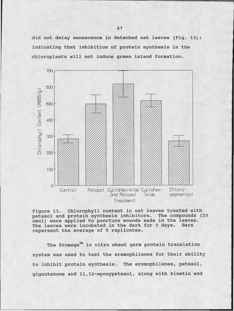
67did not delay senescence in detached oat leaves (Fig. 13); indicating that inhibition of protein synthesis in the chloroplasts will not induce green island formation.
I5S
CJ
o"
6
Reiasol Cycloheximide Cyclohex- Chloro-and Petasol Imide amphenIcoI
Control
TreatmentFigure 13. Chlorophyll content in oat leaves treated with petasol and protein synthesis inhibitors. The compounds (20 nmol) were applied to puncture wounds made in the leaves.The leaves were incubated in the dark for 3 days. Bars repersent the average of 5 replicates.
The Promega™ in vitro wheat germ protein translation system was used to test the eremophilanes for their ability to inhibit protein synthesis. The eremophilanes, petasol, gigantenone and 11,12-epoxypetasol, along with kinetin and
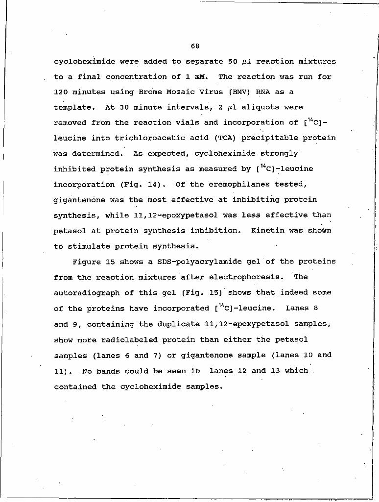
68cycloheximide were added to separate 50 /Ltl reaction mixtures to a final concentration of I znM. The reaction was run for 120 minutes using Brome Mosaic Virus (BMV) RNA as a template. At 30 minute intervals, 2 fil aliquots were removed from the reaction vials and incorporation of [14C]- leucine into trichloroacetic acid (TCA) precipitable protein was determined. As expected, cycloheximide strongly inhibited protein synthesis as measured by [14C]-leucine incorporation (Fig. 14). Of the eremophilanes tested, gigantenone was the most effective at inhibiting protein synthesis, while 11,12-epoxypetasol was less effective than petasol at protein synthesis inhibition. Kinetin was shown to stimulate protein synthesis.
Figure 15 shows a SDS-polyacrylamide gel of the proteins from the reaction mixtures after electrophoresis. The autoradiograph of this gel (Fig. 15) shows that indeed some of the proteins have incorporated [14C]-leucine. Lanes 8 and 9, containing the duplicate 11,12-epoxypetasol samples, show more radiolabeled protein than either the petasol samples (lanes 6 and 7) or gigantenone sample (lanes 10 and 11). No bands could be seen in lanes 12 and 13 which - contained the cycloheximide samples.
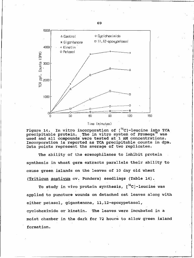
69
o CycIoheximide nil. 12-epoxypetasol
A Control x Gigantenone + Kinetin o Petasol
J5 3000
□ ---- D
.__cr' ..o.. 120
Time (minutes)Figure 14. In vitro incorporation of [14C] -leucine into TCA precipitable protein. The in vitro system of Promega™ was used and all compounds were tested at I mM concentrations. Incorporation is reported as TCA precipitable counts in dpm. Data points represent the average of two replicates.
The ability of the eremophilanes to inhibit protein synthesis in wheat germ extracts parallels their ability to cause green islands on the leaves of 10 day old wheat fTriticum aestivum cv. Pondera) seedlings (Table 14).
To study in vivo protein synthesis, [14C]-leucine was applied to puncture wounds on detached oat leaves along with either petasol, gigantenone, 11,12-epoxypetasol, cycloheximide or kinetin. The leaves were incubated in a moist chamber in the dark for 72 hours to allow green islandformation.
70
gel autoradiographFigure 15. SDS-polyacrylamide gel of proteins from the reaction mixtures and an autoradiograph of this gel. All lanes were loaded with 25 ng of protein. Lanes contain:I)standards, 2,3) control, 4,5) negative control (no BMV RNA added), 6,7) petasol, 8,9) 11,12-epoxypetasol, 10,11) gigantenone, 12,13) cycloheximide, 14,15) kinetin.
Table 14. Green island formation on Pondera wheat leaves. Test compounds (20 nmol) were applied to puncture wounds. The leaves were incubated for three days in the dark, then green islands were measured. Values represent the average of three replicates. Plus/minus values represent one S.D. on either side of the mean.
TreatmentGreen Island Size (mm2)
Control __Gigantenone 49 ±5Petasol 14 ±411,12-epoxypetasol 5 ±1Cycloheximide 113 ±8Kinetin 250 ±15
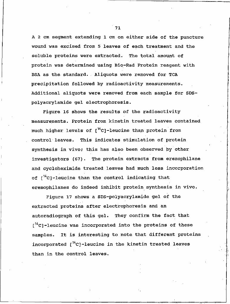
71A 2 cm segment extending I cm on either side of the puncture wound was excised from 5 leaves of each treatment and the soluble proteins were extracted. The total amount of protein was determined using Bio-Rad Protein reagent with BSA as the standard. . Aliquots were removed for TCA precipitation followed by radioactivity measurements. Additional aliquots were removed from each sample for SDS- polyacrylamide gel electrophoresis.
Figure 16 shows the results of the radioactivity measurements. Protein from kinetin treated leaves contained much higher levels of [14C]-leucine than protein from control leaves. This indicates stimulation of protein synthesis in vivo; this has also been observed by other investigators (67). The protein extracts from eremophilane and cycloheximide treated leaves had much less incorporation of [14C]-leucine than the control indicating that eremophilanes do indeed inhibit protein synthesis in vivo.
Figure 17 shows a SDS-polyacrylamide gel of the extracted proteins after electrophoresis and an autoradiograph of this gel. They confirm the fact that [14C]-leucine was incorporated into the proteins of these samples. It is interesting to note that different proteins incorporated [14C]-leucine in the kinetin treated leaves than in the control leaves.
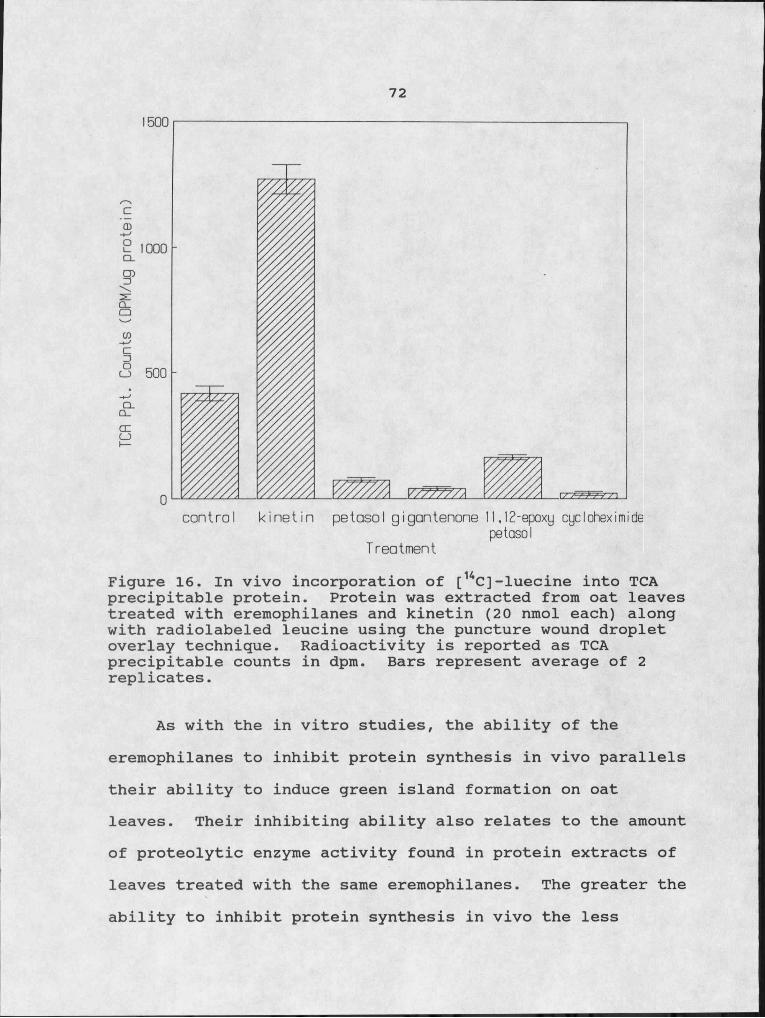
72
Figure 16. In vivo incorporation of [14C]-Iuecine into TCA precipitable protein. Protein was extracted from oat leaves treated with eremophilanes and kinetin (20 nmol each) along with radiolabeled leucine using the puncture wound droplet overlay technique. Radioactivity is reported as TCA precipitable counts in dpm. Bars represent average of 2 replicates.
As with the in vitro studies, the ability of the eremophilanes to inhibit protein synthesis in vivo parallels their ability to induce green island formation on oat leaves. Their inhibiting ability also relates to the amount of proteolytic enzyme activity found in protein extracts of leaves treated with the same eremophilanes. The greater the ability to inhibit protein synthesis in vivo the less
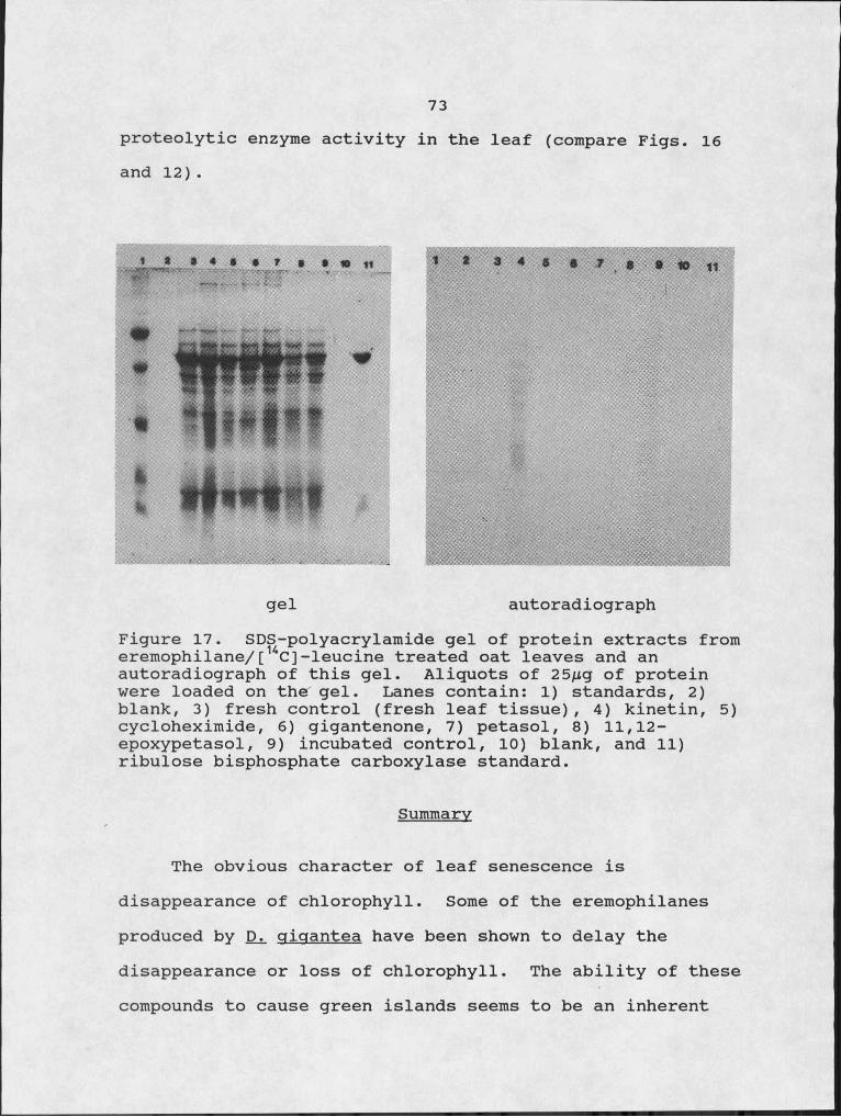
73proteolytic enzyme activity in the leaf (compare Figs. 16 and 12).
gel autoradiographFigure 17. SDS-polyacrylamide gel of protein extracts from eremophilane/[14C]-leucine treated oat leaves and an autoradiograph of this gel. Aliquots of 25/xg of protein were loaded on the gel. Lanes contain: I) standards, 2) blank, 3) fresh control (fresh leaf tissue), 4) kinetin, 5) cycloheximide, 6) gigantenone, 7) petasol, 8) 11,12- epoxypetasol, 9) incubated control, 10) blank, and 11) ribulose bisphosphate carboxylase standard.
Summary
The obvious character of leaf senescence is disappearance of chlorophyll. Some of the eremophilanes produced by Di giaantea have been shown to delay the disappearance or loss of chlorophyll. The ability of these compounds to cause green islands seems to be an inherent
74property of the molecules themselves.
Several probable mechanisms of leaf senescence have been proposed (68, and refs therein). It seems to be accepted by most investigators that senescence is an active, "programmed" process. This would indicate that it is controlled by the nucleus. Either proteolytic enzymes are produced de novo or are released from the vacuole via a nucleic acid product that travels to the tonoplast membrane to initiate release. In either case inhibitors acting on specific stages of protein synthesis would interfere with the increase of proteolytic enzyme levels. Martin and Thimann (43) presented evidence based on delayed senescence by protein synthesis inhibitors to strongly support this theory of dark-induced senescence.
The eremophilanes petasol, gigantenone, and to some extent, 11,12-epoxypetasol were shown to inhibit protein synthesis both in vitro and in vivo in monocot systems.Their ability to inhibit protein synthesis parallels their ability to induce green island formation on the detached leaves of monocots. The data presented here strongly suggests that the mode of action of eremophilanes in delaying senescence is inhibition of protein synthesis. The exact mechanism of how the eremophilanes inhibit protein synthesis is at this point unknown. However, because the mRNA is supplied in the in vitro system, the inhibition seems to be at the translational level, not at the
75transcriptional level. Additional experiments, comparing eremophilane action to known inhibitors of protein synthesis could establish whether eremophilanes inhibit initiation of protein synthesis or elongation of nascent peptides. Radiolabeled, amino acids and tRNA could be used in combination with the eremophilanes to look at the reaction . sequence by which amino acids are transferred from aminoacyl tRNA into the polypeptides on ribosomes.
76
CHAPTER V
METABOLISM OF EREMOPHILANES IN PLANTA
Introduction
Understanding the fate of toxins in plants is at a very primitive stage. Studies on in planta metabolism of a toxin are complicated by (I) the small quantity of pure toxin that is usually available, (2) the fact that toxins usually represent only a small fraction of the total carbon and nitrogen content of the culture, and hence radiolabeling to high specific activity is difficult, and (3) the difficulty of introducing toxins into a plant in sufficent quantity and at the right place and time in the development of the plant.
There are other reasons, aside from intrinsic interest, why knowledge of toxin metabolism is useful. Is the toxin molecule isolated from the pathogen really a pre-toxin that interacts with plant metabolites to become an active toxin? Is the toxin systemic in the plant or localized to the area of infection? Does the plant have the ability to recognize and metabolize the toxin? Does metabolism of the toxin lead to inactivation? Information on toxin metabolism in planta could answer these questions and potentially provide uses in plant disease control.
77The fate of toxins in the diseased plant can be varied.
Tabtoxin appears to require processing. In the diseased plant, nonspecific peptidases cleave off threonine or serine releasing the biologically active tabtoxinine-^-lactam (69). Evidence indicated that these peptidases were of plant origin. Conversely, fusaric acid, a toxin produced by Fusarium spp., was broken down to smaller non-toxic molecules and finally to carbon dioxide and water when applied to tomato cuttings (38) .
The objective of this study was to study the fate of eremophilanes in planta. [14C]-Petasol was used to trace petasol in the plant. TLC and autoradiography were used to analyze leaf extracts and determine the fate of petasol. A single metabolite labeled "conversion product" was isolated, purified, and tested for biological activity.
Materials and Methods
Plant MaterialAvena sativa cv. Park oats and Phaseolus aureous were
grown in vermiculite under enviromentalIy controlled conditions of 18 hours light, 6 hours dark/day with a constant temperature of 26°C.
T14Cl-Labeled Eremophilanes[14C]-Sodium acetate (30 /LtCi) was added to the modified
M-ID medium and the medium was inoculated with Di. gigantea.
78After incubating the culture for 6 weeks, petasol, gigantenone, and 11,12 epoxypetasol were purified from culture filtrates as previously described (28,37). Their specific activity ranged from 5-21 DPM//ig. The eremophilanes from several different cultures were pooled to obtain sufficient quantities but this led to nonuniformity in the specific activity of the individual eremophilane molecules.
Translocation of EremophilanesThe apical 5 cm of leaves from 10 day old oat seedlings
were excised and placed in a moist chamber. Puncture wounds were made in the center of the leaves with a 22 gauge needie. Each puncture wound was overlayed with [14C]- petasol (10 yitg at 21 dpm/jug) solubilized in 10% methanol. Leaves were incubated either in the dark or under conditions of intermittent light (12 hrs/day at 6 /LtE/m2/sec) . After incubation the leaves were cut into I cm segments as follows: base = 1.0 to 2.0 cm basipetal to puncture: middle = 0.5 cm basipetal to 0.5 acropetal to puncture; and tip = 1.0 cm to 2.0 cm acropetal to puncture. Leaf segments were placed in separate scintillation vials and 0.5 ml perchloric acid:hydrogen peroxide (1:2) digest solution was added and the vials were taken to IOO0C for 30 min. The digests were solubilized with 10 ml Aquasol and counted for 50 minutes in a Packard Tri-Carb scintillation counter. Cpm were converted to dpm using a quench correction curve.
79Leaf Treatment and Extraction
The apical 6 cm of leaves from 10 day old oat seedlings were excised and placed in a moist chamber. Using a 22 gauge needle, three punctures were made in each leaf I cm apart starting 2 cm from the base of the excised leaf. Each puncture was overlayed with 5 /zg of [14C]-eremophilane solubilized in 10% methanol. When mung beans were used, two punctures were made on each side of the detached leaf. The leaves were incubated for 3 days in the dark. After incubation the leaves were cut into small segments («1 mm) and ground in methanol:chloroform:water (M:C:W) (12:5:3)(v/v/v) using a Tenbrock homogenizer. The homogenate was filtered through several layers of cheesecloth to remove debris. This filtrate was referred to as "crude leaf extract".
Purification of Conversion Product"Crude leaf extract" was partitioned by the addition of
0.25 volumes of chloroform and 0.35 volumes of water. The solution was shaken vigorously to form an emulsion, then spun in a swinging bucket rotor at I5Oxg for 5 minutes to effect a phase separation. The aqueous phase was extracted 3 times with equal volumes of I-butanol. The radioactivity of each extract was checked by solubilizing an aliquot with 10 jul Aquasol and counting in a scintillation counter as described above. Pigmented samples were bleached by the
80addition of 0.5 ml of 5% sodium hypochlorite. The butanol extract was subjected to thin layer chromatography (TLC), on silica gel 60 with fluorescent indicator (E. Merck) with a I-butanol:acetic acid:water (B:A:W) (4:1:5)(v/v/v) solventsystem. Bands were visualized by placing plates under UV light (254 nm). Each band was analyzed for radioactivity by scraping the edge of the band («1 cm2) , placing the silica directly into a scintillation vial, adding 0.5 ml methanol to solubilize the compound, and finally adding 10 ml of Aquasol. The vials were counted in a scintillation counter. The band(s) containing radioactivity were scraped from the plate and eluted with methanol. The eluted fraction was further fractionated using TLC on silica gel 60 with a 1- butanol:formic acid:water (B:FA:W)(75:15:10)(v/v/v) solvent system. Bands of radioactivity were identified as described above. The eluted sample was purified using HPLC. Reverse phase HPLC was performed with a Waters Model 6000A pump equipped with a Waters Model U6K injector and coupled to a Waters Model 440 UV absorbance detector hooked to an Omniscribe D5000 chart recorder. The column used was a Waters Novapak C18 (5 /nm, 3.9 X 150 mm) with a methanol:water (2:8)(v/v) mobile phase at a flow rate of 0.5 ml/min. The absorbance of the effluent was monitored at 254 nm. Effluent representing peaks was collected and aliquots were taken to determine radioactivity. The aliquots were placed in separate scintillation vials and 10 ml Aquasol was

81added to each vial. The vials were counted using standard scintillation techniques.
Chlorophyll RetentionThe apical 5 cm of leaves from 10 day old oat seedlings
were excised and placed in a moist chamber. Puncture wounds were made in the center of the leaf. Petasol (I /Ltg) or 5 jug of conversion product solubilized in 10% methanol was applied to the wound. The leaves were incubated in the dark for 72 hours. Chlorophyll content in I cm segments, cut 0.5 cm basipetal to 0.5 cm acropetal to puncture, from each leaf was determined as previously described (Chapter III).
Mass SpectrometryFast atom bombardment mass spectra (FAB-MS) were
obtained on a VG Mass Lab Trio2 automated mass spectrometer coupled with a Hewlett Packard 5890 gas chromatograph (GC) using a P-5 microcolumn (length = 30 meters). A glycerol matrix was used. FAB-MS was kindly performed by Mr. Joe Sears, Montana State University.
Nuclear Magnetic Resonance SpectrometryProton nuclear magnetic resonance spectra (1H-NMR) were
recorded on a Varian XL-400 spectrometer. Chemical shifts were taken in ppm units relative to trimethylsiIane (0 ppm) with DMS0-d6 and chloroform-d6 as solvents. NMR spectrometry was kindly performed by Dr. Fumio Sugawara, RIKEN Institute Japan.
82Amino Acid Analysis
Amino acid analysis was performed by Dr. David Teplowf. California Institue of Technology, Pasadena, California.The instrumentation used was an Applied Biosystems Model 420 derivatizer coupled to a a Model 130 HPLC system. Data was acquired and analyzed by a Model 920 software system. NOR- leucine was used as an internal standard.
Results and Discussion
Translocation of Eremophilanes in the Leaf[14C]-Petasol was applied to the puncture wounds made
in the center of detached oat leaves. The leaves were incubated in a moist chamber in the dark or under intermittent light conditions (6 juE/m2/sec, 12 hr/day) for 3 days. After incubation, I cm segments were cut from the base, middle and tip sections of each leaf. The middle segment represented green island tissue, both base and tip sections were chlorotic tissues. The leaf segments were placed in scintillation vials and digested, then 10 ml of Aquasol was added to the vials and they were counted. As seen in Table 15, most of the radioactivity remained in the green island tissue. There was very little radiolabel found outside the green island indicating that petasol i,s not translocated in planta.
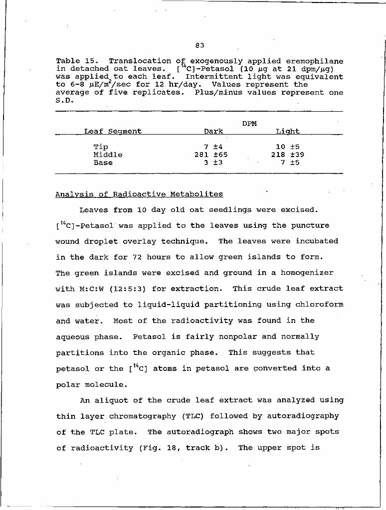
83Table 15. Translocation of exogenously applied ereitiophilane in detached oat leaves. [14C]-Petasol (10 fig at 21 dpm/jLig) was applied to each leaf. Intermittent light was equivalent to 6-8 juE/m2/sec for 12 hr/day. Values represent the average of five replicates. Plus/minus values represent one S.D.
Leaf Secrment Dark DPM LiahtTip 7 ±4 10 ±5Middle 281 ±65 218 ±39Base 3 ±3 7 ±5
Analysis of Radioactive MetabolitesLeaves from 10 day old oat seedlings were excised.
[14C]-Petasol was applied to the leaves using the puncture wound droplet overlay technique. The leaves were incubated in the dark for 72 hours to allow green islands to form.The green islands were excised and ground in a homogenizer with M:C:W (12:5:3) for extraction. This crude leaf extract was subjected to liquid-liquid partitioning using chloroform and water. Most of the radioactivity was found in the aqueous phase. Petasol is fairly nonpolar and normally partitions into the organic phase. This suggests that petasol or the [14C] atoms in petasol are converted into a polar molecule.
An aliquot of the crude leaf extract was analyzed using thin layer chromatography (TLC) followed by autoradiography of the TLC plate. The autoradiograph shows two major spots of radioactivity (Fig. 18, track b). The upper spot is
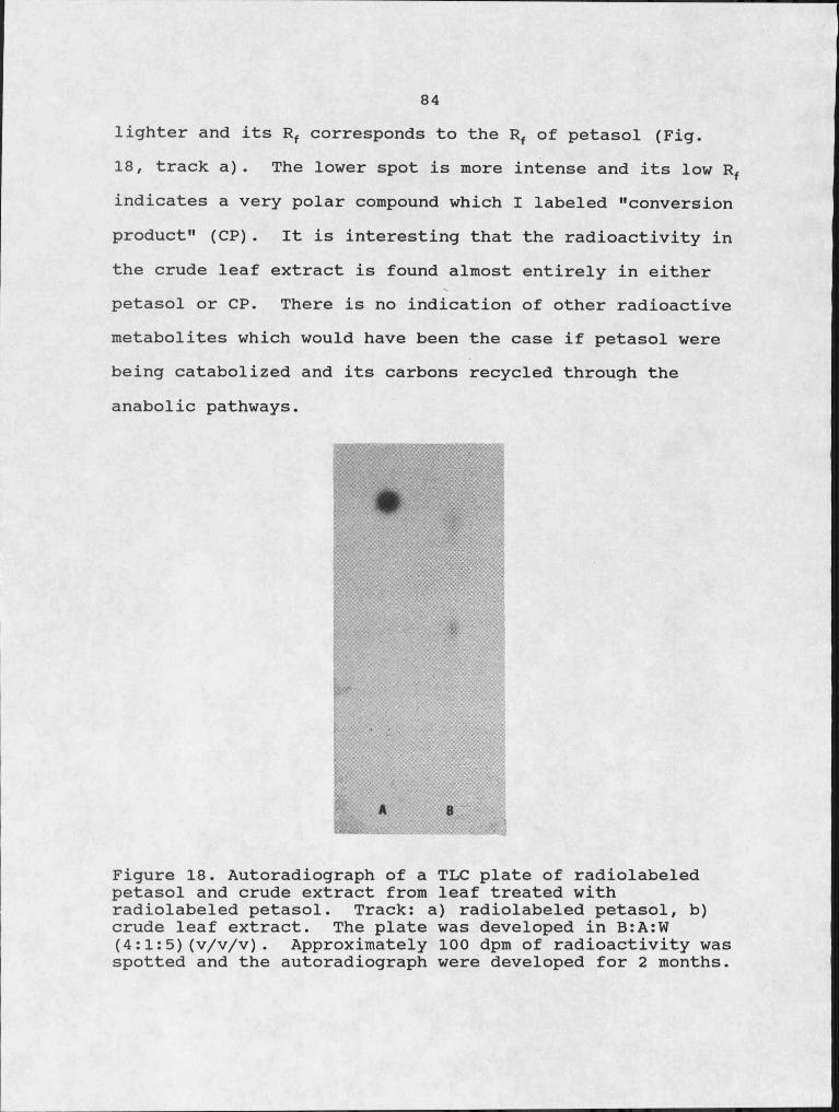
84lighter and its Rf corresponds to the Rf of petasol (Fig.18, track a). The lower spot is more intense and its low Rf indicates a very polar compound which I labeled "conversion product" (CP). It is interesting that the radioactivity in the crude leaf extract is found almost entirely in either petasol or CP. There is no indication of other radioactive metabolites which would have been the case if petasol were being catabolized and its carbons recycled through the anabolic pathways.
Figure 18. Autoradiograph of a TLC plate of radiolabeled petasol and crude extract from leaf treated with radiolabeled petasol. Track: a) radiolabeled petasol, b) crude leaf extract. The plate was developed in B:A:W (4:1:5)(v/v/v). Approximately 100 dpm of radioactivity was spotted and the autoradiograph were developed for 2 months.
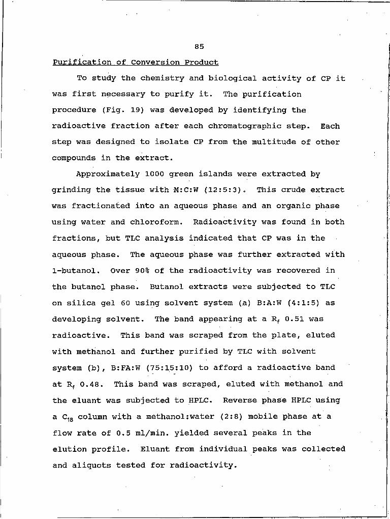
85Purification of Conversion Product
To study the chemistry and biological activity of CP it was first necessary to purify it. The purification procedure (Fig. 19) was developed by identifying the radioactive fraction after each chromatographic step. Each step was designed to isolate CP from the multitude of other compounds in the extract.
Approximately 1000 green islands were extracted by grinding the tissue with M :C:W (12:5:3). This crude extract was fractionated into an aqueous phase and an organic phase using water and chloroform. Radioactivity was found in both fractions, but TLC analysis indicated that CP was in the aqueous phase. The aqueous phase was further extracted with I-butanol. Over 90% of the radioactivity was recovered in the butanol phase. Butanol extracts were subjected to TLC on silica gel 60 using solvent system (a) B:A:W (4:1:5) as developing solvent. The band appearing at a Rf 0.51 was radioactive. This band was scraped from the plate, eluted with methanol and further purified by TLC with solvent system (b), B:FA:W (75:15:10) to afford a radioactive band at Rf 0.48. This band was scraped, eluted with methanol and the eluant was subjected to HPLC. Reverse phase HPLC using a C18 column with a methanol:water (2:8) mobile phase at a flow rate of 0.5 ml/min. yielded several peaks in the elution profile. Eluant from individual peaks was collected and aliquots tested for radioactivity.
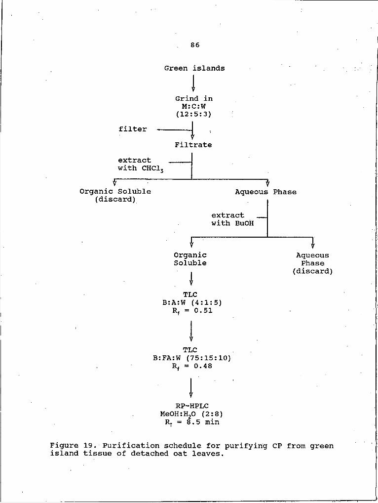
86
Green islands
IGrind in M: C: W
(12:5:3)filter
Filtrateextract with CHCl3
fOrganic Soluble (discard) Aqueous Phase
extract with BuOH
OrganicSoluble
AqueousPhase
(discard)
TLCB:A:W (4:1:5)
Rz = 0.51
VTLC
B:FA:W (75:15:10) Rf = 0.48
IRP-HPLC
MeOH:H2O (2:8) Rt = 8.5 min
Figure 19. Purification schedule for purifying CP from green island tissue of detached oat leaves.

87The peak with an Rt of 8.5 min. was radioactive. The purity of CP was established by re-injection into the HPLC and checking with TLC using three different solvent systems.
Structural Analysis of Conversion ProductFAB-MS analysis established a. M+l of 482 for CP (Fig.
20). The odd molecular weight of CP would indicate that an atom with an odd atomic number is present in, the molecule. Dealing with an organic system, the most likely candidates would be nitrogen or phosphorus.
The 1H-NMR spectra (Fig. 21) of CP contained all the peaks present in a petasol. 1H-NMR (Fig. 22) spectra plus several extra peaks. The assignment of peaks (Table 16) shows that every proton of petasol is accounted for in CP with the exception of the C-3 hydroxyl proton. This hydroxyl proton presumably exchanges with the deuterium in the solvent and is not seen in either the petasol of CP spectra. However, the CP spectra contains several other peaks indicating more protons in the molecule which would presumably be attached through the C-3 hydroxyl. The assignment of these peaks is difficult because of the low signal to noise ratio. However, the 1H-NMR data indicates that the petasol molecule is an intact moiety of the CPmolecule.
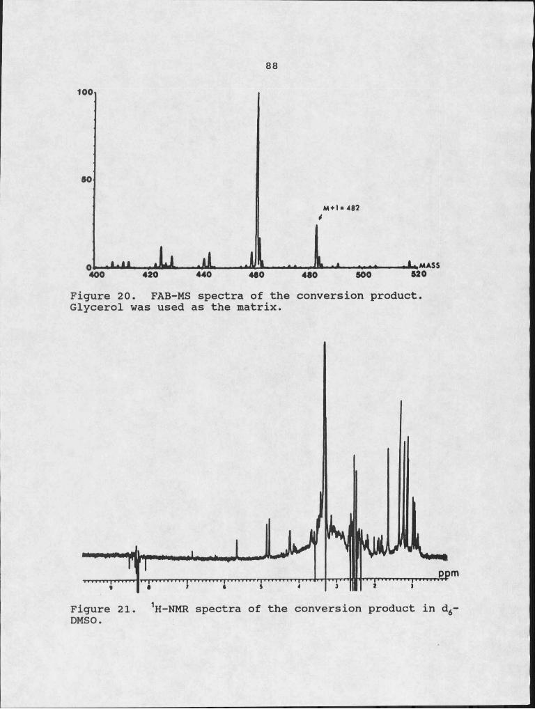
88
M*1 * 482
Figure 20. FAB-MS spectra of the conversion product. Glycerol was used as the matrix.
Figure 21. 1H-NMR spectra of the conversion product in d6 DMSO.
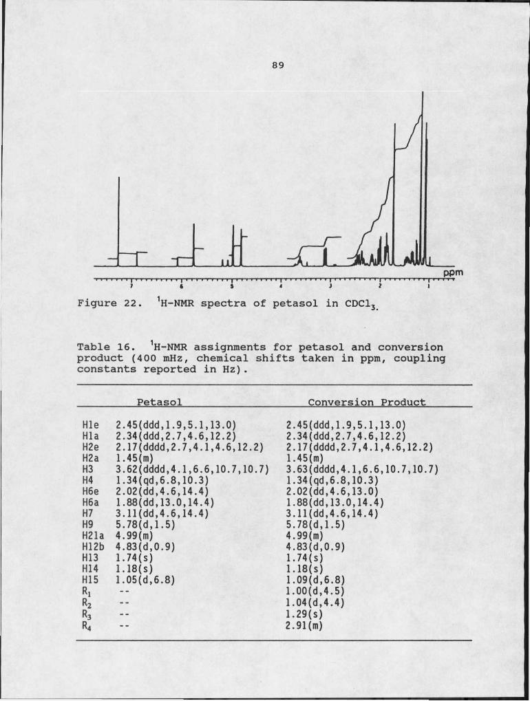
89
Figure H-NMR spectra of petasol in CDCl
Table 16. 1H-NMR assignments for petasol and conversion product (400 mHz, chemical shifts taken in ppm, coupling constants reported in Hz).
Petasol___________________Conversion ProductHle 2.45(ddd,I.9,5.I,13.0)Hla 2.34(ddd,2.7,4.6,12.2)H2e 2.17(dddd,2.7,4.1,4.6,12.2) H2a 1.45(m)H3 3.62(dddd,4.I,6.6,10.7,10.7) H4 I.34(qd,6.8,10.3)H6e 2.02(dd,4.6,14.4)H6a 1.88(dd,13.0,14.4)H7 3.11(dd,4.6,14.4)H9 5.78(d,1.5)H21a 4.99(m)H12b 4.83(d,0.9)H13 1.74(s)H14 I.18(s)H15 1.05(d,6.8)R1R2R3R4
2.45(ddd,I.9,5.I,13.0) 2.34(ddd,2.7,4.6,12.2)2.17(dddd,2.7,4.1,4.6,12.2) 1.45(m)3.63(dddd,4.1,6.6,10.7,10.7) 1.34(qd,6.8,10.3) 2.02(dd,4.6,13.0)1.88(dd,13.0,14.4) 3.11(dd,4.6,14.4)5.78(d,I.5)4.99(m)4.83(d,0.9)174(s)1. 18(s)1.09(d,6.8)1.00(d,4.5)1.04(d,4.4)1.29(s)2.91(m)

90If petasol is intact in CP it should be recoverable
from CP after mild hydrolysis. CP was hydrolyzed with 6 N formic acid for 3 hours at IOO0C. Hydrolysate products were analyzed using TLC followed by autoradiography of the TLC plate. As can be seen in Figure 23, a radioactive spot (track c) with the same Rf as petasol (track a) was seen in the hydrolysate. When the plate was sprayed with ninhydrin a pink spot developed at the origin of track c. The radioactive spot at the origin in track b is pure CP. This data confirms the results from the 1H-NMR indicating the presence of petasol in the CP molecule. The ninhydrin positive spot at the origin of the hydrolysate track indicates that the remaining part of the molecule contains a-amino acids. This would be in agreement with the FAB-MS results indicating the presence of nitrogen in the CP molecule.
Purified CP was hydrolyzed with 6 N HCL for 24 hrs at IOO0C and the hydrolysate was subjected to amino acid analysis. Table 17 shows the results from the amino acid analysis of CP hydrolysate. The amino acids present in the highest quantities were glutamic acid (glu), serine (ser), glycine (gly) and isoleucine (ile). As the amount of CP hydrolysate analyzed was increased on a percent of sample basis, levels of glu, ser, and gly all increased while ile levels remained constant (data not shown), indicating that ile is a background contaminant. The amino acid analysis

91suggests that glu, ser, and gly are all part of the CP molecule.
Figure 23. Autoradiograph of TLC plate showing the products from hydrolysis of CP. Track A) radiolabeled petasol, B)CP, C) hydrolysate of CP. Approximately 200 dpm was of each sample was spotted and autoradiograph was developed for 5 weeks.
Table 17. Chromatogram report from amino acid analysis of conversion product hydrolysate. NOR leucine (100 pmol) was used as an internal standard.
Peak IDRet.Time(min)
Cal.Time(min)
Peak Area (uA)
pmolbyarea
pmolcorrected Int Std
Aspartic Acid 1.31 1.29 17947 12.37 16.30Glutamic Acid 1.72 1.71 63937 42.66 56.23Serine 2.57 2.57 42010 36.54 48.17Glycine 3.02 3.02 45240 38.77 51.10Alanine 4.76 4.75 27843 18.72 24.68Valine 8.77 8.70 20288 12.09 15.93Isoleucine 11.03 10.97 61437 36.46 48.06Leucine 11.27 11.19 13810 9.22 12.16NOR Leu 11.62 11.54 139243 75.87 100.00
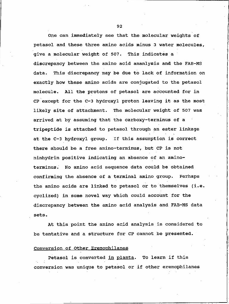
92One can immediately see that the molecular weights of
petasol and these three amino acids minus 3 water molecules, give a molecular weight of 507. This indicates a discrepancy between the amino acid ananlysis and the FAB-MS data. This discrepancy may be due to lack of information on exactly how these amino acids are conjugated to the petasol molecule. All the protons of petasol are accounted for in CP except for the C-3 hydroxyl proton leaving it as the most likely site of attachment. The molecular weight of 507 was arrived at by assuming that the carboxy-terminus of a tripeptide is attached to petasol through an ester linkage at the C-3 hydroxyl group. If this assumption is correct there should be a free amino-terminus, but CP is not ninhydrin positive indicating an absence of an amino- terminus. No amino acid sequence data could be obtained confirming the absence of a terminal amino group. Perhaps the amino acids are linked to petasol or to themselves (i.e. cyclized) in some novel way which could account for the discrepancy between the amino acid analysis and FAB-MS data sets.
At this point the amino acid analysis is considered to be tentative and a structure for CP cannot be presented.
Conversion of Other EremonhilanesPetasol is converted in planta. To learn if this
conversion was unique to petasol or if other eremophilanes
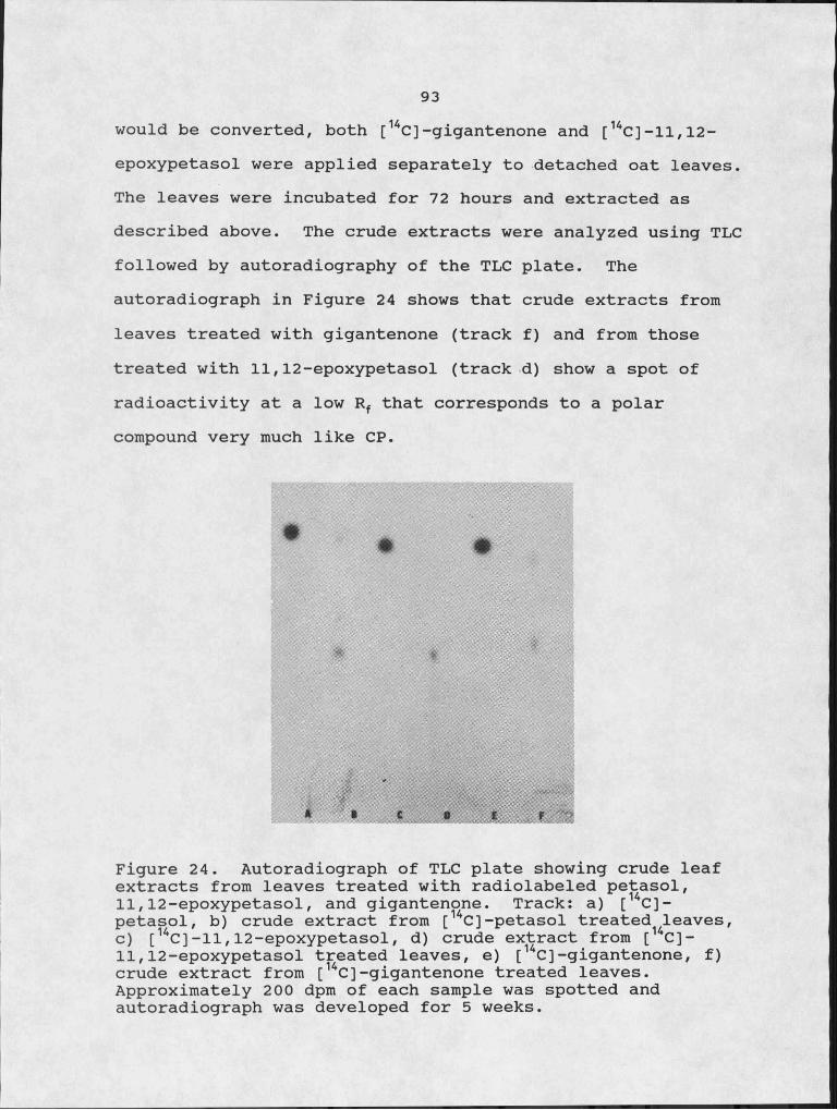
93would be converted, both [ 14C ] -gigantenone and [14C]-11,12-
epoxypetasol were applied separately to detached oat leaves.
The leaves were incubated for 72 hours and extracted as
described a b o v e . The crude extracts were analyzed using TLC
followed by autoradiography of the TLC plate. The
autoradiograph in Figure 24 shows that crude extracts from
leaves treated with gigantenone (track f) and from those
treated with 11,12-epoxypetasol (track d) show a spot of
radioactivity at a low R f that corresponds to a polar
compound very much like CP.
S #
Figure 24. Autoradiograph of TLC plate showing crude leaf extracts from leaves treated with radiolabeled petasol,11.12- epoxypetasol, and gigantenone. Track: a) [14C]- petasol, b) crude extract from [ 14C]-petasol treated leaves, c) [14C ] -11,12-epoxypetasol, d) crude extract from [14C]-11.12- epoxypetasol treated leaves, e) [ 14C]-gigantenone, f ) crude extract from [14C]-gigantenone treated leaves. Approximately 200 dpm of each sample was spotted and autoradiograph was developed for 5 weeks.

94
This would indicate that both gigantenone and 11,12- epoxypetasol are derivatized in vivo. 11,12-epoxypetasol has very little green island forming ability yet is converted like petasol and gigantenone; biological activity does not seem to be a prerequisite for conjugation.
Conversion of Petasol in DicotsPetasol causes necroses on most dicots tested. Is
petasol derivatized in dicots? [14C]-Petasol was applied to puncture wounds made on detached mung bean leaves. After incubation (72 hours) the leaves were extracted as described for the oat leaves. Crude extracts were analyzed using TLC followed by autoradiography. The autoradiograph in Figure 25 shows that the crude extract (track c) had a spot of radioactivity with an Rf of 0.40, corresponding to the Rf of CP (track b).
Petasol is converted in this dicot in planta. The darker spot in track c at Rf 0.65 corresponds to petasol.The intensity and size of this spot indicates that petasol may not be converted as efficiently in dicots as it seems to be in monocots, however, no quantitative data was collected to support this observation.
95
* I C
Figure 25. Autoradiograph of TLC plate of radiolabeled p e t a s o l , conversion p r o d u c t , and crude leaf extract from [ 14C]-petasol treated mung bean leaves. Track: a) radiolabeled p e t a s o l , b) conversion product, c) crude extract from [u C] -petasol treated mung bean leaves. Approximately 200 dpm of each sample was spotted and autoradiograph was developed for 5 weeks.
Biological Activity of Conversion Product
Pure samples of CP were tested for its ability to
induce green island formation. CP was applied to detached
oat leaves using the puncture wound droplet overlay
technique. The leaves were incubated in a moist chamber for
72 hours in the dark. A I cm segment, 0.5 cm basipetal to
0.5 cm acropetal to the punture, was excised and chlorophyll
content was determined. As seen in Figure 26, CP did not
delay senescence in the detached oat leaves. The conversion
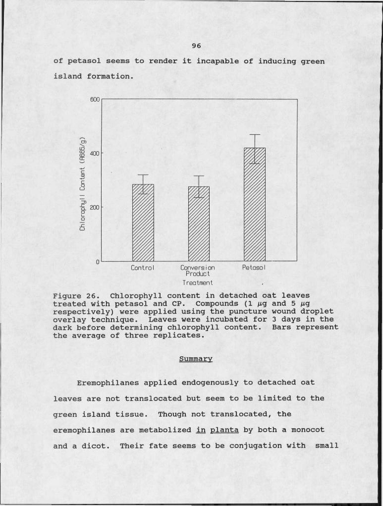
96of petasol seems to render it incapable of inducing green island formation.
PetasolConversionProductControlTreatment
Figure 26. Chlorophyll content in detached oat leaves treated with petasol and CP. Compounds (I /Ltg and 5 /ig respectively) were applied using the puncture wound droplet overlay technique. Leaves were incubated for 3 days in the dark before determining chlorophyll content. Bars represent the average of three replicates.
Summary
Eremophilanes applied endogenously to detached oat leaves are not translocated but seem to be limited to the green island tissue. Though not translocated, the eremophilanes are metabolized in planta by both a monocot and a dicot. Their fate seems to be conjugation with small

97peptides, but no structure or mechanism of conjugation can be proposed at this time. Conjugation seems to inactivate the biologically active eremophilanes, with respect to ability to induce green islands.
Conjugation is not unique to the eremophilanes, it has been reported for other biologically important compounds such as the phytohormone indoleacetic acid (IAA) (12). IAA has been found conjugated with sugars, amino acids, and inositol. These conjugates are not biologically active, but may be important as endogenous reservoirs of important growth regulators (12).
98
CHAPTER IV
CONCLUSION
This study has focused on the biological aspects of the eremophilanes produced by JX glgantea. The objectives have, in part, been met. The results give a greater understanding of these interesting molecules, operating at the interface of pathogen and plant.
Eremophilane production, by IX. glgantea, was increased with the addition of host plant leaf material to the medium, suggesting that the host can induce toxin production by the pathogen (Fig. 2). The organic extracts of host plant material increased petasol production (Fig. 3) ,. indicating the possibility of host specific factors, recognized by the fungus, which lead to increased toxin production. That host plant material was the only plant material that would increase eremophilane production has not been proven. The "amino acid fraction" of host plant material also increased eremophilane production in the fungus (Fig. 3), this is presumably due to increasing the concentration of leucine in the medium. L-Ieucine, a possible precursor in eremophilane synthesis, greatly stimulated eremophilane production (Fig. 2). Leucine content in the "amino acid fraction" was not determined.

99
L-Ieucine stimulated eremophilane production. However, [14C]-leucine, labeled at the carboxyl carbon, did not lead to a high specific radioactivity in petasol (Fig. 6), due to the fact that the carboxyl carbon of leucine is cleaved off as the molecule is metabolized to form mevalonic acid (45). This suggests that uniformly labeled [14C]-leucine could be used to obtain petasol with high specific radioactivity.
Eremophilane production began in the early growth stage of the fungus (Fig. 5), indicating that these compounds are not waste products of secondary metabolism, but could have a possible role in the physiology of the fungus.
Earlier studies on the structure-activity relationships of eremophilanes indicated the necessity of an epoxide functionality for. activity (11). The results reported here show petasol, a nonepoxidated eremophilane, to be highly active in all bioassay tests (Tables 2,3 and 4). While this work refutes previous findings, no other distinct functional group or structural characteristic could be correlated to activity. However, it was noted that the eremophilanes with higher oxidation states were generally less active.Molecular modeling may be useful in revealing characteristics of these molecules that could be correlated to activity.
It is generally accepted that pathogen induced green islands are due to the production of '‘cytokinin-like" compounds, but the chemical evidence supporting this

100speculation is incomplete (18). The eremophilanes represent
-a unique class of pathogen-produced compounds, with absolute chfemidal identity, known to induce green islands.i 'Comparison of eremophilane action to kinetin in several bioassays showed distinct differences in their physiological effects (Tables 5,6,7 and Fig. 9). In addition, kinetin stimulated protein synthesis in vivo while the eremophilanes inhibited protein synthesis in vivo (Fig. 16), indicating that eremophilane induced green islands are not due to increased levels of cytokinins in the plant tissue.
Protein synthesis has been shown to be required for plant senescence (43). The eremophilanes inhibited protein synthesis both in vitro and in vivo though they were not as effective as cycloheximide in this respect (Figs. 14 and 16). These compounds seem to induce green islands by blocking, at the translational level, processes (i.e. protein synthesis) that lead to increased proteolytic activity. However, 11,12-epoxypetasol which was also shown to inhibit protein synthesis (Fig. 14), did. not induce green islands very effectively (Fig. 8). This paradox can be explained by examining the activities of petasol and gigantenone, strong inducers of green islands. Both of these compounds were more effective than 11,12-epoxypetasolin inhibiting protein synthesis. This suggests that there
■ ■ ■is a threshold to the amount of protein synthesis required for dark induced, senescence. This observation is supported

101by data showing that the size of green islands produced by eremophilane treatment is directly related to the ability of that eremophilane to inhibit protein synthesis (compare Fig. 14 and Table 14). Additional evidence is seen in the proteolytic enzyme activity levels in treated leaves (Fig. 12). Again, for the eremophilanes, ability to inhibit protein synthesis is inversely related to proteolytic enzyme activity levels. These data also show another difference in cytokinin versus eremophilane green island formation. As mentioned earlier, kinetin stimulated protein synthesis in vivo, yet levels of proteolytic enzyme activity were lower in kinetin treated leaves than in control leaves (Fig. 12). The proteins produced in kinetin treated leaves were also electrophoretically different than proteins produced in control leaves (Fig. 17). Thus, it appears that the cytokinins and eremophilanes both induce green islands but do so through widely different mechanisms.
The exact mechanism of how eremophilanes inhibit protein synthesis is unknown. However, binding sites for eremophilanes seem to be present within the plant cells (Fig.11). Perhaps the eremophilane binding site is on the ribosomes and eremophilane binding interferes with binding of ittRNA to the ribosomes and subsequent protein synthesis.
Many of the phytohormone-1ike responses were seen when eremophilanes were applied at low concentrations (IO"5 to IO"6 M) (37) . Eremophilanes inhibited protein

102
synthesis at IO'3 M but, even at this higher concentration, inhibition was not complete (Fig.14). The mixed phytohormone-1ike response of plant tissues to these eremophilanes could be caused by hormone imbalances resulting from decreased protein synthesis. Also, the possibility exists that eremophilanes could selectively inhibit certain proteins, this could also lead to imbalances in plant metabolism.
The selectivity of the eremophilanes towards certain plant species make them candidates for probing molecular events. The eremophilanes could be useful tools for "dissecting" the biochemical apparatus of protein synthesis and for examining the differences in protein synthesis between plant species. The eremophilanes may be excellent tools for future research in understanding protein synthesis.
In studying the fate of the eremophilanes once they enter the plant several observations were made. Petasol, gigantenone, and 11,12-epoxypetasol, were converted to polar, water soluble products (Fig. 24). This conversion occurred in both monocots and dicots (Figs. 24 and 25). Chemically, these conversion products appear to be amino acid conjugates. The exact amino acid composition is not known and some variation in composition between different samples was observed. The discrepancies encountered in the elucidation of their structures lead to speculation on the

103amino acid composition. It is possible that the enzyme(s) responsible for conjugation is not stringent for the amino acids that it uses and may use any combination of related amino acids to conjugate the eremophilane. This would result in a family of conversion products with almost identical chromatographic properties but slightly different spectral characteristics. These compounds would be <difficult to separate but, the high sensitivity of the spectral analyses would "see" the different molecules and conflicting data would be obtained.
The conversion product fails to elicit green islands when applied to detached oat leaves, but its complete bioactivities are unknown. Drechsler reported the importance of secondary development in the sporulation of D. qjqantea (21). Knowing that an eremophilane has already been reported to be a sporulation agent (65), one could speculate that this fungal-plant product is involved in the sporulation of EJ2. qjqantea.
The possible role that eremophilanes play in plant pathogenesis is difficult to ascertian as no specific studies were carried out directly examining this aspect of the toxins. However, the eremophilane induced green islands are reminiscent of descriptions of the early stages of symptom development (i.e. small oval flecks surrounded by dark green zones). Necrosis, characteristic of the later stages of disease development, was not seen on monocots
104treated with eremophilanes indicating that eremophilanes may not have a direct effect in pathogenesis. Perhaps the effect of these compounds is an indirect effect involved in sporulation, as described in the previous paragraph. Many possibilities exist, but the role of eremophilanes in plant pathogenesis remains to be understood.
While the results presented here are for the most part, descriptive in nature, they provide the necessary ground work for studies focused on understanding the interactions between pathogen and host at the molecular level. The understanding of these interactions at the molecular level is at its infancy, but phytotoxins offer exciting potential for advances in this area. Additional work studying the differential response of monocots and dicots and the potential of these compounds for selective herbicidal uses remain for future, investigators.
REFERENCES CITED
106
REFERENCES CITED
1. Aducci, P., G. Crosetti, R. Federico and A. Ballio.1980. Fusicoccin receptors: evidence for endogenous ligand. Planta 148:208-210.2. Aducci, P., A. Ballio and M. Marru. 1986. Incubation of corn coleoptiies with auxin enhances in vitro fusicoccin binding. Planta 167:129-132.3. Agrios, G.N. 1978. "Plant Pathology", 2nd Ed. pp. 42-45. Academic Press, New. York.4. Anonymous. 1967. Statistics and resolution. Section H O in Handbook for Preparation of Samples for Liquid Scintillation Counting. Nuclear-Chicago Corporation, Des Plaines, Illinois.5. Bhullar, B.J., J.M. Daly and D.W. Rehfeld. 1975. Inhibition of dark C0? fixation and photosynthesis in leaf discs of corn susceptible to the host-specific toxin produced by Helminthosporium mavdis. race T. Plant Physiol. 56:1-7.6. "Biological Control of Weeds with Plant Pathogens"; R. Charudattan and H.L. Walker, eds., 1982. pp. 38. John Wiley and Sons, New York.7. Bradford, M.M. 1976. A rapid and sensitive method for the quantitation of microgram quantities of protein utilizing the principle of protein-dye binding. Anal.Biochem. 72:248-254.8. Buchanan, B. 1980. Role of light in the regulation of chloroplast enzymes. Ann. Rev. Plant Physiol. 31:341-374.9. Burk, L.G. and R.D. Durbin. 1978. The reaction of Nicotiana species to tentoxin: a new technique for identifying the cytoplasmic parent. J. Heredity 69:117-120.10. Byther, R.S. and G.W. Steiner. 1972. Use of helminthosporoside to select sugarcane seedlings resistant to eye spot disease. Phytopath. 62:466-470.
10711. Capasso, R., N.S. Iacobellis, A. Bottalico and G. Randazzo. 1984. Structure-toxicity relationships of the eremophilanes phomenone and PR-toxin. Phytochemistry 23:2781-2784.12. Cohen, J.D. and R.S. Bandurski. 1982. Chemistry and physiology of the bound auxins. Ann. Rev. Plant Physiol. 33:403-30.13. Couch, H.B. 1962. "Diseases of Turfgrasses". pp. 10-11. Reinhold Publishing Corp., New York.14. Dale, J.L. 1959. A leaf spot disease on bermudagrass. Arkansas Farm Research: Nov.-Dec. pp. 7.15. Dalling, M.J. 1987. Proteolytic enzymes and leaf senescence, pp. 54-70 in "Plant Senescence: its Biochemistry and Physiology". W. W. Thomson, E. A. Nothnagel and R. C.Huffaker, eds. Amer. Spc. Plant Physiol., Maryland.16. Day, P.R. 1979. Discussion of paper by Hanchey and Wheeler, pp. 209-210 in "Recognition Factors in Relation to Disease Specificity". J.M. Daly and I. Uritani, eds. Japan Scientific Societies Press, Tokyo.17. Dekhuijzen, H.M. and R.C. Staples. 1968. Mobilization factors in uredospores and broad bean leaves infected with bean rust fungus. Contr. Boyce Thompson Inst. PI. 24:39-52.18. Dekhuijzen, H.M. 1976. Endogenous cytokinins in healthy and diseased plants, pp. 538-540 in Physiological Plant Pathology, Encyclopedia of Plant Physiology, New Series,Vol. 4. R. Heitefuss and P. H. Williams, eds. Springer- Verlag, New York.19. Dhar, T.K., K. Siddiqui and E. Ali. 1982. Structure of phaseolinone, a novel phytotoxin from Macroohomina phaseolina. Tetrahedron Lett. 23:5459-5462.20. Douglas, T.J. and L.G. Paleg. 1974. Plant growth retardants as inhibitors of sterol biosynthesis in tobacco seedlings. Plant Physiol. 54:238-245.21. Drechsler, C. 1928. Zonate eyespot of grasses caused by Helminthosoorium aiqantea. J. Agric. Res. 37:473-492.22. Duke, S.0. 1986. Naturally occuring chemical compounds as herbicides. Rev. Weed Sci. 2:15-44.23. Durbin, R.D. 1981. Applications, pp. 495-505 in "Toxins in Plant Disease". R.D. Durbin, ed. Academic Press, New York.
10824. Filner, P. 1965. Semi-conservative replication of DNA in a higher plant cell. Exper. Cell Res. 39:33-39.25. Fletcher, R.A. and D. McCallagh. 1971. Cytokinin induced chlorophyll formation in cucumber cotyledons. Planta 101:88- 90.26. Fletcher, R.A. 1969. Retardation of leaf senescence by benzyladenine in intact bean plants. Planta 89:1-7.27. Fox, J.E. and J.L. Erion. 1975. A cytokinin binding protein from higher plant ribosomes. Biochem. Biophys. Res. Comm. 64:694-700.28. Fu, Y. 1989. Isolation and characterization of novel phytotoxins from pathogenic fungi. PhD. Thesis. Cornell Univ., Ithaca, New York.29. Gaumann, E. 1954. Toxins and plant dieases. Endeavour 13:198-204.30. Gengenbach, B.G. and C.E. Green. 1975. Selection of T- cytoplasm maize callus cultures resistant toHelminthosporium maydis race T pathotoxin. Crop Sci. 15:645- 649.31. Griffin, D.H. 1981. "Fungal Physiology", pp. 323-336. John Wiley and Sons, New York.32. Hiscox, J.D. and G.F. Israelstam. 1979. A method for extraction of chlorophyll from leaf tissue without maceration. Can. J. Bot. 57:1332-1334.33. Howard, G.A. and R.R. Traut. 1973. Separation and radioautography of microgram quantities of ribosomal proteins by two-dimensional polyacrylamide gel electrophoresis. FEES Lett. 29:177-180.34. Jacobs, M. and R. Hertel. 1978. Auxin binding to subcellular fractions from cucurbita hypocotyls:.in vitro evidence for an auxin transport carrier. Planta 142:1-10.35. Kende, H- 1971. The cytokinins. Int. Rev. Cytol. 31:301- 338.36. Kenfield, D.S. and G.A. Strobel. 1981. a-Galactoside binding proteins from plant membranes: isolation, chacterization, and relation to helminthosporoside binding proteins of sugarcane. Plant Physiol. 67:1174-1180.
10937. Kenfield, D., F. Sugawara, G. Bunkers, Y.H. Wu, G. Strobel, Y. Fu, J. Clardy. 1989. Gigantenone, a novel sesquiterpene phytohormone mimic. Experientia7 in press.38. Kern H. 1972. Phytotoxins produced by Fusaria. pp. 35-48 in "Phytotoxins in Plant Diseaes". R.K.S. Wood, A. Ballio and A. Graniti7 eds. Academic Press, New York.39. Kiraly7 Z.7 M. El Hammady7 and B.I. Pozar. 1967. Increased cytokinin activity of rust-infected bean and broad bean leaves. Phytopath. 52:171-172.40. Laemmli7 U.K. 1970. Cleavage of structural proteins during the assembly of the head of bacteriophage T4. Nature (bond.) 227:680-685.41. Leegood7 R.C. and D.A. Walker. 1983. Chloroplasts. pp. 185-210 in "Isolation of Membranes and Organelles from Plant Cells". J.L. Hall and A.L. Moore7 eds. Academic Press7 New York.42. Marre E. 1979. Fusicoccin: a tool in plant physiology. Ann. Rev. Plant Physiol. 30:273-288.43. Martin7 C. and K.V. Thimann. 1972. The role of protein synthesis in the senescence of leaves. Plant Physiol. 49:64- 71.44. Matern7 U., R. Bier and G.A. Strobel. 1984. A novel plant glycolipid serves as an activator of toxin production in Helminthosoorium sacchari. Biochem. International. 4:655- 661.45. McCorkindale7 N.J. 1976. The biosynthesis of terpenes and steriods. pp. 369-422 in "The Filamentous Fungi7 Vol.II Biosynthesis and Metabolism". J.E. Smith and D.R. Berry7 eds. John Wiley and Sons7 New York.46. Mitchell,,R.E. 1984. The relevance of non-host-specific toxins in the expression of virulence by pathogens. Ann.Rev. Phytopath. 22:215-245.47. Moule7 Y., S. Moreou and J.F. Bousquet. 1977. Relationships between the chemical structure and the biological properties of some eremophilane compounds related to PR toxin. Chem. Biol. Inter. 17:185-192.48. Muller, K. and A.C. Leopold. 1966. Correlative aging and transport of P32 in corn leaves under the influence of kinetin. Planta 68:167-185.
H O49. Peterson, L.W. and R.C. Huffaker. 1975. Loss of ribulose I,5-diphosphate carboxylase and increase in proteolytic activity during senescence of detached primary barley leaves. Plant Physiol. 55:1009-1015.50. Pinder, A.R. 1977. The chemistry of the eremophilanes and related sesquiterpenes. Fortschr. Chem. Org. Naturst. 34:81-186.51. Pinkerton, F. and G. Strobel. 1976. Serinol as an activator of toxin production in attenuated cultures of Helminthosnorium sacchari. Proc. Natl. Acd. Sci. 73:4007- 4011.52. Pjon, C.J. 1981. Effects of cycloheximide and light on leaf senescence in maize and hydrangea. Plant Cell Physiol. 22:847-854.53. Pozar, B.I. and Z. Kiraly. 1966. Phloem transport in rust infected plants and the cytokinin-directed longdistance movement of nutrients. Phytopath. Z. 56:297-309.54. Riche, C., C. Pascard-Billy, M. Devys, A. Gaudemer and M. Barbier. 1974. Structure crystalline et moleculaire de la phomenone, phytoxine produite par Ie champignon Phoma exiaua var. non-oxvdabilis. Tetrahedron Lett. 32:2765-2766.55. Robinson, T. 1980. "The Organic Constituents of Higher Plants",4th ed. pp. 133-190. Cordus Press, Mass.56. Ruzicka, L. 1953. The isoprene rule and the biogenesis of terpenic compounds. Experientia 9:357-396.57. Samaddar, K.P. and R.P. Scheffer. 1971. Early effects of Helminthosoorium victoriae toxin on plasma membranes and counteraction by chemical treatments. Physiol. Plant Pathol. 1:319-328.58. Scheffer, R.P. and O.C. Yoder. 1972. Host specific toxins and selective toxicty. pp. 217-236 in "Phytotoxins in Plant Disease". R.K.S. Wood, A. Ballio, and A. Graniti, eds. Academic Press, New York.59. Siegel, M.R. 1981. Sterol inhibiting fungicides: effects on sterol biosynthesis and sites of action. Plant Dis. 65:986-989.60. Stoessl, A. 1981. Structure and biogenetic relations: fungal non-host specific, pp. 109-219 in "Toxins in Plant Disease". R.D. Durbin, ed. Academic Press, New York.
Ill
61. Strobel, G.A. 1973. The helminthosporoside-binding protein of sugarcane. J. Biol. Chem. 248:1321-1328.62. Strobel, G.A. 1974. Phytotoxins produced by plant parasites. Ann. Rev. Plant Physiol. 25:541-566.63. Strobel, G.A. 1979. The relationship between membrane ATPase activity in sugarcane and heat-induced resistance to helminthosporoside. Biochem. et Biophys. Acta 554:460-468.64. Sugawara, F., G. Strobel, L.E. Fisher, G.D. VanDuyne, and J. Clardy. 1985. Bipolaroxin, a selective phytotoxin produced by Bipolaris cvnodontis. Proc. Natl. Acad. Sci. USA 82:8291-8294.65. Tanaka, S., K. Wada, S. Marumo and H. Hattori. 1984. Structure of sporogen AO-1, a sporogenic substance. Tetrahedron Lett. 25:5907-5910.66. TeBeest, D.O. and G.E. Templeton. 1985. Mycoherbicides: progress in the biological control of weeds. Plant Dis. 69:1-10.67. Tepfer, D.A. and D.E. Fosket. 1978. Hormone-mediated translational control of protein synthesis in cultured cells of Glvcine max. Develop. Biol. 62:486-497.68. Thimann, K.V. 1980. The senescence of leaves, pp. 86-109 in "Senescence in Plants". K. V. Thimann, ed. CRC Press, Florida.69. Uchytil, T.F. and R.D. Durbin. 1980. Hydrolysis of tabtoxins by plant and microbial enzymes. Experientia 36: 301-302.70. Wei, R.D., P.E. Still, E.B. Smalley, H.K. Schnoes and F.M. Strong. 1973. Isolation and partial characterization of a mycotoxin from Penicillium rocmeforti. Appl. Envir. Microbiol. 25: 111-114.71. Wei, R., H.K. Schnoes, P.A. Hart and F.M. Strong. 1975. The structure of PR-toxin, amycotoxin from Penicillium rocmeforti. Tetrahedron Lett. 31:109-114.72. Wickremashinghe, R.L. 1974. The mechanisms of operation of climatic factors in the biogenesis of tea flavor. Phytochemistry. 13:2057-2063.
11273. Williimsf A.G., J.W.T. Wimpenny and C.J. Lawson. 1979. Preliminary studies on the composition and rheological properties of the extracellular polysaccharide synthesized by Pseudomonas PBI (NClB 11264). Biochim. Biophys. Acta 585:611-619.
APPENDICES
APPENDIX ARECIPE FOR MODIFIED M-ID MEDIUM
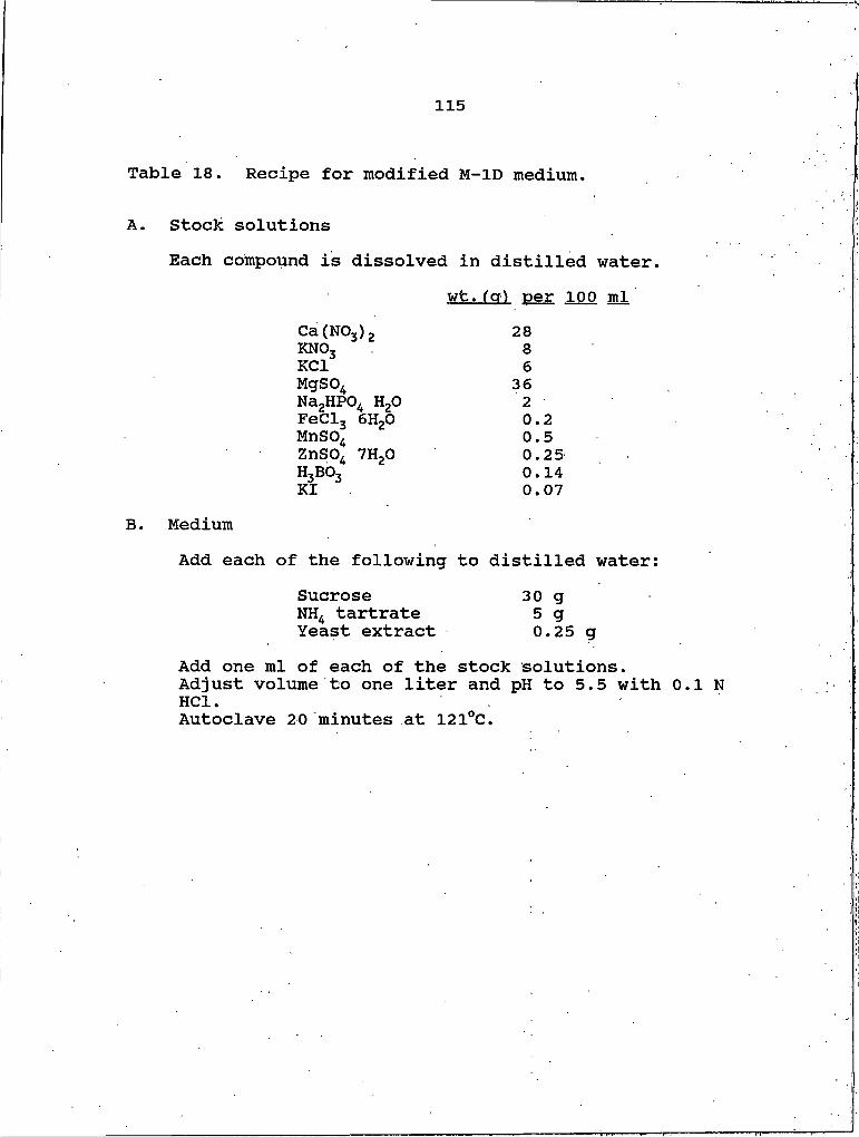
115
Table 18. Recipe for modified M-ID medium.
A. Stock solutionsEach compound is dissolved in distilled water.
wt.(a) per 100 mlCa(NO3)2 28KNO3 8KCl 6Mgso4 36Na2HPO4 H2O 2FeCl3 GH2O 0.2MnSO4 0.5ZnSO, THpO 0.25H3BO34 0.14KI 0.07
B. MediumAdd each of the following to distilled water:
Sucrose 30 gNH4 tartrate 5 gYeast extract 0.25 g
Add one ml of each of the stock solutions.Adjust volume to one liter and pH to 5.5 with 0.1 N HCl.Autoclave 20 minutes at 12I0C.
116
APPENDIX BISOLATION OF PLANT FRACTIONS
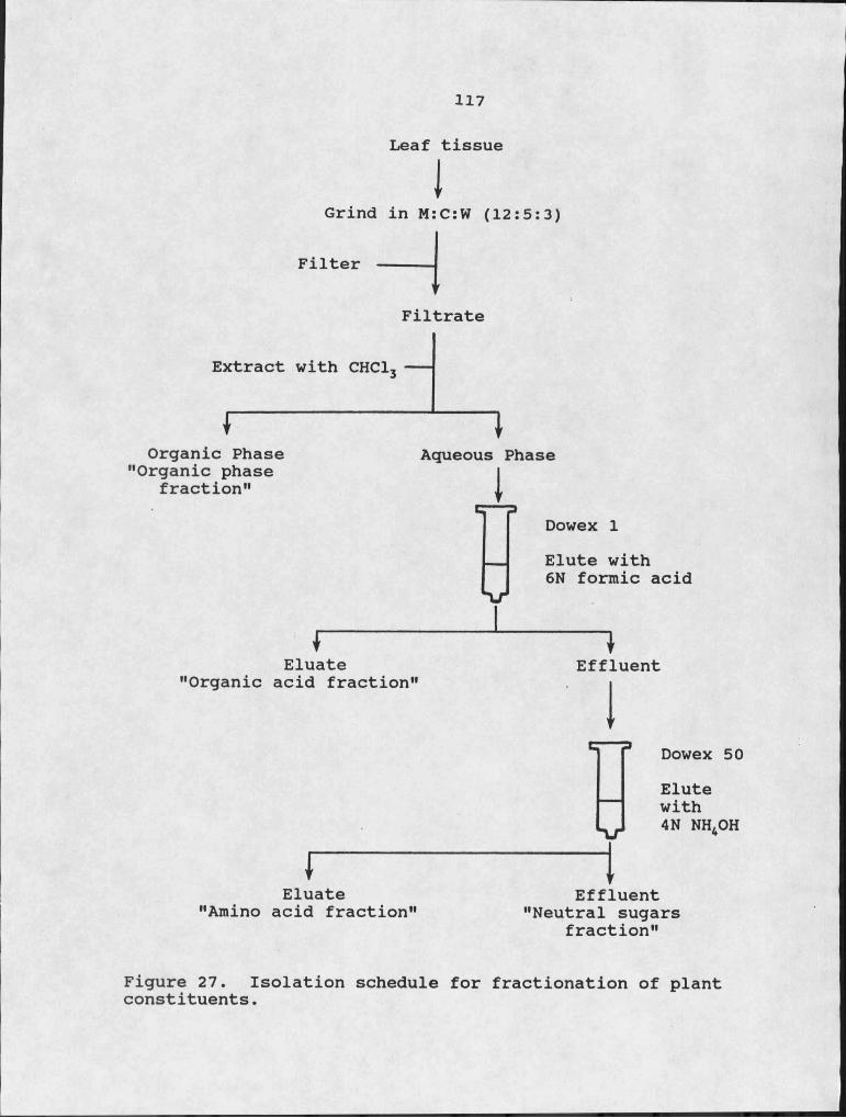
117
Leaf tissue
IGrind in M:C:W (12:5:3)
Filter -----\ f
Filtrate
Extract with CHCl3
rOrganic Phase
"Organic phase fraction"
IAqueous Phase
Dowex I
XrElute with 6N formic acid
rEluate
"Organic acid fraction"
rEluate
"Amino acid fraction"
Effluent
IDowex 50Elutewith4 N NH4OH
Effluent "Neutral sugars
fraction"
Figure 27. Isolation schedule for fractionation of plant constituents.

MONTANA STATE UNIVERSITY LIBRARIES
3 1762 101 54584 4