Evolution of substrate specificity in bacterial AA10 lytic polysaccharide monooxygenases

2878 0892-6638/88/0002-2878/$01 .50. © FASEB
In vitro cytocidal effect of novel lytic peptides on
Plasmodium falciparum and Trypanosoma cruzi1JESSE M. JAYNES,’ CATHERINE A. BURTON,’ STEPHEN B. BARR,1 GALE W. JEFFERS,T
GORDON R. JULIAN,’ KENNETH L. WHITE,t FRED M. ENRIGHT,1 THOMAS R. KLEI,1
AND ROGER A. LAINE’
‘Department of Biochemistry,I Veterinary Science,and tAnimal Science, Louisiana State University, Baton Rouge,
Louisiana 70803, USA and Louisiana State University Experiment Station, Baton Rouge, Louisiana 70803, USA
ABSTRACT
Plasmodium falciparum and Trypanosoma cruzi were killedby two novel lytic peptides (SB-37 and Shiva-1) in vitro.
Human erythrocytes infected with P falciparum, andVero cells infected with T cruzi, were exposed to these
peptides. The result, in both cases, was a significantdecrease in the level of parasite infection. Furthermore,the peptides had a marked cytocidal effect on trypo-
mastigote stages of T cruzi in media, whereas host
eukaryotic cells were unaffected by the treatments. Inview of the worldwide prevalence of these protozoandiseases and the lack of completely suitable treatments,lytic peptides may provide new and unique chemother-apeutic agents for the treatment of these infections.-JAYNES, J. M.; BURTON, C. A.; BARR, S. B.; JEFFERS,
G. W.; JULIAN, G. R.; WHITE, K. L.; ENRIGHT, F. M.;KLEI, T R.; LAINE, R. A. In vitro cytocidal effect ofnovel lytic peptides on Plasmodium falciparum andTrypanosoma cruzi. FASEBJ 2: 2878-2883; 1988.
Key Words: lyticpepeides- in vitrocytocidaleffects ma-
laria. Chagas’disease
THE WORLD HEALTH ORGANIZATION (WHO) has identi-fied malaria and Chagas’ disease, which are caused by
Plasmodium sp. and Trypanosoma cruzi, respectively, asposing significant health hazards for 2200 mfflion people,or 46% of the world’s population (1). In recent years,chemotherapy has been an important factor in reduc-ing the mortality caused by malaria; however, in manycountries, the prevalence of this disease is increasing ata rate of approximately 10 million new cases a year (2).This is due primarily to the development of drug re-sistance by Plasmodium s/i. and to vector resistance to in-secticides (3). Treatment of Chagas’ disease has focusedon the utilization of purine derivatives (e.g., allo-purine), which disrupt normal nucleic acid metabolism(4). However, the deleterious effects of these drugs arenot limited to the parasite, but are also toxic to hostcells (5). Therefore, the established chemoetherapeutictreatments are less than completely effective or ideal for
these diseases. Vector eradication programs, thus far,have also been unable to control these protozoan-caused diseases, and the development of usable vac-cines is not imminent (6). With this recognizable per-sistent threat to a large proportion of the world’s popu-lation, a search for novel chemotherapeutic agentsbased on nontraditional modes of action is important.
Many workers describe peptides or proteins capableof lysing organisms or cells (7-9). The pioneering workconducted by Hultmark et al. and Andreu et al. de-scribes the humoral defense system used by Hyalophoracecropia, the giant silk moth, as a protective mechanismagainst bacterial infection (10-15). Specialized proteinsin the insect’s hemolymph after induction by eitherlive or heat-killed bacteria are capable of membraneperturbation, which results in bacterial cell lysis.Among these proteins is a type known as the cecropins.The three principal cecropins, A, B, and D, are highlyhomologous (16), small, basic proteins that each con-tain a comparatively long hydrophobic region. Theirprimary mode of action appears to be one of membranedisruption and subsequent lysis owing to the targetcell’s loss of osmotic integrity (17). Perhaps these typesof lytic proteins will also be found to play key roles inproviding protection from disease in other organisms.Indeed, similar types of peptides have been isolatedfrom amphibians by Gibson and co-workers (18) and byGiovannini and co-workers, and were designated PGSand Gly10Lys22-PGS (19), respectively. Somewhat later,the same peptides were described by Zasloff and called
magainins (20).Although the antibacterial effect of lytic peptides
from insects has been well documented, there are noreports of their potential effectiveness against lowereukaryotic cells.
This paper describes the in vitro effects of two syn-thetic lytic peptides on the limitation of growth andmultiplication of P falciparum and T cruzi. One peptideis a closely related derivative of cecropin B, SB-37, withminor changes made in the sequence by substitution of
‘Approved for publication by the Director of the LouisianaAgricultural Experiment Station as Manuscript Number: 88-12-2080.

HKWKVFKKIEEGRNIRNGIVKAGPAIAVLGEAKALGKWKVFKKIEI&RNIRNGIVKAGPAIAVLGEAKALG
Cecropin B
SB-37
CYTOCIDAL EFFECT OF LYTIC PEPTIDES 2879
Met11 with Val and addition of an NH2-terminal,MetPro. These changes were made to plan for subse-quent gene construction to produce a CNBr-cleavablerepeat peptide. The other is a distinct peptide, Shiva-1,which was designed with significant differences in se-quence homology (about 60% different) to test whetheror not the lytic properties of cecropin-like peptides arehighly sequence-dependent. However, the charge dis-tribution and the amphipathic and hydrophobic prop-erties of the natural molecule were conserved (Fig. 1).
METHODS
Peptide synthesis
Cecropin B and the two lytic peptide analogs were syn-thesized on a Biosearch Sam Two peptide synthesizerusing MBHA (4-methyl benzhydral amine) resin with
a COOH-terminal amide. All reagents used for thesebiosyntheses were obtained from Biosearch in SanRafael, CA. After extraction and Sephadex columnchromatography, the purity of the peptides was deter-mined by HPLC on a Varian 5000 HPLC unit. AWaters Bondepak C18 column, 8 mm x 10 cmRadial-Pak cartridge, employing the Radial Compres-sion Module-100, was used. HPLC profiles of thesepeptides indicated a purity of more than 95% (Fig. 2).To determine that the syntheses progressed to comple-
tion, amino-terminus sequence analysis was performedon all peptides with an Applied Biosystems 470-A gasphase protein sequencer. PTH-derivatized amino acids,generated from the sequencer, were analyzed in aWaters PicoTag system employing a C18 column,
3.8 mm x 15 cm (Waters Nova-Pak). All experiments
used peptides that were purified in the mannerdescribed previously.
In vitro growth of P falciparum
P. falciparum cultures were derived from Sierra-Leone1/CDC isolate and maintained in Petri plates under 5%oxygen, 3% carbon dioxide, and 82% nitrogen (21). Invitro growth of the parasites was assessed by [3H]hypo-xanthine incorporation (22), and the cultures were en-riched for ring stage parasites with the addition of 5%mannitol (23). Twenty-four hours after mannitol treat-ment, the culture was diluted to a level of 0.4% para-sitemia and 3% hematocrit; media that contained 20%human serum and 50 id/well aliquoted into microtiterplates. SB-37 and Shiva-1 were dissolved in mediawithout serum, dilutions at twice the desired final con-centration were prepared, and 50 id/well were added tothe malaria culture.
Radiolabeled hypoxanthine (50 itl/well), at 10 itCi/mlin media containing 10% serum, was added after 24 hof incubation. After an additional 24 h of growth, theparasites were harvested (Cell Harvester, Flow Labs,McLean, VA) and counted in a scintillation counter.
Duplicate slides were prepared from unlabeled parasitecultures treated as above with SB-37 at 10, 50, and 100
itM. These preparations were Geimsa-stained and thelevel of parasitemia and percentage of the parasites in
the various developmental stages were determined. Todetermine hemoglobin release from infected and unin-fected red cells, an unsynchronized culture (at 1.5%parasitemia) was harvested by centrifugation at 250 x g
MP K KII R R V GPAIPVIj9PftG
MP IRPII ___ K GPAI?JLDIbG
Amino Acid Differences Between Cecropin B and SB-37
[J Amino Acid Differences Between SB-37 and Shiva-1
SB-37
Shiva-1
Figure 1. Sequence comparison of the natural cecropin B with the novel lytic peptides SB-37 and Shiva-1. The cecropin B derivative
SB-37 was designed to have relatively minor changes from the native cecropin B peptide to facilitate future bioproduction and subsequent
purification. Shiva-1, on the other hand, was designed with significant differences in sequence homology to test whether or not the lytic
properties of cecropin-like peptides were highly sequence-dependent. The charge distribution, amphipathic, and hydrophobic properties
of the natural molecule were conserved.
SB-37
I-u..-,-
u 10 20 30 40 50
Retention Time (mm)
15
.150 iQo
0
150
0
B 200
C
0I
(5#{149} 100.0.
15.4-
15
15
0
Shlva-1
U - U - U - U
0 10 20 30 40 50
0.
D.
..-SB-37
- Shiva-1
100 120
Concentration of Peptide (MM)
Figure 3. Effect of SB-37 on the in vitro growth of P fakiparum.Ahighly significant reduction (P � 0.01) was observed in the amountof [‘H]hypoxanthine incorporated by all treated cultures whencompared with nontreated controls (n = 3).
0 20 40 60 80
A Analysis of T cruzi trypomastigotes
2880 JAYNES ET AL.
Retention Time (mm)
Figure 2. HPLC profiles of lytic peptides prepared via chemical
syntheses SB-37 (A) and Shiva-1 (B). The peptides, purified in themanner described in the Methods section, were subjected to HPLCanalysis on a linear gradient prepared from 0.01% TFA/H20 (A)and 0.1% TFA/acetonitrile (B). The profiles indicate that the pep-tides used were more than 95% homogenous.
for 10 mm and washed twice with sterile PBS. The finalpellet was resuspended to 10% in PBS. The lytic pep-tides were diluted to 200, 100, and 50 tM with PBS
and mixed with equal volumes of washed infected redcells and incubated for 30 mm at 37#{176}C.The mixtureswere then centrifuged at 250 x g for 10 mm, and the
supernatants were removed and centrifuged at 1200 x g
for 10 mm. The optical density was determined with a
Bausch & Lomb spectronic 2000 at 560 nm. Uninfectedcontrol red cells were analyzed in a similar manner.
Trypomastigotes (total of 5 x 10⁶), harvested from Verocell culture, were incubated for I h at 37#{176}Cin MEM +
10% FBS with final concentrations of 100, 50, 25, and10 /LM of SB-37 and Shiva-1 were added. The numberof parasites after treatment was determined by countingthe motile organisms microscopically with a hemo-cytometer. For microscopic analysis, T cruzi (1000trypomastigotes/ml media) was incubated with 100 tMfinal concentration of Shiva-1 or medium for only 60mm at 37#{176}C.The parasites were centrifuged and thesupernatant was discarded. The parasite pellet wasfixed in 2% paraformaldehyde and 2.5% glutaralde-hyde in 0.1 M sodium cacodylate buffer for 15 mm.The suspension was observed at 100% magnificationwith Nomarski differential interference optics.
Culture of T cruzi-infected Vero cells
Vero cell monolayers were cultured for 24 h in eightchamber microscope slides with 100 cells/cm2 in RPMI1640 with 10% fetal bovine serum. The monolayerswere infected with T cruzi cell culture-derived trypo-mastigotes at a ratio of two parasites per Vero cell. Theparasites were allowed to internalize within the Verocells for 24 h. At this time, a set of slides was fixed andstained with Wright’s stain and was designated as the24-h control culture. The media from the remainingslides were removed and fresh media (control) or mediacontaining SB-37 or Shiva-1 (100 iM each) were addedto the cultures. These cultures were incubated for anadditional 24 and 48 h, after which they were fixed,stained, and counted.
Cultures receiving a second exposure were treated byremoving the media after the first 24 h of incubation,and by adding fresh media only or media containing100 tM SB-37 and culturing for an additional 24 and48 h. Numbers of parasites per infected cell were deter-
mined by counting the total number of intracellular
parasites in no fewer than 200 infected cells and bydividing by the number of infected cells counted.
... SB-37
0,0
0,
0,0..-.
2Ez
0,
E00,0,0,0.
0
0
0,C0U00.
B
0
0,.4
-
CE
200;0.>
00
0 20 40 60 80 100 120
Concentration of Peptide (tM)
-- SB-37
... Shlva-1
0 20 40 60 80 100 120
Concentration of Peptide (M)
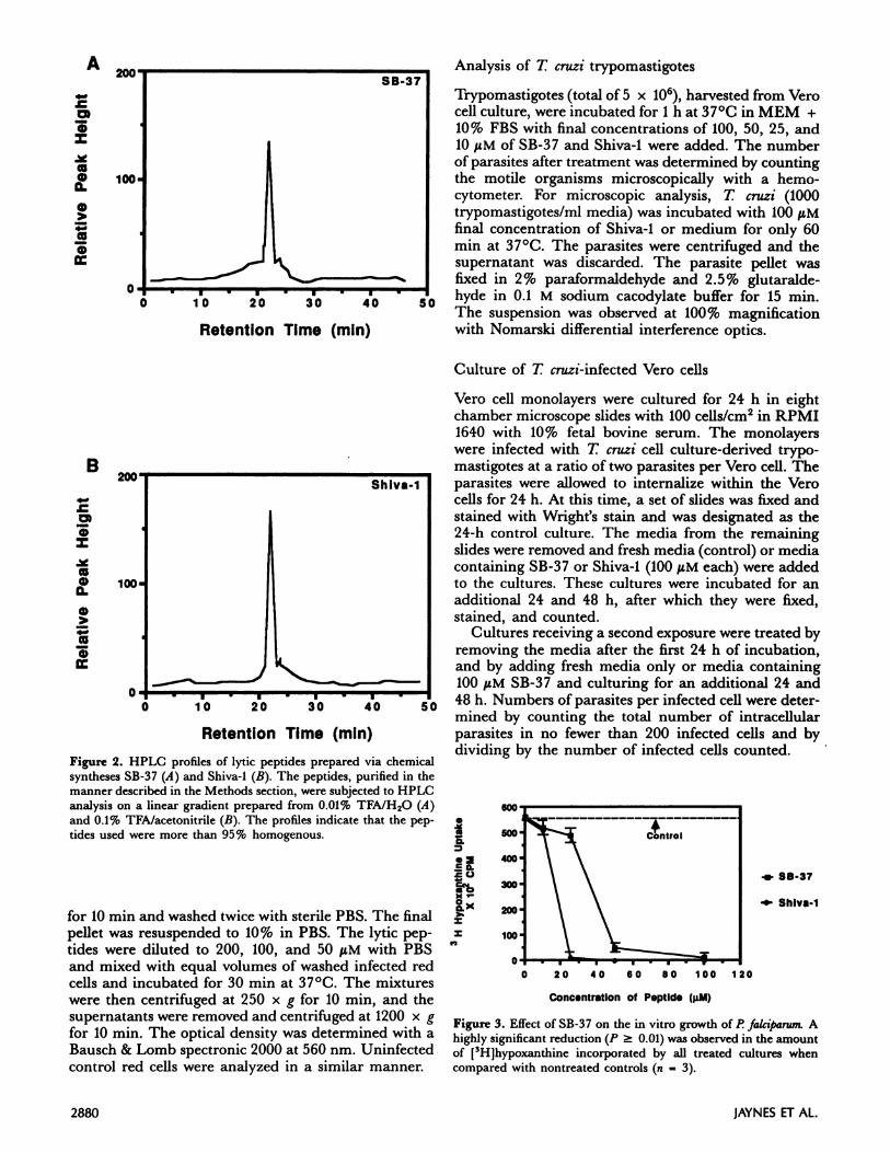
Figure 5. Viability of trypomastigotes after incubation with SB-37
and Shiva-l. A highly significant reduction (P 0.01) was observedin the number of intact parasites in all samples treated with peptidewhen compared with nontreated controls (n 4). A signficant
reduction occurred at all concentrations of peptide tested.
0 20 40 60 80 100 120
Concentration of Peptid. (iiM)
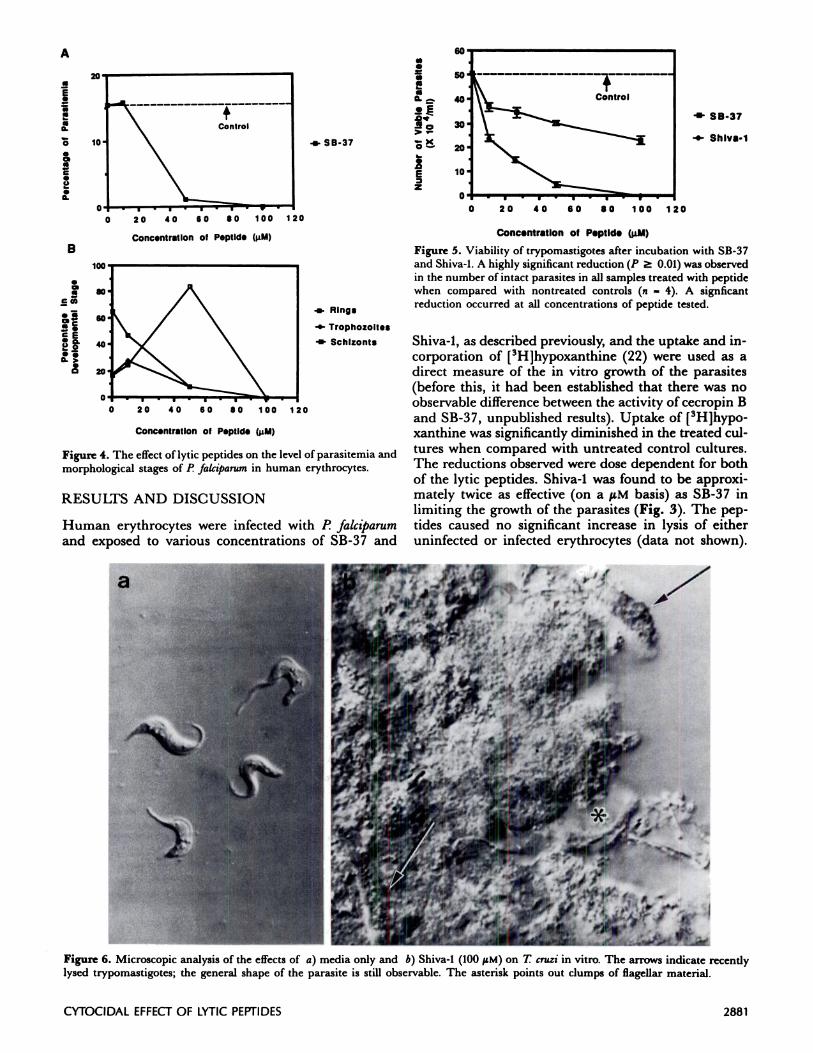
Figure 6. Microscopic analysis of the effects of a) media only and b)Shiva-l (100 tiM) on T cruzi in vitro. The arrows indicate recentlylysed trypomastigotes; the general shape of the parasite is still observable. The asterisk points out clumps of flagellar material.
A
CYTOCIDAL EFFECTOF LYTICPEPTIDES 2881
... Rings
4 Irophozoites
- Schizonts
Figure 4. The effect of lytic peptides on the level of parasitemia andmorphological stages of P falciparumin human erythrocytes.
RESULTS AND DISCUSSION
Human erythrocytes were infected with P falciparum
and exposed to various concentrations of SB-37 and
Shiva-1, as described previously, and the uptake and in-corporation of [3H]hypoxanthine (22) were used as adirect measure of the in vitro growth of the parasites(before this, it had been established that there was noobservable difference between the activity of cecropin Band SB-37, unpublished results). Uptake of [3H]hypo-xanthine was significantly diminished in the treated cul-
tures when compared with untreated control cultures.The reductions observed were dose dependent for bothof the lytic peptides. Shiva-1 was found to be approxi-mately twice as effective (on a tM basis) as SB-37 inlimiting the growth of the parasites (Fig. 3). The pep-tides caused no significant increase in lysis of eitheruninfected or infected erythrocytes (data not shown).

A
4,
aU4)
U-C
8)8)
4-
8)4)
150.
B
4)C.)
4)4-U4)
‘I-C
U,4)
4-
8)15I-150.
20 30 40
Hours Post-infection
50
. Control
.- SB-37
.- Shlva-1
= Control
- SB-37
20 30 40 50 60 70 80
Hours Post-Infection
2882 JAYNES ET AL.
An irrelevant 22 amino acid peptide, synthesized in thesame manner as described above and at similar concen-trations, was found to be inactive in reducing the up-take of [3H]hypoxanthine by infected erythrocytes.
To ascertain the effect of SB-37 on the developmentalstages of P falciparum, infected human erythrocyteswere cultured in the presence of three different concen-trations of SB-37 (10, 50, and 100 tM, respectively).After 24 h of exposure, duplicate blood smears weremicroscopically examined. The percentage of parasi-tized cells and the distribution of the various develop-mental stages of the organism were determined. As theSB-37 concentration is increased, the level of host cellparasitemia is decreased; the most dramatic reductionoccurs at the 50 ILM concentration of lytic peptide (Fig.4A). Also, there is a concomitant increase in the numberof the remaining parasites at the ring stage of develop-ment, which suggests that as the concentration of SB-37is raised to 50 tM, there is an arrest in the life cycle ofthe organism at this developmental stage (Fig. 4B), or,alternatively, that the peptides are least effective againstcells that contain this early developmental form. At 1001wM concentration of lytic peptide, there was no detec-table parasitemia.
Similar effects of the lytic peptides were observed onthe protozoan T cruzi. Trypomastigotes, harvestedfrom Vero cell culture, were exposed to various concen-trations of SB-37 and Shiva-I for 1 h at 37#{176}Cand werefound to be killed in a dose-responsive manner.
However, under these conditions, Shiva-1 was ap-proximately 10-fold more effective (on a tM basis) thanSB-37 in destroying the trypomastigotes. The numberof intact parasites was also dramatically reduced intreated samples when compared with untreated con-trols (Fig. 5), with many of the remaining parasites ap-pearing lysed or damaged (Fig. 6). However, the intacttrypomastigotes that remained in the treated sampleswere infectious when exposed to Vero cells, whichestablished a reduced level of parasitemia (parasiteswithin Vero cells; data not shown).
To determine the effect of SB-37 and Shiva-1 onT cruzi after internalization of the parasite, T cruzi-
infected Vero cells were treated with a single exposureof the peptides. The numbers of parasites per infectedcell were significantly decreased 24 h after exposure tothe lytic peptides. However, 48 h after treatment, therewas no significant difference between the number ofparasites per infected cell in treated samples and un-treated infected control cultures (Fig. 7A). This resultcan be accounted for by the multiplication of survivingparasites. A second exposure of T cruzi-infected Verocells to the same concentration of SB-37 showed a moremarked reduction in the numbers of parasites per in-fected cell after 24 and 48 h compared with culturesthat received only a single treatment of SB-37 (Fig.7B). There was no observable reduction in the numbersof control Vero cells that were treated with either of thepeptides.
This report has focused on the in vitro effect of novellytic peptides on two pathogenic protozoa. Surprising-ly, Shiva-1, the peptide that is the most divergent from
Figure 7. Effects of SB-37 and Shiva-l (100 tiM each) on T. cruzi-infected Vero cells with single exposure (A) and double exposure oflytic peptides (B). Parasitemia is expressed as the numbers of para-sites per infected cell. A highly significant reduction (P 0.01) inthe numbers of parasites per infected cell was found when control
nontreated infected cultures (n = 5) were compared with 24-h pep-
tide cultures treated with peptide (a 7) (A). However, nosignificant differences were noted in the numbers of intracellularparasites after 48 h in cultures that received a single exposure to thelytic peptides. When infected monolayers received a second ex-
posure (B) to SB-37 or fresh media at 24 and 48 h, a highlysignificant reduction (P � 0.01) in the number of intracellularparasites was observed in the cells treated with SB-37 (a 14)
compared with infected control cultures (n 14) at all time
periods.
the sequence of the parent molecule, is also the mostbiologically active against these parasites (Shiva-1 isalso more active on bacteria than either cecropin B orSB-37; to be reported elsewhere). However, the chargedistribution, amphipathic, and hydrophobic propertiesof the natural cecropin B lytic peptide were conservedin Shiva-1, and thus it would seem that these physicalcharacteristics are the parameters to be judiciously con-trolled in the design of new lytic peptide analogs.
At present, the exact mechanism of action of thesepeptides is unknown. The data suggest, however, thatalterations of the eukaryotic host cell membrane thatare caused by these parasites may increase the tendencyof infected cells to undergo peptide-induced lysis. Addi-tional studies, using different synthetic peptide analogs,are currently under way and should provide informa-tion on how the lytic effects are exerted.

CYTOCIDAL EFFECT OF LYTIC PEPTIDES 2883
Because of the global significance of protozoan dis-eases, it is of the utmost importance to develop and
evaluate novel chemotherapeutic agents for an-tiprotozoan activity.
Our report illustrates the effectiveness of syntheticlytic peptides in limiting the level of infection byP fakiparum and T cruzi in vitro. Further experimenta-tion is under way to determine if the use of such lyticpeptides can be extended to the clinical treatment ofthese and other recalcitrant diseases (J. M. Jaynes,G. W. Jeffers, G. R. Julian, K. L. White, and E M.Enright, manuscript submitted).
The authors with to acknowledge the expert technical assistance
in peptide synthesis and purification provided by Dr. V. Rao and
Judith M. Ball. Research was supported, in part, by funds from
Louisiana State University Agricultural Experiment Station, Helix
International Inc., Baton Rouge, LA, and by National Institutes of
Health grant DK 33755-03 to R. A. L.
REFERENCES
1. WORLD HEALTH ORGANIZATION TECHNICAL REPORT
SERIES, No. 711 Advances inMalaria Chemotherapy:reportof a WHO ScientftcGroup. 1984: 9-59.
2. BJ#{246}RKMAN,A.; WILLCOX, R. M.; HENSON, A. P.:BENGTSSON, E. In vivo response of Plasmodium falci-parum to different doses of chloroquine in semi-immune children in Liberia, West Africa. Ann. Trop.Med. Parasitol. 80: 155-167; 1986.
3. WORLD HEALTH ORGANIZATION, SEVENTH PROGRAMME
REPORT. Tropical Disease Research. 1984: 2/3-2/29.4. WORLD HEALTH ORGANIZATION, SEVENTH PROGRAMME
REPORT. TropicalDiseaseReserach.1984: 6/3-6/20.5. DOCAMPO, R.; CASELLAS, A. M.; MADEIRA, E. D.;
CARDONI, R. L.; MORENO, S. N.; MASON, R. P.Oxygen-derived radicals from Trypanosoma cruzi-stimulated human neutrophils. FEBSLett. 155: 25-30;1983.
6. PERLMAN, P. Are they approaching reality? Nature (Lon-don) 328: 205-206; 1987.
7. BHAKDI, S.; TRANUM-JENSEN, J. Mechanism of comple-ment cytolysis and the concept of channel-formingproteins. Philos. Trans. R. Soc. London B (Biol.) 306:311-324; 1984.
8. DONOVAN, J. J.; SIMON, M. I.; DRAPER, R. K.;MONTAL, M. Diptheria toxin forms transmembranechannels in planar-lipid bilayers. Proc. Nati. Acad. Sci.USA 78: 172-176; 1981.
9. KEHOE, M.; TIMMIS, K. N. Cloning and expression inEscherichia coli of the streptolysin 0 determinant fromStaphylococcuspyogenes: characterization of the clonedstreptolysin determinant and demonstration of the ab-sence of the substantial homology with determinants ofother thiol-activated toxins. Infect. Immun. 43:804-810; 1984.
10. HULTMARK, D.; STEINER, H.; RASMUSON, T.; BOMAN,H. G. Insect immunity: purification and properties of
three inducible bactericidal proteins from hemolymphof immunized pupae of Hyalophora cecropia. Eur. J.Bioc/zem. 106: 7-16; 1980.
11. BOMAN, H. G.; STEINER, H. Humoral immunity inCecropia pupae. Curr. Top. Microbiol. Immunol. 94/95:75-91; 1981.
12. HULTMARK, D.; ENGSTR#{246}M, A.; ANDERSSON, K.;STEINER, H.; BENNICH, H.; BOMAN, H. G. Insect im-munity: attacins, a family of antibacterial proteinsfrom Hyalophora cecropia. EMBO J. 2: 571 -76; 1983.
13. VAN HOFSTEN, P.; FAYE, I.; KOCKUM, K.; LEE, J.-Y.;XANTHOPOULOS, K. G.; BOMAN, I. A.; BOMAN, H. G.;ENGSTR6M, A.; ANDREU, D.; MERRIFIELD, R. B.Molecular cloning, cDNA sequencing, and chemicalsynthesis of cecropin B from Hyalophora cecropia. Proc.Nati. Acad. Sci. USA 82: 2240-2243; 1985.
14. BOMAN, H. G.; FAYE, I.; VAN H0F5TEN, P.; KOCKUM,K.; Lss,J.-Y.; XANTHOP0ULOS, K. G.; BENNICH, H.;ENGSTR#{246}M,A.; MERRIFIELD, R. B.; ANDREU, D. Onthe primary structure of lysozyme, cecropins, and atta-cins from Hyalophora cecropia. Dcv. Comp. Imrnunol. 9:551-558; 1985.
15. ANDREU, D.; MERRIFIELD, R. B.; STEINER, H.; BOMAN,H. G. N-terminal analogues of cecropin A: synthesis,antibacterial activity, and conformational properties.Biochemistry. 24: 1683-1688; 1985.
16. HULTMARK, D.; ENGSTR6M, A.; BENNICH, H.; KAPUR,
R.; BOMAN, H. G. Insect immunity: isolation andstructure of cecropin D and four minor antibacterialcomponents from Cecropia pupae. Eur. J. Biochem. 127:207-217; 1982.
17. JAYNES, J. M.; XANTHOPOULOS, K. G.; DESTEFANO-
BELTRAN, L.; DODDS, J. H. Increasing bacterial dis-ease resistance in plants utilizing antibacterial genesfrom insects. BioEssays 6: 263-270; 1986.
18. GIBSoN, B. W.; POULTER, L.; WILLIAMS, D. H.; MAC-(HO, J. E. Novel peptide fragments originating fromPGL and caerulein and xenopsin precursors fromXenopus laevis.J. Biol. Chem. 261: 5341-5349; 1986.
19. GI0vANNINI, M. G.; POULTER, L.; GIBSON, B. W.; WIL-LIAMS, D. H. Biosynthesis and degradation of peptidesfrom Xenopus laevisprohormones. Biochem. J. 243:113-120; 1987.
20. ZASLOFF, M. Magainins, a class of antimicrobial pep-tides from Xenopus skin: isolation, characterization oftwo active forms, and partial cDNA sequence of aprecursor. Proc. Natl. Acad. Sci. USA 84: 5449-5453;1987.
21. TRAGER, W.;JENSEN,J. B. Human malaria parasites incontinuous culture. Science 193: 673-675; 1976.
22. CHULAY,J. D.; HAYNES,J. D.; RIGGS, C.L. Plasmodiumfalciparum: Assessment of in vitro growth by[‘H]hypoxanthine incorporation. Exp. Parasitol. 55:138-146; 1983.
23. LAMBROS, C.; VANDENBERG, J. P. Synchronization ofPlasmodiumfalciparum erythrocytic stages in culture. J.Parasitol. 65: 138-146; 1979.
Received for publication January 25, 1988.
Accepted for publication May 19, 1988.
Copyright © 2022 FDOKUMEN