A critical role for PfCRT K76T in Plasmodium falciparum verapamil-reversible chloroquine resistance

A well-conserved Plasmodium falciparum var gene shows anunusual stage-specific transcript pattern
Sue A. Kyes1,*, Zoe Christodoulou1, Ahmed Raza2, Paul Horrocks1, Robert Pinches1, J.Alexandra Rowe2,†, and Chris I. Newbold1,†
1Molecular Parasitology Group, Weatherall Institute of Molecular Medicine, Headington, OxfordOX3 9DS, UK2Institute of Cell, Animal and Population Biology, University of Edinburgh, Edinburgh EH9 3JT,UK
SummaryThe var multicopy gene family encodes Plasmodium falciparum erythrocyte membrane protein 1(PfEMP1) variant antigens, which, through their ability to adhere to a variety of host receptors, arethought to be important virulence factors. The predominant expression of a single cytoadherentPfEMP1 type on an infected red blood cell, and the switching between different PfEMP1 types toevade host protective antibody responses, are processes thought to be controlled at thetranscriptional level. Contradictory data have been published on the timing of var genetranscription. Reverse transcription-polymerase chain reaction (RT-PCR) data suggested thattranscription of the predominant var gene occurs in the later (pigmented trophozoite) stages,whereas Northern blot data indicated such transcripts only in early (ring) stages. We investigatedthis discrepancy by Northern blot, with probes covering a diverse var gene repertoire. We confirmthat almost all var transcript types were detected only in ring stages. However, one type, the well-conserved varCSA transcript, was present constitutively in different laboratory parasites and doesnot appear to undergo antigenic variation. Although varCSA has been shown to encode achondroitin sulphate A (CSA)-binding PfEMP1, we find that the presence of full-length varCSAtranscripts does not correlate with the CSA-binding phenotype.
IntroductionPlasmodium falciparum is responsible for nearly all malaria-specific mortality and for a highproportion of overall malaria morbidity. The particular virulence of this species is partlyattributed to modifications to the host erythrocyte membrane during asexual infection.Parasite proteins inserted into the infected red blood cell membrane mediate adhesion to avariety of host receptors on vascular endothelium, uninfected red blood cells, placentalsyncytiotrophoblast cells, platelets and dendritic cells (reviewed by Kyes et al., 2001). Thedownstream effects of these interactions are thought to underlie much of the pathogenesis ofsevere disease. One major group of parasite proteins involved is a variant antigen family,collectively termed P. falciparum erythrocyte membrane protein 1 (PfEMP1) and encodedby the var multicopy gene family (Baruch et al., 1995; Smith et al., 1995; Su et al., 1995).PfEMP1 proteins are also targets of host protective antibodies (Bull et al., 1998), butantigenic variation between PfEMP1-types leads to evasion of the host immune responseduring chronic infection.
© 2003 Blackwell Publishing Ltd*For correspondence. [email protected]; Tel. (+44) 1865 222 303; Fax (+44) 1865 222 444..†These authors contributed equally to this work.
Europe PMC Funders GroupAuthor ManuscriptMol Microbiol. Author manuscript; available in PMC 2010 May 14.
Published in final edited form as:Mol Microbiol. 2003 June ; 48(5): 1339–1348.
Europe PM
C Funders A
uthor Manuscripts
Europe PM
C Funders A
uthor Manuscripts

Placental malaria is a special case of infected red blood cell adhesion, causing maternalanaemia and low birth-weight in the first pregnancies of women who already have partialantimalarial immunity (Brabin, 1983). In primigravidae, infected red blood cells canconcentrate in the placenta, adhering to syncytiotrophoblast cells via chondroitin sulphate A(CSA; Fried and Duffy, 1996) or hyaluronic acid (Beeson et al., 2000). In subsequentpregnancies, a relative resistance develops, possibly caused by specific antibodiesrecognizing a restricted repertoire of antigens on the surface of infected red blood cells inthe placenta (Fried et al., 1998; Buffet et al., 1999; Ricke et al., 2000; Lekana Douki et al.,2002). This situation contrasts with that in infections of non-pregnant hosts, where therepertoire of variant antigens expressed is extremely large (e.g. Marsh et al., 1986; Aguiar etal., 1992). It has thus become an important goal to define the subset of variant antigensexpressed in the placenta, as they may form the basis of a specific vaccine against placentalmalaria.
Although there is evidence for non-PfEMP1-mediated ring-stage parasite adhesion thatcorrelates with CSA binding at the pigmented trophozoite stage (Pouvelle et al., 2000), thevar gene family is the best studied variant antigen responsible for adhesive events. Eachhaploid parasite genome contains ≈60 var genes (Gardner et al., 2002), which are highlydiverse in sequence when compared within a single genome and between isolates (Freitas-Junior et al., 2000; Taylor et al., 2000; Fowler et al., 2002). The highly variable 5′ exon(3.5–10 kbp exon 1) codes for the extracellular portion of the protein plus a transmembranesegment, and the semi-conserved 3′ exon (1–1.5kb exon 2) codes for a cytoplasmic region.The genes for two different CSA-binding PfEMP1 variants have been identified in ITlineage laboratory parasites (equivalent to FCR3, Robson et al., 1992; var-CS2, Reeder etal., 1999; and FCR3.varCSA, Buffet et al., 1999). Although var-CS2 appears to be unique tothe IT/FCR3 lineage, FCR3.varCSA is well conserved between isolates (Rowe et al., 2002;Salanti et al., 2002) and is referred to here as varCSA. Its unusually high degree ofconservation between isolates makes varCSA a strong candidate for an antiplacental malariavaccine. However, transcripts for varCSA are found in both placental and non-placentalisolates (Fried and Duffy, 2002), raising some doubt as to how strictly varCSA is limited toplacental isolates.
Because of the importance of PfEMP1 in pathogenesis and immune evasion, much effort hasbeen expended on determining the molecular mechanisms controlling var gene expression.However, investigations on the timing of transcription within the cell cycle and on the rangeof var genes transcribed within any one parasite population have led to contradictory results.Reverse transcription-polymerase chain reaction (RT-PCR) and nuclear run-on data (Chen etal., 1998a; Scherf et al., 1998) suggested that many different var gene variants aretranscribed in early (10 h after invasion; ring) stages of the asexual life cycle, and exclusivetranscription of the gene encoding the single cytoadherent variant occurs in later (24 h afterinvasion; pigmented trophozoite) stages. Our Northern blot data (Smith et al., 1998; Kyes etal., 2000) indicated that full-length transcripts encoding the cytoadherent variant are presentonly in ring (3–18 h after invasion) stages, and correlated well with the stage specificity ofPfEMP1-mediated binding phenotypes, starting at about 16 h after invasion (Gardner et al.,1996).
We tried unsuccessfully to reproduce the RT-PCR results described above in 3D7, thelaboratory parasite used for the Malaria Genome Sequencing Project, but recentdevelopments have allowed us to reinterpret our data. We reported previously a comparisonof 3D7 ring versus pigmented trophozoite var transcripts (Taylor et al., 2000), using RT-PCR to amplify variant-specific sequence tags representing a short fragment within the mostconserved region of exon 1 [Duffy-binding-like domain alpha (DBLα); for a description, seeSmith et al., 2000]. We found multiple var types at low frequency in both stages, but a much
Kyes et al. Page 2
Mol Microbiol. Author manuscript; available in PMC 2010 May 14.
Europe PM
C Funders A
uthor Manuscripts
Europe PM
C Funders A
uthor Manuscripts

higher frequency of one type (3D7AFBR4; accession no. AF133860) was seen in pigmentedtrophozoites. This type did not correlate with the major full-length var transcript detected onNorthern blots. We now know through data from the Malaria Genome Sequencing Projectthat 3D7AFBR4 happens to be the variant-specific tag for 3D7 varCSA, and also that the3D7 varCSA is truncated, lacking exon 2 (3D7chr5var; Rowe et al., 2002). In this case, thepredominance of a single transcript type detected by RT-PCR in pigmented trophozoitesclearly cannot relate to PfEMP1 expression, as the 3D7 varCSA is a pseudogene.
We thus resolved to re-examine the timing of var transcription, paying particular attention tothe varCSA gene.
ResultsRelative and absolute levels of full-length var transcripts
To investigate the discrepancy between Northern blot and RT-PCR data regarding thetiming of var gene transcription, we first re-examined the design of our published timecourse Northern blots (Kyes et al., 2000). These data compared equivalent amounts of totalRNA, showing relative transcript levels at each time point. However, accumulated RNAlevels increase at least fourfold from rings to pigmented trophozoite stages (Newbold et al.,1982; data not shown), so this approach may have given a misleading picture of absolute vartranscript levels over time. Therefore, we repeated the time course analysis, but preparedRNA from known numbers of cells, at 4 h intervals, from a single culture of tightlysynchronized, unselected A4 parasites (A4ICAM-U). Blots comparing total RNA fromequivalent numbers of cells at each time point were hybridized at low stringency with thecomplex var exon 2 probe. Confirming our previous reports (Kyes et al., 2000), full-lengthvar transcripts were only detected in ring stages (8–20 h after invasion), not in pigmentedtrophozoites (after 20 h; Fig. 1A). In stages 24 h after invasion and later, an ≈3 kb transcriptis presumed to represent transcripts of Pf60 (Bonnefoy et al., 1997), another multigenefamily with high similarity to var exon2, or aberrant exon 2 transcripts (Su et al., 1995).
Complex var exon 2 is not a universal probe for var genesThe discrepancy in observed timing of var gene transcription was not resolved by comparingabsolute var transcript levels (per equivalent number of cells) with relative levels (perequivalent amounts of RNA) on Northern blots, so we tested whether the complex var exon2 probe detects all var genes. In analysing var gene transcripts in the IT lineage, we hadalready noticed that R29var1 (Rowe et al., 1997) did not hybridize with a ‘generic’ probe,varC, which was based on the exon 2 sequence and presumed to be semi-conserved betweenall var gene types (Rubio et al., 1996). Indeed, sequence analysis showed that the exon 2 ofR29var1 cannot be amplified with the varC primers. Therefore, for a more comprehensivedetection of var transcripts, we had devised the ‘complex var exon 2’ probe, by mixing varCwith a cloned R29var1 exon 2 fragment. However, of the published IT/FCR3 lineage vargenes, the exon 2 of FCR3.varCSA is also unique. To be certain that we could detect thisparticular var transcript, and any other possible var genes with related exon 2 types, wedesigned primers to amplify FCR3.varCSA exon 2 and hybridized the labelled fragment tothe A4ICAM-U time course blots (Fig. 1B). Two distinct transcript patterns were detected. Inring-stage parasites (8–16 h after invasion), barely detectable transcripts of 9–13 kb werepresent, similar to those detected by the complex var exon 2 probe. In late rings andpigmented trophozoites, however, a single large transcript of ≈15 kb was present at highlevels (from 20 h after invasion). The same transcript was present only at very low levelsduring ring stages. Again, the 3 kb transcript in pigmented trophozoites probably representsPf60/aberrant exon 2. Thus, the conflict in published results on var transcriptional timing
Kyes et al. Page 3
Mol Microbiol. Author manuscript; available in PMC 2010 May 14.
Europe PM
C Funders A
uthor Manuscripts
Europe PM
C Funders A
uthor Manuscripts
could lie in the specific type of var gene being investigated, as distinguished by differentexon 2 probes.
The single 15 kb var transcript detected in all asexual stages is identical to FCR3.varCSAWe also characterized the 15 kb transcript detected by the varCSA exon 2 probe. Itcorresponds to the relatively large size of FCR3.varCSA (Buffet et al., 1999), and itsidentity was confirmed by high-stringency hybridization of the same blot with variousprobes specific for FCR3.varCSA exon 1 (Fig. 1C; DBL7 is representative; DBL4 + DBL5probe was also tested; data not shown). These probes only detected the 15 kb band, and itwas present in all asexual stages. The pigmented trophozoite stages appear to have muchhigher levels of transcripts per infected red cell than ring stages. The 3 kb (Pf60/aberrantexon 2) band is not detected with this exon 1 probe, as expected.
The unique timing of varCSA transcript detection occurs in several different laboratoryparasites
The varCSA gene transcript profile that we observed in this time course could be a uniquefeature of the A4ICAM-U parasite, so we compared this parasite with the genetically relatedFCR3CSA-U and with unrelated TM180. On blots loaded with equivalent levels of RNA perlane and probed at high stringency with varCSA DBL4 + DBL5, a similar pattern is found inA4ICAM-U and FCR3CSA-U parasites. The 15 kb varCSA transcript is present at low levels inearly to mid-rings and at much higher levels in late rings/pigmented trophozoites (Fig. 1Eand F). With slightly less stringent washing conditions (60°C instead of 65°C toaccommodate differences in levels of sequence similarity between TM180 and FCR3varCSA genes), TM180 parasites have two transcripts, a 12 kb band detected only in ringsand the 15 kb band detected in all stages (Fig. 1G). The 12 kb transcripts are ring stagespecific, detected with the complex var exon 2 probe from 6 to 18 h after invasion, but notfrom 24 to 48 h after invasion (Fig. 1H).
We compared two further distinct parasite genotypes, 3D7 and HB3, for their var genetranscription pattern. Mid- to late-ring and pigmented trophozoite stage RNA samples wereloaded with equivalent amounts of total RNA per lane, as well as equivalent cell numbersper lane (data not shown). As before, the complex var exon 2 probe detects transcripts ofvarying sizes, restricted to ring stages in all parasites (Fig. 2A). This probe detects no full-length var transcripts in pigmented trophozoite stages, regardless of how much RNA wasloaded (data not shown). The varCSA exon 2 probe (Fig. 2B) detects multiple var transcriptsin ring stages for the three unrelated parasites, and a 15 kb transcript in A4ICAM-U and HB3,in both rings and pigmented trophozoites. Hybridization with the varCSA DBL7 probedetects only a 15 kb band in A4ICAM-U and HB3, matching the timing of the 15 kb banddetected by the varCSA exon 2 probe (Fig. 2C). No full-length transcript for the varCSAgene was detected with either probe in 3D7, at either stage, as expected because of thetruncation of this gene in 3D7 (Rowe et al., 2002).
A4 and FCR3 parasites with ICAM-1 or CSA binding phenotypes contain varCSAtranscripts in ring and pigmented trophozoite stages
The A4ICAM-U, HB3 and TM180 parasites had never been selected for CSA binding. TheA4 clone had originally been selected for intercellular adhesion molecule-1 (ICAM-1)binding, but this particular culture had not been selected for any binding phenotype for atleast 20 cycles before RNA extraction. We therefore compared RNA from related parasitesbefore and after selection for specific phenotypes. We compared A4 with low (A4ICAM-U)and high (A4ICAM-1C1) ICAM-1 binding phenotypes, and FCR3CSA before (FCR3CSA-U)and after (FCR3CSA+) selection on CSA. The complex var exon 2 probe (Fig. 3A) detectedtwo bands in A4ICAM-U and A4ICAM-IC1. The large (13 kb) band corresponds to previously
Kyes et al. Page 4
Mol Microbiol. Author manuscript; available in PMC 2010 May 14.
Europe PM
C Funders A
uthor Manuscripts
Europe PM
C Funders A
uthor Manuscripts

reported ICAM-1-binding PfEMP1 transcript A4var (Smith et al., 1998). A diffuse band ataround 9 kb probably represents multiple var transcripts of a similar size range. The 13 kbband was enriched by ≈13% (relative to the diffuse band) in the higher ICAM-1-bindingparasite, A4ICAM-IC1 (as judged by densitometry), corresponding well with the modestincrease in ICAM-1 binding. The same probe detected two bands in FCR3CSA-U (9.5 kb andbarely visible 14 kb), and two bands (equal intensities of 9.5 kb and 14 kb) in FCR3CSA+.The varCSA DBL7 probe (Fig. 3B) detects a 15 kb transcript in both rings and pigmentedtrophozoites of all parasites, regardless of the binding phenotype.
Parasites containing full-length transcripts for the varCSA gene do not necessarily bind toCSA
All parasites tested, except for 3D7, which has a truncated version of the gene, seemed tocontain the varCSA-type transcripts either in all asexual stages or mainly in pigmentedtrophozoite stages. It remained possible that all parasites bind CSA to some extent, thusexplaining the apparent universal presence of the conserved varCSA transcripts. Weperformed standard infected red blood cell binding assays to ICAM-1, CSA and 1% BSAspotted on plastic dishes (Fig. 3C). Several parasites bound to ICAM-1 significantly [>1000infected RBC mm−2; TM180 (not shown), A4ICAM-U, A4ICAM-1C1]. Three had low bindingto ICAM-1 [50–600 infected RBC mm−2; 3D7 and HB3 (not shown); FCR3CSA-U]. Asdescribed above, FCR3CSA-U bound to both ICAM-1 (low) and CSA (>1000 mm−2), butFCR3CSA+ bound only to CSA (>8000 iRBC mm−2). No parasite other than FCR3CSA
(unselected and CSA selected) bound significantly to CSA. These results confirm thatFCR3.varCSA transcript levels are not upregulated in parasites selected for enhanced CSAbinding, and show that parasites containing full-length varCSA transcripts do not necessarilybind to CSA.
DiscussionOur study of var gene transcripts in laboratory parasites was designed to resolve an apparentdiscrepancy between two methods of analysing RNA. Our Northern blot data had suggestedthat full-length var transcripts are present only in ring stages (Kyes et al., 2000), and thatthese transcripts correlate with the PfEMP1 type expressed (Smith et al., 1998). RT-PCRdata had suggested that many var types are transcribed in ring stages, and that only the majorPfEMP1-encoding type is transcribed in pigmented trophozoites (Chen et al., 1998b; Scherfet al., 1998). Recent investigations, which focus on single-cell RT-PCR with increasedsensitivity of var gene detection, have suggested that many var transcripts can still bedetected in pigmented trophozoite stages, and that truly exclusive var gene transcriptionnever occurs (Duffy et al., 2002). Although it is powerful for determining which vartranscripts are present, we felt that RT-PCR is too sensitive for making generalizations abouttranscription timing. For example, sporozoite stage-specific transcripts can be detected byRT-PCR in asexual stages (Fidock et al., 1994; Chen et al., 1998b; Scherf et al., 1998),suggesting that either stage specificity is not controlled at the transcription level or RT-PCRis too sensitive to distinguish a low background level from full specific activation. Critically,our own RT-PCR experiments on the naturally occurring varCSA ‘knock-out’, 3D7, showedthat the major var transcript detected in pigmented trophozoites is the varCSA pseudogene(Taylor et al., 2000). Therefore, until we understand more about how to interpret RT-PCRdata, Northern blot analysis paired with phenotype data is essential for unravellingtranscriptional control processes. We have concentrated here on verifying our own Northernblot results.
We focused on two possible alternative interpretations of our previously published data: thatthe RNA sample amounts for pigmented trophozoites were insufficient to detect a lowrelative level of var transcripts, or that the probe was not detecting all var transcripts. We
Kyes et al. Page 5
Mol Microbiol. Author manuscript; available in PMC 2010 May 14.
Europe PM
C Funders A
uthor Manuscripts
Europe PM
C Funders A
uthor Manuscripts

began with testing the timing of steady-state full-length var transcripts within asexual stages,in phenotypically unselected A4 parasites. Time course Northern blots comparing eitherrelative transcript levels (in equivalent amounts of total RNA) or absolute levels (in RNAfrom equivalent numbers of cells) were similar for the majority of var transcript types.However, we did find a bias in our ‘generic’ var probe. The use of a new type of var probe,varCSA exon 2, led to the observation of more var transcript types and two different steady-state transcript patterns. One pattern, identical to our previous findings, is exhibited bymultiple var transcripts of varied sizes, detected only in rings. The other pattern isrepresented by a unique 15 kb transcript, detected either in all stages or predominantly inpigmented trophozoites. We confirmed, by hybridization with gene-specific probes at highstringency, that the unique 15 kb var transcript is the highly conserved varCSA gene. Thus,in unselected A4 parasites, there are two steady-state var transcript patterns, and the varCSAgene pattern is different from that for many other var genes.
As this result could be explained by some unique feature of the A4 parasite, and as thevarCSA gene is highly conserved between isolates (Rowe et al., 2002; Salanti et al., 2002),we investigated whether the varCSA transcripts are present in both ring and pigmentedtrophozoite stages in other genetically related and unrelated laboratory parasites. Wecompared equal amounts of total RNA in full 48 h time courses and showed that the timingof varCSA transcript detection in A4 and related FCR3CSA is similar (low levels in earlyrings, high levels at late rings and in pigmented trophozoites). In the unrelated TM180parasite, varCSA transcripts are present at similar levels in both rings and pigmentedtrophozoites. Two-point time courses showed that 3D7 does not transcribe detectable levelsof a full-length version of the pseudogene, 3D7chr5var, and that, in HB3, similar levels ofvarCSA are detected in rings and pigmented trophozoites. This demonstrates that thevarCSA gene could have two slightly different steady-state transcript patterns, depending onthe parasite lineage, with transcripts detectable from early ring stage onwards or mainly inpigmented trophozoites.
Most strikingly, the CSA-binding phenotype did not correlate with the presence of full-length varCSA transcripts. Only the FCR3CSA-U and FCR3CSA+ infected red blood cellsbound to CSA. In these two parasite lines, a sixfold increase in CSA binding did notcorrelate with any change in the level of the varCSA transcript. Additionally, the varCSAtranscripts were detected in A4, TM180 and HB3, which did not bind to CSA. Therefore, allparasites tested with an intact varCSA gene contain full-length varCSA transcripts at somepoint in the 48 h life cycle, regardless of binding phenotype.
One var transcript appears to correlate well with CSA binding in FCR3CSA-U andFCR3CSA+. Our starting culture of FCR3CSA-U bound to both ICAM-1 and CSA andcontained at least three var transcripts in ring stages, of 9.5 kb, 14 kb and 15 kb. After CSAselection of FCR3CSA-U, the enrichment of the 14 kb var transcript correlates with anincrease in CSA binding and a decrease in ICAM-1 binding. Combined with our data that allparasites can contain the 15 kb varCSA transcripts regardless of CSA-binding phenotype,this suggests that the var transcript unique to FCR3CSA+ is a 14 kb band. We are currentlycharacterizing this transcript. In retrospect, our findings reconcile data from Duffy et al.(2002), which suggested the presence of varCSA transcripts in both a CSA-selected IT/FCR3 lineage parasite and its non-CSA-binding parent, and indicated a novel var transcripttype in the CSA-selected parasites (Duffy et al., 2002). The novel var transcript agrees inrelative size (and stage specificity) with the ‘14 kb’ band that we identified in FCR3.CSACSA-selected parasites, possibly representing the same var gene.
The different pattern of steady-state transcripts for the 15 kb varCSA gene suggests that it isunique in its regulation compared with the other var genes detected by the complex var exon
Kyes et al. Page 6
Mol Microbiol. Author manuscript; available in PMC 2010 May 14.
Europe PM
C Funders A
uthor Manuscripts
Europe PM
C Funders A
uthor Manuscripts

2 probe, and is in a class of its own for studying rules of var gene transcriptional regulationand switching. This is supported by the difference in its upstream regulatory sequence(Vazquez-Macias et al., 2002) compared with other var genes (Voss et al., 2000).
Preliminary Northern blot data (not shown) suggest that the full-length conserved varCSAgene is transcribed in ring-stage field isolates from children, and RT-PCR detects thesetranscripts in non-pregnant donors (Fried and Duffy, 2002). VarCSA transcripts aretherefore not strictly a characteristic of isolates from pregnant women. This does not rule outthe possibility that the varCSA-encoded PfEMP1 is directly involved in CSA binding, but itdoes strongly suggest that the varCSA-type gene will be detected by RT-PCR in manyisolates, whatever the stage and phenotype. In theory, the ring stages will have a higherrelative level of the relevant PfEMP1-encoding var transcripts. In practice, pigmentedtrophozoites yield more RNA, so both stages should be valid for investigating repertoires ofvar transcription by RT-PCR. However, our data have important implications for theinterpretation of field study data. The most conservative view cautions against correlatingthe presence of varCSA-type transcripts with CSA binding.
Experimental proceduresLaboratory parasites, phenotypes and culturing
Most laboratory parasites were from local cryopreserved stocks. All parasite genotypes wereconfirmed by PCR. The A4 parasites are genetically identical to FCR3 (Robson et al., 1992).A4 was originally derived from ITO4 selected for binding to ICAM-1 (Roberts et al., 1992).The A4 parasites used in these experiments termed A4ICAM (unselected) or A4ICAM-U hadnot been selected for any binding phenotype for over 20 cycles in culture. A4ICAM-1C1 waschosen as a control for high ICAM-1 binding; it had been derived from a high ICAM-1binding culture of A4 by limiting-dilution cloning. The parasite FCR3CSA was obtainedfrom the Malaria Research and Reference Reagent Resource Center (MR4; depositor A.Scherf) as a positive control for a CSA-binding parasite, with FCR3.varCSA transcription.This frozen parasite stock was thawed, cultured for several weeks, then cryopreserved forstorage. After thawing again, it was cultured for three cycles and analysed for RNA andbinding phenotype. These parasites, after a total of two thaws and 22 cycles in culture,bound not only to CSA but also to ICAM-1. At 28 cycles (from the MR4 source), theparasites were panned once on CSA, cultured for seven cycles and analysed again for RNAand binding phenotype (a total of 35 cycles in culture with one CSA selection; referred to asFCR3CSA+). A parallel unselected culture was compared (at 35 cycles; FCR3CSA-U).Panning on CSA bound to plastic (Sigma C9819, 1 mg ml−1 in phosphate-buffered saline;1× PBS: 0.01 M Na-phosphate, 0.0027 M KCl, 0.138 M NaCl, pH 7.4), was performed asdescribed by Roberts et al. (1992). TM180, HB3 and 3D7 are unrelated, are not related toIT/FCR3 and had all been grown for at least 10 cycles with no particular phenotypeselection.
Parasites were cultured and sorbitol synchronized with standard techniques (Trager andJensen, 1976; Lambros and Vanderberg, 1979). All cultures were mycoplasma negative(ATCC mycoplasma PCR test kit, according to instructions).
RNA preparation and Northern blotsParasites were sorbitol synchronized then cultured for at least 2 h before harvesting the firstring-stage RNA samples. Cultures were at 3–10% parasitaemia, and 200–500 μl of packedinfected red blood cells were processed for each RNA sample. Cells were spun directly fromwarm culture medium, the supernatant removed, and the appropriate volume of TRIzol(Invitrogen) was added. Samples were processed as described by Kyes et al. (2000).
Kyes et al. Page 7
Mol Microbiol. Author manuscript; available in PMC 2010 May 14.
Europe PM
C Funders A
uthor Manuscripts
Europe PM
C Funders A
uthor Manuscripts

For the A4ICAM-U time course, samples were taken every 4 h, from the same culture at 9%parasitaemia. The samples were resuspended in equivalent volumes of formamide, androughly 2 × 10⁷ cell equivalents of total RNA were loaded per lane on the agarose gels inFig. 1A–D.
For the comparison of ring (16 h after invasion) and pigmented trophozoite (30–36 h afterinvasion) RNA from different parasite lineages, samples were first electrophoresed on asmall 1% agarose gel [containing 1× TBE (0.089 M Tris, 0.089 M boric acid, 2 mM EDTA)and 5 mM guanidine thiocyanate (Goda and Minton, 1995)] to compare relative RNAcontent, then on 0.8% agarose–1× TBE–5 mM GSCN gels for blotting. Aliquots of 3–6 μgof total RNA were loaded per lane and transferred to Hybond-N+ (Amersham).
PCR and probesStandard PCR conditions for amplification from P. falciparum genomic DNA were used toproduce probe fragments. Primers were varC as reported by Rubio et al. (1996), varCSAexon 2 (spanning amino acids 3313–3536, referring to map in Buffet et al., 1999) andvarCSA individual DBL fragments [DBL4, DBL5 and DBL7, as reported by Buffet et al.(1999)]. Perkin-Elmer Amplitaq DNA polymerase was used with manufacturer’s buffer, 2mM MgCl2. Reactions were cycled: 95°C for 3 min, followed by 30 cycles of 94°C for 30 s,50°C for 30 s, 65°C for 1 min. Template for all amplification was A4 genomic DNA. For thevarC primers, this results in a mix of var exon 2 fragments. All double-stranded DNAfragments were purified on Qiagen PCR columns, according to the manufacturer’sinstructions. The R29var1 exon 2 plasmid was derived by first cloning a larger PCRfragment amplified from A4 genomic DNA, then subcloning a DraI fragment. Its identity toR29var1 exon 2 was confirmed by hybridization to Northern blots containing RNA fromR29var1-transcribing parasites and by restriction mapping, and the sequence (GenBankaccession no. AJ535777) matches the partial exon 2 sequence (reported previously,accession no. Y13403). The complex var exon 2 probe was a mix of varC and plasmidR29var1 exon 2. Aliquots of 20–40 ng of DNA were α-32P-dATP labelled (Megaprime;Amersham, according to the manufacturer’s instructions).
HybridizationsPrehybridization (>1 h) and hybridizations (overnight) were performed in 7% SDS–0.5 MNaPO4–2mM EDTA, pH 7.2 (Church and Gilbert, 1984) Low-stringency hybridizations forvarC and exon 2 probes were at 50°C; high stringency for varCSA DBL probes was at 60°C.Washes were at 5°C more than the hybridization temperature, in 0.5× SSC 0.1% SDS,except where noted otherwise. Exposures to autoradiography film ranged from 1 to 7 days.Band sizes given are approximate and are only indicated to identify bands according torelative size. Absolute size is difficult to determine, as most var transcripts are well abovethe range of the molecular size markers. GELDOC software variously puts the varCSA transcriptsize at 14.3–15.2 kb, depending on the gel.
Densitometry was performed using Bio-Rad GELDOC software.
Binding assayAll laboratory parasites were cryopreserved, thawed, then analysed for binding phenotypeaccording to methods described by Roberts et al. (1992). CSA (in 1× PBS, 10 μg ml−1 and100 μg ml−1; Sigma C8529), ICAM-1-FC (40 μg ml−1; Berendt et al., 1992) and 1% bovineserum albumin (BSA) were placed, 3 μl per spot on a Petri dish, incubated at 4°C overnight,then blocked with 1% BSA in 1× PBS for 1 h at 37°C. Parasites were adjusted to 5–6%parasitaemia, resuspended in binding medium (pH 7), then incubated for 1 h on the Petridishes (with gentle mixing every 10 min). Non-adherent cells were removed by gentle
Kyes et al. Page 8
Mol Microbiol. Author manuscript; available in PMC 2010 May 14.
Europe PM
C Funders A
uthor Manuscripts
Europe PM
C Funders A
uthor Manuscripts

washing with 1× PBS; plates were fixed in 2% glutaraldehyde, stained with Giemsa, then airdried. Parasite binding levels were calculated as numbers of infected red blood cells mm−2,with duplicate plates in each experiment and triplicate spots per plate. BSA binding levelswere subtracted as background for each spot. RNA samples were collected at both ring (16–20 h before assay) and pigmented trophozoite stages (at time of binding assay). Bindingassays were repeated several times in subsequent cycles, and the data shown arerepresentative.
AcknowledgmentsThe following reagent was obtained through the Malaria Research and Reference Reagent Resource Center,Division of Microbiology and Infectious Disease, NIAID, NIH: FCR3.CSA, from A. Scherf. This work was fundedby The Wellcome Trust [J.A.R. career development fellowship (055167) and senior research fellowship, and C.I.N.Programme grant].
ReferencesAguiar JC, Albrecht GR, Cegielski P, Greenwood BM, Jensen JB, Lallinger G, et al. Agglutination of
Plasmodium falciparum-infected erythrocytes from east and west African isolates by human serafrom distant geographic regions. Am J Trop Med Hyg. 1992; 47:621–632. [PubMed: 1449203]
Baruch DI, Pasloske BL, Singh HB, Bi X, Ma XC, Feldman M, et al. Cloning the P. falciparum geneencoding PfEMP1, a malarial variant antigen and adherence receptor on the surface of parasitizedhuman erythrocytes. Cell. 1995; 82:77–87. [PubMed: 7541722]
Beeson JG, Rogerson SJ, Cooke BM, Reeder JC, Chai W, Lawson AM, et al. Adhesion ofPlasmodium falciparum-infected erythrocytes to hyaluronic acid in placental malaria. Nature Med.2000; 6:86–90. [PubMed: 10613830]
Berendt AR, McDowall A, Craig AG, Bates PA, Sternberg MJ, Marsh K, et al. The binding site onICAM-1 for Plasmodium falciparum-infected erythrocytes overlaps, but is distinct from, the LFA-1-binding site. Cell. 1992; 68:71–81. [PubMed: 1370656]
Bonnefoy S, Bischoff E, Guillotte M, Mercereau-Puijalon O. Evidence for distinct prototypesequences within the Plasmodium falciparum Pf60 multigene family. Mol Biochem Parasitol. 1997;87:1–11. [PubMed: 9233669]
Brabin BJ. An analysis of malaria in pregnancy in Africa. Bull World Health Org. 1983; 61:1005–1016. [PubMed: 6370484]
Buffet PA, Gamain B, Scheidig C, Baruch D, Smith JD, Hernandez-Rivas R, et al. Plasmodiumfalciparum domain mediating adhesion to chondroitin sulfate A: a receptor for human placentalinfection. Proc Natl Acad Sci USA. 1999; 96:12743–12748. [PubMed: 10535993]
Bull PC, Lowe BS, Kortok M, Molyneux CS, Newbold CI, Marsh K. Parasite antigens on the infectedred cell surface are targets for naturally acquired immunity to malaria. Nature Med. 1998; 4:358–360. [PubMed: 9500614]
Chen Q, Barragan A, Fernandez V, Sundstrom A, Schlichtherle M, Sahlen A, et al. Identification ofPlasmodium falciparum erythrocyte membrane protein 1 (PfEMP1) as the rosetting ligand of themalaria parasite P. falciparum. J Exp Med. 1998a; 187:15–23. [PubMed: 9419207]
Chen Q, Fernandez V, Sundstrom A, Schlichtherle M, Datta S, Hagblom P, et al. Developmentalselection of var gene expression in Plasmodium falciparum. Nature. 1998b; 394:392–395.[PubMed: 9690477]
Church GM, Gilbert W. Genomic sequencing. Proc Natl Acad Sci USA. 1984; 81:1991–1995.[PubMed: 6326095]
Duffy MF, Brown GV, Basuki W, Krejany EO, Noviyanti R, Cowman AF, et al. Transcription ofmultiple var genes by individual, trophozoite-stage Plasmodium falciparum cells expressing achondroitin sulphate A binding phenotype. Mol Microbiol. 2002; 43:1285–1293. [PubMed:11918813]
Fidock DA, Bottius E, Brahimi K, Moelans II, Aikawa M, Konings RN, et al. Cloning andcharacterization of a novel Plasmodium falciparum sporozoite surface antigen, STARP. MolBiochem Parasitol. 1994; 64:219–232. [PubMed: 7935600]
Kyes et al. Page 9
Mol Microbiol. Author manuscript; available in PMC 2010 May 14.
Europe PM
C Funders A
uthor Manuscripts
Europe PM
C Funders A
uthor Manuscripts

Fowler EV, Peters JM, Gatton ML, Chen N, Cheng Q. Genetic diversity of the DBLalpha region inPlasmodium falciparum var genes among Asia-Pacific isolates. Mol Biochem Parasitol. 2002;120:117–126. [PubMed: 11849711]
Freitas-Junior LH, Bottius E, Pirrit LA, Deitsch KW, Scheidig C, Guinet F, et al. Frequent ectopicrecombination of virulence factor genes in telomeric chromosome clusters of P. falciparum.Nature. 2000; 407:1018–1022. [PubMed: 11069183]
Fried M, Duffy PE. Adherence of Plasmodium falciparum to chondroitin sulfate A in the humanplacenta. Science. 1996; 272:1502–1504. [PubMed: 8633247]
Fried M, Duffy PE. Two DBLgamma subtypes are commonly expressed by placental isolates ofPlasmodium falciparum. Mol Biochem Parasitol. 2002; 122:201–210. [PubMed: 12106874]
Fried M, Nosten F, Brockman A, Brabin BJ, Duffy PE. Maternal antibodies block malaria. Nature.1998; 395:851–852. [PubMed: 9804416]
Gardner JP, Pinches RA, Roberts DJ, Newbold CI. Variant antigens and endothelial receptor adhesionin Plasmodium falciparum. Proc Natl Acad Sci USA. 1996; 93:3503–3508. [PubMed: 8622966]
Gardner MJ, Hall N, Fung E, White O, Berriman M, Hyman RW, et al. Genome sequence of thehuman malaria parasite Plasmodium falciparum. Nature. 2002; 419:498–511. [PubMed:12368864]
Goda SK, Minton NP. A simple procedure for gel electrophoresis and northern blotting of RNA.Nucleic Acids Res. 1995; 23:3357–3358. [PubMed: 7545288]
Kyes S, Pinches R, Newbold C. A simple RNA analysis method shows var and rif multigene familyexpression patterns in Plasmodium falciparum. Mol Biochem Parasitol. 2000; 105:311–315.[PubMed: 10693754]
Kyes S, Horrocks P, Newbold C. Antigenic variation at the infected red cell surface in malaria. AnnuRev Microbiol. 2001; 55:673–707. [PubMed: 11544371]
Lambros C, Vanderberg JP. Synchronization of Plasmodium falciparum erythrocytic stages in culture.J Parasitol. 1979; 65:418–420. [PubMed: 383936]
Lekana Douki JB, Traore B, Costa FT, Fusai T, Pouvelle B, Sterkers Y, et al. Sequestration ofPlasmodium falciparum-infected erythrocytes to chondroitin sulfate A, a receptor for maternalmalaria: monoclonal antibodies against the native parasite ligand reveal pan-reactive epitopes inplacental isolates. Blood. 2002; 100:1478–1483. [PubMed: 12149234]
Marsh K, Sherwood JA, Howard RJ. Parasite-infected-cell-agglutination and indirectimmunofluorescence assays for detection of human serum antibodies bound to antigens onPlasmodium falciparum-infected erythrocytes. J Immunol Methods. 1986; 91:107–115. [PubMed:3522743]
Newbold CI, Boyle DB, Smith CC, Brown KN. Stage specific protein and nucleic acid synthesisduring the asexual cycle of the rodent malaria Plasmodium chabaudi. Mol Biochem Parasitol.1982; 5:33–44. [PubMed: 6174863]
Pouvelle B, Buffet PA, Lepolard C, Scherf A, Gysin J. Cytoadhesion of Plasmodium falciparum ring-stage-infected erythrocytes. Nature Med. 2000; 6:1264–1268. [PubMed: 11062539]
Reeder JC, Cowman AF, Davern KM, Beeson JG, Thompson JK, Rogerson SJ, et al. The adhesion ofPlasmodium falciparum-infected erythrocytes to chondroitin sulfate A is mediated by P.falciparum erythrocyte membrane protein 1. Proc Natl Acad Sci USA. 1999; 96:5198–5202.[PubMed: 10220443]
Ricke CH, Staalsoe T, Koram K, Akanmori BD, Riley EM, Theander TG, et al. Plasma antibodiesfrom malaria-exposed pregnant women recognize variant surface antigens on Plasmodiumfalciparum-infected erythrocytes in a parity-dependent manner and block parasite adhesion tochondroitin sulfate A. J Immunol. 2000; 165:3309–3316. [PubMed: 10975848]
Roberts DJ, Craig AG, Berendt AR, Pinches R, Nash G, Marsh K, et al. Rapid switching to multipleantigenic and adhesive phenotypes in malaria. Nature. 1992; 357:689–692. [PubMed: 1614515]
Robson K, Walliker D, Creasey A, McBride J, Beale G, Wilson R. Cross-contamination ofPlasmodium cultures. Parasitol Today. 1992; 8:38–39.
Rowe JA, Moulds JM, Newbold CI, Miller LH. P. falciparum rosetting mediated by a parasitevarianterythrocyte membrane protein and complementreceptor 1. Nature. 1997; 388:292–295. [PubMed:9230440]
Kyes et al. Page 10
Mol Microbiol. Author manuscript; available in PMC 2010 May 14.
Europe PM
C Funders A
uthor Manuscripts
Europe PM
C Funders A
uthor Manuscripts

Rowe JA, Kyes SA, Rogerson SJ, Babiker HA, Raza A. Identification of a conserved Plasmodiumfalciparum var gene implicated in malaria in pregnancy. J Infect Dis. 2002; 185:1207–1211.[PubMed: 11930336]
Rubio JP, Thompson JK, Cowman AF. The var genes of Plasmodium falciparum are located in thesubtelomeric region of most chromosomes. EMBO J. 1996; 15:4069–4077. [PubMed: 8670911]
Salanti A, Jensen AT, Zornig HD, Staalsoe T, Joergensen L, Nielsen MA, et al. A sub-family ofcommon and highly conserved Plasmodium falciparum var genes. Mol Biochem Parasitol. 2002;122:111–115. [PubMed: 12076777]
Scherf A, Hernandez-Rivas R, Buffet P, Bottius E, Benatar C, Pouvelle B, et al. Antigenic variation inmalaria: in situ switching, relaxed and mutually exclusive transcription of var genes during intra-erythrocytic development in Plasmodium falciparum. EMBO J. 1998; 17:5418–5426. [PubMed:9736619]
Smith JD, Chitnis CE, Craig AG, Roberts DJ, Hudson-Taylor DE, Peterson DS, et al. Switches inexpression of Plasmodium falciparum var genes correlate with changes in antigenic andcytoadherent phenotypes of infected erythrocytes. Cell. 1995; 82:101–110. [PubMed: 7606775]
Smith JD, Kyes S, Craig AG, Fagan T, Hudson-Taylor D, Miller LH, et al. Analysis of adhesivedomains from the A4VAR Plasmodium falciparum erythrocyte membrane protein-1 identifies aCD36 binding domain. Mol Biochem Parasitol. 1998; 97:133–148. [PubMed: 9879893]
Smith JD, Subramanian G, Gamain B, Baruch DI, Miller LH. Classification of adhesive domains in thePlasmodium falciparum erythrocyte membrane protein 1 family. Mol Biochem Parasitol. 2000;110:293–310. [PubMed: 11071284]
Su XZ, Heatwole VM, Wertheimer SP, Guinet F, Herrfeldt JA, Peterson DS, et al. The large diversegene family var encodes proteins involved in cytoadherence and antigenic variation ofPlasmodium falciparum-infected erythrocytes. Cell. 1995; 82:89–100. [PubMed: 7606788]
Taylor HM, Kyes SA, Harris D, Kriek N, Newbold CI. A study of var gene transcription in vitro usinguniversal var gene primers. Mol Biochem Parasitol. 2000; 105:13–23. [PubMed: 10613695]
Trager W, Jensen JB. Human malaria parasites in continuous culture. Science. 1976; 193:673–675.[PubMed: 781840]
Vazquez-Macias A, Martinez-Cruz P, Castaneda-Patlan MC, Scheidig C, Gysin J, Scherf A, et al. Adistinct 5′ flanking var gene region regulates Plasmodium falciparum variant erythrocyte surfaceantigen expression in placental malaria. Mol Microbiol. 2002; 45:155–167. [PubMed: 12100556]
Voss TS, Thompson JK, Waterkeyn J, Felger I, Weiss N, Cowman AF, et al. Genomic distribution andfunctional characterisation of two distinct and conserved Plasmodium falciparum var gene 5′flanking sequences. Mol Biochem Parasitol. 2000; 107:103–115. [PubMed: 10717306]
Kyes et al. Page 11
Mol Microbiol. Author manuscript; available in PMC 2010 May 14.
Europe PM
C Funders A
uthor Manuscripts
Europe PM
C Funders A
uthor Manuscripts

Fig. 1.Two patterns of var transcription. RNA time courses through the 48 h life cycle for variousparasites. Stage indicated in hours after invasion. ‘DNA>’ indicates shadow of cross-hybridization with genomic DNA in the RNA preparation, sometimes visible in the ethidiumbromide gel picture.A–D. Absolute var transcript levels during asexual life cycle in A4ICAM-U parasite. TotalRNA was compared on duplicate Northern blots, with ≈2 × 10⁷ infected red blood cellequivalents per lane.A–C. Probes indicated below each blot.D. Ethidium bromide-stained gel picture shown for comparison of total RNA amounts. LaneM, Invitrogen 0.24–9.5 kb RNA marker.E–H. Relative var transcript levels in A4ICAM-U, FCR3CSA-U and TM180 parasites. TotalRNA compared with equal amounts per lane (3–5 μg). Ethidium bromide-stained gelpictures below each blot show relative loading levels.E. A4ICAM-U.F. FCR3CSA-U.G and H. TM180 (same blot).E, F and G hybridized with varCSA DBL4 + DBL5; TM180 blot washed at lower stringency(60°C) than A4 and FCR3 blots (65°C).H. Hybridized at low stringency with complex var exon 2 probe.
Kyes et al. Page 12
Mol Microbiol. Author manuscript; available in PMC 2010 May 14.
Europe PM
C Funders A
uthor Manuscripts
Europe PM
C Funders A
uthor Manuscripts
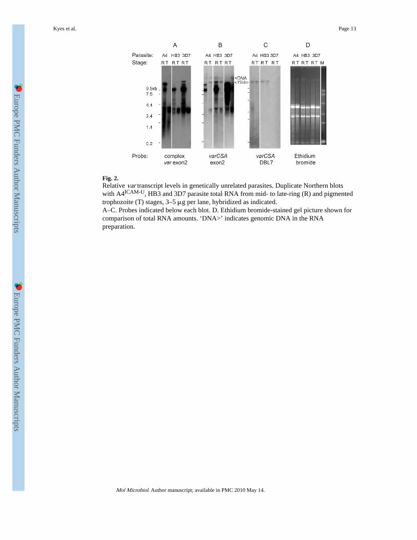
Fig. 2.Relative var transcript levels in genetically unrelated parasites. Duplicate Northern blotswith A4ICAM-U, HB3 and 3D7 parasite total RNA from mid- to late-ring (R) and pigmentedtrophozoite (T) stages, 3–5 μg per lane, hybridized as indicated.A–C. Probes indicated below each blot. D. Ethidium bromide-stained gel picture shown forcomparison of total RNA amounts. ‘DNA>’ indicates genomic DNA in the RNApreparation.
Kyes et al. Page 13
Mol Microbiol. Author manuscript; available in PMC 2010 May 14.
Europe PM
C Funders A
uthor Manuscripts
Europe PM
C Funders A
uthor Manuscripts

Fig. 3.Detection of constitutive varCSA transcript in parasites that do not bind to CSA. Parasites ofthe same genotype, with different binding phenotypes, were sampled for RNA at ring stage(≈ 16 h after invasion). Twenty hours later, pigmented trophozoite-stage parasites wereharvested for both RNA and binding assays. Binding data shown in (C) were from the samecultures sampled for RNA (except for A4ICAM-IC1, the binding assay was performed twocycles after the RNA was collected).A, B and D. Northern blot with FCR3CSA-U (U, unselected), FCR3CSA+ (CSA+, CSAselected), A4ICAM-U (U, unselected) and A4ICAM-IC1 (IC1, ICAM-1 binding), 3–5 μg oftotal RNA per lane. Ring (R) and pigmented trophozoite stages (T). Probes indicated beloweach blot.D. Ethidium bromide pattern shows relative total RNA levels. ‘DNA>’ indicates cross-hybridization with genomic DNA.C. Binding assay. Binding is presented as the number of infected red blood cells mm−2
(iRBC mm−2), with standard deviation. 10 μg ml−1 CSA (grey bars), 100 μg ml−1 CSA(black bars) and 40 μg ml−1 ICAM-1-FC (open bars).
Kyes et al. Page 14
Mol Microbiol. Author manuscript; available in PMC 2010 May 14.
Europe PM
C Funders A
uthor Manuscripts
Europe PM
C Funders A
uthor Manuscripts
Copyright © 2022 FDOKUMEN