A genomic glimpse of aminoacyl-tRNA synthetases in malaria parasite Plasmodium falciparum
14
BioMed Central Page 1 of 14 (page number not for citation purposes) BMC Genomics Open Access Research article A genomic glimpse of aminoacyl-tRNA synthetases in malaria parasite Plasmodium falciparum Tarun Kumar Bhatt 1 , Charu Kapil 1,2 , Sameena Khan 1,3 , Mohamad Aman Jairajpuri 2 , Vinay Sharma 3 , Daniele Santoni 4,5 , Francesco Silvestrini 5 , Elisabetta Pizzi 5 and Amit Sharma* 1 Address: 1 Structural and Computational Biology Group, International Centre for Genetic Engineering and Biotechnology, New Delhi 110 067, India, 2 Department of Biosciences, Jamia Millia Islamia University, Jamia Nagar, New-Delhi 110 025, India, 3 Department of Bioscience and Biotechnology, Banasthali Vidyapith University, Banasthali, Rajasthan 304 022, India, 4 Barcelona Institute for Research in Biomedicine, Barcelona Science Park, C/Samitier 1-5, Barcelona 08015, Catalonia, Spain and 5 Dipartimento di Malattie Infettive, Parassitarie ed Immunomediate, Istituto Superiore di Sanità, Viale Regina Elena, 299, 00161 Rome, Italy Email: Tarun Kumar Bhatt - [email protected]; Charu Kapil - [email protected]; Sameena Khan - [email protected]; Mohamad Aman Jairajpuri - [email protected]; Vinay Sharma - [email protected]; Daniele Santoni - [email protected]; Francesco Silvestrini - [email protected]; Elisabetta Pizzi - [email protected]; Amit Sharma* - [email protected] * Corresponding author Abstract Background: Plasmodium parasites are causative agents of malaria which affects >500 million people and claims ~2 million lives annually. The completion of Plasmodium genome sequencing and availability of PlasmoDB database has provided a platform for systematic study of parasite genome. Aminoacyl-tRNA synthetases (aaRSs) are pivotal enzymes for protein translation and other vital cellular processes. We report an extensive analysis of the Plasmodium falciparum genome to identify and classify aaRSs in this organism. Results: Using various computational and bioinformatics tools, we have identified 37 aaRSs in P. falciparum. Our key observations are: (i) fraction of proteome dedicated to aaRSs in P. falciparum is very high compared to many other organisms; (ii) 23 out of 37 Pf-aaRS sequences contain signal peptides possibly directing them to different cellular organelles; (iii) expression profiles of Pf-aaRSs vary considerably at various life cycle stages of the parasite; (iv) several PfaaRSs posses very unusual domain architectures; (v) phylogenetic analyses reveal evolutionary relatedness of several parasite aaRSs to bacterial and plants aaRSs; (vi) three dimensional structural modelling has provided insights which could be exploited in inhibitor discovery against parasite aaRSs. Conclusion: We have identified 37 Pf-aaRSs based on our bioinformatics analysis. Our data reveal several unique attributes in this protein family. We have annotated all 37 Pf-aaRSs based on predicted localization, phylogenetics, domain architectures and their overall protein expression profiles. The sets of distinct features elaborated in this work will provide a platform for experimental dissection of this family of enzymes, possibly for the discovery of novel drugs against malaria. Published: 31 December 2009 BMC Genomics 2009, 10:644 doi:10.1186/1471-2164-10-644 Received: 3 July 2009 Accepted: 31 December 2009 This article is available from: http://www.biomedcentral.com/1471-2164/10/644 © 2009 Bhatt et al; licensee BioMed Central Ltd. This is an Open Access article distributed under the terms of the Creative Commons Attribution License (http://creativecommons.org/licenses/by/2.0 ), which permits unrestricted use, distribution, and reproduction in any medium, provided the original work is properly cited.
-
Upload
independent -
Category
Documents
-
view
0 -
download
0
Transcript of A genomic glimpse of aminoacyl-tRNA synthetases in malaria parasite Plasmodium falciparum

BioMed CentralBMC Genomics
ss
Open AcceResearch articleA genomic glimpse of aminoacyl-tRNA synthetases in malaria parasite Plasmodium falciparumTarun Kumar Bhatt1, Charu Kapil1,2, Sameena Khan1,3, Mohamad Aman Jairajpuri2, Vinay Sharma3, Daniele Santoni4,5, Francesco Silvestrini5, Elisabetta Pizzi5 and Amit Sharma*1Address: 1Structural and Computational Biology Group, International Centre for Genetic Engineering and Biotechnology, New Delhi 110 067, India, 2Department of Biosciences, Jamia Millia Islamia University, Jamia Nagar, New-Delhi 110 025, India, 3Department of Bioscience and Biotechnology, Banasthali Vidyapith University, Banasthali, Rajasthan 304 022, India, 4Barcelona Institute for Research in Biomedicine, Barcelona Science Park, C/Samitier 1-5, Barcelona 08015, Catalonia, Spain and 5Dipartimento di Malattie Infettive, Parassitarie ed Immunomediate, Istituto Superiore di Sanità, Viale Regina Elena, 299, 00161 Rome, Italy
Email: Tarun Kumar Bhatt - [email protected]; Charu Kapil - [email protected]; Sameena Khan - [email protected]; Mohamad Aman Jairajpuri - [email protected]; Vinay Sharma - [email protected]; Daniele Santoni - [email protected]; Francesco Silvestrini - [email protected]; Elisabetta Pizzi - [email protected]; Amit Sharma* - [email protected]
* Corresponding author
AbstractBackground: Plasmodium parasites are causative agents of malaria which affects >500 millionpeople and claims ~2 million lives annually. The completion of Plasmodium genome sequencing andavailability of PlasmoDB database has provided a platform for systematic study of parasite genome.Aminoacyl-tRNA synthetases (aaRSs) are pivotal enzymes for protein translation and other vitalcellular processes. We report an extensive analysis of the Plasmodium falciparum genome to identifyand classify aaRSs in this organism.
Results: Using various computational and bioinformatics tools, we have identified 37 aaRSs in P.falciparum. Our key observations are: (i) fraction of proteome dedicated to aaRSs in P. falciparumis very high compared to many other organisms; (ii) 23 out of 37 Pf-aaRS sequences contain signalpeptides possibly directing them to different cellular organelles; (iii) expression profiles of Pf-aaRSsvary considerably at various life cycle stages of the parasite; (iv) several PfaaRSs posses very unusualdomain architectures; (v) phylogenetic analyses reveal evolutionary relatedness of several parasiteaaRSs to bacterial and plants aaRSs; (vi) three dimensional structural modelling has providedinsights which could be exploited in inhibitor discovery against parasite aaRSs.
Conclusion: We have identified 37 Pf-aaRSs based on our bioinformatics analysis. Our data revealseveral unique attributes in this protein family. We have annotated all 37 Pf-aaRSs based onpredicted localization, phylogenetics, domain architectures and their overall protein expressionprofiles. The sets of distinct features elaborated in this work will provide a platform forexperimental dissection of this family of enzymes, possibly for the discovery of novel drugs againstmalaria.
Published: 31 December 2009
BMC Genomics 2009, 10:644 doi:10.1186/1471-2164-10-644
Received: 3 July 2009Accepted: 31 December 2009
This article is available from: http://www.biomedcentral.com/1471-2164/10/644
© 2009 Bhatt et al; licensee BioMed Central Ltd. This is an Open Access article distributed under the terms of the Creative Commons Attribution License (http://creativecommons.org/licenses/by/2.0), which permits unrestricted use, distribution, and reproduction in any medium, provided the original work is properly cited.
Page 1 of 14(page number not for citation purposes)

BMC Genomics 2009, 10:644 http://www.biomedcentral.com/1471-2164/10/644
BackgroundAminoacylation is the process of adding an aminoacylgroup to the 3' end (CCA) of the tRNA molecule. tRNA isaminoacylated with a specific amino acid by aminoacyl-tRNA synthetase (aaRSs). aaRSs are responsible for attach-ing correct amino acid onto the cognate tRNA molecule ina two-step reaction. The amino acid is first activated withATP forming an aminoacyladenylate intermediate. Onceactivated, this amino acid is transferred to the 3' end of itscorresponding tRNA molecule to be processed during pro-tein synthesis. All aaRSs require divalent cation MgCl2 fortheir aminoacylation reaction [1,2].
Reaction:
1. amino acid + ATP → aminoacyl-AMP + PPi
2. aminoacyl-AMP + tRNA → aminoacyl-tRNA + AMP
The aaRSs are divided into two major classes based onstructural topology of their active sites. Class I aaRSs rep-resent 11 amino acids, including Arg, Cys, Gln, Glu, Ile,Leu, Lys, Met, Val, Trp and Tyr. Class II aaRSs includes 10amino acids - Ala, Asp, Asn, Gly, His, Lys, Phe, Pro, Serand Thr. Core domains of class I enzymes are character-ized by a Rossmann fold which consists of α-helices andβ-pleated sheets. This domain contains two conservedmotifs ('HIGH' and 'KMSKS') which are directly involvedin ATP binding. Catalytic domain of class II enzymes hasa unique fold with a central core of anti-parallel β strandsflanked by α helices [3]. There are three weakly conservedmotifs, two of them are involved in ATP binding while thethird one plays a role in homo dimerization. Class Ienzymes bind ATP in an extended conformation whileclass II do so in a bent conformation. The two aaRS classeshave different modes of aminoacylation - class I enzymesaminoacylate the 2'OH of the cognate tRNA whereas classII enzymes aminoacylate 3'OH of the tRNA (with theexception of PheRS) [4]. All known aaRSs are multido-main proteins with complex modular architectures [5]. Inaddition, eukaryotic aaRSs are distinguished by the pres-ence of appended domains at either the N- or C-terminuswhich are generally absent from their bacterial/archaealcounterparts [6]. These appendages to the catalytic coresof several aaRSs are non-catalytic and instead function tomediate protein- protein interactions or act as generalRNA-binding domains [7-9].
In mammalian cells, some aaRSs are present as a largermulti- aaRS complex (MSC) composed of nine syn-thetases (arginyl-, aspartyl-, glutamyl-, glutaminyl-, leu-cyl-, lysyl-, isoleucyl-, methionyl- and prolyl-tRNAsynthetases) [10-12]. The MSC is composed of a mixtureof class I and class II aaRSs along with three non- aaRSproteins p38, p43 and p18. It is not clear why certain
aaRSs exist as a complex while some are in free form. MSCmight help in efficient protein synthesis by preventingmixing of charged tRNAs with cellular pool and byincreasing local concentration of tRNA near the site ofprotein synthesis [13].
The accuracy of tRNA aminoacylation reaction is criticalin ensuring fidelity in protein translation [14]. To achievethis accuracy, some aaRS enzymes possess a proofreading(editing) mechanism that hydrolyzes tRNAs aminoa-cylated with the non-cognate amino acid [15]. For exam-ple, editing domains may be found attached to alanyl-tRNA synthetase (AlaRS), leucyltRNA synthetase (LeuRS)and so on [16-21]. In other cases, the editing domain isnot attached to aaRS but rather functions as an individualprotein [22,23]. For example, YbaK protein from Haemo-philus influenza is capable of efficiently editing Cys-tRN-APro [24]. ThrRS has been shown to have another editingdomain called NTD which can cleave the bond betweenD-amino acid and tRNA [25].
Recently it has been shown that aaRSs are not onlyinvolved in protein synthesis but also perform many non-catalytic and non-canonical roles in RNA processing/traf-ficking, apoptosis, rRNA synthesis, angiogenesis andinflammation [26-30]. These versatile properties of aaRSsare the outcome of their differential cellular localization,nucleic acid binding properties, protein-protein interac-tions and collaboration (fusion) with additionaldomains. In case of malaria parasite, apicoplast proteinsand pathways have already received particular attention asdrug targets [31]. In this work we present a study of aaRSsfrom P. falciparum - the most virulent agent of humanmalaria. Our aim for this study was to use bioinformaticstools to (a) discover special and unusual modules presentin parasite aaRSs which are potentially absent fromhuman homologues, and (b) to identify potential newdrug targets based on this protein family.
Results and DiscussionSequence extraction and analysisWe exploited current annotation available in PlasmoDB[32] to identify the repertoire of aaRSs in P. falciparumgenome. According to Enzyme Commission (EC) 37 pro-teins in PlasmoDB (see additional file 1) are annotated asbelonging to the EC group 6.1.1. (EC number providedfor aaRSs). Although in many cases current annotationsallow an assignment to Class I or II of aaRSs, for someannotations are still preliminary. Due to this, we usedHidden Markov Models (HMMs) for identifying aaRSs inP. falciparum. For each aaRS a set of known sequences wasutilized to construct 20 HMMs (see methods for details).For each database search a score distribution was obtainedand 4 cutoffs were considered to identify aaRS. Results arereported in Table 1. We observed that 2 proteins anno-
Page 2 of 14(page number not for citation purposes)

BMC Genomics 2009, 10:644 http://www.biomedcentral.com/1471-2164/10/644
tated as belonging to EC group 6.1.1.- in PlasmoDB arenot found by HMMs - PF14_0401 annotated as MetRS isinstead a generic tRNA binding protein as elucidated inthe genome re-annotation process, while the second one(PFC0470w) is still mis-annotated as ValRS. A total of 18Pf-aaRSs can be classified within the 10 aaRSs that defineclass I. All members of this class are represented in the P.falciparum proteome. The annotations of these sequencesare summarized in additional file 1. Similar to class I Pf-aaRSs, the class II Pf-aaRSs have a total of 18 sequences for10 different amino acid synthetases. Four genes arepresent in P. falciparum for PheRS but these likely encodefor 1 heterodimeric and 2 monomeric versions of PheRS.
In order to carry out comparative analyses of aaRSs of P.falciparum with those of other species we considered aaRSsequences from several organisms representing threedomains of life (see methods section). As expected, wefound variable number of aaRSs in different species. M.jannaschii (archaebacteria) and M. tuberculosis (bacteria)have the lowest aaRSs count amongst other organisms likeE. coli, S. cerevisiae, D. discoidium, P. falciparum, O. sativa,R. norvegicus, D. melanogaster, and H. sapiens. Humanbears the highest number of aaRSs in this analysis (Figure1a). Our analysis also shows that P. falciparum has thehighest aaRS fraction (relative to its proteome size) whencompared with bacteria, yeast and human counterparts(Figure 1b). The number of individual aaRS varies in dif-ferent species. For example, when individual aaRSs from
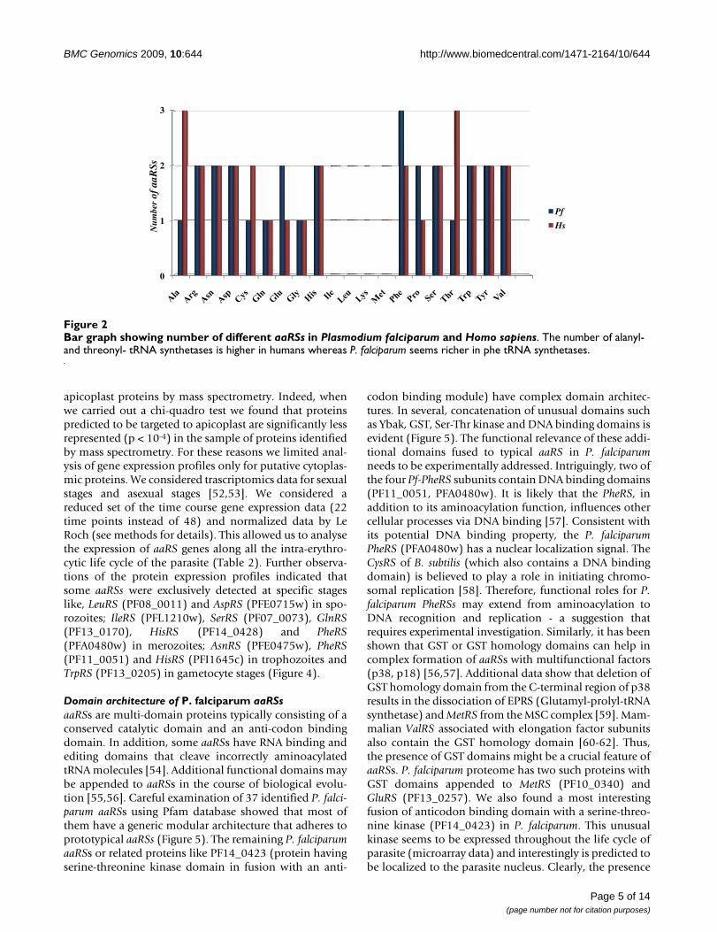
human and P. falciparum were compared it was evidentthat AlaRS and ThrRS were higher in number in humans(Figure 2). Presence of more than one copy of each aaRSin an organism may indicate additional biological, tem-poral or spatial roles for these enzymes as several aaRSsalso perform non-canonical functions [33]. In this workwe describe in detail the 37 Pf-aaRSs.
Indirect pathways of aminoacylationIt was earlier believed that 20 aaRSs were necessary for theincorporation of 20 amino acids in proteins. But surpris-ingly, some archaea, bacteria and chloroplasts lack GlnRSand AsnRS enzymes [34-38]. Interestingly, these organ-isms use an alternate pathway based on tRNA dependentamino acid transformation. A non-discriminating GluRScharges tRNAGln with glutamic amino acid and then a sec-ond enzyme called tRNA-dependent amidotransferase(AdT) amidates glutamate to make glutamine. A corre-sponding reaction occurs in case of asparagine residues. Incase of P. falciparum, occurrence of glutamate-tRNA syn-thetase (PF13_0257, MAL13P1.281) and amidotrans-ferase subunit A (PFD0780w) & subunit B (PFF1395c)together indicates presence of both direct and indirectpathways for aminoacylation [39,40]. Both subunits ofamidotransferase have apicoplast targeting signals sug-gesting an indirect pathway for aminoacylation in P. falci-parum apicoplast. The expression of Pf-AdT subunit A ispredicted in all life cycle stages of parasite based on pro-teomic and microarray data. We therefore feel that this
Table 1: Results of database searches by HMM models of aaRS@.
cutoff HMMAla HMMArg HMMAsn HMMAsp HMMCys HMMGln HMMGlu HMMGly HMMHis HMMIle
c > 50 PF13_0354 PFL0900c PFE0475w* PFB0525w* PF10_0149 PF13_0170 MAL13P1.281 PF14_0198 PF14_0428 PF13_0179
20< c < 50 PFB0525w* PFA0145cPFE0475w*
PF13_0257* PF13_0257* PFI1645c
10< c < 20 PFE0715w*
5< c <10 PFI0680c PFE0715w* PFL1210w
Cutoff HMMLeu HMMLys HMMMet HMMPhe HMMPro HMMSer HMMThr HMMTrp HMMTyr HMMVal
c > 50 PF08_0011 PF13_0262 PF10_0340 PFA0480w PFL0670c PF07_0073 PF11_0270 PF13_0205 MAL8P1.125
PF14_0589
20< c < 50 PFF1095w PF14_0166 PF10_0053 PFF0180w PFI1240c PFL0770w PFL2485c PF11_0181
10< c < 20 PFL1540cPF11_0051
5< c <10
* hits with more than one assignment to HMM model.@Two proteins PF14_0401 and PFC0470w were not found by HMMs which were annotated as tRNA binding protein and ValRS in PlasmoDB respectively.
Page 3 of 14(page number not for citation purposes)

BMC Genomics 2009, 10:644 http://www.biomedcentral.com/1471-2164/10/644
pathway must also be active in the parasite apicoplast. Wecould not find sequence homologues of enzymesinvolved in indirect aminoacylation of cysteine residues[41-43] in the proteome of P. falciparum.
The multi-synthetase complex (MSC)In mammalian cells, some aaRSs are present as a largermulti-aaRS complex (MSC). A constituent of the MSC -protein p43 - has sequence homologue (PF14_0401 -EMAP-II-like cytokine) in P. falciparum although there isno evidence for presence of MSC in malaria parasites.Interestingly, p43 is not only required for stability of theMSC complex but also functions as a proinflammatorycytokine [44-46]. Role of p43 homolog in P. falciparum isunknown, but evidence from other organisms indicatesthat MSC functions in protein stability, efficient proteintranslation and protein elongation [47]. Sequence iden-tity between P. falciparum p43 and its human homolog is~24% and based on microarray data p43 seems to be
expressed at asexual life cycle stages of P. falciparum. Amitochondrial targeting signal was also predicted for par-asite p43 but the role of p43 in parasite remains to beexplored experimentally.
Targeting of aaRSs in the parasiteaaRSs are not only involved in protein synthesis but alsoin various other cellular activities including intron splic-ing, translational regulation and tRNA channeling. Diver-sified roles for aaRSs necessitate their presence (transit)into various cellular compartments. We therefore ana-lyzed P. falciparum aaRS sequences for presence of puta-tive signal sequences predicted by MITOPROT,PredictNLS and PATS for mitochondria, nucleus and api-coplast respectively. We found that 23 P. falciparum aaRSshave signal peptides, possibly for directing them to differ-ent cellular organelles. Another 14 aaRSs from P. falci-parum may be resident in the parasite cytoplasm (Figure3a). Apicoplast is known to have protein synthesismachinery which may use aaRSs [48]. Trafficking ofnuclear encoded aaRSs to the apicoplast may explain why~20 out of 37 Pf-aaRSs have apicoplast targeting signals.Our data indicate that out of total ~20 Pf-aaRSs bearingapicoplast targeting signals, ~12 aaRSs may be exclusive tothis organelle. Others are predicted to be shared betweenapicoplast, nucleus and mitochondria (Figure 3b). It hasbeen earlier shown that some tRNAs need to be aminoa-cylated in the nucleus before they can be exported to thecytoplasm, an observation indicating occurrence of ami-noacylation reaction (mediated by aaRSs) inside thenucleus [49]. In P. falciparum, we found 10 aaRSs withnuclear localization signals but only one is predicted to beexclusively resident in the nucleus (PFA0480w- PheRS).Interestingly, we found no Pf-aaRS sequences with specificPEXEL (Plasmodium export element) motifs. This motif isfound in parasite proteins that are exported beyond theparasitophorous vacuole membrane [50,51].
Expression profiles of P. falciparum aaRSsIn order to study expression of aaRS during life cycle of themalaria parasite, we took advantage of available transcrip-tomics and proteomics data from PLASMODB. Firstly, weanalyzed proteomic data from several independent exper-iments and compared them with transcriptomics data byLe Roch [52]. The latter sets of data were obtained usingthe affimetrix technology and hence provide a quantita-tive measure of mRNA levels in the parasite. Our resultsare provided in Table 2. Interestingly, we found thatmRNA levels of potential apicoplast proteins (AP in thetable) are lower on average (mean1 = 44.6; mean2 = 41.5;gam = 91.3; spor = 58.1) than those of potential cytoplas-mic proteins (mean1 = 259; mean2 = 264.8; gam = 174.8;spor = 73.8). Proteomic data confirmed that while thecytoplasmic aaRS are found in almost all stages, the apico-plast aaRS are rarely found in the parasite. This could bein part due to experimental limits in the identification of
(a) Predictied number of aaRSs present in Plasmodium falci-parum (Pf), Rattus norvegicus (Rn), Saccharomyces cerevisiae (Sc), Drosophila melanogaster (Dm), Homo sapiens (Hs), Oryza sativa (Os), Dictyostelium discoidium (Dd), Mycobacterium tuberculosis (Mtb), Escherichia coli (Ec), and Methanocalclococcus jannaschii (Mj)Figure 1(a) Predictied number of aaRSs present in Plasmo-dium falciparum (Pf), Rattus norvegicus (Rn), Saccharo-myces cerevisiae (Sc), Drosophila melanogaster (Dm), Homo sapiens (Hs), Oryza sativa (Os), Dictyostelium discoidium (Dd), Mycobacterium tuberculosis (Mtb), Escherichia coli (Ec), and Methanocalclococcus jannas-chii (Mj). (b) Diagram representing fraction of proteome (in percentage) dedicated to the aaRS proteins in various organ-isms.
0
5
10
15
20
25
30
35
40
45
Mj Mtb Ec Sc Dd Pf Os Rn Dm Hs
Number of aaRS in different organisms
0
0.1
0.2
0.3
0.4
0.5
0.6
0.7
0.8
Os Rn Hs Dd Dm Mj Sc Mt Ec Pf
aaR
Ss %
in p
rote
ome
Fraction of aaRSs in total proteome
a)
b)
aaR
S co
unt
Page 4 of 14(page number not for citation purposes)
BMC Genomics 2009, 10:644 http://www.biomedcentral.com/1471-2164/10/644
apicoplast proteins by mass spectrometry. Indeed, whenwe carried out a chi-quadro test we found that proteinspredicted to be targeted to apicoplast are significantly lessrepresented (p < 10-4) in the sample of proteins identifiedby mass spectrometry. For these reasons we limited anal-ysis of gene expression profiles only for putative cytoplas-mic proteins. We considered trascriptomics data for sexualstages and asexual stages [52,53]. We considered areduced set of the time course gene expression data (22time points instead of 48) and normalized data by LeRoch (see methods for details). This allowed us to analysethe expression of aaRS genes along all the intra-erythro-cytic life cycle of the parasite (Table 2). Further observa-tions of the protein expression profiles indicated thatsome aaRSs were exclusively detected at specific stageslike, LeuRS (PF08_0011) and AspRS (PFE0715w) in spo-rozoites; IleRS (PFL1210w), SerRS (PF07_0073), GlnRS(PF13_0170), HisRS (PF14_0428) and PheRS(PFA0480w) in merozoites; AsnRS (PFE0475w), PheRS(PF11_0051) and HisRS (PFI1645c) in trophozoites andTrpRS (PF13_0205) in gametocyte stages (Figure 4).
Domain architecture of P. falciparum aaRSsaaRSs are multi-domain proteins typically consisting of aconserved catalytic domain and an anti-codon bindingdomain. In addition, some aaRSs have RNA binding andediting domains that cleave incorrectly aminoacylatedtRNA molecules [54]. Additional functional domains maybe appended to aaRSs in the course of biological evolu-tion [55,56]. Careful examination of 37 identified P. falci-parum aaRSs using Pfam database showed that most ofthem have a generic modular architecture that adheres toprototypical aaRSs (Figure 5). The remaining P. falciparumaaRSs or related proteins like PF14_0423 (protein havingserine-threonine kinase domain in fusion with an anti-
codon binding module) have complex domain architec-tures. In several, concatenation of unusual domains suchas Ybak, GST, Ser-Thr kinase and DNA binding domains isevident (Figure 5). The functional relevance of these addi-tional domains fused to typical aaRS in P. falciparumneeds to be experimentally addressed. Intriguingly, two ofthe four Pf-PheRS subunits contain DNA binding domains(PF11_0051, PFA0480w). It is likely that the PheRS, inaddition to its aminoacylation function, influences othercellular processes via DNA binding [57]. Consistent withits potential DNA binding property, the P. falciparumPheRS (PFA0480w) has a nuclear localization signal. TheCysRS of B. subtilis (which also contains a DNA bindingdomain) is believed to play a role in initiating chromo-somal replication [58]. Therefore, functional roles for P.falciparum PheRSs may extend from aminoacylation toDNA recognition and replication - a suggestion thatrequires experimental investigation. Similarly, it has beenshown that GST or GST homology domains can help incomplex formation of aaRSs with multifunctional factors(p38, p18) [56,57]. Additional data show that deletion ofGST homology domain from the C-terminal region of p38results in the dissociation of EPRS (Glutamyl-prolyl-tRNAsynthetase) and MetRS from the MSC complex [59]. Mam-malian ValRS associated with elongation factor subunitsalso contain the GST homology domain [60-62]. Thus,the presence of GST domains might be a crucial feature ofaaRSs. P. falciparum proteome has two such proteins withGST domains appended to MetRS (PF10_0340) andGluRS (PF13_0257). We also found a most interestingfusion of anticodon binding domain with a serine-threo-nine kinase (PF14_0423) in P. falciparum. This unusualkinase seems to be expressed throughout the life cycle ofparasite (microarray data) and interestingly is predicted tobe localized to the parasite nucleus. Clearly, the presence
Bar graph showing number of different aaRSs in Plasmodium falciparum and Homo sapiensFigure 2Bar graph showing number of different aaRSs in Plasmodium falciparum and Homo sapiens. The number of alanyl- and threonyl- tRNA synthetases is higher in humans whereas P. falciparum seems richer in phe tRNA synthetases.
0
1
2
3
Pf
Hs Num
ber
ofaa
RSs
Page 5 of 14(page number not for citation purposes)

BMC Genomics 2009, 10:644 http://www.biomedcentral.com/1471-2164/10/644
of unusual domain fusions in P. falciparum aaRSs suggestsmultiple functional roles for many of these P. falciparumenzymes as has been shown in other organisms.
PhylogeneticsOverall the percentage identity between matching humanand P. falciparum aaRS domains varies from 17 to 51.Clearly, Pf-aaRSs which have low sequence identity withhuman counterparts might serve as good drug targets. Inorder to study evolutionary relationships of P. falciparumaaRSs with other species, phylogenetic trees were devel-oped in PHYML using maximum likelihood method. Foreach type of P. falciparum aaRS a separate tree was con-structed (see additional file 2). aaRS sequences from 102different species were used for multiple sequence align-ments. As an example, phylogenetic tree of TyrRS fromvarious species (including two sequences from P. falci-parum) was constructed. Interestingly, one Pf-TyrRS
(MAL8P1.125) clustered with human TyrRS whereas thesecond Pf-TyrRS (PF11_0181) clustered with bacterialTyrRS indicating different evolutionary origins (Figure6a). Based on distance matrices, several P. falciparumaaRS sequences clustered as being closer to plants (A. thal-iana) or to bacteria (E. coli) (Figure 6b). It is alreadyknown that apicomplexan parasites like P. falciparumhouse a secondary endosymbiotic plastid, possiblyhijacked by lateral genetic transfer from an alga. There-fore, the P. falciparum aaRS sequences which are evolu-tionary close to bacteria and plants are likely to be theoutcome of horizontal gene transfer from the plastid. P.falciparum contains ~12 such aaRS sequences which clus-ter with bacterial or plant sequences. Functional andstructural characterization of these bacterial/plant-likeaaRS may be relevant in focusing efforts at using aaRS asdrug targets.
(a) Percentage predicted distribution of Pf-aaRSs in different organelles within the parasiteFigure 3(a) Percentage predicted distribution of Pf-aaRSs in different organelles within the parasite. (b) A schematic of all Pf-aaRSs and their predicted cellular localization. Detailed information regarding gene IDs can be found in additional file 1. Pf-aaRSs predicted to be common between apicoplast & mitochondria, mitochondria & nucleus and apicoplast & nucleus are marked with diamond, triangle and square shapes respectively.
�������
����� ���
���������
��������
��������
���������
���������
���������
���������
�������
�������
���������
���������
���������
��������
���������� ��������� ��������� ��!�����
���������� ���������� ���������
b)a)
"#��$#�
��������
��!����
�������
��%�����
��%�����
������ ���
���������
�������
���������
��!����
�������
���������
��&�������
�&���'�"()&�
���������
���������
���������
����������
"*�%*+
�!,-.-/01!�
"*�%*+��!,-.-/01!�
��!-���+,��!,-.-/01!�
"*�%*+���!-���+,
��!-���+,
�2,-���+
��)� %��*��1 �-��!3�,!-/
~38%
~32%
~19%
~2.7% each
Page 6 of 14(page number not for citation purposes)

BMC Genomics 2009, 10:644 http://www.biomedcentral.com/1471-2164/10/644
Homology modeling and structure comparisonsTo date, no crystal structures have been obtained for anyaaRS from P. falciparum. Hence, we performed homologymodeling of several P. falciparum aaRSs using homolo-gous structures available in PDB. Known structural tem-plates (≥ 40% identity) were used for molecular modelingof several P. falciparum aaRSs including the two TyrRSs(PF11_0181, MAL8P1.125), the PheRS (PFA0480w),ThrRS (PF11_0270), LysRS (PF13_0262), MetRS(PF10_0340) and TrpRS (PF13_0205). The programAlign2D (sequence alignment module in Modeller) wasused to perform dynamic programming-based globalalignments of the target and template sequences. This pro-gram uses variable gap penalty for structural loops andcore regions using information derived from templatestructures. We found key differences in the conservedmotifs in various aaRSs. For example, the class I motif'KYSKS' in P. falciparum TyrRS (PF11_0181) and 'KMSKS'in MAL8P1.125 differs from 'KLGKS' of human mito-chondrial TyrRS (2PID) and 'KMSSS' of human cytoplas-mic (1N3L) respectively. Similarly, class I motif 'HIGH'has subtle sequences variations between P. falciparum andH. sapiens TyrRSs (Figure 7a, Table 3). Using the aboveprocedures, we could generate structural models for sev-eral Pf-aaRSs. Stereo-chemical qualities of the generatedprotein models were assessed using PROCHECK (85-90%residues are in allowed regions of Ramachandran plot).The overall superimposed three-dimension models werevisualized in CHIMERA and PYMOL (Figure 7b). Manysequence insertions were observed for P. falciparumenzymes when compared to their homologous [63]. Loca-tion of insertions in P. falciparum TyrRS between well-con-
served secondary structures suggests ability of TyrRSanticodon binding core to accommodate larger sequenceinserts with minimum disruption to the catalytic domain.Direct comparison of modeled P. falciparum aaRSs withhuman aaRSs revealed several other important structuraldifferences. For example, numerous insertions are presentin the loop regions linking various α-helices (α10 to α13)in anticodon binding domain of P. falciparum TyrRSs(PF11_0181 and MAL8p1.125) when compared to itshuman homologous (2PID and 1N3L) respectively. Struc-tural differences between TyrRS (from P. falciparum) andhuman counterparts are summarized in Table 3 andshown in Figure 7c. These subtle structural changes thatmanifest as partial conservation of important motifs in P.falciparum aaRSs reflect evolutionary divergence, and maybe useful for exploitation of parasite-specific features asdrug targets.
ConclusionAminoacyl-tRNA synthetases (aaRSs) link RNA with pro-tein translation. Besides their key role in protein synthesis,aaRSs are also integral to various other cellular processes.aaRS enzymes have been the focus for antimicrobial drugdiscovery [64,65]. An example of clinical application ofan aaRS inhibitor is provided by the antibiotic mupirocin(marketed as Bactroban), which selectively inactivatesbacterial isoleucyl-tRNA synthetase [66]. Similarly, it hasbeen shown that the broad-spectrum antifungal 5-fluoro-1,3-dihydro-1-hydroxy-2,1-benzoxaborole (AN2690)inhibits yeast cytoplasmic leucyl-tRNA synthetase byblocking editing site of the enzyme [67,68]. Therefore,presence of distinct or tinkered P. falciparum aaRS lends an
Diagrammatic representation of Pf-aaRS protein expression which are specifically expressed in different life stages of the para-site based on mass spectrometry data [82]Figure 4Diagrammatic representation of Pf-aaRS protein expression which are specifically expressed in different life stages of the parasite based on mass spectrometry data [82].
��4$����� 5�+/)�6
��4������� 5�.%)�6
��4&���� 5'!+)�6
��4������� 5�1�)�6
��4������ 5&�%)�6
��4������� 5�%1)�6
��4������� 57�/)�6
��4������� 5'!+)�6
��4������ 5�.%)�6
��4������� 5�%*)�6
��4$����� 5�+�)�6
Sporozoite Merozoite
TrophozoiteGametocyte
Page 7 of 14(page number not for citation purposes)

BMC Genomics 2009, 10:644 http://www.biomedcentral.com/1471-2164/10/644
opportunity for their exploitation as new drug targetsagainst malaria. In this study, we have extensively ana-lyzed aaRS sequences from Plasmodium species in terms oftheir mRNA/protein expression profiles, their cellularlocalization, their organelle targeting and their uniquesequence/domain attributes. We have discovered severaldistinct aaRSs in P. falciparum with no clear human coun-terparts in terms of their overall domain structures. Wehave also highlighted deviations of some highly con-served sequence motifs and active site sequence clusters.Our analyses clearly show that a larger fraction of P. falci-
parum proteome is devoted to aaRS when compared withmany other organisms. The phylogenetic data hint at evo-lutionary closeness of some Pf-aaRSs to bacteria andplants - this further supports the fact of secondary endo-symbiosis in this apicomplexan. We hope that our in-depth phylogenetic, protein targeting, domain architec-ture, protein expression profiling and homology mode-ling data on Pf-aaRSs can be used as a platform forexperimental studies of this important protein family inmalaria parasites.
Table 2: Transcriptomic and proteomic data for aaRSs in P. falciparum@
ID aaRS mean1 asex mean2 asex Gam spor TG§ T Me G Sp Me* Oocyst/Spor*
MAL13P1.281 AP-GluRs 4.8 10.9 39.7 6.5PF08_0011 AP-LeuRs 51.4 59.1 85.4 371.6 +PF10_0053 AP-MetRs 18.7 16.5 19 19PF11_0181 AP-TyrRs 13.2 2.6 0 0PF14_0166 AP-LysRs 60.3 48.0 166.4 48.3PFE0475w AP-AsnRs 104.6 92.5 222.8 149.6 +PFE0715w AP-AspRs 17.2 26.3 21.1 0 +PFI0680c AP-ArgRs 15.7 17.9 34.7 1.1PFI1240c AP-ProRs 10.8 14.0 19.9 4.2PFI1645c AP-HisRs 27.4 21.2 8.7 15 +PFL0770w AP-SerRs 117.6 110.9 431.3 69PFL1210w AP-IleRs 124.0 109.5 132.3 71 +PFL1540c AP-PheRs 13.7 9.7 6 0.1
mean 44.6 41.5 91.3 58.1
PF11_0270 ThrRs 580.0 582.1 181.2 154.6 + + + + + + +PF13_0354 AlaRs 123.6 129.9 75.5 27.5 + + +PF10_0149 CysRs 138.1 115.6 29.6 44.5 + +MAL8P1.125 TyrRs 225.6 260.0 134.3 259.2 + + + + + + +PF07_0073 SerRs 190.9 193.8 364.2 22.5 + + +PF10_0340 MetRs 389.3 387.5 479.9 3.4 + + + + +PF11_0051 PheRs 126.2 126.5 46.9 1.6 + +PF13_0170 GlnRs 558.8 570.8 272.3 214.7 + +PF13_0179 IleRs 192.1 188.1 152.8 0 + + + + + + +PF13_0205 TrpRs 112.6 189.4 42.8 171.8 + +PF13_0257 GluRs 330.0 334.5 205.6 139 + + + + + +PF13_0262 LysRs 1048.8 971.6 591 281.9 + + + + + + +PF14_0198 GlyRs 93.4 80.1 102.4 0 + + + + +PF14_0428 HisRs 35.4 51.1 0 0 + + +PF14_0589 ValRs 48.0 33.2 5.8 0 + + + + +PFA0145c AspRs 359.2 359.2 361 21.4 + + +PFA0480w PheRs 49.5 20.2 28.4 10.4 + + +PFB0525w AsnRs 575.3 686.7 493.1 106.2 + + + + + +PFL0670c ProRs 105.4 108.8 50. 5 0 + + + + + +PFL0900c ArgRs 118.3 120.8 37.1 76.2 + + + + +PFL2485c TrpRs 38.2 51.1 16.5 15.9
Mean 259.0 264.8 174.8 73.8
@ Gene expression data are by Le Roch[52]. Mean1 and mean2 asex refer to mean values of mRNA abundance along asexual stages synchronized by sorbitol and by temperature respectively. In the last two columns, mass spectrometry data from purified merozoites (Me*; Leiden Malaria Group, unpublished data) and mosquito stages (Oocyst/Spor*; oocysts, oocystderived sporozoites and salivary gland sporozoites from Anopheles stephensi infected with NF54 strain of P. falciparum) are shown.TG§ proteomic data are obtained by Trophozoites, Gametocytes early and Gametocytes late by Lasonder lab. [88]. T (trophozoites), Me (merozoites), G (gametocytes), Sp (sporozoites) columns refer to data by multi-dimensional protein identification technology in four stages of the parasite life cycle.
Page 8 of 14(page number not for citation purposes)

BMC Genomics 2009, 10:644 http://www.biomedcentral.com/1471-2164/10/644
MethodsSequence extractionThe P. falciparum genome database PlasmoDB Release 5.4was used for the present analyses. Sequence sets of all theaaRSs from other organisms includes P. berghei, P. chabaudi,P. falciparum, P. knowlesi, P. yoelii, P. vivax, H. sapiens, M.tuberculosis, D. discoidium, M. jannaschii, R. norvegicus, C.parvum, B. bovis, S. cerevisiae, D. melanogaster, Y. pestis, T.aquaticus, S. pneumoniae, S. entrica, E. coli, A. thaliana, A.pisum, A. salmonicida, B. cereus, B. thuringiensis, B. afzelii, B.burgdorferi, B. garinii, B. valaisiana, Bradyrhizobium, B. penn-sylvanicus, C. acidaminovorans, H. defensa, C. taiwanensis, E.fergusonii, F. bacterium, F. novicida, F. tularensis, F. alni, G.tenuistipitata, H. arsenicoxydans, A. cellulolyticus, A. chloroph-enolicus, A. ferrooxidans, Algoriphagus, A. muciniphila, Anaer-omyxobacter, A. thermophilum, B. ambifaria, B. indica, B.mycoides, B. taurus, B. tribocorum, C. atlanticus, Caulobacter,C. aurantiacus, C. cellulolyticum, Citrobacter, C. pinensis, C.Ruthia, Cyanothece, D. desulfuricans, D. hafniense, Diaphoro-bacter, D. shibae, D. turgidum, E. cuniculi, E. lenta, E. rumi-nantium, Exiguobacterium, G. diazotrophicus, Geobacillus, M.maris, N. multipartita, Nocardioides, O. terrae, P. abelii, P.atlantica, P. denitrificans, P. ingrahamii, P. lavamentivorans, R.castenholzii, S. arenicola, S. fumaroxidans, X. autotrophicus, V.vadensis, V. paradoxus, T. whipplei, T. auensis, S. stellata, Ch.parvum, S. heliotrinireducens, Silicibacter, S. putrefaciens, S.usitatus, Thauera, X. laevis, Theileria annulata, Vibrio fischeri,W. succinogenes, X. tropicalis, Zeamays. Additional sequences
Representation of unusual domain architectures in Pf-aaRSs and related proteinsFigure 5Representation of unusual domain architectures in Pf-aaRSs and related proteins. A generic aaRS is also shown on top. Domain name abbreviations are YB, Ybak associating domain; TS-II, class II tRNA synthetase; AC, anti-codon binding site; ED, editing domain; GST, glutathione-Stransferase C-terminal region; RBD, S4 RNA binding domain; TS, tRNA synthetase core domain; STK, serine-thre-onine kinase; FTS, phenylalanine-tRNA synthetase; PTS, pro-linetRNA synthetase; VTS, valine-tRNA synthetase; MTS, methionine-tRNA synthetase; YTS, tyrosine-tRNA syn-thetase; ETS, glutamate-tRNA synthetase.
���
��� ������������������
���������������� �������� ��
� !"��"�# �$
����%%&'� (��)�� ��
�*'*� ��"+�,
� -�� !���#"�
.�/*� !"��0+1 ��
� ��%%� !!���0! *'* *'*
�'*&�� !!��!+!
� /�� !#���0)
��
(a) Evolutionary tree was constructed using the PHYML based on maximum likelihood methodFigure 6(a) Evolutionary tree was constructed using the PHYML based on maximum likelihood method. P. falciparum TyrRSs (PlasmoDB id -MAL8p1.125 and PF11_0181) are labeled as green triangles. One of the TryRSs (MAL8p1.125) is evolu-tionarily closer to H. sapiens whereas the other TyrRS (PF11_0181) is closer to E. coli. Total of 102 species were considered for the evolutionary analysis and were taken from three domains of life. (b) List of Pf-aaRS sequences evolutionarily closer to their E. coli and A. Thaliana counterparts.
a)
E. coli (Bacteria)
PFL2485c (TrpRS)
PFE0475w (AsnRS)
PF08_0011 (LeuRS)
PF11_0181 (TyrRS)
PF13_0170 (GlnRS)
PF13_0257 (GluRS)
PFL0770w (SerRS)
PFB0525w (AsnRS)
A. thaliana (Plants)
PF13_0205 (TrpRS )
PF14_0198 (GlyRS)
PF14_0428 (HisRS)
PF07_0073 (SerRS)
b)
Page 9 of 14(page number not for citation purposes)

BMC Genomics 2009, 10:644 http://www.biomedcentral.com/1471-2164/10/644
Page 10 of 14(page number not for citation purposes)
Left and right panels of the figure represent sequence and structural comparison of bacterial type Plasmodium TyrRS (PF11_0181) with human mitochondrial TyrRS (2PID) and the cytosolic Plasmodium TyrRS (Mal8p1.125) with human cytosolic TyrRS (1N3L)Figure 7Left and right panels of the figure represent sequence and structural comparison of bacterial type Plasmodium TyrRS (PF11_0181) with human mitochondrial TyrRS (2PID) and the cytosolic Plasmodium TyrRS (Mal8p1.125) with human cytosolic TyrRS (1N3L). a) A structure-based sequence alignment of the catalytic domain of Plasmodium TyrRSs with human TyrRSs. Insertions in Pf and human sequences are colored in light blue and orange respectively. Class I syn-thetase conserved motifs are colored red. Residues involved in tRNA recognition and catalysis are indicated in green (same residues in Pf and Hs) and violet & boxed (different in Pf and Hs). The secondary structural elements are shown above the sequence alignments. Conserved residues are indicated by asterisk below the sequence alignment. (b) Superposition of Pf-TyrRS and Hs-TyrRS depicting the structural differences. Pf-Tyr is colored grey and Hs-TyrRS is colored tan. Insertions in Pf-TyrRSs are highlighted in blue whereas Hs-TyrRS insertions are in orange. Motif 1 in Pf (PF11_0181 - HLGN and Mal8p1.125 - HIAQ) and Hs (2PID - HVGH and 1N3L - HVAY) TyrRSs has been encircled red whereas Motif 2 in Pf (PF11_0181 - KLGKS and Mal8p1.125 - KMSKS) and Hs (2PID - KYSKS and 1N3L - KMSSS) is encircled green. (c) Snapshot of the active sites of Pf and Hs TyrRSs (superimposed) structures. Non-conserved active site residues colored violet are encircled.
�
��
�������������
�
������ �
a) TyrRS-PF11_0181(Pf) 2PID(Hs) TyrRS-Mal8p1.125(Pf) 1N3L(Hs)
23
�3

BMC Genomics 2009, 10:644 http://www.biomedcentral.com/1471-2164/10/644
were obtained based on sequence similarity via NCBIBLAST [69] and ENSEMBL [70] databases. Knownsequence motifs of aaRSs have been used as templates toretrieve sequences of aaRS from other organisms. SomeaaRS sequences were manually annotated based on thepresence of signature motifs. Protein domains and motifsin the predicted aaRSs were identified using following pro-grams - Superfamily [71], SMART [72] and MotifScan avail-able at expasy web server. The following databases - Pfam[73], TIGR, PIR, EBI and PlasmoDB were also extensivelyused. Hidden Markov Model (HMM) for each of the 20aaRS were constructed by the software package SequenceAlignment and Modeling System version 2.2.1 (SAM) [74]exploiting sequences in the aaRS database [75]. HMM pro-files were then used to carry out database search vs P. falci-parum proteins. A score was assigned to each protein bycalculating the probability that the corresponding sequenceis generated by the HMM model, hence for each databasesearch a score distribution was obtained. The score distribu-tions were normalized and 4 ranges of values were consid-ered to identify aaRS (c > 5, 10 < c < 20, 20 < c < 50, c < 50).
Expression and LocalizationThe prediction of signal sequences for cellular localizationin P. falciparum was performed using various availableonline web-servers - MITOPROT [76], PredictNLS [77]and PATS [78] for mitochondria, nucleus and apicoplastrespectively. PEXEL motif prediction was been carried outby querying PlasmoDB. To identify specific gene expres-sion profiles, we have combined information from differ-ent data sets. For the spotted oligonucleotide array data,only half of the 48 time points of the intra-erythrocyticcycle are shown for simplicity, and ratios (versus a com-mon reference) were log2-transformed prior to clusteranalysis. For the photolithography data, CEL files weredownloaded from website and transferred into Biocon-ductor package for analysis using a robust multi-arrayaveraging algorithm (RMA) for background adjustmentand quantiles normalization [79]. Genes whose expres-
sion level was less than 10 (too close to background) orthe logP was greater than -0.5 (too few probes per gene)were removed from dataset. Total intensity values for eachtime point were converted to mean-centered ratios bydividing the total intensity by the average intensity for thatgene across all experimental conditions and were thenlog2-transformed prior to clustering. These data manipu-lations were necessary because the oligo-nucleotide arraydata was collected as the intensity ratio between the exper-imental sample and a common reference, while the pho-tolithography data was collected as the total signalintensity at each spot. Gene expression patterns where theminimum percentage of existing values was less than 80%were eliminated from rest of the analysis. The remainingmissing values were replaced by using the KNN-imputa-tion method [80].
Phylogenetic analysisTo explore the evolutionary relationships amongst aaRSsphylogenetic analyses were performed for each P. falci-parum aaRS on an expanded set of 102 sequences. Multi-ple sequence alignments of these sequences wereobtained from CLUSTALW with default parameters (per-formed locally) in PHYLIP format [81]. These MSAs wereused as seed sequences to run PHYML_v2.4.4 using Jones-Taylor-Thornton (JTT) model [82]. The resulting file wasfurther used in MEGA4.2 for visualization of trees [83].
Model Building and ValidationWe used Sali's Modeller8v2 [84] tool for building variousP. falciparum aaRSs models. The stereo-chemical quality ofmodeled proteins was verified by PROCHECK [85]. Struc-tural mapping of active site residues and other motifs wasperformed using CHIMERA [86] and PYMOL [87].
Authors' contributionsTKB, CK and SK carried out the computational experi-ments and data analysis and wrote the paper; MAJ and VScontributed to the manuscript writing; DS and EP carried
Table 3: Structural differences between tyrosyl-tRNA synthetases from human & P. falciparum
Hs-TyrRS (2PID)* Pf-TyrRS(PF11_0181)@ Hs-TyrRS (1N3L)! Pf-TyrRS (MAL8P1.125)$
Motif 1 HVGH HLGN HAVY HIAQMotif 2 KLGKS KYSKS KMSSS KMSKS
Residues involved in tyrosine and A73 recognition
Ser200 Arg229 Gly46 Gly67
Gln202 Glu231 Arg93 Val116Met252 Gln279 Ala340 Lys370Ile274 Leu301 Tyr341 Val371
Insertions ---- Arg157-Glu175---- Glu316-Leu321 Met104-Ser107 Glu142-Lys146---- Asn369-Lys422 Asn356-Lys360
* human mitochondrial@ P. falciparum bacteria-like! human cytosolic$ P. falciparum human-like
Page 11 of 14(page number not for citation purposes)

BMC Genomics 2009, 10:644 http://www.biomedcentral.com/1471-2164/10/644
out HMM construction and database search by HMM; FSperformed analysis of transcriptomic and proteomic data;AS designed the study and supervised the work. Allauthors have read and approved the final manuscript.
Additional material
AcknowledgementsTKB, CK and AS are supported by grants from the Department of Biotech-nology, Govt. Of India. SK is supported by MEPHITIS grant. This work has been conducted as part of MEPHITIS project and partially funded by the European Commission (Grant Agreement no: HEALTH-F3-2009-223024).
References1. Ibba M, Soll D: Aminoacyl-tRNA synthesis. Annu Rev Biochem
2000, 69:617-650.2. Ibba M, Soll D: The renaissance of aminoacyl-tRNA synthesis.
EMBO Rep 2001, 2:382-387.3. Eriani G, Delarue M, Poch O, Gangloff J, Moras D: Partition of
tRNA synthetases into two classes based on mutually exclu-sive sets of sequence motifs. Nature 1990, 347:203-206.
4. Burbaum JJ, Schimmel P: Structural relationships and the classi-fication of aminoacyl- tRNA synthetases. J Biol Chem 1991,266:16965-16968.
5. Wolf YI, Aravind L, Grishin NV, Koonin EV: Evolution of aminoa-cyl-tRNA synthetases-analysis of unique domain architec-tures and phylogenetic trees reveals a complex history ofhorizontal gene transfer events. Genome Res. 1999,9(8):689-710.
6. Mirande M: Aminoacyl tRNA synthetase family from prokary-otes and eukaryotes: structural domains and their implica-tions. Prog Nucleic Acid Res Mol Biol 1991, 40:95-142.
7. Cahuzac B, Berthonneau E, Birlirakis N, Guittet E, Mirande M: Arecurrent RNA binding domain is appended to eukaryoticaminoacyl-tRNA synthetases. EMBO J 2000, 19:445-452.
8. Robinson JC, Kerjan P, Mirande M: Macromolecular assemblageof aminoacyl-tRNA synthetases: quantitative analysis of pro-tein-protein interactions and mechanism of complex assem-bly. J Mol Biol 2000, 304:983-994.
9. Guigou L, Shalak V, Mirande M: The tRNA-interacting factor p43associates with mammalian arginyl-tRNA synthetase butdoes not modify its tRNA aminoacylation properties. Bio-chemistry 2004, 43:4592-4600.
10. Hausmann CD, Ibba M: Aminoacyl-tRNA synthetase com-plexes: molecular multitasking revealed. FEMS MicrobiologyReviews 2008, 32:705-721.
11. Bandyopadhyay AK, Deutscher MP: Complex of aminoacyl-trans-fer RNA synthetases. J Mol Biol 1971, 60:113-122.
12. Kerjan P, Cerini C, Semeriva M, Mirande M: The multienzymecomplex containing nine aminoacyl-tRNA synthetases isubiquitous from Drosophila to mammals. Biochem Biophys Acta1994, 1199:293-297.
13. Wolfson A, Knight R: Occurrence of the aminoacyl-tRNA syn-thetases in high-molecular weight complexes correlateswith the size of substrate amino acids. FEBS Lett 2005,579:3467-3472.
14. Schimmel P, Schmidt E: Residues in a class I tRNA synthetasewhich determine selectivity of amino acid recognition in thecontext of tRNA. Biochemistry 1995, 34:11204-11210.
15. Lin L, Schimmel P: Mutational analysis suggests the same designfor editing activities of two tRNA synthetases. Biochemistry1996, 35:5596-5601.
16. Sokabe M, Okada A, Yao M, Nakashima T, Tanaka I: Molecular basisof alanine discrimination in editing site. Proc Natl Acad Sci USA2005, 102:11669-11674.
17. Sokabe M, Ose T, Nakamura A, Tokunaga K, Nureki O, Yao M, Tan-aka I: The structure of alanyl-tRNA synthetase with editingdomain. Proc Natl Acad Sci U S A. 2009, 106(27):11028-11033.
18. Tardif KD, Liu M, Vitseva O, Hou YM, Horowitz J: Misacylation andediting by Escherichia coli valyl-tRNA synthetase: evidencefor two tRNA binding sites. Biochemistry 2001, 40:8118-8125.
19. Betha AK, Williams AM, Martinis SA: Isolated CP1 domain ofEscherichia coli leucyl- tRNA synthetase is dependent onflanking hinge motifs for amino acid editing activity. Biochem-istry 2007, 46:6258-6267.
20. Zhao MW, Zhu B, Hao R, Xu MG, Eriani G, Wang ED: Leucyl-tRNAsynthetase from the ancestral bacterium Aquifex aeolicuscontains relics of synthetase evolution. Embo J 2005,24:1430-1439.
21. Ambrogelly A, Ahel I, Polycarpo C: Methanocaldococcus jannas-chii prolyl-tRNA synthetase charges tRNAPro with cysteine.J Biol Chem 2002, 277:34749-34754.
22. Ruan B, Söll D: The bacterial YbaK protein is a Cys-tRNAPro
and Cys-tRNACys deacylase. J Biol Chem 2005, 280:25887-25891.23. Chong YE, Yang XL, Schimmel P: Natural homolog of tRNA syn-
thetase editing domain rescues conditional lethality causedby mistranslation. J Biol Chem 2008, 283:30073-30078.
24. An S, Musier-Forsyth K: Trans-editing of Cys-tRNAPro by Hae-mophilus influenzae YbaK protein. J Biol Chem 2004,279:42359-42362.
25. Dwivedi S, Kruparani SP, Sankaranarayanan R: A D-amino acidediting module coupled to the translational apparatus inarchaea. Nat Struct Mol Biol 2005, 12:556-7.
26. Ko YG, Kang YS, Kim EK, Park SG, Kim S: Nucleolar localizationof human methionyl-tRNA synthetase and its role in ribos-omal RNA synthesis. J Cell Biol 2000, 149:567-574.
Additional file 1List of Pf-aaRSs categorized into class I, class II, and related proteins. Gene ID, gene location, description of product and its length are given.Click here for file[http://www.biomedcentral.com/content/supplementary/1471-2164-10-644-S1.PDF]
Additional file 2Phylogenetic trees of aaRSs from P. falciparum. The evolutionary tree was constructed by the method PHYML using the MEGA 4.0. P. falci-parum aaRSs are labeled green triangles. 102 species considered for the evolutionary analysis are taken from the three domains of life viz. P. berghei, P. chabaudi, P. falciparum, P. knowlesi, P. yoelii, P. vivax, H. sapiens, M. tuberculosis, D. discoidium, M. jannaschii, R. nor-vegicus, C. parvum, B. bovis, S. cerevisiae, D. melanogaster, Y. pes-tis, T. aquaticus, S. pneumoniae, S. entrica, E. coli, A. thaliana, A. pisum, A. salmonicida, B. cereus, B. thuringiensis, B. afzelii, B. burg-dorferi, B. garinii, B. valaisiana, Bradyrhizobium, B. pennsylvani-cus, C. acidaminovorans, H. defensa, C. taiwanensis, E. fergusonii, F. bacterium, F. novicida, F. tularensis, F. alni, G. tenuistipitata, H. arsenicoxydans, A. cellulolyticus, A. chlorophenolicus, A. ferrooxi-dans, Algoriphagus, A. muciniphila, Anaeromyxobacter, A. ther-mophilum, B. ambifaria, B. indica, B. mycoides, B. taurus, B. tribocorum, C. atlanticus, Caulobacter, C. aurantiacus, C. cellulo-lyticum, Citrobacter, C. pinensis, C. Ruthia, Cyanothece, D. desul-furicans, D. hafniense, Diaphorobacter, D. shibae, D. turgidum, E. cuniculi, E. lenta, E. ruminantium, Exiguobacterium, G. diazo-trophicus, Geobacillus, M. maris, N. multipartita, Nocardioides, O. terrae, P. abelii, P. atlantica, P. denitrificans, P. ingrahamii, P. lava-mentivorans, R. castenholzii, S. arenicola, S. fumaroxidans, X. autotrophicus, V. vadensis, V. paradoxus, T. whipplei, T. auensis, S. stellata, Ch. parvum, S. heliotrinireducens, Silicibacter, S. putrefa-ciens, S. usitatus, Thauera, X. laevis, Theileria annulata, Vibrio fischeri, W. succinogenes, X. tropicalis, Zeamays.Click here for file[http://www.biomedcentral.com/content/supplementary/1471-2164-10-644-S2.PDF]
Page 12 of 14(page number not for citation purposes)
http://www.ncbi.nlm.nih.gov/entrez/query.fcgi?cmd=Retrieve&db=PubMed&dopt=Abstract&list_uids=2203971
http://www.ncbi.nlm.nih.gov/entrez/query.fcgi?cmd=Retrieve&db=PubMed&dopt=Abstract&list_uids=2203971
http://www.ncbi.nlm.nih.gov/entrez/query.fcgi?cmd=Retrieve&db=PubMed&dopt=Abstract&list_uids=2203971
http://www.ncbi.nlm.nih.gov/entrez/query.fcgi?cmd=Retrieve&db=PubMed&dopt=Abstract&list_uids=1894595
http://www.ncbi.nlm.nih.gov/entrez/query.fcgi?cmd=Retrieve&db=PubMed&dopt=Abstract&list_uids=1894595
http://www.ncbi.nlm.nih.gov/entrez/query.fcgi?cmd=Retrieve&db=PubMed&dopt=Abstract&list_uids=2031086
http://www.ncbi.nlm.nih.gov/entrez/query.fcgi?cmd=Retrieve&db=PubMed&dopt=Abstract&list_uids=2031086
http://www.ncbi.nlm.nih.gov/entrez/query.fcgi?cmd=Retrieve&db=PubMed&dopt=Abstract&list_uids=2031086
http://www.ncbi.nlm.nih.gov/entrez/query.fcgi?cmd=Retrieve&db=PubMed&dopt=Abstract&list_uids=5572099
http://www.ncbi.nlm.nih.gov/entrez/query.fcgi?cmd=Retrieve&db=PubMed&dopt=Abstract&list_uids=5572099
http://www.ncbi.nlm.nih.gov/entrez/query.fcgi?cmd=Retrieve&db=PubMed&dopt=Abstract&list_uids=8161568
http://www.ncbi.nlm.nih.gov/entrez/query.fcgi?cmd=Retrieve&db=PubMed&dopt=Abstract&list_uids=8161568
http://www.ncbi.nlm.nih.gov/entrez/query.fcgi?cmd=Retrieve&db=PubMed&dopt=Abstract&list_uids=8161568
http://www.ncbi.nlm.nih.gov/entrez/query.fcgi?cmd=Retrieve&db=PubMed&dopt=Abstract&list_uids=7669778
http://www.ncbi.nlm.nih.gov/entrez/query.fcgi?cmd=Retrieve&db=PubMed&dopt=Abstract&list_uids=7669778
http://www.ncbi.nlm.nih.gov/entrez/query.fcgi?cmd=Retrieve&db=PubMed&dopt=Abstract&list_uids=7669778
http://www.ncbi.nlm.nih.gov/entrez/query.fcgi?cmd=Retrieve&db=PubMed&dopt=Abstract&list_uids=8611551

BMC Genomics 2009, 10:644 http://www.biomedcentral.com/1471-2164/10/644
27. Martinis SA, Plateau P, Cavarelli J, Florentz C: Aminoacyl-tRNAsynthetases: a family of expanding functions. EMBO J 1999,18:4591-4596.
28. Cherniack AD, Garriga G, Kittle JD, Akins RA, Lambowitz AM: Func-tion of Neurospora mitochondrial tyrosyl-tRNA synthetasein RNA splicing requires an idiosyncratic domain not foundin other synthetases. Cell 1990, 62:745-755.
29. Sampath P, Mazumder B, Seshadri V, Gerber CA, Chavatte L, KinterM, Ting SM, Dignam JD, Kim S, Driscoll DM, Fox PL: Noncanonicalfunction of glutamyl-prolyl-tRNA synthetase: gene-specificsilencing of translation. Cell 2004, 119:147-148.
30. Herzog W, Muller K, Huisken J, Stainier DYR: Genetic evidencefor a noncanonical function of seryl-tRNA synthetase in vas-cular development. Circulation Research 2009, 104:1260-1266.
31. Ramya TNC, Karmodiya K, Surolia A, Surolia N: 15-Deoxyspergua-lin Primarily Targets the Trafficking of Apicoplast Proteinsin Plasmodium falciparum. J Biol Chem 2007, 282:6388-6397.
32. Stoeckert CJ Jr, Fischer S, Kissinger JC, Heiges M, Aurrecoechea C,Gajria B, Roos DS: PlasmoDB v5: new looks, new genomes.Trends Parasitol 2006, 22:543-546.
33. Francklyn C, Perona JJ, Puetz J, Hou YM: Aminoacyl-tRNA syn-thetases: Versatile players in the changing theater of trans-lation. RNA 2002, 8:1363-1372.
34. Feng L, Sheppard K, Tumbula-Hansen D, Söll D: Gln-tRNAGln for-mation from Glu-tRNAGln requires cooperation of an aspar-aginase and a Glu-tRNAGln kinase. J Biol Chem 2005,280:8150-8155.
35. Tumbula DL, Becker HD, Chang WZ, Söll D: Domain-specificrecruitment of amide amino acids for protein synthesis.Nature 2000, 407:106-110.
36. Sheppard K, Akochy PM, Salazar JC, Söll D: The Helicobacter pyloriamidotransferase GatCAB is equally efficient in glutamine-dependent transamidation of Asp-tRNAAsn and Glu-tRNA-Gln. J Biol Chem 2007, 282:11866-11873.
37. Schön A, Kannangara CG, Gough S, Söll D: Protein biosynthesis inorganelles requires misaminoacylation of tRNA. Nature 1988,331:187-190.
38. Jahn D, Kim YC, Ishino Y, Chen MW, Söll D: Purification and func-tional characterization of the Glu-tRNA(Gln) amidotrans-ferase from Chlamydomonas reinhardtii. J Biol Chem 1990,265:8059-8064.
39. Kim SI, Stange-Thomann N, Martins O, Hong KW, Söll D, Fox TD: Anuclear genetic lesion affecting Saccharomyces cerevisiaemitochondrial translation is complemented by a homolo-gous Bacillus gene. J Bact 1997, 179:5625-5627.
40. Sheppard K, Söll D: On the evolution of the tRNA-dependentamidotransferases, GatCAB and GatDE. J Mol Biol 2008,377:831-844.
41. Hauenstein SI, Perona JJ: Redundant synthesis of cysteinyl-tRNACys in Methanosarcina mazei. J Biol Chem 2008,283:22007-22017.
42. Sauerwald A, Zhu W, Major TA, Roy H, Palioura S, Jahn D, WhitmanWB, Yates JR, Ibba M, Söll D: RNA-dependent cysteine biosyn-thesis in archaea. Science 2005, 307:1969-1972.
43. Fukunaga R, Yokoyama S: Structural insights into the secondstep of RNA-dependent cysteine biosynthesis in Archaea:crystal structure of Sep-tRNA:Cys-tRNA synthase fromArchaeoglobus fulgidus. J Mol Biol 2007, 370:128-141.
44. Quevillon S, Agou F, Robinson JC, Mirande M: The p43 componentof the mammalian multi-synthetase complex is likely to bethe precursor of the endothelial monocyte-activatingpolypeptide II cytokine. J Biol Chem 1997, 272:32573-32579.
45. Behrensdorf HA, van de Craen M, Knies UE, Vandenabeele P, ClaussM: The endothelial monocyte-activating polypeptide II(EMAP II) is a substrate for caspase-7. FEBS Lett 2000,466:143-147.
46. Faisal W, Symonds P, Panjwani S, Heng Y, Murray JC: Cell-surfaceassociated p43/endothelial-monocyte-activating-polypep-tide-II in hepatocellular carcinoma cells induces apoptosis inT-lymphocytes. Asian J Surg 2007, 30:13-22.
47. Shalak V, Kaminska M, Mitnacht-Kraus R, Vandenabeele P, Clauss M,Mirande M: The EMAPII cytokine is released from the mam-malian multisynthetase complex after cleavage of its p43/proEMAPII component. J Biol Chem 2001, 276:23769-23776.
48. Hopkins J, Fowler R, Krishna S, Wilson I, Mitchell G, Bannister L: Theplastid in Plasmodium falciparum asexual blood stages: a
three-dimensional ultrastructural analysis. Protist 1999,150:283-295.
49. Lund E, Dahlberg JE: Proofreading and aminoacylation oftRNAs before export from the nucleus. Science 1998,282:2082-2085.
50. Cooke BM, Lingelbach K, Bannister LH, Tilley L: Protein traffickingin Plasmodium falciparum -infected red blood cells. Trends Par-asitol 2004, 20:581-589.
51. Hiller NL, Bhattacharjee S, van Ooij C, Liolios K, Harrison T: A host-targeting signal in virulence proteins reveals a secretome inmalarial infection. Science 2004, 306:1934-1937.
52. Le Roch KG, Zhou Y, Blair PL, Grainger M, Moch JK, Haynes JD, DeLa, Vega P, Holder AA, Batalov S, Carucci DJ, Winzeler EA: Discov-ery of gene function by expression profiling of the malariaparasite life cycle. Science 2003, 301:1487-1488.
53. Bozdech Z, Llinas M, Pulliam BL, Wong ED, Zhu J, DeRisi JL: Thetranscriptome of the intraerythrocytic developmental cycleof Plasmodium falciparum. PLoS Biol 2003, 1:e5.
54. O'Donoghue P, Luthey-Schulten Z: Evolution of Structure inAminoacyl-tRNA synthetases. Microbiology and Molecular BiologyReviews 2003, 67:550-573.
55. Salazar JC, Ambrogelly A, Crain PF, McCloskey JA, Söll D: A trun-cated aminoacyl-tRNA synthetase modifies RNA. Proc NatlAcad Sci USA 2004, 101:7536-7541.
56. An S, Musier-Forsyth K: Cys-tRNA(Pro) editing by Haemo-philus influenzae YbaK via a novel synthetase.YbaK.tRNAternary complex. J Biol Chem 2005, 280:34465-72.
57. Dou X, Limmer S, Kreutzer R: DNA-binding of phenylalanyl-tRNA synthetase is accompanied by loop formation of thedouble-stranded DNA. Journal of Molecular Biology 2001,305:451-458.
58. Seror SJ, Casaregola S, Vannier F, Zoauri N, Dahl M, Boye E: Amutant cysteinyl-tRNA synthetase affecting timing of chro-mosomal replication initiation in B. subtilis and conferringresistance to a protein kinase C inhibitor. EMBO J 1994,13:2472-2480.
59. Kim JY, Kang YS, Lee JW, Kim HJ, Ahn YH, Park H, Ko YG, Kim S:p38 is essential for the assembly and stability of macromo-lecular tRNA synthetase complex: implications for its physi-ological significance. Proc Natl Acad Sci USA 2002, 99:7912-7916.
60. Bec G, Kerjan P, Zha XD, Waller JP: Valyl-tRNA synthetase fromrabbit liver. I. Purification as a heterotypic complex in asso-ciation with elongation factor 1. J Biol Chem 1989,264:21131-21137.
61. Bec G, Kerjan P, Waller JP: Reconstitution in vitro of the valyl-tRNA synthetase-elongation factor (EF) 1 beta gamma deltacomplex. Essential roles of the NH2- terminal extension ofvalyl-tRNA synthetase and of the EF-1 delta subunit in com-plex formation. J Biol Chem 1994, 269:2086-2092.
62. Brandsma M, Kerjan P, Dijik J, Janssen GM, Moller W: Valyl-tRNAsynthetase from Artemia. Purification and association withelongation factor 1. Eur J Biochem 1995, 233:277-282.
63. Kumar R, Musiyenko A, Oldenburg A, Adams B, Barik S: Post-trans-lational generation of constitutively active cores from largerphosphatases in the malaria parasite, Plasmodium falci-parum: implications for proteomics. BMC Mol Biol 2004, 5:6.
64. He CY, Shaw MK, Pletcher CH, Striepen B, Tilney LG, Roos DS: Aplastid segregation defect in the protozoan parasite Toxo-plasma gondii. EMBO J 2001, 20:330-339.
65. Waller RF, McFadden GI: The apicoplast: a review of thederived plastid of apicomplexan parasites. Curr Issues Mol Biol2005, 7:57-79.
66. Fichera ME, Roos DS: A plastid organelle as a drug target in api-complexan parasites. Nature 1997, 390:407-409.
67. Boyce JM: MRSA patients: proven methods to treat coloniza-tion and infection. J Hosp Infect 2001, 48:S9-S14.
68. Rock FL, Mao W, Yaremchuk A, Tukalo M, Crépin T, Zhou H, ZhangYK, Hernandez V, Akama T, Baker SJ, Plattner JJ, Shapiro L, MartinisSA, Benkovic SJ, Cusack S, Alley MRK: An Antifungal Agent Inhib-its an Aminoacyl-tRNA Synthetase by Trapping tRNA in theEditing Site. Science 2007, 316:1759-1761.
69. Altschul SF, Madden TL, Schaffer AA, Zhang J, Zhang Z, Miller W, Lip-man DJ: Gapped BLAST and PSI-BLAST: a new generation ofprotein database search programs. Nucleic Acids Res 1997,25:3389-3402.
Page 13 of 14(page number not for citation purposes)
http://www.ncbi.nlm.nih.gov/entrez/query.fcgi?cmd=Retrieve&db=PubMed&dopt=Abstract&list_uids=2143700
http://www.ncbi.nlm.nih.gov/entrez/query.fcgi?cmd=Retrieve&db=PubMed&dopt=Abstract&list_uids=2143700
http://www.ncbi.nlm.nih.gov/entrez/query.fcgi?cmd=Retrieve&db=PubMed&dopt=Abstract&list_uids=2143700
http://www.ncbi.nlm.nih.gov/entrez/query.fcgi?cmd=Retrieve&db=PubMed&dopt=Abstract&list_uids=3340166
http://www.ncbi.nlm.nih.gov/entrez/query.fcgi?cmd=Retrieve&db=PubMed&dopt=Abstract&list_uids=3340166
http://www.ncbi.nlm.nih.gov/entrez/query.fcgi?cmd=Retrieve&db=PubMed&dopt=Abstract&list_uids=1970821
http://www.ncbi.nlm.nih.gov/entrez/query.fcgi?cmd=Retrieve&db=PubMed&dopt=Abstract&list_uids=9287027
http://www.ncbi.nlm.nih.gov/entrez/query.fcgi?cmd=Retrieve&db=PubMed&dopt=Abstract&list_uids=9405472
http://www.ncbi.nlm.nih.gov/entrez/query.fcgi?cmd=Retrieve&db=PubMed&dopt=Abstract&list_uids=9405472
http://www.ncbi.nlm.nih.gov/entrez/query.fcgi?cmd=Retrieve&db=PubMed&dopt=Abstract&list_uids=9405472
http://www.ncbi.nlm.nih.gov/entrez/query.fcgi?cmd=Retrieve&db=PubMed&dopt=Abstract&list_uids=9851929
http://www.ncbi.nlm.nih.gov/entrez/query.fcgi?cmd=Retrieve&db=PubMed&dopt=Abstract&list_uids=9851929
http://www.ncbi.nlm.nih.gov/entrez/query.fcgi?cmd=Retrieve&db=PubMed&dopt=Abstract&list_uids=8194536
http://www.ncbi.nlm.nih.gov/entrez/query.fcgi?cmd=Retrieve&db=PubMed&dopt=Abstract&list_uids=8194536
http://www.ncbi.nlm.nih.gov/entrez/query.fcgi?cmd=Retrieve&db=PubMed&dopt=Abstract&list_uids=2556394
http://www.ncbi.nlm.nih.gov/entrez/query.fcgi?cmd=Retrieve&db=PubMed&dopt=Abstract&list_uids=2556394
http://www.ncbi.nlm.nih.gov/entrez/query.fcgi?cmd=Retrieve&db=PubMed&dopt=Abstract&list_uids=2556394
http://www.ncbi.nlm.nih.gov/entrez/query.fcgi?cmd=Retrieve&db=PubMed&dopt=Abstract&list_uids=8294461
http://www.ncbi.nlm.nih.gov/entrez/query.fcgi?cmd=Retrieve&db=PubMed&dopt=Abstract&list_uids=8294461
http://www.ncbi.nlm.nih.gov/entrez/query.fcgi?cmd=Retrieve&db=PubMed&dopt=Abstract&list_uids=8294461
http://www.ncbi.nlm.nih.gov/entrez/query.fcgi?cmd=Retrieve&db=PubMed&dopt=Abstract&list_uids=7588756
http://www.ncbi.nlm.nih.gov/entrez/query.fcgi?cmd=Retrieve&db=PubMed&dopt=Abstract&list_uids=7588756
http://www.ncbi.nlm.nih.gov/entrez/query.fcgi?cmd=Retrieve&db=PubMed&dopt=Abstract&list_uids=7588756
http://www.ncbi.nlm.nih.gov/entrez/query.fcgi?cmd=Retrieve&db=PubMed&dopt=Abstract&list_uids=9389481
http://www.ncbi.nlm.nih.gov/entrez/query.fcgi?cmd=Retrieve&db=PubMed&dopt=Abstract&list_uids=9389481
http://www.ncbi.nlm.nih.gov/entrez/query.fcgi?cmd=Retrieve&db=PubMed&dopt=Abstract&list_uids=9254694

BMC Genomics 2009, 10:644 http://www.biomedcentral.com/1471-2164/10/644
Publish with BioMed Central and every scientist can read your work free of charge
"BioMed Central will be the most significant development for disseminating the results of biomedical research in our lifetime."
Sir Paul Nurse, Cancer Research UK
Your research papers will be:
available free of charge to the entire biomedical community
peer reviewed and published immediately upon acceptance
cited in PubMed and archived on PubMed Central
yours — you keep the copyright
Submit your manuscript here:http://www.biomedcentral.com/info/publishing_adv.asp
BioMedcentral
70. Stalker J, Gibbins B, Meidl P, Smith J, Spooner W, Hotz HR, Cox AV:The Ensembl Web Site: Mechanics of a Genome Browser.Genome Res 2004, 14:951-955.
71. Wilson D, Pethica R, Zhou Y, Talbot C, Vogel C, Madera M, ChothiaC, Gough J: SUPERFAMILY - Comparative Genomics,Datamining and Sophisticated Visualisation. Nucleic Acids Res2009, 37:380-386.
72. Letunic I, Doerks T, Bork P: SMART 6: Recent updates and newdevelopments. Nucleic Acids Res 2008, 37:229-232.
73. Bateman A, Birney E, Cerruti L, Durbin R, Etwiller L, Eddy SR, Grif-fiths-Jones S, Howe KL, Marshall M, Sonnhammer EL: The Pfamprotein families database. Nucleic Acids Res 2002, 30:276-280.
74. Hughey R, Krogh A: Hidden Markov models for sequences anal-ysis: Extension and analysis of the basic method. ComputerApplications in the Biosciences 1996, 12:95-107.
75. Szymanski M, Deniziak MA, Barciszewski J: Aminoacy-tRNA syn-thetases database. Nucleic Acids Res 2001, 29:288-290.
76. Claros MG, Vincens P: Computational method to predict mito-chondrially imported proteins and their targetingsequences. Eur J Biochem 1996, 241:779-786.
77. Cokol M, Nair R, Rost B: Finding nuclear localization signals.EMBO reports 2000, 1:411-415.
78. Zuegge J, Ralph S, Schmuker M, McFadden GI, Schneider G: Deci-phering apicoplast targeting signals - feature extractionfrom nuclear-encoded precursors of Plasmodium falciparumapicoplast proteins. Gene 2001, 280:19-26.
79. Irirzarry RA, Hobbs B, Collin F, Beazer-Barclay YD, Antonellis KJ,Scherf U, Speed TP: Exploration, normalization and summariesof high densities of oligonucleotide array probe level data.Biostatistics 2003, 4:249-264.
80. Troyanskaya O, Cantor M, Sherlock G: Missing value estimationmethod for DNA microarrays. Bioinformatics 2001, 17:520-525.
81. Thompson JD, Higgins DG, Gibson TJ: CLUSTAL W: improvingthe sensitivity of progressive multiple sequence alignmentthrough sequence weighting, position-specific gap penaltiesand weight matrix choice. Nucleic Acids Res 1994, 22:4673-4680.
82. Stéphane G, Franck L, Patrice D, Olivier G: PHYML Online--a webserver for fast maximum likelihood-based phylogeneticinference. Nucleic Acids Res 2005:W557-W559.
83. Kumar S, Dudley J, Nei M, Tamura K: MEGA: A biologist-centricsoftware for evolutionary analysis of DNA and proteinsequences. Briefings in Bioinformatics 2008, 9:299-306.
84. Renom MA, Stuart A, Fiser A, Sánchez R, Melo F, Sali A: Compara-tive protein structure modeling of genes and genomes. AnnuRev Biophys Biomol Struct 2000, 29:291-325.
85. Laskowski RA, MacArthur MW, Moss DS, Thornton JM: PRO-CHECK: a program to check the stereochemical quality ofprotein structures. J Appl Cryst 1993, 26:283-291.
86. Pettersen EF, Goddard TD, Huang CC, Couch GS, Greenblatt DM,Meng EC, Ferrin TE: UCSF Chimera - A Visualization Systemfor Exploratory Research and Analysis. J Comput Chem 2004,25:1605-1612.
87. DeLano WL: The PyMOL Molecular Graphics System. DeLanoScientific, San Carlos, CA, USA 2002 [http://www.pymol.org].
88. Lasonder E, Ishihama Y, Andersen JS, Vermunt AM, Pain A, SauerweinRW, Eling WM, Hall N, Waters AP, Stunnenberg HG, Mann M: Anal-ysis of the Plasmodium falciparum proteome by high-accuracymass spectrometry. Nature 2002, 419:537-542.
Page 14 of 14(page number not for citation purposes)
http://www.ncbi.nlm.nih.gov/entrez/query.fcgi?cmd=Retrieve&db=PubMed&dopt=Abstract&list_uids=8744772
http://www.ncbi.nlm.nih.gov/entrez/query.fcgi?cmd=Retrieve&db=PubMed&dopt=Abstract&list_uids=8744772
http://www.ncbi.nlm.nih.gov/entrez/query.fcgi?cmd=Retrieve&db=PubMed&dopt=Abstract&list_uids=8944766
http://www.ncbi.nlm.nih.gov/entrez/query.fcgi?cmd=Retrieve&db=PubMed&dopt=Abstract&list_uids=8944766
http://www.ncbi.nlm.nih.gov/entrez/query.fcgi?cmd=Retrieve&db=PubMed&dopt=Abstract&list_uids=8944766
http://www.ncbi.nlm.nih.gov/entrez/query.fcgi?cmd=Retrieve&db=PubMed&dopt=Abstract&list_uids=7984417
http://www.ncbi.nlm.nih.gov/entrez/query.fcgi?cmd=Retrieve&db=PubMed&dopt=Abstract&list_uids=7984417