Trans-diamminedichloroplatinum(II), a reversible RNA-protein cross-linking agent. Application to the...
9
5200 Biochemistry 1987, 26, 5200-5208 trans-Diamminedichloroplatinum( 11), a Reversible RNA-Protein Cross-Linking Synthetase/ tRNA Complex+ Agent. Application to the Ribosome and to an Aminoacyl-tRNA Mikhail A. Tukalo,* Marie-Dominique Kubler, Daniel Kern, MarylZne Mougel, Chantal Ehresmann, Jean-Pierre Ebel, Bernard Ehresmann, and Richard Gieg6* Laboratoire de Biochimie, Institut de Biologie Moleculaire et Cellulaire du CNRS, 67084 Strasbourg Cedex. France Received December 29, 1986; Revised Manuscript Received March 26, 1987 ABSTRACT: A new approach allowing detection of contact points between RNAs and proteins has been developed using trans-diamminedichloroplatinum(I1) as the cross-linking reagent. The advantage of the method relies on the fact that the coordination bonds between platinum and the potential acceptors on proteins and nucleic acids (mainly S of cysteine or methionine residues; N of imidazole rings in histidine residues; N7 of guanine, N1 of adenine, and N3 of cytosine residues) can be reversed, so that the cross-linked oligonucleotides or peptides in contact within a complex can be analyzed directly. The method was worked out with the ribosome from Escherichia coli and the tRNAVa'/valyl-tRNA synthetase system from the yeast Saccharomyces cerevisiae. In the first system the platinum approach permitted detection of ribosomal proteins cross-linked to 16s rRNA within the 30s subunits (mainly S18 and to a lower extent S3, S4, S11, and S 13/S 14); in the second system major oligonucleotides of tRNAVa'cross-linked to valyl-tRNA synthetase were detected in the anticodon stem and loop, in the variable loop, and in the 3' terminal amino acid accepting region. These results are discussed in light of the current knowledge on ribosome and tRNAs and of potential applications of the methodology. Protein biosynthesis is a complex machinery based on co- ordinated and highly tuned interactions of numerous molecules. Comprehension of its mechanism requires a detailed knowledge of the structure and spatial arrangement of all interacting components. One of the greatest challenges for structural investigations of this process is to provide the contact regions between the interacting macromolecules. Among them, two types of ribonucleoprotein complexes have been extensively studied: the aminoacyl-tRNA synthetase/tRNA system [for reviews see Ofengand (1982) and Kisselev (1985)] and the ribosome [for a review see Noller and Lake (1984)l. Cross- linking methodologies have been widely used to study these systems, and they yield much information about the neigh- borhoods or contact areas between macromolecules. Among them, ultraviolet irradiation is the simplest method able to induce covalent RNA-protein cross-links in yields sufficient to analyze cross-linked regions. In the tRNA field the method permitted determination of oligonucleotides in proximity of aminoacyl-tRNA synthetases (Budzik et al., 1975; Renaud et al., 1979; Ackerman et al., 1985). In the ribosome field the precise location of several proteins on 1 6 s or 23s rRNA could be detected within intact subunits (Zwieb & Brima- combe, 1979; Ehresmann et al., 1980; Maly et al., 1980). Bifunctional reagents can also generate bridges between RNAs and proteins and were widely employed to investigate the topography of the ribosome [for a review see Brimacombe et al. (1983)l. Nevertheless, only a few of them allowed de- termination of the precise binding sites of proteins on the RNA molecules in the ribosome (Golinska et al., 1981; Wower & Brimacombe, 1983; Chiaruttini et al., 1982; Kyriatsoulis et al., 1986). Although the two former techniques present methodological advantages, they suffer from several limita- tions: (i) a certain lack of specificity; (ii) often low cross- linking yields in the photochemical method; (iii) destabilization of higher order structures or assemblies; (iv) low yields and steric problems linked to the length of the probes when bi- functional reagents are used. In both methods, photochemical and chemical, the cross-links are essentially irreversible. In the present paper we describe a novel methodology using trans-diamminedichloroplatinum(I1) as the cross-linking reagent (Figure 1). This approach presents intrinsic advan- tages over the more classical methods outlined before. Binding positions are specific on both RNA (Rubin et al., 1983) and protein (Petsko, 1985). Because of the chemical properties of the coordination linkages between platinum and the ac- ceptors on the macromolecules, cross-links either can be kept stable under certain solvent conditions or can easily be reversed by the addition of stronger acceptors. These properties fa- cilitate the analysis of the cross-linked regions. Due to the small size of the probe, the access of the reagent to the macromolecular complexes is only partly limited by steric hindrance. Consequently the entire RNA and protein(s) can be mapped by the reagent, and residues on the two partners in a 7-A vicinity (the distance spanned by platinum) can be detected. In what follows, examples will be given where the platinum methodology was applied on the Escherichia coli ribosome and the valyl-tRNA synthetase/tRNAVa' system from the yeast Saccharomyces cerevisiae. +This work was partly supported by grants from the Centre National de la Recherche Scientifique (CNRS), the MinistEre de la Recherche et de la Technologie (MRT), and the UniversitC Louis Pasteur, Strasbourg. M.A.T. was supported by FEBS. *Present address: Institute of Molecular Biology and Genetics, Aca- demy of Sciences of the Ukrainian SSR, Kiev 252627, USSR. 0006-2960187 10426-5200$0 1.5010 EXPERIMENTAL PROCEDURES General. trans-Diamminedichloroplatinum(I1) from Sigma (St. Louis, MO) was used. Aqueous stock solutions of trans-DDP' (1 mM) were freshly prepared before experiments. 0 1987 American Chemical Society
-
Upload
independent -
Category
Documents
-
view
5 -
download
0
Transcript of Trans-diamminedichloroplatinum(II), a reversible RNA-protein cross-linking agent. Application to the...

5200 Biochemistry 1987, 26, 5200-5208
trans-Diamminedichloroplatinum( 11), a Reversible RNA-Protein Cross-Linking
Synthetase/ tRNA Complex+ Agent. Application to the Ribosome and to an Aminoacyl-tRNA
Mikhail A. Tukalo,* Marie-Dominique Kubler, Daniel Kern, MarylZne Mougel, Chantal Ehresmann, Jean-Pierre Ebel, Bernard Ehresmann, and Richard Gieg6*
Laboratoire de Biochimie, Institut de Biologie Moleculaire et Cellulaire du CNRS, 67084 Strasbourg Cedex. France Received December 29, 1986; Revised Manuscript Received March 26, 1987
ABSTRACT: A new approach allowing detection of contact points between RNAs and proteins has been developed using trans-diamminedichloroplatinum(I1) as the cross-linking reagent. The advantage of the method relies on the fact that the coordination bonds between platinum and the potential acceptors on proteins and nucleic acids (mainly S of cysteine or methionine residues; N of imidazole rings in histidine residues; N 7 of guanine, N 1 of adenine, and N 3 of cytosine residues) can be reversed, so that the cross-linked oligonucleotides or peptides in contact within a complex can be analyzed directly. The method was worked out with the ribosome from Escherichia coli and the tRNAVa'/valyl-tRNA synthetase system from the yeast Saccharomyces cerevisiae. In the first system the platinum approach permitted detection of ribosomal proteins cross-linked to 1 6 s r R N A within the 3 0 s subunits (mainly S18 and to a lower extent S3, S4, S11, and S 13/S 14); in the second system major oligonucleotides of tRNAVa' cross-linked to valyl-tRNA synthetase were detected in the anticodon stem and loop, in the variable loop, and in the 3' terminal amino acid accepting region. These results are discussed in light of the current knowledge on ribosome and tRNAs and of potential applications of the methodology.
P ro te in biosynthesis is a complex machinery based on co- ordinated and highly tuned interactions of numerous molecules. Comprehension of its mechanism requires a detailed knowledge of the structure and spatial arrangement of all interacting components. One of the greatest challenges for structural investigations of this process is to provide the contact regions between the interacting macromolecules. Among them, two types of ribonucleoprotein complexes have been extensively studied: the aminoacyl-tRNA synthetase/tRNA system [for reviews see Ofengand (1982) and Kisselev (1985)] and the ribosome [for a review see Noller and Lake (1984)l. Cross- linking methodologies have been widely used to study these systems, and they yield much information about the neigh- borhoods or contact areas between macromolecules. Among them, ultraviolet irradiation is the simplest method able to induce covalent RNA-protein cross-links in yields sufficient to analyze cross-linked regions. In the tRNA field the method permitted determination of oligonucleotides in proximity of aminoacyl-tRNA synthetases (Budzik et al., 1975; Renaud et al., 1979; Ackerman et al., 1985). In the ribosome field the precise location of several proteins on 16s or 23s rRNA could be detected within intact subunits (Zwieb & Brima- combe, 1979; Ehresmann et al., 1980; Maly et al., 1980). Bifunctional reagents can also generate bridges between RNAs and proteins and were widely employed to investigate the topography of the ribosome [for a review see Brimacombe et al. (1983)l. Nevertheless, only a few of them allowed de- termination of the precise binding sites of proteins on the RNA molecules in the ribosome (Golinska et al., 1981; Wower &
Brimacombe, 1983; Chiaruttini et al., 1982; Kyriatsoulis et al., 1986). Although the two former techniques present methodological advantages, they suffer from several limita- tions: (i) a certain lack of specificity; (ii) often low cross- linking yields in the photochemical method; (iii) destabilization of higher order structures or assemblies; (iv) low yields and steric problems linked to the length of the probes when bi- functional reagents are used. In both methods, photochemical and chemical, the cross-links are essentially irreversible.
In the present paper we describe a novel methodology using trans-diamminedichloroplatinum(I1) as the cross-linking reagent (Figure 1). This approach presents intrinsic advan- tages over the more classical methods outlined before. Binding positions are specific on both RNA (Rubin et al., 1983) and protein (Petsko, 1985). Because of the chemical properties of the coordination linkages between platinum and the ac- ceptors on the macromolecules, cross-links either can be kept stable under certain solvent conditions or can easily be reversed by the addition of stronger acceptors. These properties fa- cilitate the analysis of the cross-linked regions. Due to the small size of the probe, the access of the reagent to the macromolecular complexes is only partly limited by steric hindrance. Consequently the entire RNA and protein(s) can be mapped by the reagent, and residues on the two partners in a 7-A vicinity (the distance spanned by platinum) can be detected.
In what follows, examples will be given where the platinum methodology was applied on the Escherichia coli ribosome and the valyl-tRNA synthetase/tRNAVa' system from the yeast Saccharomyces cerevisiae.
+This work was partly supported by grants from the Centre National de la Recherche Scientifique (CNRS), the MinistEre de la Recherche et de la Technologie (MRT), and the UniversitC Louis Pasteur, Strasbourg. M.A.T. was supported by FEBS.
*Present address: Institute of Molecular Biology and Genetics, Aca- demy of Sciences of the Ukrainian SSR, Kiev 252627, USSR.
0006-2960187 10426-5200$0 1.5010
EXPERIMENTAL PROCEDURES
General. trans-Diamminedichloroplatinum(I1) from Sigma (St. Louis, MO) was used. Aqueous stock solutions of trans-DDP' (1 mM) were freshly prepared before experiments.
0 1987 American Chemical Society

R N A - P R O T E I N C R O S S - L I N K I N G W I T H P T ( I I ) V O L . 2 6 , N O . 1 6 , 1 9 8 7 5201
in buffer C (20 mM sodium phosphate, pH 7.4, containing 6 mM magnesium acetate and 25 mM NaC1). Proteins were labeled within the 30s subunits (1 nmol) or in the cross-linked 16s rRNA-protein complexes with 1 mCi of [1251]iodine ac- cording to precedures described elsewhere (Millon et al., 1980). The excess of iodine was eliminated by extensive dialysis against buffer C, followed by ethanol precipitation.
For the determination of cross-linked oligonucleotides of tRNAVa’ to valyl-tRNA synthetase the following standard conditions were used: 10 mM potassium phosphate buffer, pH 7.2, containing 5 mM MgS04, 0.005% (w/v) OGD, 20 pM 2-mercaptoethanol or DTE, 0.35 mM trans-DDP, 1.3 pM tRNAVa’, and 2.6 pM valyl-tRNA synthetase. In a typical experiment, 300 pL of cross-linking medium was incubated for 2.5 h at room tem- perature in the dark. Since chlorine ions can compete with macromolecular components for coordination with platinum, incubations were conducted in the absence of such ions. The presence of small amounts of 2-mercaptoethanol is also noted. Although harmful for platination, this reagent is needed to maintain the native conformation of valyl-tRNA synthetase.
For ribosome experiments, 30s subunits (2 pM) were preincubated in buffer C for 30 min at 37 OC, then shifted at room temperature, and diluted to 0.5 pM in the same buffer in the presence of the platinum derivative (0.3 mM in standard experiments). Platination was conducted for 1 h at room temperature in the dark. Since 2-mercaptoethanol can co- ordinate to platinum, all reagents as well as macromolecule or ribosome solutions were carefully deprived of thiols.
Specific Features in Ribosome Experiments. Isolation of the cross-linked 16s rRNA-protein complexes was conducted as follows. After completion of the platination reaction, 1 volume of two times concentrated dissociation buffer (20 mM sodium phosphate buffer, pH 7.0, containing 100 mM LiCl, 1% SDS, and 2 mM EDTA) was added. Free 16s rRNA and cross-linked 16s rRNA-protein complexes were then separated from unbound proteins and fractionated through a 5-25% sucrose gradient in the same dissociation buffer by a 20-h centrifugation at 10 OC and 26000 rpm in a SW28 rotor (Fellner, 1969). The RNA or the RNA-protein complexes were detected by their absorbance at 260 nm. In the case of complexes, detection of cross-links could also be achieved by the measure of the radioactivity comigrating with the RNA peak after platination of 1251-labeled 30s subunits. The RNA or the cross-linked complexes were precipitated twice with 3 volumes of ethanol, washed several times with ethanol in order to remove all SDS, and dissolved in water.
For isolation of cross-linked proteins, platinum adducts were reversed by treatment of the 16s rRNA-protein complex with thiourea (1 M) for 1 h at 37 OC. At the same time, 16s rRNA was totally digested by 0.5 unit of RNase TI and 0.04 pg of RNase A/pg of RNA. Proteins were precipitated with 5 volumes of acetone, dissolved in 8 M urea, and identified by electrophoresis on the two-dimensional polyacrylamide gel system of Mets and Bogorad (1 974), in the presence of 20 pg of total 30s proteins as carrier. Unlabeled proteins were visualyzed by Coomassie Blue staining and the iodinated proteins by autoradiography.
Specipc Features in tRNAIAminoacy1-tRNA Synthetase Experiments. Aliquots of cross-linked complexes from 300 pL of incubation media were isolated at room temperature on a DEAE-cellulose microcolumn (1 00 pL) by stepwise elution in buffer containing increasing concentrations of NaCl. Complexes elute at 0.5 M NaCl (20 mM potassium phosphate, pH 7.2, 0.05% (w/v) OGD). All these operations took less
Cross-Linking Conditions.
N 0 d2 -
cys i3t:;?J [RNA-protein] Met -
NK--->cl \ ~
-7- .>g / CL- - - - - --NH Np o& i”3 thiourea
His
FIGURE 1 : Possible coordinations of trans-DDP with guanosine in R N A and possible acceptors in proteins. The conformation of the coordination adduct with guanosine is that found in the crystal structure of tRNAPhe (Rubin et al., 1983).
[CI-~~PIATP (410 Ci/mmol), [y-32P]ATP (3200 Ci/mmol), [3H]ATP (50 Ci/mmol), and [1251]iodine (16 mCi/pg of iodine) from Amersham were used. RNase TI was from Sankyo (Tokyo, Japan), and polynucleotide kinase from E. coli cells infected with phage T4 was from P-L Biochemicals (St. Goar, Germany). tRNA-nucleotidyltransferase from commercial baker’s yeast was prepared according to the procedure of Rether et al. (1974). RNase kits for oligo- nucleotide sequencing were from P-L Biochemicals, and OGD was from Sigma.
Biological Materials. Brewers’ yeast tRNAVa1 was purified by countercurrent distribution (Dirheimer & Ebel, 1967) followed by conventional column chromatography on BD- cellulose and Sepharose eluted by a reverse ammonium sulfate gradient. Radioactive 32P 3’-end-labeled tRNAVa1 was used as a marker (about 10000 Cherenkov counts of marker tRNA was added to 1 pg of unlabeled tRNAVa1); before the labeling procedure, the tRNA was deprived of its CCA 3’-terminus, and labeling was achieved by the restoration of the CCA triplet in the presence of CTP, [cY-~~PIATP, and tRNA-nucleoti- dyltransferase, as described by Silberklang et al. (1977) and Vlassov et al. (1 98 1). Some experiments were conducted with tritiated tRNAVa’ (400 cpm/pmol) labeled at the terminal A with [3H]ATP with tRNA-nucleotidyltransferase and the adenosine exchange conditions described by Sternbach and Cramer (1977). Valyl-tRNA synthetase from baker’s yeast was purified as described elsewhere (Kern et al., 1975, 1977).
Ribosomes were prepared from E . coli MRE 600. The cells were harvested in the middle of the logarithmic growth phase, washed in buffer A (20 mM Tris-HC1, pH 7.5, 60 mM NH4Cl, 6 mM magnesium acetate, 6 mM 2-mercaptoethanol) and ground with alumina. Cell debris were removed by cen- trifugation for 10 min at 9000 rpm, and the ribosomes were fractionated by a 15-h centrifugation at 19000 rpm in a Beckman SW28 rotor, through a linear 10-30% sucrose gra- dient in buffer A. The fractions containing the ribosomes were pooled and precipitated with 0.65 volume of ethanol at 0 OC for 30 min. Ribosomes were dissociated by resuspension in buffer B (20 mM Tris-HC1, pH 7.5, 10 mM magnesium acetate, 60 mM NH4Cl, 100 mM NaCl), and the ribosomal subunits were fractionated on a 10-30% linear sucrose gradient in buffer B at 22000 rpm for 16 h in a SW28 rotor. 30s subunits were collected, precipitated as above, and resuspended
’ Abbreviations: trans-DDP, trans-diamminedichloroplatinum(I1); OGD, octyl P-D-glucopyranoside; BD-cellulose, benzoylated DEAE- cellulose; DEAE-cellulose, (diethylaminoethyl)cellulose; DTE, dithio- threitol; pCMB, p-(ch1oromercuri)benzoate; SDS, sodium dodecyl sul- fate: Tris, tris(hydroxymethy1)aminomethane; tRNAaa, transfer ribo- nucleic acid specific for the amino acid aa; rRNA, ribosomal ribonucleic acid; cpm, counts per minute; rpm, revolutions per minute; RNP, ribo- nucleic protein particle.

5202 B I O C H E M I S T R Y T U K A L O E T A L .
than 1 h. After 3-fold dilution with water, complexes were treated by RNase TI (2 units for 1 pg of tRNA) for 4 h at room temperature. Valyl-tRNA synthetase with cross-linked oligonucleotides was rapidly purified (in less than 30 min) by gel filtration at 4 OC on a Sephadex GlOO superfine column (14 X 0.6 cm2) equilibrated in a 10 mM potassium phosphate buffer at pH 7.2 containing 0.1 M NaCl and 0.05% (w/v) OGD. This buffer contains chlorine ions for preventing un- specific re-cross-linking after isolation of the cross-linked complexes. The isolated complexes were dialyzed in Sartorius microcollodion bags (SM 132 02) twice for 1 h in the cold against 200 mL of H 2 0 containing 0.05% OGD. Dialyzed material was lyophilized. Bound oligonucleotides were 5'- end-labeled by T4 polynucleotide kinase and [ y-32P]ATP according to a procedure derived from that of Garret-Wheeler et al. (1984). After labeling, radioactive oligonucleotides (in e 2 5 pL) were split from valyl-tRNA synthetase by treatment in 1 M thiourea (1 h at 37 OC, 50-pL final volume) and fractionated by 20% polyacrylamide/8.3 M urea gel electro- phoresis. After autoradiography, bands were cut off, eluted according to Maxam and Gilbert (1977), and sequenced by the rapid gel electrophoretic method using RNase statistical hydrolysis of the oligonucleotides.
Complexes between tRNAVa1 and valyl-tRNA synthetase can be isolated by nitrocellulose disk filtration (Bonnet & Ebel, 1975) Filtrations were done with Sartorius filters (SM 11306, pore size 0.45 pM) in 10 mM potassium phosphate buffer, pH 6.0, containing 5 mM 2-mercaptoethanol and 0.1 mg/mL bovine serum albumin. For detection of cross-linked complexes this buffer was supplemented by 0.5 M KCl.
RESULTS AND DISCUSSION Principle of the Platinum Cross-Linking Methodology.
DDP isomers are coordination compounds of square-planar geometry formed by two ammine ligands strongly coordinated to platinum and two more labile chlorines that can be easily displaced by stronger ligands containing sulfur or nitrogen atoms. The difference of behavior of the cis and trans isomers with biological compounds results from different spatial lo- cations of potential donor groups on macromolecules or macromolecular assemblies. Both isomers are known to form DNA interstrand cross-links (Roberto & Pascoe, 1972; Filipski et al., 1980), but only cis-DDP induces intrastrand adducts, a property linked to its antitumoral activity (Rosenberg et al., 1969; Filipski et al., 1980). Chemical, spectroscopic, and crystallographic evidence have shown that DDP isomers primarily exchange their chlorines with N7 atoms of guanine residues (Rhodes et al., 1974; Stone et al., 1974; Kelman & Buchbinder, 1978; Royer-Pokora et al., 1981; Girault et al., 1982; Caradonna et al., 1982; Eastman, 1983; Dewan, 1984; Sherman et al., 1985). This holds true for DNA and RNA. Other studies pointed to additional coordination of square- planar platinum compounds with N1 of guanine and N3 of cytosine residues (Vlassov & Kazakov, 1982). As to trans- DDP, which has been introduced into the monoclinic tRNAPhe crystals, the interpretation of electron density maps clearly showed the direct coordination of platinum to N7 of Gm34 and the hydrogen-bond network between the two trans-ammine groups with nearby oxygens of the same nucleotide (Jack et al., 1977). Interestingly, the second chlorine of trans-DDP is in an orientation allowing an easy exchange with potential donor groups (Figure 1). Other studies on tRNAPhe crystals soaked with trans- or cis-DDP confirmed this view: platinum coordinates to N7 of several G residues and to N1 of A73 (Rubin et al., 1983). On the protein side, platinum binds at neutral pH on sulfur atoms of cysteines and methionines and
to the unprotonated imidazole ring of histidines (Petsko et al., 1978; Petsko, 1985).
Because of the chemical properties of the coordination bridges between platinum and the acceptors on the macro- molecules, cross-links either can be kept stable under certain solvent conditions or can easily be reversed by the addition of stronger acceptors such as thiourea (Filipski et al., 1979). Taking advantage of the platinum chemistry, it is thus possible to work out a simple methodology to study close contacts between RNA and proteins. This methodology is outlined in Figure 2. According to the problem to be solved, three ex- perimental ways can be defined. For RNP particles like the ribosome, detection of proteins in contact with the RNA is done after dissociation of the particle, recovery, and isolation of cross-linked RNP, reversion of the cross-links by thiourea treatment, and analysis of the liberated proteins by poly- acrylamide gel electrophoresis. An example of this strategy will be given in the forthcoming section. For complexes be- tween an isolated protein and a single nucleic acid (e.g., be- tween a ribosomal protein and a rRNA or between an amin- oacyl-tRNA synthetase and a tRNA) it is also possible to detect those oligonucleotides (or oligopeptides) in close vicinity. The present paper will illustrate this possibility by charac- terizing tRNA oligonucleotides in contact with an amino- acyl-tRNA synthetase. In that case the method consists, after platination, of isolating the cross-linked complex on a DEAE-cellulose column, digesting the nucleic acid and end- labeling the oligonucleotides still cross-linked on the protein, reversing the cross-links by thiourea, and analyzing the lib- erated oligonucleotides by rapid sequencing techniques. As seen in Figure 2, an analogous methodology applies for the analyses of cross-linked peptides.
Ribosomal Proteins Cross-Linked to 16s rRNA within 30s Subunits. Platination Data. The capability of RNA-protein cross-linking of trans-DDP was tested within E. coli ribosomal 30s subunits. Platination was conducted on renatured particles as described above in the presence of various amounts of reagents, the molar ratio between trans-DDP and 30s subunits varying between 0 and 1200. To analyze the cross-linked proteins, the incubation mixtures were submitted immediately after platination to sucrose gradient centrifugation under dissociating conditions. In such a centrifugation, 30s subunits are dissociated and the free 16s rRNA and RNA with cross-linked protein(s) migrate inside the gradient, while un- bound proteins remain on the top (Millon et al., 1980). Figure 3 displays the effect of increasing concentrations of trans-DDP on the integrity and the shape of the 16s rRNA peak. Up to a 600 molar excess of trans-DDP over 30s subunits (under the conditions described in Experimental Procedures), the RNA migrates as a sharp peak within the gradient. Under these conditions, no RNA aggregation or degradation induced by platinum treatment is detected. Figure 4 shows the frac- tionation of the cross-linked RNA-protein complexes from '''I 30s subunits treated with trans-DDP under the above con- ditions. One radioactive peak comigrating with 16s rRNA is observed, confirming the presence of cross-linked proteins in the RNA peak. However, when trans-DDP is in large excess ([trans-DDP]/[30S] >> 600), a second RNA peak appears, migrating faster than 16s rRNA and corresponding probably to cross-linked RNA duplexes (Figure 3). Large amounts of RNA degradation were also induced in the presence of these high platinum concentrations.
To identify the cross-linked proteins, conditions were chosen allowing both an appreciable yield of cross-linking and neg- ligible amounts of intermolecular RNA adducts as well as of

R N A - P R O T E I N C R O S S - L I N K I N G WITH P T ( I I ) V O L . 2 6 , N O . 1 6 , 1 9 8 7 5203
nucleic acid characterixa tion RNP ( 1 ) t-DDP Partic'es (2) dissociation with
(ribosomes) + crosslinks (3) centrifugation proteins 1 particle with crosslinked (4) thiourcg or proteins
by PAGE
protein + nucleic acid
(1) t-DDP (2) DEAEcellulose 1
I1 cross1 in ked complexes
I 111
(3) nuclease digestion
(4) Sephadex GlOO
(3') protease digestion
protein-oligonucleotide complexes
(5 ) end-labeling of the
(6) thiourea nucleic acid I
ol igonucleot ide analysis by sequencing
nucleic acid - peptide complexes
(6') thiourea
* peptide analysis by
sequencing
FIGURE 2: Flow chart showing the methodology using from-DDP to identify proteins cross-linked to nucleic acids (I) or oligonucleotides cross-linked to proteins (11) (or oligopeptides cross-linked to nucleic acids (111)).
I 1 I R=O
0.5 - b
a U
0.25 -
- R=600 :.
IO 20 IO 20 IO 20 IO 2 0
Fraction number FIGURE 3: Isolation of the cross-linked 16s rRNA-protein complexes by centrifugation of the platinated 30s subunits through a sucrose gradient in dissociating conditions. The concentration of the 30s subunits was 0.5 p M . R = [trans-DDP]/[3OS subunit].
IO 20 Fraction number
FIGURE 4: Separation of the cross-linked 16s rRNA-1251-labeled protein complexes from unbound proteins on the dissociation gradient used in Figure 3 ([trans-DDP]/[30S subunit] = 600): (-) absorbance at 260 nm; (.--e) lZ5I radioactivity.
s( sn
A
52
e . 4
FIGURE 5: Identification by two-dimensional polyacrylamide gel electrophoresis of 12SI-labeled proteins cross-linked to 16s RNA in the presence of nonlabeled protein: (A) fractionation pattern of the proteins visualized by Coomassie Blue staining; (B) autoradiography of the cross-linked protein as isolated in Figure 3 ([trum-DDP]/[30S subunit] = 600).
RNA degradations. A good compromise permitting us to reach such an aim is a [rrans-DDP]/[30S subunit] ratio of 600 (see panel 3 in Figures 3 and 4). The identification of cross-linked proteins could be done, starting either with io- dinated 30s subunits or with cold particles and postlabeling of the protein within the isolated 16s rRNA-protein complex. For convenience, and since both approaches gave similar re- sults, only data obtained on postlabeled material will be re- ported. Doing the experiments in such a way presents the additional advantage of handling lower amounts of radioac- tivity. The results are shown in Figure 5, which displays a standard fractionation of ribosomal proteins (panel A) and the comparative autoradiography of cross-linked iodinated proteins (panel B). By visual inspection it appears that protein S18 is the main adduct. Other proteins significantly cross- linked to 16s rRNA are S3, S4, S11, and S13/S14. It is worth mentioning, however, that quantitation of the extent of cross-links is only rough since the amount of iodination is protein dependent. To overcome this difficulty, an experiment

5204 B 1 O C H E M IS T R Y T U K A L O E T A L .
was designed starting with iodinated 30s subunits so that radioactivities of control and cross-linked proteins could be compared. In agreement with the interpretation of data shown in Figure 5, 15% of protein S18 was cross-linked to the RNA. For the other proteins this percentage was lower but never- theless significant: 8% for S13/S14 (these two proteins could not be separated by the analytical method used), 5% for S3, 4% for SI 1, and 2% for S4 (average of three experiments). Compared to other methodologies using protein-RNA cross-linking in the ribosome [for a review see Brimacombe et al. (1983)], these numbers are very high, especially for S18. This should greatly facilite the isolation of the corresponding protein-Pt-oligonucleotide adducts in sufficient amounts to analyze the nucleotide and peptide sequences involved in the cross-links. Besides the major proteins discussed before, several other proteins (S2, S5, S7, S10, S20, and S21) were revealed, being cross-linked to 1 6 s rRNA by longer autoradiography exposition. Nevertheless, the yield of cross-linking is too low (less than 1%) and too variable to be taken into account.
Specificity of Cross-Linking in the Ribosome. In the ri- bosomal field, cross-linking reagents are mainly topographical probes providing information about the various components of the particle. This leads to the question of the cross-linking specificity. Unfortunately no simple method is available to test directly this specificity, in contrast with tRNA/amino- acyl-tRNA synthetase systems (see below) where cross-linking can be correlated with loss of enzymatic activity. However, several indirect arguments can be put forward. (i) Under the above-mentioned experimental conditions (600 M excess of trans-DDP), the same cross-linked proteins were found either after 1 -h incubation (standard conditions) or after shorter incubations (1 5-45 min), but at a lower extent. (ii) Experi- ments have been performed in which platination was conducted on 30s subunits incubated in buffers containing 5 or 10 mM magnesium ions and different monovalent salts: 30 mM NaCI, 30 mM N H Q , 350 mM KCI (which have been used by Traub et al. (1971) to reconstitute active ribosomes from isolated RNA and proteins). In all cases no differences in the cross-linking pattern were detected. This suggests that no conformational changes have been induced in the 30s particles or, if so, they are too discrete for modifying the RNA-protein neighborhood in the ribosome. Moreover, high ionic strength (350 m M KCI) is known to minimize unspecific protein bindings. (iii) Detailed analysis of the amino acid content of 30s ribosomal proteins [see Giri et al. (1984)] shows that residues potentially able to react with trans-DDP (cysteines, histidines, methionines) represent an average value of 5% over the total number of amino acids (Table I). Strikingly, protein S18, which has the lowest percentage of potential reacting residues (2.7%; 1 cysteine and 1 methionine for 74 amino acids), presents by far the highest cross-linking yield (15%). I t has also been noticed that protein S18 is not a primary ribosomal protein, able to bind individually to isolated 16s rRNA. Moreover, a screening of all 30s proteins (Table I) shows that none of the proteins displaying high amounts of reactive residues (15%), with the exception of the S13/S14 couple, are cross-linked to 16s rRNA. This is a strong ar- gument in favor of the specificity of cross-linking within the 30s particle. (iv) The possibility of unspecific cross-linking of platinated proteins during experimental handling (disso- ciation step of the subunit and fractionation of cross-linked complexes) has been tested. Platinated total 30s proteins were mixed with 1 6 s rRNA in dissociation buffer. After incuba- tion, the mixture was submitted to gradient centrifugation and the RNA peak was subjected to the same experimental pro-
Table I: Cysteine, Histidine, and Methionine Contents in E . coli 3 0 s Ribosomal Protein and Percentage of Cross-Linking of These Potential Sites with 1 6 s rRNA through Platinum Coordinations within the 30s Particle
no. of c y s + His + Met (total no. of % Pt-sensitive rRNA
5% protein/ 16s
protein amino acids) amino acids cross-links S6 13 (135) 9.6 0 S19 7 (91) 7.7 0 S I 7 6 (83) 7.2 0 s 2 0 6 (86) 7.0 0 s 5 10 (166) 6.0 <1 s 2 14 (240) 5.8 < I S I 2 7 (123) 5.7 0 SI5 5 (87) 5.7 0 s 1 0 5 (103) 4.8 <1 s11 6 (128) 4.7 4 S8 6 (129) 4.6 0 S13 6 (117) 5.1 S14 4 (98) 4.1 s 7 7 (177) 3.9 < 1 S16 3 (82) 3.7 0 s 3 8 (232) 3.4 5 s 4 7 (203) 3.4 2 s 9 4 (128) 3.1 0 s1 16 (557) 2.9 0 s 2 1 2 (70) 2.9 < 1 S18 2 (74) 2.7 15
ga
“Since proteins S13 and S14 cannot be separated under the experi- mental conditions used in this work, only a mean value of cross-linking can be given.
tocol (iodination, identification of cross-linked proteins). In these experiments, the dissociated ribosomal components were added to give the same amount of 30s subunits as that used in a standard assay. No significant yield of cross-linking could be detected, showing that unspecific cross-linking can be considered as negligible. (v) A region of 16s rRNA cross- linked to protein S18 with trans-DDP in the 30s subunit has been identified (Ehresmann et al., to be submitted for pub- lication). This region is located in the central domain of the RNA molecule, which contains the binding site of protein S18 as shown by both in vitro site-directed mutagenesis of 16s rRNA and ribosome reconstitution experiments (Stark et al., 1984). Taken together, these features strongly suggest that the major cross-links described here are specific.
The possibility that some of the proteins cross-linked to the RNA, especially minor species, originate from protein-Pt- protein adducts in protein-Pt-protein-Pt-RNA complexes cannot be entirely ruled out, until the oligonucleotides attached to a single specific protein have been identified in isolated RNP complexes (11, Figure 2 ) . But this possibility is also unlikely for the following reasons. (i) By use of a protein-protein cross-linking reagent, 14.5 8, in length, no cross-links could be detected between the proteins found here, except for the S3/S4 and S4/S13 couples (Traut et al., 1979). Also for S4/S13, cross-linking by platinum (which usually spans a distance of 7 A) is unlikely, since S4 and SI 3 are located far apart as shown by immune electron microscopy (Lake, 1985). (ii) The size of almost all 30s proteins and the distances between their centers of mass were calculated from neutron scattering data (Ramakrishnan et al., 1984). Considering these results and immune electron microscopy data, it appears that distances between proteins possibly cross-linked by trans-DDP are too large (except perhaps for S3/S4 and S13/S14) to reasonably allow interprotein cross-links through the 7-A length of the platinum coordinations.
Oligonucleotides of Yeast tRNAVa’ Cross-Linked to Va- lyl-tRNA Synthetase. The standard conditions used to define
R N A - P R O T E I N C R O S S - L I N K I N G W I T H P T ( I I ) V O L . 2 6 , N O . 1 6 , 1 9 8 7 5205
x
1 2 3 4 Incubation time (hours)
01 0 2 0 3 04 05 [ P t ] x 1 0 6 M
FIGURE 6: Cross-linking at 24 OC of yeast tRNAVa' with valyl-tRNA synthetase promoted by trans-DDP. The conditions were as described in Experimental Procedures, except that the buffer was at pH 8.0 and 20 pM 2-mercaptoethanol, 0.25 mM trans-DDP, 1.5 pM synthetase, and 1.5 pM [3H]tRNAVa' were present. Complexes were measured on nitrocellulose disks after filtration of 10-pL aliquots under various conditions. (A) Cross-linking as a function of incubation time: (A) dissociating conditions after addition of KC1 (500 mM final concentration); (m, A) nondissociating conditions (measure of total complex, Le., cross-linked and non-cross-linked association), either in the presence of a stoichiometric amount of tRNA (m) or after addition of a 3-fold stoichiometric excess of tRNA (A). (B) Percentage of cross-linking after 3-h incubation for various trans-DDP concen- trations measured under dissociating conditions (see above).
oligonucleotides of tRNAVa1 cross-linked to valyl-tRNA synthetase through platinum bridges were worked out after detailed studies on the extent and specificity of the platination reaction as well as the stability of the macromolecules under the incubation conditions.
Conditions for Formation of Cross-Linked Complexes. Complexes between tRNAVa' and valyl-tRNA synthetase were assayed by nitrocellulose disk filtration. To discriminate be- tween cross-linked and non-cross-linked species, filtration of platination mixtures was done under two conditions: (i) a t low ionic strength, where both types of complexes are retained on the filter; (ii) a t high ionic strength (0.5 M KCl), under dissociating conditions where only the coordinated species are retained. As a prerequisite, and in order to optimize the extent of specific cross-linking, various factors were tested. As a result, it was found that extensive reduction of valyl-tRNA synthetase followed by dialysis under nitrogen is useful as well as the presence of small amounts of thiol group reductors (20-40 pM 2-mercaptoethanol or DTE). The nonionic de- tergent OGD also leads to a higher tRNA cross-linking to the synthetase by dissociating enzyme aggregates. As expected, increasing the temperature stimulates the cross-linking rate, but this leads to a concomitant decrease of the stability of the coordination complex (half-life of 50 h at 2 O C and 3 h a t 24
I
I 0
[ tRNA] /[enzyme]
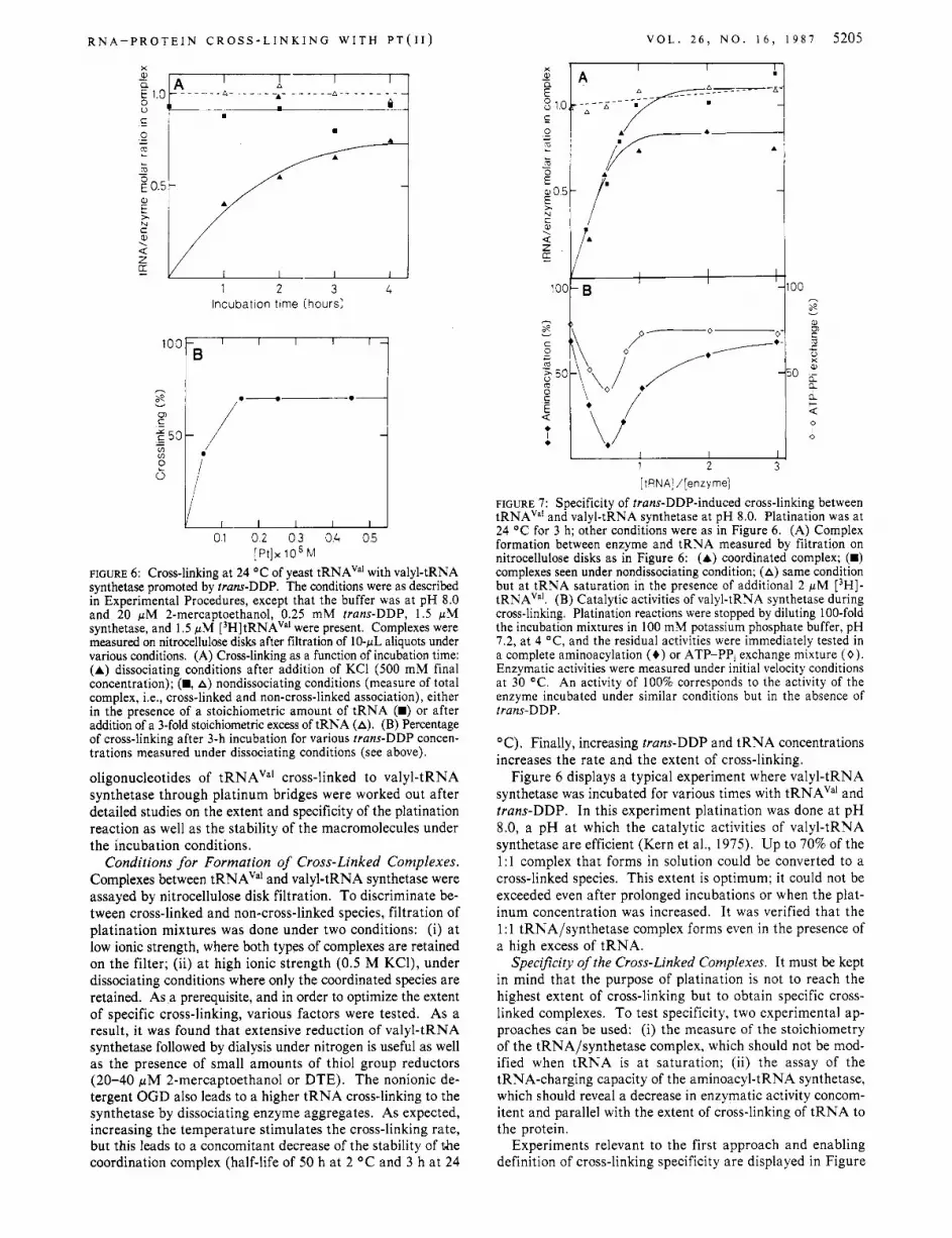
FIGURE 7: Specificity of trans-DDP-induced cross-linking between tRNAVa' and valyl-tRNA synthetase at pH 8.0. Platination was at 24 O C for 3 h; other conditions were as in Figure 6 . (A) Complex formation between enzyme and tRNA measured by filtration on nitrocellulose disks as in Figure 6: (A) coordinated complex; (m) complexes seen under nondissociating condition; (A) same condition but at tRNA saturation in the presence of additional 2 pM [3H]- tRNAVa'. (B) Catalytic activities of valyl-tRNA synthetase during cross-linking. Platination reactions were stopped by diluting 100-fold the incubation mixtures in 100 mM potassium phosphate buffer, pH 7.2, at 4 "C, and the residual activities were immediately tested in a complete aminoacylation (+) or ATP-PPI exchange mixture (0) . Enzymatic activities were measured under initial velocity conditions at 30 OC. An activity of 100% corresponds to the activity of the enzyme incubated under similar conditions but in the absence of trans-DDP.
"C). Finally, increasing trans-DDP and tRNA concentrations increases the rate and the extent of cross-linking.
Figure 6 displays a typical experiment where valyl-tRNA synthetase was incubated for various times with tRNAVa1 and trans-DDP. In this experiment platination was done at pH 8.0, a pH at which the catalytic activities of valyl-tRNA synthetase are efficient (Kern et al., 1975). Up to 70% of the 1:l complex that forms in solution could be converted to a cross-linked species. This extent is optimum; it could not be exceeded even after prolonged incubations or when the plat- inum concentration was increased. It was verified that the 1 : 1 tRNA/synthetase complex forms even in the presence of a high excess of tRNA.
Specificity of the Cross-Linked Complexes. It must be kept in mind that the purpose of platination is not to reach the highest extent of cross-linking but to obtain specific cross- linked complexes. To test specificity, two experimental ap- proaches can be used: (i) the measure of the stoichiometry of the tRNA/synthetase complex, which should not be mod- ified when tRNA is a t saturation; (ii) the assay of the tRNA-charging capacity of the aminoacyl-tRNA synthetase, which should reveal a decrease in enzymatic activity concom- itent and parallel with the extent of cross-linking of tRNA to the protein.
Experiments relevant to the first approach and enabling definition of cross-linking specificity are displayed in Figure

5206 B I 0 C H E M I S T R Y
G U
U A
PG
T U K A L O E T A L .
7A. In the presence of an excess of valyl-tRNA synthetase, the tRNA appears to be essentially incorporated in its specific site, since the stoichiometry of the total complex is near 1 .O after addition of an excess of tRNA. When tRNA is in excess, up to 0.9 mol of tRNAVa' can be incorporated/mol of synthetase. Again, the amount of total complex does not exceed significantly the theoretical 1:l stoichiometry, even after further addition of tRNA when the platination reaction is
' completed. This also points to the specificity of cross-linking. The second approach (measure of catalytic activities) was
widely used to demonstrate the specificity of tRNA/synthetase complexes cross-linked via Schiff base formation between periodate-oxidized 3' terminal adenosine of tRNA and lysine residues of the protein (Baltzinger et al., 1979; Hountondji et al., 1986). In the case of the platination reaction, the use of this approach (Figure 7B) presents some difficulties because platination interferes with enzymatic activities even in the absence of cross-linking (20-3096 inactivation). The reason for that relies on the reaction of trans-DDP with essential thiol groups required in valyl-tRNA synthetase for amino acid activation and tRNA charging.* In spite of these difficulties, the decrease of valyl-tRNA synthetase activities at low tRNA concentration likely reflects the specific coordination of tRNAVa' with the synthetase, although part of the enzyme inactivation could be the consequence of reactions between essential amino acids and platinum. This effect could even be enhanced as the result of a conformational change of the synthetase upon tRNA binding (Zacca" et al., 1978). Unexpectedly, when the concentration of tRNA increases, the enzymatic activities are less affected, and for a large excess of tRNA they become similar to those observed in the absence of tRNA. A likely explanation is that a high concentration of tRNA decreases the concentration of free platinum able to react with the enzyme, so that inactivation mainly expresses covalent association.
Characterization of Cross-Linked Oligonucleotides. In the following, cross-linking conditions were used enabling specific incorporation of the tRNA in its binding site on the amino- acyl-tRNA synthetase. However, pH 7.2 was chosen, which is a suboptimal value for specific cross-linking. This choice was dictated in order to facilitate the structural analysis of tRNA because at this pH the extent of alkaline-type hydrolysis of the nucleic acid during platination is decreased. A more acidic pH (in the range around pH 6.0) was also avoided. Although such condition would strengthen the association between tRNA and synthetase (Bonnet & Ebel, 1975) as well as the chemical stability of RNA molecules, it was not retained because it favors some unspecific associations (results not shown).
The output of a typical experiment is displayed on Figure 8, which is the autoradiography of a polyacrylamide gel showing oligonucleotides of yeast tRNAva1 cross-linked to valyl-tRNA synthetase. To demonstrate the potentiality of the platinum method to map regions of a RNA in vicinity with a protein, some oligonucleotides of tRNAVa' were sequenced and the data confronted with those obtained from other ex- perimental approaches. In the case of the experiment shown in Figure 8, eight major bands were eluted from the gel and the oligonucleotides sequenced. Their location in the sequence
* When valyl-tRNA synthetase (5 pM) is incubated with [14C]pCMB (1 1 pM), two thiol groups are statistically modified and both the amino acid activation and the tRNA aminoacylation activities of the enzyme are decreased by 50%. These enzymatic activities are totally abolished after modification of four thiols. Under optimal modification conditions 12 thiols can react with pCMB.
-0 xc
13=;
-5
m - 4
bpb -3
-2
- 1
FIGURE 8: Major oligonucleotides in yeast tRNAVa1 cross-linked to valyl-tRNA synthetase. The figure represents the autoradiography of a polyacrylamide gel electrophoresis experiment. After sequencing, bands could be assigned as follows: 1 , A35-G39; 2, A35-C40; 3, A68-C74; 4, A68-A76; 5, A43453; 6, A35-G53; 7, m'A58-A76; 8, A35457.
'7 C D G A
G w
5 -u C G
'U %G
C U G
c A rW *C
U 30G
C
:$ I 35
FIGURE 9: Contacts in yeast tRNAVa1 with its cognate valyl-tRNA synthetase. The oligonucleotides cross-linked to the synthetase are indicated in boxes (standard RNase TI fragments are assigned as in Figure 8 and shown in heavy lines; other regions belonging to cross-linked oligonucleotides are encircled by doted lines). Potential cross-linking sites with trans-DDP are indicated (-0). The arrows show the phosphates in tRNAVa' protected against ethylnitrosourea alkylation in the complex (Vlassov et al., 1983). Sequence data on tRNAVa' are according to Bonnet et al. (1974) and numbering of residues according to that of yeast tRNAPhc.
of tRNAVa1 is shown in Figure 9. Three of them (1,4,5) are typical RNase TI cleavage products. Some minor bands (Le., band 3 in Figure 8) correspond to degradation products of RNase TI oligonucleotides originating in fragile CpA se- quences (Carbon et al., 1978; Moras et al., 1985). Other sequenced oligonucleotides correspond to RNA fragments A35-G57 (band 8) and m1A58-A76 (band 7). Since N7 atoms in G residues are potential cross-linking sites, this could mean that these internal G's are bridged to valyl-tRNA synthetase. This is a likely explanation for G39 and G42,

R N A - P R O T E I N C R O S S - L I N K I N G W I T H P T ( I I )
which are within a sequence containing phosphate groups protected against alkylation by ethylnitrosourea in the tRNA/synthetase complex (Vlassov et al., 1983). An alter- native explanation would be that RNase TI cannot split at all sites due to steric hindrance effects in the complex.
When the present data are compared with other interaction studies on yeast tRNAVai, it appears that the major RNase TI fragments bound to valyl-tRNA synthetase (Le., A 3 5 4 3 9 in the anticodon region, A43-G53 covering the variable loop, and A68-A76 containing the amino acid accepting region) were also found involved in the recognition process by other means, either in protection experiments against phosphate alkylation (Vlassov et al., 1983), by ultraviolet cross-linking (Renaud et al., 1979), or by other methods [for a review see Kisselev (1985)l. Note the absence of a G residue in the cross-linked terminal A68-A76 oligonucleotide, showing that strong coordination sites for platinum exist on other residues. It is likely that this oligonucleotide binds to the synthetase at the level of the CCA end through coordination with either N3 of the cytosines or N 1 of adenosine, in good agreement with crystallographic studies on yeast tRNAPhe crystals soaked with DDP (Rubin et al., 1983).
It might appear unexpected that no major cross-linked ol- igonucleotide is found in the 5’ half of tRNAVd although major contact points were found in that region by other methods (Renaud et al., 1979; Vlassov et al., 1983). This could mean either that no counterpart on the protein exists for coordination with platinum or that trans-DDP is too large for bridging the nucleic acid to the protein in this region. Indeed, this part of tRNA, in particular the region around residue 8 at the junction of the amino acid and D stem, is in intimate contact with the synthetase, as shown by protection experiments using a chemical probe of very small size (Vlassov et al., 1983). Methodological difficulties could also explain this fact. Indeed, because of the higher content of G residues in this part of the tRNA, many very short RNase TI cleavage products are generated, which could have escaped isolation after electro- phoresis.
General Conclusion. This work demonstrates the versatility of trans-DDP as a tool for investigating nucleic acid/protein complexes. In the field of R N P particles it enables rapid detection of proteins in vicinity of the RNA. Here it was shown that protein S18 is the major adduct to 16s rRNA within the 30s subunit of the E. coli ribosome. In another study trans-DDP permitted us to find cross-linking of initiation factor IF 3 with two domains of 16s rRNA inside the small ribosomal subunit (Ehresmann et al., 1986). At a more subtle level of structural analysis, trans-DDP permits definition of contact points between proteins and nucleic acids. The present study illustrates this potentiality for tRNA sites in the tRNAVa’/valyl-tRNA synthetase system from yeast. Prelim- inary experiments in the tRNAA”P/aspartyl-tRNA synthetase system have strengthened this view. In that case, a major cross-linked oligonucleotide covers the variable-loop region of tRNAASp, in good agreement with the conclusion from other approaches (Romby et al., 1985). All these studies, however, only yielded information about regions of tRNA in contact with the synthetase. Working with a combination of RNases it should be possible to detect the exact nucleotide of RNA in contact with the interacting protein. Conversely, the use of proteases and adequate analytical tools should enable de- tection of peptides and even amino acids at the protein side in the vicinity of the interacting nucleic acid.
ACKNOWLEDGMENTS We thank G. K. Matsuka, D. Moras, P. Romby, and V. V.
V O L . 2 6 , N O . 1 6 , 1 9 8 7 5207
Vlassov for suggestions and stimulating discussions and F. Eyerman for technical help.
Registry No. trans-DDP, 1491 3-33-8; aminoacyl-tRNA synthetase, 9028-02-8.
REFERENCES
Ackerman, E. J., Joachimiak, A., Klinghofer, V., & Sigler,
Baltzinger, M., Fasiolo, F., & Remy, P. (1979) Eur. J . Bio-
Bonnet, J., & Ebel, J. P. (1975) Eur. J . Biochem. 58, 193-201. Bonnet, J., Ebel, J . P., Dirheimer, G., Shershneva, L. P.,
Krutilina, A. I., Venkstern, P. V., & Bayev, A. A. (1974) Biochimie 56, 1211-1213.
Brimacombe, R., Maly, P., & Zwieb, C. (1983) Prog. Nucleic Acid Res. Mol. Biol. 28, 1-48.
Budzik, G. P., Lam, S. S . M., Schoemaker, H. J. P., & Schimmel, P. R. (1975) J . Biol. Chem. 250, 4433-4439.
Caradonna, J . P., Lippard, S. J., Gait, M. J., & Singh, M. (1982) J . Am. Chem. SOC. 104, 5793-5800.
Carbon, P., Ehresmann, C., Ehresmann, B., & Ebel, J. P. (1978) FEBS Lett. 94, 152-156.
Chiaruttini, C., Expert-BezanGon, A., Hayes, D., & Ehres- mann, B. (1982) Nucleic Acids Res. 10, 7657-7676.
Dewan, J. C. (1984) J . Am. Chem. SOC. 106, 7239-7244. Dirheimer, G., & Ebel, J. P. (1967) Bull. SOC. Chim. Biol.
Eastman, A. (1983) Biochemistry 22, 3927-3933. Ehresmann, B., Backendorf, C., Ehresmann, C., Millon, R.,
& Ebel, J . P. (1980) Eur. J . Biochem. 104, 255-262. Ehresmann, C., Moine, H., Mougel, M., Dondon, J., Grun-
berg-Manago, M., Ebel, J. P., & Ehresmann, B. (1986) Nucleic Acids Res. 14, 4803-4821.
P. B. (1985) J . Mol. Biol. 181, 93-102.
chem. 97, 481-499.
49, 1679-1687.
Fellner, P. (1969) Eur. J . Biochem. 11, 12-27. Filipski, J., Kohn, K. W., Prather, R., & Bonner, W. M.
(1979) Science (Washington, D.C.) 204, 181-183. Filipski, J., Kohn, K. W., & Bonner, W. M. (1980)
Chem.-Biol. Interact. 32, 321-330. Garrett-Wheeler, E., Lockard, R. E., & Kumar, A. (1984)
Nucleic Acids Res. 12, 3405-3423. Girault, J. P., Chottard, G., Lallemand, J . Y., Chottard, J.
C. (1982) Biochemistry 21, 1352-1356. Giri, L., Hill, W. E., & Wittmann, H. G. (1984) Adu. Protein
Chem. 36, 1-77. Golinska, B., Millon, R., Backendorf, C., Olomucki, M., Ebel,
J. P., & Ehresmann, B. (1981) Eur. J . Biochem. 115,
Hountondji, C., Lederer, F., Dessen, P., & Blanquet, S . (1986)
Jack, A., Ladner, J. E., Rhodes, D., Brown, R. S., & Klug,
Kelman, A. D., & Buchbinder, M. (1978) Biochimie 60,
Kern, D., GiegE, R., Robbe-Saul, S., Boulanger, Y . , & Ebel,
Kern, D., Dietrich, A,, Fasiolo, F., Renaud, M., GiegE, R.,
Kisselev, L. L. (1985) Prog. Nucleic Acid Res. Mol. Biol. 32,
47 9-48 4.
Biochemistry 25, 16-2 1.
A. (1977) J . Mol. Biol. I l l , 315-328.
893-899.
J. P . (1975) Biochimie 57, 1167-1 176.
& Ebel, J . P. (1977) Biochimie 59, 453-462.
237-266.

5208 Biochemistry 1987, 26, 5208-521 2
Kyriatsoulis, A., Maly, P., Greuer, B., Brimacombe, R., Stoffler, G., Frank, R., & Block, H. (1986) Nucleic Acids Res. 14, 1171-1185.
Lake, J. A. (1985) Annu. Rev. Biochem. 54, 507-530. Maly, P., Rinke, J., Ulmer, E., Zwieb, C., & Brimacombe,
Maxam, A. M., & Gilbert, W. (1977) Proc. Natl. Acad. Sci. U.S.A. 74, 560-564.
Mets, L. J . , & Bogorad, L. (1974) Anal. Biochem. 57, 200-2 10.
Millon, R., Olomucki, M., Le Gall, J. Y ., Golinska, B., Ebel, J. P., & Ehresmann, B. (1980) Eur. J . Biochem. 110,
Moras, D., Dock, A. C., Dumas, P., Westhof, E., Romby, P., Ebel, J. P., & GiegC, R. (1985) J . Biomol. Struct. Dyn. 3, 479-493.
Noller, H. F., & Lake, J. A. (1984) in Membrane Structure and Function (Bittar, E., Ed.) Vol. 6, pp 218-297, Wiley, New York.
Ofengand, J . (1982) in Protein Biosynthesis in Eukaryotes (Perez-Bercoff, R., Ed.) pp 1-67, Plenum, New York.
Petsko, G. A. (1985) Methods Enzymol. 114, 147-157. Petsko, G. A., Philips, D. C., Williams, R. J . P., & Wilson,
I . A. (1978) J . Mol. Biol. 120, 345-360. Ramakrishnan, V., Capel, M., Kjeldgaard, M., Engelman, D.
M., & Moore, P. B. (1984) J . Mol. Biol. 174, 265-284. Renaud, M., Dietrich, ?.., GiegC, R., Remy, P., & Ebel, J . P.
(1979) Eur. J . Biochrm. 101, 475-483. Rether, B., Bonnet, J., & Ebel, J. P. (1974) Eur. J . Biochem.
Rhodes, D., Piper, P. W., & Clark, B. F. C. (1974) J . Mol.
Roberto, J. J . , & Pascoe, J . M. (1972) Nature (London) 235,
Romby, P., Moras, D., Bergdoll, M., Dumas, P., Vlassov, V. V., Westhof, E., Ebel, J. P., & GiegC, R. (1985) J . Mol.
R. (1980) Biochemistry 19, 4179-4188.
485-492.
50, 281-288.
Biol. 89, 469-475.
282-284.
Biol. 184, 455-47 1. Rosenberg, B., Van Camp, L., Trosko, J. E., & Mansour, V.
H. (1969) Nature (London) 222, 385-386. Royer-Pokora, B., Gordon, L. K., & Haseltine, W. A. (1981)
Nucleic Acids Res. 9 , 4595-4609. Rubin, J. R., Sabat, M., & Sundaralingam, M. (1983) Nucleic
Acids Res. 11, 6571-6586. Sherman, S. E., Gibson, D., Wang, A. H. J., & Lippard, S .
J. (1985) Science (Washington, D.C.) 230, 412-417. Silberklang, M., Gillum, A. M., & RajBhandary, U. L. (1977)
Nucleic Acids Res. 4 , 4091-4108. Stark, M. J . R., Gregory, R. J., Gourse, R. L., Thurlow, D.
L., Zwieb, C., Zimmermann, R. A., & Dahlberg, A. E. (1984) J . Mol. Biol. 178, 303-322.
Sternbach, H., & Cramer, F. (1977) Hoppe-Seyler's Z . Physiol. Chem. 358, 3 12.
Stone, P. J., Kelman, A. D,, & Sinex, F. M. (1974) Nature (London) 251, 736-737.
Traub, P., Mitzushima, S., Lowry, C. V., & Nomura, M. (1971) Methods Enzymol. 20, 391-407.
Traut, R. R., Lambert, J . M., Bioleau, G., & Kenny, J. W. (1979) in Ribosomes, Structure, Function, and Genetics (Chambliss, G., et al., Eds.) pp 89-1 10, University Park Press, Baltimore, MD.
Vlassov, V. V., & Kazakov, S . A. (1982) Biokhimia 8,
Vlassov, V. V., Gieg6, R., & Ebel, J. P. (1981) Eur. J . Bio-
Vlassov, V. V., Kern, D., Romby, P., GiegC, R., & Ebel, J .
Wower, I., & Brimacombe, R. (1983) Nucleic Acids Res. 11,
ZaccG, G., Morin, P., Jacrot, B., Moras, D., Thierry, J. C.,
Zwieb, C., & Brimacombe, R. (1979) Nucleic Acids Res. 6,
499-506.
chem. 119, 51-59.
P. (1983) Eur. J . Biochem. 132, 537-544.
14 19-1 437.
& GiegC, R. (1978) J . Mol. Biol. 129, 483-500.
1775-1790.
Organization and Evolution of the Rat Tyrosine Hydroxylase Genet Elaine R. Brown, George T. Coker, 111, and Karen L. O'Malley*
Department of Anatomy and Neurobiology, Washington University Medical School, St . Louis, Missouri 631 10 Received January 26, 1987: Revised Manuscript Received March 5, 1987
ABSTRACT: This report describes the organization of the rat tyrosine hydroxylase (TH) gene and compares its structure with the human phenylalanine hydroxylase gene. Both genes are single copy and contain 13 exons separated by 12 introns. Remarkably, the positions of 10 out of 12 intron/exon boundaries are identical for the two genes. These results support the idea that these hydroxylase genes are members of a gene family which has a common evolutionary origin. We predict that this ancestral gene would have encoded exons similar to those of TH prior to evolutionary drift to other members of this gene family.
Tyrosine hydroxylase (TH),' phenylalanine hydroxylase (PH), and tryptophan hydroxylase comprise a family of en- zymes known as the aromatic amino acid hydroxylases. These three mammalian enzymes are iron-containing mixed-function oxidases which require a reduced pterin cofactor and molecular
This work was supported in part by National Institutes of Health Grant NS21318-01 to K.L.O. and Neurobiology Training Grant NS07071 to E.R.B. Funding was also provided by Monsanto Corp. Grant 442785. The National Bionet Resource is funded by National Institutes of Health Grant RR-01685-02.
* Correspondence should be addressed to this author.
oxygen. All three exhibit cross-specificity for their aromatic amino acid derivatives. Their hydroxylation reactions occur by a similar mechanism which, in all cases, can be inhibited by p-chlorophenylalanine (Gal et al., 1970 Kaufman & Fisher, 1974). Along with similar biochemical properties, these en- zymes also share immunological characteristics, indicating a common antigenic determinant (Friedman et al., 1972; Kaufman & Fisher, 1974; Chikaraishi et al., 1983).
' Abbreviations: TH, tyrosine hydroxylase; PH, phenylalanine hy- droxylase; kb, kilobase(s); bp, base pair(s).
0006-2960/87/0426-5208$01.50/0 0 1987 American Chemical Society