In vitro and in vivo behaviour of zinc-doped phosphosilicate glasses

Misacylation of pyrrolysine tRNA in vitro and in vivo
Sarath Gundllapalli1,$, Alexandre Ambrogelly1,$, Takuya Umehara1, Darrick Li1, CarlaPolycarpo1,#, and Dieter Söll1,2
1 Department of Molecular Biophysics and Biochemistry, Yale University, New Haven, CT 06520-8114 USA
2 Department of Chemistry, Yale University, New Haven, CT 06520-8114 USA
AbstractMethanosarcina barkeri inserts pyrrolysine (Pyl) at an in-frame UAG codon in its monomethylaminemethyltransferase gene. Pyrrolysyl-tRNA synthetase acylates Pyl onto tRNAPyl, the ambersuppressor pyrrolysine tRNA. Here we show that M. barkeri Fusaro tRNAPyl can be misacylatedwith serine by the M. barkeri bacterial-type seryl-tRNA synthetase in vitro and in vivo in Escherichiacoli. Compared to the M. barkeri Fusaro tRNA, the M. barkeri MS tRNAPyl contains two basechanges; a G3:U70 pair, the known identity element for E. coli alanyl-tRNA synthetase (AlaRS).While M. barkeri MS tRNAPyl cannot be alanylated by E. coli AlaRS, mutation of the MStRNAPyl A4:U69 pair into C4:G69 allows aminoacylation by E. coli AlaRS both in vitro and invivo.
KeywordstRNAPyl; pyrrolysine; mischarging; Methanosarcina barkeri
1. IntroductionA number of deviations from the universality of the genetic code are known [1]. One of themis the recoding of in-frame UAG codons as pyrrolysine (Pyl) sense codons in theMethanosarcinaceae, a group of methanogenic archaea, and in two evolutionarily unrelatedbacteria [2,3]. The co-translational insertion of Pyl is contingent on the presence of the specialamber suppressor tRNAPyl (encoded by pylT) [4], and an unusual aminoacyl-tRNA synthetase(aaRS), pyrrolysyl-tRNA synthetase (PylRS, encoded by pylS), specific only for this modifiedamino acid [4–6]. PylRS specifically recognizes Pyl and tRNAPyl [5,6] and generates Pyl-tRNAPyl both in vitro and in vivo. Transformation of E. coli with the archaeal pylT and pylSgenes showed that both gene products are also functional in a bacterial setting [6–9].Monitoring of UAG read-through efficiency by various reporter systems indicated that the stopcodon is suppressed only when tRNAPyl and PylRS are present and the E. coli strain is grownin a culture media containing an exogenous source of Pyl [N-ε-cyclopentyloxycarbonyl-L-lysine (Cyc), a structural analog of Pyl] [6–9].
Correspondence to: Dr. Dieter Söll, Department of Molecular Biophysics and Biochemistry, Yale University, P.O. Box 208114, 266Whitney Avenue, New Haven, CT 06520-8114, USA, Tel: (203) 432-6200, Fax: (203) 432-6202, E-mail: [email protected].$S.G. and A.A. contributed equally to this work#Present address: Instituto de Bioquimica Medica, Universidade Federal do Rio de Janeiro, Rio de Janeiro, Brasil.Publisher's Disclaimer: This is a PDF file of an unedited manuscript that has been accepted for publication. As a service to our customerswe are providing this early version of the manuscript. The manuscript will undergo copyediting, typesetting, and review of the resultingproof before it is published in its final citable form. Please note that during the production process errors may be discovered which couldaffect the content, and all legal disclaimers that apply to the journal pertain.
NIH Public AccessAuthor ManuscriptFEBS Lett. Author manuscript; available in PMC 2009 October 15.
Published in final edited form as:FEBS Lett. 2008 October 15; 582(23-24): 3353–3358. doi:10.1016/j.febslet.2008.08.027.
NIH
-PA Author Manuscript
NIH
-PA Author Manuscript
NIH
-PA Author Manuscript

The molecular basis of PylRS specificity towards tRNAPyl was attributed to the unusualsecondary/tertiary structure of the tRNA [8,10] previously seen only in the bovinemitochondrial tRNASer [11,12]. A series of specific interactions mediated by nucleotideslocated in the acceptor stem and flanking the tRNAPyl
CUA anticodon were also shown tocontribute to the recognition of tRNAPyl by PylRS [8,10]. In contrast to most aaRSs, PylRSdoes not recognize its tRNA substrate via interaction with nucleotides of the anticodon [8,10,13]. This observation made possible the insertion of Cyc at a UGA codon upon engineering ofa complementary UCA anticodon in tRNAPyl [10]. Crystallization of Methanosarcina mazeiPylRS in a complex with Pyl and Cyc allowed the visualization of the enzyme’s active site andshed some light on Pyl recognition by archaeal [13,14] and bacterial PylRS [15].
In addition to Pyl and Cyc recognition, recent work describes the engineering of the PylRSactive site to accommodate the lysine analogs, N-benzyloxycarbonyl lysine and N-acetyllysine, so as to allow their incorporation into proteins in human [16] and E. coli [17] cells.
The tRNAPyl sequences of two M. barkeri strains are known; M. barkeri Fusaro and M.barkeri MS tRNAPyl differ in two bases (see Fig. 1), but both share the unique tRNA structurewith the one found in the Bos taurus mitochondrial tRNASer species [18]. In these tRNAs thejunction between the acceptor stem and D stem is shortened by one nucleotide, the anticodonstem consists of six base pairs instead of the classical five pairs, and the D loop is significantlyshorter compared to the canonical cloverleaf structures [5,18]. The M. barkeri MS tRNAPyl
has a G3:U70 base pair; this pair was shown to be the primary identity element for tRNAAla
recognition by E. coli AlaRS [19,20]. Since the PylRS and tRNAPyl are assumed to be afunctional orthogonal pair for genetic code expansion, we wanted to study this further byprobing the aminoacylation of tRNAPyl by non-cognate aaRSs.
2. Materials and Methods2.1 General
Oligonucleotide synthesis, DNA sequencing and Edman degradation were performed by theKeck Foundation Biotechnology Resource Laboratory at Yale University. [14C]Serine (155mCi/mmol) and [14C]alanine (150 mCi/mmol) were from Amersham Biosciences (Uppsala,Sweden).
2.2 Construction of tRNA, minihelix, and enzyme constructsM. barkeri strain Fusaro and strain MS tRNAPyl were produced and purified as described in[5]. Minihelices were constructed based on the sequences of M. barkeri MS tRNAAla
(tRNAAla), M. barkeri MS tRNAPyl (tRNAPyl), and an M. barkeri MS tRNAPyl mutant(A4:U69→C4:G69) (tRNAPyl C4:G69). The minihelices are shown in Fig. 4B with theirdesignated names (indicated in parentheses in the previous sentence). The pGF-1b plasmidcontaining the E. coli suppressor tRNALys gene was obtained from W. McClain (Universityof Wisconsin, Madison). The pET-15b plasmids containing the M. barkeri Fusaro bacterialand methanogenic SerRS genes were as described [21]. The pET15 E. coli AlaRS constructwas a gift from M. Frugier (IBMC, Strasbourg, France).
2.3 Aminoacylation AssaysAminoacylation was performed at 37° C in 100 mM Hepes-KOH pH 7.2, 20 mM MgCl2, 5mM KCl, 5 mM dithiotreitol, 5 mM ATP, 300 μM [14C]Ser or [14C]Ala, 2 μM tRNA transcript,and 1 μM enzyme. 20 μL aliquots were taken at 1, 5, 20, 40, and 60 min and spotted ontoWhatman filters (3MM). The filters were washed twice with 10% TCA for 15 min each, andonce with 5% TCA for 15 min. The filters were then rinsed with ethanol and allowed to dry.Radioactivity was measured with a Beckman Coulter LS 6500 scintillation counter.
Gundllapalli et al. Page 2
FEBS Lett. Author manuscript; available in PMC 2009 October 15.
NIH
-PA Author Manuscript
NIH
-PA Author Manuscript
NIH
-PA Author Manuscript
2.4 TrpA mutant auxotrophyTo determine charging of a tRNAPyl mutant with alanine in vivo, an E. coli trpA94 strain (ambermutation in position 94 of trpA) was used. Active TrpA protein can only be made by theinsertion of Ala or Gly into this position [22]. Cells were transformed with pGF-1b plasmidcarrying the tRNAPyl C4:G69 mutant and plated on M9 minimal medium supplemented withampicillin, either with or without Trp and grown at 37°C.
2.5 Suppression efficiencyRead-through efficiency using the dual luciferase/β-galactosidase reporter system wasperformed as indicated earlier [9].
2.6 Suppression of a UAG codon in an E. coli folA reporter geneA mutant E. coli folA gene (encoding dihydrofolate reductase, DHFR) containing a UAG codonin place of codon 3 was amplified from genomic DNA, cloned into the pCR 2.1-TOPO plasmidvector (Invitrogen, Carlsbad, CA), sequenced, and subcloned into pRSFDuet-1. E. coli BL21(DE3) competent cells were cotransformed with wild-type M. barkeri Fusaro tRNAPyl (clonedin pTECH), the folA reporter construct (cloned in pRSF) and the M. barkeri bacterial typeSerRS in pET15b. The resulting strains were grown in Luria broth at 37° C. Production of therecombinant DHFR was induced with 1 mM isopropyl-β-D-thiogalactoside when cells reachedan A600 of 0.6. Cells were harvested after 17 h. DHFR was purified using a Ni-NTA affinitycolumn and then blotted onto a PVDF membrane. The first eight amino acids were identifiedby Edman degradation. Background was determined in the same conditions but in the absenceof any overexpressed proteins.
BL21(DE3) competent cells were alsocotransformed with wild-type M. barkeri FusarotRNAPyl (cloned in pTECH), the folA reporter construct (cloned in pRSF) and M. barkeri PylRSin pET15b. Cells were grown in the presence of Cyc, recombinant DHFR was purified andsequenced by Edman degradation as indicated above.
3. Results and Discussion3.1 Mischarging of M. barkeri Fusaro tRNAPyl with serine in vitro
The unique secondary structure of M. barkeri Fusaro tRNAPyl is only shared by mammalianmitochondrial tRNASer
UGA [23] (Fig. 1). Therefore, we investigated whether tRNAPyl couldbe a substrate for the two M. barkeri SerRSs. M. barkeri contains two serS genes that encodetwo different types of SerRSs: a bacterial-type SerRS present in most organisms, and a highlydivergent SerRS only found in some methanogenic archaea [24]. The methanogenic andbacterial-type SerRSs use overlapping identity elements to recognize the same set oftRNASer species [21]. In vitro aminoacylation reactions showed that the bacterial-type SerRSwas able to acylate up to 15% of the available M. barkeri tRNAPyl transcript with serine after60 min (Fig. 2). In contrast, the methanogenic SerRS showed no detectable charging oftRNAPyl (Fig. 2).
3.2 Mischarging of M. barkeri Fusaro tRNAPyl with serine in vivoWe then determined whether the mischarging of tRNAPyl with serine observed in vitro alsooccurs in vivo. To do so, we measured the level of UAG read-through in E. coli in the presenceof the two M. barkeri SerRSs using a plasmid-based reporter system that contains in frame thegenes encoding β-galactosidase (lacZ) and luciferase (luc). The two genes are transcribed asa single mRNA unit; an in frame UAG codon is located between the two open reading frames.In the absence of read-through only the β-galactosidase is produced. Upon read-through a β-galactosidase-luciferase fusion protein is synthesized. Luciferase enzymatic activity directly
Gundllapalli et al. Page 3
FEBS Lett. Author manuscript; available in PMC 2009 October 15.
NIH
-PA Author Manuscript
NIH
-PA Author Manuscript
NIH
-PA Author Manuscript

correlates to the frequency of read-through, and when normalized to β-galactosidase activityallows sample-to-sample comparison. Read-through efficiency is expressed as the ratio ofluciferase to β-galactosidase enzymatic activities observed with the in frame UAG-containingreporter plasmid over the same ratio obtained when the stop codon is a standard lysine codon.This reporter system, previously used to study internal ribosome reentry sites [25], permits toreliably measure read-through even when it occurs at very low level.
We quantified UAG suppression efficiency in the presence of tRNAPyl and each one of thetwo M. barkeri SerRSs and compared these values to those obtained with the appropriatenegative and positive controls. When tRNAPyl was co-expressed with the bacterial type SerRS,read-through to up to 8.6% was detected and only to about 3.3% with the methanogenic SerRS(Fig. 3). In the absence of any overexpressed enzyme, background read-through was measuredas 1.3% (Fig. 3). These results indicated that when expressed in E. coli the two M. barkeriSerRSs are able to misacylate M. barkeri tRNAPyl with serine at a low but detectable level. Togauge the significance of such read-through we compared it with UAG suppression observedin the presence of PylRS, tRNAPyl and Cyc, a structural analog of Pyl (Fig. 3). Read-throughin the presence of the bacterial-type SerRS accounted for only 11% of that observed with PylRSand Cyc. The in vivo observations are in line with our above in vitro results that showed theability of the M. barkeri bacterial-like SerRS to serylate tRNAPyl. More surprising was the factthat we could detect read-through activity with the methanogenic enzyme while noaminoacylation could be detected in vitro. We attribute this apparent discrepancy to the highersensitivity of the in vivo assay for very low mischarging activities. Neither the methanogen-type M. barkeri SerRS nor E. coli SerRS formed significant amounts of Ser-tRNAPyl, andsuppression was entirely dependent on the presence of tRNAPyl (Fig. 3).
3.3 Direct evidence for serine incorporation into proteinNext we sought to confirm the direct insertion of serine into a reporter protein in E. coli. Weutilized an E. coli folA (encoding DHFR) reporter gene containing an in-frame UAG codon atposition 3 and a six-histidine codon repeat at the 3′ end. E. coli cells were co-transformed withtRNAPyl, the folA gene reporter and either M. barkeri bacterial-type serS, or an empty plasmidfor background determination. For each condition, a DHFR-His6 was purified by Ni-NTAchromatography and SDS gel electrophoresis. N-terminal sequencing of the purified DHFR-His6 from cells co-transformed with the bacterial-type serS revealed the presence of Ser atposition 3 in the protein. Pro, Gln and Val were also detected at the third position albeit inmuch smaller amounts. Pro was found to be at position 3 of the protein sample extracted fromthe cells lacking tRNAPyl (negative control).
To ensure that our reporter system was functioning properly we measured the incorporation ofCyc at position 3 of the recombinant DHFR-His6 in cells grown on Cyc and containing thetRNAPyl:PylRS pair. Expression of the DHFR-His6 was significantly higher than in theprevious experiments and allowed purification of the protein to homogeneity. Amino acid 3of the purified DHFR-His6 was identified as Cyc, in line with earlier mass spectroscopy data[17]. No other amino acid was detected in position 3 (data not shown).
3.4 Misacylation of M. barkeri MS tRNAPyl with alanine in vitroM. barkeri MS tRNAPyl differs from M. barkeri Fusaro tRNAPyl at two positions: the firstnucleotide of the variable loop is U44 instead of C44, and in the acceptor stem MS tRNAPyl
contains a G3 instead of A3 (Fig. 1). The G3A deviation results in the formation of the wobblebase pair G3:U70. A G3:U70 pair is also the primary identity element for tRNAAla recognitionby AlaRS [19,20].
Gundllapalli et al. Page 4
FEBS Lett. Author manuscript; available in PMC 2009 October 15.
NIH
-PA Author Manuscript
NIH
-PA Author Manuscript
NIH
-PA Author Manuscript

We then tested whether M. barkeri MS tRNAPyl could be a substrate for E. coli AlaRS. Asseen in Fig. 4A, the M. barkeri MS tRNAPyl is not misacylated with alanine by E. coli AlaRSin vitro, possibly the result of a tRNA anti-determinant whose presence prevents AlaRSrecognition. A good candidate for an anti-determinant in wild-type MS tRNAPyl is the A4:U69base pair, as insertion of A4:U69 into tRNAAla disrupts the acceptor stem helix and reducesrecognition by AlaRS [19]. We tested whether this particular base pair plays a role in precludingMS tRNAPyl from being misacylated with alanine by E. coli AlaRS. Inspection of the M.barkeri MS tRNAAla sequence revealed a C4:G69 base pair underneath the G3:U70 mainidentity element. We decided to replace M. barkeri MS tRNAPyl A4:U69 with a C4:G69 pair.AlaRS is one of the few aaRSs shown to be able to charge stem-loop helices derived from itscognate tRNAAla [26]. We then constructed three minihelices (Fig. 4B) based on (i) M.barkeri MS tRNAAla (tRNAAla), (ii) acceptor and T-stem sequences of M. barkeri MStRNAPyl (tRNAPyl), and (iii) a C4:G69 M. barkeri MS tRNAPyl mutant (tRNAPyl C4:G69).The M. barkeri MS tRNAPyl minihelix, like the full length tRNAPyl is not misacylated by E.coli AlaRS. The control M. barkeri MS wild-type tRNAAla stem-loop helix was charged closeto 50%. The C4:G69 mutant minihelix was acylated with Ala but remained a poor substratefor AlaRS since it was charged to only 2% (Fig. 4C).
3.5 Misacylation of M. barkeri MS tRNAPyl with alanine in vivoTo determine whether M. barkeri MS tRNAPyl and its C4:G69 mutant can be misacylated withAla in vivo, we made use of the E. coli trpA94 amber mutant strain that will only grow in theabsence of exogenously added Trp is the amber codon is suppressed by either Gly or Ala[22]. We transformed this strain with plasmids containing either the M. barkeri MS wild typetRNAPyl, the C4:G69 tRNAPyl mutant, an E. coli tRNAAla amber suppressor, or an emptyvector. The transformed strains were plated on M9 minimal medium with or without Trp (Fig.5).
After incubation at 37 C for 72 h, the E. coli strain trpA94 transformed with the tRNAPyl
C4:G69 mutant showed growth on plates without Trp, albeit at a slower rate (72 h) comparedto the E. coli tRNAAla amber suppressor (24–48 h) (Fig. 5). This result indicated themisacylation of the mutant tRNAPyl with Ala, and the production of a sufficient amount ofactive TrpA enzyme, permitting cell survival. The cells transformed with M. barkeri MS wild-type tRNAPyl or with the empty plasmid did not show any growth after 72 h (Fig. 5), confirmingour in vitro results. Lastly, as expected, all transformants grew on minimal plates supplementedwith Trp (Fig. 5).
4. OutlookThe current study demonstrates two instances of tRNAPyl misacylation, the formation of Ser-tRNAPyl and Ala-tRNAPyl. M. barkeri bacterial-type SerRS recognizes its three tRNASer
isoacceptors via interaction of amino acids in the long α-helix domain with nucleotides of theextended variable loop of the tRNASer; this specificity mechanism is shared with E. coli SerRS[21]. Bovine mitochondrial tRNASer lacks such an extended variable loop and presents a non-canonical structure; a mitochondrial SerRS:tRNASer complex requires an additional N-terminal enzyme domain interacting with nucleotides of the D-and T-loops [23]. Future studieswill establish details of the interaction between SerRS and tRNAPyl, and should also providemore insight into possible uses of misacylated suppressor tRNAPyl protein synthesis.
AcknowledgementsWe thank O. Namy for the reporter system, and E.J. Murgola for the E. coli trpA94 mutant strain. This work wassupported by grants from the National Institute for General Medical Sciences and from the National ScienceFoundation.
Gundllapalli et al. Page 5
FEBS Lett. Author manuscript; available in PMC 2009 October 15.
NIH
-PA Author Manuscript
NIH
-PA Author Manuscript
NIH
-PA Author Manuscript

References1. Ambrogelly A, Palioura S, Söll D. Natural expansion of the genetic code. Nat Chem Biol 2007;3:29–
35. [PubMed: 17173027]2. Krzycki JA. The direct genetic encoding of pyrrolysine. Curr Opin Microbiol 2005;8:706–712.
[PubMed: 16256420]3. Herring S, Ambrogelly A, Gundllapalli S, O’Donoghue P, Polycarpo CR, Söll D. The amino-terminal
domain of pyrrolysyl-tRNA synthetase is dispensable in vitro but required for in vivo activity. FEBSLett 2007;581:3197–3203. [PubMed: 17582401]
4. Srinivasan G, James CM, Krzycki JA. Pyrrolysine encoded by UAG in Archaea: charging of a UAG-decoding specialized tRNA. Science 2002;296:1459–1462. [PubMed: 12029131]
5. Polycarpo C, Ambrogelly A, Bérubé A, Winbush SM, McCloskey JA, Crain PF, Wood JL, Söll D. Anaminoacyl-tRNA synthetase that specifically activates pyrrolysine. Proc Natl Acad Sci USA2004;101:12450–12454. [PubMed: 15314242]
6. Blight SK, Larue RC, Mahapatra A, Longstaff DG, Chang E, Zhao G, Kang PT, Green-Church KB,Chan MK, Krzycki JA. Direct charging of tRNACUA with pyrrolysine in vitro and in vivo. Nature2004;431:333–335. [PubMed: 15329732]
7. Polycarpo C, Herring S, Bérubé A, Wood JL, Söll D, Ambrogelly A. Pyrrolysine analogues assubstrates for pyrrolysyl-tRNA synthetase. FEBS Lett 2006;580:6695–6700. [PubMed: 17126325]
8. Ambrogelly A, Gundllapalli S, Herring S, Polycarpo C, Frauer C, Söll D. Pyrrolysine is not hardwiredfor cotranslational insertion at UAG codons. Proc Natl Acad Sci USA 2007;104:3141–3146. [PubMed:17360621]
9. Namy O, Zhou Y, Gundllapalli S, Polycarpo CR, Denise A, Rousset JP, Söll D, Ambrogelly A. Addingpyrrolysine to the Escherichia coli genetic code. FEBS Lett 2007;581:5282–5288. [PubMed:17967457]
10. Herring S, Ambrogelly A, Polycarpo CR, Söll D. Recognition of pyrrolysine tRNA by theDesulfitobacterium hafniense pyrrolysyl-tRNA synthetase. Nucleic Acids Res 2007;35:1270–1278.[PubMed: 17267409]
11. Polycarpo C, Ambrogelly A, Ruan B, Tumbula-Hansen D, Ataide SF, Ishitani R, Yokoyama S, NurekiO, Ibba M, Söll D. Activation of the pyrrolysine suppressor tRNA requires formation of a ternarycomplex with class I and class II lysyl-tRNA synthetases. Mol Cell 2003;12:287–294. [PubMed:14536069]
12. Théobald-Dietrich A, Frugier M, Giegé R, Rudinger-Thirion J. Atypical archaeal tRNA pyrrolysinetranscript behaves toward EF-Tu as a typical elongator tRNA. Nucleic Acids Res 2004;32:1091–1096. [PubMed: 14872064]
13. Yanagisawa T, Ishii R, Fukunaga R, Kobayashi T, Sakamoto K, Yokoyama S. Crystallographicstudies on multiple conformational states of active-site loops in pyrrolysyl-tRNA synthetase. J MolBiol 2008;378:634–652. [PubMed: 18387634]
14. Kavran JM, Gundllapalli S, O’Donoghue P, Englert M, Söll D, Steitz TA. Structure of pyrrolysyl-tRNA synthetase, an archaeal enzyme for genetic code innovation. Proc Natl Acad Sci USA2007;104:11268–11273. [PubMed: 17592110]
15. Lee MM, Jiang R, Jain R, Larue RC, Krzycki J, Chan MK. Structure of Desulfitobacteriumhafniense PylSc, a pyrrolysyl-tRNA synthetase. Biochem Biophys Res Commun 2008;374:470–474.[PubMed: 18656445]
16. Mukai T, Kobayashi T, Hino N, Yanagisawa T, Sakamoto K, Yokoyama S. Adding L-lysinederivatives to the genetic code of mammalian cells with engineered pyrrolysyl-tRNA synthetases.Biochem Biophys Res Commun 2008;371:818–822. [PubMed: 18471995]
17. Neumann H, Peak-Chew SY, Chin JW. Genetically encoding N-ε-acetylly-sine in recombinantproteins. Nat Chem Biol 2008;4:232–234. [PubMed: 18278036]
18. Watanabe Y, Kawai G, Yokogawa T, Hayashi N, Kumazawa Y, Ueda T, Nishikawa K, Hirao I, MiuraK, Watanabe K. Higher-order structure of bovine mitochondrial tRNASerUGA: chemicalmodification and computer modeling. Nucleic Acids Res 1994;22:5378–5384. [PubMed: 7529407]
19. Hou YM, Schimmel P. A simple structural feature is a major determinant of the identity of a transferRNA. Nature 1988;333:140–145. [PubMed: 3285220]
Gundllapalli et al. Page 6
FEBS Lett. Author manuscript; available in PMC 2009 October 15.
NIH
-PA Author Manuscript
NIH
-PA Author Manuscript
NIH
-PA Author Manuscript

20. McClain WH, Foss K. Changing the identity of a tRNA by introducing a G-U wobble pair near the3′ acceptor end. Science 1988;240:793–796. [PubMed: 2452483]
21. Korencic D, Polycarpo C, Weygand-Durasevic I, Söll D. Differential modes of transfer RNASerrecognition in Methanosarcina barkeri. J Biol Chem 2004;279:48780–48786. [PubMed: 15364939]
22. Murgola EJ. tRNA, suppression, and the code. Ann Rev Genet 1985;19:57–80. [PubMed: 2417544]23. Chimnaronk S, Jeppesen MG, Suzuki T, Nyborg J, Watanabe K. Dual-mode recognition of
noncanonical tRNAsSer by seryl-tRNA synthetase in mammalian mitochondria. EMBO J2005;24:3369–3379. [PubMed: 16163389]
24. Kim HS, Vothknecht U, Hedderich R, Celic I, Söll D. Sequence divergence of seryl-tRNA synthetasesin archaea. J Bacteriol 1998;180:6446–6449. [PubMed: 9851985]
25. Namy O, Rousset JP, Napthine S, Brierley I. Reprogrammed genetic decoding in cellular geneexpression. Mol Cell 2004;13:157–168. [PubMed: 14759362]
26. Hou YM, Francklyn C, Schimmel P. Molecular dissection of a transfer RNA and the basis for itsidentity. Trends Biochem Sci 1989;14:233–237. [PubMed: 2669241]
Gundllapalli et al. Page 7
FEBS Lett. Author manuscript; available in PMC 2009 October 15.
NIH
-PA Author Manuscript
NIH
-PA Author Manuscript
NIH
-PA Author Manuscript

Fig. 1.Cloverleaf representation of M. barkeri Fusaro tRNAPyl, Bos taurus mitochondrial tRNASer,M. barkeri MS tRNAPyl and M. barkeri tRNAAla UGC. Differences in M. barkeri MStRNAPyl are indicated by arrows. The boxes indicate the unusual structure shared by the tRNAs.
Gundllapalli et al. Page 8
FEBS Lett. Author manuscript; available in PMC 2009 October 15.
NIH
-PA Author Manuscript
NIH
-PA Author Manuscript
NIH
-PA Author Manuscript

Fig. 2.Misacylation of M. barkeri Fusaro tRNAPyl with serine in vitro. tRNAPyl and bacterial-typeSerRS (●), tRNAPyl and methanogenic type SerRS (▲), no enzyme (◆).
Gundllapalli et al. Page 9
FEBS Lett. Author manuscript; available in PMC 2009 October 15.
NIH
-PA Author Manuscript
NIH
-PA Author Manuscript
NIH
-PA Author Manuscript

Fig. 3.Incorporation of serine and Cyc in a β-galactosidase/luciferase fusion reporter protein. Barsrepresent UAG read-through efficiency: M. barkeri tRNAPyl and either M. barkeri bacteriallike SerRS (B-SerRS), M. barkeri methanogenic SerRSs (M-SerRS), E. coli SerRS (Ec-SerRS)or an empty expression vector (pET15 empty). As positive control, M. barkeri tRNAPyl andM. barkeri PylRS (Mb-PylRS) in the presence of Cyc was measured. Negative controls includemeasurements with either M-SerRS or B-SerRS without PylT.
Gundllapalli et al. Page 10
FEBS Lett. Author manuscript; available in PMC 2009 October 15.
NIH
-PA Author Manuscript
NIH
-PA Author Manuscript
NIH
-PA Author Manuscript

Fig. 4.Misacylation of M. barkeri MS tRNAPyl with alanine in vitro. (A) In vitro aminoacylation ofwild type M. barkeri MS tRNAPyl. Total E. coli tRNA with E. coli AlaRS (■), M. barkeri MStRNAPyl with E. coli AlaRS (▲), no enzyme (◆). (B) Minihelices based on the sequences of,from left to right, M. barkeri MS tRNAAla (tRNAAla), M. barkeri MS tRNAPyl (tRNAPyl), M.barkeri MS C4:G69 tRNAPyl mutant (tRNAPyl C4:G69). (C) In vitro aminoacylation ofminihelices. MS tRNAAla minihelix with E. coli AlaRS (●), MS tRNAPyl minihelix with theE. coli AlaRS (▲), MS tRNAPyl C4:G69 mutant minihelix with E. coli AlaRS (◆).
Gundllapalli et al. Page 11
FEBS Lett. Author manuscript; available in PMC 2009 October 15.
NIH
-PA Author Manuscript
NIH
-PA Author Manuscript
NIH
-PA Author Manuscript
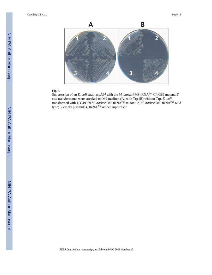
Fig. 5.Suppression of an E. coli strain trpA94 with the M. barkeri MS tRNAPyl C4:G69 mutant. E.coli transformants were streaked on M9 medium (A) with Trp (B) without Trp. E. colitransformed with 1, C4:G69 M. barkeri MS tRNAPyl mutant; 2, M. barkeri MS tRNAPyl wildtype; 3, empty plasmid; 4, tRNAAla amber suppressor.
Gundllapalli et al. Page 12
FEBS Lett. Author manuscript; available in PMC 2009 October 15.
NIH
-PA Author Manuscript
NIH
-PA Author Manuscript
NIH
-PA Author Manuscript
Copyright © 2022 FDOKUMEN