Optimization, In-Vitro and In-Vivo Evaluation of Floating Tablet ...
Upload
independentCategory
view
1download
0
In Vitro and In Vivo Isolation and Characterization ofDuvenhage VirusPenelope Koraka1*, Byron E. E. Martina1, Jouke M. Roose1, Pieter-Paul A. M. van Thiel2, Geert van
Amerongen1, Thijs Kuiken1, Albert D. M. E. Osterhaus1
1 Department of Virology, Erasmus Medical Center, Rotterdam, The Netherlands, 2 Department of Infectious Diseases, Tropical Medicine and AIDS, Academic Medical
Center, University of Amsterdam, Amsterdam, The Netherlands
Abstract
A fatal human case of Duvenhage virus (DUVV) infection in a Dutch traveller who had returned from Kenya was reported in2007. She exhibited classical symptoms of rabies encephalitis with distinct pathological findings. In the present study wedescribe the isolation and characterization of DUVV in vitro and its passage in BALB/c mice. The virus proved to beneuroinvasive in both juvenile and adult mice, resulting in about 50% lethality upon peripheral infection. Clinical signs ininfected mice were those of classical rabies. However, the distribution of viral antigen expression in the brain differed fromthat of classical rabies virus infection and neither inclusion bodies nor neuronal necrosis were observed. This is the firststudy to describe the in vitro and in vivo isolation and characterization of DUVV.
Citation: Koraka P, Martina BEE, Roose JM, van Thiel P-PAM, van Amerongen G, et al. (2012) In Vitro and In Vivo Isolation and Characterization of DuvenhageVirus. PLoS Pathog 8(5): e1002682. doi:10.1371/journal.ppat.1002682
Editor: Matthias Johannes Schnell, Thomas Jefferson University, United States of America
Received November 4, 2011; Accepted March 22, 2012; Published May 24, 2012
Copyright: � 2012 Koraka et al. This is an open-access article distributed under the terms of the Creative Commons Attribution License, which permitsunrestricted use, distribution, and reproduction in any medium, provided the original author and source are credited.
Funding: The authors received no specific funding for this article.
Competing Interests: None of the authors declare conflict of interest apart from Albert Osterhaus, who is a part time employee (CEO) of Viroclinics BV (fordetails go to www.erasmusmc.nl). The stating competing interest do not alter the authors’adherence to all PLoS Pathogens policies on sharing data andmaterials.
* E-mail: [email protected]
Introduction
Infection with Duvenhage virus (DUVV) causes lethal enceph-
alitis in humans and animals. Although DUVV infection is
prevalent among bats in Africa, reports of human infections are
rare and limited to three fatal cases to date, two from South Africa
and one from Kenya [1–3]. The clinical manifestations of human
rabies encephalitis, caused by any of the lyssaviruses, are typically
divided into four stages: 1) prodromal phase (local neuropathic
reactions at the inoculation site); 2) acute neurological phase (signs
of aggression, fear for water and air, fluctuating consciousness,
weakness and inspiratory spasms); 3) comatous phase; and 4)
death. No effective treatment is available for rabies to date. The
prototype virus of the lyssavirus genus; rabies virus (RABV) has a
world-wide distribution and is usually transmitted through the bite
of a rabid carnivore. Bat species are important reservoirs for
RABV in North and South America. Ten additional virus species
have been recognized within the Lyssavirus genus, which are
mainly carried by bats (with the notable exception of Mokola virus)
and are geographically more restricted. African lyssaviruses
include Lagos bat virus, (LBV) Mokola virus, (MOKV) and
DUVV. European bat lyssaviruses 1 and 2 (EBLV 1 and 2
respectively), Irkut (IRKV), Aravan (ARAV), Khujant (KHUV)
and West Caucasian bat virus (WCBV) cause sporadic cases in
Europe and Asia. Australian bat lyssavirus (ABLV) is restricted to
Australia. DUVV although genetically closely related to RABV,
causes different lesions in humans: RABV infection is associated
with eosinophilic cytoplasmic inclusion bodies in neurons (Negri
bodies) while inflammation is usually not prominent [4,5]. In
contrast, Negri bodies have not been observed in human DUVV
infections while extensive inflammation and cell death were found
[1,2]. Similarly, extensive cell death and neuronal damage has
been described in human cases of EBLV infection [6]. These
differences in lesions suggest differences in the pathogenesis of the
infection by the different members of the lyssavirus genus.
However, data from human cases of DUVV infections should
not be generalized since only three human cases have been
described to date. DUVV has not been previously propagated in
vitro and in vivo, hence the limited studies on DUVV pathogenesis.
The present study describes the isolation and characterization of
the DUVV that caused a fatal infection in a Dutch traveler who
had visited Kenya in 2007 [2,3]. Furthermore we describe passage
of the virus in BALB/c mice, thus describing an animal model to
further study the pathogenesis of DUVV infection. In the present
paper we compare the virulence of DUVV in mice with that of
two rabies viruses: the highly pathogenic, wild-type silver-haired
bat rabies virus (SHBRV) and the laboratory adapted, attenuated
Pasteur rabies virus (RABV-PV). Since different rabies virus
isolates vary considerably in their pathogenic potential and our
knowledge of DUVV pathogenesis is limited, we chose to compare
DUVV-NL07 with two very different rabies virus isolates in order
to be able to place DUVV in the spectrum of rabies pathogenesis.
Results
Isolation and genome sequence of DUVV-NL07Samples taken for diagnostic purposes from different parts of the
human brain [2] were inoculated onto N2a cells and the cultures
were followed for 28 days. Virus was isolated from a sample taken
from the thalamus as shown by the increase of viral RNA over
PLoS Pathogens | www.plospathogens.org 1 May 2012 | Volume 8 | Issue 5 | e1002682
time in the absence of cytophathic changes. The culture
supernatant (primary isolate) was subsequently used for RNA
isolation and sequencing of the complete genome of the virus
(DUVV-NL07). To confirm that the primary isolate could be
propagated in mice, intracranial inoculation of 3-day old BALB/c
mice (n = 12) was performed using 100–200 TCID50 of the
primary isolate of DUVV-NL07, which resulted in 100%
mortality within 5 days post infection (DPI). Virus could be
recovered from the brain of infected animals and after three in vivo
passages the virus was still 100% lethal in newborn mice. The
complete nucleotide sequence of the primary isolate was deposited
in GenBank (accession number JN986749). We next determined
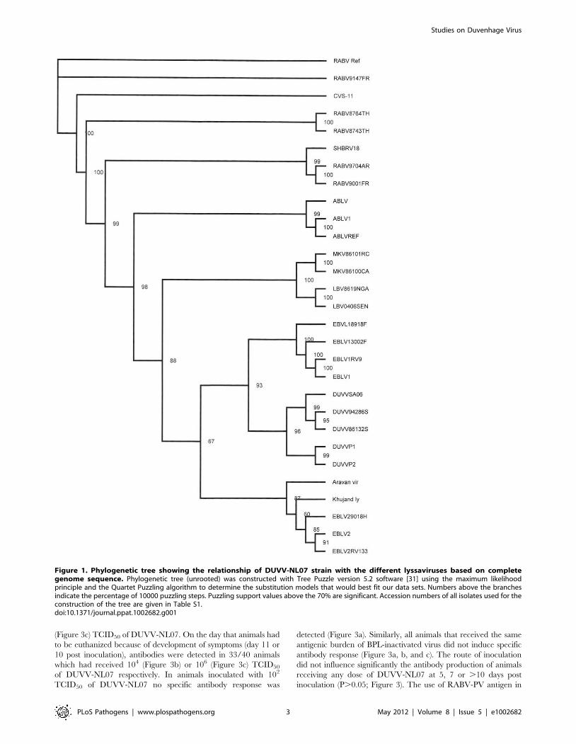
the phylogenetic relationship of DUVV-NL07 with other mem-
bers of the genus Lyssavirus. A phylogenetic tree constructed with
whole genome sequences of members of all 11 lyssavirus species
confirmed that DUVV-NL07 indeed belongs to DUVV species
(Figure 1). The DUVV-NL07 isolate proved to be 93% identical
on nucleotide level to the other three DUVV isolates from South
Africa for which the complete genome sequences are available.
The identity between the three South African isolates is up to
99%. The position of DUVV-NL07 outside the South African
cluster suggests a genetic variant that circulates in Kenya. When
the deduced amino acid sequences of the individual genes were
compared with published sequences, the highest identity of
DUVV-NL07 with other DUVV isolates was observed in the
nucleoprotein and matrix proteins (99% identity with eight
published sequences of each gene) followed by the phosphoprotein
and the polymerase (96% identity with seven and three published
sequences of the respective genes). The glycoprotein G was the
most divergent protein (95% identity with six published sequenc-
es). An earlier in-frame initiator was seen in the matrix protein
resulting in 12 additional amino acids at the N-terminal of matrix
protein, similar to what has been described for ABLV [7]. The
sequence of the primary DUVV-NL07 isolate was also compared
with the partial sequence of the N protein obtained directly from
the brain material of the patient [2]. These sequences were 99%
identical on the nucleotide level with only three nucleotide
differences reported between the sequence deposited from the
original brain material and the primary isolate of DUVV-NL07
(position 542: G vs A, position 545: Y vs C, position 551: T vs C).
Virus stocks were prepared by two additional passages of the
primary isolate on human neuroblastoma cells (SK-N-SH). The
nucleotide sequence of the passaged virus (P3) was 98% identical
to the primary isolate. The differences between primary isolate
and P3 virus were mainly silent with only one amino acid
substitution (a phenylalanine was substituted into a tyrosine in the
P3 virus) in the L gene. All subsequent experiments described here
were carried out with the primary isolate that had been passaged
twice on SK-N-SH cells.
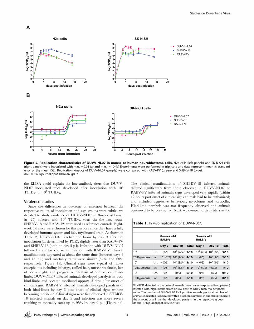
In vitro and in vivo characteristics of DUVV-NL07In the focal infection experiment, the replication kinetics of the
three different viruses were compared on N2a cells infected at an
m.o.i. of 0.01. DUVV-NL07 and RABV-PV showed peak virus
titres by day 3 post infection (DPI; Figure 2a), a day earlier
compared to SHBRV-18. Infection with any of the three viruses
proved to be non-cytopathic in these cells, which remained
persistently infected for the 17 days follow up period, although
virus titers started to decrease from 7 DPI onwards. In contrast,
peak virus titers were measured by 4 DPI in SK-N-SH cells for
DUVV-NL07 and SHBRV-18 viruses and by 7 DPI for RABV-
PV (Figure 2a). In addition, similar titers were obtained from
DUVV-NL07 and RABV-PV in SK-N-SH cells compared to
slightly higher titers obtained from SHBRV-18 infected N2a cells.
In contrast, infection of SK-N-SH cells with DUVV-NL07
resulted in almost 2log10 higher titers compared to the titers
obtained from RABV-PV and SHBRV-18 infected cells. To study
the short term dynamics of DUVV-NL07 replication, a one-step
growth curve was performed on mouse and human neuroblastoma
cells at an m.o.i. of 10. The eclipse period for all three viruses was
between 14 and 16 hours on N2a cells. Slightly different eclipse
periods were found on human neuroblastoma cells for DUVV-
NL07 and SHBRV-18 (between 14 and 16 hours) compared to
RABV-PV (between 16 and 18 hours) (Figure 2b). At 24 hours,
100% of the cells were infected (Figure S1 and S2) and high titers
were detected in the culture supernatant (Figure 2b). Similar titers
were found on SK-N-SH cells and N2a cells.
Neuroinvasion studies and serum antibody developmentSeveral studies have demonstrated that neuro-invasiveness and
neurovirulence of RABV are strain-dependent [8–10]. We sought
to determine the kinetics and minimum dose of DUVV-NL07 that
can cause disease upon peripheral inoculation in 3-week and 8-
week old BALB/c mice. We found that DUVV-NL07 was
neuroinvasive for both 3-week and 8-week old mice inoculated
with 106 TCID50 or 104 TCID50 either i.m or s.c (Table 1). Virus
was detected in the brains of animals as early as 7 days post
inoculation and clinical signs (including ruffled hair, hunched
position and muscle weakness) were apparent from 10 days post
inoculation onward. On day 10 post inoculation seven out of 60
inoculated animals developed hind limb paralysis. As shown in
Table 1, more animals that received 106 TCID50 developed
paralysis compared to animals receiving 104 TCID50. There was
no strong association between development of paralysis and route
of inoculation, age or level of viral RNA in the brain. Virus was
not detected in the brain samples of animals inoculated with 102
TCID50. In addition, no viral RNA was detected in samples taken
from the site of inoculation (muscle for i.m. inoculation or skin for
s.c. inoculations) and the draining lymph nodes, early after
inoculation (3–7 DPI.). In order to assess whether virus replication
was necessary to induce antibody response or whether the
inoculated antigen burden alone was sufficient to induce antibody
response, we compared antibody production in animals inoculated
i.m. and s.c. with DUVV-NL07 and BPL-inactivated virus
controls. As depicted in Figure 3, on day 7 antibodies could be
measured in 30/40 mice inoculated with 104 (Figure 3b) or 106
Author Summary
Lyssaviruses have been known for centuries to cause lethalencephalitis in animals and humans, representing a seriouspublic health problem especially in developing countries.Little is known about the way that lyssaviruses in general,and Duvenhage virus in particular cause disease. Studies ofpathogenesis have been hampered by the fact that thevirus has not yet been propagated and characterizedextensively. In this paper, we describe the characterizationof Duvenhage virus in vitro. Further, we characterized thevirus in BALB/c mice. We compared Duvenhage virus witha wild type rabies virus (silver-haired bat rabies virus) andwe found that while in vitro the differences of these twoviruses were not significant, the in vivo characteristics ofthese two viruses differed significantly. Histological anal-yses of infected mouse brains suggest that differences invirulence may be associated with difference in tropism.Elucidating the differences in pathogenesis betweendifferent lyssaviruses might help us in the design of noveltreatment protocols.
Studies on Duvenhage Virus
PLoS Pathogens | www.plospathogens.org 2 May 2012 | Volume 8 | Issue 5 | e1002682
(Figure 3c) TCID50 of DUVV-NL07. On the day that animals had
to be euthanized because of development of symptoms (day 11 or
10 post inoculation), antibodies were detected in 33/40 animals
which had received 104 (Figure 3b) or 106 (Figure 3c) TCID50
of DUVV-NL07 respectively. In animals inoculated with 102
TCID50 of DUVV-NL07 no specific antibody response was
detected (Figure 3a). Similarly, all animals that received the same
antigenic burden of BPL-inactivated virus did not induce specific
antibody response (Figure 3a, b, and c). The route of inoculation
did not influence significantly the antibody production of animals
receiving any dose of DUVV-NL07 at 5, 7 or .10 days post
inoculation (P.0.05; Figure 3). The use of RABV-PV antigen in
Figure 1. Phylogenetic tree showing the relationship of DUVV-NL07 strain with the different lyssaviruses based on completegenome sequence. Phylogenetic tree (unrooted) was constructed with Tree Puzzle version 5.2 software [31] using the maximum likelihoodprinciple and the Quartet Puzzling algorithm to determine the substitution models that would best fit our data sets. Numbers above the branchesindicate the percentage of 10000 puzzling steps. Puzzling support values above the 70% are significant. Accession numbers of all isolates used for theconstruction of the tree are given in Table S1.doi:10.1371/journal.ppat.1002682.g001
Studies on Duvenhage Virus
PLoS Pathogens | www.plospathogens.org 3 May 2012 | Volume 8 | Issue 5 | e1002682
the ELISA could explain the low antibody titers that DUVV-
NL07 inoculated mice developed after inoculation with 106
TCID50 or 104 TCID50.
Virulence studiesSince the differences in outcome of infection between the
respective routes of inoculation and age groups were subtle, we
decided to study virulence of DUVV-NL07 in 8-week old mice
(n = 25) infected with 106 TCID50 virus via the i.m. route.
SHBRV-18 and RABV-PV were used as reference controls. Eight-
week old mice were chosen for this purpose since they have a fully
developed immune system and fully myelinated brain. As shown in
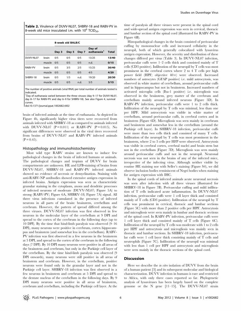
Table 2, DUVV-NL07 reached the brain by day 9 after i.m
inoculation (as determined by PCR), slightly later than RABV-PV
and SHBRV-18 (both on day 5 p.i). Infection with DUVV-NL07
followed a similar course as infection with RABV-PV: clinical
manifestations appeared at about the same time (between days 8
and 15 p.i.) and mortality rates were similar (52% and 60%
respectively; Figure 4a). Clinical signs were typical of rabies
encephalitis including lethargy, ruffled hair, muscle weakness, loss
of body-weight, and progressive paralysis of one or both hind-
limbs. DUVV-NL07 infected animals developed paralysis in both
hind-limbs and became moribund approx. 3 days after onset of
clinical signs. RABV-PV infected animals developed paralysis of
both hind-limbs by day 3 post onset of clinical signs without
becoming moribund. Clinical signs were first observed in SHBRV-
18 infected animals on day 5 and infection was more severe
resulting in mortality rates up to 95% by day 9 p.i. (Figure 4a).
The clinical manifestations of SHBRV-18 infected animals
differed significantly from those observed in DUVV-NL07 or
RABV-PV infected animals: signs developed very rapidly (within
12 hours post onset of clinical signs animals had to be euthanized)
and included aggressive behaviour, myoclonus and torticollis.
Hind-limb paralysis was not frequently observed and animals
continued to be very active. Next, we compared virus titers in the
Figure 2. Replication characteristics of DUVV-NL07 in mouse or human neuroblastoma cells. N2a cells (left panels) and SK-N-SH cells(right panels) were inoculated with m.o.i = 0.01 (a) and m.o.i. = 10 (b) Experiments were performed in triplicate and data represent mean 6 standarderror of the mean (SE). Replication kinetics of DUVV-NL07 (purple) were compared with RABV-PV (green) and SHBRV-18 (blue).doi:10.1371/journal.ppat.1002682.g002
Table 1. In vivo replication of DUVV-NL07.
8-week oldBALB/c
3-week oldBALB/c
Day 7 Day 10 Total Day 7 Day 10 Total
106 i.m. 2(0/5) 107 (2/5)1 2/10 107 (2/5) 106 (3/5)1 5/10
TCID50/mouse s.c. 107 (2/5) 107 (2/5)1 4/10 2(0/5) 108 (2/5)1 2/10
104 i.m. 2(0/5) 106 (3/5)1 3/10 2(0/5) 108 (1/5)1 1/10
TCID50/mouse s.c. 2(0/5) 108 (1/5)1 1/10 104 (1/5) 2(0/5) 1/10
102 i.m. 2(0/5) 2(0/5) 0/10 2(0/5) 2(0/5) 0/10
TCID50/mouse s.c. 2(0/5) 2(0/5) 0/10 2(0/5) 2(0/5) 0/10
Viral RNA detected in the brain of animals (mean values expressed in copies/ml)infected with high, intermediate or low dose of DUVV-NL07 via peripheralroute. The number of DUVV-NL07 RNA positive animals per total number ofanimals inoculated is indicated within brackets. Numbers in superscript indicatethe amount of animals that developed paralysis in the respective groups.doi:10.1371/journal.ppat.1002682.t001
Studies on Duvenhage Virus
PLoS Pathogens | www.plospathogens.org 4 May 2012 | Volume 8 | Issue 5 | e1002682
Figure 3. Development of RABV-specific antibodies over time in three- and eight-week old mice. BALB/c mice were inoculated i.m or s.cwith (a) 102 TCID50, (b) 104 TCID50 (c) 106 TCID50 of DUVV-NL07 or DUVV-NL07 BPL control. Dotted lines indicate threshold level of the assay.doi:10.1371/journal.ppat.1002682.g003
Studies on Duvenhage Virus
PLoS Pathogens | www.plospathogens.org 5 May 2012 | Volume 8 | Issue 5 | e1002682
brain of infected animals at the time of euthanasia. As depicted in
Figure 4b, significantly higher virus titers were recovered from
animals infected with SHBRV-18 as compared to animals infected
with DUVV-NL07 (P = 0.011) or RABV-PV (P = 0.015). No
significant differences were observed in the viral titers recovered
from brains of DUVV-NL07 and RABV-PV infected animals
(P = 0.45).
Histopathology and immunohistochemistryMost wild type RABV strains are known to induce few
pathological changes in the brain of infected humans or animals.
The pathological changes and tropism of DUVV for brain
compartments are unknown. HE and LFB-staining of brains from
DUVV-NL07, SHBRV-18 and RABV-PV infected animals
showed no evidence of necrosis or demyelination. Staining with
anti-RABV-NP antibodies showed extensive antigen expression in
infected brains. Antigen expression was seen as characteristic
granular staining in the cytoplasm, axons and dendritic processes
of infected neurons of moderate (DUVV-NL07; Figure 5A) to
strong (RABV-PV; Figure 6A, SHBRV-18; Figure 7A) signal. All
three virus infections cumulated in the presence of infected
neurons in all parts of the brain: brainstem, cerebellum and
cerebrum. However, the pattern of spread differed among the
three viruses. DUVV-NL07 infection was first observed in few
neurons in the molecular layer of the cerebellum at 9 DPI and
spread to the cortex of the cerebrum in the following days (up to
10 DPI). By the time that hind-limb paralysis was observed (9–16
DPI), many neurons were positive in cerebrum, cortex hippocam-
pus and brainstem (and somewhat less in the cerebellum). RABV-
PV infection was first observed in a few neurons in the brainstem
at 5 DPI, and spread to the cortex of the cerebrum in the following
days (7 DPI). By 10 DPI many neurons were positive in all areas of
the brainstem and cerebrum, but only in the Purkinje cell layer of
the cerebellum. By the time hind-limb paralysis was observed (9
DPI onwards), many neurons were still positive in all areas of
brainstem and cerebrum. However, in the cerebellum, positive
neurons were found only in the granular layer and not in the
Purkinje cell layer. SHBRV-18 infection was first observed in a
few neurons in brainstem and cerebrum at 5 DPI and spread to
the dentate nucleus of the cerebellum in the following days. By 9
DPI many neurons were positive in all areas of brainstem,
cerebrum and cerebellum, including the Purkinje cell layer. At the
time of paralysis all three viruses were present in the spinal cord
and wide-spread antigen expression was seen in cervical, thoracic
and lumbar section of the spinal cord (illustrated for RABV-PV in
Figure 6B).
Histopathological changes in the brain consisted of perivascular
cuffing by mononuclear cells and increased cellularity in the
neuropil, both of which generally colocalized with lyssavirus
antigen expression. However, the severity and distribution of these
changes differed per virus (Table 3). In DUVV-NL07 infection,
perivascular cuffs were 1–2 cells thick and consisted mainly of T
cells (CD3 positive). Infiltration of the neuropil by T cells was most
prominent in the cerebral cortex where 2 to 4 T cells per high
power field (HPF; objective 406) were observed. Increased
numbers of astrocytes (GFAP positive) i.e. mild astrocytosis, was
observed in white matter of cerebellum, around perivascular cuffs
and in hippocampus but not in brainstem. Increased numbers of
activated microglia cells (Iba-1 positive) i.e. microgliosis was
observed in the brainstem, grey matter of the cerebrum and
cerebellum mainly around infected neurons (Figure 5D). In
RABV-PV infection, perivascular cuffs were 1 to 2 cells thick.
Infiltration of the neuropil by T cells was minimal, less than one
per HPF. Mild astrocytosis was visible in white matter of
cerebellum, around perivascular cuffs, in cerebral cortex and in
brainstem (Figure 6D). Microgliosis was seen mainly in cerebrum
and brainstem and somewhat less in the cerebellum (around the
Purkinje cell layer). In SHBRV-18 infection, perivascular cuffs
were more than two cells thick and consisted of many T cells.
Infiltration of the neuropil by T cells was most prominent in the
brainstem, where 2 to 3 cells per HFP were observed. Astrocytosis
was visible in cerebral cortex, cerebral nuclei and brain stem but
not in the cerebellum (Figure 7D). Microgliosis was seen mainly
around perivascular cuffs and not in the neuropil. Neuronal
necrosis was not seen in the brains of any of the infected mice,
irrespective of the infecting virus. Although neither visible by
routine HE staining nor with Seller’s staining (Figure S4), we did
observe inclusion bodies reminiscent of Negri bodies when staining
for antigen expression with IHC.
In the spinal cords of infected animals acute neuronal necrosis
was seen after infection with all three viruses (illustrated for
SHBRV-18 in Figure 7B). Perivascular cuffing and mild infiltra-
tion of T cells indicated acute inflammation. In DUVV-NL07
infection, perivascular cuffs were 1 cell layer thick and consisted
mainly of T cells (CD3 positive). Infiltration of the neuropil by T
cells was prominent in cervical, thoracic and lumbar sections
(Figure 5C) with more than 2 positive cells per HPF. Astrocytosis
and microgliosis were seen mainly in lumbar and thoracic sections
of the spinal cord. In RABV-PV infection, perivascular cuffs were
1 cell layer thick and consisted mainly of T cells (Figure 6C).
Infiltration of the neuropil by T cells was moderate with 1 to 2 cells
per HPF and astrocytosis and microgliosis was mainly seen in
thoracic and lumbar sections. In SHBRV-18 infection, perivascu-
lar cuffs were 1 cell layer thick consisting mainly of T cells and
neutrophils (Figure 7C). Infiltration of the neuropil was minimal
with less than 1 cell per HPF and astrocytosis and microgliosis
were seen mainly in the thoracic sections of the spinal cord.
Discussion
Here we describe the in vitro isolation of DUVV from the brain
of a human patient [3] and its subsequent molecular and biological
characterization. DUVV infection in humans is rare and restricted
to Africa, with only three cases reported so far. Phylogenetic
analysis of lyssaviruses has been largely based on the complete
genome or the N gene [11–15]. The DUVV-NL07 strain
Table 2. Virulence of DUVV-NL07, SHBRV-18 and RABV-PV in8-week old mice inoculated i.m. with 106 TCID50.
8-week old BALB/C
Day 3 Day 5 Day 7Day ofeuthanasia1 Total
DUVV-NL07 brain 0/5 0/5 0/5 13/25 13/40
muscle 0/5 0/5 0/5 n.d. 0/15
RABV-PV brain 0/5 3/5 2/5 15/25 30/40
muscle 0/5 0/5 0/5 4/5 4/20
SHBRV-18 brain 0/5 1/5 n.d. 19/20 20/30
muscle 0/5 0/5 n.d. 5/5 5/15
The number of positive animals (viral RNA) per total number of animals tested isindicated.1Day of euthanasia varied between the three viruses (day 9–17 for DUVV-NL07,day 8–17 for RABV-PV and day 6–9 for SHBRV-18). See also Figure 4, survivalcurves.doi:10.1371/journal.ppat.1002682.t002
Studies on Duvenhage Virus
PLoS Pathogens | www.plospathogens.org 6 May 2012 | Volume 8 | Issue 5 | e1002682
described in this study clusters differently from the bat and human
DUVV’s reported so far (Figure 1) [1]. At the time of analysis
there were only three complete genome sequences and eight N
gene sequences available from other DUVV isolates. Although the
variation among DUVV strains (including the DUVV-NL07
isolate) is low compared to other lyssavirus species [11,16],
DUVV-NL07 is the most divergent among DUVV isolates both
on the nucleotide and amino acid level. Further studies are needed
to elucidate the biological relevance of the observed differences
between DUVV isolates as has been proposed for different RABV
isolates [17,18].
The one-step growth curve experiment did not reveal any
significant differences in replication kinetics between the different
viruses, neither on mouse or human neuroblastoma cells.
Therefore, the replication kinetics are not an in vitro correlate
of virulence. However, we cannot exclude that different amounts
of defective interfering particles in the different virus stocks may
have influenced the replication kinetics of these viruses. In order to
study the spreading potential of the respective viruses, focal
experiments were performed. Reduced virus titers were observed
in mouse neuroblastoma cell lines as compared to human cell
lines. The mechanisms underlying these differences are not clear.
It is possible that the two different cell lines differ in viral entry-
receptor density or qualitative or quantitative differences in anti-
viral response to infection. Alternatively, differences in the rate of
cell death due to prolonged culture could also explain the
differences observed in the spreading potential of the three viruses
in human and mouse cells.
In the case report of van Thiel et al. [2] the crude brain material
from the patient did not cause disease in outbred mice with the
mouse inoculation test. Consistently, virus or RNA could not be
detected in inoculated animals. Similarly, virus was not isolated
Figure 4. Virulence of lyssaviruses in eight-week old BALB/c mice. (A): survival curves of animals infected with DUVV-NL07 (n = 25), RABV-PV(n = 25) and SHBRV-18 (n = 20). Animals were infected i.m. with 106 TCID50 of DUVV-NL07 (squares, dotted line), RABV-PV (triangles, straight line) andSHBRV-18 (circles, dotted line) and followed for clinical signs for 20 days. (B): Infectious viral titres recovered from the brains of mice infected i.m with106 TCID50 of DUVV-NL07, RABV-PV or SHBRV-18. *: statistically different (two-tailed, Mann-Whitney test).doi:10.1371/journal.ppat.1002682.g004
Studies on Duvenhage Virus
PLoS Pathogens | www.plospathogens.org 7 May 2012 | Volume 8 | Issue 5 | e1002682

from newborn inbred mice (BALB/c and C57/Bl6) inoculated
with the same crude brain material (data not shown). Even though
we could not determine the infectious virus titer in the brain
material of the patient by endpoint dilution, the quantitative PCR
data suggest that the brain sample used for inoculation of mice
contained approximately 10 TCID50/ml of infectious virus. Our
data confirm the low sensitivity of the mouse inoculation test
[19,20]. Therefore, prior in vitro propagation of the virus proved
to be necessary to further study the characteristics of the virus. We
have shown that DUVV-NL07 is pathogenic for BALB/c mice
upon i.c. and peripheral inoculation. DUVV-NL07 replicated well
in mouse and human neuroblastoma cells as well as in hamster
fibroblasts (BHK-21-C13, data not shown). Further, the virus
proved to be both neuroinvasive and neurovirulent in juvenile and
adult BALB/c mice upon infection via the i.m or s.c routes. It was
previously shown that neuroinvasiveness of a bat-derived RABV
was dependent on the route of the inoculation [17]. The authors
speculated that the increased neuroinvasion observed in their
study was likely a reflection of the peripheral cell tropism. It is
plausible that bat-derived lyssaviruses, such as DUVV-NL07 could
replicate better in sites such as the dermis or the subcutaneous
space compared to muscles since the natural route of infection via
bat scratches would not implicate direct inoculation of the virus
into the muscle. In our studies we did not see differences in
neuroinvasion between the two different routes of inoculation,
neither could we detect replicating virus at the site of inoculation
early after infection. This suggests that neuroinvasion might not be
dependent on abundant peripheral virus replication. Given the
nature of transmission via usually superficial bat-scratches, as was
the case in the human DUVV-NL07 infection, it is generally
believed that bat-derived lyssaviruses are neuroinvasive at low
doses. Indeed, peripheral infection with as little as 104 TCID50 of
DUVV-NL07 caused encephalitis in a substantial number of mice.
Further comparisons revealed that neither route of infection, nor
age of the mice led to marked differences in the clinical outcome,
antibody response or amount of viral RNA recovered from the
brain.
Comparison of in vitro characteristics and in vivo neuroinvasive
characteristics and virulence of DUVV-NL07 (after peripheral
inoculation) with those of RABV-PV (a mouse adapted strain of
RABV) and SHBRV-18, a wild-type highly pathogenic bat-
derived RABV strain that had been recovered from a human
rabies case [17], showed clear similarities with RABV-PV. In
contrast, SHBRV-18 infection exhibited a different tropism for
Figure 5. Histopathology of 8-week old BALB/c mice infected i.m. with 106 TCID50 of DUVV-NL07. (A) Neo-cortical neurons stained withanti-NP rabies antibody, (B) HE staining of spinal cord section illustrating perivascular cuffing (objective 406), (C) CD3+ cells infiltrating the neuropil ofthe spinal cord, (D) activated microglia (Iba1 staining; 406 objective) in the brainstem. Examples of positively stained cells are indicated by blockarrows.doi:10.1371/journal.ppat.1002682.g005
Studies on Duvenhage Virus
PLoS Pathogens | www.plospathogens.org 8 May 2012 | Volume 8 | Issue 5 | e1002682
neuronal cells, different clinical manifestations and nearly 100%
lethality. These data indicate that DUVV-NL07 has intermediate
neuroinvasiveness and virulence while exhibiting characteristics of
some of the fixed RABV strains [21]. Similar to RABV-PV and
SHBRV-18, the major pathological change observed after
DUVV-NL07 infections was perivascular cuffing, whereas no
neuronal necrosis (in the brain) was observed. Different lines of
research in mouse models have indicated that several aspects of
RABV pathogenesis are largely strain dependent, such as
activation of the immune system, infiltration of leukocytes into
infected brains, gene expression patterns and cell death (reviewed
by Fu and Jackson [22]). Some studies have suggested that level of
expression of G protein inversely correlates with pathogenicity
[8,23]. However, more recent data suggest that G protein
expression levels are not critical for pathogenicity [24] and that
it is the capacity of the G protein to promote survival or death
signals in the infected neurons that contributes to pathogenicity
[25]. It remains to be seen if the G protein of DUVV-NL07 would
have the capacity to induce such signals and confer a pathogenic
or attenuated phenotype.
Our findings are in agreement with previous studies suggesting
that inflammation is not a significant determinant of pathogenesis
of DUVV-NL07 or bat-derived RABV [10,23]. Also tropism of
lyssaviruses for different brain compartments has been shown to be
largely strain dependent [4,26,27]. The mouse cerebellum did not
seem to be an important site of replication for DUVV-NL07, since
only few cells stained positive for NP-antigen at all time points
analysed after infection. The infection appeared largely localized
in certain areas of the cerebral cortex and hippocampus even in
mice that had reached advanced stages of paralysis. In contrast,
both RABV-PV and SHBRV-18 showed a clearly different
pattern of spread into the CNS, with Purkinje cells and the
dentate nucleus in the cerebellum being the primary site of
replication and more than 70% of neurons throughout the brain
being infected at the time of euthanasia. Interestingly these
histopathological findings largely paralleled the differences in
clinical signs observed between infections with the respective virus
strains. Overall, all animals exhibited signs of aggression reflecting
infection of the cerebrum by the three viruses. Extensive infection
of the cerebellum by RABV-PV and SHBRV-18 may have
contributed to paralysis whereas infection of the dentate nucleus
by SHBRV-18 correlated with the characteristic signs of torticollis.
Given the differential tropism patterns of the three viruses for
neurons in the cerebellum, it would be interesting to compare the
receptor usage of DUVV-NL07 with RABV-PV and SHBRV-18.
Despite the differences seen in the brain, the pathology of the
spinal cord of the infected animals seemed similar between the
three viruses. Inflammation and necrosis were of similar extent
Figure 6. Histopathology of 8-week old BALB/c mice infected i.m. with 106 TCID50 of RABV-PV. (A) Purkinje cell layer of the cerebellumstained with anti-NP rabies antibody, (B) Extensive rabies virus antigen expression in the spinal cord (anti-NP rabies staining; 106objective). (C) CD3+staining in a perivascular cuff of the spinal cord. (D) astrocytosis in the cerebrum of infected animals (GFAP staining; 406 objective). Examples ofpositively stained cells are indicated by block arrows.doi:10.1371/journal.ppat.1002682.g006
Studies on Duvenhage Virus
PLoS Pathogens | www.plospathogens.org 9 May 2012 | Volume 8 | Issue 5 | e1002682
between the three viruses and antigen expression was wide-spread
in all sections of the spinal cord. These findings suggest that the
differences in pathogenesis of the three viruses are most likely
linked to the brain.
In the three human cases of DUVV infection documented so far,
the clinical manifestations did not differ from those of classical
rabies. We therefore speculate that the severity of clinical outcome
seen in rabies is probably attributable to infection or dysfunction of
(yet unidentified) highly specialized compartments of the brain,
common to all lyssaviruses. It should be noted that the original
sequence of the virus from crude brain material was not available.
However, the sequence of the primary isolate was 98% identical (on
the nucleotide level) and only one amino acid substitution with the
P3 virus used for the in vitro and in vivo studies described in this
paper. Studies using a molecular clone of DUVV would be needed
to further determine the significance of the single amino acid
substitution in the L gene after in vitro passage of DUVV. It would
be interesting to evaluate the implications of such differences in
patient management and identify treatment protocols.
Both the in vitro and in vivo systems for DUVV-NL07
propagation described in the present study will be valuable tools
to further elucidate the pathogenesis of infections with bat
lyssaviruses and possible treatment options.
Materials and Methods
Ethics statementAll animal experiments described in this paper have been
conducted according to Dutch guidelines for animal experimen-
tation and approved by the Animal Welfare Committee of the
Erasmus Medical Centre, Rotterdam, The Netherlands. All efforts
were made to minimize animal suffering.
Cells and virusesMouse neuroblastoma (N2a) cells and baby hamster kidney
(BHK-21 clone 13), a kind gift from Dr. F. Cliquet, (AFSSA,
Nancy France), were grown in DMEM supplemented with
5% heat inactivated fetal bovine serum (HI-FBS) and GMEM
supplemented with 10% HI- FBS, respectively. Human
neuroblastoma cells (SK-N-SH; a kind gift from Dr. C.
Prenaud; Institute Pasteur, Paris France) were cultured in
DMEM with Glutamax supplemented with 10% HI-FBS. All
media were supplemented with antibiotics (100 U penicillin,
100 mg/ml streptomycin) and 2 mM L-glutamine. Cell culture
reagents were obtained from LONZA (Lonza Benelux BV,
Breda, The Netherlands). All cell lines tested negative for
mycoplasma.
Figure 7. Histopathology of 8-week old BALB/c mice infected i.m. with 106 TCID50 of SHBRV-18. (A) The dentate nucleus of thecerebellum stained with anti-NP rabies antibody, (B) necrotic neurons in the spinal cord (HE staining; 406objective). (C) CD3+ cells infiltrating theneuropil of the spinal cord. (D) astrocytosis in the brainstem of infected animals (GFAP staining; 406objective). Examples of positively stained cellsare indicated by block arrows.doi:10.1371/journal.ppat.1002682.g007
Studies on Duvenhage Virus
PLoS Pathogens | www.plospathogens.org 10 May 2012 | Volume 8 | Issue 5 | e1002682

The SHBRV-18 (a kind gift of Dr. B Dietzschold, Tomas
Jefferson University, Philadelphia, USA), RABV strain Pasteur
(RABV-PV), and DUVV isolated from a Dutch patient (DUVV-
NL07, described in this study) were grown to high titres on SK-N-
SH cells. SHBRV-18 was originally isolated from the brain of an
infected human and subsequently passaged in vivo and in vitro to
obtain variant SHBRV-18 [18]. The passage history of RABV-PV
has not been documented adequately. Virus titrations were
performed in BHK-21-C13 cells as previously described [28] and
titres were calculated with the Karber-Kaplan method [29]. Viruses
were inactivated with beta-propiolactone (BPL). To this end, BPL
was introduced to a final dilution of 1:4000 (v/v) and incubated
overnight at 4uC to ensure complete viral inactivation. Subsequent-
ly, BPL was inactivated for 1 h at 37uC. Inactivated viruses did not
replicate on BHK-21-C13 cells in virus titration assays.
Sequencing of DUVV-NL07 isolateRNA was isolated from the primary DUVV-NL07 isolate with
the Qiagen RNeasy mini kit according to the instructions of the
manufacturer (Qiagen Benelux, Venlo, The Netherlands). cDNA
was synthesized using random hexamer primers (Invitrogen,
Breda, The Netherlands) or oligo dT primer (Invitrogen) and
superscript III RT enzyme (Invitrogen) according to the instruc-
tions of the manufacturer. Twelve sets of primers spanning the
complete genome sequence of RABV-PV were designed in areas
conserved with other members of plylogroup I lyssaviruses
including the previously known DUVV sequences [14]. Primers
were designed using the Primer select module of DNASTAR
software (DNASTAR, Madison WI, USA) and adjusted manually
to obtain highest identity with the known DUVV sequences.
Primer sequences are available from the authors upon request.
cDNA was amplified using Taq DNA polymerase (Invitrogen) and
DNA fragments were purified from gel and cloned into the pCR4-
TOPO vector (Invitrogen). Colonies were sequenced using M13
primers in an ABI3130XL sequencer. Sequences were analysed
using the SeqMan module of DNASTAR software and aligned so
that the complete DUVV-NL07 genome was obtained from the
consensus sequence of at least five colonies.
Detection and quantification of viral RNAReal-time PCR for the detection of viral RNA was done using
the Taq-Gold TaqMan kit (Applied Biosystems, Nieuwerkerk aan
den Ijssel, The Netherlands) and primers/probe combinations
previously described [2,30]. RNA copy numbers were quantified
using a standard curve of in-vitro transcribed RNA of known
quantities. The detection limit of the qPCR used to determine
virus load in the brains of infected animals was 1000 copies/ml.
Replication kinetics of DUVV-NL07, RABV-PV and SHBRV-18 viruses
The replication kinetics of DUVV-NL07, RABV-PV, and
SHBRV-18 were studied in vitro using two type of experiments: a
one-step growth curve experiment in which all cells were infected
at high m.o.i., and a focal-infection experiment using a low m.o.i.
To this end, mouse or human neuroblastoma cells were inoculated
in suspension with either of the three viruses at an m.o.i. of 0.01
(focal infection) or m.o.i. of 10 (one step growth curve) for one
hour at 37uC. Cells were washed five times with serum free
medium, resuspended in growth medium, seeded in plates and
incubated at 37uC (T = 0). Supernatant samples were collected in
triplicate at the indicated time points, centrifuged at 1000 rpm for
5 min to remove cell debris and stored in 280uC until
determination of virus titers on BHK-21-C13 cells.
Detection of lyssavirus specific antibodies in mouseserum samples
High-binding COSTAR 96-well ELISA plates (Sigma Aldrich,
St. Luis, MO, USA) were coated overnight at 4uC with BPL-
inactivated RABV-PV antigen. Plates were washed four times with
PBS containing 0.05% Tween-20 (PBS-T) to remove unbound
antigen and blocked for one hour at 37uC with PBS containing
0.1% (w/v) bovine serum albumin (Sigma) and 0.2% (w/v) skim
milk powder (ELK Campina, Eindhoven, The Netherlands)
(ELISA buffer). After a washing step, plates were incubated for
one hour at 37uC with mouse serum samples serially diluted (2-
fold) in ELISA buffer. After removal of unbound antibodies with a
Table 3. Overview of histopathological changes observed in the brain and spinal cord of animals infected with DUVV-NL07, RABV-PV or SHBRV-18 virus at the time of paralysis.
DUVV-NL07 RABV-PV SHBRV-18
perivascular cuffs (brain) 1–2 CD3+ ,1 CD3+ .2 CD3+
perivascular cuffs (spinal cord) 1–2 CD3+ ,1 CD3+ .2 CD3+
Cells infiltrating neuropil 2–4 CD3+ (cortex) ,1 CD3+ 2–3 CD3+ (brainstem)
.2 CD3+ (spinal cord) 1–2 CD3+ (spinal cord) 1 CD3+ (spinal cord)
astrocytosis Cerebellum Cerebrum Cerebrum
(GFAP+) Brainstem Brainstem Brainstem
Perivascular cuffs Perivascular cuffs
Spinal cord Spinal cord Spinal cord
microgliosis Cerebellum Cerebellum
(Iba1+) Cerebrum Cerebrum
Brainstem Brainstem
Perivascular cuffs
Spinal cord Spinal cord Spinal cord
Sections were stained with antibodies against rabies virus nucleoprotein, CD3 (T cells) GFAP (activated astrocytes) and Iba1 (activated microglial cells) as described inthe materials and methods. Numbers indicate numbers of positive cells per high power field (objective 406) when complete brain or spinal cord sections werescreened.doi:10.1371/journal.ppat.1002682.t003
Studies on Duvenhage Virus
PLoS Pathogens | www.plospathogens.org 11 May 2012 | Volume 8 | Issue 5 | e1002682
washing step plates were incubated with a rabbit anti-mouse IgG
HRPO conjugate (DAKO, Glosturp, Denmark) for one hour at
37uC. After a final washing step plates were developed with tetra-
methyl-benzidine substrate for 10 min at RT and the reaction was
stopped by adding 0.5 N of sulphuric acid. Absorbance was
measured at 450 nm with an Infinite F200 TECAN instrument
(TECAN Benelux, Giessen, The Netherlands). Antibody ratio was
calculated as O.D. sample/cutoff (where cutoff: mean O.D. of the
negative controls+36 the standard deviation).
Animal studiesNeuroinvasive characteristics and virulence of DUVV-NL07
was studied in newborn (3 days old), 3-week or 8-week old BALB/
c mice. Newborn mice were euthanized when they reached
humane end-points (both hind-limb paralysis). Three-week or 8-
week old mice were euthanized on day 3, 5, 7, 10 (n = 5 for each
time point) and on the humane end-point (n = 25). Mice were
anesthetized with isoflurane prior to inoculation via the indicated
route (intracranial; i.c. intramuscular; i.m. or subcutaneous; s.c).
Animals were euthanized under anaesthesia by cervical dislocation
at the indicated time points and samples were collected
immediately for further processing. Brain samples were collected
for immunohistochemistry, virus isolation and quantification
whereas blood was collected for determination of antibody levels.
Eight-week old BALB/c mice were inoculated i.m. with RABV-
PV or SHBRV-18 and euthanized on day 3, 5, 7 (n = 5 for each
time point) and at the time of the humane end point (n = 25 for
RABV-PV and n = 20 for SHBRV-18). Samples were collected as
described for DUVV-NL07. Animals were housed in cages of 5
animals per cage, had 12-hour day-night cycle and had constant
access to food and water. All animal experiments were approved
by the Animal Ethics Committee of Erasmus MC, The Nether-
lands.
Histology and immunohistochemistryBrains and spinal cords were removed and fixed in 10% neutral-
buffered formalin, embedded in paraffin and sectioned at 4 mm.
Spinal cords were divided in parts of approx. 1 cm thick to obtain
cervical, thoracic and lumbar sections. Slides were stained with
hematoxylin and eosin (HE) and Luxor Fast blue (LFB) for analysis
by light microscopy. Immunohistochemical analysis for virus
nucleoprotein and cell-type markers was performed on brain and
spinal cord sections using the streptavidin-biotin-peroxidase
technique. Briefly, sections were deparaffinized in xylane, re-
hydrated in descending concentrations of ethanol and incubated
for 10 min in 3% H2O2 diluted in PBS to block endogenous
peroxidase activity. Antigen exposure was performed by incuba-
tion for 15 min at 121uC in citrate buffer (0.01 M, pH 6.0).
Primary antibodies included goat anti-rabies NP antibody (1:500
Rabies polyclonal DFA Reagent; CHEMICON), anti-mouse CD3
(T cell marker; 1:1000 DAKO), rabbit anti-Iba1 (microglial
marker, 1:500; WAKO) and rabbit anti-GFAP (astrocyte marker,
1:500; ZYMED). A streptavidin-biotin-peroxidase kit (UltraVision
Large Volume Detection System Anti-polyvalent, HRP Lab
Vision, USA) was used as secondary antibody (goat anti-
polyvalent/streptavidin enzyme complex) and 3-amino-9-ethyl
carbazole (AEC, Sigma) was used as a substrate. Sections were
counterstained with Mayer’s hematoxylin and mounted with
Kaiser’s glycerin-gelatin. Sections incubated without the primary
antibody (omission control), isotype controls and use of negative
brain tissue confirmed specificity of staining. Sections from
animals inoculated with RABV-PV were considered as positive
controls. One complete brain section from all infected animals (as
demonstrated by positive viral antigen staining) was screened at
high power field (objective 406, approximately 30 high power
fields/cerebellum, 30 high power fields/cerebrum and 30 high
power fields/brainstem) for determination of CD3, GFAP and
Iba1 positive cells. One cervical, one thoracic and one lumbar
section of the spinal cord of nine representative animals (n = 3 for
DUVV, n = 3 for RABV-PV and n = 3 for SHBRV-18) were
screened at high power field as described for the brain sections
(approximately eight high power fields per spinal cord section).
StatisticsSurvival curves were made with the Kaplan-Meier method and
analyzed with a two-tailed logrank test (Graph Pad version 4).
Viral and antibody titers were compared with a two-tailed, non-
parametric Mann-Whitney test (Graph Pad version 4).
Supporting Information
Figure S1 Amount of infected mouse neuroblastoma cells
24 hours after infection. N2a cells were infected (m.o.i. = 10) with
(a) DUVV-NL07, (b) RABV-PV and (c) SHBRV-18 and stained
with anti-NP FITC antibody.
(PDF)
Figure S2 Amount of infected human neuroblastoma cells
24 hours after infection. SK-N-SH cells were infected (m.o.i. = 10)
with (a) DUVV-NL07, (b) RABV-PV and (c) SHBRV-18 and
stained with anti-NP FITC antibody.
(PDF)
Figure S3 Amount of infected human neuroblastoma cells seven
days after infection. SK-N-SH cells were infected (m.o.i. = 0.01)
with (a) DUVV-NL07, (b) RABV-PV and (c) SHBRV-18 and
stained with anti-NP FITC antibody.
(PDF)
Figure S4 Seller’s staining of brain sections from 8-week old
mice. BALB/C mice were infected i.m. with 106 TCID50 of
DUVV-NL07 (S4a) or RABV-PV (S4b) or SHBRV-18 (S4c).
Seller’s staining was performed as described in ‘‘Laboratory
Techniques in rabies’’ Forth Edition, World Health Organization,
Geneva 1996.
(PDF)
Table S1 Virus isolates and their respective accession numbers
used to construct the phylogenetic tree depicted in Figure 1.
(DOC)
Acknowledgments
The authors would like to thank the anonymous reviewers for their critical
comments and Peter van Run for excellent technical assistance.
Author Contributions
Conceived and designed the experiments: PK BEEM ADMEO. Performed
the experiments: PK BEEM JMR GvA. Analyzed the data: PK BEEM
TK. Contributed reagents/materials/analysis tools: PPAMvT. Wrote the
paper: PK BEEM TK ADMEO.
References
1. Paweska JT, Blumberg LH, Liebenberg C, Hewlett RH, Grobbelaar AA, et al.
(2006) Fatal human infection with rabies-related Duvenhage virus, South Africa.
Emerg Infect Dis 12: 1965–1967.
2. van Thiel PP, de Bie RM, Eftimov F, Tepaske R, Zaaijer HL, et al. (2009) Fatal
human rabies due to Duvenhage virus from a bat in Kenya: failure of treatment
with coma-induction, ketamine, and antiviral drugs. PLoS Negl Trop Dis 3: e428.
Studies on Duvenhage Virus
PLoS Pathogens | www.plospathogens.org 12 May 2012 | Volume 8 | Issue 5 | e1002682
3. van Thiel PP, van den Hoek JA, Eftimov F, Tepaske R, Zaaijer HJ, et al. (2008)
Fatal case of human rabies (Duvenhage virus) from a bat in Kenya: TheNetherlands, December 2007. Euro Surveill 13: pii: 8007.
4. Hicks DJ, Nunez A, Healy DM, Brookes SM, Johnson N, et al. (2009) Comparative
pathological study of the murine brain after experimental infection with classicalrabies virus and European bat lyssaviruses. J Comp Pathol 140: 113–126.
5. Tobiume M, Sato Y, Katano H, Nakajima N, Tanaka K, et al. (2009) Rabiesvirus dissemination in neural tissues of autopsy cases due to rabies imported into
Japan from the Philippines: immunohistochemistry. Pathol Int 59: 555–566.
6. Roine RO, Hillbom M, Valle M, Haltia M, Ketonen L, et al. (1988) Fatalencephalitis caused by a bat-borne rabies-related virus. Clinical findings. Brain
111(Pt 6): 1505–1516.7. Gould AR, Hyatt AD, Lunt R, Kattenbelt JA, Hengstberger S, et al. (1998)
Characterisation of a novel lyssavirus isolated from Pteropid bats in Australia.Virus Res 54: 165–187.
8. Morimoto K, Hooper DC, Spitsin S, Koprowski H, Dietzschold B (1999)
Pathogenicity of different rabies virus variants inversely correlates with apoptosisand rabies virus glycoprotein expression in infected primary neuron cultures.
J Virol 73: 510–518.9. Prehaud C, Lay S, Dietzschold B, Lafon M (2003) Glycoprotein of
nonpathogenic rabies viruses is a key determinant of human cell apoptosis.
J Virol 77: 10537–10547.10. Wang ZW, Sarmento L, Wang Y, Li XQ, Dhingra V, et al. (2005) Attenuated
rabies virus activates, while pathogenic rabies virus evades, the host innateimmune responses in the central nervous system. J Virol 79: 12554–12565.
11. Kuzmin IV, Mayer AE, Niezgoda M, Markotter W, Agwanda B, et al. (2010)Shimoni bat virus, a new representative of the Lyssavirus genus. Virus Res 149:
197–210.
12. Kuzmin IV, Hughes GJ, Botvinkin AD, Orciari LA, Rupprecht CE (2005)Phylogenetic relationships of Irkut and West Caucasian bat viruses within the
Lyssavirus genus and suggested quantitative criteria based on the N genesequence for lyssavirus genotype definition. Virus Res 111: 28–43.
13. Kissi B, Tordo N, Bourhy H (1995) Genetic polymorphism in the rabies virus
nucleoprotein gene. Virology 209: 526–537.14. Delmas O, Holmes EC, Talbi C, Larrous F, Dacheux L, et al. (2008) Genomic
diversity and evolution of the lyssaviruses. PLoS One 3: e2057.15. Bourhy H, Kissi B, Tordo N (1993) Molecular diversity of the Lyssavirus genus.
Virology 194: 70–81.16. Markotter W, Kuzmin I, Rupprecht CE, Nel LH (2008) Phylogeny of Lagos bat
virus: challenges for lyssavirus taxonomy. Virus Res 135: 10–21.
17. Morimoto K, Patel M, Corisdeo S, Hooper DC, Fu ZF, et al. (1996)Characterization of a unique variant of bat rabies virus responsible for newly
emerging human cases in North America. Proc Natl Acad Sci U S A 93:5653–5658.
18. Dietzschold B, Morimoto K, Hooper DC, Smith JS, Rupprecht CE, et al. (2000)
Genotypic and phenotypic diversity of rabies virus variants involved in human
rabies: implications for postexposure prophylaxis. J Hum Virol 3: 50–57.
19. Bordignon J, Brasil-Dos-Anjos G, Bueno CR, Salvatiera-Oporto J, Davila AM,
et al. (2005) Detection and characterization of rabies virus in Southern Brazil by
PCR amplification and sequencing of the nucleoprotein gene. Arch Virol 150:
695–708.
20. Robardet E, Picard-Meyer E, Andrieu S, Servat A, Cliquet F (2011)
International interlaboratory trials on rabies diagnosis: an overview of results
and variation in reference diagnosis techniques (fluorescent antibody test, rabies
tissue culture infection test, mouse inoculation test) and molecular biology
techniques. J Virol Methods 177: 15–25.
21. Wang X, Zhang S, Sun C, Yuan ZG, Wu X, et al. (2011) Proteomic profiles of
mouse neuro n2a cells infected with variant virulence of rabies viruses.
J Microbiol Biotechnol 21: 366–373.
22. Fu ZF, Jackson AC (2005) Neuronal dysfunction and death in rabies virus
infection. J Neurovirol 11: 101–106.
23. Yan X, Prosniak M, Curtis MT, Weiss ML, Faber M, et al. (2001) Silver-haired
bat rabies virus variant does not induce apoptosis in the brain of experimentally
infected mice. J Neurovirol 7: 518–527.
24. Wirblich C, Schnell MJ (2011) Rabies virus (RV) glycoprotein expression levels
are not critical for pathogenicity of RV. J Virol 85: 697–704.
25. Prehaud C, Wolff N, Terrien E, Lafage M, Megret F, et al. (2010) Attenuation of
rabies virulence: takeover by the cytoplasmic domain of its envelope protein. Sci
Signal 3: ra5.
26. Li XQ, Sarmento L, Fu ZF (2005) Degeneration of neuronal processes after
infection with pathogenic, but not attenuated, rabies viruses. J Virol 79:
10063–10068.
27. Kojima D, Park CH, Tsujikawa S, Kohara K, Hatai H, et al. (2010) Lesions of
the central nervous system induced by intracerebral inoculation of BALB/c mice
with rabies virus (CVS-11). J Vet Med Sci 72: 1011–1016.
28. Cliquet F, Aubert M, Sagne L (1998) Development of a fluorescent antibody
virus neutralisation test (FAVN test) for the quantitation of rabies-neutralising
antibody. J Immunol Methods 212: 79–87.
29. Karber G (1931) Beitrag zur behandlung kollektiver Reihenversuche. Arch exp
Pharmkol 162: 480–483.
30. Wakeley PR, Johnson N, McElhinney LM, Marston D, Sawyer J, et al. (2005)
Development of a real-time, TaqMan reverse transcription-PCR assay for
detection and differentiation of lyssavirus genotypes 1, 5, and 6. J Clin Microbiol
43: 2786–2792.
31. Schmidt HA, Strimmer K, Vingron M, von Haeseler A (2002) TREE-PUZZLE:
maximum likelihood phylogenetic analysis using quartets and parallel comput-
ing. Bioinformatics 18: 502–504.
Studies on Duvenhage Virus
PLoS Pathogens | www.plospathogens.org 13 May 2012 | Volume 8 | Issue 5 | e1002682
Copyright © 2022 FDOKUMEN