In Search of Health-Promoting Microbes: In Vitro and In Vivo ... - Helda
99
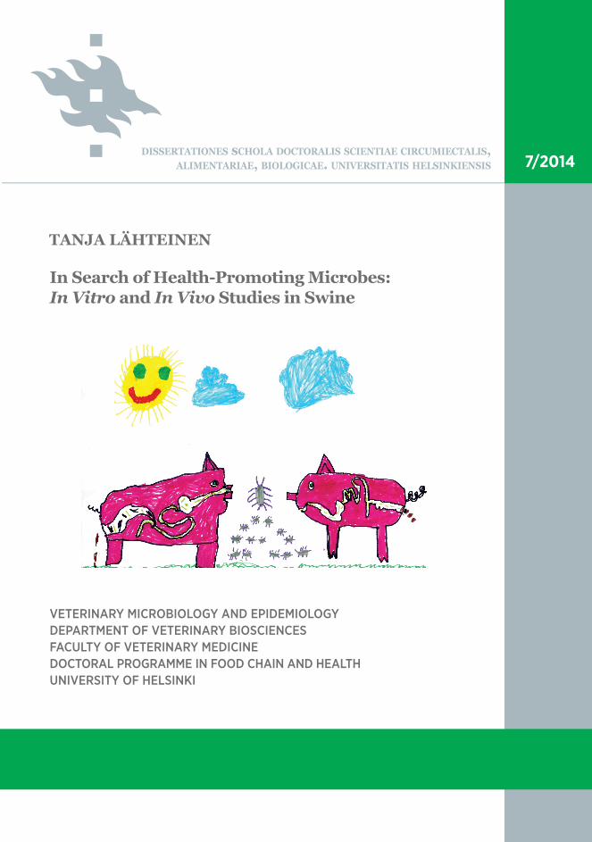
In Search of Health-Promoting Microbes: In Vitro and In Vivo Studies in Swine VETERINARY MICROBIOLOGY AND EPIDEMIOLOGY DEPARTMENT OF VETERINARY BIOSCIENCES FACULTY OF VETERINARY MEDICINE DOCTORAL PROGRAMME IN FOOD CHAIN AND HEALTH UNIVERSITY OF HELSINKI TANJA LÄHTEINEN DISSERTATIONES sCHOLA DOCTORALIS SCIENTIAE CIRCUMIECTALIS, ALIMENTARIAE, BIOLOGICAE. UNIVERSITATIS HELSINKIENSIS 7/2014
-
Upload
khangminh22 -
Category
Documents
-
view
2 -
download
0
Transcript of In Search of Health-Promoting Microbes: In Vitro and In Vivo ... - Helda
In Search of Health-Promoting Microbes:In Vitro and In Vivo Studies in Swine
VETERINARY MICROBIOLOGY AND EPIDEMIOLOGYDEPARTMENT OF VETERINARY BIOSCIENCESFACULTY OF VETERINARY MEDICINEDOCTORAL PROGRAMME IN FOOD CHAIN AND HEALTHUNIVERSITY OF HELSINKI
TANJA LÄHTEINEN
DISSERTATIONES sCHOLA DOCTORALIS SCIENTIAE CIRCUMIECTALIS, ALIMENTARIAE, BIOLOGICAE. UNIVERSITATIS HELSINKIENSIS 7/2014
7/2014Helsinki 2014 ISSN 2342-5423 ISBN 978-951-51-0336-9
Recent Publications in this Series
1/2014 Yanping TianStrains of Potato Virus Y and Their Molecular Signatures Recognized By Resistance Genes and Monoclonal Antibodies2/2014 Emad JaberPathobiology of Heterobasidion-Conifer Tree Interaction: Molecular Analysis of Antimicrobial Peptide Genes (Sp-AMPs)3/2014 Douwe HoornstraTracking Cereulide Producing Bacillus cereus in Foods, Papermaking and Biowaste Management4/2014 Jouni KvistFunctional Genomics of the Glanville Fritillary Butterfl y5/2014 Liwei LiuNew Bioactive Secondary Metabolites from Cyanobacteria6/2014 Christina LiedertAdhesion and Survival Tools of the Bacterium Deinococcus geothermalis
YEB
TA
NJA
LÄ
HT
EIN
EN
In Search
of Health
-Prom
oting M
icrobes: In V
itro and In
Vivo Stu
dies in Sw
ine
cover.indd 1cover.indd 1 15.10.2014 10:40:1415.10.2014 10:40:14

Department of Veterinary BiosciencesFaculty of Veterinary Medicine
University of HelsinkiFinland
IN SEARCH OF HEALTH-PROMOTING MICROBES IN VITRO AND I N VIVO STUDIES IN SWINE
Tanja Lähteinen
ACADEMIC DISSERTATION
To be presented, with the permission of the Faculty of Veterinary Medicine of the University of Helsinki, for public examination in Walter auditorium, EE-
building, Agnes Sjöberginkatu 2, Helsinki, on November 14th 2014, at 12 noon.
Helsinki 2014

Supervisors: Professor Airi Palva Department of Veterinary Biosciences University of Helsinki, Finland
Professor Johanna Björkroth Department of Food Hygiene and Environmental Health University of Helsinki, Finland
University lecturer Joanna Koort Department of Veterinary Biosciences University of Helsinki, Finland
Reviewers: Professor Tapani Alatossava Department of Food and Environmental Sciences University of Helsinki, Finland
Docent Maria Saarela VTT Technical Research Centre of Finland Espoo, Finland
Opponent: Professor Atte von Wright Institute of Public Health and Clinical Nutrition Department of Clinical Nutrition University of Eastern Finland, Finland
Published in Dissertationes Schola Doctoralis Scientiae Circumiectalis, Alimentarie, Biologicae
ISBN 978-951-51-0336-9 (paperback)ISSN 2342-5423 (print)
ISBN 978-951-51-0337-6 (PDF)ISSN 2342-5431 (Online) http://ethesis.helsinki.fi
Cover illustration: Veera & Teemu Lähteinen
Layout: Tinde Päivärinta/PSWFolders Oy
Hansaprint Helsinki 2014

To all the bacteria that got stressed-out,
damaged or even consumed during the course of this work.

Contents
AbstractList of original publicationsAbbreviations1 Introduction ......................................................................................................................................1
2 Literature review ..............................................................................................................................3 2.1 Th e gastrointestinal tract of swine ..........................................................................................3 2.2 Th e genus Lactobacillus............................................................................................................4 2.2.1 Overview .......................................................................................................................4 2.2.2 Lactobacilli in the gut microbiota of swine ...............................................................4 2.2.3 Lactobacilli as animal probiotics ................................................................................5 2.2.4 Lactobacilli as vaccine vectors ....................................................................................7 2.2.5 Surface layer proteins of lactobacilli ..........................................................................7 2.3 Discovering probiotic microbes .............................................................................................9 2.3.1 How to choose the best strains? ..................................................................................9 2.3.2 In vitro studies .............................................................................................................10 2.3.2.1 Tolerance of low pH and bile ........................................................................10 2.3.2.2 Adhesion .........................................................................................................11 2.3.2.3 Pathogen inhibition .......................................................................................13 2.3.2.4 Immunological eff ects ...................................................................................15 2.3.2.5 Antibiotic resistance ......................................................................................16 2.3.3 In vivo studies ..............................................................................................................17 2.3.3.1 Eff ects on productivity parameters ..............................................................23 2.3.3.2 Eff ects on gut microbiota ..............................................................................24 2.3.3.3 Eff ects on host immune function .................................................................25 2.3.3.4 Eff ects on disease occurrence .......................................................................26 2.3.4 Correlation of in vitro and in vivo studies ...............................................................28
3 Aims of the study ..........................................................................................................................30
4 Materials and methods .................................................................................................................31 4.1 Bacterial strains and culture conditions (I-IV) ..................................................................31 4.2 Identifi cation of lactic acid bacteria (I, III) .........................................................................31 4.3 In vitro assays for examining probiotic properties .............................................................35 4.3.1 Acid and bile tolerance (I) .........................................................................................35 4.3.2 Bacterial adhesion (I, IV) ..........................................................................................35 4.3.3 Inhibition of pathogen adhesion (IV)......................................................................36 4.3.4 Antimicrobial activity (I, III, IV) .............................................................................37 4.3.5 Stimulation of cytokine production by dendritic cells (IV) .................................37 4.3.6 Antimicrobial susceptibility testing (III) .................................................................38

4.4 In vivo feeding trial (II, III) ...................................................................................................38 4.4.1 Trial design and sample collection (II, III) .............................................................38 4.4.2 Detection of the supplemented strains in intestinal and fecal samples (II, III) .39 4.4.3 Quantifi cation of total bacteria and the genus Lactobacillus from digesta and fecal samples (II, III) .............................................................................40 4.4.4 Detection of serum immunoglobulins by ELISA (II) ............................................41 4.4.5 Cytokine gene expression in the intestinal mucosa (II, III)..................................41 4.5 Th e surface layer proteins of Lactobacillus amylovorus strains (IV) ................................42 4.5.1 Genes encoding the S-layer proteins: Detection and expression analysis (IV) ..42 4.5.2 Adherence of the S-layer protein-coated cell wall fragments (IV) ......................42 4.6 Statistical analyses (I-III) .......................................................................................................43
5 Results and discussion ..................................................................................................................44 5.1 Isolation and identifi cation of lactic acid bacteria (I, III) .................................................44 5.2 In vitro probiotic properties ..................................................................................................44 5.2.1 Acid and bile tolerance (I) .........................................................................................44 5.2.2 Adhesion (I, IV) ..........................................................................................................45 5.2.3 Inhibition of pathogen adhesion (IV)......................................................................46 5.2.4 Antimicrobial activity against intestinal pathogens (I, IV) ..................................47 5.2.5 Correlations between the properties tested (I) .......................................................52 5.2.6 Stimulation of cytokine production by dendritic cells (IV) .................................52 5.2.7 Antibiotic susceptibility (III) ....................................................................................53 5.3 In vivo feeding trial (II, III) ...................................................................................................54 5.3.1 Animal performance and growth (II, III) ...............................................................54 5.3.2 Detection of the supplemented strains in the intestine and feces (II, III) ..........55 5.3.3 Eff ects of the supplementations on the bacterial and lactobacillar counts (II, III) ..........................................................................................................................57 5.3.4 Immunological eff ects of the supplementations (II, III) .......................................58 5.4 Th e surface layer proteins of Lactobacillus amylovorus strains (IV) ................................59 5.4.1 Genomic characterization and expression analysis of L. amylovorus S-layer proteins (IV)...................................................................................................59 5.4.2 Th e role of L. amylovorus S-layer proteins in adherence (IV) ..............................60
6 Conclusions and future aspects ..................................................................................................62
7 Acknowledgements ........................................................................................................................64
8 References .......................................................................................................................................65

Abstract
Lactobacilli are important members of the commensal microbiota of both man and animals, contributing to the health and well-being of the host. Several Lactobacillus strains are known to possess health-promoting characteristics, warranting their recognition as probiotics, defi ned by WHO as “live microorganisms which, when administered in adequate amounts, confer a health benefi t on the host”. Dietary supplementation with lactobacilli to enhance the health and productivity of the host has generated much interest in the production animal sector, especially aft er the prohibition of in-feed antimicrobials. In the swine rearing industry, disturbances in gastro-intestinal (GI) health, such as diarrhea, are very common, causing signifi cant fi nancial losses and compromising animal welfare. Th e use of lactobacilli as dietary probiotics to maintain and restore a balanced intestinal microbiota during various stressful situations, like the weaning of piglets, might help to prevent the development of GI infections. In addition to their use as probiotics, lactobacilli are also considered as good candidates for antigen carriers in vaccine applications. In particular, those strains carrying a surface (S) layer are attractive vaccine vectors, as the production of the antigenic epitope in each protein subunit as part of the S-layer lattice would enable the expression of a large number of antigens on the surface of the bacterial cell. Th is, in turn, would be expected to enhance the immune response generated by the vaccine. While the use of lactobacilli in a variety of ways to maintain and improve the health and productivity of swine has been widely examined, there are several aspects such as the selection process of the strains used as well as the functional mechanisms behind the observed eff ects that still remain to be clarifi ed. Th us, comprehensive studies regarding the use of lactobacilli as a probiotic and / or vaccine vector in various animal species are warranted.
Th e main aim of this work was to characterize the probiotic potential of lactobacilli for use in swine production, by using both in vitro and vivo methods. A total of 94 lactic acid bacterial (LAB) isolates, originating from porcine small intestine and feces, were fi rst screened for selected properties considered as important for putative probiotic microbes. In general, the isolates tolerated well low pH and bile, and showed highly variable adhesion capacities towards porcine enterocytes collected from fi ve diff erent intestinal sections. While the LAB isolates adhered more effi ciently to large intestinal enterocytes, compared to those collected from the small intestine, the isolation site of the strain had no infl uence on the adhesion preferences of the strains towards enterocytes of diff erent origins. Th e spent culture fi ltrates collected from the isolates inhibited the growth of several intestinal pathogens. While this inhibition was mainly due to organic acids, some of the isolates appeared to produce also other inhibitory substances. Th e predominating phylotypes identifi ed among the isolates were Lactobacillus reuteri and Lactobacillus salivarius, of which the former generally had the best adhesion capacity, whereas the latter was one of the best inhibitors of pathogen growth. However, the properties assessed showed extensive variability, even between strains of the same species.
With respect to the porcine lactobacilli evaluated in the fi rst part of this work, six strains were selected for use in a multispecies bacterial supplementation, which was assessed in a feeding trial performed in recently weaned piglets. Additionally, a Lactobacillus strain possessing a surface (S) layer, namely Lactobacillus brevis ATCC 8287, was used in the feeding trial as a monostrain supplementation. While both supplementations did induce some alterations on the mRNA levels of selected cytokines in the intestinal mucosa, more pronounced eff ects were evident with the multispecies supplementation. Th e L. brevis supplementation induced a non-signifi cant increase

in piglet body weight, but no such eff ect was observed for the multispecies supplementation. None of the supplemented strains could be isolated alive from feces, although the L. brevis strain was detected in the large intestinal digesta as well as in the mucosa of small and large intestines using techniques unable to diff erentiate between dead and live cells. Based on these results, it seems that the ability of these strains to survive and colonize within the porcine gut appears to be limited, although both types of supplementations exerted some immunomodulatory eff ects in the intestinal mucosa. While these supplementations may be suitable for use as probiotics in swine, additional studies will be needed to explore the eff ects of the strains on piglet health and immune status in more detail. Additionally, the suitability of the L. brevis strain for use as a vaccine vector will need to be further assessed.
In the fi nal part of this work, S-layer protein-carrying Lactobacillus amylovorus strains of swine origin, as well as the type strain (DSM 20531T) were characterized for certain probiotic properties. Additionally, the role of the S-layer proteins of each strain in adherence to IPEC-1 cells was addressed. While none of the L. amylovorus strains adhered to porcine intestinal mucus, more variability was observed in their adherence to IPEC-1 cells, with some strains demonstrating a good adhesion capacity to these cells. Interestingly, the adhesion effi ciency of the strains to IPEC-1 cells did not strictly correlate with their ability to inhibit adhesion of an Escherichia coli strain to the same cells. Th us, apart from competition for binding sites, other mechanisms are also involved in the ability of lactobacilli to inhibit pathogen adhesion. Th e extent of cytokine induction by the strains in human MoDC was of varying intensity, and did not clearly deviate towards Th 1 or Th 2 phenotypes. Instead, the induced cytokine response included Th 1 favoring (IL-12), Th 2 favoring (IL-10) as well as proinfl ammatory (TNF-α, IL-6, IL-1β, IP-10/CXCL10) cytokines. In all of the strains, one major S-layer protein (named SlpA) was recognized, and these proteins were found to share a high sequence similarity with the L. acidophilus NCFM SlpA protein. In addition, two of the strains carried additional S-layer like proteins on their surfaces (named SlpB and SlpC). Unexpectedly, none of the major S-layer proteins was found to solely mediate adhesion of the strains to IPEC-1 cells.

List of original publications
Th is thesis is based on the following original publications referred to in the text by their Roman numerals (I-IV):
I Lähteinen T, Malinen E, Koort JMK, Mertaniemi-Hannus U, Hankimo T, Karikoski N, Pakkanen S, Laine H, Sillanpää H, Söderholm H and Palva A. (2010) Probiotic properties of Lactobacillus isolates originating from porcine intestine and feces. Anaerobe 16(3):293-300.
II Lähteinen T, Lindholm A, Rinttilä T, Junnikkala S, Kant R, Pietilä TE, Levonen K, von Ossowski I, Solano-Aguilar G, Jakava-Viljanen M and Palva A. (2014) Eff ect of Lactobacillus brevis ATCC 8287 as a feeding supplement on the performance and immune function of piglets. Vet. Immunol. Immunopathol. 158(1-2):14-25
III Lähteinen T, Rinttilä T, Koort JMK, Kant R, Levonen K, Jakava-Viljanen M, Björkroth J and Palva A. Eff ect of a multispecies Lactobacillus formulation as a feeding supplement on the performance and immune function of piglets. (Submitted)
IV Hynönen U, Kant R, Lähteinen T, Pietilä TE, Beganović J, Smidt H, Uroić K, Åvall-Jääskeläinen S, Palva A. (2014) Functional characterization of probiotic surface layer protein-carrying Lactobacillus amylovorus strains. BMC Microbiol. 14(1):199 doi: 10.1186/1471-2180-14-199
Th e original publications are reprinted with the kind permission of the publishers. In addition, some unpublished results are presented.

Abbreviations
ADWG Average daily weight gainATCC American Type Culture CollectionAUC Area under the growth curveARP Area reduction percentageBMDC Bone marrow derived dendritic cellBSA Bovine serum albuminCD Cluster of diff erentiationcDNA Complementary deoxyribonucleic acidCFU Colony forming unitsCXCL (C-X-C motif) ligandCTLA Cytotoxic T-lymphocyte antigenCWF Cell wall fragmentDAPI 4’, 6-diaminido-2-phenylindole dilactateDC Dendritic cellDC-SIGN Dendritic cell specifi c C-type lectin intercellular adhesion molecule 3-grabbing non-integrin DFI Daily feed intakeDMEM Dulbecco’s modifi ed eagle mediumDMSO Dimethyl sulphoxideDNA Deoxyribonucleic acidDSM Deutsche Sammlung von MikroorganismenECM Extracellular matrixEFSA European Food Safety AuthorityEHEC Enterohemorrhagic Escherichia coliELISA Enzyme-linked-immunosorbent-assayETEC Enterotoxigenic Escherichia coliFAO Food and Agriculture Organization of the United NationsFCR Feed conversion ratioFCS Fetal calf serumFEEDAP Panel on Additives and Products or Substances used in Animal FeedFISH Fluorescence in situ hybridisationGI GastrointestinalGM-CSF Granulocyte macrophage-colony stimulating factor HBSS Hanks balanced salt solutionHCl Hydrochloric acidHEPES 4-(2-hydroxyethyl)-1-piperazineethanesulfonic acidHPRT1 Hypoxanthine phosphoribosyltransferase 1HRP Horse radish peroxidaseIEC Intestinal epithelial cellIFA Indirect immunofl uorescence assayIFN InterferonIg ImmunoglobulinIL InterleukinIL-12-RB2 Interleukin 12 receptor beta 2 subunit

IP Interferon gamma-induced proteinISR 16S-23S intergenic spacer regionkDa KilodaltonLAB Lactic acid bacteriaLB Luria Bertani mediumMIC Minimum inhibitory concentration MoDC Monocyte derived dendritic cellMOI Multiplicity of infectionMPN Most probable numbermRNA Messenger ribonucleic acidMRS de Man Rogosa Sharpe mediumMyD-88 Myeloid diff erentiation primary response gene 88NaOH Sodium hydroxideNF- B Nuclear factor BOD Optical densityPAGE Polyacrylamide gel electrophoresispBD Porcine beta-defencin PBMC Peripheral blood mononuclear cellPBS Phosphate buff ered salinePCR Polymerase chain reactionPFGE Pulsed-fi eld gel electrophoresispI Isoelectric pointPPIB Peptidyl-prolyl cis-trans isomerase BqPCR Quantitative polymerase chain reactionQPS Qualifi ed presumption of safetyRNA Ribonucleic acidRPL32 Ribosomal protein L32rpm Revolutions per minuterRNA ribosomal ribonucleic acidSDS Sodium dodecyl sulphateS-layer Surface layerSCF Spent culture fi ltrateSlp Surface layer proteinTh T helper cellTreg Regulaory T cellTEER Transepithelial electric resistanceTGF Transforming growth factorTLR Toll-like receptorTNF Tumor necrosis factorTSA Tryptic soy agarTSB Tryptic soy brothUPGMA Unweighted pair group method with arithmetic averages WGS Whole genome sequencingWHO World Health Organization

1
1 Introduc on
Th e main aim of livestock farming is to produce safe foodstuff s for human consumption. While the importance of animal health and welfare is being increasingly appreciated in animal production, the intensive management practices of modern animal farming oft en subject the animals to severe stress, leading to health problems and reduced productivity. For example, diarrhea and other disturbances in gastro-intestinal (GI) health are very common in the swine rearing industry, where they are responsible for signifi cant fi nancial losses and compromised animal welfare (Reid and Friendship, 2002). In addition to direct health problems to the host animal, pathogenic bacteria can also be transferred along the food chain, thus endangering also human health. In an attempt to manage the problems caused by GI and other infections, sub-therapeutic levels of antibiotics have long been incorporated into animal feed (Dibner and Richards, 2005); this both eff ectively promotes the growth of the animals and reduces animal mortality and morbidity (Cromwell, 2002). However, the worldwide concern about the growing problem of antibiotic resistance and about the transfer of resistance genes from animal to human microbiota (Aarestrup, 2002) has led to a gradual discontinuation of growth-promoting antibiotics throughout Europe. For example, Sweden prohibited the use of antibiotic feed additives in 1986 (Wierup, 2001), Denmark in 2000 (Aarestrup et al., 2010), and from the beginning of 2006, this prohibition has been implemented throughout the European Union. Aft er the withdrawal of these antibiotics, increases in mortality and morbidity, especially due to enteric infections, have been reported in pig farms (Callesen, 2002; Casewell et al., 2003). Additionally, the use of antibiotics as therapeutic agents has increased (Callesen, 2002; Casewell et al., 2003). Although these negative consequences on the pig production may be only transient (Aarestrup et al., 2010), it is clear that alternative ways are urgently needed to promote the health of production animals and to reduce the increasing use of therapeutic antibiotics.
Various dietary strategies have been claimed to improve gut health and disease resistance of production animals, including swine (Roselli et al., 2005; Th acker, 2013). One such approach involves the supplementation of animal feeds with benefi cial microbes. Th e concept that harmful gut microbes could be suppressed and displaced by benefi cial ones to improve the health of the host fi rst appeared over a century ago. Since then, the concept of benefi cial microbes, which are oft en called “probiotics”, has evolved considerably, as have the defi nition for these microbes (Isolauri et al., 2002; Dobrogosz et al., 2010). According to the World Health Organization, probiotics are defi ned as “live microorganisms which, when administered in adequate amounts, confer a health benefi t on the host” (WHO/FAO, 2001).
One of the health benefi ts associated with probiotic consumption is modifi cation of the gastro-intestinal (GI) microbiota in such a way that there is increased resistance to colonization by pathogenic bacteria (McCracken and Gaskins, 1999; Lalles et al., 2007; Ohashi and Ushida, 2009). Th e commensal GI tract microbiota is a highly complex community, consisting of an immense number of diff erent species of microbes. In humans, the gut microbiota has generally been estimated to consist of around 500-1000 diff erent microbial species (Yatsunenko et al., 2012), and estimations of the species diversity of swine intestinal microbiota have been in the same range (Leser et al., 2002; Lamendella et al., 2011). Th e gut microbiota is of crucial importance to the host, for example providing protection against extrinsic microbes. Th is is emphasized by the fact that germfree animals, devoid of commensal microbiota, are much more susceptible to infections, especially to gastro-intestinal infections. For example, the lethal dose of Salmonella enteritidis in a germfree mouse is as low as ten bacteria delivered per os, whereas
Introduction

2
in conventional mice 109 bacteria are required to evoke a lethal infection (Collins and Carter, 1978). Th e colonization resistance provided by the commensal intestinal microbiota is illustrated also by studies conducted with newly hatched chickens. In commercial poultry settings, the development of the commensal gut microbiota during the fi rst weeks of life is delayed, e.g. rendering the hatchlings highly vulnerable to Salmonella infections (Schneitz, 2005). However, providing the gut contents of an adult bird to the chicks has been shown to be a very eff ective way to protect the young birds against Salmonella, and other intestinal infections (Nurmi and Rantala, 1973; Schneitz, 2005; Dobrogosz et al., 2010), and this phenomenon is referred to as “competitive exclusion” (CE). In other production animals, the development of the commensal microbiota aft er birth is not as prone to disturbances as it is in poultry, and thus the practice of CE is not as widespread elsewhere. However, probiotic products containing defi ned bacterial strains and aimed at increasing the disease resistance of the animals are being used also in several other species, including swine (Bernardeau et al., 2006; Gaggia et al., 2010).
Several diff erent types of microbes, i.e. bacteria, yeasts and molds, have been evaluated as animal probiotics, but the genus Lactobacillus has been one of the most widely applied genera (Nousiainen et al., 2004; Gaggia et al., 2010). Lactobacilli are prominent members of the commensal intestinal microbiota of both humans and animals, and are considered to be benefi cial for the host. In piglets, a reduction in the abundance of intestinal lactobacilli has been observed to occur around weaning (Konstantinov et al., 2006a; Su et al., 2008b) and this decline has been considered to predispose the piglets to GI disturbances, e.g. diarrhea. Th e restoration of a balanced gut microbiota aft er the application of probiotic microbes could help to prevent the development of GI infections around weaning. Indeed, positive health eff ects including reductions in diarrhea prevalence have been described in swine aft er consumption of lactobacilli and other probiotics, although positive eff ects have not been detected in all studies that have been performed (Nousiainen et al., 2004; Lalles et al., 2007; Bosi and Trevisi, 2010; Kenny et al., 2011). Th e functional mechanisms behind the putative health-promoting eff ects of probiotic lactobacilli are for the most part unclear. Consequently, research aimed at revealing the putative positive health eff ects of probiotic consumption as well as the molecular mechanisms leading to these outcomes is very important and can be used to guide the selection of bacterial strains used in probiotic feed additives intended not only for swine but also for other animals.
Introduction

3
2 Literature review2.1 The gastrointes nal tract of swineTh e digestive tract of swine is classifi ed as monogastric and shares many anatomical and physiological similarities with the human GI-tract (Miller and Ullrey, 1987; Heinritz et al., 2013). Anatomically the porcine GI-tract can be divided into four parts: the esophagus, the stomach, the small intestine (which is further divided into the duodenum, the jejunum and the ileum) as well as the large intestine (which is further divided into the cecum, the colon and the rectum; Figure 1). At the time of birth, the lengths of the porcine small and large intestine are around four and one meters, respectively, while at maturity the corresponding measures lie in the range from 18 to 23 meters and four to seven meters (McCance, 1974; Miller and Ullrey, 1987).
Similarly to humans, pigs are omnivorous colon fermenters. However, while humans lack a distinct cecum, pigs exhibit also signifi cant cecal fermentation. Th is diff erence is illustrated by the fact that the short chain fatty acids (SCFA) produced by the large intestinal microbiota of swine may provide up to 30% of the host energy requirements for maintenance (Rerat et al., 1987), whereas in humans only about 10% of this requirement is provided by SCFA produced in the colon (Bergman, 1990). Th e diet of feral pigs is very diverse, consisting mainly of vegetation like fruits, grasses, forbs, corn, roots and tubers, but also of animal matter including both invertebrates and small vertebrates (Baber and Coblentz, 1987; Taylor, 1999). In commercial pig production, a highly palatable and concentrated feed is provided to maximize growth effi ciency; the main ingredients in swine fodder are diff erent grains like maize and barley and various sources of proteins such as soybean and fi sh meal as well as whey powder.
Like in all mammals, the GI microbiota of swine is a highly complex community consisting mainly of bacteria, but also of archae and eukaryotic microbes as well as viruses. Th e total number of microbial cells in the GI tract is estimated to be around 1014 cells, which is ten times more than the number of cells of the host organism (Luckey, 1972; Savage, 1977). Th e GI microbiota is crucial for the health and normal development of the host; it provides resistance to colonization by harmful microbes, infl uences intestinal structure and physiology and promotes the development of the immune system (reviewed by Shanahan (2002)). In general, the GI-tract microbiota of swine resembles that of humans; e.g. the predominant phyla in both of these host species are Firmicutes and Bacteroidetes (Mahowald et al., 2009; Isaacson and Kim, 2012; Heinritz et al., 2013). However, there are some interesting diff erences between the intestinal microbiota of swine and of humans. For example, LAB and in particular lactobacilli, are among the most abundant phylotypes in the gut microbiota of swine, while in the human intestine, lactobacilli are present at considerably lower levels (Heinritz et al., 2013).
Literature review
Esophagus
Stomach
Duodenum
Jejunum m Ileum IlCecum CC
Rectum
Colon
Figure 1. A schematic representation of the GI-tract of swine

4
2.2 The genus Lactobacillus2.2.1 Overview
Th e genus Lactobacillus is an important group of LAB belonging to the phylum Firmicutes. Members of the genus Lactobacillus are Gram-positive rod shaped bacteria that obtain the energy they need for the maintenance and macromolecule synthesis from substrate-level phosphorylation (i.e. fermentation), forming lactic acid as the primary metabolic end product. Th is highly heterogeneous genus consists of over 150 species inhabiting diverse ecological niches. Lactobacilli are ubiquitous in natural environments and are present in all places where substrates rich in carbohydrates are available, i.e. in soil, sewage and plant material. Lactobacilli are also essential members of the commensal microbiota of humans and animals, e.g. they can be found in the GI tract and are present in feces of several diff erent host species. For centuries, lactobacilli have been used as starter cultures in the production of fermented foods, particularly in dairy products, but also in vegetable and meat products. In addition to foods intended for human consumption, Lactobacillus fermentation can also be used to produce animal feeds, such as silage (Holzer et al., 2003; Meieregger et al., 2011). Furthermore, as lactobacilli have been associated with various health-promoting properties, they are widely used as probiotics for both humans and animals (Barrangou et al., 2012).
Two basic fermentative pathways are utilized by lactobacilli. Th e homofermentative pathway (Embden-Meyerhof-Parnas pathway) produces almost exclusively lactic acid as the end product, whereas in the heterofermentative pathway (phosphoketolase pathway) also CO2 and ethanol are produced. Based on these pathways, lactobacilli can be divided into three distinct groups; (1) obligatory homofermentative species, (2) facultative heterofermentative species, which can use both of the pathways, and (3) obligatory heterofermentative species (Hammes and Hertel, 2009).
Th e various environmental habitats of lactobacilli are refl ected in the high genomic diversity of the genus. Th e size of the Lactobacillus genome ranges usually from 1.8 to 3.3 Mbp (Canchaya et al., 2006; Kant et al., 2011a; Lukjancenko et al., 2012), the smallest reported so far being that of Lactobacillus iners which is only 1.3 Mbp (Macklaim et al., 2011). Th e G+C content of the Lactobacillus genomes is typically low (Barrangou et al., 2012) but also diverse, ranging from 32% to 55% (Axelsson, 2004). Th e taxonomy of lactobacilli is known to be complex and constantly changing; the information obtained from new studies and the recent availability of whole genome sequences (WGS) have enabled more conclusive analyses of the phylogenetic relationships of lactobacilli species (Makarova et al., 2006; Claesson et al., 2008; Kant et al., 2011a). It is evident that the phylogeny of lactobacilli species does not correlate well with their phenotypes (Felis and Dellaglio, 2007), and further revisions of the taxonomy have been proposed (Axelsson, 2004; Makarova et al., 2006).
2.2.2 Lactobacilli in the gut microbiota of swine
At birth, the sterile environment of the womb changes to a situation in which there is a constant microbial exposure, leading to colonization of the newborn animal. Th e establishment of the host microbiota proceeds sequentially, as a succession of microbial populations, until a relatively stable climax community is formed (Savage, 1977; Isaacson and Kim, 2012). In most suckling animals, lactobacilli are one of the fi rst colonizers of the gut (Savage, 1977; Berg, 1996), and this has been confi rmed in swine as well (Smith, 1965; Fuller et al., 1978; Ducluzeau, 1983; Konstantinov et al., 2006a). Th e colonizing bacteria, including lactobacilli, originate from the rearing environment, like maternal feces (Tannock et al., 1990; Nousiainen et al., 2004). Th e
Literature review

5
microbial succession of diff erent intestinal compartments and feces of piglets has been examined in several studies (Kenworthy and Crabb, 1963; Tannock et al., 1990; Swords et al., 1993; Naito et al., 1995; Melin et al., 1997; Konstantinov et al., 2004b; Konstantinov et al., 2006a), although the experiments have been generally conducted using culture-based methods, which are unable to detect the majority of gut microbes (Vaughan et al., 2000). Substantial individuality in the colonization process of the piglet intestine has been described during the fi rst two weeks of life, this being refl ected by diff erences in the gut microbiota of littermates and penmates (Th ompson et al., 2008). In addition, the developing microbiota of piglets is infl uenced also by management practices (Davis, 2012).
During the suckling period, the numbers of lactobacilli in the ileum of piglets seem to remain rather constant (Konstantinov et al., 2006a), but marked changes in both the abundance and community structure of lactobacilli occur at weaning. Weaning, which occurs at approximately three to four weeks of age in commercial swine production units, exposes the piglets to severe social, environmental and nutritional stresses. Th e weaning transition causes a signifi cant decrease in the overall GI tract Lactobacillus population (Franklin et al., 2002; Konstantinov et al., 2006a; Pieper et al., 2006; Su et al., 2008a), and changes in the diversity and structure of lactobacilli community have also been described (Bateup et al., 1998; Janczyk et al., 2007; Su et al., 2008a). However, at least some of these changes are transient (Janczyk et al., 2007).
Investigations of the GI tract microbial composition of post-weaning swine conducted with high-throughput molecular techniques have revealed lactobacilli as being among of the most abundant phylotypes recognized in the intestine (Pryde et al., 1999; Leser et al., 2002; Hill et al., 2005) and feces (Kim et al., 2011). Th e cell count of lactobacilli in the porcine intestine, based on bacterial culture and fl uorescent in situ hybridization (FISH), is at the level of 108 CFU per gram of digesta (Konstantinov et al., 2004a; Castillo et al., 2006). Th e predominant Lactobacillus species in the swine intestine include e.g. L. amylovorus, L. reuteri, L. johnsonii, L. mucosae and L. salivarius (Leser et al., 2002; Hill et al., 2005; Konstantinov et al., 2006a; Mann et al., 2014). Although the GI tract microbiota of adult swine is relatively stable over time, dietary and environmental factors can induce changes in the composition of the microbiota, including the lactobacilli community (Fuller, 1989; Bauer et al., 2006; Rist et al., 2013).
2.2.3 Lactobacilli as animal probio cs
Th e lactobacilli in the GI tract have long been considered as being benefi cial for the host (Tannock, 1990), and several species of lactobacilli are known to possess properties considered important for probiotic microbes (Reid, 1999). Consequently, the oral supplementation of these bacteria is hypothesized to improve the gut health of the host (Fuller, 1989; Meieregger et al., 2011), and the use of lactobacilli as probiotics for both man and animals has been extensively studied in the last decades (Simon et al., 2001; Nousiainen et al., 2004; Bernardeau et al., 2006; de Vrese and Schrezenmeir, 2008; Turpin et al., 2010). Although the use of microbial feed supplements for farm animals was explored as early as 1925, this practice was not exploited commercially until the 1960s and 1970s, coinciding with increased concerns over the widespread and uncontrolled use of antibiotic growth promoters (Fuller, 1999; Reid and Friendship, 2002; Meieregger et al., 2011). In swine, L. acidophilus was one of the fi rst microbes reported to stimulate the growth when supplemented into the feed (King, 1968). Currently, around 20 diff erent species of microbes are authorized in the EU as feed additives for animal nutrition, including several Lactobacillus species, such as L. brevis, L. plantarum, L. rhamnosus and L. salivarius (EU, 2014).
Literature review

6
Several health eff ects have been proposed for benefi cial microbes, but as the goals of probiotic consumption depend on the target host species, some of these are not relevant in the veterinary fi eld, or at least not in production animals (Table 1). On the other hand, some eff ects desirable in production animals may be irrelevant or even unwanted in humans. Th ese include e.g. increased body weight gain and improvements in feed effi ciency (Simon et al., 2001; Bernardeau and Vernoux, 2013). In addition to benefi cial eff ects for the host animal itself, one further aim of using probiotic microbes in production animals would be to reduce the carriage of microbes which are not harmful to the hosts themselves, but can lead to human infection if they pass in to the food chain (Reid and Friendship, 2002; Doyle and Erickson, 2012).
Th e mechanisms behind the health enhancing eff ects of probiotics are largely unknown, but several modes of action have been proposed (reviewed by Fooks and Gibson (2002); Vanderpool et al. (2008); Ohashi and Ushida (2009) and Kenny et al. (2011)). Th ese include e.g. 1) eff ects on intestinal microbiota, such as stimulation of indigenous lactobacilli or other benefi cial bacteria, 2) inhibition of harmful microbes via competition for nutrients or receptors for adhesion, or via production of antagonistic substances, 3) stimulation and / or modulation of the host immune function, 4) regulation of enterocyte functions, like the maintenance of the epithelial barrier 5) stimulation and / or modulation of intestinal nutrition physiology, like absorption and secretion activity, or enhancement of short-chained fatty acid (SCFA) production.
Table 1. Proposed health eff ects of probiotics and their relevance in the veterinary fi eld, as estimated by the author.
Proposed eff ect (adapted from de Vrese and Schrezenmeir (2008) and Chassard et al. (2011)
Relevance in veterinary medicine
Very well-established eff ects with valid scientifi c proofPrevention and alleviation of certain types of diarrhea (e.g. rotavirus, antibiotic-associated, traveler’s)
High
Alleviation of lactose intolerance LowWell-established eff ects / eff ects observed in certain target groupsModulation of the microbiota (usually intestinal) HighImmunomodulation / - regulation HighPrevention of respiratory tract infections HighBenefi cial eff ects in infl ammatory diseases of the GI tract (e.g. infl ammatory bowel disease, bacterial overgrowth)
Intermediate (mainly comp. animals1)
Prevention and alleviation of allergies / atopic diseases in infants Intermediate (mainly comp. animals)
Treatment of urogenital infections Intermediate (mainly comp. animals)
Eff ects not well-established with insuffi cient scientifi c proofNormalization of passing stool and stool consistency (e.g. constipation) Intermediate (mainly
comp. animals)Alleviation of autoimmune diseases Intermediate (mainly
comp. animals)Prevention of cancer Intermediate (mainly
comp. animals)Prevention of ischemic heart disease LowReducing of blood cholesterol LowCaries prevention Intermediate (mainly
comp. animals)1 companion animals
Literature review

7
2.2.4 Lactobacilli as vaccine vectors
In addition to the use of lactobacilli as probiotics, interest in their use as delivery vectors for vaccine antigens has increased during the last decades (Bermudez-Humaran et al., 2011; Wells, 2011a). Many of the properties considered important for probiotics (see section 2.2.1.) also apply to the use of bacteria as potential vaccine delivery vectors, thus several Lactobacillus species are good candidates for both applications (Mercenier, 1999).
One important advantage of using live bacterial vaccine vectors would be the ability to administer these vaccines mucosally, e.g. per os. Mucosal administration is expected to generate improved and more appropriate local immune response than the traditional parenteral route, giving better protection against infectious agents that mostly enter the body mucosally (Mercenier, 1999; Fujkuyama et al., 2012). In addition, mucosal administration is more convenient and cheaper than the parenteral route (Mercenier, 1999; Holmgren et al., 2003). Traditionally, certain pathogenic bacteria, e.g. Salmonella and Listeria, have been extensively investigated for use as vaccine vectors (Detmer and Glenting, 2006; Mohamadzadeh et al., 2008), but the comparatively lower intrinsic immunogenicity of lactobacilli compared to attenuated pathogens is considered favorable, since it may result in fewer side-eff ects in the vaccinated host (Pouwels et al., 1998; Mercenier, 1999; Wells and Mercenier, 2008). At the same time, however, lactobacilli are able to stimulate and / or modulate host immune responses, as demonstrated by several studies performed in both humans and animals (reviewed by Corthesy et al. (2007) and Wells (2011b)). Th is adjuvant-like property has been demonstrated during infections, e.g. as increased pathogen-specifi c Ig-titers (Link-Amster et al., 1994; Kaila et al., 1995; Vlasova et al., 2013), and also as enhancement or modulation of the immune response stimulated by vaccination (Licciardi and Tang, 2011; Maidens et al., 2013). However, studies performed on pigs are extremely scarce; one study examining the eff ect of L. rhamnosus supplementation on porcine reproductive and respiratory syndrome (PRRS) vaccination could not detect any impact on the resulting immune response (Kritas and Morrison, 2007).
Th e Lactobacillus species mostly explored as vaccine vectors include L. plantarum, L. casei, L. helveticus and L. acidophilus (Wells, 2011a). Various antigens have been successfully expressed in lactobacilli (Mohamadzadeh et al., 2008; Wells, 2011a), including also those originating from pathogens infecting swine, e.g. transmissible gastroenteritis virus (Ho et al., 2005), classical swine fever virus and porcine parvovirus (Xu et al., 2011). In addition, lactobacilli have been investigated as carriers for DNA vaccines (Li et al., 2007). Most of the immunization studies performed with recombinant lactobacilli have been conducted in mouse models (Mohamadzadeh et al., 2008; Wells, 2011a), but promising results have been obtained also in swine (Xu et al., 2011).
2.2.5 Surface layer proteins of lactobacilli
Surface (S) layers are cell envelope structures found on the outermost surface of many bacteria and most archaea, completely covering the cells (Sleytr et al., 2014). In addition, several Lactobacillus species including e.g. L. amylovorus, L. crispatus, L. acidophilus, L. brevis and L. helveticus are known to possess an S-layer (Hynönen and Palva, 2013). Th ese crystalline bidimensional arrays are composed of identical protein or glycoprotein subunits assembled to form a regular porous structure, which can be aligned in square, oblique or hexagonal symmetry, although only the last two of these possible arrangements have so far been observed in S-layers of lactobacilli (Åvall-Jääskelainen and Palva, 2005). Th e S-layer proteins of lactobacilli are among the smallest known, with molecular weights ranging from 25 to 71 kDa (Hynönen and Palva, 2013), while in other
Literature review
8
bacterial species the size of these proteins is much larger, up to 200 kDa (Sara and Sleytr, 2000). Th e thickness of the S-layer is generally 5-20 nm, and the size of the pores, which occupy 30% to 70% of the S-layer surface, is in the range of approximately 2-8 nm (Sleytr et al., 2014). Th e sequence similarities between S-layer proteins of diff erent bacterial species are generally low (Sleytr et al., 2014), and this is the case also in lactobacilli; homology is found only between genes of related species (Hynönen and Palva, 2013). Despite this, some common features are present in the amino acid composition of S-layer proteins, such as their high content of hydrophobic amino acids as well as the low amount of sulfur containing amino acids (Sleytr et al., 2014). A characteristic which is specifi c to lactobacillar S-layers is the higher abundance of positively charged amino acid residues compared to that of negatively charged residues, leading to high theoretical isoelectric point (pI) values (i.e. 9.35-10.4) (Åvall-Jääskelainen and Palva, 2005).
Th e S-layer subunit proteins attach to each other and to the underlying cell wall structures by non-covalent interactions. It is possible to achieve complete detachment of the S-layer and disintegration into the monomer subunits e.g. with high concentrations of chaotropic agents disrupting the interactions of non-covalent forces (Sleytr et al., 2014). Isolated S-layer protein subunits have a high intrinsic propensity to recrystallize into regular lattices e.g. on solid supports or even in suspension aft er the removal of the disrupting agent, thus they are very poorly water-soluble. Two distinct structural regions have been identifi ed in S-layer proteins; the fi rst being involved in the binding of the S-layer to the cell wall, and the other being responsible for the S-layer assembly (Sleytr et al., 2014). Th e locations of these regions in the S-layer protein vary between diff erent species of lactobacilli (Hynönen and Palva, 2013).
Although no common biological role for S-layers has been identifi ed, various functions have been postulated, including protection from environmental factors, cell shape maintenance, binding or sieving of large molecules and modulation of the host immune responses (Hynönen and Palva, 2013; Sleytr et al., 2014). In addition, mediation of adhesion to host structures has commonly been proposed as a function of lactobacillar S-layers. Attempts to produce completely S-layer negative Lactobacillus mutants have been unsuccessful (Boot et al., 1996a; Martinez et al., 2000; Palva A. unpublished results), emphasizing the necessity of this structure for its host cell. For this reason, protein level methods (e.g. labelled subunits or recombinantly expressed proteins) have to be used when examining the role of S-layers in bacterial adhesion. In some Lactobacillus species, S-layers have been shown to mediate the adhesion to diff erent targets, like extracellular matrix (ECM) proteins and epithelial cells (Mobili et al., 2010; Hynönen and Palva, 2013).
Th e structural properties of the S-layer, as well as the self-assembly tendency of the subunits mean that these structures are attractive candidates for a wide range of applications, including vaccine carriers. Th e ability to produce the antigenic epitope in each S-layer subunit as part of the overall lattice structure would enable bacteria with such chimeric S-layers to display a large number (e.g., ~5×10⁵/bacterium) of antigenic molecules on their cell surface (Sleytr et al., 2007). Small model peptides have already been successfully expressed in each monomeric subunit of the S-layer of L. brevis ATCC 8287 (Åvall-Jääskeläinen et al., 2002) and L. acidophilus ATCC 4356 (Smit et al., 2002). In the future, increasing knowledge about the structure and biology of Lactobacillus S-layers will help in the utilization of these structures in the development of effi cient vaccines for veterinary use.
Literature review

9
2.3 Discovering probio c microbes2.3.1 How to choose the best strains?
Th e vast species and strain diversity of lactobacilli and other potentially benefi cial microbes ensures that there is no shortage of candidate probiotics. However, as it is impossible to test large numbers of diff erent strains in in vivo feeding trials, some kind of preliminary selection is necessary. Th e commonly stated selection criteria for probiotic microbes involve several features related to safety aspects as well as functional and technological properties (Table 2).
Th e fi rst step in the development of a probiotic product is the isolation of the candidate strain(s). Th e origin of the strain is an important factor to be considered, e.g. for human probiotics, strains of human origin are preferred (Collins and Gibson, 1999; Saarela et al., 2000; Ouwehand et al., 2011). It has been claimed that at least some of the functional properties of probiotics, such as adhesion to the intestinal epithelium, are host species specifi c, indicating that strains isolated from the intended target species would show better performance as compared to those isolated from other species (Bengmark, 1998; Saarela et al., 2000). However, the results of in vitro studies assessing the host specifi city of lactobacilli adherence have been confl icting, with both supporting fi ndings (Fuller, 1973; Barrow et al., 1980; Mäyrä-Mäkinen et al., 1983; Nemcova et al., 1997), as well as negative reports (Rinkinen et al., 2000; Nikoskelainen et al., 2001; Rinkinen et al., 2003). Nevertheless, since host species specifi c diversifi cation of lactobacilli strains has been observed (Oh et al., 2010; Frese et al., 2011; Guinane et al., 2011), it appears prudent to prefer strains originally isolated from the target species.
Safety is obviously one of the most important requirements for a possible probiotic (Saarela et al., 2000; von Wright, 2005; Chassard et al., 2011). While lactobacilli have a long history of safe use in food products, they are living micro-organisms, and theoretically could be capable of evoking unwanted side-eff ects, at least in susceptible individuals. As reviewed by Bernardeau et al. (2006) and Sanders et al. (2010), cases of lactobacillar infections, e.g. bacteremia, peritonitis and pneumonia, have been reported in humans, but considering the huge quantities of probiotic products consumed, these infections seem to be extremely rare occurring mainly in immunocompromised patients. On the other hand, as far as the author is aware, there are no reports of animal infections caused by lactobacilli, even though Weissella confusa (basonym Lactobacillus confusus) has been isolated from an otitis sample obtained from a dog (Björkroth et al., 2002) as well as from a case of systemic infection in a mona monkey (Cercopithecus mona) (Vela et al., 2003). With respect to the assessment of safety, an unequivocal taxonomic identifi cation of the strain is an important prerequisite (WHO/FAO, 2002; Vankerckhoven et al., 2008), as a number of bacterial species, including several lactobacilli, have received a qualifi ed presumption of safety (QPS) status in the European Union (EFSA, 2007). Micro-organisms to be used as feed additives are authorized according to the European Parliament and Council Regulation (EC) No 1831/2003, and this protocol requires thorough safety assessment, including toxicity studies performed using the target species (von Wright, 2005; Anadón et al., 2006; Meieregger et al., 2011). However, a full safety assessment is required only for strains without a QPS status (Meieregger et al., 2011; Salminen and von Wright, 2012).
Although non-viable microbes have demonstrated some health enhancing eff ects (Ouwehand and Salminen, 1998), it is generally assumed that viability is important for the functional properties of probiotic microbes (Chassard et al., 2011; Ouwehand et al., 2011). Consequently, the candidate strain has to be able to survive industrial manufacturing conditions,
Literature review

10
as well as to maintain high viable cell numbers in the fi nal product during storage (Saarela et al., 2000; Meieregger et al., 2011). Th is requirement makes the technological properties of the strain (Table 2) extremely important in the selection process, and it has been claimed that the current probiotics have been chosen mostly based on these characteristics (Lacroix and Yildirim, 2007). For the most part, probiotics of intestinal origin are highly sensitive to many environmental stresses, such as the extremes of temperature, oxygen stress and the mechanical shearing forces encountered during industrial processing (Lacroix and Yildirim, 2007; Meieregger et al., 2011), Th ese are factors which pose signifi cant challenges to the process optimization. Moreover, in addition to aff ecting the cell yield and viability of the strain, manufacturing procedures and also the food matrix into which the bacteria are to be incorporated may have an infl uence on the functional properties of the strain (Pessi et al., 1998; Deepika et al., 2009; Grzeskowiak et al., 2011; Deepika et al., 2012), further emphasizing the importance of the optimization of production process.
It is important to recognize that the characteristics of a bacterial strain are specifi c to that particular strain, meaning that even strains belonging to the same species can have divergent properties (Pineiro and Stanton, 2007; Marteau, 2011). Th us, properties known to exist in one strain cannot be extrapolated to all strains of the same species. Consequently, appropriate in vitro and in vivo experiments need to be performed for each candidate strain to guide the selection process, and the fi nal validation of the health benefi ts can only be obtained in carefully controlled clinical trials (Chassard et al., 2011).
Table 2. Commonly used criteria for selection of probiotic microbes. Adapted from Saarela et al., (2000); Chassard et al., (2011); Meieregger et al., (2011)
Safety aspects Functional properties Technological propertiesOrigin (preferably target species)Identifi cation QPS statusAntibiotic resistanceVirulence factorsToxicity (e.g. acute and chronic)
GI tract survival (acid, bile)AdhesionPathogen inhibitionImmuno-stimulation / -modulationMetabolic activitiesAnti-carcinogenesis / -mutagenesis
Easily propagatedMaintains high viabilitySustains production processesStable during storageFavorable / no adverse eff ects on product quality
2.3.2 In vitro studies2.3.2.1 Tolerance of low pH and bile
Probiotics are usually consumed orally, and their expected biological functions occur in the intestine. Aft er ingestion, the probiotic encounters the acidic environment of the stomach; the gastric pH of a suckling piglet can reach values below three, and even lower values, less than two, have been recorded aft er weaning (Moughan et al., 1991; Snoeck et al., 2004). Aft er transit through the stomach, the acid stress is followed by exposure to bile in the duodenum. Apart for their digestive functions, both of these factors are also antimicrobial defense mechanisms of the GI-tract, causing stress to transiting microbes (Dunne et al., 2001; Upadrasta et al., 2011). Exposure to an acidic environment, leading to a reduction in bacterial cytoplasmic pH, can decrease the activity of pH sensitive enzymes and damage the cell membrane and
Literature review

11
macromolecules, such as DNA and proteins (van de Guchte et al., 2002; Cotter and Hill, 2003). Th e antimicrobial eff ect of bile on the other hand is mainly due to the disruption on cell membrane integrity, but it may also infl uence macromolecular stability (Begley et al., 2005).
Th e acid and bile tolerance of lactobacilli isolated from various sources, including swine, have been extensively assessed (e.g. De Angelis et al. (2006); Yun et al. (2009); Guo et al. (2010); Zhang et al. (2013)), and while lactobacilli of intestinal origin usually show high tolerance of low pH and bile (Morelli, 2000), extensive strain-to-strain variability is evident. Th e in vitro experiments used in these tests have been based on exposing the strains to low pH or to bile for a limited time period, aft er which the bacterial population surviving the stress has been estimated, e.g. by plate count or turbidity measurements. Additionally, methods based on growth curve parameters have also been used (Morelli, 2000). Typically, the lowest pH values used in the experiments have been pH 2, and the bile concentration has ranged from 0.1% to 5%, but most oft en below 0.5%. Interestingly, diff erences according to the origin of the bile have been reported, as lactobacilli and bifi dobacteria were inhibited more by porcine bile than by bovine bile (Dunne et al., 2001). Nonetheless, the ability of these kinds of static experiments, using constant pH and / or bile concentration, to accurately predict the in vivo GI-tract survivability of the strains has been criticized (Morelli, 2000), as several factors related to the food matrix, ingestion and digestion have been observed to aff ect strain survival (Conway et al., 1987; Charteris et al., 1998; Upadrasta et al., 2011). Th us, dynamic models possibly capable of simulating the GI-tract conditions more precisely have been developed (Marteau et al., 1997; Mainville et al., 2005; Ceuppens et al., 2012; Van den Abbeele et al., 2012).
2.3.2.2 Adhesion
Th e intestinal motility causes a constant fl ow of the gut luminal contents, fl ushing the microbiota towards the distal intestine, and ultimately out of the host. In order to resist this fl ow and to maintain their population density at constant levels, bacteria need to either multiply rapidly, and / or to adhere to intestinal surfaces (Fuller, 1999; Morelli, 2000). Th us, adhesiveness is oft en considered to be an important characteristic for a candidate probiotic, as adhesive strains are generally assumed to have better abilities to at least temporarily colonize the intestine of the host, and to exist in an intimate contact with host cells, leading to more effi cient functionality (Blum et al., 1999; Gueimonde and Salminen, 2006; Chassard et al., 2011).
Several in vitro methods have been used for the assessment of the adherence of lactobacilli to intestinal structures (Blum et al., 1999; Morelli, 2000; Gueimonde and Salminen, 2006; Velez et al., 2007; Van Tassell and Miller, 2011). Th e main diff erence between these methods is the component of the intestinal mucosa being used as the substratum of adhesion. A schematic representation of the intestinal epithelium is shown in Figure 2. Th e intestinal epithelial cells, i.e. enterocytes, are covered by a mucus layer, a continuous viscous gel matrix consisting mainly of complex glycoproteins called mucins (Van Tassell and Miller, 2011). Th is layer protects the underlying cells, creating a physical barrier to bacteria-host interactions. Th us, mucus adhesion might be the fi rst step in bacterial colonization and interaction with the host. Th e ability of lactobacilli to adhere to porcine mucus has been assessed in several studies, and both commercially available mucins as well as mucus isolated from freshly collected intestines have been used (Li et al., 2008; Macias-Rodriguez et al., 2009; Iniguez-Palomares et al., 2011; Carasi et al., 2014).
Literature review

12
Th e most widely applied method for the assessment of bacterial adhesion to intestinal epithelial cells is to use tissue culture cells as the target of adhesion (Lahtinen and Ouwehand, 2009). Th e human derived carcinoma cell lines Caco-2 and HT29 (Rousset, 1986) have commonly been used in adhesion assays (Ouwehand and Salminen, 2003), including also those performed with lactobacilli originating from swine (Kim et al., 2007; Li et al., 2008; Zhang et al., 2013). However, the porcine derived cell lines IPEC-1 (Gonzalez-Vallina et al., 1996) and IPEC-J2 (Rhoads et al., 1994) have been reported to support the adhesion of swine pathogenic Escherichia coli strains better than the human derived cell line INT-407 (Koh et al., 2008). Th erefore, these swine specifi c cell lines might also be more appropriate for the adhesion assays performed with porcine lactobacilli. While it is known that these tissue culture cell models do show morphological and functional diff erentiation and possess the characteristics of mature enterocytes (Chantret et al., 1988; Gonzalez-Vallina et al., 1996; Diesing et al., 2011), it is still somewhat unclear how well they represent intact intestinal epithelial cells. Freshly harvested intestinal tissue pieces and cells have consequently been postulated to represent a closer model of the intestinal epithelium (Morelli, 2000; Ouwehand et al., 2002). Both intestinal pieces as well as isolated enterocytes have been used to examine the adhesion of lactobacilli isolated from swine (Barrow et al., 1980; Mäyrä-Mäkinen et al., 1983; Lin et al., 2007; Guo et al., 2010). However, even this approach has its disadvantages, including practical diffi culties such as availability and preservation of specimens. In addition, variability between animals from which the tissue samples are collected is likely to exist, and this would likely cause variations in bacterial adhesion as well. Furthermore, when using enterocytes collected from the intestine, the separation of cells from each other exposes the basolatelar sides of the cells to bacteria, which is not a relevant target of adhesion in the intact intestine (Ouwehand and Salminen, 2003). Th e use of whole intestinal tissue pieces preserves the intestinal wall architecture and may include also the covering mucus in addition to the enterocytes, providing perhaps a more natural model of the intestinal wall.
In addition to the intestinal cells and mucus, also ECM components, including type IV collagen, laminin and fi bronectin, have been used as the target of adhesion studies performed with lactobacilli (Styriak et al., 2003; Jakava-Viljanen and Palva, 2007).
Th e results of a large number of studies assessing the adhesion of lactobacilli to intestinal structures highlight their highly variable and strain specifi c adhesion pattern, swine lactobacilli being no exception (Barrow et al., 1980; Mäyrä-Mäkinen et al., 1983; Kim et al., 2007; Lin et al., 2007; Guo et al., 2010; Zhang et al., 2013). However, the lack of standardization in the assay protocols complicates comparison of the results obtained in the diff erent studies. Several factors have been observed to aff ect the results of adhesion studies; these include e.g. the buff er used, the pH, the presence of spent culture supernatant, the incubation time as well as the growth phase and concentration of the bacteria (Blum et al., 1999; Ouwehand and Salminen, 2003). Th us there is an obvious need for standardized methods to evaluate lactobacillar adhesion to intestinal structures. However, as at present it is impossible to say which method most accurately models the in vivo intestine, it is probably sensible to use more than one method when assessing the adhesion ability of lactobacilli and other bacteria.
Literature review

13
Figure 2. Schematic representation of the components of the intestinal mucosa and submucosa. Components of the extracellular matrix are indicated with an asterisk. Th e Figure is reproduced from Velez et al. (2007) with permission granted by John Wiley and Sons.
2.3.2.3 Pathogen inhibi on
Th e ability to inhibit pathogenic microbes is considered to be highly desirable property for probiotics, as this characteristic is anticipated to confer better gut health for the host (Vandenbergh, 1993; Servin and Coconnier, 2003; De Vuyst and Leroy, 2007). Generally, the in vitro studies have assessed the ability of probiotic microbes to inhibit two diff erent aspects of pathogen function: their adhesion to intestinal structures, and their growth (Fernandez et al., 2003; Servin and Coconnier, 2003).
As adhesion to the intestinal epithelium is the fi rst step in the pathogenesis of many GI-tract infections (Gyles and Prescott, 2010), inhibition of pathogen adhesion by probiotic microbes could be assumed to reduce the occurrence of these diseases (Holzapfel et al., 1998). While the mechanisms by which probiotics, including lactobacilli, inhibit the adhesion of pathogens have been poorly characterized, several possible modes of action have been proposed, e.g. competition for adhesion sites and steric hindrance, as well as stimulation of mucin production by intestinal cells (Servin and Coconnier, 2003; Servin, 2004). Th e in vitro methods applied to assess inhibition of pathogen adhesion by probiotics have used the same substrata for adhesion as the regular adhesion assays, i.e. swine intestinal mucus (Jin et al., 2000; Collado et al., 2007), tissue culture cells (Bogovic Matijasic et al., 2006; Zhang et al., 2013) including IPEC-1 (Roselli et al., 2007), isolated enterocytes (Spencer and Chesson, 1994), as well as resected ex vivo jejunal tissue (Bogovic Matijasic et al., 2006). Th ree experimental set-ups have commonly been used: i) competition (simultaneous addition of the probiotic and the pathogen), ii) exclusion (addition of the probiotic prior to the pathogen) and iii) displacement (addition of the pathogen prior to the probiotic). Similarly to adhesiveness, inhibition of pathogen adhesion by lactobacilli displays extensive variability; this is a species and / or strain dependent property, being furthermore
Literature review

14
aff ected by the assay methodology (Spencer and Chesson, 1994; Collado et al., 2007; Roselli et al., 2007; Zhang et al., 2013).
Th e ability of lactobacilli to inhibit the growth of other microbes is well-established, and this property has for a long time been utilized in food preservation. Inhibition of pathogen growth would be highly desirable also for lactobacilli when they are used as probiotics (Saarela et al., 2000). Th e microbial growth inhibition caused by lactobacilli is primarily due to the production of organic acids, mainly lactic acid, and concomitant lowering of the pH (Nes et al., 2012). Th e inhibitory action of lactic acid is likely caused by the undissociated form of this acid, which is favored at low pH, and which can cross the cell membrane entering the cell cytoplasm. Subsequently, as the pH inside the cell is higher than outside the cell, the molecule will dissociate, disrupting intracellular pH homeostasis and aff ecting metabolic processes of the cell (Brul and Coote, 1999; Nes et al., 2012). Lactic acid has also been shown to permeabilize the outer membrane of Gram-negative bacteria, and sensitizing them to other antimicrobial compounds, like lysozyme (Alakomi et al., 2000). In addition to weak organic acids (such as lactic acid), also other metabolic products produced by lactobacilli, such as free fatty acids, ammonia, ethanol, diacetyl and hydrogen peroxide, may contribute to the growth inhibition, although to a lesser extent (Vandenbergh, 1993; Nes and Johnsborg, 2004). Furthermore, certain strains of lactobacilli produce specifi c antimicrobial compounds, including bacteriocins and reuterin (Nes et al., 2012). While reuterin has a broad antimicrobial spectrum, acting against Gram-positive and Gram-negative bacteria, fungi, protozoa and even viruses (Nes et al., 2012), bacteriocins are primarily targeted against closely related bacteria, although a broader target specifi city including also pathogenic bacteria has been recognized for some members of this group of antimicrobials (Jack et al., 1995; Servin, 2004; Nes et al., 2012).
Th e in vitro screening of antimicrobial properties has usually been based on assessing the growth inhibition of selected bacteria, or so called “indicator organisms”, in solid or in liquid medium (Cabo et al., 1999; Papagianni et al., 2006). Th e agar diff usion assay, resembling the disc diff usion assay for antibiotic susceptibility, has been a very commonly applied method for investigating the antimicrobial properties of lactobacilli (De Mitchell and Kenworthy, 1976; du Toit et al., 2000; Klose et al., 2010a; Klose et al., 2010b). In this method, the growth inhibition of the indicator organism is quantifi ed by measuring the growth-free zone around the lactobacilli spotted on the agar, or in wells containing the spent culture fi ltrate (SCF) (Davidson and Parish, 1989). Several factors, including the diff usion rate of the antimicrobial compounds and the composition of the agar are known to aff ect the results of agar based methods (Davidson and Parish, 1989; Piddock, 1990). In addition, measurement of the inhibition zones can be diffi cult and subjective. Unexpectedly, when the same Lactobacillus strains were tested with both the agar spot and the well diff usion methods, the results obtained did not correlate well with each other (Hernández et al., 2005). Consequently, liquid-medium methods, which eliminate the diff usion related problems, have been proposed to be more accurate and reliable (Cabo et al., 1999). In these assays, the indicator organism is grown in liquid media supplemented with lactobacillar SCF (Kim et al., 2007; Bernardeau et al., 2009; Guo et al., 2010), or alternatively co-cultured in the same test tube with the Lactobacillus strain (Drago et al., 1997; Annuk et al., 2003; Fernandez et al., 2003). Growth inhibition of the indicator organism can be quantifi ed in several ways, including plate count (Drago et al., 1997; Moslehi-Jenabian et al., 2011) and bioluminescence (Vesterlund et al., 2004), but measurement of optical density is a simple and widely applied method (Davidson and Parish, 1989; Turcotte et al., 2004). Turbidometry has oft en been used as an end-point method, when the quantifi cation of the growth inhibition is performed aft er a
Literature review
15
lengthy incubation period (e.g. overnight), at the stationary growth phase of the indicator (Daba et al., 1991; Parente et al., 1995; Cabo et al., 1999; Lee et al., 2003). However, kinetic recording of the indicator growth over the whole incubation period gathers more information on the eff ects caused by the inhibitory compounds (Davidson and Parish, 1989; Skyttä and Mattila-Sandholm, 1991), and several growth curve parameters can be used to quantify the growth inhibition of the indicator organism (Adams and Hall, 1988; Mattila and Sandholm, 1989; Skyttä and Mattila-Sandholm, 1991). While the methods involving liquid media have some advantages over agar based methods, their results may also be aff ected by several factors, such as the inoculum size of the indicator and incubation parameters (Davidson and Parish, 1989; Piddock, 1990).
Th e ability of porcine lactobacilli to inhibit the growth of various swine pathogens, including diff erent strains of E. coli, Salmonella, Listeria, Staphylococcus, Brachyspira and Clostridium have been observed in several studies (du Toit et al., 2000; Chang et al., 2001; De Angelis et al., 2006; Kim et al., 2007; Lin et al., 2007; Guo et al., 2010; Klose et al., 2010a; Klose et al., 2010b). However, as in the case of adhesion assays, the results from diff erent studies are diffi cult to compare due to methodological diff erences. Furthermore, as pathogen inhibition in the complex gut environment is likely to be aff ected by the whole intestinal microbiota, fermentation models simulating the GI-tract conditions, including possible changes in the microbiota composition aft er the addition of lactobacilli, have been developed (Chassard et al., 2011).
2.3.2.4 Immunological e ects
Th e co-evolution of the commensal microbiota with its host has led to a fi nely tuned crosstalk between the partners in this symbiotic relationship (Artis, 2008; Neish, 2009). Th is dynamic interaction occurs for the most part at the epithelial surfaces, and it is orchestrated by the immune system. Commensal microbiota, including lactobacilli, is known to regulate the immune system functions of the host, with both immunostimulatory as well as anti-infl ammatory eff ects being observed (Corthesy et al., 2007; Wells, 2011b; van Baarlen et al., 2013). Additionally, lactobacilli and other commensals have an important role in the establishment of immune tolerance in the intestine (Round et al., 2010; Finamore et al., 2012). Benefi cial immunomodulatory properties would be a highly desirable characteristic for lactobacilli aimed to be used as probiotics, as these could lead to e.g. increased disease resistance via enhanced immune response against pathogens and / or alleviation of allergic or infl ammatory symptoms (Saarela et al., 2000; Meijerink and Wells, 2010; Chassard et al., 2011).
Th e immunomodulatory actions of lactobacilli can be triggered by several mechanisms, including infl uencing the maturation and functions of dendritic cells (DCs) (Borchers et al., 2009; Wells, 2011b). Th ese sentinel cells have a central role in the activation of immune responses, as they are the most potent antigen presenting cells, and have the ability to activate naïve T-cells (Kelsall et al., 2002; Mildner and Jung, 2014). Additionally, DCs direct the activation of T cells in such a way to result in the triggering of an appropriate type of immune response (e.g. Th 1 / Th 2 / Th 17 / Treg) and this guidance occurs mainly through the secretion of cytokines, as well as via costimulatory signals provided by cell surface molecules (Mildner and Jung, 2014). In vitro co-culture assays have been extensively used in assessing the eff ects of lactobacilli on DC maturation and cytokine production. Since intestinal DCs are diffi cult to obtain, these experiments have usually been conducted with DCs derived from peripheral blood monocytes (MoDCs), or from bone marrow (BMDCs) (Borchers et al., 2009). It appears that there are at least some diff erences in the responses of DCs originating from the diff erent compartments of the body (Hart et al., 2004; O’Mahony et al., 2006; Fink and Frokiaer, 2008), so it remains to be
Literature review

16
determined how well the results obtained with peripheral DCs can be extrapolated to those of intestinal origin. Considering the species origin of the cells, human and murine DCs have by far been the most extensively applied, with swine-derived DCs seldom being used. Th e priming eff ect of lactobacilli on DCs can be estimated by measuring the production of selected cytokines (e.g. interleukin (IL) 10, IL-12 or tumor necrosis factor (TNF) α) or the expression of certain surface markers (e.g. cluster of diff erentiation (CD) molecules like CD40, CD80 and CD86) (Mohamadzadeh et al., 2005; Smits et al., 2005; Konstantinov et al., 2008b; Verbeek et al., 2010; Gad et al., 2011). In addition, co-incubation of the lactobacilli-primed DCs with Th cells has been used to reveal the resulting modulations of T cell diff erentiation (Mohamadzadeh et al., 2005; Smits et al., 2005; Konstantinov et al., 2008b).
In addition to modifying the functions of immune cells like DCs, lactobacilli can interact with intestinal epithelial cells (IECs) (Borchers et al., 2009; van Baarlen et al., 2013). While the intestinal epithelium is a crucial barrier between the gut lumen and underlying host tissues, it is also an active player in the regulation of intestinal homeostasis and immunity (Artis, 2008). Lactobacilli and other commensals have been shown to infl uence the functions of the IEC, such as their production of signaling molecules e.g. cytokines (Artis, 2008; Borchers et al., 2009). Th e in vitro experiments assessing the possible alterations in IEC cytokine expression induced by lactobacilli have mainly used the same tissue culture cells as the adhesion studies, namely Caco-2 cells (Morita et al., 2002; Roselli et al., 2006) and HT- 29 cells (McCracken et al., 2002), as well as two cell lines originating from swine, IPEC-J2 (Liu et al., 2010), and PIE (Hosoya et al., 2011). Furthermore, as it is evident that in the intestinal environment a close interaction occurs also between IECs and lamina propria DCs, in vitro experiments combining these two cell types in a transwell culture system have also been conducted (Zeuthen et al., 2008; Villena et al., 2014).
Similarly to the other potentially probiotic characteristics of lactobacilli, considerable species- and strain-specifi c diff erences have been observed in the in vitro immunomodulative properties of lactobacilli (reviewed e.g. by Borchers et al. (2009); Meijerink and Wells (2010); Wells (2011b)). Priming of DCs towards the production of regulatory T cells (Smits et al., 2005), Th 2 cells (Konstantinov et al., 2008b) as well as Th 1 cells (Mohamadzadeh et al., 2005) has been described, and the bacterial dose used aff ects these responses (Smits et al., 2005; Konstantinov et al., 2008b; Gad et al., 2011). While epithelial cell lines have been unresponsive to some strains of lactobacilli (O’Hara et al., 2006; Candela et al., 2008), stimulation of these cells by lactobacilli can result to production of pro- and / or anti-infl ammatory cytokines (Roselli et al., 2006; Zeuthen et al., 2008). Moreover, lactobacilli have been observed to modulate the infl ammatory responses of DCs and IECs induced by pathogenic bacteria (Vizoso Pinto et al., 2009; Villena et al., 2014), indicating possible advantageous outcomes for the host.
2.3.2.5 An bio c resistance
Resistance to antibiotics is an ancient protection mechanism of bacteria (D’Costa et al., 2011), which nowadays is widely disseminated in bacterial communities due to the modern over-use of antibiotics. In addition to pathogenic bacteria, also commensals and environmental bacteria as well as those used in the food industry are known to harbor antibiotic resistance determinants (Riesenfeld et al., 2004; Sommer et al., 2010; Devirgiliis et al., 2013), and the horizontal transfer of resistance genes from these reservoirs to pathogens poses a major threat to both human and animal health. Consequently, evaluating the antibiotic resistance patterns and mechanisms exhibited by potentially probiotic bacteria is an integral part of the safety assessment (Saarela et al., 2000; von Wright, 2005; Clementi and Aquilanti, 2011). According to the FEEDAP Panel
Literature review

17
(Panel on Additives and Products or Substances used in Animal Feed), strains carrying an acquired antibiotic resistance resulting from genes located in transmissible elements are not acceptable for use as feed additives (EFSA, 2012).
A broad variety of in vitro techniques, including both agar and broth-based methods, have been used to assess the antibiotic resistance of lactobacilli (Salminen et al., 2006; Klare et al., 2007; Mayrhofer et al., 2008; Rabia and Shah, 2011; Mayrhofer et al., 2014), limiting the comparability and interpretation of the results. However, the technical guidance of the FEEDAP panel includes instructions for methods suitable for determination of minimum inhibitory concentration (MIC), as well as the microbiological breakpoints for selected antibiotics categorizing potential probiotics, including lactobacilli, as resistant (EFSA, 2012).
Although lactobacilli are naturally resistant to a wide range of antibiotics, this resistance is generally not due to transmissible elements but instead it is of the intrinsic type, not considered to be a safety concern (Saarela et al., 2000; Rabia and Shah, 2011). For instance, intrinsic resistance towards vancomycin is common among species of lactobacilli (Saarela et al., 2000; Gueimonde et al., 2013). However, also resistance genes located in mobile genetic elements potentially able to be transferred to other microbes have been found in lactobacilli (Rabia and Shah, 2011; Devirgiliis et al., 2013; Gueimonde et al., 2013), and transmission of some of the elements to other bacteria has been shown to occur (Feld et al., 2008; Devirgiliis et al., 2009).
In addition to the safety concerns regarding lactobacillar antibiotic resistance, this property can also be utilized when isolating lactobacilli from complex communities, e.g. the GI-tract. For example, in an in vivo feeding trial, the re-isolation of the supplemented strain among the commensal gut microbiota can be highly challenging. Th e usage of strains harboring resistance to particular antibiotics would enable antibiotic selection in the bacterial culture, increasing the probability of detecting the right strain(s) among the indigenous lactobacilli (Casey et al., 2007; De Angelis et al., 2007; Walsh et al., 2008).
2.3.3 In vivo studies
Aft er the preliminary selection of the candidate strain(s) by in vitro methods, the functionality of the putative probiotic strain has to be validated in in vivo trials. Th ese should be preferably performed in the intended target host species, although mechanistic studies focusing on molecular level functions of the strain are oft en performed in rodent models (Mileti et al., 2009; Pagnini et al., 2010; Castillo et al., 2013). Th e authorization process of microbial feed additives, based on Regulation (EC) No. 1831/2003, requires that the effi cacy of the supplement is established, and usually three target animal studies showing statistically signifi cant benefi ts on relevant parameters are required. Th e FEEDAP panel has issued technical guidance on the performance of effi cacy trials on target animals (EFSA, 2011).
To be able to deduce whether the strain tested actually “works”, i.e. confers desirable outcomes to the host, specifi c measurable endpoints have to be defi ned. In production animals, and to some extent also in companion animals as well as in humans, the use of probiotic microbes diff er from the use of most pharmaceuticals, as the main aim is to maintain and improve health rather than to cure a clinical disease. However, animal health and well-being is a complex concept which is not always easily measured (Clark et al., 1997; Meieregger et al., 2011). In addition, in the case of production animals, productivity is of major importance, and while this aspect does not necessarily strictly correlate with animal well-being (Frazer, 1993; Clark et al., 1997), it needs to be taken into account in probiotic feeding trials. Indeed, the measures most commonly used in feeding trials performed in production animals relate to the productivity of the animal, but
Literature review

18
also other endpoints, like alterations in the composition of the intestinal microbiota and in the immune function of the host are commonly used (Bosi and Trevisi, 2010; Kenny et al., 2011; Meieregger et al., 2011).
Numerous feeding trials using potentially probiotic Lactobacillus strains have been performed in swine, and as can be seen from Table 3, the results have been highly variable. Interpretation of these results is hampered by the fact that several aspects in the study design, including those related to the supplemented microbe or to the host animal, can aff ect the outcomes of in vivo feeding trials (Bernardeau et al., 2006; Lim and Tan, 2009; Rijkers et al., 2010).
While the bacterial species and strain used are obviously important variables in probiotic feeding trials, the bacterial dose administered to the animals can also infl uence the results obtained (Li et al., 2012; Suo et al., 2012; Wen et al., 2012; Zhu et al., 2014). However, probiotic functionality in terms of dose-dependence has not been extensively investigated in feeding trials performed in swine, and the dosages of bacteria used have typically been in the range from 108 to 1010 CFU, with these being administered daily or at diff erent time intervals, e.g. once a week (Table 3). In addition, although using only single strain supplementation has been a common approach, multi-strain or multi-species preparations have also been investigated in several studies (Table 3). Th e combination of probiotic strains possessing characteristics that complement each other might help to overcome the problems related to species– and strain–specifi city of the benefi cial properties (Collado and Salminen, 2009). Moreover, as host related factors also contribute to the host–microbe interaction, the performance of a probiotic microbe is likely to vary between diff erent host individuals. Consequently, multistrain /–species products might function more eff ectively and more consistently than a monostrain product. Since most of the studies that have been performed using combinations of diff erent strains were not specifi cally designed to compare the eff ects of a multistrain supplementation to those of the same strains applied singly, it is diffi cult to evaluate the possible diff erences in benefi cial outcomes between these approaches. However, there are some indications that multistrain products may perform better that their monostrain counterparts (Nousiainen et al., 2004; Timmerman et al., 2004; Collado and Salminen, 2009; Chapman et al., 2011).
In addition to factors related to the strain(s) used in the supplementation, there are also several practical issues that can infl uence the results of an in vivo feeding trial. Th e production environment and management practices are known to impact on the gut microbiota composition of swine (Davis, 2012), thus these factors probably also aff ect the interplay between the supplemented strain and the commensal microbiota. In addition, the hygiene level of the production facility can play an important role, since exposure to pathogenic bacteria and the subsequent infection incidence can be very variable in diff erent environments. Consequently, the most pronounced eff ects of probiotic supplementation might be observed in facilities with lower level of hygiene and / or in circumstances infl icting stress on the animals (Fuller, 1989; Th omke and Elwinger, 1998; Kritas and Morrison, 2007), and the same has been proposed also for in-feed antimicrobials (Fuller, 1989; Dritz et al., 2002; Kenny et al., 2011). In addition, the time frame of probiotic application, including the age of the animals used and the length of the trial, has diff ered in the various experiments performed, and is likely to have an impact on the observed outcomes of probiotic supplementation (Simon et al., 2001; Taras et al., 2007; Gaggia et al., 2010).
Literature review

19
Tabl
e 3. E
xam
ples
of b
acte
rial f
eedi
ng tr
ials
perfo
rmed
in sw
ine,
estim
atin
g th
e eff e
cts o
f eith
er a
mon
ostr
ain
Lact
obac
illus
pre
para
tion
or a
mul
tistr
ain
prep
arat
ion,
cont
aini
ng at
leas
t one
Lac
toba
cillu
s str
ain.
Bact
eria
l str
ains
use
d1D
aily
dos
age /
anim
alA
ge2 o
r cat
egor
yM
ain
eff e
cts o
bser
ved
Refe
renc
eL.
acid
ophi
lus
2x10
12 C
FU2
days
Incr
ease
d nu
mbe
rs o
f lac
toba
cilli
and
colif
orm
s in
the
stom
ach,
tend
ed to
redu
ce g
row
thPo
llman
n et
al.,
1980
L. a
cidop
hilu
s LAC
-300
3x10
8 CFU
/3x
109 C
FUN
eona
tal a
nd w
eane
dIn
crea
sed
AD
WG
, no
eff e
ct o
n FC
RAb
e et a
l., 1
995
L. b
revi
s 1E1
5x10
9 CFU
Neo
nata
lD
ecre
ased
num
bers
of E
. col
i and
colif
orm
s in
the s
mal
l in
testi
ne
Incr
ease
d vi
llous
hei
ght :
cryp
t dep
th ra
tio
Cer
tain
chan
ges i
n th
e int
estin
al le
ucoc
yte p
opul
atio
ns
Geb
ert e
t al.,
201
1
L. ca
sei s
ubsp
. cas
ei CE
CT
4043
109 C
FU/g
feed
321
day
sIn
crea
sed
AD
WG
and
DFI
D
ecre
ased
num
bers
of f
ecal
colif
orm
sG
uerr
a et a
l., 2
007
L. fe
rmen
tum
I500
72x
109 C
FU3
wee
ksIn
crea
sed
AD
WG
and
FCR
Incr
ease
d pr
opor
tion
of D
C4+
cells
in b
lood
In
crea
sed
expr
essio
n of
TN
F-α
and
IFN
-γ in
the i
leum
N
o eff
ect
on
seru
m cy
toki
ne le
vels
Wan
g et
al.,
2009
L. ga
sseri
LF22
1L.
gasse
ri K7
(a
pplie
d se
para
tely
)
5x10
10 C
FU
21 d
ays
Both
stra
ins i
sola
ted
from
fece
s and
adhe
red
to th
e sm
all i
ntes
tinal
muc
osa;
high
var
iabi
lity
betw
een
diff e
rent
hos
t ind
ivid
uals
Tran
sient
incr
ease
in th
e num
bers
of f
ecal
lact
obac
illi
Bogo
vic M
atija
sic
et al
., 20
06
L. p
arac
asei
2x10
9 CFU
Neo
nata
l and
36
days
No
eff e
ct o
n gr
owth
, blo
od im
mun
oglo
bulin
co
ncen
trat
ions
or i
mm
une c
ell n
umbe
rs
No
chan
ge in
tota
l cou
nts o
f sel
ecte
d ba
cter
ial g
roup
s
Her
ich
et al
., 20
02
L. p
lant
arum
ZJ3
161x
109 / 5
x109 /
1x10
10
CFU
35 d
ays
Incr
ease
d A
DW
G an
d FC
RIn
crea
sed
villo
us h
eigh
t in
the s
mal
l int
estin
eRe
duce
d in
cide
nce o
f dia
rrhe
a Be
tter p
ork
qual
ityD
ose-
depe
nden
ce o
f the
obs
erve
d eff
ect
s
Suo
et al
., 20
12
Literature review

20
Bact
eria
l str
ains
use
d1D
aily
dos
age /
anim
alA
ge2 o
r cat
egor
yM
ain
eff e
cts o
bser
ved
Refe
renc
eL.
saliv
ariu
s B1
5x10
9 – 1
,5x1
010 C
FU
(incr
easin
g do
se
adm
inist
ered
on
days
0,
7 an
d 11
)
Neo
nata
lIn
crea
sed
num
bers
of i
ntra
epith
elia
l lym
phoc
ytes
and
IgA-
prod
ucin
g ce
lls
Incr
ease
d ex
pres
sion
of IL
-6 in
the i
leum
and
TLR
2 in
th
e duo
denu
m an
d ile
um
Zhan
g et
al.,
2011
L. so
briu
s S1
5x10
9 – 1
,5x1
010 C
FU
(incr
easin
g do
se
adm
inist
ered
on
days
0,
7 an
d 11
)
7 da
ysM
inor
chan
ges i
n th
e mic
robi
al co
mm
unity
of t
he
larg
e int
estin
eSu
et al
., 20
08a
L. sa
livar
ius B
1 Ba
cillu
s sub
tilis
RJG
P16
(app
lied
sepa
rate
ly o
r in
com
bina
tion)
5x10
9 – 1
,5x1
010 C
FU
(incr
easin
g do
se
adm
inist
ered
on
days
0,
7 an
d 11
)
Neo
nata
lIn
crea
sed
inte
stina
l exp
ress
ion
of T
LR-2
, pBD
-2 an
d IL
-6
Co-
adm
inist
ratio
n of
the s
trai
ns m
odifi
ed re
spon
ses
Incr
ease
d nu
mbe
rs o
f IgA
-pro
duci
ng ce
lls
Den
g et
al.,
2013
L. rh
amno
sus G
G
L. so
briu
s 001
T
(app
lied
sepa
rate
ly)
1010
CFU
21 d
ays
Cros
s-re
activ
ity o
f ser
um Ig
A; s
uppl
emen
tatio
n str
ain-
spec
ifi c I
gA fo
und
in th
e con
trol g
roup
as w
ell
Casin
i et a
l., 2
007
L. p
lant
arum
423
L.
saliv
ariu
s 241
(a
pplie
d se
para
tely
or i
n co
mbi
natio
n)
2x10
10 CF
U1x
1010
CFU
1.
5x10
10 CF
U (i
n to
tal;
com
bina
tion)
dosin
g on
ce a
wee
k
Neo
nata
l and
28
days
Both
stra
ins a
dher
ed to
the s
mal
l int
estin
al m
ucos
a, w
ith d
iff er
ing
loca
lizat
ion
whi
ch w
as aff
ect
ed b
y th
e ag
e of t
he p
igle
ts
Mar
é et a
l., 2
006
L. fe
rmen
tum
appl
ied
sepa
rate
ly o
r in
com
bina
tion
with
: L.
gasse
ri,
L. re
uter
i, an
d L.
acid
ophi
lus
109 /
1010
/ 10
11 C
FU
(sep
arat
ely)
108 C
FU (i
n to
tal;
com
bina
tion)
4 w
eeks
Incr
ease
d A
DW
G an
d ap
pare
nt d
iges
tibili
ty o
f cru
de
prot
ein,
no
eff e
ct o
n FC
R
Tran
sient
incr
ease
d in
seru
m o
valb
umin
-spe
cifi c
IgG
le
vels
Dos
e-de
pend
ence
of t
he o
bser
ved
eff e
cts
Yu et
al.,
2008
L. p
lant
arum
4.1
L.
reut
eri 3
S7
(app
lied
in co
mbi
natio
n)
1010
CFU
(in
tota
l)So
ws a
nd p
igle
tsBo
th st
rain
s iso
late
d fro
m so
w an
d pi
glet
fece
sD
ecre
ased
num
bers
of f
ecal
Ent
erob
acte
riace
aeIn
crea
sed
num
bers
of f
ecal
lact
obac
illi i
n pi
glet
s
De A
ngeli
s et a
l.,
2007
Literature review
Tabl
e 3 co
nt.
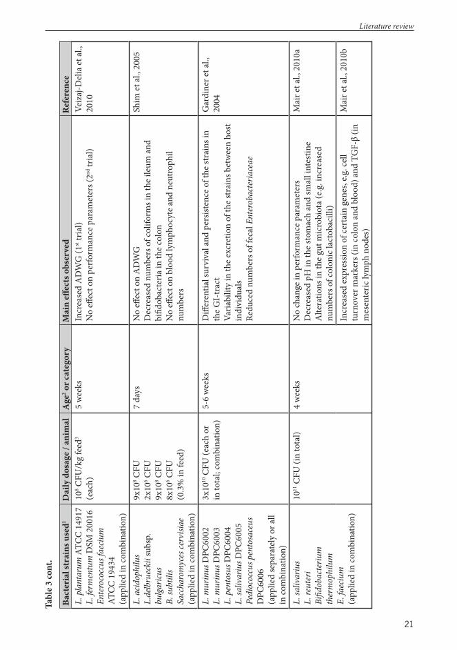
21
Bact
eria
l str
ains
use
d1D
aily
dos
age /
anim
alA
ge2 o
r cat
egor
yM
ain
eff e
cts o
bser
ved
Refe
renc
eL.
pla
ntar
um A
TCC
1491
7 L.
ferm
entu
m D
SM 2
0016
En
tero
cocc
us fa
eciu
m
ATCC
194
34
(app
lied
in co
mbi
natio
n)
108 C
FU/k
g fe
ed3
(eac
h)5
wee
ksIn
crea
sed
AD
WG
(1st tr
ial)
N
o eff
ect
on
perfo
rman
ce p
aram
eter
s (2nd
tria
l) Ve
izaj
-Del
ia et
al.,
2010
L. a
cidop
hilu
s L.
delb
ruec
kii s
ubsp
. bu
lgaric
us
B. su
btili
s Sa
ccha
rom
yces
cerv
isiae
(a
pplie
d in
com
bina
tion)
9x10
8 CFU
2x10
6 CFU
9x10
8 CFU
8x10
6 CFU
(0
.3%
in fe
ed)
7 da
ysN
o eff
ect
on
AD
WG
D
ecre
ased
num
bers
of c
olifo
rms i
n th
e ile
um an
d bi
fi dob
acte
ria in
the c
olon
N
o eff
ect
on
bloo
d ly
mph
ocyt
e and
neu
troph
il nu
mbe
rs
Shim
et al
., 20
05
L. m
urin
us D
PC60
02L.
mur
inus
DPC
6003
L. p
ento
sus D
PC60
04
L. sa
livar
ius D
PC60
05
Pedi
ococ
cus p
ento
sace
us
DPC
6006
(a
pplie
d se
para
tely
or a
ll in
com
bina
tion)
3x10
10 C
FU (e
ach
or
in to
tal;
com
bina
tion)
5-6
wee
ksD
iff er
entia
l sur
viva
l and
per
siste
nce o
f the
stra
ins i
n th
e GI-
trac
t Va
riabi
lity
in th
e exc
retio
n of
the s
trai
ns b
etw
een
host
indi
vidu
als
Redu
ced
num
bers
of f
ecal
Ent
erob
acte
riace
ae
Gar
dine
r et a
l.,
2004
L. sa
livar
ius
L. re
uter
i Bi
fi dob
acte
rium
th
erm
ophi
lum
E.
faec
ium
(app
lied
in co
mbi
natio
n)
1011
CFU
(in
tota
l)4
wee
ksN
o ch
ange
in p
erfo
rman
ce p
aram
eter
s D
ecre
ased
pH
in th
e sto
mac
h an
d sm
all i
ntes
tine
Alte
ratio
ns in
the g
ut m
icro
biot
a (e.g
. inc
reas
ed
num
bers
of c
olon
ic la
ctob
acill
i)
Mai
r et a
l., 2
010a
Incr
ease
d ex
pres
sion
of ce
rtai
n ge
nes,
e.g. c
ell
turn
over
mar
kers
(in
colo
n an
d bl
ood)
and
TGF-
β (in
m
esen
teric
lym
ph n
odes
)
Mai
r et a
l., 2
010b
Literature review
Tabl
e 3 co
nt.

22
Bact
eria
l str
ains
use
d1D
aily
dos
age /
anim
alA
ge2 o
r cat
egor
yM
ain
eff e
cts o
bser
ved
Refe
renc
eL.
mur
inus
DPC
6002
L. p
ento
sus D
PC60
04
L. sa
livar
ius D
PC60
05
P. pe
ntos
aceu
s DPC
6006
(a
pplie
d in
com
bina
tion)
5x10
9 CFU
(in
tota
l)6
wee
ksD
iff er
entia
l sur
viva
l of t
he st
rain
s thr
ough
the G
I-tr
act,
as w
ell as
adhe
renc
e to
the i
ntes
tine w
all
Incr
ease
d nu
mbe
rs o
f ile
al E
nter
obac
teria
ceae
Incr
ease
d IL
-8 ex
pres
sion
in th
e ile
umD
ecre
ased
CD
25 an
d CT
LA-4
indu
ctio
n on
imm
une
cells
Wal
sh et
al.,
2008
L. a
cidop
hilu
s KN
U 3
1 B.
subt
ilis K
NU
42
S. ce
rvici
ae K
NU
55
Aspe
rgill
us o
ryza
r KN
U
48
(app
lied
in co
mbi
natio
n)
104 /1
010 C
FU10
6 /1011
CFU
104 /1
06 CFU
10
5 /109
(0.3
/ 0.
6% in
feed
)
Wea
ned
pigl
ets
Incr
ease
d A
DW
G, D
FI, F
CR an
d ap
pare
nt d
iges
tibili
ty
Dec
reas
ed n
umbe
rs o
f fec
al C
lostr
idiu
m
Dos
e-de
pend
ent e
ff ect
on
inte
stina
l mor
phol
ogy
(vill
us h
eigh
t and
vill
us h
eigh
t : cr
ypt r
atio
)
Choi
et al
., 20
11a
L. a
cidop
hilu
s KN
U 3
1 B.
subt
ilis K
NU
42
S. ce
rvici
ae K
NU
55
Aspe
rgill
us or
yzar
KN
U 4
8 (a
pplie
d in
com
bina
tion)
1010
CFU
10
11 C
FU10
9 CFU
10
9 CFU
(0.3
% in
feed
)
Wea
ned
pigl
ets
Incr
ease
d A
DW
G, D
FI an
d FC
R D
ecre
ased
num
bers
of f
ecal
colif
orm
s and
Clo
strid
ium
, in
crea
sed
num
bers
of f
ecal
lact
obac
illi
Th e p
rodu
ctio
n m
etho
d of
the p
robi
otic
pre
para
tion
infl u
ence
d th
e out
com
es
Choi
et al
., 20
11b
L. a
cidop
hilu
s C3
L. p
lant
arum
1K8
L.
pla
ntar
um 3
K2
E. fa
eciu
m 6
H2
P. pe
ntos
aceu
s D7
(app
lied
in th
ree d
iff er
ent
mix
ture
s)
108 C
FU
108 C
FU10
8 CFU
10
10 C
FU10
8 CFU
(600
ppm
in fe
ed)
21-2
3 da
ysIn
crea
sed
AD
WG
, DFI
and
FCR
in th
e fi rs
t tw
o w
eeks
aft
er w
eani
ng (n
o di
ff ere
nce b
etw
een
the v
ario
us
mix
ture
s)
Incr
ease
d nu
mbe
rs o
f lac
tic ac
id b
acte
ria in
the
dige
sta,
but n
o eff
ect
on
E. co
li co
unts
Dec
reas
e in
diar
rhea
inci
denc
e
Gia
ng et
al.,
2010
1 L.
= L
acto
bacil
lus
2 Age
at th
e sta
rt o
f tria
l 3 Fe
ed co
nsum
ptio
n no
t rep
orte
d
Literature review
Tabl
e 3 co
nt.

23
2.3.3.1 E ects on produc vity parameters
A high level of biological functioning can be considered as one aspect of animal well-being (Frazer, 1993), thus the assessment of certain performance parameters to detect changes in productivity is frequently used to estimate the possible eff ects of bacterial supplementation. Productivity measures are also extremely important for the producer, as higher productivity yields better fi nancial returns. Th e average daily weight gain (ADWG) is the most commonly utilized measurement of productivity, used mainly in fattening farm animals (Meieregger et al., 2011). Other parameters that are oft en used are the daily feed intake (DFI), as well as the feed conversion ratio (FCR), which quantifi es the relation between DFI and ADWG, measuring the effi ciency in converting food mass into body mass. Depending on the production category of the animal, the usual range for FCR is 1 : 1.5-4 for swine and poultry, and 1 : 6-7 for cattle (Meieregger et al., 2011), with high-performing animals showing lower ratios indicating that less feed is needed to produce the same amount of meat. In production animals not in the fattening category, other parameters, such as those related to reproduction (e.g. the litter size and weight, sow weight loss aft er birth) can be used to assess the eff ects of probiotic supplementation (Böhmer et al., 2006).
Th e mechanisms behind the improved performance caused by bacterial supplementation are not fully understood, but some of the putative actions of probiotics possibly contributing to enhanced host growth are summarized in Figure 3. Lactobacilli and other benefi cial microbes have been proposed to contribute to the nutrition of the host e.g. by synthesizing vitamins and breaking down nutritional components that the host is unable to metabolize, possibly improving nutrient bioavailability (Kopp-Hoolihan, 2001; Turpin et al., 2010; Kenny et al., 2011; Ezema, 2013). However, as the cell numbers of supplemented bacteria are low compared to that of the commensal microbiota, the relevance of their possible contribution to nutrition may be minor. Modifi cation of the host gut physiology, such as increasing the production of growth factors, might also be a mechanism through which lactobacilli can enhance host growth (Turpin et al., 2010). In addition, stabilization of the gut microbiota resulting in a reduced pathogen load has been postulated to induce increased growth (Kenny et al., 2011).
In swine, highly variable results have been obtained on studies assessing the ability of various Lactobacillus supplementations to benefi cially aff ect the productivity, with some studies showing improved performance while others have not detected any benefi ts (Table 3). With regard to growth promotion, the percentage improvements in the weight gain of starter piglets has oft en ranged from around 2 to 10% (Fuller, 1989; Meieregger et al., 2011), and while several studies have failed to achieve statistical signifi cance, a general tendency towards an elevated growth rate in piglets supplemented with probiotics has been reported in numerous studies, as reviewed by Simon et al. (2001). Th e FEEDAP panel has issued scientifi c opinions regarding the ability of certain Lactobacillus preparations to enhance swine growth performance and some products have been concluded to increase the growth of piglets (EFSA, 2008, 2013). Studies performed in growing-fi nishing pigs are rarer, and the responses in growth enhancement have usually been of lower magnitude than those observed in young piglets (Nousiainen et al., 2004).
Literature review

24
Figure 3. Probiotics can improve host growth by increasing biodisponibility of macronutrients (1), degradation of antinutrient factors (2), enhancement of mineral absorption (3), and production of growth factors (4). Th e other squares indicate the mechanism involved in host growth. Th e Figure is reprinted from Turpin et al. (2010) with permission granted by Elsevier.
2.3.3.2 E ects on gut microbiota
Th e GI-tract microbiota is known to be a signifi cant factor aff ecting the health and well-being of the host, and positive eff ects on this complex community are believed to be one of the mechanisms behind probiotic action (Simon et al., 2001; Gueimonde and Salminen, 2006; Bosi and Trevisi, 2010). Consequently, the possible alterations of the gut microbiota attributable to lactobacilli consumption have been commonly monitored in porcine feeding trials. While it is not so straightforward to determine what kind of changes in the GI-tract microbiota can be considered as favorable for the host (Simon et al., 2001), an increase in the numbers of “benefi cial” microbes, such as lactic acid bacteria and bifi dobacteria is oft en interpreted as a positive outcome aft er lactobacilli supplementation (Simon et al., 2001; Nousiainen et al., 2004; Meieregger et al., 2011). Additionally, a reduction in the numbers of potentially pathogenic microbes, like enterobacteria including E. coli, has been proposed to be an advantageous change (Simon et al., 2001; Nousiainen et al., 2004; Meieregger et al., 2011). Both of these responses have been described in swine aft er supplementation with lactobacilli (Table 3). However, since a large part of these studies have been based on bacterial culture methods, the results are biased towards cultivable species, and provide only a partial picture of the changes occurring in the intestinal microbiota. Th e use of culture-independent molecular techniques allows a more complete description of the possible eff ects bacterial supplementation might have on the intestinal microbiota composition.
Literature review
25
In addition to general changes in the composition of the intestinal microbiota, also the fate of the administered strain is of interest. Survivability of the feeding strain through the GI-tract, and possibly even colonization of the host intestine have been stated as necessary characteristics for a probiotic microbe (Vaughan et al., 1999; Marco et al., 2006). While the GI-transit survivability has been demonstrated for certain strains of lactobacilli in feeding trials performed in swine (Table 3), this factor has not been investigated in the majority of the trials. Moreover, colonization of the intestine by the supplemented strain has been evaluated even more rarely in swine and only for short periods of time (Pedersen and Tannock, 1989; Gardiner et al., 2004; De Angelis et al., 2007). While permanent colonization of the host intestine by an allochthonous (i.e. exogenous) strain is easily achieved in gnotobiotic animals (Servin and Coconnier, 2003; Smith et al., 2007), the colonization resistance exerted by the commensal microbiota seems to be strong as the exogenous strain administered usually disappears from the intestine aft er discontinuation of consumption (Bezkorovainy, 2001; Ouwehand and Salminen, 2003; Isolauri et al., 2004). Even the adhesive L. rhamnosus strain GG does not seem to permanently colonize the human GI-tract (Bezkorovainy, 2001), nonetheless its persistence has been reported in colonic mucosa even although it was not recovered from fecal samples (Alander et al., 1999). In addition, the age of the host at the time of probiotic administration may be an important factor, as L. rhamnosus strain GG was found to persist for as long as 24 months in babies when administered to their mothers during late pregnancy (Schultz et al., 2004). Th is implies that application of probiotics in the neonatal period, when the commensal microbiota is not yet fully developed, may be optimal for intestinal colonization with the desired bacterial strain. Interestingly, a strain of L. acidophilus (LAB20) isolated from a dog was found to persist in the intestine of a dog at least for six weeks (Tang and Saris, 2013), thus there probably exist diff erences in colonization ability attributable to the bacterial strain as well as the host species.
2.3.3.3 E ects on host immune func on
Th e in vivo immune modulating actions of lactobacilli have been demonstrated in several trials, performed for the most part in mouse models as well as in humans, and have included shift s in the balance of diff erent T cell types, stimulation of regulatory immune mechanisms (e.g. generation of Treg cells), alterations in cytokine expression patterns, increases in the production of antibodies and in the activity of certain immunological cells (e.g. natural killer cells, macrophages and other phagocytes) as well as modifi cations in the functions of the intestinal barrier (Borchers et al., 2009; Meijerink and Wells, 2010; Wells, 2011b; van Baarlen et al., 2013). In lactobacilli feeding trials performed in swine, the most commonly measured immunological parameter has been the expression levels of cytokine genes in the intestine or other tissues, but also immune cell populations in the gut mucosa or peripheral blood as well as immunoglobulin concentrations have been investigated in some of the trials (Table 3).
While lactobacilli and other probiotic microbes obviously do have the capability to modify host immune responses, the question still remain open; are the observed eff ects actually benefi cial for the host? When taking into account the complexity of the immune system, interpretation of the data obtained from the in vivo studies is highly problematic and mere immunostimulation or some other alteration does not necessarily represent a positive health eff ect. A more detailed understanding of the immune system mechanisms are needed to unravel the consequences of the changes that probiotic microbes induce in the host immune function.
Literature review

26
2.3.3.4 E ects on disease occurrence
As a reduced occurrence of diseases, especially of diarrheal diseases, is one of the health claims for benefi cial microbes, this outcome has been frequently explored in probiotic feeding trials, both in humans and animals (Fuller, 1989; Simon et al., 2001). Reductions in disease incidence or prevalence would lead to improved animal welfare and oft en also to increased productivity. GI infections are very common in the pig production, causing signifi cant fi nancial losses. Diarrheal diseases especially in the neonatal and post-weaning period are a major problem, which traditionally has been combated with the use of therapeutic and in-feed antimicrobials (Bomba et al., 2012). While there are several diff erent bacteria and viruses that can cause neonatal and post-weaning diarrhea in piglets, diff erent enterotoxigenic E. coli (ETEC) strains possessing F4, F5, F6, F7 and F18 fi mbriae are those most commonly detected (Melkebeek et al., 2013).
Two diff erent study designs can be applied to assess the ability of a bacterial supplement to reduce disease occurrence in vivo. In fi eld trials, the effi cacy of the potential probiotic is estimated in regular production farms, i.e. under “real life conditions”. However, for practical reasons, these kinds of studies are very challenging to perform, and a large number of animals is needed to achieve signifi cant and biologically relevant results. Th erefore, studies in which the animals are deliberately challenged with infectious organisms to cause a disease are oft en used to estimate the disease preventive ability of lactobacilli and other probiotic microbes. In the challenge studies that have been performed in swine, diff erent E. coli strains have been the most oft en used pathogens, but strains of Salmonella have also been used (Table 4). In several studies, the supplemented probiotic strain has been able to alleviate the symptoms and reduce the fecal excretion of the pathogen, as well as to modify the pathogen induced immune responses of the host animal (Table 4). Th e mechanisms behind the protective eff ect of probiotic bacteria against pathogens remain unresolved, although several have been postulated, including competition for adhesion sites or nutrients, production of antibacterial substances that inhibit pathogen growth and survival, prevention of pathogen actions by agglutinating with the pathogen and inhibition of the production of pathogen toxins (Fuller, 1989; Nousiainen et al., 2004; Turpin et al., 2010). In the future, more detailed studies will help to reveal the interactions between the supplemented probiotic and the pathogen that occur in the complex environment of the host gut microbiota.
Literature review
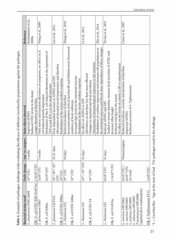
27
Tabl
e 4. E
xam
ples
of p
atho
gen
chal
leng
e tria
ls ev
alua
ting
the e
ffi ca
cy o
f diff
eren
t Lac
toba
cillu
s pre
para
tions
agai
nst t
he p
atho
gen.
Bact
eria
l str
ains
use
d1D
aily
dos
age
Age
2 or c
ateg
ory
Mai
n eff
ect
s obs
erve
dRe
fere
nce
L. a
myl
ovor
us D
SM 1
6698
Ch3 : E
. col
i ETE
C K8
8 O
149
Fac
1010
CFU
1.5x
1010
CFU
3 w
eeks
Impr
oved
AD
WG
Re
duce
d ET
EC le
vels
in th
e ile
um
Long
er d
urat
ion
of d
iarr
hea
Kons
tant
inov
et al
., 20
08a
L. fe
rmen
tum
I500
7
Ch:
E. c
oli K
88ac
2x10
9 CFU
2x10
9 CFU
3 w
eeks
Onl
y m
inor
and
tran
sient
alle
viat
ion
of sy
mpt
oms,
no eff
ect
on
E.
coli
indu
ced
grow
th d
epre
ssio
n M
odul
atio
n of
the E
. col
i ind
uced
alte
ratio
ns in
the e
xpre
ssio
n of
TN
F-α
and
IFN
-γ in
the s
mal
l int
estin
e
Wan
g et
al.,
2009
L. p
lant
arum
CJL
P243
Ch:
E. c
oli E
TEC
K88a
c
108 /
109 /
1010
CF
U10
9 CFU
19-2
1 da
ysD
ose-
depe
nden
t inc
reas
e in
AD
WG
and
DFI
A
llevi
atio
n of
elev
ated
rect
al te
mpe
ratu
re an
d di
arrh
ea
Mod
ulat
ion
of th
e ser
um cy
toki
ne re
spon
se
Lee e
t al.,
201
2
L. rh
amno
sus G
G
Ch:
E. c
oli E
TEC
K88a
c
1011
CFU
1010
CFU
19 d
ays
Redu
ced
inci
denc
e of d
iarr
hea
Incr
ease
d nu
mbe
rs o
f fec
al la
ctob
acill
i and
bifi
doba
cter
ia D
ecre
ased
nu
mbe
rs o
f fec
al co
lifor
ms
Incr
ease
d sI
gA le
vels
in th
e int
estin
al m
ucos
a M
odul
atio
n of
the s
erum
cyto
kine
resp
onse
Zhan
g et
al.,
2010
L. rh
amno
sus G
G
Ch:
E. c
oli E
TEC
F4
1010
/ 10
12 C
FU 10
10 C
FU
22 d
ays
No
eff e
ct o
n A
DW
G o
r DFI
A
llevi
atio
n on
dia
rrhe
a; lo
w d
ose m
ore e
ffi ci
ent
Chan
ges i
n in
testi
nal a
nd fe
cal m
icro
biot
a At
tenu
atio
n of
imm
unol
ogic
al re
spon
ses i
n th
e int
estin
e
Li et
al.,
2012
Mod
ulat
ion
of cy
toki
ne ex
pres
sion
and
imm
une c
ell p
opul
atio
ns in
th
e int
estin
al m
ucos
a and
blo
od; d
ose-
depe
nden
ce o
f the
se ch
ange
sZh
u et
al.,
2014
L. rh
amno
sus G
G
Ch:
E. c
oli O
149:
Fac
6x10
9 CFU
1.5x
1010
CFU
21 d
ays
Redu
ced
AD
WG
and
DFI
Te
nded
to w
orse
n di
arrh
ea, i
ncre
ase f
ecal
excr
etio
n of
ETE
C an
d de
crea
se v
illus
hei
ght
Redu
ced
tota
l IgA
in th
e ser
umN
o eff
ect
on
the l
evels
of c
olon
ic L
AB
or en
tero
bact
eria
Trev
isi et
al.,
2011
L. m
urin
us D
PC60
02L.
mur
inus
DPC
6003
L. p
ento
sus D
PC60
04
L. sa
livar
ius D
PC60
05
P. pe
ntos
aceu
s DPC
6006
(app
lied
all i
n co
mbi
natio
n)
Ch:
S. T
yphi
mur
ium
PT1
2
4x10
9 CFU
/ 4x
1010
CFU
(in
tota
l)
1x10
8 CFU
Wea
ned
pigl
ets
Redu
ced
inci
denc
e, se
verit
y an
d du
ratio
n of
dia
rrhe
aIm
prov
ed A
DW
GRe
duce
d ex
cret
ion
of S
. Typ
him
uriu
m
Case
y et
al.,
2007
1 L.
= L
acto
bacil
lus
2 Age
at th
e sta
rt o
f tria
l 3Ch
: pat
hoge
n us
ed in
the c
halle
nge
Literature review

28
2.3.4 Correla on of in vitro and in vivo studies
As the purpose of the in vitro tests is to predict which of the candidate probiotic strains would perform best in the target animal species, the correlation of the results obtained in these screening tests to the in vivo fi ndings is very important. However, it is a very diffi cult task to systematically and extensively evaluate this matter. Additionally, studies which have estimated the correlation of in vitro and in vivo results are, for the most part, based on experiments performed in humans or mouse, and only few studies executed in swine have evaluated this topic.
Th e tests for tolerance of low pH and bile are intended to demonstrate which strains will be able to survive the transit through the host GI-tract. Indeed, several studies performed both in humans (Pochart et al., 1992; Jacobsen et al., 1999; Dunne et al., 2001) and swine (Gardiner et al., 2004; De Angelis et al., 2007) have shown that strains exhibiting in vitro tolerance to low pH and bile are able to survive intestinal passage. Additionally, the in vitro survival results obtained in a dynamic model of the human GI-tract correlated well with in vivo data (Marteau et al., 1997). However, appropriate in vivo comparison of strains with diff ering performances in the in vitro stress tolerance tests has not usually been performed in these studies. Furthermore, those strains that are known to survive human GI-tract transit in vivo, e.g. L. rhamnosus GG (Goldin et al., 1992; Alander et al., 1999), have sometimes performed poorly in in vitro tests (Mattila-Sandholm et al., 1999; Morelli, 2000; Mainville et al., 2005). Th us, at least some of the in vitro test conditions used in the stress tolerance assays may well be too harsh as compared to the in vivo situation. Additionally, as the food matrix protects the supplemented bacteria during gastric passage (Conway et al., 1987; Charteris et al., 1998), strains with poor in vitro acid tolerance may well be able to tolerate the in vivo GI-tract environment.
Good adhesiveness to the intestinal wall structures has been considered to be an important property for probiotic microbes mainly for three reasons: adherent strains might 1) more easily colonize the host intestine, 2) exclude pathogens by competing for adhesion receptors, 3) induce immune modulatory actions via their close contact with host cells (Ouwehand and Salminen, 2003; Lahtinen and Ouwehand, 2009). With regard to colonization, as already discussed in section 2.2.3.2, it seems however that even adhesive strains cannot permanently colonize the host intestine. Based on studies performed in humans (Jacobsen et al., 1999; Morelli, 2000) and in various animals (i.e. chickens (Fuller, 1978), mouse (Zarate et al., 2002) and fi sh (Sugimura et al., 2011)) it seems that in vitro adhesion ability of a bacterial strain may predict in vivo adhesion and / or persistence in the host intestine. However, the adhesion ability by itself is apparently not suffi cient to ensure a prolonged persistence of the strain in the gut environment. Th is is illustrated by results obtained in mouse showing that also non-adherent strains can temporarily colonize the host intestine (Hautefort et al., 2000). On the other hand, studies performed in humans (Morelli, 2000) and swine (Nousiainen et al., 2004) have revealed that adhesive strains do not necessarily succeed in colonization. Consequently, the correlation between in vitro adhesiveness and transient gut colonization is not clear. Interestingly, when comparing isogenic variants of L. crispatus diff ering in their in vitro aggregation and adhesion abilities, the low-adhesive mutant was not detected in the feces or biopsies of human volunteers, while the more adhesive parent strain was found (Cesena et al., 2001), and the same was observed also in experiments conducted in mice (Voltan et al., 2007). Future studies with a similar approach of using isogenic strains diff ering only in their adhesion ability may help clarify the correlation of in vitro adhesiveness to in vivo colonization capacity.
Literature review

29
While in vivo challenge studies performed in swine have shown that lactobacilli and other probiotics are able to exclude pathogens, and there is some evidence that lactobacilli could use the same attachment sites as pathogens (Lebeer et al., 2008), the importance of probiotic adhesion in pathogen exclusion is still rather unclear. A non-adhering mutant of the strain L. plantarum 299v was, in contrast to the wild type, unable to inhibit the adhesion of an E. coli strain to HT-29 cells, which indicates that adhesion ability may be important for pathogen exclusion (Mack et al., 2003). However, it appears that even if a strain has a high adherence ability, it may not necessarily inhibit pathogen adhesion to cultured intestinal epithelial cells (Bibiloni et al., 1999), and also other mechanisms have been proposed to be involved in pathogen exclusion, including coaggregation with the pathogen (Lebeer et al., 2008).
In addition to the colonization ability and / or pathogen exclusion properties, adhesiveness is also considered important for the immunomodulatory actions of a probiotic strain. Obviously, stimulation of the immune system requires an intimate contact between the bacteria and host cells, and this typically occurs via specifi c receptors, e.g. pattern recognition receptors (PRRs) binding to certain structures on the microbial surface, inducing signaling pathways that trigger immunological responses (Murphy, 2012). As reviewed by Lebeer et al. (2010), these kinds of interactions also take place between probiotic bacteria and the host. However, this does not reveal whether adhesion to enterocytes is necessary to obtain the immunological actions of probiotics. While studies performed in humans have given some indications that highly adhesive strains have a greater eff ect on the immune system than their less adhesive counterparts (Ouwehand and Salminen, 2003), it cannot be concluded for certain that these immunological diff erences are specifi cally due to the diff ering adherence properties of the strains. Th us far, few studies have been performed with isogenic strains; in one such study the aggregating and adhesive strain L. crispatus M247 induced changes at the mRNA levels of Toll-like receptor (TLR) 2 and TLR 4 in the colonic mucosa of mice, while the less adhesive mutant strain had no eff ect on these parameters (Voltan et al., 2007). Th is observation indicates that an aggregative and / or adhesive phenotype is important in modulating murine enterocyte responsiveness, but for the most part, the role of adhesiveness in probiotic immunomodulation still remains to be elucidated.
Th e comparison of in vitro and in vivo immunomodulatory properties of probiotic bacteria is complicated by the extreme complexity of the host immune system, as only a few parts of this vast network can be examined in laboratory experiments. However, at least some correlations seem to exist between certain in vitro measures and in vivo immune functionality. In chickens, provision of lactobacilli strains which enhanced the proliferation of suboptimally stimulated lymphocytes in vitro also increased the specifi c humoral immune responses in vivo (Koenen et al., 2004). In mouse models of experimentally induced infl ammatory colitis, the strains inducing in PBMCs a higher release of IL-10, considered as anti-infl ammatory, and a lower release of IL-12, viewed as Th 1 stimulating, showed better in vivo protective ability against colitis (Foligne et al., 2007). Additionally, in vitro induction of infl ammatory cytokine production on DCs predicted the ability of diff erent Lactobacillus species to confer protection from induced colitis in mice (Mileti et al., 2009). In the latter study, strains displaying immunostimulatory properties exacerbated the development of induced colitis, emphasizing the need to characterize properly the in vivo immunomodulative properties of diff erent strains in various disease conditions. While these results indicate that the in vitro immunomodulatory responses observed for lactobacilli may indeed be predictive of in vivo functions, more comparative studies will be needed before any fi rm conclusions can be made.
Literature review
30
3 Aims of the study
Th e objective of this study was to explore the possibility of using lactobacilli as health-promoting microbes and/or a recombinant vaccine vector in swine; to obtain host-specifi c Lactobacillus strains originating from the swine intestine as well as to characterize certain properties associated with probiosis of the strains and to evaluate the suitability of selected Lactobacillus strains for use as a viable probiotic / vaccine vector in swine. Additionally, the potential role of the surface layer proteins of Lactobacillus amylovorus strains in adhesion was examined. Th is study was part of a project aiming to develop a Lactobacillus vectored vaccine against porcine post-weaning diarrhea and edema disease caused by E. coli F18+ strains.
Th e specifi c aims of the subprojects were:1. to examine the in vitro probiotic properties of lactic acid bacterial strains isolated from
the feces and intestine of swine,2. to evaluate the potential eff ects of Lactobacillus brevis ATCC 8287 as a feeding
supplement on the performance and immune function in piglets, 3. to evaluate the potential eff ects of a multispecies Lactobacillus supplementation
(containing L. amylovorus, L. johnsonii, L. mucosae, L. salivarius and two strains of L. reuteri) on the performance and intestinal cytokine expression in piglets and
4. to characterize the putative probiotic properties of eight L. amylovorus strains and to assess the role of the surface layer proteins of the strains in adherence to porcine intestinal epithelium cells in vitro
Aims of the study
31
4 Materials and methods4.1 Bacterial strains and culture condi ons (I-IV)A total of 94 LAB were isolated (I) from the digesta and feces of swine, with some of these Lactobacillus strains being used in further studies (III, IV). Digesta samples were from the duodenum, jejunum and ileum of six swine, and fecal samples from the rectum of two sows. For the isolation of swine Lactobacillus strains the samples were cultured on de Man Rogosa Sharpe (MRS) or Rogosa liquid and solid media (Difco, Becton-Dickinson (BD), USA) incubated anaerobically (Anaerocult A, Merck, Germany) at 37°C for 48 h.
Six LAB isolates identifi ed as surface layer-carrying L. amylovorus strains were selected for Study IV (Table 5). Th e seven Lactobacillus strains used in the porcine feeding trial (II, III; Table 5) were selected on the basis of the in vitro characteristics associated with probiotic properties.
Additionally, several strains from culture collections were used (Table 5). Th e pathogens used in Study I and IV were selected based on food hygienic relevance and / or signifi cance to swine health. Furthermore, two Escherichia coli strains were used in the gene expression analysis in Study IV (Table 5).
All bacterial strains were stored at −80°C; the LAB strains in MRS broth (Difco) and the pathogens and E. coli strains in tryptic soy broth (TSB; Difco) or in Luria-Bertani (LB) broth (Difco), containing 15% glycerol.
Th e Lactobacillus strains (I-IV) were grown from stocks anaerobically in MRS broth at 37°C for 24h, and, when needed, subcultured in MRS solid media at 37°C for 48h.
Th e pathogenic bacteria used in the growth inhibition assay (I, IV) were grown from stocks in blood agar (Tammer-Tutkan maljat OY, Finland) or tryptic soy agar (TSA; Difco) at 37°C for 24 h and subcultured in TSB. Th e enterotoxigenic E. coli (ETEC) strain used in the adhesion assays (IV) was cultured in LB broth (Difco). Th e broth cultures of the pathogens were incubated at 37°C with agitation (200 rpm). Th e E. coli strains used in the genome expression analyses (IV) were cultivated in LB broth or in M9ZB medium (Studier, 1991) with added kanamycin (30 μg/ml) when appropriate.
4.2 Iden ca on of lac c acid bacteria (I, III)Th e putative identifi cation of the porcine LAB isolates as members of the genus Lactobacillus (I) was performed with Gram-staining and a polymerase chain reaction-enzyme-linked-immunosorbent-assay (PCR-ELISA) as described by Jakava-Viljanen and Palva (2007).
LAB isolates confi rmed as S-layer positive (n=9) were identifi ed by 16S ribosomal RNA (rRNA) gene sequencing as described by Jakava-Viljanen and Palva (2007). For the remaining isolates, preliminary identifi cation down to the species level was performed by sequencing the fi rst 500 base pairs of the 16S rRNA gene. For selected isolates (n=11), sequencing of the whole 16S rRNA gene was performed in order to confi rm the preliminary identifi cation. Th e chromosomal DNA was extracted with standard methods, and the amplifi cation PCR product purifi ed with the Qiaquick PCR purifi cation kit (Qiagen Co., Germany). Th e primers used in the 16S rRNA sequencing analyses are listed in Table 6. Sequencing was performed with an ABI Prism 310 or 3700 Genetic Analyzer (Applied Biosystems Inc., USA), and the Basic Local Alignment Search Tool (BLAST) program was used for comparing the 16S sequences to the gene bank database.
Materials and methods

32
In addition to sequencing of the 16S rRNA gene, whole genome sequencing (WGS) was performed to those Lactobacillus isolates used in Study III. Th e WGS was performed in the Institute of Biotechnology with the method described by Kant et al. (2011b).
Table 5. Strains used in this work.
Strain Origin, reference or source StudyLactobacillus strainsL. brevis ATCC 8287 Green fermented olives, ATCCa IIL. mucosae GLR 1167 (LAB 4) Swine feces, DVBc IIIL. salivarius GRL 1169 (LAB 33) Swine ileum, DVBc IIIL. johnsonii GRL 1171 (LAB 81) Swine ileum, DVBc IIIL. reuteri GRL 1168 (LAB 26) Swine duodenum, DVBc IIIL. reuteri GRL 1170 (LAB 49) Swine jejunum, DVBc IIIL. amylovorus GRL 1112 (LAB 2) Swine feces (Jakava-Viljanen and Palva,
2007), DVBcIII, IV
L. amylovorus GRL 1114 (LAB 8) Swine feces (Jakava-Viljanen and Palva, 2007), DVBc
IV
L. amylovorus GRL 1115 (LAB 13) Swine ileum (Jakava-Viljanen and Palva, 2007), DVBc
IV
L. amylovorus GRL 1116 (LAB 16) Swine jejunum (Jakava-Viljanen and Palva, 2007), DVBc
IV
L. amylovorus GRL 1117 (LAB 31) Swine jejunum (Jakava-Viljanen and Palva, 2007), DVBc
IV
L. amylovorus GRL 1118 (LAB 52) Swine jejunum (Jakava-Viljanen and Palva, 2007), DVBc
IV
L. amylovorus DSM 16698 Swine feces (Konstantinov et al., 2006b), DVBc
IV
L. amylovorus DSM 20531T Fermented corn silage, DSMb IVIntestinal pathogensEscherichia coli ERF 2014; O141, F18+ DVBc I, IVEscherichia coli F4+ (ETEC) (Roselli et al., 2007) IVEscherichia coli ATCC 43894; O157 (EHEC) ATCCa I, IVSalmonella Typhimurium ATCC14028 ATCCa I, IVListeria monocytogenes R14-2-2 DVBc I, IVYersinia enterocolitica R5-9-1 DVBc I, IVStrains used in gene expression studiesEscherichia coli DH5αF’ (Woodcock et al., 1989) IVEscherichia coli BL21 (DE3) EMD Millipore IV
a American Type Culture Collectionb Deutsche Sammlung von Mikroorganismenc Culture collection of the Department of Veterinary Biosciences / Veterinary Microbiology and Epidemiology, University of Helsinki, Finland
Materials and methods

33
Tabl
e 6. P
rimer
s use
d in
this
wor
k.
Olig
onuc
leot
ide s
eque
nce (
5´- 3
´)T m
°CTa
rget
gen
e / sp
ecie
sRe
fere
nce
Stud
yPr
imer
s use
d in
16S
rRN
A am
plifi
catio
n (I
)f A
GAG
TTTG
ATCC
TGG
CTCA
G
r ACG
GCT
ACCT
TGTT
ACG
ACTT
5516
S rR
NA
/ un
iver
sal
Dor
é et a
l., 1
998
I
Prim
ers u
sed
in 1
6S rR
NA
sequ
enci
ng: fi
rst 5
00 b
ase p
airs
(I)
f AG
AGTT
TGAT
CCTG
GCT
CAG
r C
ACCG
CTAC
ACAT
GG
AG
5016
S rR
NA
/ un
iver
sal
16S
rRN
A /
Lact
obac
illus
D
oré e
t al.,
199
8H
eilig
et al
., 20
02I
Addi
tiona
l prim
ers u
sed
in w
hole
16S
rRN
A se
quen
cing
f CAG
C(AC
)GCC
GCG
GTA
AT(A
T)C
r CCC
CGTC
AAT
TCCT
TTG
AGTT
T f A
AAC
TCA
AAG
GA
ATTG
ACG
GG
Gr A
CGG
CTAC
CTTG
TTAC
GAC
TT
5016
S / 1
8S rR
NA
/ un
iver
sal
16S
/ 18S
rRN
A /
univ
ersa
l16
S / 1
8S rR
NA
/ un
iver
sal
16S
rRN
A /
univ
ersa
l
Apaj
alah
ti et
al.,
2001
Th is
study
Dor
é et a
l., 1
998
UB1
Prim
ers u
sed
in P
CR sc
reen
ing
of fe
cal i
sola
tes (
II, I
II)
f CTG
ATG
GTA
CAA
AGG
CAG
GTT
r C
CAG
CAG
CATA
GAC
TGTT
GA
60
S-lay
er /
L. b
revi
sTh
is stu
dyII
f TAT
GTC
TGG
CTTA
AA
AAG
CACT
TGr T
CAAT
GTA
CTA
ACTC
CTG
ACTT
C59
ISR
/ L. a
myl
ovor
usTh
is stu
dyII
I
f ACG
GAC
TTG
ACG
TTG
GTT
TAr C
CGA
AGCC
ATCT
TTTA
AAT
TTG
A59
16S
rRN
A /
L. m
ucos
aeTh
is stu
dyII
I
f ACG
AA
ACTT
TCTT
ACAC
CGA
ATr G
ATCA
TGCG
ATCC
TTAG
AGAT
A62
16S
rRN
A /
L. sa
livar
ius
Th is
study
III
f CTT
GA
ATA
ACA
AGCC
AAG
CATA
r CCC
ATCA
TTG
CCTT
TTAT
CA57
ISR
/ L. j
ohns
onii
Th is
study
III
f AAC
GG
AAC
CTAC
ACAT
CGA
Ar C
CTTC
ATA
ACTT
AAC
CTA
AAC
AAT
C62
ISR
/ L. r
eute
riTh
is stu
dyII
I
Materials and methods

34
Tabl
e 6 co
nt.
Olig
onuc
leot
ide s
eque
nce (
5´- 3
´)T m
°CTa
rget
gen
e / sp
ecie
sRe
fere
nce
Stud
yPr
imer
s use
d in
real
-tim
e qPC
R an
alys
is (I
I, II
I)f T
CCTA
CGG
GAG
GCA
GCA
GT
r GG
ACTA
CCAG
GG
TATC
TAAT
CCTG
TT58
16S
rRN
A /
tota
l bac
teria
Nad
karn
i et a
l., 2
002
II, I
II
f AG
CAG
TAG
GG
AAT
CTTC
CA
r CAC
CGCT
ACAC
ATG
GAG
6016
S rR
NA
/ La
ctob
acill
iRi
nttil
ä et a
l., 2
004
III
Prim
ers u
sed
in S
-laye
r gen
e am
plifi
catio
n (I
V)
f GCG
GCC
ATG
GAT
GCC
GTT
CAAT
CAG
CTAC
T r C
GG
CTCG
AGA
AAG
TTTG
AAG
CCTT
AAC
GTA
AG62
slp
A / L
. am
ylov
orus
DSM
166
98
Th is
study
IV
f GCG
GCC
ATG
GCT
GAC
AGCA
ATG
AA
ACTG
G
r GCG
GCT
CGAG
TTG
AGCT
GCG
CTCA
AGTT
AG63
slpB
/ L. a
myl
ovor
us D
SM 1
6698
Th is
study
IV
f CG
TAG
CCAT
GG
CAG
ATAC
TAAT
GCT
GTA
AAC
A A
AAT
G
r TCG
CACT
CGAG
CTTC
TTTG
CGG
CAG
CTT
62
slpC
/ L. a
myl
ovor
us D
SM 1
6698
Th is
study
IV
f AG
TACC
CATG
GCT
GCT
ACTA
CTAT
TAAC
GCT
GG
TT
r TCG
CACT
CGAG
AA
AGTT
TGA
AGCC
TTTA
CGTA
A G
T63
slp
A / L
. am
ylov
orus
DSM
203
51T
Th is
study
IV
f GCG
GCC
ATG
GAC
GTT
AAC
ACTA
ACAT
TGTT
T r G
CGG
CTCG
AGG
AAG
TTTG
CCTT
CTTT
ACG
T58
slpA
/ L. a
myl
ovor
us G
RL 1
112
Th is
study
IV
f GTC
ATCC
ATG
GCT
ACTA
CAG
TTA
ATAT
CAAC
GG
TA
AC
r TCG
CACT
CGAG
AA
AGTT
TGA
AGCC
TTA
ACG
T60
slp
A / L
. am
ylov
orus
GRL
111
4Th
is stu
dyIV
f GCG
GCC
ATG
GCT
GAC
GCT
ACTA
CAAC
TACT
ACr G
CGG
CTCG
AGA
AAG
TTTG
CAAC
CTTA
ACG
TA60
slpA
/ L. a
myl
ovor
us G
RL 1
115
Th is
study
IV
f CTA
CGCC
ATG
GCT
GAC
GCT
ACTA
CAAC
TACT
AC T
G
r GTC
TGCT
CGAG
AA
AGTT
TGA
AGCC
TTTA
CGTA
A G
T60
slp
A / L
. am
ylov
orus
GRL
111
6Th
is stu
dyIV
r CTA
CGCC
ATG
GCT
GTA
GCA
ATTA
AGG
GTA
ACA
CT
r GTA
GTC
TCG
AGG
AAG
TTTG
CCTT
CTTA
ACG
TAT
TG64
slp
A / L
. am
ylov
orus
GRL
111
7Th
is stu
dyIV
f CTA
CGCC
ATG
GAC
ACAC
CAAC
AGTT
GAT
ACTG
r G
TAG
TCTC
GAG
TTG
AGCT
GCG
CTCA
AGTT
60
slpB
/ L. a
myl
ovor
us G
RL 1
117
Th is
study
IV
f GCG
GCC
ATG
GCT
GAT
GCT
ACTA
ACAG
TAAT
GC
r CG
GCT
CGAG
AA
AGTT
TGA
AGCC
TTA
ACG
TAAG
62slp
A /
L. a
myl
ovor
us G
RL 1
118
Th is
study
IV
1 unbu
blish
ed
Materials and methods

35
4.3 In vitro assays for examining probio c proper es4.3.1 Acid and bile tolerance (I)
Th e ability of the LAB isolates to tolerate low pH and bile was determined by exposing each isolate (OD600: 1) to either pH-adjusted MRS broth (pH 2.0 or pH 4.0) or MRS broth containing 0.3% ox gall. Th e isolates were incubated in these stress conditions for three hours at 37°C in a microwell plate, aft er which a 7-step dilution series (dilution factor of 25) was generated in buff ered MRS (acid tolerance test) or regular MRS (bile tolerance test) on the same microwell plate. Five parallel series were used for each isolate. Following anaerobic incubation, the wells were examined for growth and the OD600 values were measured (iEMS Reader MF; Th ermo Labsystems, Finland). Th e size of the bacterial population surviving the exposure to each stress condition was estimated by the most probable number (MPN) method in MS Excel as described by Briones and Reichardt (1999).
4.3.2 Bacterial adhesion (I, IV)
For the adhesion assay performed in Study I, intestinal epithelial cells were collected from healthy pigs aged two to three months. Th e entire intestine was divided to the anatomical sections (duodenum, jejunum, ileum, proximal colon and distal colon), and the sections were washed with PBS until clean. Th e enterocytes were removed by mechanical disruption, washed two to three times by centrifugation, and resuspended in Hanks Balanced Salt Solution (HBSS; Gibco BRL, Life Technologies Inc., USA) supplemented with lactalbumin (Difco), fetal bovine serum (Gibco BRL), dimethyl sulphoxide (DMSO; Sigma-Aldrich, USA) and glycerol, and stored at -80°C until used in the adhesion assay. For the assay, the enterocyte suspension and the OD-adjusted LAB isolates (OD600: 1) were incubated in a microwell plate, aft er which the unattached bacteria were removed by washing. Bacterial adhesion was quantifi ed by examining Giemsa-stained microscopic slides under a light microscope (Leica; Microsystems GmbH, Germany) and calculating the average number of attaching bacteria per enterocyte, using twenty visually undamaged enterocytes. In the case when the number of adhering bacteria in one enterocyte exceeded twenty, it was scored as twenty in the calculations, because in such cases it was too diffi cult to calculate the exact number of bacteria. For each intestinal section, a positive and a negative control strain were selected from the isolates and the consistency of the adherence level of these was monitored in individual test runs.
For the mucus adhesion assay conducted in Study IV, porcine intestinal mucus was isolated from the small intestine of a two-months-old piglet with a protocol modifi ed from MacAdam et al. (2000). Briefl y, the intestinal sections washed with protease inhibiting saline solution were scraped gently to collect the mucus, aft er which the insoluble material and cellular debris were separated by centrifugation. Th e crude mucus was homogenized, concentrated by fi ltration, and clarifi ed by centrifugation and several fi ltration steps. Purifi cation of the mucus was performed by gel fi ltration chromatography monitoring the A280 nm values of 5 ml fractions. Protein-containing fractions were dialyzed against water and assayed for total protein by the method of Bradford (Bio-Rad Protein Assay, Bio-Rad, USA) using bovine serum albumin (BSA) as a standard, and for glycoproteins using the Crypton™ Glycoprotein Staining Kit (Th ermo Scientifi c, USA), with porcine gastric mucins (Sigma), horse radish peroxidase (HRP) and soybean trypsin inhibitor (Th ermo Scientifi c) as standards. Th e void volume fractions with high glycoprotein content were pooled, lyophilized and stored at -20°C until used in the adhesion assay. In addition to the porcine intestinal mucus, porcine gastric mucins (type II, Sigma) were used in the adhesion
Materials and methods

36
assay. For the assay, the mucus was immobilized into polystyrene microtiter plate wells (Th ermo Scientifi c). Th e L. amylovorus cells used in the mucus adhesion assay were labelled with a nucleic acid binding fl uorescent stain by incubating the bacteria with SYTO®9 (Molecular Probes, USA), aft er which the OD-adjusted (OD600: 0.1, 0.25, 0.5 and 1.0) bacterial suspension was added to the mucus coated wells. Aft er incubation and removing the unbound bacteria by washing, the bound bacteria were lysed with sodium dodecyl sulfate (SDS) in NaOH, and the input (added) and output (remaining) fl uorescence values were measured in a microplate reader (Victor Multilabel Plate Reader; Perkin Elmer, USA). Th e background fl uorescence from mucus-coated wells without bacteria (for outputs) and from wells fi lled with PBS (for inputs) were subtracted from the values. Th e adherence was expressed as the proportion (%) of the original fl uorescence added.
Th e cell adhesion assays conducted in Study IV were performed using IPEC-1 cells, a non-transformed continuous cell line (Gonzalez-Vallina et al., 1996). Th e cells were cultured at 39 ºC and 5% CO2 using Dulbecco’s modifi ed eagle medium/Ham’s F12 Nutrient Mixture (DMEM/Ham’s F-12 [1:1]) supplemented with fetal calf serum (FCS; Integro, Netherlands), insulin-transferrin-selenium, HEPES (all PAN-Biotech, Germany) and epidermal growth factor (BD). In the assay, the cells were seeded (2 x 10⁵ /ml) into a Transwell system (Th incerts™, 1 μm pore size, diameter 10 mm; Greiner bio-one, Germany) and allowed to diff erentiate for 4-5 days, until the transepithelial electric resistance (TEER) value was ≥1 kΩcm2. Th e L. amylovorus strains used in the cell adhesion assay were metabolically labelled with 3H-thymidine, and added to the wells aft er OD-adjustment (OD600: 0.25, 0.5 or 1). Aft er incubation, the unbound bacteria were removed by washing, the bound bacteria lysed with SDS in NaOH, and the radioactivity of the lysed samples (output) was measured by liquid scintillation counting. Th e input radioactivity values were determined by liquid scintillation counting of the OD-adjusted cell suspensions in DMEM/Ham’s F-12 [1:1] treated with SDS in NaOH. Th e adherence was expressed as the proportion (%) of the original radioactivity added aft er subtracting the background radioactivity from IPEC-1 cells incubated without bacteria (for outputs) and from DMEM/Ham’s F-12 [1:1] medium (for inputs).
4.3.3 Inhibi on of pathogen adhesion (IV)
Th e ability of the L. amylovorus strains to inhibit the adhesion of the E. coli ETEC F4+ strain to IPEC-1 cells was investigated in the adhesion inhibition assay (IV). Th e ETEC strain was metabolically labelled with 3H-thymidine, and the expression of F4 fi mbriae was confi rmed with an agglutination test. Th ree diff erent setups were used in the adhesion inhibition assay, with diff ering orders of addition of the bacterial cells to the IPEC-1 cells: 1) exclusion, where the lactobacilli (OD600: 6) were allowed to adhere for 1 hour before adding the ETEC strain (OD600: 0.6); 2) displacement, where the ETEC strain was allowed to adhere for 1 hour, and the unbound bacteria were removed with washing before the addition of lactobacilli; 3) competition, where both bacteria species were added simultaneously. In all of the assays, the cells were lysed aft er incubation and washing by SDS in NaOH, aft er which the radioactivity was measured by liquid scintillation counting. Th e proportion of adherent ETEC cells (%) in the presence or absence of the L. amylovorus strains was calculated as in the IPEC-1 cell adhesion experiments, and the inhibition (%) was calculated according to the formula: [adherence (no La) – adherence (with La)] / adherence (no La) x 100 %, where La indicates L. amylovorus.
Materials and methods

37
4.3.4 An microbial ac vity (I, III, IV)
Th e ability of the LAB isolates (I) and the L. amylovorus strains (IV) to inhibit the growth of selected intestinal pathogens was investigated with a turbidometric assay essentially as described by Skyttä and Mattila-Sandholm (1991), using fi ltration-sterilized (0.22 μm) spent culture fi ltrates (SCF) of the strains. Th e inhibitory eff ects of the SCF were examined by following the growth of the pathogens in TSB media supplemented (10% vol/vol) with the fi ltrates using an automatic reader Bioscreen C (Growth Curves Oy/Ab, Finland). Growth rate of the pathogens was quantifi ed using the area under the growth curve (AUC) value of the logarithmic growth phase, which was automatically processed by the soft ware (BioLink (Growth Curves Oy/Ab) or Research Express (Transgalactic Ltd., Finland)). Inhibition caused by the SCF was expressed as an area reduction percentage (ARP) calculated with the following formula: (AUCCo – AUCSCF) / AUCCo × 100, in which AUCCo represents pathogen growth in TSB media supplemented with 10% MRS medium and AUCSCF represents pathogen growth in TSB media supplemented with the culture fi ltrate. For each LAB strain assayed, one to three independent experiments with fresh SCF were performed, and in each assay two to three parallel wells were used.
In order to estimate the relationship between the ARP value and CFU counts, ten-fold dilution series prepared from each of the pathogens were incubated in the Bioscreen microwell plate, the ARP values of the dilutions were plotted against the corresponding logCFU reductions and linear regression was used to generate regression formulas for the pathogens, as described by Skyttä and Mattila-Sandholm (1991).
Two diff erent approaches were adopted to monitor the eff ect of the fi ltrate pH on the pathogen growth inhibition. In the fi rst approach, the culture fi ltrates collected from the LAB strains were categorized into six pH groups, and for each group two diff erent pH controls were used in the Bioscreen assay: MRS medium with pH adjusted to the group mean either with lactic acid or with HCl. Th e second approach involved using both pH-adjusted (pH 6.2) and non-adjusted culture fi ltrates.
To verify the lack of antagonistic eff ects of the Lactobacillus strains used in Study III towards each other, the ability of each strain to grow in the presence of the SCF collected from the other strains was monitored with the turbidometric Bioscreen assay with a similar experiment setup as described for the intestinal pathogens.
4.3.5 S mula on of cytokine produc on by dendri c cells (IV)
Monocyte-derived dendritic cells (MoDC) were generated from purifi ed and in vitro cultured human monocytes using a method described by (Veckman et al. (2004) with minor modifi cations. Briefl y, peripheral blood mononuclear cells were fi rst isolated from leukocyte-rich buff y coats by two density gradient centrifugations and magnetic beading using anti-CD3 and anti-CD19 beads (Dynal Invitrogen, Life Technologies, USA). Th ereaft er, the monocytes were allowed to adhere to 24-well plates (Falcon, BD, Franklin Lakes, New Jersey) in the presence of RPMI 1640 (Sigma) supplemented with HEPES, penicillin, streptomycin and L-glutamine, and diff erentiation was induced by maintaining the cells in RPMI 1640 (with supplements as described above) containing FCS, human recombinant granulocyte macrophage-colony stimulating factor (GM-CSF; Gibco Life Technologies, USA), and human recombinant IL-4 (Gibco Life Technologies). MoDCs were used on day 7 in the experiments, with cells of four donors being were used in each experiment. Th e OD-normalized L. amylovorus strains were added to human moDCs (multiplicity of infection (MOI) 1, 10, and 100), using medium without bacteria as a control.
Materials and methods

38
Aft er incubation, the cell culture supernatants were collected and stored at -20°C until analyzed by Bio-Rad’s Bio-Plex Pro Cytokine assay using the Bio-Plex -200 platform (Bio-Rad, Hercules, CA). Quantifi cation of human TNF-α, IL-1β, IL-6, IL-10, and IL-12 was performed according to the manufacturer’s instructions. Human IP-10/CXCL10 was measured separately with the OptEIA ELISA kit (BD, Franklin Lakes, New Jersey) using samples diluted with sample matrix RPMI 1640 medium.
4.3.6 An microbial suscep bility tes ng (III)
Th e antibiotic resistance of the Lactobacillus strains used in Study III was investigated against fi ve antibiotics: G-penicillin, kanamycin, vancomycin, tobramycin and enrofl oxacin. Eight-step dilution series (dilution factor of two) were prepared from the antibiotics in MRS to the honeycomb microwell plates (Growth Curves Oy/Ab), with the highest concentration of the antibiotic being as follows: 16 μg/ml for G-penicillin, 1024 μg/ml for kanamycin and vancomycin, 512 μg/ml for tobramycin and 128 μg/ml for enrofl oxacin. Turbidity-adjusted lactobacilli (Mac Farland standard: 0.5) were inoculated (10% vol/vol) into the wells, and the growth of the strains was followed for 48h with a Bioscreen C (Growth Curves Oy/Ab).
4.4 In vivo feeding trial (II, III)4.4.1 Trial design and sample collec on (II, III)
Th e bacterial strains used in the porcine feeding trial are listed in Table 5. For the trial, the cell density of L. brevis ATCC 8287 was adjusted to 1010 CFU/ml, and the cell density of each strain (n=6) included in the multispecies supplement was adjusted to 1.67 x 10⁹ CFU/ml (total bacterial cell count 1010 CFU/ml). Th e density adjusted cell suspensions were aliquoted as 1 ml glycerol stocks, and stored at -80°C until used in the trial.
Th irty commercially-bred piglets of both genders (12 Finnish Landrace, 6 Finnish Yorkshire, and 12 backcrossing of Finnish Landrace X Finnish Yorksire sow and Finnish Landrace or Finnish Yorkshire boar) with mean body weights of 9.6 ± 1.7 kg were used in the trial. Aft er weaning at 4-5 weeks of age, the piglets were transported to the animal facilities of the National Veterinary and Food Research Institute EELA (later Finnish Food Safety Authority Evira). Based on litter origin, the piglets were divided into three groups (n=10), each of which was housed in similar but separate holding facilities and allowed to acclimatize for 12 days. During the three week feeding trial, the two treatment groups received daily a piece of wheat bun containing the thawed aliquot of the supplemented bacteria (L. brevis or the multispecies preparation), while the control group was provided with a wheat bun to which a 1-ml aliquot of PBS and glycerol had been added. Th e feeding trial was approved by the ethics committee of National Veterinary and Food Research Institute EELA (No 5/03).
Th e health status of the piglets as well as any signs of possible GI disturbances were monitored daily. Individual weighing as well as sample collection was performed according to the timeline shown in Figure 4. Serum was separated from the blood samples (II), and stored at -80°C. Fecal samples (II, III) were processed immediately for bacterial culturing or stored at -80°C for real-time qPCR. Aft er slaughtering, the intestinal luminal pH was measured (II, III) aft er which digesta samples were collected (II, III) from four compartments of the intestine; the jejunum (2-4 meters cranially from the ileocecal junction), the ileum (2 meters cranially from the ileocecal junction), the cecum, and the spiral colon, and stored at -80°C. For the pathological investigation (II, III), pieces of intestinal walls were cut from the same compartments and
Materials and methods

39
immersed in 10% buff ered formalin. Additionally, biopsies for immunofl uorescence studies (II) were also cut from the same locations, as well as from the jejunal and ileal Peyer’s patches, and stored at -80°C. For the cytokine assay (II, III), mucosal samples from the four intestinal compartments were scraped from the intestine wall with a scalpel, resuspended in the RNA stabilization reagent RNAlater (Ambion, USA), and stored at -20°C.
slaughter
day -12 -8 0 2 9 20, 21,22,23
sample W B F W W W, F, B, IC, IM, IB
feeding
timelineweek 1 week 2 week 3
Figure 4. Weighing and sample collection schedule during the porcine feeding trial. W = weighing, F = fecal sample, B = blood sample, IC = intestinal contents (digesta) sample, IM = intestinal mucosal sample, IB = intestinal biopsy sample
4.4.2 Detec on of the supplemented strains in intes nal and fecal samples (II, III)
Culture techniques were used to investigate if the lactobacilli strains fed to the piglets would be able to survive through the porcine intestine (II, III). Prior to culturing, pooled fecal samples were generated by combining either all ten individual pre-feeding samples within the study groups or two to four individual samples per group from pigs that had been slaughtered on the same day. Pooled samples were cultured in MRS liquid and solid media, and colonies with a morphology resembling the supplemented strains were selected; 14 colonies from the pre-feeding samples from each group, as well as 30 (control and multispecies groups) and 42 (L. brevis group) colonies from the slaughter samples. Th e isolates were identifi ed with species-specifi c PCR analysis using Dynazyme II DNA Polymerase and primers listed in Table 6.
To confi rm the identity of the fecal isolates, some of the PCR products were further selected for sequencing (III). Th e purifi ed PCR products were sequenced with the original amplifi cation primers and Big Dye Terminator v3.1 Cycle Sequencing Reaction Kit (Applied Biosystems, USA). Aft er precipitating and washing, the PCR products were resuspended in Template suppression reagent (Applied Biosystems, USA) and evaluated by ABIPrism 310 Genetic Analyzer (Applied Biosystems). Th e sequences obtained were analyzed by comparison with the GenBank sequence fi le.
To determine whether the fecal isolates represented the same strains as those fed to the piglets, selected isolates were further analyzed with pulsed-fi eld gel electrophoresis (PFGE), essentially as previously described by Vihavainen and Björkroth (2009) (III). Agarose embedded bacterial cells were lysed with lysozyme (Sigma, USA), mutanolysin (Sigma) and proteinase K (Finnzymes), aft er which the DNA was digested with SmaI endonuclease (New England Biolabs, USA). Th e separation of restriction fragments was performed on SeaKem Gold agarose (Lonza Rockland) at 14°C and 6 V/cm with the PFGE switching and running times as follows: 0.5-6 s and 16 h (L. reuteri); 0.5-10 s and 16 h (L. johnsonii); 0.5-15 s and 18 h (L. mucosae); 0.5-20 s and
Materials and methods

40
20 h (L. amylovorus and L. salivarius). Images of ethidium bromide-stained gels were captured, and the pattern similarity was estimated visually. Th e image of L. reuteri run was imported into BioNumerics (version 5.1; Applied Maths, Sint-Martens-Latem, Belgium), the gel was normalized against the low-range PFG marker (New England Biolabs), and a dendrogram of PFGE patterns was constructed using the unweighted pair group method with arithmetic averages (UPGMA) and the band based Dice similarity coeffi cient with a position tolerance of 1.8%.
Th e levels of the fed L. brevis strain in the digesta samples as well as in the fecal samples taken at slaughter were analyzed with real-time qPCR (II) using the same S-layer gene-fl anking primers as employed in the analysis of the isolates cultured from the fecal samples. Th e bacterial cell lysis, DNA isolation and real-time qPCR amplifi cations were performed as described in section 4.4.3. Ten-fold dilution series of 0.1 pg to 10 ng of L. brevis genomic DNA were used as standards in conjunction with the intestinal DNA samples in the same real-time qPCR run.
Indirect immunofl uorescence assay (IFA) was used to localize the L. brevis cells in diff erent intestinal compartments (II). Th in-sections cut from the Tissue-Tek O. C. T. (Sakura Finetek, USA) embedded frozen intestinal wall pieces were fi xed with paraformaldehyde and treated with a blocking solution containing BSA (Sigma Life Sciences, USA) and incubated with rabbit anti-SlpA serum. Aft er removing unbound antibodies by washing, further incubation with goat anti-rabbit Alexa Fluor 488 F(ab’)2 Fragment secondary antibody (Life Technologies, USA) was performed, aft er which the sections were embedded in ProLong Gold Antifade Reagent (Life Technologies, USA) including DAPI (4’, 6-diaminido-2-phenylindole dilactate) nucleic acid stain for recognizing epithelial cell DNA. A Leica DM 4000 B microscope (Leica Microsystems, USA) equipped with epifl uorescence excitation modules was used to examine the stained sections. Th e presence of L. brevis was determined by two independent investigators counting those fl uorescent bacteria that resembled the L. brevis cell staining morphology in 20 randomly selected fi elds (in three to fi ve sections) at 400x magnifi cation. Controls were obtained by omitting the primary antibody. For histological clarity parallel sections were stained with hematoxylin and eosin. Confocal images were taken with a Leica TCS SP5 microscope (Leica Microsystems, Germany) using the Leica Application Suite Advanced Fluorescence Lite 2.6.0 (LAS AF Lite; Leica, Germany) soft ware. Th e images were studied with the public domain soft ware Image J.
4.4.3 Quan ca on of total bacteria and the genus Lactobacillus from digesta and fecal samples (II, III)
Th e fecal and intestinal digesta samples collected from the piglets at slaughter were analyzed for the abundance of total bacteria (II, III) and the genus Lactobacillus (III) with real-time qPCR. Bacterial cells were collected from the digesta or feces by centrifugation, lysed by incubation with proteinase K, and disrupted by bead beating twice in a FastPrep®-24 cell disruption instrument (MP Biomedicals, Inc., USA) with sterile glass beads (Ø=0.1 mm; Scientifi c industries inc., USA). Genomic DNA was isolated from the samples by phenol-chloroform-isoamyl alcohol extraction and chloroform-isoamyl alcohol purifi cation with standard methods. Th e template DNA was diluted 1:1000 or 1:100 for the real-time qPCR amplifi cations performed with the primers listed in Table 6, and SYBR Green Master Mix (Roche Diagnostics, Germany). A melting curve analysis was carried out in conjunction with each amplifi cation run by slowly cooling from 95°C to 60°C, with fl uorescence collection at 0.3°C intervals. Ten-fold dilution series of 0.1 pg to 10 ng of L. crispatus genomic DNA were used as a standard in conjunction with the intestinal DNA samples in the same real-time qPCR run.
Materials and methods

41
4.4.4 Detec on of serum immunoglobulins by ELISA (II)
Serum samples taken before the start of the feeding trial and at slaughter were analyzed for total IgA and IgG concentrations using a commercial pig IgA or IgG ELISA Quantitation Kit (Bethyl Laboratories, USA), according to the manufacturer’s instructions. Two-fold serial dilutions were prepared from the serum samples (from 1:400 to 1:6400 for IgA and from 1:20 000 to 1:160 000 for IgG). Th e dilutions of HRP conjugate used in the assays were 1:120 000 for IgA and 1:20 000 for IgG. Th e optical density at 450 nm was measured with a Multiskan EX device (Th ermo Scientifi c, Finland) and Sigmoid logistics was utilized to calculate the Ig concentrations of the samples from the linear range of the calibration curve (Ascent Soft ware for iEMS and Multiskan EX; Th ermo Scientifi c, Finland).
L. brevis-specifi c IgG antibodies were determined from the serum samples taken at slaughter with a whole-cell ELISA method essentially as described by Lindholm et al. (2004). Briefl y, frozen pellets of L. brevis cells were resuspended in serum samples diluted in PBS (1:10) and then incubated. Th e unbound antibodies were removed by washing, and 1:1000 diluted HRP-conjugated goat anti-pig IgG (Bethyl Laboratories) was added. Aft er incubation and washing, the cells were resuspended in substrate buff er and loaded on microwell titer plates. 3,3’,5,5’-Tetramethylbenzidine (TMB; Sigma, Germany) was used as the substrate solution as described by Laitinen et al. (2002). Th e concentration values of specifi c IgG were expressed as arbitrary units (AU) per milligram total IgG.
4.4.5 Cytokine gene expression in the intes nal mucosa (II, III)
Preliminary analysis of the expression of selected immunological genes in the intestinal mucosal samples of fi ve piglets from both the control group and the L. brevis supplementation group (II) was performed essentially as described by Solano-Aguilar et al. (2008). Th e genes chosen for the analysis included those of cytokines (IL-1β, IL-2, IL-4, IL-6, IL-8, IL-10, IL-12p35, IL12p40, IL-18, TNF-α, IFN-α, IFN- ), immunological receptors (TLR-9, IL-12-RB2), and the adapter protein MyD-88 as well as the transcription factor NF- B. Total RNA was extracted from the homogenized mucosal samples with a phenol/chloroform-based method (Trizol reagent; Invitrogen, USA), aft er which the samples were treated with DNase in the presence of RNase inhibitor (Ambion, USA). Th e absence of genomic DNA was confi rmed with a real-time qPCR amplifi cation of the ribosomal protein L32 (RPL32) housekeeping gene. Quantifi cation of the RNA was performed using Bioanalyzer 2100 and the RNA 6000 Kit (Agilent Technologies, USA), and 7.5-10 μg of RNA was used for cDNA synthesizing with SuperScript II RT (Invitrogen) and oligo(dT) (Invitrogen), with 50 ng of this cDNA being used for the real-time qPCR amplifi cations performed with an iCycler iQ Real-Time Detection System (Bio-Rad, USA). Th e primers and probes used in the real-time qPCR runs can be found in the DGIL Porcine Translational Research Database available at http://www.ars.usda.gov/services/docs.htm?docid=6065. Data for each gene assayed were adjusted for the housekeeping gene RPL32, and the adjusted values were analyzed using the CT method (Livak and Schmittgen, 2001), with control group piglets as the comparison group.
Th e gene expression levels of eight selected cytokines, i.e., IL-1β, IL-4, IL-6, IL-8, TGF-β1, IFN-α, IFN-γ and TNF, were further analyzed (II, III) with the QuantiGene 2.0 Plex Assay (Aff ymetrix/Panomics, USA), according to the manufacturer’s instructions. Th is assay combines branched DNA signal amplifi cation and multianalyte bead technologies, and enables quantifi cation of mRNA signals without an RNA isolation step. Homogenization of the samples
Materials and methods

42
was done in a homogenization solution (QuantiGene 2.0 Sample Processing Kit; Aff ymetrix/Panomics) using a FastPrep®-24 instrument (MP Biomedicals, Inc.) with a CryoPrep adapterTM (MP Biomedicals, Inc.) and zirconium–silica beads (Ø=0.1 mm; Biospec Products, USA). Aft er incubation at 65°C, the supernatants were collected with centrifugation and used in the assay. Samples were run with a Bio-Plex 200 array system (Bio-Rad Laboratories, USA) using the low RP1 target values. Aft er subtracting the background fl uorescence from the values obtained for the test samples, each gene marker was normalized to the geometric means of hypoxanthine phosphoribosyltransferase 1 (HPRT1) and peptidyl-prolyl cis-trans isomerase B (PPIB) housekeeping genes. Normalized values were averaged for treatment groups, and the fold change calculated using the control group as the comparison.
4.5 The surface layer proteins of Lactobacillus amylovorus strains (IV)4.5.1 Genes encoding the S-layer proteins: Detec on and expression analysis
(IV)
Th e putative slp genes were identifi ed in silico in the draft genomes of the L. amylovorus strains based on homology with L. acidophilus slp gene sequences. Standard SDS-PAGE analysis of the L. amylovorus strains was used to estimate the molecular weights of the S-layer proteins. Th e expressed slp genes were identifi ed on the bases of these molecular weights, and additionally, using aminoterminal sequencing and/or peptide mapping analysis performed at the Institute of Biotechnology (University of Helsinki, Finland).
4.5.2 Adherence of the S-layer protein-coated cell wall fragments (IV)
Th e role of the L. amylovorus Slp:s in adherence to IPEC-1 cells was examined using a protein presentation system based on purifi ed L. amylovorus cell wall fragments (CWF) as S-layer protein carriers. Th e S-layer proteins used in the adhesion assays were produced by cloning the expressed slp genes as NcoI-XhoI –fragments in E. coli DH5αF’ and expressing the proteins in E. coli BL21 (DE3) as C-terminal hexahistidine tag-fusions, as described in the pET system manual (Merck KGaA, Germany) and as previously reported by Åvall-Jääskeläinen et al. (2008). Th e oligonucleotide sequences used in the PCR reactions, carried out with Phusion High-Fidelity DNA polymerase (Th ermo Scientifi c), are listed in Table 6. Aft er purifi cation with His Trap HP columns (GE Healthcare, UK), the pooled protein fractions were dialyzed against deionized water, centrifuged and stored in aliquots at -80°C until used to coat the cell wall fragments (CWF) purifi ed from the L. amylovorus cells as described by Åvall-Jääskeläinen et al. (2008). Before coating, the recombinant S-layer proteins were solubilized in guanidine hydrochloride (GuHCl), dialyzed, and centrifuged to remove large protein aggregates. Aft er incubating the S-layer proteins with the CWF (ratio 1:4 (W/W)), the coated CWF were collected by centrifugation and analyzed by SDS-PAGE in order to estimate the success of the coating. Th e absence of large protein aggregates was verifi ed by examining the uranyl acetate stained preparations by JEOL 1200-EX II transmission electron microscope. Th e uncoated CWF used as controls were labeled by EZ-Link Sulfo-NHS-LC-Biotin (Th ermo Scientifi c, Waltham, MA) according to the manufacturer’s instructions. Th e Slp-coated or uncoated CWF were added to the IPEC-1 cells grown on Th incert™ wells and incubated, aft er which the unbound material was removed by washing and the IPEC-1 cells were fi xed with paraformaldehyde. Th e Slp-coated CWF were detected by indirect immunofl uorescence staining with Slp-specifi c immunoglobulins (GE Healthcare, UK) and AlexaFluor488-conjugated secondary antibodies
Materials and methods

43
(Life Technologies), and the uncoated cell walls were detected by staining with AlexaFluor488-conjugated streptavidin (Life Technologies). Th e bottoms of the Th incert™ wells were observed in a Leica DM 4000B epifl uorescence microscope (Leica Microsystems, Wetzlar, Germany). Th e mean number of adherent CWF was quantifi ed from 20 randomly selected fi elds of 3.5 x 104 μm2, and representative photographs were taken with the Olympus DP70 digital camera system with the cellP imaging soft ware (Olympus Corp., Tokyo, Japan).
4.6 Sta s cal analyses (I-III)SPSS versions 12.0.1 (I), 18.0 (II) or 21.0 (III) for Windows (SPSS Inc., Chicago, IL, USA) were used for statistical analysis of the data. As the tests of normality showed that the data did not follow a normal distribution (I), or the datasets were too small to assess the distribution (II , III), nonparametric tests were utilized in the analyses. Th e p-values ≤ 0.05 were considered signifi cant.
In order to estimate diff erences between several test groups, the Kruskall-Wallis test (for independent samples) or Friedman’s test (for dependent samples) was applied (I), with suitable post hoc analysis. When appropriate, the Bonferoni correction was used when estimating statistical signifi cance. Diff erences between two test groups were estimated with the Mann-Whitney U test (I, II, III), or, for related samples within the groups, the Wilcoxon Signed-Rank test (II). For categorical data, cross-tabulation with Pearson Chi-Square test or Fisher’s exact test was used (I). Principal component analysis (PCA) was used to reduce the dimensions of the data when the antimicrobial assay results were interpreted (I), and regression formulas for the relationships between ARPs and CFU counts were calculated for each indicator organism used in the antimicrobial activity tests (I, IV).
Materials and methods

44
5 Results and discussion5.1 Isola on and iden ca on of lac c acid bacteria (I, III)In total, 29, 26, 31 and 8 LAB strains were isolated from the swine duodenum, jejunum, ileum and feces, respectively. Th e isolates were putatively identifi ed as Lactobacillus strains with a PCR-ELISA assay, and partial sequencing of the 16S rRNA gene confi rmed this identity for 89 isolates, while fi ve of the isolates were identifi ed as Streptococcus alactolyticus (GenBank accession numbers for the best BLAST hits are shown in Table 1 in I). Th e identifi cation of 11 isolates was further confi rmed with sequencing of the whole 16S rRNA gene. Th e largest identifi cation groups were L. reuteri (n=46), L. salivarius (n=14), L. mucosae (n=10), L. johnsonii (n=8) and L. amylovorus (n=6), all of which have previously been identifi ed as abundant phylotypes in the swine intestinal microbiota (Leser et al., 2002; Mann et al., 2014).
For the swine originating strains used in the in vivo feeding trial (Study III), the WGS confi rmed the preliminary identifi cation based on 16S rRNA sequencing (Kant et al., 2011b, personal communication Kant R.).
5.2 In vitro probio c proper es5.2.1 Acid and bile tolerance (I)
Th e results of each stress tolerance test were categorized initially into four classes: good, moderate, poor and no survival (Table 7). For the Pearson Chi-Square test analysis of the acid tolerance test results, the categories of moderate and good survival were combined, as were the categories of poor or no survival to minimize the number of cells with an expected count less than fi ve in the cross tabulation table.
Th e majority of the isolates tolerated exposure to bile and pH 4.0 well with no signifi cant reductions in MPN values, whereas more variation in tolerance to pH 2.0 was observed (Table 7). Th is is in line with previous studies performed with lactobacilli of swine origin, reporting typically good tolerance to pH 3.0 to 4.0, whereas pH 2.0 has been more poorly tolerated by the isolates (De Angelis et al., 2006; Yun et al., 2009; Guo et al., 2010; Zhang et al., 2013). Considering the infl uence of the isolation site of the strain on the acid and bile tolerance, strains sourced from the human GI tract have been reported to tolerate low pH and bile better than those originating from other environments (Morelli, 2000; Upadrasta et al., 2011). However, to the knowledge of the author, the putative diff erences between strains isolated from diff erent intestinal compartments have not been investigated previously. In this work, the intestinal isolates tended to tolerate pH 2.0 better than the fecal isolates (Pearson Chi-Square p=0.062), but the tolerance to pH 4.0 (Pearson Chi-Square p=0.247) or to bile (Pearson Chi-Square p=0.943) did not diff er according to the isolation sites. At the species level, the largest identifi cation group, namely L. reuteri, tolerated pH 2.0 (Fisher’s exact test p<0.0005) and bile (Fisher’s exact test p=0.004) better than the others, but no such diff erence was observed with respect to pH 4.0 tolerance (Fisher’s exact test p=0.361). However, there was extensive variation between strains of the same species, a phenomenon observed also in other studies comparing multiple strains of one single species isolated from various environments, including swine intestine and diff erent foodstuff s (Parente et al., 2010; Zhang et al., 2013). Although specifi c strains of L. reuteri isolated from swine have been previously shown to be resistant to exposure to low pH and bile (Lee et al., 2009; Guo et al., 2010; Seo et al., 2010), no systematic comparison to other species of lactobacilli has been performed.
Results and discussion

45
Table 7. Proportion (%) of the lactobacilli isolates in each stress survival category according to the origins of the isolates. Th e values in brackets represent the number of isolates.
Stress survival categoryGooda Moderateb Poorc Nod
Isolation sitepH 2.0
pH 4.0
Bile pH 2.0
pH 4.0
Bile pH 2.0
pH 4.0
Bile pH 2.0
pH 4.0
Bile
Duodenum 41.4 (12)
62.1 (18)
86.2 (25)
20,7 (6)
37.9 (11)
13.8 (4)
6.9 (2)
0 0 31.0 (9)
0 0
Jejunum 15.4 (4)
57.7 (15)
80.8 (21)
26.9 (7)
30.8 (8)
19.2 (5)
11.5 (3)
7.7 (2)
0 46.2 (12)
3.8 (1)
0
Ileum 26.7 (8)
56.7 (17)
83.3 (25)
30.0 (9)
36.7 (11)
16.7 (5)
3.3 (1)
6.6 (2)
0 40.0 (12)
0 0
Feces 0 75.0 (6)
87.5 (7)
12.5 (1)
25.0 (2)
12.5 (1)
0 0 0 87.5 (7)
0 0
a no reduction in the MPN value compared with the controlb a maximum of 3 logs reduction in the MPN valuec a maximum of 6 logs reduction in the MPN valued more than 6 logs reduction in the MPN value
5.2.2 Adhesion (I, IV)
Th e adhesion capacity of the LAB isolates to swine enterocytes collected from the small intestine (i.e. duodenum, jejunum and ileum) as well as from the large intestine (i.e. proximal and distal colon) was very variable (Table 2 in I), with better adherence to the colonic enterocytes compared to those from small intestinal origin (Friedman test p<0.001; Fig. 1 in I). Examples of adherent and non-adherent strains are shown in Figure 5. Th e high strain-to-strain variability with respect to lactobacillar adherence has also been observed in previous studies (Lin et al., 2007; Guo et al., 2010), but the possible variation in adhesion to enterocytes collected from diff erent intestinal sections has rarely been addressed before this study. Barrow et al. (1980) observed that two swine originating LAB isolates (identifi ed as L. fermentum and Streptococcus salivarius) adhered to the squamous epithelial cells of the mouth, esophagus and stomach, but not to the columnar epithelium of the stomach, duodenum, ileum or cecum. In addition, Li et al. (2008) compared intestinal mucus isolated from the three compartments of the small intestine (i.e. duodenum, jejunum and ileum) in terms of lactobacillar adhesion, and adhesion to ileal mucus was found to be more effi cient than that to duodenal or jejunal mucus. In addition to the origin of the enterocytes, also the isolation site of the strain could aff ect its adherence pattern, and better adhesion to that part of the intestine from which the strain was originally isolated might be anticipated. While some previous studies have described these kinds of adhesion preferences (Barrow et al., 1980; Jin et al., 1996), in this study the origin of the isolate did not seem to infl uence its adhesion properties to enterocytes collected from the diff erent intestinal sections.
At the species level, those isolates identifi ed as L. reuteri showed a generally good adherence capacity, with a statistically signifi cantly higher level of adhesion to duodenal enterocytes compared to L. amylovorus and L. mucosae (Kruskall-Wallis p=0.05; Fig. 2A in I). In addition, the adhesion of L. reuteri isolates to the proximal colon cells was better than that of L. johnsonii or L. amylovorus isolates (Kruskall-Wallis p=0.024; Fig. 2D in I) and to the distal colon cells better than that of L. johnsonii isolates (Kruskall-Wallis p=0.032; Fig. 2E in I). Although certain strains of L. reuteri have shown very good in vitro adherence to diff erent intestinal cells (Guo et
Results and discussion
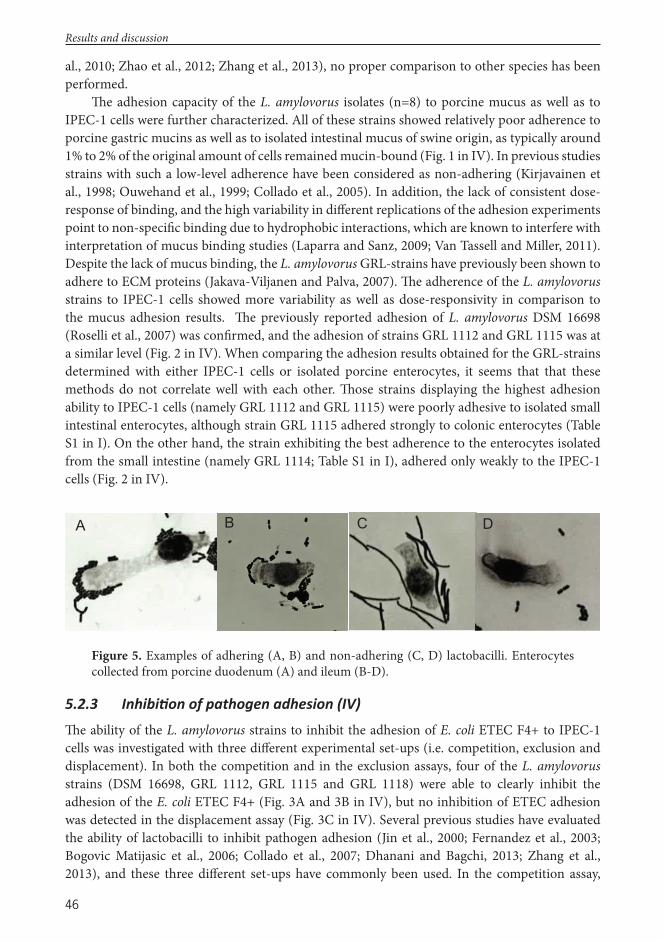
46
al., 2010; Zhao et al., 2012; Zhang et al., 2013), no proper comparison to other species has been performed.
Th e adhesion capacity of the L. amylovorus isolates (n=8) to porcine mucus as well as to IPEC-1 cells were further characterized. All of these strains showed relatively poor adherence to porcine gastric mucins as well as to isolated intestinal mucus of swine origin, as typically around 1% to 2% of the original amount of cells remained mucin-bound (Fig. 1 in IV). In previous studies strains with such a low-level adherence have been considered as non-adhering (Kirjavainen et al., 1998; Ouwehand et al., 1999; Collado et al., 2005). In addition, the lack of consistent dose-response of binding, and the high variability in diff erent replications of the adhesion experiments point to non-specifi c binding due to hydrophobic interactions, which are known to interfere with interpretation of mucus binding studies (Laparra and Sanz, 2009; Van Tassell and Miller, 2011). Despite the lack of mucus binding, the L. amylovorus GRL-strains have previously been shown to adhere to ECM proteins (Jakava-Viljanen and Palva, 2007). Th e adherence of the L. amylovorus strains to IPEC-1 cells showed more variability as well as dose-responsivity in comparison to the mucus adhesion results. Th e previously reported adhesion of L. amylovorus DSM 16698 (Roselli et al., 2007) was confi rmed, and the adhesion of strains GRL 1112 and GRL 1115 was at a similar level (Fig. 2 in IV). When comparing the adhesion results obtained for the GRL-strains determined with either IPEC-1 cells or isolated porcine enterocytes, it seems that that these methods do not correlate well with each other. Th ose strains displaying the highest adhesion ability to IPEC-1 cells (namely GRL 1112 and GRL 1115) were poorly adhesive to isolated small intestinal enterocytes, although strain GRL 1115 adhered strongly to colonic enterocytes (Table S1 in I). On the other hand, the strain exhibiting the best adherence to the enterocytes isolated from the small intestine (namely GRL 1114; Table S1 in I), adhered only weakly to the IPEC-1 cells (Fig. 2 in IV).
Results and discussion
B C D A
Figure 5. Examples of adhering (A, B) and non-adhering (C, D) lactobacilli. Enterocytes collected from porcine duodenum (A) and ileum (B-D).
5.2.3 Inhibi on of pathogen adhesion (IV)
Th e ability of the L. amylovorus strains to inhibit the adhesion of E. coli ETEC F4+ to IPEC-1 cells was investigated with three diff erent experimental set-ups (i.e. competition, exclusion and displacement). In both the competition and in the exclusion assays, four of the L. amylovorus strains (DSM 16698, GRL 1112, GRL 1115 and GRL 1118) were able to clearly inhibit the adhesion of the E. coli ETEC F4+ (Fig. 3A and 3B in IV), but no inhibition of ETEC adhesion was detected in the displacement assay (Fig. 3C in IV). Several previous studies have evaluated the ability of lactobacilli to inhibit pathogen adhesion (Jin et al., 2000; Fernandez et al., 2003; Bogovic Matijasic et al., 2006; Collado et al., 2007; Dhanani and Bagchi, 2013; Zhang et al., 2013), and these three diff erent set-ups have commonly been used. In the competition assay,

47
simultaneous addition of the Lactobacillus and the pathogen will result in competition for the previously empty binding sites, while the other two set-ups explore the possible displacement of previously bound bacteria. In the exclusion assay, Lactobacillus cells are allowed to bind to the target before the addition of the pathogen, while the reversed order of adding the diff erent bacteria is used in the displacement assay. When considering that lactobacilli are normal residents of the swine intestinal microbiota, i.e. they are continuously present, whereas the presence of pathogens is usually transient in the swine gut, it may be that the exclusion assay which assesses the ability of lactobacilli to protect intestinal cells from becoming colonized by pathogens is the most appropriate simulation of the in vivo situation. Moreover, it might be anticipated that the greatest inhibition of pathogen adhesion by lactobacilli would be obtained in the exclusion and / or competition assays, while displacement of pathogens already attached to the intestinal structures would be less likely to occur. Indeed, this was observed with the L. amylovorus strains used here, as inhibition of ETEC adhesion was observed in the competition and exclusion assays, but none of the strains were able to remove previously bound ETEC from IPEC-1 cells. A higher inhibition of pathogen adhesion in the exclusion and / or competition assays compared to the displacement assay has been noted also in some previous studies (Jin et al., 2000; Fernandez et al., 2003; Bogovic Matijasic et al., 2006; Zhang et al., 2011), but not in others (Collado et al., 2007; Dhanani and Bagchi, 2013).
As competition for the same adhesion sites is believed to be one mechanism behind pathogen exclusion, the ability to inhibit the adhesion of pathogens could be expected to correlate with adhesiveness of the strain. To some extent, this was the case with the L. amylovorus strains used in this study, as three (namely DSM 16698, GRL 1112 and GRL 1115) of the four strains which inhibited ETEC adhesion also exhibited adhesion ability to IPEC-1 cells. Th e inhibition of pathogen adhesion demonstrated by the poorly adherent strain GRL1118 suggests that in addition to competition for adhesion receptors other mechanisms must also be involved in the inhibition. Th ese could include coaggregation with the pathogen or secreted inhibitory factors (Lebeer et al., 2008). Th e culture fi ltrate of the strain GRL 1118 did not inhibit the growth of the ETEC strain more than those of the other GRL strains (see 5.2.4), but the possible production of substances specifi cally capable of inhibiting adherence was not investigated.
5.2.4 An microbial ac vity against intes nal pathogens (I, IV)
Th e spent culture fi ltrates collected from the LAB isolates were assayed against six indicator pathogens (I, unpublished results). Th e growth inhibitions caused by the SCF were quantifi ed using the area under the growth curve (AUC) value, which combines several points of time in the indicator growth curve, and has been shown to be a reliable parameter for determining changes occurring during bacterial growth (Skyttä and Mattila-Sandholm, 1991). Th e log CFU reductions in pathogen numbers caused by the culture fi ltrates were estimated using the regression formulas generated for each pathogen.
In accordance with previous studies estimating the antimicrobial properties of porcine lactobacilli (du Toit et al., 2000; Chang et al., 2001; De Angelis et al., 2006; Kim et al., 2007; Lin et al., 2007; Guo et al., 2010; Klose et al., 2010a; Klose et al., 2010b), a variable degree of inhibition in the growth of the indicator pathogens was caused by the SCF collected from the porcine LAB (I, unpublished results). When the diff erent indicator pathogens were compared against each other, E. coli ETEC F4+ and E. coli EHEC were inhibited statistically signifi cantly more than the other pathogens (Friedman test p<0.001; Fig. 6). Th e culture fi ltrates of the eight L.
Results and discussion

48
amylovorus strains were also assayed against the same pathogens (IV), and similarly, the growth of E. coli ETEC F4+ was the most effi ciently inhibited by these SCF (Fig. 4 in IV). As far as is known, the magnitude of growth inhibition exhibited by diff erent indicator pathogens has not been systematically compared previously, but strains of E. coli have been extensively inhibited by lactobacilli also in other studies (Guo et al., 2010; Klose et al., 2010a).
E. coli F18 E. coli F4 E. coli EHEC S. Typhimurium Y. enterocoliticaE. coli F4 0.000E. coli EHEC 0.002 0.000S.Typhimurium 0.504 0.000 0.000Y. enterocolitica 1.000 0.000 0.001 0.880L. monocytogenes 1.000 0.000 0.016 0.107 1.000
Figure 6. A box-and-wishkers plot illustrating the log CFU reductions achieved by the six indicator pathogens cultured in TSB media supplemented with the SCF collected from the porcine LAB isolates. Each box shows the median, quartiles and extreme values within a category. Friedman’s test post hoc values for pairwise comparisons (SPSS, 2012) with a Bonferroni correction for multiple comparisons are shown; statistically signifi cant values in bold.
Results and discussion

49
Results and discussion
For most of the indicator pathogens, the isolation site of the LAB strain did not infl uence the growth inhibition caused by the SCF, except for E. coli EHEC and L. monocytogenes, both of which were inhibited more by the fecal isolates (Kruskal-Wallis p=0.037 (E. coli EHEC) and p=0.016 (L. monocytogenes); Fig. 7). However, the small number of fecal isolates as well as the fact that these isolates represented diff erent Lactobacillus species compared to the intestinal isolates may have infl uenced this fi nding. At the species level, when comparing the fi ve largest Lactobacillus identifi cation groups, a statistically signifi cant diff erence was found in the inhibition of all the indicator pathogens (Kruskall-Wallis p<0.001). Generally, the isolates identifi ed as L. salivarius, L. johnsonii or L. amylovorus inhibited the indicator pathogens more than the isolates identifi ed as L. reuteri or L. mucosae (Fig. 8). Apparently, no systematic comparisons of diff erent species of lactobacilli in terms of pathogen growth inhibition have been performed previously, but three L. salivarius strains have been found to inhibit several swine pathogens, including fi ve E. coli strains and a Brachyspira pilosicoli strain, generally more than the other Lactobacillus species investigated (Klose et al., 2010a)
Th e pH of the culture fi ltrates collected from the LAB isolates (I), and from the L. amylovorus strains (IV) varied from 3.62 to 5.36 and from 3.8 to 4.5, respectively. In order to assess the eff ect of the pH value of the SCF on the pathogen growth inhibition, two diff erent experimental set-ups were used. In Study I, the pathogen growth inhibition evoked by the SCF was compared to those caused by the pH controls (MRS medium with pH adjusted with lactic acid or with HCl). It was evident that most of the growth inhibition caused by the SCF was due to lactic acid and low pH, but certain fi ltrates inhibited the growth of the indicators more than the pH controls (Table S1 in I). Th us, some of the LAB isolates appeared to produce other inhibitory substances in addition to organic acids. On the other hand, the pathogen growth inhibition caused by the pH-adjusted (pH 6.2) SCF of the L. amylovorus strains (IV) was low, indicating that most of the pathogen growth inhibition was due to acidity of the SCF (data not shown). Alternatively, inhibitory compounds with activity at acidic but not at neutral pH might have been produced, as has been observed for Lactocin S produced by Lactobacillus sakei (Mortvedt-Abildgaard et al., 1995). Th ese results are in line with previous studies showing that neutralization of the SCF oft en, although not invariably, abolishes the growth inhibition of pathogens caused by the fi ltrate (Lin et al., 2007; Guo et al., 2010; Klose et al., 2010a).

50
Results and discussion
Duo
denu
mJe
junu
mIle
umJe
junu
m1.
000
/ 1.0
00Ile
um1.
000
/ 1.0
001.
000
/ 1.0
00Fe
ces
0.05
7 / 0
.012
0.05
0 / 0
.031
0.03
1 / 0
.021
Fig
ure
7. A
box
-and
-wish
kers
plo
t ill
ustr
atin
g th
e lo
g CF
U r
educ
tions
ach
ieve
d by
the
six in
dica
tor
path
ogen
s cu
lture
d in
TSB
med
ia s
uppl
emen
ted
with
the
SCF
colle
cted
from
the
porc
ine
LAB
isola
tes
orig
inat
ing
from
the
duod
enum
, jeju
num
, ile
um a
nd fe
ces o
f sw
ine.
Each
box
show
s the
med
ian,
qua
rtile
s and
ext
rem
e va
lues
w
ithin
a c
ateg
ory.
Krus
kall-
Wal
lis t
est
post
hoc
valu
es f
or p
airw
ise c
ompa
rison
s (D
unn’s
196
4) w
ith a
Bon
ferr
oni c
orre
ctio
n fo
r mul
tiple
com
paris
ons a
re sh
own
for
E. co
li EH
EC an
d L.
mon
ocyt
ogen
es; s
tatis
tical
ly si
gnifi
cant
val
ues i
n bo
ld.
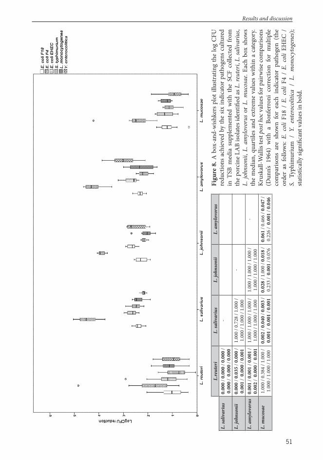
51
Results and discussion
L.re
uter
iL.
saliv
ariu
sL.
john
soni
iL.
am
ylov
orus
L. sa
livar
ius
0.00
0 / 0
.000
/ 0.
000
/ 0.
000
/ 0.
000
/ 0.0
00-
L. jo
hnso
nii
0.00
0 / 0
.035
/ 0.
000
/ 0.
001
/ 0.
000
/ 0.0
011.
000
/ 0.7
28 /
1.00
0 /
1.00
0 / 1
.000
/ 1.
000
-
L. a
myl
ovor
us0.
001
/ 0.0
01 /
0.00
1 /
0.00
2 /
0.00
0 /
0.00
11.
000
/ 1.0
00 /
1.00
0 /
1.00
0 / 1
.000
/ 1.
000
1.00
0 / 1
.000
/ 1.
000
/ 1.
000
/ 1.0
00 /
1.00
0-
L. m
ucos
ae1.
000
/ 0.3
84 /
1.00
0 /
1.00
0 / 1
.000
/ 1.
000
0.00
2 / 0
.040
/ 0.
003
/ 0.
001
/ 0.
001
/ 0.0
010.
028
/ 1.0
00 /
0.01
8 /
0.23
3 /
0.00
1 / 0
.076
0.06
1 / 0
.466
/ 0.
047
/ 0.
226
/ 0.
001
/ 0.0
46
Figu
re 8
. A b
ox-a
nd-w
ishke
rs p
lot i
llustr
atin
g th
e lo
g CF
U
redu
ctio
ns a
chie
ved
by th
e six
indi
cato
r pat
hoge
ns c
ultu
red
in T
SB m
edia
sup
plem
ente
d w
ith t
he S
CF c
olle
cted
fro
m
the p
orci
ne L
AB
isola
tes i
dent
ifi ed
as L
. reu
teri,
L. s
aliv
ariu
s, L.
john
soni
i, L.
am
ylov
orus
or
L. m
ucos
ae. E
ach
box
show
s th
e m
edia
n, q
uart
iles a
nd e
xtre
me
valu
es w
ithin
a c
ateg
ory.
Krus
kall-
Wal
lis te
st po
st ho
c val
ues f
or p
airw
ise co
mpa
rison
s (D
unn’s
196
4) w
ith a
Bon
ferr
oni
corr
ectio
n fo
r m
ultip
le
com
paris
ons
are
show
n fo
r ea
ch i
ndic
ator
pat
hoge
n (th
e or
der
as f
ollo
ws:
E. c
oli F
18 /
E. c
oli F
4 /
E. c
oli E
HEC
/
S. T
yphi
mur
ium
/ Y
. en
tero
colit
ica /
L.
mon
ocyt
ogen
es);
statis
tical
ly si
gnifi
cant
val
ues i
n bo
ld.
52
5.2.5 Correla ons between the proper es tested (I)
Th e possible association between the antimicrobial activity, adherence to the enterocytes and stress tolerance of the LAB strains was investigated with the Spearman’s rank-correlation test. For this analysis, a relative survival was calculated from the stress tolerance data of each isolate with the following formula: MPN (stress) / MPN (control), and the logarithmic of this value was used in the correlation analysis. Additionally, an overall average adhesion was calculated for each isolate from the adhesion values to enterocytes of diff erent intestinal origins. In an attempt to reduce the dimensions of the antimicrobial activity data, a principal components analysis (PCA) was performed. In the correlation matrix, all variables had correlation coeffi cients greater than 0.68, indicating that the correlations between the diff erent variables were suffi cient to include all of the variables in the analysis. Th e overall Kaiser-Meyer-Olkin (KMO) measure (0.89) and the individual KMO measures (all greater than 0.7) showed the adequacy of sampling, and the Bartlett’s test of sphericity was statistically signifi cant (p<0.005), thus the inhibitory data was suitable for PCA. In PCA, only one component with an eigenvalue greater than one was revealed, and this component explained 85.1% of the total variance.
In the correlation tests, only negative correlations were found between the diff erent properties, and all of them were relatively weak. Th e PCA values of the antimicrobial activity tests correlated negatively with the averaged adhesion (Spearman’s rho -0.346, p=0.001), indicating that the strains showing the highest adhesion ability generally exhibited lower inhibitory capacity of the indicator pathogens. To some extent, this was also illustrated when comparing the largest identifi cation groups (see 5.2.2 and 5.2.4). Additionally, the PCA values correlated negatively with the tolerance towards pH 2 (Spearman’s rho -0.519, p<0.0005) as well as with the tolerance toeards pH 4 (Spearman’s rho -0.220, p=0.034). Th us, those strains with the greatest ability to inhibit the growth of the indicator pathogens generally seemed to show lower tolerance towards low pH.
5.2.6 S mula on of cytokine produc on by dendri c cells (IV)
Th e possible stimulation of immune signaling in human MoDCs by the L. amylovorus strains was assessed at three diff erent bacterial concentrations. Induction of cytokine production was observed at the bacteria/DC ratio of 100:1, with substantial strain-dependent diff erences between the levels of cytokines induced (Fig. 5 in IV). Th e cytokine profi les induced by the strains were not clearly skewed towards Th 1 or Th 2 phenotypes. Instead, a mixed type of response, including Th 1 favoring (IL-12), Th 2 favoring (IL-10) as well as proinfl ammatory (TNF-α, IL-6, IL-1β, IP-10/CXCL10) cytokines, was stimulated by most of the strains. Similar cytokine profi les have been demonstrated also for L. gasseri strains (Luongo et al., 2013), while induction of proinfl ammatory cytokine profi les without high IL-12 expression has also been described for some strains of this Lactobacillus species (Stoeker et al., 2011). Interestingly, the strain NCFM of L. acidophilus, which is a close relative of L. amylovorus, has been shown to stimulate the diff erentiation of human DCs towards Th 2 generation via interaction between the SlpA protein and the DC-SIGN molecule (dendritic cell specifi c C-type lectin intercellular adhesion molecule 3-grabbing non-integrin) (Konstantinov et al., 2008b). Although the SlpA proteins of the L. amylovorus strains used in this study show a high similarity with the NCFM SlpA (Additional fi le 2 in IV), the Th 2 favoring action of NCFM was not observed with the present L. amylovorus strains. On the other hand, a skewing towards Th 1 type has also been observed to be caused by L. acidophilus NCFM
Results and discussion

53
in both murine BMDCs (Weiss et al., 2010) and human MoDCs (Gad et al., 2011), emphasizing the divergent eff ects observed for the same strains with diff erent experimental designs.
By using diff erent bacterial doses in DC stimulation experiments, a clear dose-dependent eff ect has been observed (Smits et al., 2005; Konstantinov et al., 2008b; Gad et al., 2011). For example, the ratio of IL-10 to IL-12 can even be reversed with increasing bacterial loads (Konstantinov et al., 2008b). Smits et al. (2005) observed optimal in vitro priming of regulatory DCs cells with MOI values of 1, whereas higher concentrations of bacteria evoked lower eff ect. Th is contrasts with the results of the present study, in which no notable induction of any of the cytokines measured was observed at the lower MOI values of 10 and 1, as compared to the negative control (data not shown).
5.2.7 An bio c suscep bility (III)
To evaluate if antibiotics could be used as selective agents in the bacterial culture of porcine fecal samples in Study III, the susceptibilities of the strains used in the feeding trial to fi ve antibiotics was tested with the broth microdilution method (Table 8). All of the strains were sensitive to G-penicillin, which seems to be a common feature among lactobacilli (Klare et al., 2007; Nawaz et al., 2011; Gueimonde et al., 2013). Four strains expressed resistance towards vancomycin, concurring with previous results obtained for human and food isolates (Klare et al., 2007; Nawaz et al., 2011) and also with the fact that many species of lactobacilli are intrinsically resistant to this antibiotic (Saarela et al., 2000; Gueimonde et al., 2013). Similarly, lactobacilli oft en exhibit resistance towards inhibitors of nucleic acid synthesis, such as fl uoroquinolones (Karapetkov et al., 2011; Gueimonde et al., 2013), and this was also the case for the porcine strains examined in this study, although the MIC values were somewhat variable. Th e most extensive variation was observed in the susceptibility of the strains to kanamycin and tobramycin; previously the resistance of lactobacilli isolated from diff erent food and animal sources towards aminoglycosides (i.e. gentamycin, tobramycin and kanamycin) has also been described to be variable (Nawaz et al., 2011; Bujnakova et al., 2014; Klose et al., 2014). No single antibiotic, which could have been used as a selective agent for isolation of the six strains from the porcine fecal samples, could be identifi ed amongst the tested antibiotic agents.
Table 8. Minimal inhibitory concentration (MIC) values (μg/ml) of the Lactobacillus strains for selected antibiotics assayed using microdilution method, with two parallel wells for each dilution series of the antibiotics.
Strain Penicillin G Enrofl oxacin Kanamycin Tobramycin VancomycinL. mucosae GLR 1167 <0.125 16 32 8 512L. salivarius GRL 1169 <0.125 2 256 64 >1024L. johnsonii GRL 1171 <0.125 32 256 128 <8L. reuteri GRL 1168 <0.125 16 32 128 512L. reuteri GRL 1170 <0.125 8 64 8 512L. amylovorus GRL 1112 <0.125 8 64 64 <8
Results and discussion

54
5.3 In vivo feeding trial (II, III)5.3.1 Animal performance and growth (II, III)
During the adaptation period, signs of mild diarrhea were recorded in some of the piglets, and a total of seven piglets (three in both the control and multispecies supplementation groups and one in the L. brevis group) had to be treated with the combination of trimethoprim and sulfadiazine (Tribrissen, Schering-Plough A/S, Denmark). On the other hand, all piglets remained healthy during the actual feeding trial, exhibiting no clinical signs of diarrhea or any other adverse eff ects.
When evaluating the possible benefi cial health eff ects of bacterial feed supplements in meat-producing animals, weight gain during the feeding trial is a commonly used measure, as improved growth would be highly desirable outcome of probiotic supplementation. In swine, enhanced growth rates have been observed both with monostrain (Abe et al., 1995; Guerra et al., 2007) and multistrain/-species (Choi et al., 2011a; Choi et al., 2011b) supplementations. On the other hand, a failure to promote growth has also been reported with both types of supplementation (Shim et al., 2005; Mair et al., 2010a). In the present study, no statistically signifi cant diff erences in the mean body weight (BW) or ADWG could be detected between the control and the two supplementation groups at any of the measurement points, although piglets receiving L. brevis supplementation had a numerically greater fi nal body weight than the piglets in the other two groups (Table 9). Th is concurs with an earlier study, in which a diff erent L. brevis strain (1E1) also induced a statistically non-signifi cant growth increase in neonatal piglets (Gebert et al., 2011). Th e high variability in growth promotion observed aft er various bacterial supplementations can be attributed to several factors, e.g. inherent characteristics of the species or strain being used. Additionally, host-specifi c properties like age and health status can aff ect the responses of the host to bacterial supplementation (Taras et al., 2007; Gaggia et al., 2010). Furthermore, it has been speculated that the hygienic conditions in experimental institutes may be too favorable to allow the appearance of any clear health benefi ts or growth promotion aft er bacterial supplementation, as the environmental pathogen load in these sites is presumably lower compared to that present in commercial intensive rearing units (Simon et al., 2001).
Table 9. Eff ects of a monostrain (L. brevis) and a multistrain Lactobacillus supplementations on the growth of piglets and digesta pH. All values represent mean ± standard deviation.
Growth pH
GroupInitial BW Final BW ADWG Jejunum Ileum Cecum Colon
Control 9.48 ± 1.88
20.26 ± 3.58
0.44 ± 0.09 6.4 ± 0.4 6.5 ± 0.3 6.0 ± 0.2 6.0 ± 0.2
L. brevis9.39 ± 1.66
21.21 ± 4.10
0.48 ± 0.11 6.4 ± 0.3 6.7 ± 0.4 6.0 ± 0.2 6.0 ± 0.3
Multispecies10.04 ±
1.5520.66 ±
4.490.42 ± 0.12 6.5 ± 0.2 6.6 ± 0.2 5.9 ± 0.2 6.1 ± 0.2
p-value1 0.627 0.619 0.415 0.967 0.237 0.642 0.779
1 P-values for Kruskall-Wallis test
Results and discussion

55
5.3.2 Detec on of the supplemented strains in the intes ne and feces (II, III)
To investigate the survivability of the strains fed to the piglets, bacterial colonies isolated from the fecal samples were analyzed with species-specifi c PCR (II, III). A total of 144 colonies (56 from the L. brevis group and 44 from the control and multispecies groups, respectively) were analyzed by L. brevis S-layer specifi c PCR; however, no positive results were found. On the other hand, each of the bacterial species included in the multispecies formulation was found among the fecal isolates analyzed (44 colonies from both the control and multispecies supplementation groups) (Table 3 in III). However, in PFGE, none of the strains isolated from the supplementation group fecal samples had an identical SmaI pattern (genotype) to the strains which had been fed to the piglets, even though one L. reuteri isolate from the slaughter sample did resemble L. reuteri GRL 1170 (Fig. 9).
Th is inability to isolate any of the strains fed to the piglets from the fecal samples was unexpected and surprising. Previously, using a similar technique, the same L. brevis strain that was used in the present trial was isolated from human stool samples in a small-scale intervention study (Rönkä et al., 2003), and as well from feces of mice that had received the strain via an oral gavage (unpublished data from the laboratory of A. Palva). In addition, the Lactobacillus strains included in the multispecies supplementation had all been originally isolated from the intestine and feces of swine, and based on the in vitro screening tests for tolerance of low pH and bile it was expected that the strains would be able to survive in the porcine GI-tract. However, as the bacterial culturing method used with the pooled fecal samples may have lacked suffi cient sensitivity to detect the strains, it cannot be concluded with certainty that the supplemented strains were unable to survive passage through the intestine of swine. Previously, certain Lactobacillus strains have been observed to survive transit through the GI tract of pigs (Table 3), but high variability have been observed in the excretion levels of the strain fed between diff erent host individuals (Gardiner et al., 2004).
To investigate the distribution of L. brevis cells in the porcine intestine in more detail, the presence of this strain in the intestinal lumen and mucosa of the piglets from the control and L. brevis supplementation groups was examined with real-time qPCR analysis of the digesta and fecal samples as well as with histological examination of cryosections using IFA (II). With both of these methods, L. brevis was found as the most abundant in the large intestine of piglets from the L. brevis supplementation group. In this group, genomic DNA of L. brevis was detected in the cecal digesta of four piglets as well as in the colonic digesta and feces of seven piglets while only one piglet from the control group had any detectable amounts of L. brevis DNA in its cecum (Fig. 1 in II). Th e inability to detect L. brevis in every piglet of the supplementation group may be due to the detection limit of the real-time qPCR assay (104 genomes g-1) or host-related genetic factors infl uencing the ability of the strain to colonize the intestinal microbiota. On the other hand, when examining the cecal and colonic cryosections, L. brevis-like cells were detected in samples from every piglet in the supplementation group and with high cell numbers, but accurate and reliable enumeration of individual cells was problematic owing to the large cell clusters that had formed (Fig. 2 in II). In addition, nonspecifi c staining of smaller-sized bacterial cells further complicated L. brevis cell counting; thus, numerical data for the large intestine cryosections is not presented. In the small intestine, genomic DNA of L. brevis was not found in the digesta of either the control or the supplementation group (<104 genomes g-1). However, L. brevis-like cells were detected in the jejunal and ileal cryosections from both groups, with the jejunum sections of the supplementation group displaying statistically signifi cantly higher cell numbers compared
Results and discussion

56
to the control group (Mann-Whitney U-test p<0.001), while no diff erence was observed in the ileum (Fig. 3 in II). Taken together, the results of the real-time qPCR analysis of the digesta and detection L. brevis-like cells with intact cell morphology in cryosections suggest that this strain may be capable of colonizing both the large, and to a lesser extent also the small, intestines of weaned piglets. However, the viability of the strain in the piglet intestine should be further confi rmed.
Interestingly, potentially indigenous L. brevis cells were also detected in a few control group piglets both in cryosections (with L. brevis SlpA-specifi c antiserum), as well as in the large intestinal digesta samples (with real-time qPCR). While this Lactobacillus species does not seem to be a prominent member of the porcine gut microbiota (Hill et al., 2002; Leser et al., 2002; Hill et al., 2005), it has been isolated previously from diff erent porcine samples (Krause et al., 1995; Martin et al., 2009; Gebert et al., 2011; Qi et al., 2011), thus the L. brevis-like cells detected in samples from the control group were in all likelihood indigenous strains of this Lactobacillus.
In an attempt to examine whether the L. brevis strain used would adhere more effi ciently to Peyer’s patches as compared to the regular intestinal wall areas, the small intestinal cryosections containing and not containing Peyer’s patches were examined for the presence of L. brevis-like cells with IFA (II). While it has been reported that several Lactobacillus species seem to be able to
Results and discussion
100
959085807570
C_0
C_0
C_0
F_S
F_S
F_0
C_S
C_S
F_0
C_S
F_S
F_S
F_S
F_S
F_S
F_S
C_S
C_S
L. reuteri GRL 1168
L. reuteri GRL 1170
Figure 9. Dendrogram representing the Dice coeffi cient-derived percentages of similarity among the distinct SmaI PFGE patterns obtained for fecal L. reuteri isolates. Th e source of isolates is listed on the right. C=control group; F=multispecies supplementation group; 0=sample taken before the start of the trial; S=sample taken at slaughter.

57
adhere specifi cally to Peyer’s patches (Perdigon et al., 2000; Plant and Conway, 2001; Edelman et al., 2002), the mucosal numbers of L. brevis-like cells were similar in both types of cryosections (Fig. 3 in II), suggesting that this strain lacks the capacity to adhere specifi cally to these tissue structures.
5.3.3 E ects of the supplementa ons on the bacterial and lactobacillar counts (II, III)
In order to reveal any major eff ects of the bacterial supplements on the GI-tract microbiota, the total numbers of bacteria (II, III) and lactobacilli (III) were quantifi ed with real-time qPCR. While increased numbers of digestal or fecal lactobacilli and / or other bacterial species considered benefi cial for the host have been detected in some previous porcine feeding trials (Pollmann et al., 1980; De Angelis et al., 2007; Choi et al., 2011b), the multispecies supplementation used in the present study did not infl uence the abundance of lactobacilli in either the intestinal contents or feces of the piglets (Fig. 2 in III). However, increased numbers of total bacteria were detected in the jejunum of the multispecies supplementation group, as compared to the control group (Mann-Whitney U-test p=0.023; Fig. 2 in III), suggesting that some changes had occurred in the indigenous microbiota of this intestinal compartment due to the multispecies supplementation. In the L. brevis supplementation group, the quantities of bacteria remained similar to the control group in all intestinal compartments investigated (Table 2 in II), which is in agreement with a previous porcine feeding trial performed with L. amylovorus DSM 16698T (Konstantinov et al., 2008a).
5.3.4 Immunological e ects of the supplementa ons (II, III)
To determine whether the L. brevis supplement could induce systemic humoral immune responses, total IgA and IgG as well as L. brevis-specifi c IgG concentrations were measured from serum samples of the L. brevis and the control group (II). While in both groups the concentrations of serum IgA and IgG increased during the feeding trial, no statistically signifi cant diff erences could be observed between the groups (Fig. 5 in II). Additionally, the levels of L. brevis-specifi c IgG remained similar in both groups (data not shown). Th ese results indicate that the L. brevis strain used does not elicit a measurable humoral immune response, or alternatively, that the length of the bacterial supplementation was too short to induce any detectable antibody response. Th e detection of L. brevis-specifi c IgG antibodies in piglets from the control group is likely due to either cross-reactivity or the stimulation of antibody production by an autochthonous L. brevis strain. Correspondingly, the presence of serum IgA antibodies that recognize L. rhamnosus GG, considered an atypical bacterium of the porcine microbiota, has been demonstrated also in piglets that did not receive this strain as a feed supplement (Casini et al., 2007). Furthermore, IgA antibodies able to recognize L. amylovorus DSM 16698T have been detected in both the supplementation and control groups (Konstantinov et al., 2008a), thus in swine it seems that it may be rather common to detect cross-reactive antibodies binding to various Lactobacillus species.
Th e potential eff ects of the L. brevis supplement on the expression of selected immunological genes were fi rst screened by investigating the intestinal mucosal samples of fi ve piglets from the L. brevis and the control groups by real-time qPCR (data not shown). However, as the RNA extracted from the samples was of low quality and quantity, an alternative method, QuantiGene 2.0 Plex Assay (Aff ymetrix/Panomics, USA), permitting mRNA quantitation without RNA extraction, was used for the analysis of the mucosal samples.
Results and discussion

58
Examination of the gene expression of eight cytokines (i.e. IL-1β, IL-4, IL-6, IL-8, TGF-β1, IFN-α, IFN-γ and TNF) in the intestinal mucosa revealed that although both types of supplementations did induce some alterations in the expression of these cytokines, more pronounced eff ects were triggered by the multispecies preparation (Fig. 4 in II; Fig. 3 in III). Th e mRNA levels of the cytokine TGF-β1 were downregulated in the ileum by both of the supplementations (Mann-Whitney U test p=0.015 (L. brevis group) and <0.001 (multispecies group)), and also in the jejunum (p<0.001) and the colon (p=0.017) by the multispecies formulation. Th is contrasts to previous results obtained for another type of multispecies supplementation containing four bacterial strains which exerted no eff ect on the expression of this cytokine in the intestine of weaned piglets (Mair et al., 2010b). Moreover, increased production of TGF- β1 in the Caco-2 cell line has been reported aft er contact with gut commensals, including Lactobacillus strains (Zeuthen et al., 2008). Despite the downregulation of the anti-infl ammatory TGF-β1 cytokine, no visual changes were observed in the pathological investigation of the porcine intestinal walls, suggesting that the supplementations had not induced any infl ammation in the intestinal wall.
Interestingly, the mRNA levels of the cytokines IL-6 and IL-4 were upregulated in the cecum by both types of supplementation (Fig. 4 in II; Fig. 3 III), although statistical signifi cance was reached only by the IL-4 change in the multispecies group (p=0.035) and the IL-6 change in the L. brevis group (p=0.023). While changes in the expression of IL-6 in the small intestinal mucosa of piglets have been reported to occur e.g. during weaning (Pie et al., 2004) as well as aft er bacterial supplementation (Zhang et al., 2011; Deng et al., 2013), there does not seem to be any data available on the expression patterns of this cytokine in the colon of piglets. With regard to the increased cecal mRNA expression of IL-4 stimulated by the multispecies supplementation, this change was mainly attributable to two individual piglets in that group, and when the Mann-Whitney U test analysis was performed without these piglets, no diff erence between the groups was any longer apparent (data not shown). However, no special features were observed in the pathological investigation of the cecum wall samples from these two individuals. It does seem that the intestinal expression of this major Th 2 cytokine has not been investigated in other bacterial feeding studies performed in swine, and little is known about the function of IL-4 in the porcine intestine. In human intestinal cell lines, IL-4 has been found to reduce the barrier function of the epithelial monolayer, resulting in its increased permeability to macromolecules (Colgan et al., 1994; Berin et al., 1999). However, there are indications that species diff erences exist in the expression and functions of IL-4 (Reddy et al., 2000; Raymond and Wilkie, 2004; Bautista et al., 2007), and diff erences between swine breeds have been observed (Verfaillie et al., 2001; de Groot et al., 2005), refl ecting possible genetic factors infl uencing immune function. Th us, the functions of IL-4 in the porcine intestine remain to be explored in further studies. In addition to the increased mRNA expression of IL-4, also the mRNA expression of IFN-α was upregulated in the cecum of the multispecies supplementation group (p=0.001). While the production of interferons has traditionally been considered to be primarily induced by viruses, also bacteria, including lactobacilli, are known to stimulate their production (Kitazawa et al., 1994; Bogdan et al., 2004).
In the colon, the multispecies formulation induced a downregulated mRNA expressions of IL-8 (p=0.008) and TNF (p=0.001). Previously, a fi ve-strain bacterial supplementation has been reported to increase the expression of IL-8 in the ileum of weaned piglets, but the large intestinal compartments were not investigated (Walsh et al., 2008). In addition, the expression of TNF in the colon and the small intestine of weaned piglets was not altered by a four-strain supplement
Results and discussion
59
(Mair et al., 2010b). Th ese dissimilarities in the results of various studies are probably due to diff erences in the bacterial strains used, as well as in the experimental designs, such as the genetic background of the animals, as these factors can aff ect the results of immunological studies (Joling et al., 1993; de Groot et al., 2005; Flori et al., 2011). One limitation of the present study was the heterogeneity of the study population, as the piglets represented a mixture of diff erent breeds as well as genders, and this may well have exerted an infl uence on the outcomes. Moreover, when considering the complexity of the mucosal immune system, it is very diffi cult to draw any fi rm conclusions about the results from cytokine expression studies, as is predicting what would be the concrete health consequences of increased or decreased gene expression of diff erent cytokines triggered by bacterial supplementation. Consequently, reasonable speculation on these phenomena will need to be based on more detailed information about the immune system networks.
5.4 The surface layer proteins of Lactobacillus amylovorus strains (IV)5.4.1 Genomic characteriza on and expression analysis of L. amylovorus
S-layer proteins (IV)
In the in silico analysis of the L. amylovorus genomes, several slp genes were revealed in each of the strains. Th e presence of multiple S-layer protein genes is common in lactobacilli, and has previously been reported e.g. in strains of L. acidophilus (Boot et al., 1995) as well as those closely related to L. acidophilus, including L. amylovorus (Boot et al., 1996b), and also in strains of L. brevis (Jakava-Viljanen et al., 2002) and L. crispatus (Sillanpää et al., 2000). Th e L. amylovorus slp genes with high homology to L. acidophilus NCFM slpA and slpB (Konstantinov et al., 2008b) were also named slpA and slpB, respectively, and the third type of slp-like gene detected was designated slpC. For both slpA and slpB, most of the strains carried only one gene homologue, but two distinct homologues of slpA and slpB were detected in GRL 1117 and DSM20531, respectively. Th ere was more variation in the number of slpC-type genes, as one to three homologues were recognized in the L. amylovorus strains (Fig. 6 in IV). A phylogenetic tree constructed based on the deduced amino acid sequences of the slpA, slpB and slpC gene products indicated that the SlpA-like sequences have diversifi ed most during the evolution, while the SlpB-type proteins have remained more similar to each other and the predicted SlpC-type proteins have formed three distinct groups (Fig. 6 in IV, the deduced amino acid sequences of the L. amylovorus Slp proteins are presented in Additional fi le 1 in IV).
Comparison of the sequence data of the slp genes with the results of the aminoterminal sequencing or the peptide mapping analysis performed for the L. amylovorus S-layer proteins revealed that the major S-layer protein bands seen in the surface protein profi les of the strains (Fig. 7 in IV) were all encoded by slpA-like genes. Th e amino acid sequences of the SlpA proteins were found to be very similar to that of the L. acidophilus NCFM SlpA protein, especially in the carboxy terminal region. Th is may refl ect the role of the C-terminal part in cell wall binding, previously described in L. acidophilus (Smit et al., 2001) and L. crispatus (Antikainen et al., 2002). Bands representing the products of slpB- and slpC-like genes were also identifi ed in the surface protein profi les of the strains: the former in strains GRL 1117 and DSM 16698 but the latter only in strain DSM 16698 (Fig. 7 in IV). While multiple S-layer genes are commonly carried by lactobacilli, simultaneous expression of more than one of these gene seems rare, but this phenomenon has been previously described for L. brevis ATCC 14869 (Jakava-Viljanen et al., 2002) and L. acidophilus NCMF (Goh et al., 2009).
Results and discussion

60
5.4.2 The role of L. amylovorus S-layer proteins in adherence (IV)
To explore if the L. amylovorus S-layer proteins have a role in the adherence capacity of the strains to IPEC-1 cells, a protein presentation system based on the propensity of S-layer proteins to recrystallize in a native manner on purifi ed CWF was developed (an electron micrograph of purifi ed CWF: Fig. 8B in IV). In an attempt to minimize the presence of Slp protein aggregates in the adhesion assay, the dilute Slp protein fraction was separated from the precipitated protein by centrifugation, as has been described previously (Åvall-Jääskeläinen et al., 2008). Th e method applied to the purifi cation of the CWF preserves the covalently attached polymeric components like teichoic acids and polysaccharides, which ensures the proper self-assembly of the recombinant Slp:s and leads to the presentation of the S-layer proteins in the native and symmetric organization observed on the surface of bacterial cells. In the adhesion assays, the detection of the Slp-coated CWF was performed with an indirect immunofl uorescence staining using Slp-specifi c antibodies. Uncoated CWF were used as negative controls, but as attempts to produce functional antibodies against purifi ed cell wall fragments were unsuccessful, these were biotinylated prior to the adhesion assay, and detected with labeled streptavidin aft er the assay.
While none of the uncoated CWF adhered to the IPEC-1 cells, at least a low level of adherence was demonstrated for all of the Slp coated CWF. However, the adherence level of the coated CWF did not correlate to that of the bacterial strain from which the proteins had originated. For example, the adhesion of CWF coated with SlpA from the strains DSM 16698 (Fig. 8D in IV), GRL 1112 or GRL 1115 was poor, even though the intact cells of these strains were adhesive to IPEC-1 cells (Fig. 2 in IV). On the other hand, the CWF coated with Slp:s from the weakly adhering strains GRL 1117 (Fig. 8H in IV), DSM 20531T (Fig. 8G in IV) and GRL 1118 did display affi nity for IPEC-1 cells. Th e SlpB of the strain DSM 16698 as well showed some adhesiveness (Fig. 8E in IV), when compared to SlpA (Fig. 8D in IV) or SlpC (Fig. 8F in IV) of the same strain.
Th ese results indicate that none of the L. amylovorus S-layer proteins examined in this study solely mediate adhesion of the strains to IPEC-1 cells. However, also other non-covalently attached cell wall components may have been removed during the preparation of the CWF, and that these structures may well have a role in the mediation of adherence. Th is methodological limitation also applies to experiments based on chemical extraction of the S-layer from the bacterial surface; while this procedure has been shown to decrease the adhesion of lactobacilli to diff erent targets (Hynönen and Palva, 2013), providing indirect evidence that the S-layer structure is involved in the adhesion process, a role for other non-covalently attached molecules removed in the extraction process cannot be excluded.
Th e highly effi cient binding of the S-layer protein originating from the weakly adhesive strain GRL 1117 indicates that some component(s) on the surface of this strain shield(s) the S-layer and thus prevents the adhesion of the strain to IPEC-1 cells. Th is kind of phenomenon has been observed in L. rhamnosus GG: the exopolysaccharide component shields the mucus-binding fi mbriae, reducing the adhesive capacity of the strain for mucus (Kankainen et al., 2009; Lebeer et al., 2009). While genes putatively participating in exopolysaccharide synthesis have been identifi ed in all of the L. amylovorus strains studied (unpublished data from the laboratory of A. Palva), no biochemical evidence of their presence has been described so far. Th us, the reason for reduced adhesiveness of the intact cells of GRL 1117 compared to that of the SlpA-protein originating from this strain remains to be explained.
Results and discussion

61
6 Conclusions and future aspects
Th e importance of the gut microbiota to the health and well-being of the host has been increasingly recognized during the last decade, and the use of microbial supplements to modulate the composition and function of this complex community has generated much interest in the fi elds of human and veterinary medicine. While various types of microbial products have been used in production animals, including swine, to maintain and improve the health and productivity of the host animal, several factors related to the selection process of the microbes as well as the functional mechanisms behind the observed eff ects still remain unrevealed. Th us, comprehensive studies regarding the use of probiotic microbes on various animal species are warranted. Th e main aim of this study was to characterize the probiotic potential of porcine lactobacilli for use in swine production. In addition, as lactobacillar S-layers are attractive candidates for antigen carriers in vaccine vectors, strains of lactobacilli possessing an S-layer were also investigated in this work.
I. Th e in vitro screening assays for selected traits commonly considered important for a putative probiotic led to the following conclusions:
1) the probiotic properties of porcine LAB strains, such as acid and bile tolerance, adherence to host structures, ability to inhibit pathogens as well as immunostimulatory patterns vary considerably, even between strains of the same species. Th is fi nding emphasizes the need for careful selection of the putative probiotic strain,
2) the porcine LAB isolates examined in this study generally adhered more effi ciently to large intestinal enterocytes, in comparison to those collected from the small intestine. While the isolation site of the strain did not aff ect its adhesion preferences towards enterocytes of diff erent origins, this may be infl uenced by the fact that the strains were isolated from intestinal digesta, and it would be interesting to explore this aspect also with strains originating from the mucosa of diff erent intestinal sections,
3) the ability to inhibit pathogen adhesion was not strictly correlated with the adhesive capacity of the porcine Lactobacillus strain. Th us, in addition to competition for the same adhesion sites, also other mechanisms are involved in the inhibition of pathogen adhesion,
4) while the SCF of the LAB isolates inhibited the growth of several pathogens, this inhibition was mainly due to low pH value of the fi ltrate. However, some of the isolates seemed to produce also other inhibitory compounds, but the nature of these substances remains to be investigated in future studies and
5) some of the properties correlated negatively with each other; for example, LAB strains showing high adhesion ability to enterocytes generally exhibited lower ability to inhibit pathogen growth. Although these correlations were only weak, this fi nding may indicate that it may be diffi cult to discover strain(s) that would possess all the characteristics considered desirable for probiotic bacteria. Indeed, no single strain showing excellent performance in all of the diff erent properties tested in this work was detected.
Conclusions and future aspects

62
II. Th e in vivo feeding trial led to the following conclusions:1) both of the supplementations used (i.e. a monostrain preparation containing
L. brevis ATCC 8287, and a multistrain preparation containing six lactobacilli of porcine origins) were well tolerated by piglets, and produced no adverse eff ects. Th e supplementations did not seem to exert any growth enhancing eff ects; however, this aspect needs to be explored in more detail in clinically relevant settings,
2) both supplementations induced some alterations of cytokine mRNA expression in the intestinal mucosa, with more pronounced changes being produced by the multistrain preparation, especially in the large intestinal area. Despite these immunological alterations in the intestinal mucosa, neither of the supplementations caused mucosal infl ammation. Th e health consequences of these changes in mucosal cytokine expression will need to be clarifi ed in future studies,
3) the supplemented strains may not be able to survive transit through the swine GI-tract; however, as the method used may have lacked suffi cient sensitivity, the gut transit tolerance of these strains will need to be investigated further in more detail. Nevertheless, it seems probable that these strains are not able to effi ciently compete with the indigenous gut microbiota of weaned piglets. In future studies, it would be interesting to investigate the ability of these strains to colonize and / or to survive passage through the GI-tract of suckling piglets, and
4) both of the supplementations examined in this work may be suitable for use as probiotics in swine, but as the effi cacy of these putative probiotics under intensive production systems cannot be evaluated based on these preliminary results, more comprehensive studies will still be needed to assess their possible eff ects on porcine health and immunity. Additionally, the potential use of the L. brevis strain as a vaccine carrier for swine pathogens is worthy of further investigations e.g. with immunization studies performed using a recombinant strain expressing suitable antigens, such as the receptor binding domain of the FedF adhesin of F18+ E. coli strains causing oedema disease and / or weaning diarrhea.
III. Th e genomic characterization of the S-layer protein-carrying L. amylovorus strains led to
the following conclusions:1) several slp –like genes are carried by each of the strains; these genes were named
slpA, slpB and slpC. In all of the strains, the major Slp protein was encoded by the slpA gene, and this protein group shared high sequence similarity with the L. acidophilus NCFM SlpA protein. In addition to the major S-layer protein SlpA, two strains carried also other S-layer-like proteins on their surface, namely SlpB and / or SlpC and
2) as the adherence level of the CWF coated with L. amylovorus Slp proteins did not correlate to that of the bacterial strains from which the proteins had originated, it seems apparent that none of the major Slp proteins alone mediate the adhesion of the strain to IPEC-1 cells. However, the putative role of other non-covalently attached cell wall components possibly removed during the preparation of the CWF should be examined in further studies.
Conclusions and future aspects

63
7 Acknowledgements
Th is work was carried out in the Department of Veterinary Biosciences, Faculty of Veterinary Medicine, University of Helsinki. Th e fi nancial support received for this research from the University of Helsinki, the Ministry of Agriculture and Forestry, the Academy of Finland, the Finnish Veterinary Foundation, the Finnish Foundation of Veterinary Research, the Finnish Graduate School on Applied Biosciences and the Walter Ehrstöm Foundation is gratefully acknowledged. I thank the heads of the Department, Lars-Axel Lindberg, Antti Sukura and Airi Palva, for providing such excellent working and educational facilities.
I am deeply grateful to my main supervisor Professor Airi Palva for her advice, guidance and support during the ups and downs of the research process and for her much-needed encouragement during the completion of this dissertation. I want to gratefully acknowledge my supervisor Professor Johanna Björkroth for her very valuable expertise and advice during the course of this work. I want to express my deep gratitude to my supervisor University lecturer Joanna Koort for her patient help and counselling; your door was always open to me, whether I needed help in research related issues or in knitting. I am also thankful to Docent Ilkka Palva for stimulating scientifi c discussions, and I’m deeply sorry that he is no longer here with us. I sincerely thank the offi cial reviewers of this thesis, Professor Tapani Alatossava and Docent Maria Saarela, for their constructive and valuable comments that helped to improve the quality of this thesis. Ewen MacDonald is gratefully acknowledged for the revision of the English language.
I am extremely grateful to all my co-authors for their contributions; without your help this work would never have been completed. I especially want to thank Erja Malinen for her generous help with the statistics and the SPSS programme, Agneta Lindholm for her serene but enthusiastic attitude which helped me to calm down even in tight situations, Ulla Hynönen for her kind encouragement and willingness to answer my endless questions, Miia Jakava-Viljanen for her invaluable help with the practical aspects of the animal trial and Gloria Solano-Aguilar for her generous help during my visit to BHNRC. Over these years, I have had the opportunity to work with exceptional people, and I want to warmly thank all of the former and present members of Airi Palva’s research group for their companionship and for creating such a pleasant and enjoyable atmosphere in the laboratory as well as around the lunch table. Special thanks to my bald-headed roommate for immunological discussions. I also want to express my gratitude to Sinikka Ahonen, Esa Pohjolainen, Anja Osola and Ulla Viitanen, the personnel in EELA (later Evira) for their help in conducting the animal trial, the staff in BHNRC (USDA) for helping me during my visit, and all the others who have off ered their skilful technical assistance throughout this work. Th e secretaries in our Department are also gratefully acknowledged for handling of numerous practical things over the years.
Finally, I want to thank all my friends and family for support and encouragement. I especially want to thank my husband Ville, without whom I would still be working on the Figures of Study I, and my children Teemu and Veera, without whom there would be much less joy in my life. Vantaa, August 2014
Acknowledgements
64
8 References
Aarestrup, F.M. 2002. Eff ects of termination of AGP use on antimicrobial resistance in food animals. In: Beyond antimicrobial growth promoters in food animal production, Foulum, Denmark, pp. 6-12.[available at http://www.who.int /gfn/links/en/FoulumWorkingPapers.pdf]
Aarestrup, F.M., Jensen, V.F., Emborg, H.D., Jacobsen, E., Wegener, H.C., 2010. Changes in the use of antimicrobials and the eff ects on productivity of swine farms in Denmark. Am. J. Vet. Res. 71, 726-733.
Abe, F., Ishibashi, N., Shimamura, S., 1995. Eff ect of administration of bifi dobacteria and lactic acid bacteria to newborn calves and piglets. J. Dairy Sci. 78, 2838-2846.
Adams, M.R., Hall, C.J., 1988. Growth-inhibition of foodborne pathogens by lactic and acetic-acids and their mixtures. Int. J. Food Sci. Technol. 23, 287-292.
Alakomi, H.L., Skyttä, E., Saarela, M., Mattila-Sandholm, T., Latva-Kala, K., Helander, I.M., 2000. Lactic acid permeabilizes gram-negative bacteria by disrupting the outer membrane. Appl. Environ. Microbiol. 66, 2001-2005.
Alander, M., Satokari, R., Korpela, R., Saxelin, M., Vilpponen-Salmela, T., Mattila-Sandholm, T., von Wright, A., 1999. Persistence of colonization of human colonic mucosa by a probiotic strain, Lactobacillus rhamnosus GG, aft er oral consumption. Appl. Environ. Microbiol. 65, 351-354.
Anadón, A., Martinez-Larranaga, M.R., Aranzazu Martinez, M., 2006. Probiotics for animal nutrition in the European Union. Regulation and safety assessment. Regul. Toxicol. Pharmacol. 45, 91-95.
Annuk, H., Shchepetova, J., Kullisaar, T., Songisepp, E., Zilmer, M., Mikelsaar, M., 2003. Characterization of intestinal lactobacilli as putative probiotic candidates. J. Appl. Microbiol. 94, 403-412.
Antikainen, J., Anton, L., Sillanpää, J., Korhonen, T.K., 2002. Domains in the S-layer protein CbsA of Lactobacillus crispatus involved in adherence to collagens, laminin and lipoteichoic acids and in self-assembly. Mol. Microbiol. 46, 381-394.
Apajalahti, J.H., Kettunen, A., Bedford, M.R., Holben, W.E., 2001. Percent G+C profi ling accurately reveals diet-related diff erences in the gastrointestinal microbial community of broiler chickens. Appl. Environ. Microbiol. 67, 5656-5667.
Artis, D., 2008. Epithelial-cell recognition of commensal bacteria and maintenance of immune homeostasis in the gut. Nat. Rev. Immunol. 8, 411-420.
Axelsson, L. 2004. Lactic Acid Bacteria: classifi cation and physiology. In: Salminen, S., von Wright, A., Ouwehand, A. (Eds.), Lactic acid bacteria: Microbiological and Functional aspects. Marcel Dekker, New York, USA, pp. 1-66.
Baber, D.W., Coblentz, B.E., 1987. Diet, nutrition, and conception in feral pigs on santa-catalina island. J. Wildl. Manage. 51, 306-317.
Barrangou, R., Lahtinen, S.J., Ibrahim, F., Ouwehand, A.C. 2012. Genus Lactobacillus. In: Lahtinen, S., Ouwehand, A.C., Salminen, S., von Wright, A. (Eds.), Lactic acid bacteria: microbiological and functional aspects. CRC Press, USA, pp. 77-92.
Barrow, P.A., Brooker, B.E., Fuller, R., Newport, M.J., 1980. Th e attachment of bacteria to the gastric epithelium of the pig and its importance in the microecology of the intestine. J. Appl. Bacteriol. 48, 147-154.
Bateup, J.M., Dobbinson, S., McConnell, M.A., Munro, K., Tannock, G.W., 1998. Molecular analysis of the composition of Lactobacillus populations inhabiting the stomach and caecum of pigs. Microb. Ecol. Health Dis. 10, 95-102.
References

65
Bauer, E., Williams, B.A., Smidt, H., Mosenthin, R., Verstegen, M.W.A., 2006. Infl uence of dietary components on development of the microbiota in single-stomached species. Nutr. Res. Rev. 19, 63-78.
Bautista, E.M., Nfon, C., Ferman, G.S., Golde, W.T., 2007. IL-13 replaces IL-4 in development of monocyte derived dendritic cells (MoDC) of swine. Vet. Immunol. Immunopathol. 115, 56-67.
Begley, M., Gahan, C.G.M., Hill, C., 2005. Th e interaction between bacteria and bile. FEMS Microbiol. Rev. 29, 625-651.
Bengmark, S., 1998. Ecological control of the gastrointestinal tract. Th e role of probiotic fl ora. Gut 42, 2-7.
Berg, R.D., 1996. Th e indigenous gastrointestinal microfl ora. Trends Microbiol. 4, 430-435. Bergman, E.N., 1990. Energy contributions of volatile fatty acids from the gastrointestinal tract in
various species. Physiol. Rev. 70, 567-590. Berin, M.C., Yang, P.C., Ciok, L., Waserman, S., Perdue, M.H., 1999. Role for IL-4 in macromolecular
transport across human intestinal epithelium. Am. J. Physiol. 276, 1046-1052. Bermudez-Humaran, L.G., Kharrat, P., Chatel, J.M., Langella, P., 2011. Lactococci and lactobacilli as
mucosal delivery vectors for therapeutic proteins and DNA vaccines. Microb. Cell. Fact. 10 Suppl 1, 4.
Bernardeau, M., Gueguen, M., Smith, D.G., Corona-Barrera, E., Vernoux, J.P., 2009. In vitro antagonistic activities of Lactobacillus spp. against Brachyspira hyodysenteriae and Brachyspira pilosicoli. Vet. Microbiol. 138, 184-190.
Bernardeau, M., Guguen, M., Vernoux, J.P., 2006. Benefi cial lactobacilli in food and feed: long-term use, biodiversity and proposals for specifi c and realistic safety assessments. FEMS Microbiol. Rev. 30, 487-513.
Bernardeau, M., Vernoux, J.P., 2013. Overview of diff erences between microbial feed additives and probiotics for food regarding regulation, growth promotion eff ects and health properties and consequences for extrapolation of farm animal results to humans. Clin. Microbiol. Infec. 19, 321-330.
Bezkorovainy, A., 2001. Probiotics: determinants of survival and growth in the gut. Am. J. Clin. Nutr. 73 Suppl, 399-405.
Bibiloni, R., Fernando, P., De Antoni, G.L., 1999. Will a high adhering capacity in a probiotic strain guarantee exclusion of pathogens from intestinal epithelia. Anaerobe 5, 519-524.
Björkroth, K.J., Schillinger, U., Geisen, R., Weiss, N., Hoste, B., Holzapfel, W.H., Korkeala, H.J., Vandamme, P., 2002. Taxonomic study of Weissella confusa and description of Weissella cibaria sp. nov., detected in food and clinical samples. Int. J. Syst. Evol. Microbiol. 52, 141-148.
Blum, S., Reniero, R., Schiff rin, E.J., Crittenden, R., Mattila-Sandholm, T., Ouwehand, A.C., Salminen, S., von Wright, A., Saarela, M., Saxelin, M., Collins, K., Morelli, L., 1999. Adhesion studies for probiotics: need for validation and refi nement. Trends Food Sci. Tech. 10, 405-410.
Bogdan, C., Mattner, J., Schleicher, U., 2004. Th e role of type I interferons in non-viral infections. Immunol. Rev. 202, 33-48.
Bogovic Matijasic, B., Narat, M., Zoric Peternel, M., Rogelj, I., 2006. Ability of Lactobacillus gasseri K 7 to inhibit Escherichia coli adhesion in vitro on Caco-2 cells and ex vivo on pigs’ jejunal tissue. Int. J. Food Microbiol. 107, 92-96.
Bomba, A., Nemcová, R., Strojný, L., Mudronová, D. 2012. Probiotics for farm animals. In: Lahtinen, S., Ouwehand, A.C., Salminen, S., von Wright, A. (Eds.), Lactic acid bacteria: microbiological and functional aspects. CRC Press, USA, pp. 633-670.
References

66
Boot, H.J., Kolen, C.P., Pouwels, P.H., 1995. Identifi cation, cloning, and nucleotide sequence of a silent S-layer protein gene of Lactobacillus acidophilus ATCC 4356 which has extensive similarity with the S-layer protein gene of this species. J. Bacteriol. 177, 7222-7230.
Boot, H.J., Kolen, C.P., Pouwels, P.H., 1996a. Interchange of the active and silent S-layer protein genes of Lactobacillus acidophilus by inversion of the chromosomal slp segment. Mol. Microbiol. 21, 799-809.
Boot, H.J., Kolen, C.P.A.M., Pot, B., Kersters, K., Pouwels, P.H., 1996b. Th e presence of two S-layer-protein-encoding genes is conserved among species related to Lactobacillus acidophilus. Microbiol-SGM 142, 2375-2384.
Borchers, A.T., Selmi, C., Meyers, F.J., Keen, C.L., Gershwin, M.E., 2009. Probiotics and immunity. J. Gastroenterol. 44, 26-46.
Bosi, P., Trevisi, P., 2010. New topics and limits related to the use of benefi cial microbes in pig feeding. Benef. Microbes 1, 447-454.
Briones, A.M., Jr., Reichardt, W., 1999. Estimating microbial population counts by ‘most probable number’ using Microsoft Excel. J. Microbiol. Methods 35, 157-161.
Brul, S., Coote, P., 1999. Preservative agents in foods. Mode of action and microbial resistance mechanisms. Int. J. Food Microbiol. 50, 1-17.
Bujnakova, D., Strakova, E., Kmet, V., 2014. In vitro evaluation of the safety and probiotic properties of Lactobacilli isolated from chicken and calves. Anaerobe 29, 118-127.
Böhmer, B.M., Kramer, W., Roth-Maier, D.A., 2006. Dietary probiotic supplementation and resulting eff ects on performance, health status, and microbial characteristics of primiparous sows. J. Anim. Physiol. Anim. Nutr. 90, 309-315.
Cabo, M.L., Murado, M.A., Gonzalez, M.P., Pastoriza, L., 1999. A method for bacteriocin quantifi cation. J. Appl. Microbiol. 87, 907-914.
Callesen, J. 2002. Eff ects of termination of AGP use on pig welfare and productivity. In: Beyond Antimicrobial Growth Promoters in Food Animal Production, Foulum, Denmark, pp. 43-46.[available at http://www.who.int/gfn/links/en/FoulumWorkingPapers.pdf]
Canchaya, C., Claesson, M.J., Fitzgerald, G.F., van Sinderen, D., O’Toole, P.W., 2006. Diversity of the genus Lactobacillus revealed by comparative genomics of fi ve species. Microbiology 152, 3185-3196.
Candela, M., Perna, F., Carnevali, P., Vitali, B., Ciati, R., Gionchetti, P., Rizzello, F., Campieri, M., Brigidi, P., 2008. Interaction of probiotic Lactobacillus and Bifi dobacterium strains with human intestinal epithelial cells: adhesion properties, competition against enteropathogens and modulation of IL-8 production. Int. J. Food Microbiol. 125, 286-292.
Carasi, P., Ambrosis, N.M., De Antoni, G.L., Bressollier, P., Urdaci, M.C., Serradell, M.L.A., 2014. Adhesion properties of potentially probiotic Lactobacillus kefi ri to gastrointestinal mucus. J. Dairy Res. 81, 16-23.
Casewell, M., Friis, C., Marco, E., McMullin, P., Phillips, I., 2003. Th e European ban on growth-promoting antibiotics and emerging consequences for human and animal health. J. Antimicrob. Chemother. 52, 159-161.
Casey, P.G., Gardiner, G.E., Casey, G., Bradshaw, B., Lawlor, P.G., Lynch, P.B., Leonard, F.C., Stanton, C., Ross, R.P., Fitzgerald, G.F., Hill, C., 2007. A fi ve-strain probiotic combination reduces pathogen shedding and alleviates disease signs in pigs challenged with Salmonella enterica Serovar Typhimurium. Appl. Environ. Microbiol. 73, 1858-1863.
References

67
Casini, L., Kostantinov, S.R., Coloretti, F., De Filippi, S., Mazzoni, M., Trevisi, P., Bosi, P., 2007. Interference of the humoral immune response against resident and nonresident intestinal commensal strains in weaning pigs. Livest. Sci. 108, 226-228.
Castillo, M., Martin-Orue, S.M., Manzanilla, E.G., Badiola, I., Martin, M., Gasa, J., 2006. Quantifi cation of total bacteria, enterobacteria and lactobacilli populations in pig digesta by real-time PCR. Vet. Microbiol. 114, 165-170.
Castillo, N.A., de LeBlanc, A.D., Galdeano, C.M., Perdigon, G., 2013. Comparative study of the protective capacity against Salmonella infection between probiotic and nonprobiotic lactobacilli. J. Appl. Microbiol. 114, 861-876.
Cesena, C., Morelli, L., Alander, M., Siljander, T., Tuomola, E., Salminen, S., Mattila-Sandholm, T., Vilpponen-Salmela, T., von Wright, A., 2001. Lactobacillus crispatus and its nonaggregating mutant in human colonization trials. J. Dairy Sci. 84, 1001-1010.
Ceuppens, S., Uyttendaele, M., Drieskens, K., Heyndrickx, M., Rajkovic, A., Boon, N., Van de Wiele, T., 2012. Survival and germination of Bacillus cereus spores without outgrowth or enterotoxin production during in vitro simulation of gastrointestinal transit. Appl. Environ. Microbiol. 78, 7698-7705.
Chang, Y.H., Kim, J.K., Kim, H.J., Kim, W.Y., Kim, Y.B., Park, Y.H., 2001. Selection of a potential probiotic Lactobacillus strain and subsequent in vivo studies. Antonie Van Leeuwenhoek 80, 193-199.
Chantret, I., Barbat, A., Dussaulx, E., Brattain, M.G., Zweibaum, A., 1988. Epithelial polarity, villin expression, and enterocytic diff erentiation of cultured human colon carcinoma cells: a survey of twenty cell lines. Cancer Res. 48, 1936-1942.
Chapman, C.M.C., Gibson, G.R., Rowland, I., 2011. Health benefi ts of probiotics: are mixtures more eff ective than single strains? Eur. J. Nutr. 50, 1-17.
Charteris, W.P., Kelly, P.M., Morelli, L., Collins, J.K., 1998. Development and application of an in vitro methodology to determine the transit tolerance of potentially probiotic Lactobacillus and Bifi dobacterium species in the upper human gastrointestinal tract. J. Appl. Microbiol. 84, 759-768.
Chassard, C., Grattepanche, F., Lacroix, C. 2011. Probiotics and health claims: Challenges for tailoring their effi cacy. In: Kneifel, W., Salminen, S. (Eds.), Probiotics and Health Claims. Blackwell Publishing, pp. 49-74.
Choi, J.Y., Kim, J.S., Ingale, S.L., Kim, K.H., Shinde, P.L., Kwon, I.K., Chae, B.J., 2011a. Eff ect of potential multimicrobe probiotic product processed by high drying temperature and antibiotic on performance of weanling pigs. J. Anim. Sci. 89, 1795-1804.
Choi, J.Y., Shinde, P.L., Ingale, S.L., Kim, Y.W., Kim, K.H., Kim, I.K., Kwon, B.J., Chae, B.J., 2011b. Evaluation of multi-microbe probiotics prepared by submerged liquid or solid substrate fermentation and antibiotics in weaning pigs. Livest. Sci. 138, 144-151.
Claesson, M.J., van Sinderen, D., O’Toole, P.W., 2008. Lactobacillus phylogenomics - towards a reclassifi cation of the genus. Int. J. Syst. Evol. Microbiol. 58, 2945-2954.
Clark, J.D., Rager, D.R., Calpin, J.P., 1997. Animal well-being. III. An overview of assessment. Lab. Anim. Sci. 47, 580-585.
Clementi, F., Aquilanti, L., 2011. Recent investigations and updated criteria for the assessment of antibiotic resistance in food lactic acid bacteria. Anaerobe 17, 394-398.
Colgan, S.P., Resnick, M.B., Parkos, C.A., Delp-Archer, C., McGuirk, D., Bacarra, A.E., Weller, P.F., Madara, J.L., 1994. IL-4 directly modulates function of a model human intestinal epithelium. J. Immunol. 153, 2122-2129.
References

68
Collado, M.C., Grzeskowiak, L., Salminen, S., 2007. Probiotic strains and their combination inhibit in vitro adhesion of pathogens to pig intestinal mucosa. Curr. Microbiol. 55, 260-265.
Collado, M.C., Gueimonde, M., Hernandez, M., Sanz, Y., Salminen, S., 2005. Adhesion of selected Bifi dobacterium strains to human intestinal mucus and the role of adhesion in enteropathogen exclusion. J. Food Prot. 68, 2672-2678.
Collado, M.C., Salminen, S. 2009. Combined probiotics and pathogen adhesion and aggregation. In: Lee, Y.K., Salminen, S. (Eds.), Handbook of Probiotics and Prebiotics. John Wiley & Sons, USA & Canada, pp. 384-391.
Collins, F.M., Carter, P.B., 1978. Growth of salmonellae in orally infected germfree mice. Infect. Immun. 21, 41-47.
Collins, M.D., Gibson, G.R., 1999. Probiotics, prebiotics, and synbiotics: approaches for modulating the microbial ecology of the gut. Am. J. Clin. Nutr. 69 Suppl, 1052-1057.
Conway, P.L., Gorbach, S.L., Goldin, B.R., 1987. Survival of lactic acid bacteria in the human stomach and adhesion to intestinal cells. J. Dairy Sci. 70, 1-12.
Corthesy, B., Gaskins, H.R., Mercenier, A., 2007. Cross-talk between probiotic bacteria and the host immune system. J. Nutr. 137 Suppl 2, 781-790.
Cotter, P.D., Hill, C., 2003. Surviving the acid test: responses of gram-positive bacteria to low pH. Microbiol. Mol. Biol. Rev. 67, 429-453.
Cromwell, G.L., 2002. Why and how antibiotics are used in swine production. Anim. Biotechnol. 13, 7-27.
D’Costa, V.M., King, C.E., Kalan, L., Morar, M., Sung, W.W.L., Schwarz, C., Froese, D., Zazula, G., Calmels, F., Debruyne, R., Golding, G.B., Poinar, H.N., Wright, G.D., 2011. Antibiotic resistance is ancient. Nature 477, 457-461.
Daba, H., Pandian, S., Gosselin, J.F., Simard, R.E., Huang, J., Lacroix, C., 1991. Detection and activity of a bacteriocin produced by Leuconostoc mesenteroides. Appl. Environ. Microbiol. 57, 3450-3455.
Davidson, P.M., Parish, M.E., 1989. Methods for testing the effi cacy of food antimicrobials. Food Technol. 43, 148-155.
Davis, E. 2012. Future challenges of administration of direct-fed microbial supplementation to swine. In: Callaway, T.R., Ricke, S.C. (Eds.), Direct-Fed Microbials and Prebiotics for Animals: Science and Mechanisms of Action. Springer, pp. 153-162.
De Angelis, M., Siragusa, S., Berloco, M., Caputo, L., Settanni, L., Alfonsi, G., Amerio, M., Grandi, A., Ragni, A., Gobbetti, M., 2006. Selection of potential probiotic lactobacilli from pig feces to be used as additives in pelleted feeding. Res. Microbiol. 157, 792-801.
De Angelis, M., Siragusa, S., Caputo, L., Ragni, A., Burzigotti, R., Gobbetti, M., 2007. Survival and persistence of Lactobacillus plantarum 4.1 and Lactobacillus reuteri 3S7 in the gastrointestinal tract of pigs. Vet. Microbiol. 123, 133-144.
de Groot, J., Kruijt, L., Scholten, J.W., Boersma, W.J., Buist, W.G., Engel, B., van Reenen, C.G., 2005. Age, gender and litter-related variation in T-lymphocyte cytokine production in young pigs. Immunology. 115, 495-505.
De Mitchell, I., Kenworthy, R., 1976. Investigations on a metabolite from Lactobacillus bulgaricus which neutralizes the eff ect of enterotoxin from Escherichia coli pathogenic for pigs. J. Appl. Bacteriol. 41, 163-174.
de Vrese, M., Schrezenmeir, J., 2008. Probiotics, prebiotics, and synbiotics. Adv. Biochem. Eng. Biotechnol. 111, 1-66.
References

69
De Vuyst, L., Leroy, F., 2007. Bacteriocins from lactic acid bacteria: Production, purifi cation, and food applications. J. Mol. Microb. Biotech. 13, 194-199.
Deepika, G., Green, R.J., Frazier, R.A., Charalampopoulos, D., 2009. Eff ect of growth time on the surface and adhesion properties of Lactobacillus rhamnosus GG. J. Appl. Microbiol. 107, 1230-1240.
Deepika, G., Karunakaran, E., Hurley, C.R., Biggs, C.A., Charalampopoulos, D., 2012. Infl uence of fermentation conditions on the surface properties and adhesion of Lactobacillus rhamnosus GG. Microb. Cell Fact. 11, 116.
Deng, J., Li, Y., Zhang, J., Yang, Q., 2013. Co-administration of Bacillus subtilis RJGP16 and Lactobacillus salivarius B1 strongly enhances the intestinal mucosal immunity of piglets. Res. Vet. Sci. 94, 62-68.
Detmer, A., Glenting, J., 2006. Live bacterial vaccines - a review and identifi cation of potential hazards. Microb. Cell. Fact. 5, 23.
Devirgiliis, C., Coppola, D., Barile, S., Colonna, B., Perozzi, G., 2009. Characterization of the Tn916 conjugative transposon in a food-borne strain of Lactobacillus paracasei. Appl. Environ. Microbiol. 75, 3866-3871.
Devirgiliis, C., Zinno, P., Perozzi, G., 2013. Update on antibiotic resistance in foodborne Lactobacillus and Lactococcus species. Front. Microbiol. 4.
Dhanani, A.S., Bagchi, T., 2013. Lactobacillus plantarum CS24.2 prevents Escherichia coli adhesion to HT-29 cells and also down-regulates enteropathogen-induced tumor necrosis factor-alpha and interleukin-8 expression. Microbiol. Immunol. 57, 309-315.
Dibner, J.J., Richards, J.D., 2005. Antibiotic growth promoters in agriculture: history and mode of action. Poult. Sci. 84, 634-643.
Diesing, A.K., Nossol, C., Panther, P., Walk, N., Post, A., Kluess, J., Kreutzmann, P., Dänicke, S., Rothkötter, H.J., Kahlert, S., 2011. Mycotoxin deoxynivalenol (DON) mediates biphasic cellular response in intestinal porcine epithelial cell lines IPEC-1 and IPEC-J2. Toxicol. Lett. 200, 8-18.
Dobrogosz, W.J., Peacock, T.J., Hassan, H.M., 2010. Evolution of the probiotic concept from conception to validation and acceptance in medical science. Adv. Appl. Microbiol. 72, 1-41.
Doré, J., Sghir, A., Hannequart-Gramet, G., Corthier, G., Pochart, P., 1998. Design and evaluation of a 16S rRNA-targeted oligonucleotide probe for specifi c detection and quantitation of human faecal Bacteroides populations. Syst. Appl. Microbiol. 21, 65-71.
Doyle, M.P., Erickson, M.C., 2012. Opportunities for mitigating pathogen contamination during on-farm food production. Int. J. Food Microbiol. 152, 54-74.
Drago, L., Gismondo, M.R., Lombardi, A., de Haen, C., Gozzini, L., 1997. Inhibition of in vitro growth of enteropathogens by new Lactobacillus isolates of human intestinal origin. FEMS Microbiol. Lett. 153, 455-463.
Dritz, S.S., Tokach, M.D., Goodband, R.D., Nelssen, J.L., 2002. Eff ects of administration of antimicrobials in feed on growth rate and feed effi ciency of pigs in multisite production systems. J. Am. Vet. Med. Assoc. 220, 1690-1695.
du Toit, M., Franz, C.M., Dicks, L.M., Holzapfel, W.H., 2000. Preliminary characterization of bacteriocins produced by Enterococcus faecium and Enterococcus faecalis isolated from pig faeces. J. Appl. Microbiol. 88, 482-494.
Ducluzeau, R., 1983. Implantation and development of the gut fl ora in the newborn animal. Ann. Rech. Vet. 14, 354-359.
Dunne, C., O’Mahony, L., Murphy, L., Th ornton, G., Morrissey, D., O’Halloran, S., Feeney, M., Flynn, S., Fitzgerald, G., Daly, C., Kiely, B., O’Sullivan, G.C., Shanahan, F., Collins, J.K., 2001. In vitro
References

70
selection criteria for probiotic bacteria of human origin: correlation with in vivo fi ndings. Am. J. Clin. Nutr. 73 Suppl 2, 386-392.
EFSA, 2007. Introduction of a Qualifi ed Presumption of Safety (QPS) approach for assessment of selected microorganisms referred to EFSA. EFSA J. 578, 1-16.
EFSA 2008. Scientifi c opinnion on the safety and effi cacy of the product Sorbifl ore, a preparation of Lactobacillus rhamnosus and Lactobacillus farciminis, as feed additive for piglets. EFSA J. 771, 1-13.
EFSA, 2011. Technical Guidance: Tolerance and effi cacy studies in target animals. EFSA J. 5, 2175. EFSA 2012. Guidance on the assessment of bacterial susceptibility to antibiotics of human or
veterinary importance. EFSA J. 10, 2740. EFSA 2013. Scientifi c opinnion on the safety and effi cacy of probiotic LACTINA (Lactobacillus
acidophilus, Lactobacillus helveticus, Lactobacillus bulgaricus, Lactobacillus lactis, Streptococcus thermophilus and Enterococcus faecium) for chickens for fattening and piglets. EFSA J. 11, 3170.
EU, 2014. European Union Register of Feed Additives pursuant to Regulation (EC) No 1831/2003 (release 27/03/2014) Edition 184. Health and Consumers Directorate General, D.G., Veterinary and International aff airs, Unit G1 – Feed [date of access 6.5.2014] [available at http://ec.europa.eu/food/food/animalnutrition/feedadditives/comm_register_feed_additives_1831-03.pdf].
Ezema, C., 2013. Probiotics in animal production: A review. J. Vet. Med. Anim. Health 5, 308-316. Feld, L., Schjorring, S., Hammer, K., Licht, T.R., Danielsen, M., Krogfelt, K., Wilcks, A., 2008. Selective
pressure aff ects transfer and establishment of a Lactobacillus plantarum resistance plasmid in the gastrointestinal environment. J. Antimicrob. Chemother. 61, 845-852.
Felis, G.E., Dellaglio, F., 2007. Taxonomy of Lactobacilli and Bifi dobacteria. Curr. Issues Intest. Microbiol. 8, 44-61.
Fernandez, M.F., Boris, S., Barbes, C., 2003. Probiotic properties of human lactobacilli strains to be used in the gastrointestinal tract. J. Appl. Microbiol. 94, 449-455.
Finamore, A., Roselli, M., Britti, M.S., Merendino, N., Mengheri, E., 2012. Lactobacillus rhamnosus GG and Bifi dobacterium animalis MB5 induce intestinal but not systemic antigen-specifi c hyporesponsiveness in ovalbumin-immunized rats. J. Nutr. 142, 375-381.
Fink, L.N., Frokiaer, H., 2008. Dendritic cells from Peyer’s patches and mesenteric lymph nodes diff er from spleen dendritic cells in their response to commensal gut bacteria. Scand. J. Immunol. 68, 270-279.
Flori, L., Gao, Y., Laloe, D., Lemonnier, G., Leplat, J.J., Teillaud, A., Cossalter, A.M., Laffi tte, J., Pinton, P., de Vaureix, C., Bouff aud, M., Mercat, M.J., Lefevre, F., Oswald, I.P., Bidanel, J.P., Rogel-Gaillard, C., 2011. Immunity traits in pigs: substantial genetic variation and limited covariation. PLoS One. 6, e22717.
Foligne, B., Nutten, S., Grangette, C., Dennin, V., Goudercourt, D., Poiret, S., Dewulf, J., Brassart, D., Mercenier, A., Pot, B., 2007. Correlation between in vitro and in vivo immunomodulatory properties of lactic acid bacteria. World J. Gastroenterol. 13, 236-243.
Fooks, L.J., Gibson, G.R., 2002. Probiotics as modulators of the gut fl ora. Brit. J. Nutr. 88 Suppl, 39-49. Franklin, M.A., Mathew, A.G., Vickers, J.R., Clift , R.A., 2002. Characterization of microbial
populations and volatile fatty acid concentrations in the jejunum, ileum, and cecum of pigs weaned at 17 vs 24 days of age. J. Anim. Sci. 80, 2904-2910.
Frazer, D. 1993. Assessing animal well-being: common sense, uncommon science. In: Th e Proceedings of the Conference on Food Animal Well-Being, West Lafayette, IN, USA, pp. 37-51
References
71
Frese, S.A., Benson, A.K., Tannock, G.W., Loach, D.M., Kim, J., Zhang, M., Oh, P.L., Heng, N.C., Patil, P.B., Juge, N., Mackenzie, D.A., Pearson, B.M., Lapidus, A., Dalin, E., Tice, H., Goltsman, E., Land, M., Hauser, L., Ivanova, N., Kyrpides, N.C., Walter, J., 2011. Th e evolution of host specialization in the vertebrate gut symbiont Lactobacillus reuteri. PLoS Genet. 7, e1001314.
Fujkuyama, Y., Tokuhara, D., Kataoka, K., Gilbert, R.S., McGhee, J.R., Yuki, Y., Kiyono, H., Fujihashi, K., 2012. Novel vaccine development strategies for inducing mucosal immunity. Expert Rev. Vaccines 11, 367-379.
Fuller, R., 1973. Ecological studies on Lactobacillus fl ora associated with crop epithelium of fowl. J. Appl. Bacteriol. 36, 131-139.
Fuller, R., 1978. Epithelial attachment and other factors controlling the colonization of the intestine of the gnotobiotic chicken by lactobacilli. J. Appl. Bacteriol. 45, 389-395.
Fuller, R., 1989. Probiotics in man and animals. J. Appl. Bacteriol. 66, 365-378. Fuller, R. 1999. Probiotics for farm animals. In: Tannock, G.W. (Ed.), Probiotics, a Critical review.
Horizon Scientifi c Press, England, pp. 15-22. Fuller, R., Barrow, P.A., Brooker, B.E., 1978. Bacteria associated with the gastric epithelium of neonatal
pigs. Appl. Environ. Microbiol. 35, 582-591. Gad, M., Ravn, P., Soborg, D.A., Lund-Jensen, K., Ouwehand, A.C., Jensen, S.S., 2011. Regulation
of the IL-10/IL-12 axis in human dendritic cells with probiotic bacteria. FEMS Immunol. Med. Microbiol. 63, 93-107.
Gaggia, F., Mattarelli, P., Biavati, B., 2010. Probiotics and prebiotics in animal feeding for safe food production. Int. J. Food Microbiol. 141 Suppl 1, 15-28.
Gardiner, G.E., Casey, P.G., Casey, G., Lynch, P.B., Lawlor, P.G., Hill, C., Fitzgerald, G.F., Stanton, C., Ross, R.P., 2004. Relative ability of orally administered Lactobacillus murinus to predominate and persist in the porcine gastrointestinal tract. Appl. Environ. Microbiol. 70, 1895-1906.
Gebert, S., Davis, E., Rehberger, T., Maxwell, C.V., 2011. Lactobacillus brevis strain 1E1 administered to piglets through milk supplementation prior to weaning maintains intestinal integrity aft er the weaning event. Benef. Microbes. 2, 35-45.
Giang, H.H., Viet, T.Q., Ogle, B., Lindberg, J.E., 2010. Growth performance, digestibility, gut environment and health status in weaned piglets fed a diet supplemented with potentially probiotic complexes of lactic acid bacteria. Livest. Sci. 129, 95-103.
Goh, Y.J., Azcarate-Peril, M.A., O’Flaherty, S., Durmaz, E., Valence, F., Jardin, J., Lortal, S., Klaenhammer, T.R., 2009. Development and application of a upp-based counterselective gene replacement system for the study of the S-Layer protein SlpX of Lactobacillus acidophilus NCFM. Appl. Environ. Microbiol. 75, 3093-3105.
Goldin, B.R., Gorbach, S.L., Saxelin, M., Barakat, S., Gualtieri, L., Salminen, S., 1992. Survival of Lactobacillus species (strain GG) in human gastrointestinal tract. Digest. Dis. Sci. 37, 121-128.
Gonzalez-Vallina, R., Wang, H., Zhan, R., Berschneider, H.M., Lee, R.M., Davidson, N.O., Black, D.D., 1996. Lipoprotein and apolipoprotein secretion by a newborn piglet intestinal cell line (IPEC-1). Am. J. Physiol. 271, G249-259.
Grzeskowiak, L., Isolauri, E., Salminen, S., Gueimonde, M., 2011. Manufacturing process infl uences properties of probiotic bacteria. Br. J. Nutr. 105, 887-894.
Gueimonde, M., Salminen, S., 2006. New methods for selecting and evaluating probiotics. Dig. Liver Dis. 38 Suppl 2, 242-247.
Gueimonde, M., Sanchez, B., C, G.d.L.R.-G., Margolles, A., 2013. Antibiotic resistance in probiotic bacteria. Front. Microbiol. 4, 202.
References

72
Guerra, N.P., Bernárdez, P.F., Méndez, J., Cachaldora, P., Castro, L.P., 2007. Production of four potentially probiotic lactic acid bacteria and their evaluation as feed additives for weaned piglets. Anim. Feed Sci. Tech. 134, 89-107.
Guinane, C.M., Kent, R.M., Norberg, S., Hill, C., Fitzgerald, G.F., Stanton, C., Ross, R.P., 2011. Host specifi c diversity in Lactobacillus johnsonii as evidenced by a major chromosomal inversion and phage resistance mechanisms. PLoS One 6, e18740.
Guo, X.H., Kim, J.M., Nam, H.M., Park, S.Y., 2010. Screening lactic acid bacteria from swine origins for multistrain probiotics based on in vitro functional properties. Anaerobe 16, 321-326.
Gyles, C.L., Prescott, J.F. 2010. Th emes in bacterial pathogenic mechanisms. In: Gyles, C.L., Prescott, J.F., Songer, G., Th oen, C.O. (Eds.), Pathogenesis of Bacterial Infections in Animals. Wiley-Blackwell, USA, pp. 3-14.
Hammes, W.P., Hertel, C. 2009. Genus Lactobacillus Beijerinck 1901, 212AL. In: De Vos, P., Garrity, G.M., Jones, D., Krieg, N.R., Ludwig, W., Rainey, F.A., Schleifer, K., Whitman, W.B. (Eds.), Bergey’s manual of systematic bacteriology. CRC Press, USA, pp. 465-511.
Hart, A.L., Lammers, K., Brigidi, P., Vitali, B., Rizzello, F., Gionchetti, P., Campieri, M., Kamm, M.A., Knight, S.C., Stagg, A.J., 2004. Modulation of human dendritic cell phenotype and function by probiotic bacteria. Gut 53, 1602-1609.
Hautefort, I., Flechon, B., Degrouard, J., Fons, M., 2000. Adhesion to the digestive mucosa is not suffi cient for durable persistence of diff erent Lactobacillus fermentum strains in the digestive tract of mice. Microb. Ecol. Health Dis. 12, 48-56.
Heilig, H.G., Zoetendal, E.G., Vaughan, E.E., Marteau, P., Akkermans, A.D., de Vos, W.M., 2002. Molecular diversity of Lactobacillus spp. and other lactic acid bacteria in the human intestine as determined by specifi c amplifi cation of 16S ribosomal DNA. Appl. Environ. Microbiol. 68, 114-123.
Heinritz, S.N., Mosenthin, R., Weiss, E., 2013. Use of pigs as a potential model for research into dietary modulation of the human gut microbiota. Nutrition Research Reviews 26, 191-209.
Herich, R., Revajova, V., Levkut, M., Bomba, A., Nemcova, R., Guba, P., Gancarcikova, S., 2002. Th e eff ect of Lactobacillus paracasei and Raft ilose P95 upon the non-specifi c immune response of piglets. Food Agr. Immunol. 14, 171-179.
Hernández, D., Cardell, E., Zárate, V., 2005. Antimicrobial activity of lactic acid bacteria isolated from Tenerife cheese: initial characterization of plantaricin TF711, a bacteriocin-like substance produced by Lactobacillus plantarum TF711. J. Appl. Microbiol. 99, 77-84.
Hill, J.E., Hemmingsen, S.M., Goldade, B.G., Dumonceaux, T.J., Klassen, J., Zijlstra, R.T., Goh, S.H., Van Kessel, A.G., 2005. Comparison of ileum microfl ora of pigs fed corn-, wheat-, or barley-based diets by chaperonin-60 sequencing and quantitative PCR. Appl. Environ. Microbiol. 71, 867-875.
Hill, J.E., Seipp, R.P., Betts, M., Hawkins, L., Van Kessel, A.G., Crosby, W.L., Hemmingsen, S.M., 2002. Extensive profi ling of a complex microbial community by high-throughput sequencing. Appl. Environ. Microbiol. 68, 3055-3066.
Ho, P.S., Kwang, J., Lee, Y.K., 2005. Intragastric administration of Lactobacillus casei expressing transmissible gastroentritis coronavirus spike glycoprotein induced specifi c antibody production. Vaccine 23, 1335-1342.
Holmgren, J., Czerkinsky, C., Eriksson, K., Mharandi, A., 2003. Mucosal immunisation and adjuvants: a brief overview of recent advances and challenges. Vaccine 21 Suppl 2, 89-95.
Holzapfel, W.H., Haberer, P., Snel, J., Schillinger, U., Huis in’t Veld, J.H., 1998. Overview of gut fl ora and probiotics. Int. J. Food Microbiol. 41, 85-101.
Holzer, M., Mayrhuber, E., Danner, H., Braun, R., 2003. Th e role of Lactobacillus buchneri in forage preservation. Trends Biotechnol. 21, 282-287.
References

73
Hosoya, S., Villena, J., Shimazu, T., Tohno, M., Fujie, H., Chiba, E., Shimosato, T., Aso, H., Suda, Y., Kawai, Y., Saito, T., Alvarez, S., Ikegami, S., Itoh, H., Kitazawa, H., 2011. Immunobiotic lactic acid bacteria benefi cially regulate immune response triggered by poly(I:C) in porcine intestinal epithelial cells. Vet. Res. 42, 111.
Hynönen, U., Palva, A., 2013. Lactobacillus surface layer proteins: structure, function and applications. Appl. Microbiol. Biotechnol. 97, 5225-5243.
Iniguez-Palomares, C., Jimenez-Flores, R., Vazquez-Moreno, L., Ramos-Clamont-Montfort, G., Acedo-Felix, E., 2011. Protein-carbohydrate interactions between Lactobacillus salivarius and pig mucins. J. Anim. Sci. 89, 3125-3131.
Isaacson, R., Kim, H.B., 2012. Th e intestinal microbiome of the pig. Anim. Health Res. Rev. 13, 100-109.
Isolauri, E., Rautava, S., Kalliomaki, M., Kirjavainen, P., Salminen, S., 2002. Role of probiotics in food hypersensitivity. Curr. Opin. Allergy Clin. Immunol. 2, 263-271.
Isolauri, E., Salminen, S., Ouwehand, A.C., 2004. Probiotics. Best Pract. Res. Clin. Gastroenterol. 18, 299-313.
Jack, R.W., Tagg, J.R., Ray, B., 1995. Bacteriocins of gram-positive bacteria. Microbiol. Rev. 59, 171-200.
Jacobsen, C.N., Nielsen, V.R., Hayford, A.E., Moller, P.L., Michaelsen, K.F., Paerregaard, A., Sandstrom, B., Tvede, M., Jakobsen, M., 1999. Screening of probiotic activities of forty-seven strains of Lactobacillus spp. by in vitro techniques and evaluation of the colonization ability of fi ve selected strains in humans. Appl. Environ. Microbiol. 65, 4949-4956.
Jakava-Viljanen, M., Palva, A., 2007. Isolation of surface (S) layer protein carrying Lactobacillus species from porcine intestine and faeces and characterization of their adhesion properties to diff erent host tissues. Vet. Microbiol. 124, 264-273.
Jakava-Viljanen, M., Åvall-Jääskeläinen, S., Messner, P., Sleytr, U.B., Palva, A., 2002. Isolation of three new surface layer protein genes (slp) from Lactobacillus brevis ATCC 14869 and characterization of the change in their expression under aerated and anaerobic conditions. J. Bacteriol. 184, 6786-6795.
Janczyk, P., Pieper, R., Smidt, H., Souff rant, W.B., 2007. Changes in the diversity of pig ileal lactobacilli around weaning determined by means of 16S rRNA gene amplifi cation and denaturing gradient gel electrophoresis. FEMS Microbiol. Ecol. 61, 132-140.
Jin, L.Z., Ho, Y.W., Ali, M.A., Abdullah, N., Ong, K.B., Jalaludin, S., 1996. Adhesion of Lactobacillus isolates to intestinal epithelial cells of chicken. Lett. Appl. Microbiol. 22, 229-232.
Jin, L.Z., Marquardt, R.R., Zhao, X., 2000. A strain of Enterococcus faecium (18C23) inhibits adhesion of enterotoxigenic Escherichia coli K88 to porcine small intestine mucus. Appl. Environ. Microbiol. 66, 4200-4204.
Joling, P., Mok, K.S., de Vries Reilingh, G., Wever, P.J., Cornelis, R.S., Oskam, J.P., Henken, A.M., 1993. An evaluation of immune competence in diff erent swine breeds. Vet. Q. 15, 9-15.
Kaila, M., Isolauri, E., Saxelin, M., Arvilommi, H., Vesikari, T., 1995. Viable versus inactivated lactobacillus strain GG in acute rotavirus diarrhoea. Arch. Dis. Child. 72, 51-53.
Kankainen, M., Paulin, L., Tynkkynen, S., von Ossowski, I., Reunanen, J., Partanen, P., Satokari, R., Vesterlund, S., Hendrickx, A.P., Lebeer, S., De Keersmaecker, S.C., Vanderleyden, J., Hämäläinen, T., Laukkanen, S., Salovuori, N., Ritari, J., Alatalo, E., Korpela, R., Mattila-Sandholm, T., Lassig, A., Hatakka, K., Kinnunen, K.T., Karjalainen, H., Saxelin, M., Laakso, K., Surakka, A., Palva, A.,
References
74
Salusjarvi, T., Auvinen, P., de Vos, W.M., 2009. Comparative genomic analysis of Lactobacillus rhamnosus GG reveals pili containing a human-mucus binding protein. Proc. Natl. Acad. Sci. 106, 17193-17198.
Kant, R., Blom, J., Palva, A., Siezen, R.J., de Vos, W.M., 2011a. Comparative genomics of Lactobacillus. Microb. Biotechnol. 4, 323-332.
Kant, R., Paulin, L., Alatalo, E., de Vos, W.M., Palva, A., 2011b. Genome Sequence of Lactobacillus amylovorus GRL1112. J. Bact. 193, 789-790.
Karapetkov, N., Georgieva, R., Rumyan, N., Karaivanova, E., 2011. Antibiotic susceptibility of diff erent lactic acid bacteria strains. Benef. Microbes 2, 335-339.
Kelsall, B.L., Biron, C.A., Sharma, O., Kaye, P.M., 2002. Dendritic cells at the host-pathogen interface. Nature Immunol. 3, 699-702.
Kenny, M., Smidt, H., Mengheri, E., Miller, B., 2011. Probiotics - do they have a role in the pig industry? Animal 5, 462-470.
Kenworthy, R., Crabb, W.E., 1963. Th e intestinal fl ora of young pigs, with reference to early weaning, Escherichia coli and scours. J. Comp. Pathol. 73, 215-228.
Kim, H.B., Borewicz, K., White, B.A., Singer, R.S., Sreevatsan, S., Tu, Z.J., Isaacson, R.E., 2011. Longitudinal investigation of the age-related bacterial diversity in the feces of commercial pigs. Vet. Microbiol. 153, 124-133.
Kim, P.I., Jung, M.Y., Chang, Y.H., Kim, S., Kim, S.J., Park, Y.H., 2007. Probiotic properties of Lactobacillus and Bifi dobacterium strains isolated from porcine gastrointestinal tract. Appl. Microbiol. Biotechnol. 74, 1103-1111.
King, J.O.L., 1968. Lactobacillus Acidophilus as a Growth Stimulant for Pigs. Veterinarian 5, 273-285. Kirjavainen, P.V., Ouwehand, A.C., Isolauri, E., Salminen, S.J., 1998. Th e ability of probiotic bacteria
to bind to human intestinal mucus. FEMS Microbiol. Lett. 167, 185-189. Kitazawa, H., Tomioka, Y., Matsumura, K., Aso, H., Mizugaki, M., Itoh, T., Yamaguchi, T., 1994.
Expression of mRNA encoding IFN alpha in macrophages stimulated with Lactobacillus gasseri. FEMS Microbiol. Lett. 120, 315-321.
Klare, I., Konstabel, C., Werner, G., Huys, G., Vankerckhoven, V., Kahlmeter, G., Hildebrandt, B., Muller-Bertling, S., Witte, W., Goossens, H., 2007. Antimicrobial susceptibilities of Lactobacillus, Pediococcus and Lactococcus human isolates and cultures intended for probiotic or nutritional use. J. Antimicrob. Chemother. 59, 900-912.
Klose, V., Bayer, K., Bruckbeck, R., Schatzmayr, G., Loibner, A.P., 2010a. In vitro antagonistic activities of animal intestinal strains against swine-associated pathogens. Vet. Microbiol. 144, 515-521.
Klose, V., Bayer, K., Kern, C., Goelss, F., Fibi, S., Wegl, G., 2014. Antibiotic resistances of intestinal lactobacilli isolated from wild boars. Vet. Microbiol. 168, 240-244.
Klose, V., Bruckbeck, R., Henikl, S., Schatzmayr, G., Loibner, A.P., 2010b. Identifi cation and antimicrobial susceptibility of porcine bacteria that inhibit the growth of Brachyspira hyodysenteriae in vitro. J. Appl. Microbiol. 108, 1271-1280.
Koenen, M.E., van der Hulst, R., Leering, M., Jeurissen, S.H.M., Boersma, W.J.A., 2004. Development and validation of a new in vitro assay for selection of probiotic bacteria that express immune-stimulating properties in chickens in vivo. FEMS Immunol. Med. Microbiol. 40, 119-127.
Koh, S.Y., George, S., Brozel, V., Moxley, R., Francis, D., Kaushik, R.S., 2008. Porcine intestinal epithelial cell lines as a new in vitro model for studying adherence and pathogenesis of enterotoxigenic Escherichia coli. Vet. Microbiol. 130, 191-197.
References

75
Konstantinov, S.R., Awati, A., Smidt, H., Williams, B.A., Akkermans, A.D., de Vos, W.M., 2004a. Specifi c response of a novel and abundant Lactobacillus amylovorus-like phylotype to dietary prebiotics in the guts of weaning piglets. Appl. Environ. Microbiol. 70, 3821-3830.
Konstantinov, S.R., Awati, A.A., Williams, B.A., Miller, B.G., Jones, P., Stokes, C.R., Akkermans, A.D., Smidt, H., de Vos, W.M., 2006a. Post-natal development of the porcine microbiota composition and activities. Environ. Microbiol. 8, 1191-1199.
Konstantinov, S.R., Favier, C.F., Zhu, W.Y., Williams, B.A., Kluss, J., Souff rant, W.B., de Vos, W.M., Akkermans, A.D.L., Smidt, H., 2004b. Microbial diversity studies of the porcine gastrointestinal ecosystem during weaning transition. Anim. Res. 53, 317-324.
Konstantinov, S.R., Poznanski, E., Fuentes, S., Akkermans, A.D., Smidt, H., de Vos, W.M., 2006b. Lactobacillus sobrius sp. nov., abundant in the intestine of weaning piglets. Int. J. Syst. Evol. Microbiol. 56, 29-32.
Konstantinov, S.R., Smidt, H., Akkermans, A.D., Casini, L., Trevisi, P., Mazzoni, M., De Filippi, S., Bosi, P., de Vos, W.M., 2008a. Feeding of Lactobacillus sobrius reduces Escherichia coli F4 levels in the gut and promotes growth of infected piglets. FEMS Microbiol. Ecol. 66, 599-607.
Konstantinov, S.R., Smidt, H., de Vos, W.M., Bruijns, S.C., Singh, S.K., Valence, F., Molle, D., Lortal, S., Altermann, E., Klaenhammer, T.R., van Kooyk, Y., 2008b. S layer protein A of Lactobacillus acidophilus NCFM regulates immature dendritic cell and T cell functions. Proc. Natl. Acad. Sci. 105, 19474-19479.
Kopp-Hoolihan, L., 2001. Prophylactic and therapeutic uses of probiotics: a review. J. Am. Diet. Assoc. 101, 229-241.
Krause, D.O., Easter, R.A., White, B.A., Mackie, R.I., 1995. Eff ect of weaning diet on the ecology of adherent lactobacilli in the gastrointestinal tract of the pig. J. Anim. Sci. 73, 2347-2354.
Kritas, S.K., Morrison, R.B., 2007. Eff ect of orally administered Lactobacillus casei on porcine reproductive and respiratory syndrome (PRRS) virus vaccination in pigs. Vet. Microbiol. 119, 248-255.
Lacroix, C., Yildirim, S., 2007. Fermentation technologies for the production of probiotics with high viability and functionality. Curr. Opin. Biotechnol. 18, 176-183.
Lahtinen, S., Ouwehand, A. 2009. Adhesion to intestinal mucus and epithelium by probiotics. In: Lee, Y.K., Salminen, S. (Eds.), Handbook of Probiotics and Prebiotics. John Wiley & Sons, USA & Canada, pp. 377-383.
Laitinen, R., Malinen, E., Palva, A., 2002. PCR-ELISA I: Application to simultaneous analysis of mixed bacterial samples composed of intestinal species. Syst. Appl. Microbiol. 25, 241-248.
Lalles, J.P., Bosi, P., Smidt, H., Stokes, C.R., 2007. Nutritional management of gut health in pigs around weaning. Proc. Nutr. Soc. 66, 260-268.
Lamendella, R., Domingo, J.W., Ghosh, S., Martinson, J., Oerther, D.B., 2011. Comparative fecal metagenomics unveils unique functional capacity of the swine gut. BMC Microbiol. 11, 103.
Laparra, J.M., Sanz, Y., 2009. Comparison of in vitro models to study bacterial adhesion to the intestinal epithelium. Lett. Appl. Microbiol. 49, 695-701.
Lebeer, S., Vanderleyden, J., De Keersmaecker, S.C., 2010. Host interactions of probiotic bacterial surface molecules: comparison with commensals and pathogens. Nat. Rev. Microbiol. 8, 171-184.
Lebeer, S., Vanderleyden, J., De Keersmaecker, S.C.J., 2008. Genes and molecules of lactobacilli supporting probiotic action. Microbiol. Mol. Biol. Rev. 72, 728-764.
Lebeer, S., Verhoeven, T.L., Francius, G., Schoofs, G., Lambrichts, I., Dufrene, Y., Vanderleyden, J., De Keersmaecker, S.C., 2009. Identifi cation of a gene cluster for the biosynthesis of a long, galactose-
References

76
rich exopolysaccharide in Lactobacillus rhamnosus GG and functional analysis of the priming glycosyltransferase. Appl. Environ. Microbiol. 75, 3554-3563.
Lee, D.Y., Seo, Y.S., Rayamajhi, N., Kang, M.L., Lee, S.I., Yoo, H.S., 2009. Isolation, characterization, and evaluation of wild isolates of Lactobacillus reuteri from pig feces. J. Microbiol. 47, 663-672.
Lee, J.S., Awji, E.G., Lee, S.J., Tassew, D.D., Park, Y.B., Park, K.S., Kim, M.K., Kim, B., Park, S.C., 2012. Eff ect of Lactobacillus plantarum CJLP243 on the growth performance and cytokine response of weaning pigs challenged with enterotoxigenic Escherichia coli. J. Anim. Sci. 90, 3709-3717.
Lee, Y.J., Yu, W.K., Heo, T.R., 2003. Identifi cation and screening for antimicrobial activity against Clostridium diffi cile of Bifi dobacterium and Lactobacillus species isolated from healthy infant faeces. Int. J. Antimicrob. Agents 21, 340-346.
Leser, T.D., Amenuvor, J.Z., Jensen, T.K., Lindecrona, R.H., Boye, M., Moller, K., 2002. Culture-independent analysis of gut bacteria: the pig gastrointestinal tract microbiota revisited. Appl. Environ. Microbiol. 68, 673-690.
Li, X.J., Yue, L.Y., Guan, X.F., Qiao, S.Y., 2008. Th e adhesion of putative probiotic lactobacilli to cultured epithelial cells and porcine intestinal mucus. J. Appl. Microbiol. 104, 1082-1091.
Li, X.Q., Zhu, Y.H., Zhang, H.F., Yue, Y., Cai, Z.X., Lu, Q.P., Zhang, L., Weng, X.G., Zhang, F.J., Zhou, D., Yang, J.C., Wang, J.F., 2012. Risks associated with high-dose Lactobacillus rhamnosus in an Escherichia coli model of piglet diarrhoea: intestinal microbiota and immune imbalances. PLoS One 7, e40666.
Li, Y.G., Tian, F.L., Gao, F.S., Tang, X.S., Xia, C., 2007. Immune responses generated by Lactobacillus as a carrier in DNA immunization against foot-and-mouth disease virus. Vaccine 25, 902-911.
Licciardi, P.V., Tang, M.L., 2011. Vaccine adjuvant properties of probiotic bacteria. Discov. Med. 12, 525-533.
Lim, A., Tan, H. 2009. Eff ects on farm animals. In: Lee, Y.K., Salminen, S. (Eds.), Handbook of Probiotics and Prebiotics. John Wiley & Sons, USA & Canada, pp. 321-376.
Lin, W.H., Yu, B., Jang, S.H., Tsen, H.Y., 2007. Diff erent probiotic properties for Lactobacillus fermentum strains isolated from swine and poultry. Anaerobe 13, 107-113.
Lindholm, A., Smeds, A., Palva, A., 2004. Receptor binding domain of Escherichia coli F18 fi mbrial adhesin FedF can be both effi ciently secreted and surface displayed in a functional form in Lactococcus lactis. Appl. Environ. Microbiol. 70, 2061-2071.
Link-Amster, H., Rochat, F., Saudan, K.Y., Mignot, O., Aeschlimann, J.M., 1994. Modulation of a specifi c humoral immune response and changes in intestinal fl ora mediated through fermented milk intake. FEMS Immunol. Med. Microbiol. 10, 55-63.
Liu, F.N., Li, G.H., Wen, K., Bui, T., Cao, D.J., Zhang, Y.M., Yuan, L.J., 2010. Porcine small intestinal epithelial cell line (IPEC-J2) of rotavirus infection as a new model for the study of innate immune responses to rotaviruses and probiotics. Viral. Immunol. 23, 135-149.
Livak, K.J., Schmittgen, T.D., 2001. Analysis of relative gene expression data using real-time quantitative PCR and the 2(-Delta Delta C(T)) Method. Methods 25, 402-408.
Luckey, T.D., 1972. Introduction to intestinal microecology. Am. J. Clin. Nutr. 25, 1292-1294. Lukjancenko, O., Ussery, D.W., Wassenaar, T.M., 2012. Comparative genomics of Bifi dobacterium,
Lactobacillus and related probiotic genera. Microb. Ecol. 63, 651-673. Luongo, D., Miyamoto, J., Bergamo, P., Nazzaro, F., Baruzzi, F., Sashihara, T., Tanabe, S., Rossi, M.,
2013. Diff erential modulation of innate immunity in vitro by probiotic strains of Lactobacillus gasseri. BMC Microbiol. 13, 298.
References

77
MacAdam, A.B., Shafi , Z.B., Marriott, C., Martin, G.P., James, S.L., 2000. Anti-mucus polyclonal antibody production, purifi cation and linkage to the surface of albumin microspheres. Int. J. Pharm. 195, 147-158.
Macias-Rodriguez, M.E., Zagorec, M., Ascencio, F., Vazquez-Juarez, R., Rojas, M., 2009. Lactobacillus fermentum BCS87 expresses mucus- and mucin-binding proteins on the cell surface. J. Appl. Microbiol. 107, 1866-1874.
Mack, D.R., Ahrne, S., Hyde, L., Wei, S., Hollingsworth, M.A., 2003. Extracellular MUC3 mucin secretion follows adherence of Lactobacillus strains to intestinal epithelial cells in vitro. Gut 52, 827-833.
Macklaim, J.M., Gloor, G.B., Anukam, K.C., Cribby, S., Reid, G., 2011. At the crossroads of vaginal health and disease, the genome sequence of Lactobacillus iners AB-1. Proc. Natl. Acad. Sci. 108 Suppl 1, 4688-4695.
Mahowald, M.A., Rey, F.E., Seedorf, H., Turnbaugh, P.J., Fulton, R.S., Wollam, A., Shah, N., Wang, C., Magrini, V., Wilson, R.K., Cantarel, B.L., Coutinho, P.M., Henrissat, B., Crock, L.W., Russell, A., Verberkmoes, N.C., Hettich, R.L., Gordon, J.I., 2009. Characterizing a model human gut microbiota composed of members of its two dominant bacterial phyla. Proc. Natl. Acad. Sci. USA 106, 5859-5864.
Maidens, C., Childs, C., Przemska, A., Dayel, I.B., Yaqoob, P., 2013. Modulation of vaccine response by concomitant probiotic administration. Br. J. Clin. Pharmacol. 75, 663-670.
Mainville, I., Arcand, Y., Farnworth, E.R., 2005. A dynamic model that simulates the human upper gastrointestinal tract for the study of probiotics. Int. J. Food Microbiol. 99, 287-296.
Mair, C., Plitzner, C., Domig, K.J., Schedle, K., Windisch, W., 2010a. Impact of inulin and a multispecies probiotic formulation on performance, microbial ecology and concomitant fermentation patterns in newly weaned piglets. J. Anim. Physiol. Anim. Nutr. 94, 164-177.
Mair, C., Plitzner, C., Pfaffl , M.W., Schedle, K., Meyer, H.H., Windisch, W., 2010b. Inulin and probiotics in newly weaned piglets: eff ects on intestinal morphology, mRNA expression levels of infl ammatory marker genes and haematology. Arch. Anim. Nutr. 64, 304-321.
Makarova, K., Slesarev, A., Wolf, Y., Sorokin, A., Mirkin, B., Koonin, E., Pavlov, A., Pavlova, N., Karamychev, V., Polouchine, N., Shakhova, V., Grigoriev, I., Lou, Y., Rohksar, D., Lucas, S., Huang, K., Goodstein, D.M., Hawkins, T., Plengvidhya, V., Welker, D., Hughes, J., Goh, Y., Benson, A., Baldwin, K., Lee, J.H., Diaz-Muniz, I., Dosti, B., Smeianov, V., Wechter, W., Barabote, R., Lorca, G., Altermann, E., Barrangou, R., Ganesan, B., Xie, Y., Rawsthorne, H., Tamir, D., Parker, C., Breidt, F., Broadbent, J., Hutkins, R., O’Sullivan, D., Steele, J., Unlu, G., Saier, M., Klaenhammer, T., Richardson, P., Kozyavkin, S., Weimer, B., Mills, D., 2006. Comparative genomics of the lactic acid bacteria. Proc. Natl. Acad. Sci. 103, 15611-15616.
Mann, E., Schmitz-Esser, S., Zebeli, Q., Wagner, M., Ritzmann, M., Metzler-Zebeli, B.U., 2014. Mucosa-associated bacterial microbiome of the gastrointestinal tract of weaned pigs and dynamics linked to dietary calcium-phosphorus. PloS One 9, e86950.
Marco, M.L., Pavan, S., Kleerebezem, M., 2006. Towards understanding molecular modes of probiotic action. Curr. Opin. Biotechnol. 17, 204-210.
Maré, L., Wolfaardt, G.M., Dicks, L.M., 2006. Adhesion of Lactobacillus plantarum 423 and Lactobacillus salivarius 241 to the intestinal tract of piglets, as recorded with fl uorescent in situ hybridization (FISH), and production of plantaricin 423 by cells colonized to the ileum. J. Appl. Microbiol. 100, 838-845.
Marteau, P., 2011. Evidence of probiotic strain specifi city makes extrapolation of results impossible from a strain to another, even from the same species. Ann. Gastroenterol. Hepatol. 2, 34-36.
References

78
Marteau, P., Minekus, M., Havenaar, R., Huis in’t Veld, J.H., 1997. Survival of lactic acid bacteria in a dynamic model of the stomach and small intestine: validation and the eff ects of bile. J. Dairy Sci. 80, 1031-1037.
Martin, R., Delgado, S., Maldonado, A., Jimenez, E., Olivares, M., Fernandez, L., Sobrino, O.J., Rodriguez, J.M., 2009. Isolation of lactobacilli from sow milk and evaluation of their probiotic potential. J. Dairy Res. 76, 418-425.
Martinez, B., Sillanpää, J., Smit, E., Korhonen, T.K., Pouwels, P.H., 2000. Expression of cbsA encoding the collagen-binding S-protein of Lactobacillus crispatus JCM5810 in Lactobacillus casei ATCC 393T. J. Bacteriol. 182, 6857-6861.
Mattila-Sandholm, T., Mättö, J., Saarela, M., 1999. Lactic acid bacteria with health claims - interactions and interference with gastrointestinal fl ora. Int. Dairy J. 9, 25-35.
Mattila, T., Sandholm, M., 1989. Antibacterial eff ect of the glucose oxidase-glucose system on food-poisoning organisms. Int. J. Food Microbiol. 8, 165-174.
Mayrhofer, S., Domig, K.J., Mair, C., Zitz, U., Huys, G., Kneifel, W., 2008. Comparison of broth microdilution, Etest, and agar disk diff usion methods for antimicrobial susceptibility testing of Lactobacillus acidophilus group members. Appl. Environ. Microbiol. 74, 3745-3748.
Mayrhofer, S., Zitz, U., Birru, F.H., Gollan, D., Golos, A.K., Kneifel, W., Domig, K.J., 2014. Comparison of the CLSI guideline and ISO/IDF standard for antimicrobial susceptibility testing of lactobacilli. Microb. Drug Resist., Ahead of print, doi:10.1089/mdr.2013.0189.
McCance, R.A., 1974. Th e eff ect of age on the weights and lengths of pigs’ intestines. J Anat 117, 475-479.
McCracken, V.J., Chun, T., Baldeon, M.E., Ahrne, S., Molin, G., Mackie, R.I., Gaskins, H.R., 2002. TNF-alpha sensitizes HT-29 colonic epithelial cells to intestinal lactobacilli. Exp. Biol. Med. 227, 665-670.
McCracken, V.J., Gaskins, H.R. 1999. Probiotics and the immune system. In: Tannock, G.W. (Ed.), Probiotics: a critical review. Horizon Scientifi c Press, England, pp. 85-112.
Meieregger, A., Mayrhuber, E., Lettner, H.P. 2011. Probiotics and Health Claims: the Perspective of the Feed Industry. In: Kneifel, W., Salminen, S. (Eds.), Probiotics and Health Claims. Blackwell Publishing, pp. 223-248.
Meijerink, M., Wells, J.M., 2010. Probiotic modulation of dendritic cells and T cell responses in the intestine. Benef. Microbes 1, 317-326.
Melin, L., Jensen-Waern, M., Johannisson, A., Ederoth, M., Katouli, M., Wallgren, P., 1997. Development of selected faecal microfl oras and of phagocytic and killing capacity of neutrophils in young pigs. Vet. Microbiol. 54, 287-300.
Melkebeek, V., Goddeeris, B.M., Cox, E., 2013. ETEC vaccination in pigs. Vet. Immunol. Immunopathol. 152, 37-42.
Mercenier, A. 1999. Lactic acid bacteria as live vaccines. In: Tannock, G.W. (Ed.), Probiotics: a critical review. Horizon Scientifi c Press, England, pp. 113-128.
Mildner, A., Jung, S., 2014. Development and function of dendritic cell subsets. Immunity 40, 642-656.
Mileti, E., Matteoli, G., Iliev, I.D., Rescigno, M., 2009. Comparison of the immunomodulatory properties of three probiotic strains of lactobacilli using complex culture systems: prediction for in vivo effi cacy. PloS One 4, e7056.
Miller, E.R., Ullrey, D.E., 1987. Th e pig as a model for human nutrition. Annu. Rev. Nutr. 7, 361-382.
References

79
Mobili, P., Gerbino, E., Tymczyszyn, E.E., Gómez-Zavaglia, A. 2010. S-layers in lactobacilli: structural characteristics and putative role in surface and probiotic properties of whole bacteria. In: Méndez-Vilas, A. (Ed.), Current Research, Technology and Education Topics in Applied Microbiology and Microbial Biotechnology. Formatex Research Center, Spain, pp. 1224-1243.
Mohamadzadeh, M., Duong, T., Hoover, T., Klaenhammer, T.R., 2008. Targeting mucosal dendritic cells with microbial antigens from probiotic lactic acid bacteria. Expert Rev. Vaccines 7, 163-174.
Mohamadzadeh, M., Olson, S., Kalina, W.V., Ruthel, G., Demmin, G.L., Warfi eld, K.L., Bavari, S., Klaenhammer, T.R., 2005. Lactobacilli activate human dendritic cells that skew T cells toward T helper 1 polarization. Proc. Natl. Acad. Sci. 102, 2880-2885.
Morelli, L., 2000. In vitro selection of probiotic lactobacilli: a critical appraisal. Curr. Issues Intest. Microbiol. 1, 59-67.
Morita, H., He, F., Fuse, T., Ouwehand, A.C., Hashimoto, H., Hosoda, M., Mizumachi, K., Kurisaki, J.I., 2002. Adhesion of lactic acid bacteria to Caco-2 cells and their eff ect on cytokine secretion. Microbiol. Immunol. 46, 293-297.
Mortvedt-Abildgaard, C.I., Nissen-Meyer, J., Jelle, B., Grenov, B., Skaugen, M., Nes, I.F., 1995. Production and pH-dependent bactericidal activity of Lactocin-S, a lantibiotic from Lactobacillus sake L45. Appl. Environ. Microbiol. 61, 175-179.
Moslehi-Jenabian, S., Vogensen, F.K., Jespersen, L., 2011. Th e quorum sensing luxS gene is induced in Lactobacillus acidophilus NCFM in response to Listeria monocytogenes. Int. J. Food Microbiol. 149, 269-273.
Moughan, P.J., Cranwell, P.D., Smith, W.C., 1991. An evaluation with piglets of bovine milk, hydrolyzed bovine milk, and isolated soybean proteins included in infant milk formulas. II. Stomach-emptying rate and the postprandial change in gastric pH and milk-clotting enzyme activity. J. Pediatr. Gastroenterol. Nutr. 12, 253-259.
Murphy, K. 2012. Th e induced responses of innate immunity. In: Murphy, K. (Ed.), Janeway’s Immunobiology. Garland Science, USA & UK, pp. 75-125.
Mäyrä-Mäkinen, A., Manninen, M., Gyllenberg, H., 1983. Th e adherence of lactic acid bacteria to the columnar epithelial cells of pigs and calves. J. Appl. Bacteriol. 55, 241-245.
Nadkarni, M.A., Martin, F.E., Jacques, N.A., Hunter, N., 2002. Determination of bacterial load by real-time PCR using a broad-range (universal) probe and primers set. Microbiology 148, 257-266.
Naito, S., Hayashidani, H., Kaneko, K., Ogawa, M., Benno, Y., 1995. Development of intestinal lactobacilli in normal piglets. J. Appl. Bacteriol. 79, 230-236.
Nawaz, M., Wang, J., Zhou, A., Ma, C., Wu, X., Moore, J.E., Millar, B.C., Xu, J., 2011. Characterization and transfer of antibiotic resistance in lactic acid bacteria from fermented food products. Curr. Microbiol. 62, 1081-1089.
Neish, A.S., 2009. Microbes in gastrointestinal health and disease. Gastroenterology 136, 65-80. Nemcova, R., Laukova, A., Gancarcikova, S., Kastel, R., 1997. In vitro studies of porcine lactobacilli
for possible probiotic use. Berl. Munch. Tierarztl. 110, 413-417. Nes, I.F., Johnsborg, O., 2004. Exploration of antimicrobial potential in LAB by genomics. Curr. Opin.
Biotechnol. 15, 100-104. Nes, I.F., Kjos, M., Diep, D.B. 2012. Antimicrobial components of lactic acid bacteria. In: Lahtinen,
S., Ouwehand, A.C., Salminen, S., von Wright, A. (Eds.), Lactic acid bacteria: microbiological and functional aspects. CRC Press, USA, pp. 258-329.
References

80
Nikoskelainen, S., Salminen, S., Bylund, G., Ouwehand, A.C., 2001. Characterization of the properties of human- and dairy-derived probiotics for prevention of infectious diseases in fi sh. Appl. Environ. Microbiol. 67, 2430-2435.
Nousiainen, J., Javanainen, P., Setälä, J., von Wright, A. 2004. Lactic acid bacteria as animal probiotics. In: Salminen, S., von Wright, A., Ouwehand, A. (Eds.), Lactic acid bacteria: Microbiological and Functional aspects. Marcel Dekker, New York, USA, pp. 547-580.
Nurmi, E., Rantala, M., 1973. New aspects of Salmonella infection in broiler production. Nature 241, 210-211.
O’Hara, A.M., O’Regan, P., Fanning, A., O’Mahony, C., Macsharry, J., Lyons, A., Bienenstock, J., O’Mahony, L., Shanahan, F., 2006. Functional modulation of human intestinal epithelial cell responses by Bifi dobacterium infantis and Lactobacillus salivarius. Immunology 118, 202-215.
O’Mahony, L., O’Callaghan, L., McCarthy, J., Shilling, D., Scully, P., Sibartie, S., Kavanagh, E., Kirwan, W.O., Redmond, H.P., Collins, J.K., Shanahan, F., 2006. Diff erential cytokine response from dendritic cells to commensal and pathogenic bacteria in diff erent lymphoid compartments in humans. Am. J. Physiol. Gastrointest. Liver Physiol. 290, G839-845.
Oh, P.L., Benson, A.K., Peterson, D.A., Patil, P.B., Moriyama, E.N., Roos, S., Walter, J., 2010. Diversifi cation of the gut symbiont Lactobacillus reuteri as a result of host-driven evolution. Isme J. 4, 377-387.
Ohashi, Y., Ushida, K., 2009. Health-benefi cial eff ects of probiotics: Its mode of action. Anim. Sci. J. 80, 361-371.
Ouwehand, A.C., Kirjavainen, P.V., Gronlund, M.M., Isolauri, E., Salminen, S.J., 1999. Adhesion of probiotic micro-organisms to intestinal mucus. Int. Dairy J. 9, 623-630.
Ouwehand, A.C., Salminen, S., 2003. In vitro adhesion assays for probiotics and their in vivo relevance: A review. Microb. Echol. Heath Dis. 15, 175-184.
Ouwehand, A.C., Salminen, S., Tolkko, S., Roberts, P., Ovaska, J., Salminen, E., 2002. Resected human colonic tissue: new model for characterizing adhesion of lactic acid bacteria. Clin. Diagn. Lab. Immunol. 9, 184-186.
Ouwehand, A.C., Salminen, S.J., 1998. Th e health eff ects of cultured milk products with viable and non-viable bacteria. Int. Dairy J. 8, 749-758.
Ouwehand, A.C., Svendsen, L.S., Leyer, G. 2011. Probiotics: from strain to product. In: Kneifel, W., Salminen, S. (Eds.), Probiotics and Health Claims. Blackwell Publishing, pp. 37-48.
Pagnini, C., Saeed, R., Bamias, G., Arseneau, K.O., Pizarro, T.T., Cominelli, F., 2010. Probiotics promote gut health through stimulation of epithelial innate immunity. Proc. Natl. Acad. Sci. 107, 454-459.
Papagianni, M., Avramidis, N., Filioussis, G., Dasiou, D., Ambrosiadis, I., 2006. Determination of bacteriocin activity with bioassays carried out on solid and liquid substrates: assessing the factor “indicator microorganism”. Microb. Cell Fact. 5, 30.
Parente, E., Brienza, C., Moles, M., Ricciardi, A., 1995. A comparison of methods for the measurement of bacteriocin activity. J. Microbiol. Methods 22, 95-108.
Parente, E., Ciocia, F., Ricciardi, A., Zotta, T., Felis, G.E., Torriani, S., 2010. Diversity of stress tolerance in Lactobacillus plantarum, Lactobacillus pentosus and Lactobacillus paraplantarum: A multivariate screening study. Int. J. Food Microbiol. 144, 270-279.
Pedersen, K., Tannock, G.W., 1989. Colonization of the porcine gastrointestinal tract by lactobacilli. Appl. Environ. Microbiol. 55, 279-283.
References

81
Pessi, T., Sutas, Y., Marttinen, A., Isolauri, E., 1998. Probiotics reinforce mucosal degradation of antigens in rats: implications for therapeutic use of probiotics. J. Nutr. 128, 2313-2318.
Piddock, L.J.V., 1990. Techniques used for the determination of antimicrobial resistance and sensitivity in bacteria. J. Appl. Bacteriol. 68, 307-318.
Pie, S., Lalles, J.P., Blazy, F., Laffi tte, J., Seve, B., Oswald, I.P., 2004. Weaning is associated with an upregulation of expression of infl ammatory cytokines in the intestine of piglets. J. Nutr. 134, 641-647.
Pieper, R., Janczyk, P., Schumann, R., Souff rant, W.B., 2006. Th e intestinal microfl ora of piglets around weaning - with emphasis on lactobacilli. Archiva Zootechnica 9, 28-40.
Pineiro, M., Stanton, C., 2007. Probiotic bacteria: legislative framework - requirements to evidence basis. J. Nutr. 137, 850-853.
Pochart, P., Marteau, P., Bouhnik, Y., Goderel, I., Bourlioux, P., Rambaud, J.C., 1992. Survival of bifi dobacteria ingested via fermented milk during their passage through the human small intestine: an in vivo study using intestinal perfusion. Am. J. Clin. Nutr. 55, 78-80.
Pollmann, D.S., Danielson, D.M., Wren, W.B., Peo, E.R., Shahani, K.M., 1980. Infl uence of Lactobacillus acidophilus inoculum on gnotobiotic and conventional pigs. J. Anim. Sci. 51, 629-637.
Pouwels, P.H., Leer, R.J., Shaw, M., Heijne den Bak-Glashouwer, M.J., Tielen, F.D., Smit, E., Martinez, B., Jore, J., Conway, P.L., 1998. Lactic acid bacteria as antigen delivery vehicles for oral immunization purposes. Int. J. Food Microbiol. 41, 155-167.
Pryde, S.E., Richardson, A.J., Stewart, C.S., Flint, H.J., 1999. Molecular analysis of the microbial diversity present in the colonic wall, colonic lumen, and cecal lumen of a pig. Appl. Environ. Microbiol. 65, 5372-5377.
Qi, X., Jin, Y., Liu, H., Wang, A., Zhao, X., 2011. Microencapsulation of Lactobacillus brevis and preliminary evaluation of their therapeutic eff ect on the diarrhea of neonatal calf. J. Anim. Vet. Adv. 10, 151-156.
Rabia, A., Shah, N.P., 2011. Antibiotic resistance of probiotic organisms and safety of probiotic dairy products. Int. Food Res. J. 18, 837-853.
Raymond, C.R., Wilkie, B.N., 2004. Th -1/Th -2 type cytokine profi les of pig T-cells cultured with antigen-treated monocyte-derived dendritic cells. Vaccine 22, 1016-1023.
Reddy, N.R., Borgs, P., Wilkie, B.N., 2000. Cytokine mRNA expression in leukocytes of eff erent lymph from stimulated lymph nodes in pigs. Vet. Immunol. Immunopathol. 74, 31-46.
Reid, G., 1999. Th e scientifi c basis for probiotic strains of Lactobacillus. Appl. Environ. Microbiol. 65, 3763-3766.
Reid, G., Friendship, R., 2002. Alternatives to antibiotic use: Probiotics for the gut. Anim. Biotechnol. 13, 97-112.
Rerat, A., Fiszlewicz, M., Giusi, A., Vaugelade, P., 1987. Infl uence of meal frequency on postprandial variations in the production and absorption of volatile fatty acids in the digestive tract of conscious pigs. J. Anim. Sci. 64, 448-456.
Rhoads, J.M., Chen, W., Chu, P., Berschneider, H.M., Argenzio, R.A., Paradiso, A.M., 1994. L-glutamine and L-asparagine stimulate Na+ -H+ exchange in porcine jejunal enterocytes. Am. J. Physiol. 266, G828-838.
Riesenfeld, C.S., Goodman, R.M., Handelsman, J., 2004. Uncultured soil bacteria are a reservoir of new antibiotic resistance genes. Environ. Microbiol. 6, 981-989.
References

82
Rijkers, G.T., Bengmark, S., Enck, P., Haller, D., Herz, U., Kalliomäki, M., Kudo, S., Lenoir-Wijnkoop, I., Mercenier, A., Myllyluoma, E., Rabot, S., Raft er, J., Szajewska, H., Watzl, B., Wells, J., Wolvers, D., Antoine, J.M., 2010. Guidance for substantiating the evidence for benefi cial eff ects of probiotics: current status and recommendations for future research. J. Nutr. 140 Suppl, 671-676.
Rinkinen, M., Mättö, J., Salminen, S., Westermarck, E., Ouwehand, A.C., 2000. In vitro adhesion of lactic acid bacteria to canine small intestinal mucus. J. Anim. Physiol. Anim. Nutr. 84, 43-47.
Rinkinen, M., Westermarck, E., Salminen, S., Ouwehand, A.C., 2003. Absence of host specifi city for in vitro adhesion of probiotic lactic acid bacteria to intestinal mucus. Vet. Microbiol. 97, 55-61.
Rinttilä, T., Kassinen, A., Malinen, E., Krogius, L., Palva, A., 2004. Development of an extensive set of 16S rDNA-targeted primers for quantifi cation of pathogenic and indigenous bacteria in faecal samples by real-time PCR. J. Appl. Microbiol. 97, 1166-1177.
Rist, V.T.S., Weiss, E., Eklund, M., Mosenthin, R., 2013. Impact of dietary protein on microbiota composition and activity in the gastrointestinal tract of piglets in relation to gut health: a review. Animal 7, 1067-1078.
Roselli, M., Finamore, A., Britti, M.S., Bosi, P., Oswald, I., Mengheri, E., 2005. Alternatives to in-feed antibiotics in pigs: Evaluation of probiotics, zinc or organic acids as protective agents for the intestinal mucosa. A comparison of in vitro and in vivo results. Anim. Res. 54, 203-218.
Roselli, M., Finamore, A., Britti, M.S., Konstantinov, S.R., Smidt, H., de Vos, W.M., Mengheri, E., 2007. Th e novel porcine Lactobacillus sobrius strain protects intestinal cells from enterotoxigenic Escherichia coli K88 infection and prevents membrane barrier damage. J. Nutr. 137, 2709-2716.
Roselli, M., Finamore, A., Britti, M.S., Mengheri, E., 2006. Probiotic bacteria Bifi dobacterium animalis MB5 and Lactobacillus rhamnosus GG protect intestinal Caco-2 cells from the infl ammation-associated response induced by enterotoxigenic Escherichia coli K88. Br. J. Nutr. 95, 1177-1184.
Round, J.L., O’Connell, R.M., Mazmanian, S.K., 2010. Coordination of tolerogenic immune responses by the commensal microbiota. J. Autoimmun. 34, 220-225.
Rousset, M., 1986. Th e human colon carcinoma cell lines HT-29 and Caco-2: two in vitro models for the study of intestinal diff erentiation. Biochimie 68, 1035-1040.
Rönkä, E., Malinen, E., Saarela, M., Rinta-Koski, M., Aarnikunnas, J., Palva, A., 2003. Probiotic and milk technological properties of Lactobacillus brevis. Int. J. Food Microbiol. 83, 63-74.
Saarela, M., Mogensen, G., Fonden, R., Mättö, J., Mattila-Sandholm, T., 2000. Probiotic bacteria: safety, functional and technological properties. J. Biotech. 84, 197-215.
Salminen, M.K., Rautelin, H., Tynkkynen, S., Poussa, T., Saxelin, M., Valtonen, V., Järvinen, A., 2006. Lactobacillus bacteremia, species identifi cation, and antimicrobial susceptibility of 85 blood isolates. Clin. Infect. Dis. 42, 35-44.
Salminen, S., von Wright, A. 2012. Probiotics: safety and effi cacy. In: Lahtinen, S., Ouwehand, A.C., Salminen, S., von Wright, A. (Eds.), Lactic acid bacteria: microbiological and functional aspects. CRC Press, USA, pp. 689-703.
Sanders, M.E., Akkermans, L.M., Haller, D., Hammerman, C., Heimbach, J., Hormannsperger, G., Huys, G., Levy, D.D., Lutgendorff , F., Mack, D., Phothirath, P., Solano-Aguilar, G., Vaughan, E., 2010. Safety assessment of probiotics for human use. Gut Microbes 1, 164-185.
Sara, M., Sleytr, U.B., 2000. S-layer proteins. J. Bacteriol. 182, 859-868. Savage, D.C., 1977. Microbial ecology of the gastrointestinal tract. Annu. Rev. Microbiol. 31, 107-133. Schneitz, C., 2005. Competitive exclusion in poultry - 30 years of research. Food Control 16, 657-667.
References

83
Schultz, M., Gottl, C., Young, R.J., Iwen, P., Vanderhoof, J.A., 2004. Administration of oral probiotic bacteria to pregnant women causes temporary infantile colonization. J. Pediatr. Gastroenterol. Nutr. 38, 293-297.
Seo, B.J., Mun, M.R., J, R.K., Kim, C.J., Lee, I., Chang, Y.H., Park, Y.H., 2010. Bile tolerant Lactobacillus reuteri isolated from pig feces inhibits enteric bacterial pathogens and porcine rotavirus. Vet. Res. Commun. 34, 323-333.
Servin, A.L., 2004. Antagonistic activities of lactobacilli and bifi dobacteria against microbial pathogens. FEMS Microbiol. Rev. 28, 405-440.
Servin, A.L., Coconnier, M.H., 2003. Adhesion of probiotic strains to the intestinal mucosa and interaction with pathogens. Best Pract. Res. Clin. Gastroenterol. 17, 741-754.
Shanahan, F., 2002. Th e host-microbe interface within the gut. Best Pract. Res. Clin. Gastroenterol. 16, 915-931.
Shim, S.B., Verstegen, M.W., Kim, I.H., Kwon, O.S., Verdonk, J.M., 2005. Eff ects of feeding antibiotic-free creep feed supplemented with oligofructose, probiotics or synbiotics to suckling piglets increases the preweaning weight gain and composition of intestinal microbiota. Arch. Anim. Nutr. 59, 419-427.
Sillanpää, J., Martinez, B., Antikainen, J., Toba, T., Kalkkinen, N., Tankka, S., Lounatmaa, K., Keränen, J., Höök, M., Westerlund-Wikstrom, B., Pouwels, P.H., Korhonen, T.K., 2000. Characterization of the collagen-binding S-layer protein CbsA of Lactobacillus crispatus. J. Bacteriol. 182, 6440-6450.
Simon, O., Jadamus, A., Vahjen, W., 2001. Probiotic feed additives - eff ectiveness and expected modes of action. J. Anim. Feed Sci. 10, 51-67.
Skyttä, E., Mattila-Sandholm, T., 1991. A quantitative method for assessing bacteriocins and other food antimicrobials by automated turbidometry. J. Microbiol. Methods 14, 77-88.
Sleytr, U.B., Egelseer, E.M., Ilk, N., Pum, D., Schuster, B., 2007. S-Layers as a basic building block in a molecular construction kit. FEBS J. 274, 323-334.
Sleytr, U.B., Schuster, B., Egelseer, E.M., Pum, D., 2014. S-layers: principles and applications. FEMS Microbiol. Rev. 38, 823-864.
Smit, E., Jager, D., Martinez, B., Tielen, F.J., Pouwels, P.H., 2002. Structural and functional analysis of the S-layer protein crystallisation domain of Lactobacillus acidophilus ATCC 4356: Evidence for protein-protein interaction of two subdomains. J. Mol. Biol. 324, 953-964.
Smit, E., Oling, F., Demel, R., Martinez, B., Pouwels, P.H., 2001. Th e S-layer protein of Lactobacillus acidophilus ATCC 4356: identifi cation and characterisation of domains responsible for S-protein assembly and cell wall binding. J. Mol. Biol. 305, 245-257.
Smith, H.W., 1965. Th e development of the fl ora of the alimentary tract in young animals. J. Pathol. Bacteriol. 90, 495-513.
Smith, K., McCoy, K.D., Macpherson, A.J., 2007. Use of axenic animals in studying the adaptation of mammals to their commensal intestinal microbiota. Semin. Immunol. 19, 59-69.
Smits, H.H., Engering, A., van der Kleij, D., de Jong, E.C., Schipper, K., van Capel, T.M.M., Zaat, B.A.J., Yazdanbakhsh, M., Wierenga, E.A., van Kooyk, Y., Kapsenberg, M.L., 2005. Selective probiotic bacteria induce IL-10-producing regulatory T cells in vitro by modulating dendritic cell function through dendritic cell-specifi c intercellular adhesion molecule 3-grabbing nonintegrin. J. Allergy Clin. Immun. 115, 1260-1267.
Snoeck, V., Cox, E., Verdonck, F., Joensuu, J.J., Goddeeris, B.M., 2004. Infl uence of porcine intestinal pH and gastric digestion on antigenicity of F4 fi mbriae for oral immunisation. Vet. Microbiol. 98, 45-53.
References

84
Solano-Aguilar, G., Dawson, H., Restrepo, M., Andrews, K., Vinyard, B., Urban, J.F., Jr., 2008. Detection of Bifi dobacterium animalis subsp. lactis (Bb12) in the intestine aft er feeding of sows and their piglets. Appl. Environ. Microbiol. 74, 6338-6347.
Sommer, M.O., Church, G.M., Dantas, G., 2010. Th e human microbiome harbors a diverse reservoir of antibiotic resistance genes. Virulence 1, 299-303.
Spencer, R.J., Chesson, A., 1994. Th e eff ect of Lactobacillus spp. on the attachment of enterotoxigenic Escherichia coli to isolated porcine enterocytes. J. Appl. Bacteriol. 77, 215-220.
Stoeker, L., Nordone, S., Gunderson, S., Zhang, L., Kajikawa, A., LaVoy, A., Miller, M., Klaenhammer, T.R., Dean, G.A., 2011. Assessment of Lactobacillus gasseri as a candidate oral vaccine vector. Clin. Vaccine Immunol. 18, 1834-1844.
Studier, F.W., 1991. Use of bacteriophage T7 lysozyme to improve an inducible T7 expression system. J. Mol. Biol. 219, 37-44.
Styriak, I., Nemcova, R., Chang, Y.H., Ljungh, A., 2003. Binding of extracellular matrix molecules by probiotic bacteria. Lett. Appl. Microbiol. 37, 329-333.
Su, Y., Yao, W., Perez-Gutierrez, O.N., Smidt, H., Zhu, W.Y., 2008a. 16S ribosomal RNA-based methods to monitor changes in the hindgut bacterial community of piglets aft er oral administration of Lactobacillus sobrius S1. Anaerobe 14, 78-86.
Su, Y., Yao, W., Perez-Gutierrez, O.N., Smidt, H., Zhu, W.Y., 2008b. Changes in abundance of Lactobacillus spp. and Streptococcus suis in the stomach, jejunum and ileum of piglets aft er weaning. FEMS Microbiol. Ecol. 66, 546-555.
Sugimura, Y., Hagi, T., Hoshino, T., 2011. Correlation between in vitro mucus adhesion and the in vivo colonization ability of lactic acid bacteria: Screening of new candidate carp probiotics. Biosci., Biotech., Biochem. 75, 511-515.
Suo, C., Yin, Y., Wang, X., Lou, X., Song, D., Gu, Q., 2012. Eff ects of Lactobacillus plantarum ZJ316 on pig growth and pork quality. BMC Vet. Res. 8, 89.
Swords, W.E., Wu, C.C., Champlin, F.R., Buddington, R.K., 1993. Postnatal changes in selected bacterial groups of the pig colonic microfl ora. Biol. Neonate 63, 191-200.
Tang, Y., Saris, P.E.J., 2013. Strain-specifi c detection of orally administered canine jejunum-dominated Lactobacillus acidophilus LAB20 in dog faeces by real-time PCR targeted to the novel surface layer protein. Lett. Appl. Microbiol. 57, 330-335.
Tannock, G.W. 1990. Th e microecology of Lactobacilli inhabiting the gastrointestinal tract. In: Marshall, K.C. (Ed.), Advances in Microbial Ecology. Plenum Press, New York, pp. 147-171.
Tannock, G.W., Fuller, R., Pedersen, K., 1990. Lactobacillus succession in the piglet digestive tract demonstrated by plasmid profi ling. Appl. Environ. Microbiol. 56, 1310-1316.
Taras, D., Wahjen, W., Simon, O., 2007. Probiotics in pigs — modulation of their intestinal distribution and of their impact on health and performance. Livest. Sci. 108, 229-231.
Taylor, R.B. 1999. Seasonal diets and food habits of feral swine. In: Proceedings of the First National Feral Swine Conference. Ft. Worth, Texas, USA, pp. 58-66.
Th acker, P.A., 2013. Alternatives to antibiotics as growth promoters for use in swine production: a review. J. Anim. Sci. Biotechnol. 4, 35.
Th omke, S., Elwinger, K., 1998. Growth promotants in feeding pigs and poultry. III. Alternatives to antibiotic growth promotants. Ann. Zootech. 47, 245-271.
Th ompson, C.L., Wang, B., Holmes, A.J., 2008. Th e immediate environment during postnatal development has long-term impact on gut community structure in pigs. ISME J. 2, 739-748.
References

85
Timmerman, H.M., Koning, C.J., Mulder, L., Rombouts, F.M., Beynen, A.C., 2004. Monostrain, multistrain and multispecies probiotics - A comparison of functionality and effi cacy. Int. J. Food Microbiol. 96, 219-233.
Trevisi, P., Casini, L., Coloretti, F., Mazzoni, M., Merialdi, G., Bosi, P., 2011. Dietary addition of Lactobacillus rhamnosus GG impairs the health of Escherichia coli F4-challenged piglets. Animal. 5, 1354-1360.
Turcotte, C., Lacroix, C., Kheadr, E., Grignon, L., Fliss, I., 2004. A rapid turbidometric microplate bioassay for accurate quantifi cation of lactic acid bacteria bacteriocins. Int. J. Food Microbiol. 90, 283-293.
Turpin, W., Humblot, C., Th omas, M., Guyot, J.P., 2010. Lactobacilli as multifaceted probiotics with poorly disclosed molecular mechanisms. Int. J. Food Microbiol. 143, 87-102.
Upadrasta, A., Stanton, C., Hill, C., Fizgerald, F., Ross, P. 2011. Improving the stress tolerance of probiotic cultures: Recent trends and future directions. In: Tsakalidou, E., Papadimitriou, K. (Eds.), Stress Responses of Lactic Acid Bacteria. Springer Science & Business Media, pp. 395-438.
Walsh, M.C., Gardiner, G.E., Hart, O.M., Lawlor, P.G., Daly, M., Lynch, B., Richert, B.T., Radcliff e, S., Giblin, L., Hill, C., Fitzgerald, G.F., Stanton, C., Ross, P., 2008. Predominance of a bacteriocin-producing Lactobacillus salivarius component of a fi ve-strain probiotic in the porcine ileum and eff ects on host immune phenotype. FEMS Microbiol. Ecol. 64, 317-327.
van Baarlen, P., Wells, J.M., Kleerebezem, M., 2013. Regulation of intestinal homeostasis and immunity with probiotic lactobacilli. Trends Immunol. 34, 208-215.
van de Guchte, M., Serror, P., Chervaux, C., Smokvina, T., Ehrlich, S.D., Maguin, E., 2002. Stress responses in lactic acid bacteria. A. van Leeuw. J. Microb. 82, 187-216.
Van den Abbeele, P., Roos, S., Eeckhaut, V., MacKenzie, D.A., Derde, M., Verstraete, W., Marzorati, M., Possemiers, S., Vanhoecke, B., Van Immerseel, F., Van de Wiele, T., 2012. Incorporating a mucosal environment in a dynamic gut model results in a more representative colonization by lactobacilli. Microb. Biotechnol. 5, 106-115.
Van Tassell, M.L., Miller, M.J., 2011. Lactobacillus adhesion to mucus. Nutrients 3, 613-636. Vandenbergh, P.A., 1993. Lactic-Acid Bacteria, their metabolic products and interference with
microbial-growth. FEMS Microbiol. Rev. 12, 221-238. Vanderpool, C., Yan, F., Polk, D.B., 2008. Mechanisms of probiotic action: Implications for therapeutic
applications in infl ammatory bowel diseases. Infl amm. Bowel Dis. 14, 1585-1596. Wang, A.N., Yu, H.F., Gao, X., Li, X.J., Qiao, S.Y., 2009. Infl uence of Lactobacillus fermentum I5007
on the intestinal and systemic immune responses of healthy and E. coli challenged piglets. Anton. Leeuw. Int. J. G. 96, 89-98.
Vankerckhoven, V., Huys, G., Vancanneyt, M., Vael, C., Klare, I., Romond, M.B., Entenza, J.M., Moreillon, P., Wind, R.D., Knol, J., Wiertz, E., Pot, B., Vaughan, E.E., Kahlmeter, G., Goossens, H., 2008. Biosafety assessment of probiotics used for human consumption: recommendations from the EU-PROSAFE project. Trends Food Sci. Tech. 19, 102-114.
Vaughan, E.E., Mollet, B., deVos, W.M., 1999. Functionality of probiotics and intestinal lactobacilli: light in the intestinal tract tunnel. Curr. Opin. Biotechnol. 10, 505-510.
Vaughan, E.E., Schut, F., Heilig, H.G., Zoetendal, E.G., de Vos, W.M., Akkermans, A.D., 2000. A molecular view of the intestinal ecosystem. Curr. Issues Intest. Microbiol. 1, 1-12.
Veckman, V., Miettinen, M., Pirhonen, J., Sirén, J., Matikainen, S., Julkunen, I., 2004. Streptococcus pyogenes and Lactobacillus rhamnosus diff erentially induce maturation and production of Th 1-type cytokines and chemokines in human monocyte-derived dendritic cells. J. Leukoc. Biol. 75, 764-771.
References

86
Weiss, G., Rasmussen, S., Fink, L.N., Jarmer, H., Nielsen, B.N., Frokiaer, H., 2010. Bifi dobacterium bifi dum actively changes the gene expression profi le induced by Lactobacillus acidophilus in murine dendritic cells. Plos One 5, e11065.
Veizaj-Delia, E., Piu, T., Lekaj, P., Tafaj, M., 2010. Using combined probiotic to improve growth performance of weaned piglets on extensive farm conditions. Livest. Sci. 134, 249-251.
Vela, A.I., Porrero, C., Goyache, J., Nieto, A., Sanchez, B., Briones, V., Moreno, M.A., Dominguez, L., Fernandez-Garayzabal, J.F., 2003. Weissella confusa infection in primate (Cercopithecus mona). Emerg. Infect. Dis. 9, 1307-1309.
Velez, M.P., De Keersmaecker, S.C., Vanderleyden, J., 2007. Adherence factors of Lactobacillus in the human gastrointestinal tract. FEMS Microbiol. Lett. 276, 140-148.
Wells, J., 2011a. Mucosal vaccination and therapy with genetically modifi ed lactic acid bacteria. Annu. Rev. Food Sci. Technol. 2, 423-445.
Wells, J.M., 2011b. Immunomodulatory mechanisms of lactobacilli. Microb. Cell Fact. 10 Suppl 1, 17. Wells, J.M., Mercenier, A., 2008. Mucosal delivery of therapeutic and prophylactic molecules using
lactic acid bacteria. Nat. Rev. Microbiol. 6, 349-362. Wen, K., Li, G., Bui, T., Liu, F., Li, Y., Kocher, J., Lin, L., Yang, X., Yuan, L., 2012. High dose and low
dose Lactobacillus acidophilus exerted diff erential immune modulating eff ects on T cell immune responses induced by an oral human rotavirus vaccine in gnotobiotic pigs. Vaccine 30, 1198-1207.
Verbeek, R., Bsibsi, M., Plomp, A., van Neerven, R.J.J., te Biesebeke, R., van Noort, J.M., 2010. Late rather than early responses of human dendritic cells highlight selective induction of cytokines, chemokines and growth factors by probiotic bacteria. Benef. Microbes 1, 109-119.
Verfaillie, T., Cox, E., To, L.T., Vanrompay, D., Bouchaut, H., Buys, N., Goddeeris, B.M., 2001. Comparative analysis of porcine cytokine production by mRNA and protein detection. Vet. Immunol. Immunopathol. 81, 97-112.
Vesterlund, S., Paltta, J., Laukova, A., Karp, M., Ouwehand, A.C., 2004. Rapid screening method for the detection of antimicrobial substances. J. Microbiol. Methods 57, 23-31.
WHO/FAO, 2001. Joint FAO/WHO Expert Consultation on Evaluation of Health and Nutritional Properties of Probiotics in Food Including Powder Milk with Live Lactic Acid Bacteria. [available at http://www.who.int/foodsafety/publications/fs_management/probiotics/en/].
WHO/FAO, 2002. Joint FAO/WHO Working Group Report on Draft ing Guidelines for the Evaluation of Probiotics in Food. [available at ft p://ft p.fao.org/es/esn/food/wgreport2.pdf].
Wierup, M., 2001. Th e Swedish experience of the 1986 year ban of antimicrobial growth promoters, with special reference to animal health, disease prevention, productivity, and usage of antimicrobials. Microb. Drug. Resist. 7, 183-190.
Vihavainen, E.J., Björkroth, K.J., 2009. Diversity of Leuconostoc gasicomitatum associated with meat spoilage. Int. J. Food Microbiol. 136, 32-36.
Villena, J., Chiba, E., Vizoso-Pinto, M.G., Tomosada, Y., Takahashi, T., Ishizuka, T., Aso, H., Salva, S., Alvarez, S., Kitazawa, H., 2014. Immunobiotic Lactobacillus rhamnosus strains diff erentially modulate antiviral immune response in porcine intestinal epithelial and antigen presenting cells. BMC Microbiol. 14, 126.
Vizoso Pinto, M.G., Rodriguez Gomez, M., Seifert, S., Watzl, B., Holzapfel, W.H., Franz, C.M., 2009. Lactobacilli stimulate the innate immune response and modulate the TLR expression of HT29 intestinal epithelial cells in vitro. Int. J. Food Microbiol. 133, 86-93.
References

87
Vlasova, A.N., Chattha, K.S., Kandasamy, S., Liu, Z., Esseili, M., Shao, L., Rajashekara, G., Saif, L.J., 2013. Lactobacilli and bifi dobacteria promote immune homeostasis by modulating innate immune responses to human rotavirus in neonatal gnotobiotic pigs. PLoS One 8, e76962.
Voltan, S., Castagliuolo, I., Elli, M., Longo, S., Brun, P., D’Inca, R., Porzionato, A., Macchi, V., Palu, G., Sturniolo, G.C., Morelli, L., Martines, D., 2007. Aggregating phenotype in Lactobacillus crispatus determines intestinal colonization and TLR2 and TLR4 modulation in murine colonic mucosa. Clin. Vaccine Immunol. 14, 1138-1148.
von Wright, A., 2005. Regulating the safety of probiotics - Th e European approach. Curr. Pharm. Design 11, 17-23.
Woodcock, D.M., Crowther, P.J., Doherty, J., Jeff erson, S., DeCruz, E., Noyer-Weidner, M., Smith, S.S., Michael, M.Z., Graham, M.W., 1989. Quantitative evaluation of Escherichia coli host strains for tolerance to cytosine methylation in plasmid and phage recombinants. Nucleic Acids Res. 17, 3469-3478.
Xu, Y., Cui, L., Tian, C., Zhang, G., Huo, G., Tang, L., Li, Y., 2011. Immunogenicity of recombinant classic swine fever virus CD8(+) T lymphocyte epitope and porcine parvovirus VP2 antigen coexpressed by Lactobacillus casei in swine via oral vaccination. Clin. Vaccine Immunol. 18, 1979-1986.
Yatsunenko, T., Rey, F.E., Manary, M.J., Trehan, I., Dominguez-Bello, M.G., Contreras, M., Magris, M., Hidalgo, G., Baldassano, R.N., Anokhin, A.P., Heath, A.C., Warner, B., Reeder, J., Kuczynski, J., Caporaso, J.G., Lozupone, C.A., Lauber, C., Clemente, J.C., Knights, D., Knight, R., Gordon, J.I., 2012. Human gut microbiome viewed across age and geography. Nature 486, 222-227.
Yu, H.F., Wang, A.N., Li, X.J., Qiao, S.Y., 2008. Eff ect of viable Lactobacillus fermentum on the growth performance, nutrient digestibility and immunity of weaned pigs. J. Anim. Feed Sci. 17, 61-69.
Yun, J.H., Lee, K.B., Sung, Y.K., Kim, E.B., Lee, H.G., Choi, Y.J., 2009. Isolation and characterization of potential probiotic lactobacilli from pig feces. J. Basic Microbiol. 49, 220-226.
Zarate, G., De Ambrosini, V.I.M., Chaia, A.P., Gonzalez, S.N., 2002. Adhesion of dairy propionibacteria to intestinal epithelial tissue in vitro and in vivo. J. Food Protect. 65, 534-539.
Zeuthen, L.H., Fink, L.N., Frokiaer, H., 2008. Epithelial cells prime the immune response to an array of gut-derived commensals towards a tolerogenic phenotype through distinct actions of thymic stromal lymphopoietin and transforming growth factor-beta. Immunology 123, 197-208.
Zhang, J., Deng, J., Wang, Z., Che, C., Li, Y.F., Yang, Q., 2011. Modulatory eff ects of Lactobacillus salivarius on intestinal mucosal immunity of piglets. Curr. Microbiol. 62, 1623-1631.
Zhang, L., Xu, Y.Q., Liu, H.Y., Lai, T., Ma, J.L., Wang, J.F., Zhu, Y.H., 2010. Evaluation of Lactobacillus rhamnosus GG using an Escherichia coli K88 model of piglet diarrhoea: Eff ects on diarrhoea incidence, faecal microfl ora and immune responses. Vet. Microbiol. 141, 142-148.
Zhang, W., Wang, H., Liu, J., Zhao, Y., Gao, K., Zhang, J., 2013. Adhesive ability means inhibition activities for lactobacillus against pathogens and S-layer protein plays an important role in adhesion. Anaerobe 22, 97-103.
Zhao, Q., Mutukumira, A., Lee, S.J., Maddox, I., Shu, Q., 2012. Functional properties of free and encapsulated Lactobacillus reuteri DPC16 during and aft er passage through a simulated gastrointestinal tract. World J. Microbiol. Biotechnol. 28, 61-70.
Zhu, Y.H., Li, X.Q., Zhang, W., Zhou, D., Liu, H.Y., Wang, J.F., 2014. Dose-dependent eff ects of Lactobacillus rhamnosus on serum interleukin-17 production and intestinal T-cell responses in pigs challenged with Escherichia coli. Appl. Environ. Microbiol. 80, 1787-1798.
Åvall-Jääskelainen, S., Palva, A., 2005. Lactobacillus surface layers and their applications. FEMS Microbiol. Rev. 29, 511-529.
References

88
Åvall-Jääskeläinen, S., Hynönen, U., Ilk, N., Pum, D., Sleytr, U.B., Palva, A., 2008. Identifi cation and characterization of domains responsible for self-assembly and cell wall binding of the surface layer protein of Lactobacillus brevis ATCC 8287. BMC Microbiol. 8, 165.
Åvall-Jääskeläinen, S., Kylä-Nikkilä, K., Kahala, M., Miikkulainen-Lahti, T., Palva, A., 2002. Surface display of foreign epitopes on the Lactobacillus brevis S-layer. Appl. Environ. Microbiol. 68, 5943-5951.
References