Thymoquinone Inhibits Murine Leukemia WEHI-3 Cells In Vivo and In Vitro
21
RESEARCH ARTICLE Thymoquinone Inhibits Murine Leukemia WEHI-3 Cells In Vivo and In Vitro Landa Zeenelabdin Ali Salim 1 , Rozana Othman 1,8 *, Mahmood Ameen Abdulla 8,2 , Karim Al-Jashamy 3 , Hapipah Mohd Ali 4,8 , Pouya Hassandarvish 5 , Firouzeh Dehghan 6 , Mohamed Yousif Ibrahim 1 , Fatima Abd Elmutaal Ahmed Omer 1 , Syam Mohan 7 1. Department of Pharmacy, Faculty of Medicine, University of Malaya, Kuala Lumpur, Malaysia, 2. Department of Molecular Medicine, Faculty of Medicine, University of Malaya, Kuala Lumpur, Malaysia, 3. Department of Pathology, Faculty of Medicine, SEGi University, Petaling Jaya, Malaysia, 4. Department of Chemistry, Faculty of Science, University of Malaya, Kuala Lumpur, Malaysia, 5. Department of Medical Microbiology, Faculty of Medicine, University of Malaya, Kuala Lumpur, Malaysia, 6. Department of Physiology, Faculty of Medicine, University of Malaya, Kuala Lumpur, Malaysia, 7. Medical Research Centre, Jazan University, Jazan, Saudi Arabia, 8. Center for Natural Products and Drug Discovery (CENAR), University of Malaya, Kuala Lumpur, Malaysia * [email protected] Abstract Background: Thymoquinone is an active ingredient isolated from Nigella sativa (Black Seed). This study aimed to evaluate the in vitro and in vivo anti-leukemic effects of thymoquinone on WEHI-3 cells. Methodology/Principal Findings: The cytotoxic effect of thymoquinone was assessed using an MTT assay, while the inhibitory effect of thymoquinone on murine WEHI-3 cell growth was due to the induction of apoptosis, as evidenced by chromatin condensation dye, Hoechst 33342 and acridine orange/propidium iodide fluorescent staining. In addition, Annexin V staining for early apoptosis was performed using flowcytometric analysis. Apoptosis was found to be associated with the cell cycle arrest at the S phase. Expression of Bax, Bcl2 and HSP 70 proteins were observed by western blotting. The effects of thymoquinone on BALB/ c mice injected with WEHI-3 cells were indicated by the decrease in the body, spleen and liver weights of the animal, as compared to the control. Conclusion: Thymoquinone promoted natural killer cell activities. This compound showed high toxicity against WEHI-3 cell line which was confirmed by an increase of the early apoptosis, followed by up-regulation of the anti-apoptotic protein, Bcl2, and down-regulation of the apoptotic protein, Bax. On the other hand, high reduction of the spleen and liver weight, and significant histopathology study of spleen and liver confirmed that thymoquinone inhibited WEHI-3 growth in the OPEN ACCESS Citation: Ali Salim LZ, Othman R, Abdulla MA, Al- Jashamy K, Mohd Ali H, et al. (2014) Thymoquinone Inhibits Murine Leukemia WEHI-3 Cells In Vivo and In Vitro. PLoS ONE 9(12): e115340. doi:10.1371/journal.pone. 0115340 Editor: Zhengqi Wang, Emory University, United States of America Received: July 17, 2014 Accepted: November 21, 2014 Published: December 22, 2014 Copyright: ß 2014 Ali Salim et al. This is an open-access article distributed under the terms of the Creative Commons Attribution License, which permits unrestricted use, distribution, and repro- duction in any medium, provided the original author and source are credited. Data Availability: The authors confirm that all data underlying the findings are fully available without restriction. All relevant data are within the paper. Funding: This research is supported by High Impact Research Grant UM.C/HIR/MOHE/SC/09 (HIR grant F000009-21001) from the Ministry of Higher Education Malaysia, and PPP grant (PG141-2012B) and UMRG grant (RP002/2012B) from the University of Malaya. The funders had no role in study design, data collection and analysis, decision to publish, or preparation of the manu- script. Competing Interests: The authors have declared that no competing interests exist. PLOS ONE | DOI:10.1371/journal.pone.0115340 December 22, 2014 1 / 21
-
Upload
universitymalaya -
Category
Documents
-
view
1 -
download
0
Transcript of Thymoquinone Inhibits Murine Leukemia WEHI-3 Cells In Vivo and In Vitro
RESEARCH ARTICLE
Thymoquinone Inhibits Murine LeukemiaWEHI-3 Cells In Vivo and In VitroLanda Zeenelabdin Ali Salim1, Rozana Othman1,8*, Mahmood Ameen Abdulla8,2,Karim Al-Jashamy3, Hapipah Mohd Ali4,8, Pouya Hassandarvish5, FirouzehDehghan6, Mohamed Yousif Ibrahim1, Fatima Abd Elmutaal Ahmed Omer1, SyamMohan7
1. Department of Pharmacy, Faculty of Medicine, University of Malaya, Kuala Lumpur, Malaysia, 2.Department of Molecular Medicine, Faculty of Medicine, University of Malaya, Kuala Lumpur, Malaysia, 3.Department of Pathology, Faculty of Medicine, SEGi University, Petaling Jaya, Malaysia, 4. Department ofChemistry, Faculty of Science, University of Malaya, Kuala Lumpur, Malaysia, 5. Department of MedicalMicrobiology, Faculty of Medicine, University of Malaya, Kuala Lumpur, Malaysia, 6. Department ofPhysiology, Faculty of Medicine, University of Malaya, Kuala Lumpur, Malaysia, 7. Medical Research Centre,Jazan University, Jazan, Saudi Arabia, 8. Center for Natural Products and Drug Discovery (CENAR),University of Malaya, Kuala Lumpur, Malaysia
Abstract
Background: Thymoquinone is an active ingredient isolated from Nigella sativa
(Black Seed). This study aimed to evaluate the in vitro and in vivo anti-leukemic
effects of thymoquinone on WEHI-3 cells.
Methodology/Principal Findings: The cytotoxic effect of thymoquinone was
assessed using an MTT assay, while the inhibitory effect of thymoquinone on
murine WEHI-3 cell growth was due to the induction of apoptosis, as evidenced by
chromatin condensation dye, Hoechst 33342 and acridine orange/propidium iodide
fluorescent staining. In addition, Annexin V staining for early apoptosis was
performed using flowcytometric analysis. Apoptosis was found to be associated
with the cell cycle arrest at the S phase. Expression of Bax, Bcl2 and HSP 70
proteins were observed by western blotting. The effects of thymoquinone on BALB/
c mice injected with WEHI-3 cells were indicated by the decrease in the body,
spleen and liver weights of the animal, as compared to the control.
Conclusion: Thymoquinone promoted natural killer cell activities. This compound
showed high toxicity against WEHI-3 cell line which was confirmed by an increase
of the early apoptosis, followed by up-regulation of the anti-apoptotic protein, Bcl2,
and down-regulation of the apoptotic protein, Bax. On the other hand, high
reduction of the spleen and liver weight, and significant histopathology study of
spleen and liver confirmed that thymoquinone inhibited WEHI-3 growth in the
OPEN ACCESS
Citation: Ali Salim LZ, Othman R, Abdulla MA, Al-Jashamy K, Mohd Ali H, et al.(2014) Thymoquinone Inhibits Murine LeukemiaWEHI-3 Cells In Vivo and In Vitro. PLoSONE 9(12): e115340. doi:10.1371/journal.pone.0115340
Editor: Zhengqi Wang, Emory University, UnitedStates of America
Received: July 17, 2014
Accepted: November 21, 2014
Published: December 22, 2014
Copyright: � 2014 Ali Salim et al. This is anopen-access article distributed under the terms ofthe Creative Commons Attribution License, whichpermits unrestricted use, distribution, and repro-duction in any medium, provided the original authorand source are credited.
Data Availability: The authors confirm that all dataunderlying the findings are fully available withoutrestriction. All relevant data are within the paper.
Funding: This research is supported by HighImpact Research Grant UM.C/HIR/MOHE/SC/09(HIR grant F000009-21001) from the Ministry ofHigher Education Malaysia, and PPP grant(PG141-2012B) and UMRG grant (RP002/2012B)from the University of Malaya. The funders had norole in study design, data collection and analysis,decision to publish, or preparation of the manu-script.
Competing Interests: The authors have declaredthat no competing interests exist.
PLOS ONE | DOI:10.1371/journal.pone.0115340 December 22, 2014 1 / 21
BALB/c mice. Results from this study highlight the potential of thymoquinone to be
developed as an anti-leukemic agent.
Introduction
A large number of medicinal plants and their purified constituents have shown
beneficial therapeutic potentials. Nigella Sativa is an annual herbaceous flowering
plant [1], found in southern Europe, northern Africa, and Asia. It is an amazing
herb with a rich historical and religious backgrounds [2]. This plant is cultivated
for its seeds and is classified to be fit for human consumption. It is a bushy, self-
branching plant with white or pale to dark blue flowers. Many studies have been
recently carried out related to the anti-cancer activities of N. sativa and some of its
active compounds, such as thymoquinone and alpha-hederin [3]. Acute and
chronic toxicity studies have recently confirmed the safety of N. sativa oil and its
most active component, thymoquinone (2-isopropyl-5-methyl-1,4-benzoqui-
none) [4]. Thymoquinone has a variety of beneficial properties including anti-
oxidative and anti-inflammatory activities [5], and has been successfully used in
treating allergic diseases in humans [6]. The effects of thymoquinone have been
demonstrated in animal models mainly when given orally [7]. Since 1960,
thymoquinone had been investigated for its anti-oxidative, anti-inflammatory and
anti-cancer activities in both in vitro and in vivo models [8, 9]. Its anti-oxidative/
anti-inflammatory effect has been reported in various disease models, including
encephalomyelitis, diabetes, asthma, carcinogenesis and gastric ulcer [10].
Moreover, thymoquinone could act as a free radical and superoxide radical
scavenger, as well as preserving the activity of various anti-oxidant enzymes such
as catalase, glutathione peroxidase and glutathione-S-transferase. The anti-cancer
effects of thymoquinone are mediated through different modes of action,
including anti-proliferation, apoptosis induction, cell cycle arrest, reactive oxygen
species (ROS) generation and anti-metastasis. In addition, thymoquinone was
found to exhibit anti-cancer activity through the modulation of multiple
molecular targets, including p53, p73, PTEN, STAT3, PPAR-c, activation of
caspases and generation of ROS [11, 12]. The anti-tumor effects of thymoquinone
have also been investigated in tumor xenograft mice models for colon, prostate,
pancreatic and lung cancers [13, 14].
Cancer is a major public health problem in the United States and many other
parts of the world. One in 4 deaths in the United States is due to cancer [15]. It is
estimated that the population of cancer survivors will increase to nearly 18 million
(8.8 million males and 9.2 million females). Death rates for leukemia, in
particular, have been declining for the past several decades; from 2004 to 2008, the
rates decreased by 0.8% per year among males and by 1.4% per year among
females. Among leukemia patients, 90% were diagnosed at the age of 20 years and
older, with acute melyoid leukimia (AML) and chronic lymphocytic leukemia
Thymoquinone in WEHI-3 Cells
PLOS ONE | DOI:10.1371/journal.pone.0115340 December 22, 2014 2 / 21
(CLL) being the most common types of leukemia occurring in adults [16].
Myelomonocytic leukemia is a form of leukemia characterized by the rapid
growth of abnormal monocytes, which are immature white blood cells produced
in the bone marrow. The monocytes surpass the red blood cells and platelets that
exist in the bone marrow, which caused development of anemia, infection or easy
bruising and bleeding. The murine WEHI-3 leukemia cell line was first established
in 1969 and showed characteristics of myelomonocytic leukemia [17]. This cell
line has been used to induce leukemia in Balb/c mice for evaluating the anti-
leukemic effects of drugs [18, 19].
Until today, there is no available information with regards to the in vitro and in
vivo effects of thymoquinone on WEHI-3 cell lines. Therefore, this study reports
the in vitro and in vivo investigation of the antitumor effects of thymoquinone on
murine WEHI-3 leukemia cells.
Materials and Methods
Chemicals and Reagents
Thymoquinone was purchased from Sigma (.99% pure). RPMI 1640, fetal
bovine serum (FBS) and penicillin-streptomycin were obtained from Bioscience
Ltd. Phosphate buffered saline (PBS), 3-(4,5-dimethylthiazol-2-yl)-2,5-diphenyl-
tetrazolium bromide (MTT), propidium iodide, acridin orange (Macalai from
Japan), Annexin V kit (TACS AnnexinV kit), caspase 3, 8 and 9 kits were obtained
from (R&D Systems); Bcl2, Bax and Hsp70 antibodies were obtained from Santa
Cruz Biotechnology Inc. (USA). The murine myelomonocytic leukemia cell line,
WEHI-3 (ATCC TIB-68), was obtained from the NIH AIDS Research and
Reference Reagent Program, Division of AIDS, NIAID, NIH, USA. The cells were
grown in 75 cm3 tissue culture flasks in RPMI 1640 medium containing 10% fetal
bovine serum, 1% penicillin-streptomycin, at 37 C̊ under a humidified 5% CO2
atmosphere. Male BALB/c mice, approximately 22–28 g, were obtained at the age
of 8 weeks from the Laboratory Animal Center, University Putra Malaysia, and
were kept in the Animal Center of University Malaya for 2 weeks before the
experiment.
Cytotoxic Assay (MTT Assay)
The cytotoxic activity of thymoquinone was evaluated using the colorimetric
MTT assay. Briefly, 26105 cells/ml were seeded on a 96-well plate in 100 ml
culture medium per well. The cells were plated in triplicate. A serial dilution of
thymoquinone was prepared in different concentrations (100, 50, 25, 12.5, 6, 3
and 1.5 mg/ml). All dilutions were transferred to the cells in the 96-well plate and
incubated for 24 h. Subsequently, 20 ml of 3-(4,5-dimethylthiazol-2-yl)-2,5-
diphenyltetrazolium bromide (MTT, 5 mg/ml) were added to the cells in the dark
and incubated for 4 h, covered with aluminium foil. After incubation, DMSO
(100 ml) was added to each well to dissolve the formazan crystals formed, and
Thymoquinone in WEHI-3 Cells
PLOS ONE | DOI:10.1371/journal.pone.0115340 December 22, 2014 3 / 21
absorbance was read at a wavelength of 570 nm using the micro plate reader. The
potency of cell growth inhibition for the test agent was expressed as the half
maximal (50%) inhibitory concentration, IC50.
Morphological Study
WEHI-3 cells (26105 cells/ml) were treated with IC50 concentration of
thymoquinone and observed under light microscope after 24 and 48 h of
exposure to thymoquinone.
Nuclear Staining with Hoechst 33342
The nuclear morphology of the cells was studied using the cell-permeable DNA-
specific dye Hoechst 33342. Approximately 26105 cells/ml were treated with IC50
of thymoquinone for 24 and 48 h. The cells were then collected and washed twice
with PBS, and Hoechst 33342 was added at a final concentration of 10 mg/ml, and
incubated for another 10 min at 37 C̊. The stained cells were then observed under
a fluorescence microscope (Lieca attached with Q-Floro Software) to examine the
degree of nuclear condensation.
AO/PI Analysis
Thymoquinone-induced cell death in myelomonocytic WEHI-3 leukemia cells
was quantified using acridine orange (AO) and propidium iodide (PI) double-
staining according to standard procedures and examined under a fluorescence
microscope (Lieca attached with Q-Floro Software). WEHI-3 cells were plated at a
concentration of 26105 cells/ml in a 25 ml culture flask (Nunc, Roskilde,
Denmark). In brief, treatment was carried out with thymoquinone at IC50
concentration. Flasks were incubated in an atmosphere of 5% CO2 at 37 C̊ for 24,
48 and 72 h. The cells were then centrifuged at 1800 rpm for 10 min. The
supernatant was discarded and the cell pellet was washed twice using cold PBS
after being centrifuged at 1800 rpm for 10 min to remove the remaining media.
Ten microlitres of fluorescent dyes containing AO (10 mg/ml) and PI (10 mg/ml)
were then added to the pellet. Freshly stained cell was dropped onto a glass slide,
covered with a cover slip and observed under the fluorescence microscope within
30 min before the fluorescence started to fade. The percentages of viable, early
apoptotic, late apoptotic and secondary necrotic cells were determined in.200
cells. AO and PI are intercalating nucleic acid-specific fluorochromes that emit
green and orange fluorescence, respectively. When AO and PI are used
simultaneously, viable cells fluoresce green and non-viable cells fluoresce orange
under the fluorescence microscope [20].
Annexin V Assay
WEHI-3 cells (26105 cells/ml) were exposed to IC50 concentration of
thymoquinone for 24, 48 and 72 h, and the Annexin V assay was performed using
Thymoquinone in WEHI-3 Cells
PLOS ONE | DOI:10.1371/journal.pone.0115340 December 22, 2014 4 / 21
the BD Pharmingen Annexin V-FITC Apoptosis Detection Kit (APO Alert
Annexin V, Clon Tech, California, USA). Briefly, treated cells were centrifuged for
10 min at 1800 rpm to remove the media. Later, the cells were rinsed with 16binding buffer supplied by the manufacturer. The rinsed cells were resuspended in
200 ml of the binding buffer. Five microlitres of Annexin V and 10 ml of
propidium iodide (Sigma, Saint Louis, Missouri, USA) were added and the cells
were then incubated at room temperature in the dark for 15 min. Flow cytometric
analysis was carried out using the flow cytometer (BD FACS Canto II). The
binding buffer supplied by the manufacturer was used to bring the reaction
volume to at least 500 ml for the flow cytometric analysis. DMSO-treated (0.1% v/
v) WEHI-3 cells were used as control.
Cell Cycle Analysis
WEHI-3 cells at a concentration of 26105 cells/ml were cultured in RPMI 1640
medium containing 10% FBS and 1% penicillin/streptomycin seeded into a 25 ml
culture flask (TPP Brand) and treated with thymoquinone at IC50 concentration
for 3, 6, 12, 24, 48 and 72 h. Following incubation, the cells were spun down at
1800 rpm for 5 min. The supernatant was discarded and the pellet was washed
with PBS twice to remove any remaining media. To restore the integrity, a fixation
of cell population for flow cytometric analysis was performed. Briefly, cell pellets
were fixed by mixing 700 ml of 90% cold ethanol and keeping it overnight at 4 C̊.
The cells were then spun down at 1800 rpm for 5 min and the ethanol was
decanted. After being washed once with PBS, cells were resuspended in 600 ml
PBS. Twenty five microlitres of RNase A (10 mg/ml) and 50 ml of PI (1 mg/ml)
were added to the fixed cells and were kept for 1 hour at 37 C̊. PI has the ability to
bind to RNA molecules, and hence, RNase enzyme was added in order to allow PI
to bind directly to the DNA. The DNA content of the cells was then analyzed
using the flow cytometer (BD FACS Canto II).
Western Blotting Analysis on Apoptosis
Extraction of Whole Protein from Cells
WEHI-3 cells at a concentration of 26105 cells/ml were cultured in RPMI 1640
medium (PAA, Coelbe, Germany) containing 10% FBS, seeded into a 75-ml
culture flask (TPP Brand) and then treated with thymoquinone at IC50
concentration for 0, 3, 6, 12 and 24 h. After incubation, the cells were spun down
at 1000 rpm for 10 min. The supernatant was then discarded and the pellet was
washed twice with PBS to remove any remaining media. Estimation of the packed
cell pellet volume was done and 20 volumes of mammalian cell lysis reagent
(Proteo JET, Fermentas Life Sciences) were added to 1 volume of packed cells. The
cells were then incubated for 10 min at room temperature on a shaker (900–
1200 rpm) and centrifugation was done at 16000–200006g for 15 min to clarify
the lysate. The resultant lysate was then transferred to a new tube and stored at
Thymoquinone in WEHI-3 Cells
PLOS ONE | DOI:10.1371/journal.pone.0115340 December 22, 2014 5 / 21
270 C̊ until analysis by sodium dodecyl sulfate-polyacrylamide gel electrophor-
esis (SDS-PAGE).
Western Blot Analysis
Forty micrograms of protein extract was separated by 10% SDS-PAGE, transferred
to a polyvinylidenedifluoride (PVDF) membrane (Bio-Rad) and blocked with 5%
non-fat milk in TBS-Tween buffer 7 (0.12 M Tris-base, 1.5 M NaCl, 0.1%
Tween20) for 1 h at room temperature. It was then incubated overnight with the
appropriate antibody at 4 C̊, by incubation with horseradish peroxidase-
conjugated secondary antibody for 30 min at room temperature. The bound
antibody was detected using peroxidase-conjugated anti-rabbit antibody
(1:10000) or anti-mouse antibody (1:10000) followed by chemiluminescence
(ECL System) and exposed to autoradiography. The primary antibodies b-actin
(1:10000), Bcl2 (1:1000), Bax (1:1000) and HSP70 (1:1000) were purchased from
Santa Cruz Biotechnology Inc, California, USA.
Establishment of the Leukemic Mice Model
These experiments were divided into two parts: Part I. The normal animals (20
BALB/c mice) were divided into 2 groups: Group I was control (10 animals);
Group II (10 mice) was treated with thymoquinone in olive oil. Part II. Forty
BALB/c mice were divided into 4 groups: Group I was injected with WEHI-3 cells
only as the control (10 animals); Group II was injected with WEHI-3 cells,
followed by treatment with vinblastine (25 mg/100 ml) in olive oil as the positive
control; Group III was injected with WEHI-3 cells, followed by treatment with
thymoquinone (100 mg/ml) in olive oil as high dose sample assay; Group IV was
injected with WEHI-3 cells, followed by treatment with thymoquinone (50 mg/kg)
in olive oil as low dose sample assay.
All animals were orally (200 ml) given the above daily dose for up to 3 weeks
before being weighed. Thymoquinone (Sigma, MO, USA) was dissolved in olive
oil (Sigma, MO, USA) to treat the mice. The BALB/c mice were randomly divided
into the 6 groups receiving the different treatments [17]. Finally, the liver and
spleen samples were isolated, weighed individually and used for histopathology
and TUNEL assays.
Hematoxylin-Eosin Staining and Histopathology
Spleen and liver tissue samples were fixed in 4% formaldehyde and embedded in
paraffin. Each tissue sample was cut into 5 mm section and stained with
hematoxylin-eosin (H&E). The histological images were photographed under a
light microscopy at 40x and 100x magnification. All tissues for histopathological
examination and identification of the leukemic cells in the tissue section were
looked at under a microscope by a pathologist [23]. For further confirmation of
the incidence of apoptosis, TUNEL assay was performed using DeadEnd
fluorometric TUNEL system (Promega, USA). The assay was conducted according
Thymoquinone in WEHI-3 Cells
PLOS ONE | DOI:10.1371/journal.pone.0115340 December 22, 2014 6 / 21
to the manufacturer’s instructions. Briefly, the tissue sections were deparaffinized
by immersing the slides in fresh xylene, followed with fixation by immersing the
slides in 4% methanol-free formaldehyde solution in PBS for 15 min at room
temperature. Proteinase K solution (20 mg/ml) was added to each slide to cover
the tissue section for permeability. The tissue sections were covered with 100 ml of
equilibration buffer followed by 50 ml of rTdT incubation buffer. The samples
were then stained by propidium iodide solution and were analyzed under a
fluorescence microscope [24].
Statistics
The results were expressed as mean ¡ SD and the differences between groups
were analyzed by one-way ANOVA. *p,0.05 was considered significant. Shapiro-
Wilk test was applied to evaluate data normality and homogeneity. P value greater
than a level of 0.05 indicated that the data came from a normally distributed
population.
Ethics Statement
Animal work in this study was carried out in strict accordance with the United
States Institute of Animal Research guidelines for the care and use of laboratory
animals [21], and was approved by the Institutional Animal Care and Use
Committee, University of Malaya (UM IACUC) (Ethic No: FAR/19/02/2013/
LZAS(R)). Throughout the experiments, all animals received humane care
according to the regulations stated in the ‘‘Guide to the care and use of
experimental animals’’ prepared by the Canadian Council for Animal Care [22].
Animals were sacrificed under anesthesia with ketamine/xylazine (0.5 mL of
100 mg/mL ketamine combined with 0.05 mL of 20 mg/mL xylazine) at a dosage
of 0.55 mL/100 g body weight.
Results
In Vitro Study
Cytotoxicity Assay (MTT Assay)
Fig. 1A shows the structure of thymoquinone. The doses of thymoquinone
(Fig. 1B) (100, 50, 25, 12.5, 6, 3 and 1.5 mg/ml) on the WEHI-3 cells were
required to induce death to the cells after 24 h. Treatments were measured using
the MTT assay. Cellular proliferation following 24 h of exposure to thymoqui-
none showed significant inhibition in thymoquinone-treated cells compared to
non-treated cells (controls). The IC50 value of thymoquinone was 2.0¡0.04 mg/
ml following 24 h of treatment. The proliferation of thymoquinone-treated cells
decreased as the thymoquinone concentration increased [25].
Thymoquinone in WEHI-3 Cells
PLOS ONE | DOI:10.1371/journal.pone.0115340 December 22, 2014 7 / 21
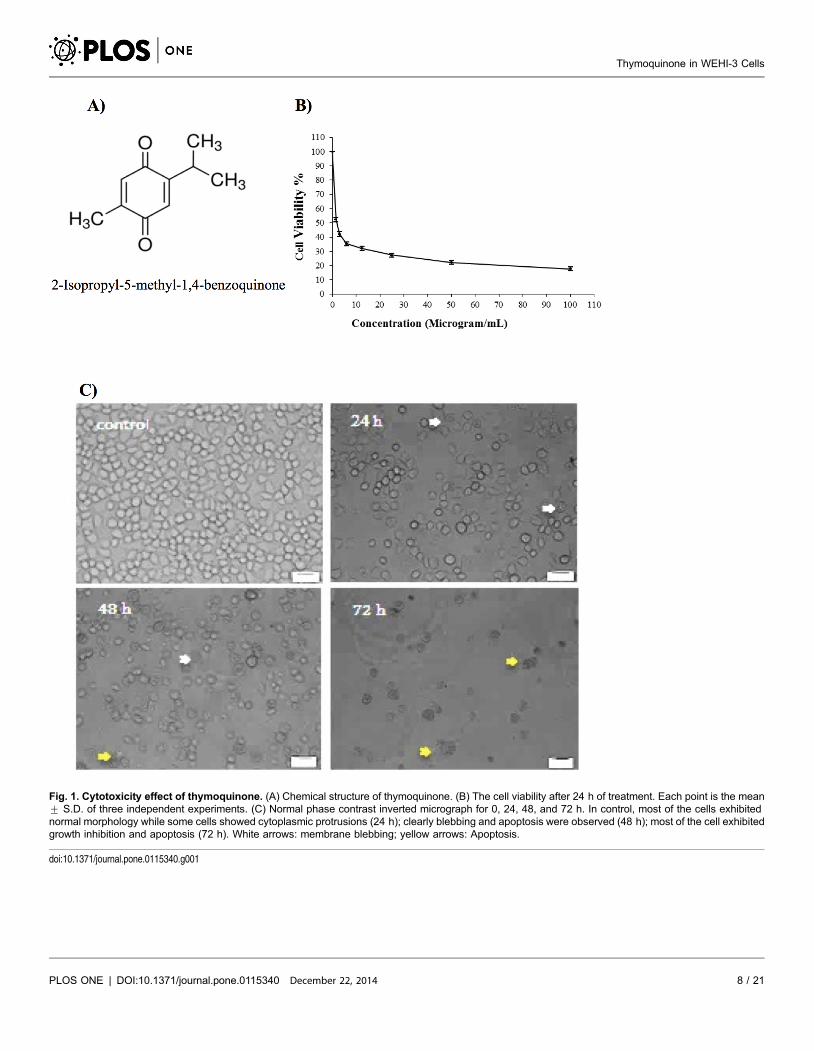
Fig. 1. Cytotoxicity effect of thymoquinone. (A) Chemical structure of thymoquinone. (B) The cell viability after 24 h of treatment. Each point is the mean¡ S.D. of three independent experiments. (C) Normal phase contrast inverted micrograph for 0, 24, 48, and 72 h. In control, most of the cells exhibitednormal morphology while some cells showed cytoplasmic protrusions (24 h); clearly blebbing and apoptosis were observed (48 h); most of the cell exhibitedgrowth inhibition and apoptosis (72 h). White arrows: membrane blebbing; yellow arrows: Apoptosis.
doi:10.1371/journal.pone.0115340.g001
Thymoquinone in WEHI-3 Cells
PLOS ONE | DOI:10.1371/journal.pone.0115340 December 22, 2014 8 / 21
Morphological Study
The thymoquinone-treated WEHI-3 cells became rounded up, shrunken in size,
and detached from the monolayer surface of the wells (Fig. 1C). The number of
cells was also found to decrease when compared to the control, and some treated
cells showed formation of apoptotic bodies which appeared to be round or oval
masses of cytoplasm, smaller than the original cells [26].
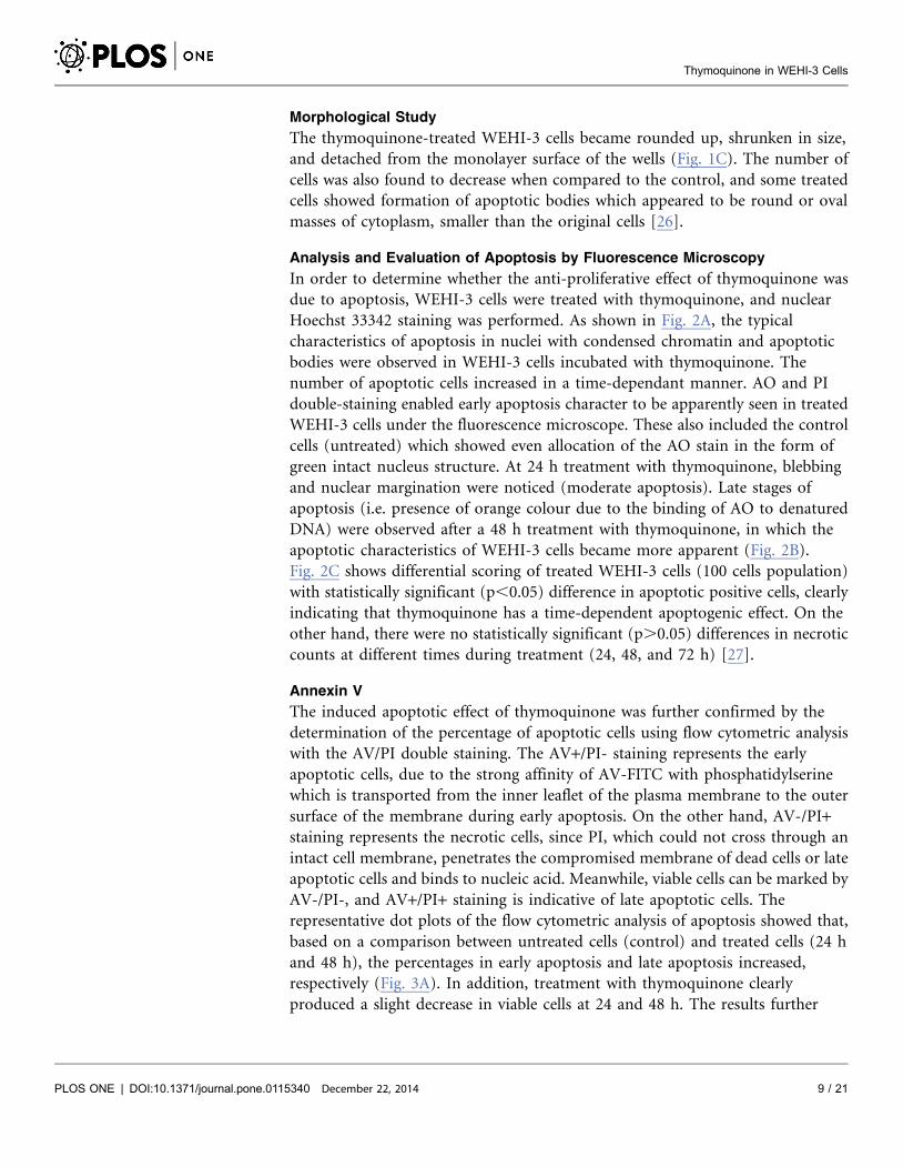
Analysis and Evaluation of Apoptosis by Fluorescence Microscopy
In order to determine whether the anti-proliferative effect of thymoquinone was
due to apoptosis, WEHI-3 cells were treated with thymoquinone, and nuclear
Hoechst 33342 staining was performed. As shown in Fig. 2A, the typical
characteristics of apoptosis in nuclei with condensed chromatin and apoptotic
bodies were observed in WEHI-3 cells incubated with thymoquinone. The
number of apoptotic cells increased in a time-dependant manner. AO and PI
double-staining enabled early apoptosis character to be apparently seen in treated
WEHI-3 cells under the fluorescence microscope. These also included the control
cells (untreated) which showed even allocation of the AO stain in the form of
green intact nucleus structure. At 24 h treatment with thymoquinone, blebbing
and nuclear margination were noticed (moderate apoptosis). Late stages of
apoptosis (i.e. presence of orange colour due to the binding of AO to denatured
DNA) were observed after a 48 h treatment with thymoquinone, in which the
apoptotic characteristics of WEHI-3 cells became more apparent (Fig. 2B).
Fig. 2C shows differential scoring of treated WEHI-3 cells (100 cells population)
with statistically significant (p,0.05) difference in apoptotic positive cells, clearly
indicating that thymoquinone has a time-dependent apoptogenic effect. On the
other hand, there were no statistically significant (p.0.05) differences in necrotic
counts at different times during treatment (24, 48, and 72 h) [27].
Annexin V
The induced apoptotic effect of thymoquinone was further confirmed by the
determination of the percentage of apoptotic cells using flow cytometric analysis
with the AV/PI double staining. The AV+/PI- staining represents the early
apoptotic cells, due to the strong affinity of AV-FITC with phosphatidylserine
which is transported from the inner leaflet of the plasma membrane to the outer
surface of the membrane during early apoptosis. On the other hand, AV-/PI+staining represents the necrotic cells, since PI, which could not cross through an
intact cell membrane, penetrates the compromised membrane of dead cells or late
apoptotic cells and binds to nucleic acid. Meanwhile, viable cells can be marked by
AV-/PI-, and AV+/PI+ staining is indicative of late apoptotic cells. The
representative dot plots of the flow cytometric analysis of apoptosis showed that,
based on a comparison between untreated cells (control) and treated cells (24 h
and 48 h), the percentages in early apoptosis and late apoptosis increased,
respectively (Fig. 3A). In addition, treatment with thymoquinone clearly
produced a slight decrease in viable cells at 24 and 48 h. The results further
Thymoquinone in WEHI-3 Cells
PLOS ONE | DOI:10.1371/journal.pone.0115340 December 22, 2014 9 / 21
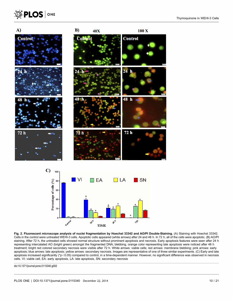
Fig. 2. Fluorescent microscope analysis of nuclei fragmentation by Hoechst 33342 and AO/PI Double-Staining. (A) Staining with Hoechst 33342.Cells in the control were untreated WEHI-3 cells. Apoptotic cells appeared (white arrows) after 24 and 48 h. In 72 h, all of the cells were apoptotic. (B) AO/PIstaining. After 72 h, the untreated cells showed normal structure without prominent apoptosis and necrosis. Early apoptosis features were seen after 24 hrepresenting intercalated AO (bright green) amongst the fragmented DNA; blebbing, orange color representing late apoptosis were noticed after 48 htreatment; bright red colored secondary necrosis were visible after 72 h. White arrows: viable cells; red arrows: membrane blebbing; pink arrows: earlyapoptosis; blue arrows: late apoptosis; yellow arrows: secondary necrosis. Images are representative of one of three similar experiments. (C) Early and lateapoptosis increased significantly (*p,0.05) compared to control, in a time-dependent manner. However, no significant difference was observed in necrosiscells. VI: viable cell, EA: early apoptosis, LA: late apoptosis, SN: secondary necrosis
doi:10.1371/journal.pone.0115340.g002
Thymoquinone in WEHI-3 Cells
PLOS ONE | DOI:10.1371/journal.pone.0115340 December 22, 2014 10 / 21
Thymoquinone in WEHI-3 Cells
PLOS ONE | DOI:10.1371/journal.pone.0115340 December 22, 2014 11 / 21
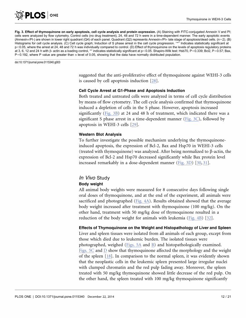
suggested that the anti-proliferative effect of thymoquinone against WEHI-3 cells
is caused by cell apoptosis induction [28].
Cell Cycle Arrest at G1-Phase and Apoptosis Induction
Both treated and untreated cells were analyzed in terms of cell cycle distribution
by means of flow cytometry. The cell cycle analysis confirmed that thymoquinone
induced a depletion of cells in the S phase. However, apoptosis increased
significantly (Fig. 3B) at 24 and 48 h of treatment, which indicated there was a
significant S phase arrest in a time-dependent manner (Fig. 3C), followed by
apoptosis in WEHI-3 cells [29].
Western Blot Analysis
To further investigate the possible mechanism underlying the thymoquinone-
induced apoptosis, the expression of Bcl-2, Bax and Hsp70 in WEHI-3 cells
(treated with thymoquinone) was analyzed. After being normalized to b-actin, the
expression of Bcl-2 and Hsp70 decreased significantly while Bax protein level
increased remarkably in a dose-dependent manner (Fig. 3D) [30, 31].
In Vivo Study
Body weight
All animal body weights were measured for 8 consecutive days following single
oral doses of thymoquinone, and at the end of the experiment, all animals were
sacrificed and photographed (Fig. 4A). Results obtained showed that the average
body weight increased after treatment with thymoquinone (100 mg/kg). On the
other hand, treatment with 50 mg/kg dose of thymoquinone resulted in a
reduction of the body weight for animals with leukemia (Fig. 4B) [32].
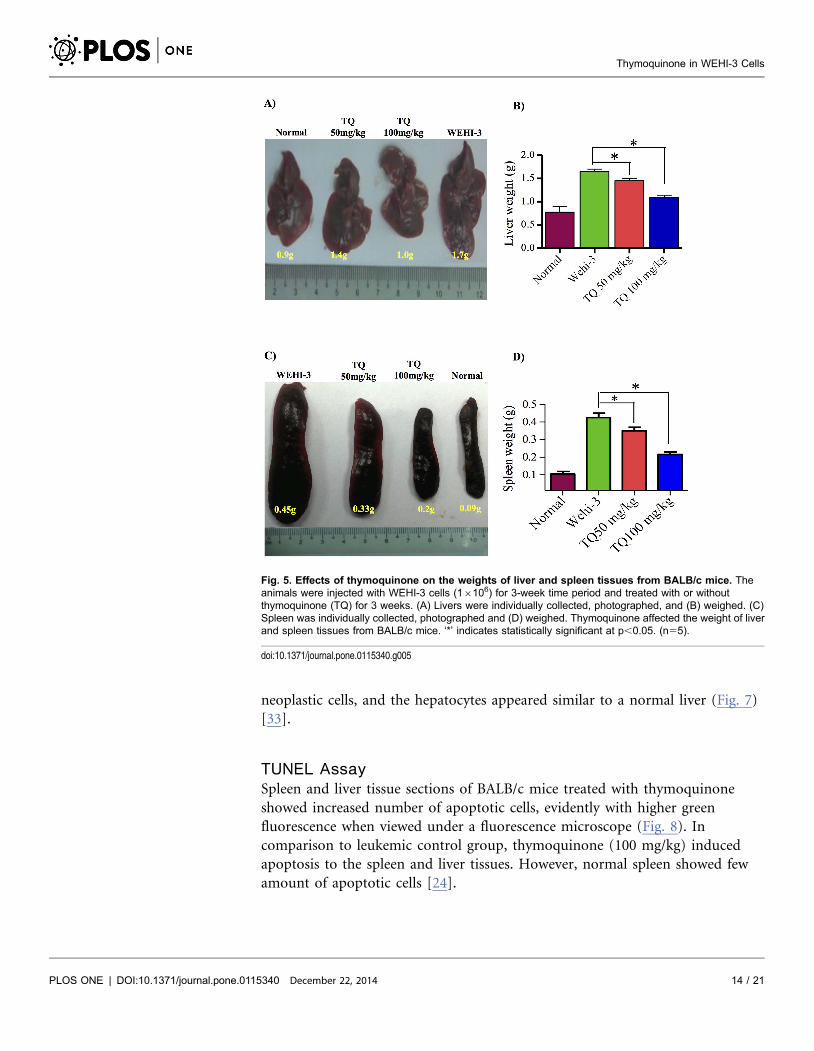
Effects of Thymoquinone on the Weight and Histopathology of Liver and Spleen
Liver and spleen tissues were isolated from all animals of each group, except from
those which died due to leukemic burden. The isolated tissues were
photographed, weighed (Figs. 5A and B) and histopathologically examined.
Figs. 5C and D show that thymoquinone affected the morphology and the weight
of the spleen [18]. In comparison to the normal spleen, it was evidently shown
that the neoplastic cells in the leukemic spleen presented large irregular nuclei
with clumped chromatin and the red pulp fading away. Moreover, the spleen
treated with 50 mg/kg thymoquinone showed little decrease of the red pulp. On
the other hand, the spleen treated with 100 mg/kg thymoquinone significantly
Fig. 3. Effect of thymoquinone on early apoptosis, cell cycle analysis and protein expression. (A) Staining with FITC-conjugated Annexin V and PI;cells were analyzed by flow cytometry. Control cells (no drug treatment), 24, 48 and 72 h were in a time-dependent manner. The early apoptotic events(Annexin+/PI-) are shown in lower right quadrant (Q4) of each panel. Quadrant (Q2) represents Annexin+/PI+ late stage of apoptosis/dead cells. (n52). (B)Histograms for cell cycle analysis. (C) Cell cycle graph; Induction of S phase arrest in the cell cycle progression. ‘‘*’’ Indicates statistically significant atp,0.05, where the arrest at 24, 48 and 72 h was individually compared to control. (D) Effect of thymoquinone on the levels of apoptosis regulatory proteinsat 3, 6, 12 and 24 h with b- actin as a loading control, ‘*’ indicates statistically significant at p,0.05. Shapiro-Wilk test: Hsb70, P50.339; Bcl2, P50.57; Bax,P50.192, where P value are greater than a level of 0.05, showing that the data have normally distributed population.
doi:10.1371/journal.pone.0115340.g003
Thymoquinone in WEHI-3 Cells
PLOS ONE | DOI:10.1371/journal.pone.0115340 December 22, 2014 12 / 21
showed its appearance to be close to that of a normal spleen, with clear red pulp
and few numbers of neoplastic cells (Fig. 6).
The histopathological examination of the liver in animal with leukemia showed
that hepatocytes were damaged and appeared as nuclei with high density of
chromatin, and the neoplastic cell nests present in the sinusoids. Treatment with
50 mg/kg of thymoquinone resulted in the reduction of hepatocyte enlargement.
However, treatment with 100 mg/kg of thymoquinone yielded a high decrease of
Fig. 4. Effects of thymoquinone on animal body weight. (A) Representative images of normal BALB/cmice; BALB/c mice injected with WEHI-3 cells (16106) and BALB/c mice injected with WEHI-3 cells (16106)and treated with thymoquinone 100 mg/kg for 3 weeks. The animals were then sacrificed, and photographed.The arrows are pointing at the size of the liver (blue arrow) and spleen (red arrow). (B) Body weight changesof BALB/c mice treated with or without thymoquinone (TQ) for 3 weeks. Values are average of five mice.
doi:10.1371/journal.pone.0115340.g004
Thymoquinone in WEHI-3 Cells
PLOS ONE | DOI:10.1371/journal.pone.0115340 December 22, 2014 13 / 21
neoplastic cells, and the hepatocytes appeared similar to a normal liver (Fig. 7)
[33].
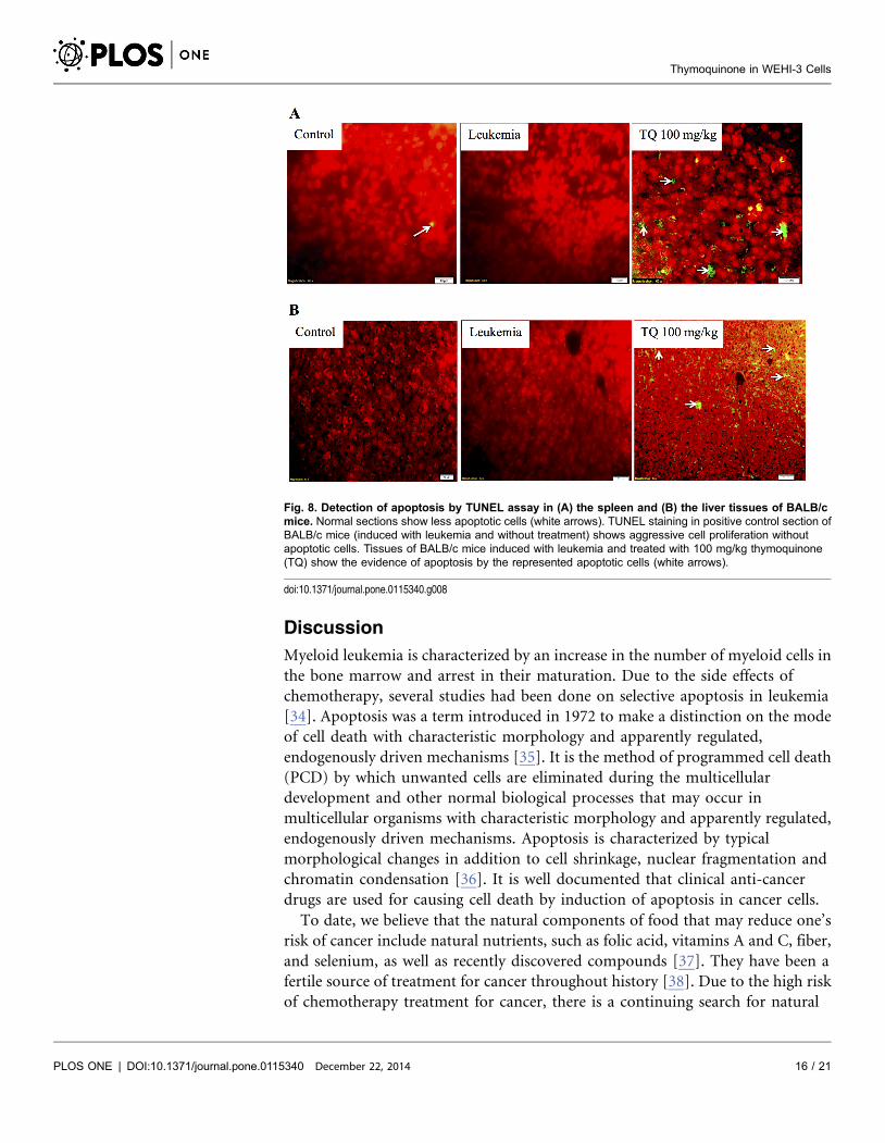
TUNEL Assay
Spleen and liver tissue sections of BALB/c mice treated with thymoquinone
showed increased number of apoptotic cells, evidently with higher green
fluorescence when viewed under a fluorescence microscope (Fig. 8). In
comparison to leukemic control group, thymoquinone (100 mg/kg) induced
apoptosis to the spleen and liver tissues. However, normal spleen showed few
amount of apoptotic cells [24].
Fig. 5. Effects of thymoquinone on the weights of liver and spleen tissues from BALB/c mice. Theanimals were injected with WEHI-3 cells (16106) for 3-week time period and treated with or withoutthymoquinone (TQ) for 3 weeks. (A) Livers were individually collected, photographed, and (B) weighed. (C)Spleen was individually collected, photographed and (D) weighed. Thymoquinone affected the weight of liverand spleen tissues from BALB/c mice. ‘*’ indicates statistically significant at p,0.05. (n55).
doi:10.1371/journal.pone.0115340.g005
Thymoquinone in WEHI-3 Cells
PLOS ONE | DOI:10.1371/journal.pone.0115340 December 22, 2014 14 / 21
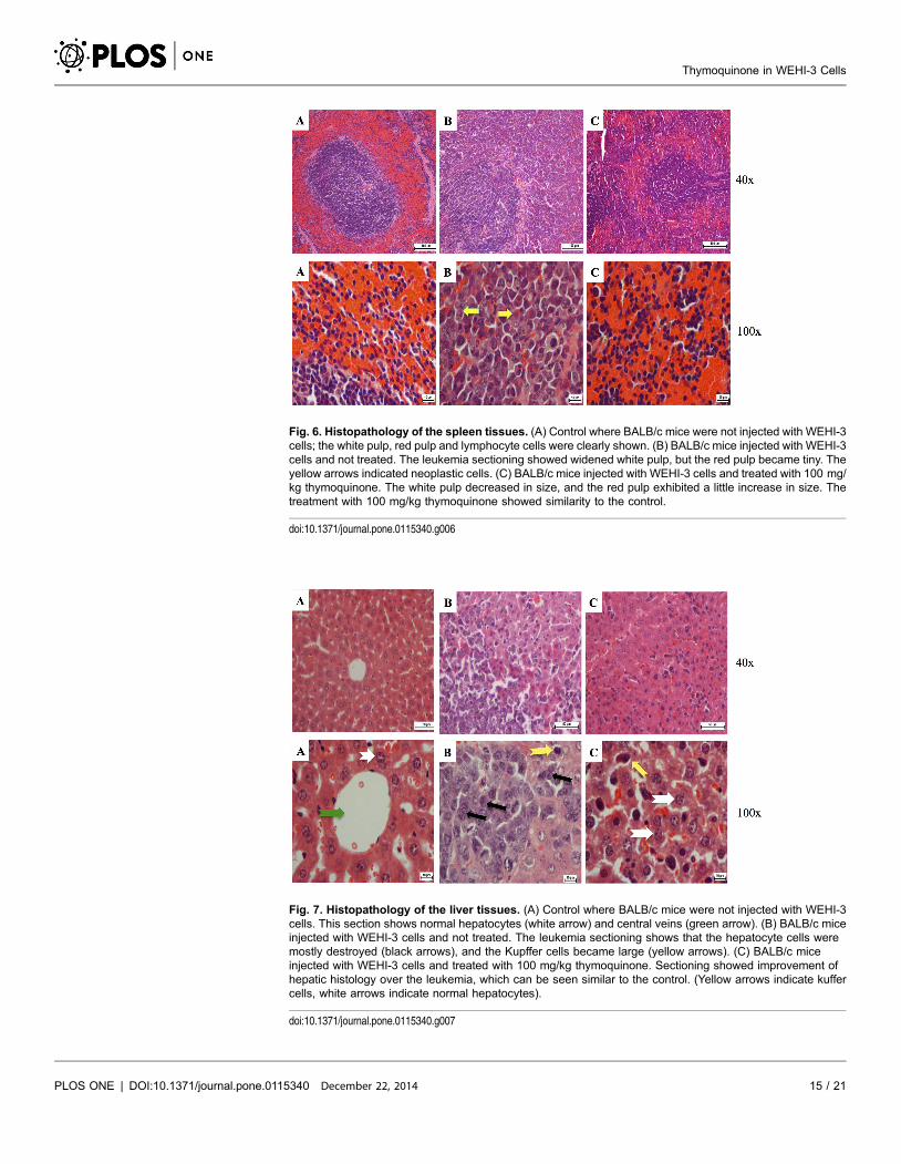
Fig. 6. Histopathology of the spleen tissues. (A) Control where BALB/c mice were not injected with WEHI-3cells; the white pulp, red pulp and lymphocyte cells were clearly shown. (B) BALB/c mice injected with WEHI-3cells and not treated. The leukemia sectioning showed widened white pulp, but the red pulp became tiny. Theyellow arrows indicated neoplastic cells. (C) BALB/c mice injected with WEHI-3 cells and treated with 100 mg/kg thymoquinone. The white pulp decreased in size, and the red pulp exhibited a little increase in size. Thetreatment with 100 mg/kg thymoquinone showed similarity to the control.
doi:10.1371/journal.pone.0115340.g006
Fig. 7. Histopathology of the liver tissues. (A) Control where BALB/c mice were not injected with WEHI-3cells. This section shows normal hepatocytes (white arrow) and central veins (green arrow). (B) BALB/c miceinjected with WEHI-3 cells and not treated. The leukemia sectioning shows that the hepatocyte cells weremostly destroyed (black arrows), and the Kupffer cells became large (yellow arrows). (C) BALB/c miceinjected with WEHI-3 cells and treated with 100 mg/kg thymoquinone. Sectioning showed improvement ofhepatic histology over the leukemia, which can be seen similar to the control. (Yellow arrows indicate kuffercells, white arrows indicate normal hepatocytes).
doi:10.1371/journal.pone.0115340.g007
Thymoquinone in WEHI-3 Cells
PLOS ONE | DOI:10.1371/journal.pone.0115340 December 22, 2014 15 / 21
Discussion
Myeloid leukemia is characterized by an increase in the number of myeloid cells in
the bone marrow and arrest in their maturation. Due to the side effects of
chemotherapy, several studies had been done on selective apoptosis in leukemia
[34]. Apoptosis was a term introduced in 1972 to make a distinction on the mode
of cell death with characteristic morphology and apparently regulated,
endogenously driven mechanisms [35]. It is the method of programmed cell death
(PCD) by which unwanted cells are eliminated during the multicellular
development and other normal biological processes that may occur in
multicellular organisms with characteristic morphology and apparently regulated,
endogenously driven mechanisms. Apoptosis is characterized by typical
morphological changes in addition to cell shrinkage, nuclear fragmentation and
chromatin condensation [36]. It is well documented that clinical anti-cancer
drugs are used for causing cell death by induction of apoptosis in cancer cells.
To date, we believe that the natural components of food that may reduce one’s
risk of cancer include natural nutrients, such as folic acid, vitamins A and C, fiber,
and selenium, as well as recently discovered compounds [37]. They have been a
fertile source of treatment for cancer throughout history [38]. Due to the high risk
of chemotherapy treatment for cancer, there is a continuing search for natural
Fig. 8. Detection of apoptosis by TUNEL assay in (A) the spleen and (B) the liver tissues of BALB/cmice. Normal sections show less apoptotic cells (white arrows). TUNEL staining in positive control section ofBALB/c mice (induced with leukemia and without treatment) shows aggressive cell proliferation withoutapoptotic cells. Tissues of BALB/c mice induced with leukemia and treated with 100 mg/kg thymoquinone(TQ) show the evidence of apoptosis by the represented apoptotic cells (white arrows).
doi:10.1371/journal.pone.0115340.g008
Thymoquinone in WEHI-3 Cells
PLOS ONE | DOI:10.1371/journal.pone.0115340 December 22, 2014 16 / 21
products as anti-cancer drugs [39]. Thymoquinone is the major bioactive
constituent present in black seed oil. Different studies reported thymoquinone to
exert anti-cancer activities both in vitro and in vivo through different mechanisms,
such as the effect of thymoquinone on cell signalling and survival pathways [40].
The in vitro study of thymoquinone against HL60 cell line had been done through
different pathways such as the p53 pathway and caspase activity [20, 41].
Numerous in vivo studies had shown promising anti-cancer effects of
thymoquinone in different cancer cell lines such as HL60 and LNM35 [42, 43].
Our previous research on thymoquinone had shown that this compound
exerted significant cytotoxic and apoptotic effects on acute lymphocytic leukaemia
CEMss cell line [20]. In the current study, it was demonstrated that cyctotoxic
activities of thymoquinone towards WEHI-3 cells were selective. Indications of
apoptosis in WEHI-3 cells treated with thymoquinone showed typical
morphological patterns of apoptosis in the form of a reduction in the number of
cells, cytoplasmic shrinkage, and membrane blebbing, observed using inverted
light microscope (Fig. 1C). DNA fragmentation was observed under fluorescence
microscopic analyses with AO/PI and Hoechst 33342 staining, where it was found
that the number of cells undergoing apoptosis was increasing significantly with
time. In addition, late apoptosis and secondary necrosis appeared intensively in
72 h of treatment [44]. Hence, early phases of apoptosis were detected using
Annexin V which was found to bind specifically to phosphatidylserine (PS)
located at the outer membrane leaflet of cells in the presence of calcium. In this
study, it was shown that there was a significant increase in the early stage of
apoptosis in time-dependant manner [45]. Bcl-2, Bax and HSP70 play a major
role in determining whether cells will undergo apoptosis under experimental
conditions that promote cell death; Bcl-2 protects cells from apoptosis, while
increased expression of Bax can induce apoptosis. The ratio of Bax: Bcl-2, rather
than Bcl-2 alone, is important for the survival of drug-induced apoptosis in
leukemia cell lines. In this study, a decrease in Bcl-2 expression was observed in
WEHI-3 cells after treatment with thymoquinone. The expression of Bax,
however, was up-regulated in WEHI-3 cells after treatment for 3, 6, 12 and 24 h
[20, 46].
Cell cycle analysis was performed to evaluate the effect of thymoquinone on the
distribution of tumor cells in G1, S and G2/M phases of the cell cycle. Recently,
many studies highlighted that cell cycle regulation is one of the important
mechanisms of anti-proliferation in cancers [31]. Abnormalities of cell cycle
regulators have been connected with many carcinogenic processes. Therefore, cell
cycle regulators in cancer cells could be targeted and changed to be useful for
treatment. Thymoquinone was shown to induce apoptosis through its ability to
arrest cells in the S phase of the cell cycle in WEHI-3 cells. The proportion of
accumulated cells was blocked at S phase. Thymoquinone caused DNA damage in
WEHI-3 cells by arresting the cell cycle at S phase. This is in accordance to several
studies that reported compounds isolated from natural resources, which arrested
cell cycle at S phase, similarly induced apoptosis [47, 48].
Thymoquinone in WEHI-3 Cells
PLOS ONE | DOI:10.1371/journal.pone.0115340 December 22, 2014 17 / 21
Since the in vitro study reported herein showed that thymoquinone induced
apoptosis against WEHI-3 cell lines, it was important to perform an in vivo study
to check its ability to induce apoptosis in living animal. Many studies had been
used to induce leukemia in BALB/c mice, for example by intraperitonial (i.p.)
injection of WEHI-3 cells, in order to evaluate the anti-leukemic effects of many
agents [19, 49, 50]. Murine monomyelocytic leukemia cells were originally derived
from the BALB/c mouse [51]. The importance of this animal model is that the
murine host systems are used for experimental tumor therapy and they carry
several beneficial factors such as the low cost, they are easily obtainable with which
cancer production is established, and they are widely accepted at experimental
end-points [50, 52].
Our previous studies showed that the in vivo model (the mice i.p. injected with
WEHI-3 cells) was well established. The BALB/c mice were i.p. injected with
WEHI-3 cells 1 day prior to the treatment with thymoquinone for 21 days, after
which the animals were sacrificed. The WEHI-3 leukemia animal model was
characterized by high peripheral monocytes and granulocytes with immature
morphology, and the spleens and livers were enlarged, when compared to the
normal ones. The in vivo effects of thymoquinone on WEHI-3 tumor cells in
BALB/c mice were also examined in normal animal where the weights of spleen
and liver were 0.09 g and 1.9 g, respectively. In untreated animal, the weights of
spleen and liver were 0.41 g and 1.9 g, respectively. However, the spleen and liver
weights for animal treated with thymoquinone (50 mg/kg) were 0.34 g and 1.6 g,
respectively, while animal treated with thymoquinone (100 mg/kg) showed that
the spleen and liver weights were 0.2 g and 1.2 g, respectively. The results
demonstrated that thymoquinone significantly decreased the size and weights of
spleen and liver in the examined animals.
In this study, toxicity was assessed by daily measurement of the body weight for
8 consecutive days following single oral doses of thymoquinone. In support to
these results, the histopathological examination indicated that after treatment, the
spleen and liver verified a pattern ranging from minimal histopathological change
to measly small neoplastic cell nests present in the sinusoid. Reduction in the
infiltration of immature myeloblastic cells into splenic red pulp was also observed.
TUNEL assay highlighted that thymoquinone significantly induced apoptosis in
spleen and liver tissues. Overall, our findings indicated that thymoquinone
significantly induced apoptosis in murine WEHI-3, in vitro, and inhibited the
spleen tumor, where there was significant difference between the control and
thymoquinone-treated groups. In addition to our current finding, more study is
needed to further evaluate the molecular mechanisms and the pathways involved
before thymoquinone can be proposed as a potential therapeutic agent for
leukemia.
Acknowledgments
The authors would like to express their utmost gratitude to the late Prof. A Hamid
bin A Hadi for his invaluable support.
Thymoquinone in WEHI-3 Cells
PLOS ONE | DOI:10.1371/journal.pone.0115340 December 22, 2014 18 / 21
Author ContributionsConceived and designed the experiments: LZAS RO SM. Performed the
experiments: LZAS MAA PH FD MYI FAEAO. Analyzed the data: LZAS RO MAA
KA PH FD MYI FAEAO SM. Contributed reagents/materials/analysis tools: LZAS
RO MAA HMA SM. Wrote the paper: LZAS RO MAA MYI SM.
References
1. Hosseini M, Zakeri S, Khoshdast S, Yousefian FT, Rastegar M, et al. (2012) The effects of Nigellasativa hydro-alcoholic extract and thymoquinone on lipopolysaccharide-induced depression likebehavior in rats. Journal of Pharmacy & Bioallied Sciences 4: 219–225.
2. Paarakh PM (2010) Nigella sativa Linn. – A comprehensive review. Indian Journal of Natural Productsand Resources 1: 409–429.
3. Ahmad A, Husain A, Mujeeb M, Khan SA, Najmi AK, et al. (2013) A review on therapeutic potential ofNigella sativa: A miracle herb. Asian Pacific Journal of Tropical Biomedicine 3: 337–352.
4. Jrah Harzallah H, Grayaa R, Kharoubi W, Maaloul A, Hammami M, et al. (2012) Thymoquinone, theNigella sativa bioactive compound, prevents circulatory oxidative stress caused by 1, 2-dimethylhydrazine in erythrocyte during colon postinitiation carcinogenesis. Oxidative Medicine andCellular Longevity 2012: 854065.
5. Lutterodt H, Luther M, Slavin M, Yin J-J, Parry J, et al. (2010) Fatty acid profile, thymoquinonecontent, oxidative stability, and antioxidant properties of cold-pressed black cumin seed oils. LWT-FoodScience and Technology 43: 1409–1413.
6. El Mezayen R, El Gazzar M, Nicolls MR, Marecki JC, Dreskin SC, et al. (2006) Effect of thymoquinoneon cyclooxygenase expression and prostaglandin production in a mouse model of allergic airwayinflammation. Immunology Letters 106: 72–81.
7. Ismail M, Al-Naqeep G, Chan KW (2010) Nigella sativa thymoquinone-rich fraction greatly improvesplasma antioxidant capacity and expression of antioxidant genes in hypercholesterolemic rats. FreeRadical Biology and Medicine 48: 664–672.
8. Ivankovic S, Stojkovic R, Jukic M, Milos M, Milos M, et al. (2006) The antitumor activity ofthymoquinone and thymohydroquinone in vitro and in vivo. Experimental Oncology 28: 220–224.
9. Shoieb AM, Elgayyar M, Dudrick PS, Bell JL, Tithof PK (2003) In vitro inhibition of growth andinduction of apoptosis in cancer cell lines by thymoquinone. International Journal of Oncology 22: 107–114.
10. Arslan SO, Gelir E, Armutcu F, Coskun O, Gurel A, et al. (2005) The protective effect of thymoquinoneon ethanol-induced acute gastric damage in the rat. Nutrition Research 25: 673–680.
11. El-Najjar N, Chatila M, Moukadem H, Vuorela H, Ocker M, et al. (2010) Reactive oxygen speciesmediate thymoquinone-induced apoptosis and activate ERK and JNK signaling. Apoptosis 15: 183–195.
12. El-Mahdy MA, Zhu Q, Wang QE, Wani G, Wani AA (2005) Thymoquinone induces apoptosis throughactivation of caspase-8 and mitochondrial events in p53-null myeloblastic leukemia HL-60 cells.International journal of Cancer 117: 409–417.
13. Torres MP, Ponnusamy MP, Chakraborty S, Smith LM, Das S, et al. (2010) Effects of thymoquinone inthe expression of mucin 4 in pancreatic cancer cells: implications for the development of novel cancertherapies. Molecular Cancer Therapeutics 9: 1419–1431.
14. Woo CC, Kumar AP, Sethi G, Tan KHB (2012) Thymoquinone: potential cure for inflammatory disordersand cancer. Biochemical Pharmacology 83: 443–451.
15. Jemal A, Siegel R, Xu J, Ward E (2010) Cancer statistics, 2010. CA: A Cancer Journal for Clinicians60: 277–300.
16. Siegel R, Naishadham D, Jemal A (2012) Cancer statistics, 2012. CA: A Cancer Journal for Clinicians62: 10–29.
Thymoquinone in WEHI-3 Cells
PLOS ONE | DOI:10.1371/journal.pone.0115340 December 22, 2014 19 / 21
17. Yang J-S, Kok L-F, Lin Y-H, Kuo T-C, Yang J-L, et al. (2006) Diallyl disulfide inhibits WEHI-3 leukemiacells in vivo. Anticancer Research 26: 219–225.
18. Chang Y-H, Yang J-S, Yang J-L, Wu C-L, Chang S-J, et al. (2009) Ganoderma lucidum extractsinhibited leukemia WEHI-3 cells in BALB/c mice and promoted an immune response in vivo. Bioscience,Biotechnology, and Biochemistry 73: 2589–2594.
19. Yu CS, Lai KC, Yang JS, Chiang JH, Lu CC, et al. (2010) Quercetin inhibited murine leukemia WEHI-3cells in vivo and promoted immune response. Phytotherapy Research 24: 163–168.
20. Salim LZA, Mohan S, Othman R, Abdelwahab SI, Kamalidehghan B, et al. (2013) Thymoquinoneinduces mitochondria-mediated apoptosis in acute lymphoblastic leukaemia in vitro. Molecules 18:11219–11240.
21. Garber JC, Barbee RY, Bielitzki JT (2011) Guide for the care and use of laboratory animals. In:Research IfLA, editor. 8th ed. United State: National Academies Press.
22. Olfert ED, Cross BM, McWilliam AA (1993) Guide to the care and use of experimental animals:Canadian Council on Animal Care Ottawa.
23. Lu H-F, Liu J-Y, Hsueh S-C, Yang Y-Y, Yang J-S, et al. (2007) (–)-Menthol Inhibits WEHI-3 LeukemiaCells In Vitro and In Vivo. In Vivo 21: 285–289.
24. Mohan S, Abdul AB, Abdelwahab SI, Al-Zubairi AS, Aspollah Sukari M, et al. (2010) Typhoniumflagelliforme inhibits the proliferation of murine leukemia WEHI-3 cells in vitro and induces apoptosis invivo. Leukemia Research 34: 1483–1492.
25. Zhang B, Chen N, Chen H, Wang Z, Zheng Q (2012) The critical role of redox homeostasis in Shikonin-Induced HL-60 cell differentiation via unique modulation of the Nrf2/ARE Pathway. Oxidative Medicineand Cellular Longevity 2012, Article ID 781516, 12 p.
26. Ebrahimi Nigjeh S, Yusoff FM, Mohamed Alitheen NB, Rasoli M, Keong YS, et al. (2012) Cytotoxiceffect of ethanol extract of microalga, Chaetoceros calcitrans, and its mechanisms in inducing apoptosisin human breast cancer cell line. BioMed Research International 2013, Article ID 783690, 8 p.
27. Abd Ghafar SA, Ismail M, Saiful Yazan L, Fakurazi S, Ismail N, et al. (2013) Cytotoxic activity of kenafseed oils from supercritical carbon dioxide fluid extraction towards human colorectal cancer (HT29) celllines. Evidence-Based Complementary and Alternative Medicine 2013, Article ID 549705, 8 p.
28. Huang L-H, Hu J-Q, Tao W-Q, Li Y-H, Li G-M, et al. (2010) Gossypol inhibits phosphorylation of Bcl-2 inhuman leukemia HL-60 cells. European Journal of Pharmacology 645: 9–13.
29. Lin C-C, Lin S-Y, Chung J-G, Lin J-P, Chen G-W, et al. (2006) Down-regulation of cyclin B1 and up-regulation of Wee1 by berberine promotes entry of leukemia cells into the G2/M-phase of the cell cycle.Anticancer Research 26: 1097–1104.
30. Fu W-Y, Chen J-P, Wang X-M, Xu L-H (2005) Altered expression of p53, Bcl-2 and Bax induced bymicrocystin-LR in vivo and in vitro. Toxicon 46: 171–177.
31. Bai Y, Mao QQ, Qin J, Zheng XY, Wang YB, et al. (2010) Resveratrol induces apoptosis and cell cyclearrest of human T24 bladder cancer cells in vitro and inhibits tumor growth in vivo. Cancer Science 101:488–493.
32. Chang Y-C, Lai T-Y, Yu C-S, Chen H-Y, Yang J-S, et al. (2011) Emodin induces apoptotic death inmurine myelomonocytic leukemia WEHI-3 cells in vitro and enhances phagocytosis in leukemia mice invivo. Evidence-Based Complementary and Alternative Medicine 2011, Article ID 523596, 13 p.
33. Alabsi AM, Ali R, Ideris A, Omar AR, Bejo MH, et al. (2012) Anti-leukemic activity of Newcastledisease virus strains AF2240 and V4-UPM in murine myelomonocytic leukemia in vivo. LeukemiaResearch 36: 634–645.
34. Bedi A, Zehnbauer BA, Barber JP, Sharkis S, Jones, et al. (1994) Inhibition of apoptosis by BCR-ABLin chronic myeloid leukemia. Blood 83: 2038–2044.
35. Wyllie AH (2010) ‘‘Where, O death, is thy sting?’’ A brief review of apoptosis biology. MolecularNeurobiology 42: 4–9.
36. Elmore S (2007) Apoptosis: a review of programmed cell death. Toxicologic Pathology 35: 495–516.
37. Salminen S, Bouley C, Boutron M-C, Cummings J, Franck A, et al. (1998) Functional food scienceand gastrointestinal physiology and function. British Journal of Nutrition 80: S147–S171.
Thymoquinone in WEHI-3 Cells
PLOS ONE | DOI:10.1371/journal.pone.0115340 December 22, 2014 20 / 21
38. Da Rocha AB, Lopes RM, Schwartsmann G (2001) Natural products in anticancer therapy. CurrentOpinion in Pharmacology 1: 364–369.
39. Nobili S, Lippi D, Witort E, Donnini M, Bausi L, et al. (2009) Natural compounds for cancer treatmentand prevention. Pharmacological Research 59: 365–378.
40. Aggarwal BB, Kunnumakkara AB, Harikumar KB, Tharakan ST, Sung B, et al. (2008) Potential ofspice-derived phytochemicals for cancer prevention. Planta Medica 74: 1560–1569.
41. Banerjee S, Padhye S, Azmi A, Wang Z, Philip PA, et al. (2010) Review on molecular and therapeuticpotential of thymoquinone in cancer. Nutrition and Cancer 62: 938–946.
42. Schneider-Stock R, Fakhoury IH, Zaki AM, El-Baba CO, Gali-Muhtasib HU (2014) Thymoquinone:fifty years of success in the battle against cancer models. Drug Discovery Today 19: 18–30.
43. Attoub S, Sperandio O, Raza H, Arafat K, Al-Salam S, et al. (2013) Thymoquinone as an anticanceragent: evidence from inhibition of cancer cells viability and invasion in vitro and tumor growth in vivo.Fundamental & Clinical Pharmacology 27: 557–569.
44. Arbab IA, Abdul AB, Sukari MA, Abdullah R, Syam S, et al. (2013) Dentatin isolated from Clausenaexcavata induces apoptosis in MCF-7 cells through the intrinsic pathway with involvement of NF-kBsignalling and G0/G1 cell cycle arrest: A bioassay-guided approach. Journal of Ethnopharmacology 145:343–354.
45. Ng K-B, Bustamam A, Sukari MA, Abdelwahab SI, Mohan S, et al. (2013) Induction of selectivecytotoxicity and apoptosis in human T4-lymphoblastoid cell line (CEMss) by boesenbergin A isolatedfrom Boesenbergia rotunda rhizomes involves mitochondrial pathway, activation of caspase 3 and G2/Mphase cell cycle arrest. BMC Complementary and Alternative Medicine 13: 41.
46. Guo F, Sigua C, Bali P, George P, Fiskus W, et al. (2005) Mechanistic role of heat shock protein 70 inBcr-Abl–mediated resistance to apoptosis in human acute leukemia cells. Blood 105: 1246–1255.
47. Anasamy T, Abdul AB, Sukari MA, Abdelwahab SI, Mohan S, et al. (2013) A phenylbutenoid dimer,cis-3-(39,49-dimethoxyphenyl)-4-[(E)-309,409-dimethoxystyryl] cyclohex-1-ene, exhibits apoptogenicproperties in T-acute lymphoblastic leukemia cells via induction of p53-independent mitochondrialsignalling pathway. Evidence-Based Complementary and Alternative Medicine 2013, Article ID 939810,14 p.
48. Yeh R-D, Chen J-C, Lai T-Y, Yang J-S, Yu C-S, et al. (2011) Gallic acid induces G0/G1 phase arrestand apoptosis in human leukemia HL-60 cells through inhibiting cyclin D and E, and activatingmitochondria-dependent pathway. Anticancer Research 31: 2821–2832.
49. Tsou M-F, Peng C-T, Shih M-C, Yang J-S, Lu C-C, et al. (2009) Benzyl isothiocyanate inhibits murineWEHI-3 leukemia cells in vitro and promotes phagocytosis in BALB/c mice in vivo Leukemia Research33: 1505–1511.
50. Lin J-P, Yang J-S, Lu C-C, Chiang J-H, Wu C-L, et al. (2009) Rutin inhibits the proliferation of murineleukemia WEHI-3 cells in vivo and promotes immune response in vivo. Leukemia Research 33: 823–828.
51. Yu F-S, Yang J-S, Lin H-J, Yu C-S, Tan T-W, et al. (2007) Berberine inhibits WEHI-3 leukemia cells invivo. In Vivo 21: 407–412.
52. Lu C-C, Yang J-S, Chiang J-H, Hour M-J, Lin K-L, et al. (2012) Novel quinazolinone MJ-29 triggersendoplasmic reticulum stress and intrinsic apoptosis in murine leukemia WEHI-3 cells and inhibitsleukemic mice. PLoS One 7: e36831; doi:10.1371/journal.pone.0036831
Thymoquinone in WEHI-3 Cells
PLOS ONE | DOI:10.1371/journal.pone.0115340 December 22, 2014 21 / 21