An Anthranilate Derivative Inhibits Glutamate Release ... - MDPI
16
Citation: Lu, C.-W.; Lin, C.-J.; Hsieh, P.-W.; Chiu, K.-M.; Lee, M.-Y.; Lin, T.-Y.; Wang, S.-J. An Anthranilate Derivative Inhibits Glutamate Release and Glutamate Excitotoxicity in Rats. Int. J. Mol. Sci. 2022, 23, 2641. https://doi.org/10.3390/ijms 23052641 Academic Editor: Jonathan Mapelli Received: 10 February 2022 Accepted: 25 February 2022 Published: 27 February 2022 Publisher’s Note: MDPI stays neutral with regard to jurisdictional claims in published maps and institutional affil- iations. Copyright: © 2022 by the authors. Licensee MDPI, Basel, Switzerland. This article is an open access article distributed under the terms and conditions of the Creative Commons Attribution (CC BY) license (https:// creativecommons.org/licenses/by/ 4.0/). International Journal of Molecular Sciences Article An Anthranilate Derivative Inhibits Glutamate Release and Glutamate Excitotoxicity in Rats Cheng-Wei Lu 1,2,† , Chen-Jung Lin 1,† , Pei-Wen Hsieh 3,4,5 , Kuan-Ming Chiu 6,7,8 , Ming-Yi Lee 6 , Tzu-Yu Lin 1,2, * and Su-Jane Wang 3,9, * 1 Department of Anesthesiology, Far-Eastern Memorial Hospital, New Taipei City 22060, Taiwan; [email protected] (C.-W.L.); [email protected] (C.-J.L.) 2 Department of Mechanical Engineering, Yuan Ze University, Taoyuan 32003, Taiwan 3 Research Center for Chinese Herbal Medicine, College of Human Ecology, Chang Gung University of Science and Technology, Taoyuan 33303, Taiwan; [email protected] 4 Graduate Institute of Natural Products, Graduate Institute of Biomedical Sciences, School of Traditional Chinese Medicine, College of Medicine, Chang Gung University, Taoyuan 33303, Taiwan 5 Department of Anesthesiology, Chang Gung Memorial Hospital, Taoyuan 33305, Taiwan 6 Cardiovascular Center, Division of Cardiovascular Surgery, Far-Eastern Memorial Hospital, New Taipei City 22060, Taiwan; [email protected] (K.-M.C.); [email protected] (M.-Y.L.) 7 Department of Nursing, Asia Eastern University of Science and Technology, New Taipei City 22060, Taiwan 8 Department of Photonics Engineering, Yuan Ze University, Taoyuan 32003, Taiwan 9 School of Medicine, Fu Jen Catholic University, New Taipei City 24205, Taiwan * Correspondence: [email protected] (T.-Y.L.); [email protected] (S.-J.W.); Tel.: +886-2-89667000 (T.-Y.L.); +886-2-29053465 (S.-J.W.) † These authors contributed equally to this work. Abstract: The neurotransmitter glutamate plays an essential role in excitatory neurotransmission; however, excessive amounts of glutamate lead to excitotoxicity, which is the most common pathogenic feature of numerous brain disorders. This study aimed to investigate the role of butyl 2-[2-(2- fluorophenyl)acetamido]benzoate (HFP034), a synthesized anthranilate derivative, in the central glutamatergic system. We used rat cerebro-cortical synaptosomes to examine the effect of HFP034 on glutamate release. In addition, we used a rat model of kainic acid (KA)-induced glutamate excitotoxicity to evaluate the neuroprotective potential of HFP034. We showed that HFP034 inhibits 4-aminopyridine (4-AP)-induced glutamate release from synaptosomes, and this inhibition was absent in the absence of extracellular calcium. HFP034-mediated inhibition of glutamate release was associated with decreased 4-AP-evoked Ca 2+ level elevation and had no effect on synaptosomal membrane potential. The inhibitory effect of HFP034 on evoked glutamate release was suppressed by blocking P/Q-type Ca 2+ channels and protein kinase C (PKC). Furthermore, HFP034 inhibited the phosphorylation of PKC and its substrate, myristoylated alanine-rich C kinase substrate (MARCKS) in synaptosomes. We also observed that HFP034 pretreatment reduced neuronal death, glutamate concentration, glial activation, and the levels of endoplasmic reticulum stress-related proteins, cal- pains, glucose-regulated protein 78 (GRP 78), C/EBP homologous protein (CHOP), and caspase-12 in the hippocampus of KA-injected rats. We conclude that HFP034 is a neuroprotective agent that prevents glutamate excitotoxicity, and we suggest that this effect involves inhibition of presynaptic glutamate release through the suppression of P/Q-type Ca 2+ channels and PKC/MARCKS pathways. Keywords: anthranilate derivative; glutamate release; glutamate excitotoxicity; neuroprotection; synaptosomes; kainic acid 1. Introduction Glutamate is a key excitatory neurotransmitter in brain development, synaptic trans- mission, synaptic plasticity, and learning and memory processes [1,2]. However, excess Int. J. Mol. Sci. 2022, 23, 2641. https://doi.org/10.3390/ijms23052641 https://www.mdpi.com/journal/ijms
-
Upload
khangminh22 -
Category
Documents
-
view
3 -
download
0
Transcript of An Anthranilate Derivative Inhibits Glutamate Release ... - MDPI

�����������������
Citation: Lu, C.-W.; Lin, C.-J.; Hsieh,
P.-W.; Chiu, K.-M.; Lee, M.-Y.; Lin,
T.-Y.; Wang, S.-J. An Anthranilate
Derivative Inhibits Glutamate
Release and Glutamate Excitotoxicity
in Rats. Int. J. Mol. Sci. 2022, 23, 2641.
https://doi.org/10.3390/ijms
23052641
Academic Editor: Jonathan Mapelli
Received: 10 February 2022
Accepted: 25 February 2022
Published: 27 February 2022
Publisher’s Note: MDPI stays neutral
with regard to jurisdictional claims in
published maps and institutional affil-
iations.
Copyright: © 2022 by the authors.
Licensee MDPI, Basel, Switzerland.
This article is an open access article
distributed under the terms and
conditions of the Creative Commons
Attribution (CC BY) license (https://
creativecommons.org/licenses/by/
4.0/).
International Journal of
Molecular Sciences
Article
An Anthranilate Derivative Inhibits Glutamate Release andGlutamate Excitotoxicity in RatsCheng-Wei Lu 1,2,† , Chen-Jung Lin 1,†, Pei-Wen Hsieh 3,4,5 , Kuan-Ming Chiu 6,7,8 , Ming-Yi Lee 6 ,Tzu-Yu Lin 1,2,* and Su-Jane Wang 3,9,*
1 Department of Anesthesiology, Far-Eastern Memorial Hospital, New Taipei City 22060, Taiwan;[email protected] (C.-W.L.); [email protected] (C.-J.L.)
2 Department of Mechanical Engineering, Yuan Ze University, Taoyuan 32003, Taiwan3 Research Center for Chinese Herbal Medicine, College of Human Ecology,
Chang Gung University of Science and Technology, Taoyuan 33303, Taiwan; [email protected] Graduate Institute of Natural Products, Graduate Institute of Biomedical Sciences,
School of Traditional Chinese Medicine, College of Medicine, Chang Gung University, Taoyuan 33303, Taiwan5 Department of Anesthesiology, Chang Gung Memorial Hospital, Taoyuan 33305, Taiwan6 Cardiovascular Center, Division of Cardiovascular Surgery, Far-Eastern Memorial Hospital,
New Taipei City 22060, Taiwan; [email protected] (K.-M.C.); [email protected] (M.-Y.L.)7 Department of Nursing, Asia Eastern University of Science and Technology, New Taipei City 22060, Taiwan8 Department of Photonics Engineering, Yuan Ze University, Taoyuan 32003, Taiwan9 School of Medicine, Fu Jen Catholic University, New Taipei City 24205, Taiwan* Correspondence: [email protected] (T.-Y.L.); [email protected] (S.-J.W.);
Tel.: +886-2-89667000 (T.-Y.L.); +886-2-29053465 (S.-J.W.)† These authors contributed equally to this work.
Abstract: The neurotransmitter glutamate plays an essential role in excitatory neurotransmission;however, excessive amounts of glutamate lead to excitotoxicity, which is the most common pathogenicfeature of numerous brain disorders. This study aimed to investigate the role of butyl 2-[2-(2-fluorophenyl)acetamido]benzoate (HFP034), a synthesized anthranilate derivative, in the centralglutamatergic system. We used rat cerebro-cortical synaptosomes to examine the effect of HFP034on glutamate release. In addition, we used a rat model of kainic acid (KA)-induced glutamateexcitotoxicity to evaluate the neuroprotective potential of HFP034. We showed that HFP034 inhibits4-aminopyridine (4-AP)-induced glutamate release from synaptosomes, and this inhibition wasabsent in the absence of extracellular calcium. HFP034-mediated inhibition of glutamate releasewas associated with decreased 4-AP-evoked Ca2+ level elevation and had no effect on synaptosomalmembrane potential. The inhibitory effect of HFP034 on evoked glutamate release was suppressedby blocking P/Q-type Ca2+ channels and protein kinase C (PKC). Furthermore, HFP034 inhibited thephosphorylation of PKC and its substrate, myristoylated alanine-rich C kinase substrate (MARCKS)in synaptosomes. We also observed that HFP034 pretreatment reduced neuronal death, glutamateconcentration, glial activation, and the levels of endoplasmic reticulum stress-related proteins, cal-pains, glucose-regulated protein 78 (GRP 78), C/EBP homologous protein (CHOP), and caspase-12in the hippocampus of KA-injected rats. We conclude that HFP034 is a neuroprotective agent thatprevents glutamate excitotoxicity, and we suggest that this effect involves inhibition of presynapticglutamate release through the suppression of P/Q-type Ca2+ channels and PKC/MARCKS pathways.
Keywords: anthranilate derivative; glutamate release; glutamate excitotoxicity; neuroprotection;synaptosomes; kainic acid
1. Introduction
Glutamate is a key excitatory neurotransmitter in brain development, synaptic trans-mission, synaptic plasticity, and learning and memory processes [1,2]. However, excess
Int. J. Mol. Sci. 2022, 23, 2641. https://doi.org/10.3390/ijms23052641 https://www.mdpi.com/journal/ijms
Int. J. Mol. Sci. 2022, 23, 2641 2 of 16
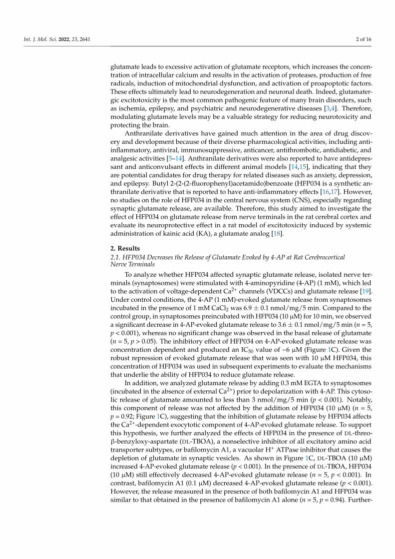
glutamate leads to excessive activation of glutamate receptors, which increases the concen-tration of intracellular calcium and results in the activation of proteases, production of freeradicals, induction of mitochondrial dysfunction, and activation of proapoptotic factors.These effects ultimately lead to neurodegeneration and neuronal death. Indeed, glutamater-gic excitotoxicity is the most common pathogenic feature of many brain disorders, suchas ischemia, epilepsy, and psychiatric and neurodegenerative diseases [3,4]. Therefore,modulating glutamate levels may be a valuable strategy for reducing neurotoxicity andprotecting the brain.
Anthranilate derivatives have gained much attention in the area of drug discov-ery and development because of their diverse pharmacological activities, including anti-inflammatory, antiviral, immunosuppressive, anticancer, antithrombotic, antidiabetic, andanalgesic activities [5–14]. Anthranilate derivatives were also reported to have antidepres-sant and anticonvulsant effects in different animal models [14,15], indicating that theyare potential candidates for drug therapy for related diseases such as anxiety, depression,and epilepsy. Butyl 2-(2-(2-fluorophenyl)acetamido)benzoate (HFP034 is a synthetic an-thranilate derivative that is reported to have anti-inflammatory effects [16,17]. However,no studies on the role of HFP034 in the central nervous system (CNS), especially regardingsynaptic glutamate release, are available. Therefore, this study aimed to investigate theeffect of HFP034 on glutamate release from nerve terminals in the rat cerebral cortex andevaluate its neuroprotective effect in a rat model of excitotoxicity induced by systemicadministration of kainic acid (KA), a glutamate analog [18].
2. Results2.1. HFP034 Decreases the Release of Glutamate Evoked by 4-AP at Rat CerebrocorticalNerve Terminals
To analyze whether HFP034 affected synaptic glutamate release, isolated nerve ter-minals (synaptosomes) were stimulated with 4-aminopyridine (4-AP) (1 mM), which ledto the activation of voltage-dependent Ca2+ channels (VDCCs) and glutamate release [19].Under control conditions, the 4-AP (1 mM)-evoked glutamate release from synaptosomesincubated in the presence of 1 mM CaCl2 was 6.9 ± 0.1 nmol/mg/5 min. Compared to thecontrol group, in synaptosomes preincubated with HFP034 (10 µM) for 10 min, we observeda significant decrease in 4-AP-evoked glutamate release to 3.6 ± 0.1 nmol/mg/5 min (n = 5,p < 0.001), whereas no significant change was observed in the basal release of glutamate(n = 5, p > 0.05). The inhibitory effect of HFP034 on 4-AP-evoked glutamate release wasconcentration dependent and produced an IC50 value of ~6 µM (Figure 1C). Given therobust repression of evoked glutamate release that was seen with 10 µM HFP034, thisconcentration of HFP034 was used in subsequent experiments to evaluate the mechanismsthat underlie the ability of HFP034 to reduce glutamate release.
In addition, we analyzed glutamate release by adding 0.3 mM EGTA to synaptosomes(incubated in the absence of external Ca2+) prior to depolarization with 4-AP. This cytoso-lic release of glutamate amounted to less than 3 nmol/mg/5 min (p < 0.001). Notably,this component of release was not affected by the addition of HFP034 (10 µM) (n = 5,p = 0.92; Figure 1C), suggesting that the inhibition of glutamate release by HFP034 affectsthe Ca2+-dependent exocytotic component of 4-AP-evoked glutamate release. To supportthis hypothesis, we further analyzed the effects of HFP034 in the presence of DL-threo-β-benzyloxy-aspartate (DL-TBOA), a nonselective inhibitor of all excitatory amino acidtransporter subtypes, or bafilomycin A1, a vacuolar H+ ATPase inhibitor that causes thedepletion of glutamate in synaptic vesicles. As shown in Figure 1C, DL-TBOA (10 µM)increased 4-AP-evoked glutamate release (p < 0.001). In the presence of DL-TBOA, HFP034(10 µM) still effectively decreased 4-AP-evoked glutamate release (n = 5, p < 0.001). Incontrast, bafilomycin A1 (0.1 µM) decreased 4-AP-evoked glutamate release (p < 0.001).However, the release measured in the presence of both bafilomycin A1 and HFP034 wassimilar to that obtained in the presence of bafilomycin A1 alone (n = 5, p = 0.94). Further-

Int. J. Mol. Sci. 2022, 23, 2641 3 of 16
more, 15 mM KCl-evoked glutamate release, a process that involves Ca2+ influx primarilythrough VDCCs [20], was also decreased by HFP034 (10 µM) (n = 5, p < 0.001; Figure 1C).
Int. J. Mol. Sci. 2022, 23, 2641 3 of 17
bafilomycin A1 (0.1 μM) decreased 4-AP-evoked glutamate release (p < 0.001). However, the release measured in the presence of both bafilomycin A1 and HFP034 was similar to that obtained in the presence of bafilomycin A1 alone (n = 5, p = 0.94). Furthermore, 15 mM KCl-evoked glutamate release, a process that involves Ca2+ influx primarily through VDCCs [20], was also decreased by HFP034 (10 μM) (n = 5, p < 0.001; Figure 1C).
Figure 1. Effect of HFP034 on 4-AP-evoked glutamate release from rat cerebro-cortical nerve termi-nals. (A) The chemical structure of HFP034. (B) Glutamate release was measured under control con-ditions or in the presence of 10 μM HFP034 added 10 min prior to the addition of 4-AP (1 mM). (C) Effect of HFP034 at different concentrations on 4-AP-evoked glutamate release, and extracellular Ca2+-free solution, glutamate transporter inhibitor DL-TBOA or vesicular glutamate transporter in-hibitor bafilomycin A1 on the effect of HFP034. Effect of HFP034 on the release of glutamate evoked by 15 mM KCl is shown in the Figure 1C. Each dot represents the value for an individual experiment. Data are presented as mean ± S.E.M. (n = 5 per group). *** p < 0.001 vs. control group; # p < 0.001 vs. DL-TBOA-treated group.
2.2. HFP034 Reduces the 4-AP-Induced Increase in (Ca2+)i and Does Not Affect Synaptosomal Membrane Potential
Figure 2A shows that the addition of 4-AP (1 mM) increased intra-terminal Ca2+ levels ((Ca2+)i) from 148.3 ± 1.2 nM to a plateau of 224.4 ± 3.2 nM. Preincubation of synaptosomes with HFP034 (10 μM) did not significantly affect basal Ca2+ levels (149.5 ± 2.1 nM, p = 1) but reduced the 4AP-evoked (Ca2+)C increase by 17% (186.6 ± 4.1 nM; n = 5, p < 0.01). In addition, we used the membrane potential-sensitive dye 3,3,3-dipropylthiadicarbocya-nine iodide (DiSC3(5)) to assess the effect of HFP034 on the synaptosomal plasma mem-brane potential. As shown in Figure 2B, the addition of 4-AP (1 mM) caused an increase
Figure 1. Effect of HFP034 on 4-AP-evoked glutamate release from rat cerebro-cortical nerve terminals.(A) The chemical structure of HFP034. (B) Glutamate release was measured under control conditionsor in the presence of 10 µM HFP034 added 10 min prior to the addition of 4-AP (1 mM). (C) Effectof HFP034 at different concentrations on 4-AP-evoked glutamate release, and extracellular Ca2+-free solution, glutamate transporter inhibitor DL-TBOA or vesicular glutamate transporter inhibitorbafilomycin A1 on the effect of HFP034. Effect of HFP034 on the release of glutamate evoked by15 mM KCl is shown in the Figure 1C. Each dot represents the value for an individual experiment.Data are presented as mean ± S.E.M. (n = 5 per group). *** p < 0.001 vs. control group; # p < 0.001 vs.DL-TBOA-treated group.
2.2. HFP034 Reduces the 4-AP-Induced Increase in (Ca2+)i and Does Not Affect SynaptosomalMembrane Potential
Figure 2A shows that the addition of 4-AP (1 mM) increased intra-terminal Ca2+ levels((Ca2+)i) from 148.3 ± 1.2 nM to a plateau of 224.4 ± 3.2 nM. Preincubation of synaptosomeswith HFP034 (10 µM) did not significantly affect basal Ca2+ levels (149.5 ± 2.1 nM, p = 1)but reduced the 4AP-evoked (Ca2+)C increase by 17% (186.6 ± 4.1 nM; n = 5, p < 0.01). Inaddition, we used the membrane potential-sensitive dye 3,3,3-dipropylthiadicarbocyanineiodide (DiSC3(5)) to assess the effect of HFP034 on the synaptosomal plasma membrane po-tential. As shown in Figure 2B, the addition of 4-AP (1 mM) caused an increase in DiSC3(5)fluorescence in synaptosomes from 0.53 ± 0.3 to 29.3 ± 1.6 fluorescence units/5 min.Preincubation of synaptosomes with HFP034 (10 µM) did not alter the basal DiSC3(5)

Int. J. Mol. Sci. 2022, 23, 2641 4 of 16
fluorescence (0.55 ± 0.2 fluorescence units/5 min) and produced no substantial change inthe 4-AP-mediated increase in DiSC3(5) fluorescence (29.5 ± 1.9 fluorescence units/5 min;p = 0.93).
Int. J. Mol. Sci. 2022, 23, 2641 4 of 17
in DiSC3(5) fluorescence in synaptosomes from 0.53 ± 0.3 to 29.3 ± 1.6 fluorescence units/5 min. Preincubation of synaptosomes with HFP034 (10 μM) did not alter the basal DiSC3(5) fluorescence (0.55 ± 0.2 fluorescence units/5 min) and produced no substantial change in the 4-AP-mediated increase in DiSC3(5) fluorescence (29.5 ± 1.9 fluorescence units/5 min; p = 0.93).
Figure 2. Effect of eupafolin on [Ca2+]i (A) and the synaptosomal membrane potential (B). HFP034 (10 μM) was added 10 min before the addition of 4-AP. Data are presented as mean ± S.E.M. (n = 5 per group). *** p < 0.001, versus control group.
2.3. HFP034 Decreases a Component of the P/Q-Type Ca2+ Channel-Coupled Glutamate Release Pathway
At rat cerebro-cortical nerve terminals, the 4-AP-evoked glutamate release is sup-ported by Ca2+ entry through N and P/Q-type VDCCs and Ca2+ release from intracellular Ca2+ stores [21,22]. Therefore, we assessed which part of the Ca2+ source contributed to the effect of HFP034 on 4-AP-evoked glutamate release. As shown in Figure 3, control 4-AP-evoked glutamate release was reduced by 10 μM HFP034 (n = 5, p < 0.001). Likewise, the blocker of N-type Ca2+ channels ω-conotoxin GVIA (ω-CgTX GVIA) (2 μM), the blocker of P/Q-type Ca2+ channels ω-agatoxin IVA (ω-Aga IVA) (0.5 μM), the inhibitor of intracel-lular Ca2+ release from the endoplasmic reticulum dantrolene (10 μM), and the inhibitor of mitochondrial Na+/Ca2+ exchange 7-chloro-5-(2-chlorophenyl)-1,5-dihydro-4,1-benzo-thiazepin-2(3H)-one (CGP37157) (10 μM) also reduced 4-AP-evoked glutamate release (p < 0.001). However, HFP034 (10 μM) could still effectively reduce release in the presence
Figure 2. Effect of eupafolin on [Ca2+]i (A) and the synaptosomal membrane potential (B). HFP034(10 µM) was added 10 min before the addition of 4-AP. Data are presented as mean ± S.E.M. (n = 5per group). *** p < 0.001, versus control group.
2.3. HFP034 Decreases a Component of the P/Q-Type Ca2+ Channel-Coupled GlutamateRelease Pathway
At rat cerebro-cortical nerve terminals, the 4-AP-evoked glutamate release is supportedby Ca2+ entry through N and P/Q-type VDCCs and Ca2+ release from intracellular Ca2+
stores [21,22]. Therefore, we assessed which part of the Ca2+ source contributed to theeffect of HFP034 on 4-AP-evoked glutamate release. As shown in Figure 3, control 4-AP-evoked glutamate release was reduced by 10 µM HFP034 (n = 5, p < 0.001). Likewise,the blocker of N-type Ca2+ channels ω-conotoxin GVIA (ω-CgTX GVIA) (2 µM), theblocker of P/Q-type Ca2+ channels ω-agatoxin IVA (ω-Aga IVA) (0.5 µM), the inhibitorof intracellular Ca2+ release from the endoplasmic reticulum dantrolene (10 µM), and theinhibitor of mitochondrial Na+/Ca2+ exchange 7-chloro-5-(2-chlorophenyl)-1,5-dihydro-4,1-benzothiazepin-2(3H)-one (CGP37157) (10 µM) also reduced 4-AP-evoked glutamaterelease (p < 0.001). However, HFP034 (10 µM) could still effectively reduce release in the

Int. J. Mol. Sci. 2022, 23, 2641 5 of 16
presence ofω-CgTX GVIA, dantrolene or CGP37157 (n = 5, p < 0.001). In contrast, furtherinhibition by HFP034 was not observed in the presence of ω-Aga IVA (n = 5, p = 0.83).These results suggest the involvement of decreased P/Q-type Ca2+ channel activity in theaction of HFP034.
Int. J. Mol. Sci. 2022, 23, 2641 5 of 17
of ω-CgTX GVIA, dantrolene or CGP37157 (n = 5, p < 0.001). In contrast, further inhibition by HFP034 was not observed in the presence of ω-Aga IVA (n = 5, p = 0.83). These results suggest the involvement of decreased P/Q-type Ca2+ channel activity in the action of HFP034.
Figure 3. Effect of HFP034 on 4-AP-evoked glutamate release in the presence of N-type Ca2+ channel blocker ω-CgTX GVIA, P/Q-type Ca2+ channel blocker ω-AgTX IVA, ryanodine receptor inhibitor dantrolene, or mitochondrial Na+/Ca2+ exchanger inhibitor CGP37157. HFP034 was added 10 min before the addition of 4-AP, and other drugs were added 10 min before this. Each dot represents the value for an individual experiment. Data are presented as mean ± S.E.M. (n = 5 per group). *** p < 0.001, versus control group. # p < 0.001, versus dantrolene- or CGP37157-treated group.
2.4. The Suppressed Protein Kinase C Pathway Is Linked to HFP034-Mediated Inhibition of Glutamate Release
In view of the demonstrated role of protein kinase C (PKC) in presynaptic modula-tion [23], we assessed the effect of HFP034 on glutamate release in the presence of the selective PKC inhibitor bisindolylmaleimide I (GF109203X) (10 μM). As shown in Figure 4A, 4-AP (1 mM)-evoked glutamate release was decreased in the presence of GF109203X (10 μM) (p < 0.001). HFP034 (10 μM) alone reduced the 4-AP-evoked release of glutamate (p < 0.001), but this inhibition was completely suppressed by pretreatment with GF109203X. There was no significant difference between the release after GF109203X alone, and that after GF109203X + HFP034 treatment (n = 5, p = 0.98). In addition, Figure 4B shows the effects of HFP034 on the phosphorylation of PKC, PKCα and MARCKS (an important presynaptic substrate for PKC) in cerebro-cortical synaptosomes. Compared to the control, 1 mM 4-AP treatment induced a significant increase in the phosphorylation of PKC, PKCα, and MARCKS induced by (p < 0.001). When synaptosomes were pretreated with HFP034 (10 μM) for 10 min before the addition of 4-AP, 4-AP-increased phosphory-lation of PKC, PKCα, and MARCKS was markedly decreased compared with that in the 4-AP group (n = 5, p < 0.05; Figure 4B).
Figure 3. Effect of HFP034 on 4-AP-evoked glutamate release in the presence of N-type Ca2+ channelblockerω-CgTX GVIA, P/Q-type Ca2+ channel blockerω-AgTX IVA, ryanodine receptor inhibitordantrolene, or mitochondrial Na+/Ca2+ exchanger inhibitor CGP37157. HFP034 was added 10 minbefore the addition of 4-AP, and other drugs were added 10 min before this. Each dot representsthe value for an individual experiment. Data are presented as mean ± S.E.M. (n = 5 per group).*** p < 0.001, versus control group. # p < 0.001, versus dantrolene- or CGP37157-treated group.
2.4. The Suppressed Protein Kinase C Pathway Is Linked to HFP034-Mediated Inhibition ofGlutamate Release
In view of the demonstrated role of protein kinase C (PKC) in presynaptic modula-tion [23], we assessed the effect of HFP034 on glutamate release in the presence of theselective PKC inhibitor bisindolylmaleimide I (GF109203X) (10 µM). As shown in Figure 4A,4-AP (1 mM)-evoked glutamate release was decreased in the presence of GF109203X (10 µM)(p < 0.001). HFP034 (10 µM) alone reduced the 4-AP-evoked release of glutamate (p < 0.001),but this inhibition was completely suppressed by pretreatment with GF109203X. Therewas no significant difference between the release after GF109203X alone, and that afterGF109203X + HFP034 treatment (n = 5, p = 0.98). In addition, Figure 4B shows the effects ofHFP034 on the phosphorylation of PKC, PKCα and MARCKS (an important presynapticsubstrate for PKC) in cerebro-cortical synaptosomes. Compared to the control, 1 mM 4-APtreatment induced a significant increase in the phosphorylation of PKC, PKCα, and MAR-CKS induced by (p < 0.001). When synaptosomes were pretreated with HFP034 (10 µM) for10 min before the addition of 4-AP, 4-AP-increased phosphorylation of PKC, PKCα, andMARCKS was markedly decreased compared with that in the 4-AP group (n = 5, p < 0.05;Figure 4B).

Int. J. Mol. Sci. 2022, 23, 2641 6 of 16Int. J. Mol. Sci. 2022, 23, 2641 6 of 17
Figure 4. (A) Effect of the PKC inhibitor GF109203X on the HFP034-mediated inhibition of 4-AP-evoked glutamate release. (B) Effect of HFP034 on PKC and MARCKS phosphorylation evoked by 4-AP. HFP034 or GF109203X was added 10 min before the addition of 4-AP. Each dot represents the value for an individual experiment. Data are presented as mean ± S.E.M. (n = 5 per group). *** p < 0.001, versus control group. # p < 0.001, versus GF109203X- or 4-AP-treated group.
2.5. HFP034 Attenuates Neuronal Death and Glutamate Elevation in the Hippocampus of Rats with KA
To evaluate whether HFP034 exerted a protective action in a rat model of KA-in-duced glutamate excitotoxicity, rats were treated with an intraperitoneal (i.p.) injection of HFP034 or dimethyl-sulfoxide (DMSO) for 30 min prior to i.p. injection of KA (15 mg/kg). As shown in Figure 5, we performed Nissl and NeuN staining of brain tissue sections 72 h after KA treatment. Nissl staining revealed significant neuronal loss in the CA1 and CA3 hippocampal regions of the KA-injected rats compared with the DMSO-treated rats (con-trol) (p < 0.001). In contrast, Nissl staining in HFP034 + KA-treated rats demonstrated a significant level of protection against hippocampal cell death. Neuronal survival in
Figure 4. (A) Effect of the PKC inhibitor GF109203X on the HFP034-mediated inhibition of 4-AP-evoked glutamate release. (B) Effect of HFP034 on PKC and MARCKS phosphorylation evoked by4-AP. HFP034 or GF109203X was added 10 min before the addition of 4-AP. Each dot representsthe value for an individual experiment. Data are presented as mean ± S.E.M. (n = 5 per group).*** p < 0.001, versus control group. # p < 0.001, versus GF109203X- or 4-AP-treated group.
2.5. HFP034 Attenuates Neuronal Death and Glutamate Elevation in the Hippocampus of Ratswith KA
To evaluate whether HFP034 exerted a protective action in a rat model of KA-inducedglutamate excitotoxicity, rats were treated with an intraperitoneal (i.p.) injection of HFP034or dimethyl-sulfoxide (DMSO) for 30 min prior to i.p. injection of KA (15 mg/kg). Asshown in Figure 5, we performed Nissl and NeuN staining of brain tissue sections 72 hafter KA treatment. Nissl staining revealed significant neuronal loss in the CA1 and CA3hippocampal regions of the KA-injected rats compared with the DMSO-treated rats (con-trol) (p < 0.001). In contrast, Nissl staining in HFP034 + KA-treated rats demonstrated

Int. J. Mol. Sci. 2022, 23, 2641 7 of 16
a significant level of protection against hippocampal cell death. Neuronal survival inHFP034 + KA-treated rats was significantly higher than that observed in KA-treated rats(n = 3, p < 0.05; Figure 5B). The NeuN findings were consistent with those of Nissl staining.Compared to the control group, the KA-treated group experienced a significant decreasein the number of NeuN-positive neurons in the CA1 and CA3 regions (p < 0.001). Thisreduction was prevented in HFP034 + KA-treated rats compared to the KA group (n = 3,p < 0.05; Figure 5C). In addition, we examined the effect of HFP034 on the concentration ofglutamate in the hippocampus of rats with KA (Figure 5D). Compared to the control group,the KA group had significantly increased glutamate levels in the hippocampus (p < 0.001).However, groups that received KA and were pretreated with HFP034 experienced a de-crease in glutamate levels in the hippocampus, which was significantly different from theKA group (n = 3, p < 0.05).
Int. J. Mol. Sci. 2022, 23, 2641 7 of 17
HFP034 + KA-treated rats was significantly higher than that observed in KA-treated rats (n = 3, p < 0.05; Figure 5B). The NeuN findings were consistent with those of Nissl staining. Compared to the control group, the KA-treated group experienced a significant decrease in the number of NeuN-positive neurons in the CA1 and CA3 regions (p < 0.001). This reduction was prevented in HFP034 + KA-treated rats compared to the KA group (n = 3, p < 0.05; Figure 5C). In addition, we examined the effect of HFP034 on the concentration of glutamate in the hippocampus of rats with KA (Figure 5D). Compared to the control group, the KA group had significantly increased glutamate levels in the hippocampus (p < 0.001). However, groups that received KA and were pretreated with HFP034 experi-enced a decrease in glutamate levels in the hippocampus, which was significantly differ-ent from the KA group (n = 3, p < 0.05).
Figure 5. Effect of HFP034 pretreatment on the neuronal cell death and glutamate levels in the hip-pocampus of rats with KA. (A) Representative images of crystal violet and NeuN staining at 3 d after i.p. KA. (B,C) Quantitative data of A showing the number of living neurons and NeuN+ cells in the hippocampal CA1 and CA3 regions. (D) The effect of HFP034 on the concentration of gluta-mate in the hippocampus of rats with KA. Each dot represents the value for an individual experi-ment. Data are presented as mean ± S.E.M. n = 3 rats for each group. *** p < 0.001, versus control group. # p < 0.001, versus KA group.
2.6. HFP034 Reduces the Levels of Endoplasmic Reticulum (ER) Stress-Related Proteins in the Hippocampus of Rats with KA
Figure 5. Effect of HFP034 pretreatment on the neuronal cell death and glutamate levels in thehippocampus of rats with KA. (A) Representative images of crystal violet and NeuN staining at 3 dafter i.p. KA. (B,C) Quantitative data of A showing the number of living neurons and NeuN+ cells inthe hippocampal CA1 and CA3 regions. (D) The effect of HFP034 on the concentration of glutamatein the hippocampus of rats with KA. Each dot represents the value for an individual experiment.Data are presented as mean ± S.E.M. n = 3 rats for each group. *** p < 0.001, versus control group.# p < 0.001, versus KA group.

Int. J. Mol. Sci. 2022, 23, 2641 8 of 16
2.6. HFP034 Reduces the Levels of Endoplasmic Reticulum (ER) Stress-Related Proteins in theHippocampus of Rats with KA
To investigate how HFP034 attenuated KA-induced neuronal death, we examinedwhether pretreatment with HFP034 contributed to a reduction in KA-induced ER stress,resulting in neuronal damage [24]. As shown in Figure 6, the levels of ER stress signaturemolecules, including calpains, glucose-regulated protein 78 (GRP 78), C/EBP homologousprotein (CHOP), and caspase-12, were increased in the hippocampus of rats treated withKA compared to the control (p < 0.001), but the level of increase decreased with HFP034pretreatment (n = 3, p < 0.05 vs. KA group).
Int. J. Mol. Sci. 2022, 23, 2641 8 of 17
To investigate how HFP034 attenuated KA-induced neuronal death, we examined whether pretreatment with HFP034 contributed to a reduction in KA-induced ER stress, resulting in neuronal damage [24]. As shown in Figure 6, the levels of ER stress signature molecules, including calpains, glucose-regulated protein 78 (GRP 78), C/EBP homologous protein (CHOP), and caspase-12, were increased in the hippocampus of rats treated with KA compared to the control (p < 0.001), but the level of increase decreased with HFP034 pretreatment (n = 3, p < 0.05 vs. KA group).
Figure 6. Effect of HFP034 pretreatment on the expression levels of ER stress-associated proteins, calpains, GRP78, CHOP, and caspase-12 in the hippocampus of rats with KA. Each dot represents the value for an individual experiment. Data are presented as mean ± S.E.M. n = 3 rats for each group. ** p < 0.01, *** p < 0.001 versus control group. * p < 0.01, # p < 0.001, versus KA group.
2.7. HFP034 Suppresses the Activation of Microglia and Astrocytes in the Hippocampus of Rats with KA
Microglial and astrocyte activation is a common pathological feature following KA-induced excitotoxic injury [25]. We examined the effect of HFP034 on KA-induced micro-glia and astrocyte activation in hippocampal sections using anti-OX42 and anti-GFAP an-tibodies, respectively. As shown in Figure 7A, we observed a significant increase in the number of OX42+ microglial cells in the CA1 and CA3 hippocampal regions of the KA group compared with the control group (p < 0.001). In contrast, HFP034-pretreated rats displayed much less OX42 staining, indicating a reduced microglial response to KA-in-duced injury. Quantification of the results showed a significant decrease in the number of OX42+ cells (n = 3, p < 0.05 vs. KA group; Figure 7B). In addition, the evaluation of GFAP staining for astrocyte activation showed a considerable response in KA-treated rats com-pared with those in the control group (p < 0.001; Figure 7A). Similarly, HFP034 pretreat-ment significantly reduced astrocyte activation induced by KA, as observed by the reduc-tion in the number of GFAP+ cells in the CA1 and CA3 regions (n = 3, p < 0.05 vs. KA group; Figure 7C).
Figure 6. Effect of HFP034 pretreatment on the expression levels of ER stress-associated proteins,calpains, GRP78, CHOP, and caspase-12 in the hippocampus of rats with KA. Each dot represents thevalue for an individual experiment. Data are presented as mean ± S.E.M. n = 3 rats for each group.** p < 0.01, *** p < 0.001 versus control group. * p < 0.01, # p < 0.001, versus KA group.
2.7. HFP034 Suppresses the Activation of Microglia and Astrocytes in the Hippocampus of Ratswith KA
Microglial and astrocyte activation is a common pathological feature following KA-induced excitotoxic injury [25]. We examined the effect of HFP034 on KA-induced microgliaand astrocyte activation in hippocampal sections using anti-OX42 and anti-GFAP antibodies,respectively. As shown in Figure 7A, we observed a significant increase in the numberof OX42+ microglial cells in the CA1 and CA3 hippocampal regions of the KA groupcompared with the control group (p < 0.001). In contrast, HFP034-pretreated rats displayedmuch less OX42 staining, indicating a reduced microglial response to KA-induced injury.Quantification of the results showed a significant decrease in the number of OX42+ cells(n = 3, p < 0.05 vs. KA group; Figure 7B). In addition, the evaluation of GFAP stainingfor astrocyte activation showed a considerable response in KA-treated rats comparedwith those in the control group (p < 0.001; Figure 7A). Similarly, HFP034 pretreatmentsignificantly reduced astrocyte activation induced by KA, as observed by the reductionin the number of GFAP+ cells in the CA1 and CA3 regions (n = 3, p < 0.05 vs. KA group;Figure 7C).

Int. J. Mol. Sci. 2022, 23, 2641 9 of 16Int. J. Mol. Sci. 2022, 23, 2641 9 of 17
Figure 7. Effect of HFP034 pretreatment on the activation of microglia and astrocyte in the hippo-campus of rats with KA. (A) Representative images of OX42 and GFAP staining at 3 d after i.p. KA. (B,C) Quantitative data of A and C showing the number of OX42+ and GFAP+ cells in the hippocam-pal CA1 and CA3 regions. Each dot represents the value for an individual experiment. Data are presented as mean ± S.E.M. n = 3 rats for each group. *** p < 0.001, versus control group. # p < 0.001, versus KA group.
3. Discussion Excessive release and accumulation of glutamate in the brain is associated with exci-
totoxicity, a key mechanism in neuronal degeneration in several acute and chronic CNS diseases [3,26]. Identifying new drugs that regulate glutamate release and provide protec-tion against glutamate excitotoxicity is therefore crucial [4,27]. In the current study, we investigated the effect of the anthranilate derivative HFP034 on glutamate release in vitro and its neuroprotective potential against KA-induced glutamate excitotoxicity in vivo. We also examined possible mechanisms underlying the effects of HFP034
In the present study, HFP034 inhibited the release of glutamate evoked by exposing synaptosomes to 4-AP. Because 4-AP induces glutamate release from neurons, two mech-anisms (exocytosis and carrier-mediated outward transport) are involved. Exocytotic glu-tamate release is the result of glutamate release from storage vesicles (Ca2+-dependent fraction), whereas glutamate transporters transport glutamate from the axoplasmic site via a reduced Na+ gradient (Ca2+-dependent fraction) [27]. We found that HFP034 reduced 4-AP-evoked glutamate release in the presence of extracellular Ca2+; however, it had no effect in the absence of extracellular Ca2+. Furthermore, depletion of glutamate in synaptic vesicles by bafilomycin A1 largely prevented the inhibition of release by HFP034; how-ever, the inhibition of release by HFP034 was insensitive to blockade of the glutamate
Figure 7. Effect of HFP034 pretreatment on the activation of microglia and astrocyte in the hip-pocampus of rats with KA. (A) Representative images of OX42 and GFAP staining at 3 d after i.p.KA. (B,C) Quantitative data of A and C showing the number of OX42+ and GFAP+ cells in thehippocampal CA1 and CA3 regions. Each dot represents the value for an individual experiment.Data are presented as mean ± S.E.M. n = 3 rats for each group. *** p < 0.001, versus control group.# p < 0.001, versus KA group.
3. Discussion
Excessive release and accumulation of glutamate in the brain is associated with exci-totoxicity, a key mechanism in neuronal degeneration in several acute and chronic CNSdiseases [3,26]. Identifying new drugs that regulate glutamate release and provide protec-tion against glutamate excitotoxicity is therefore crucial [4,27]. In the current study, weinvestigated the effect of the anthranilate derivative HFP034 on glutamate release in vitroand its neuroprotective potential against KA-induced glutamate excitotoxicity in vivo. Wealso examined possible mechanisms underlying the effects of HFP034
In the present study, HFP034 inhibited the release of glutamate evoked by expos-ing synaptosomes to 4-AP. Because 4-AP induces glutamate release from neurons, twomechanisms (exocytosis and carrier-mediated outward transport) are involved. Exocytoticglutamate release is the result of glutamate release from storage vesicles (Ca2+-dependentfraction), whereas glutamate transporters transport glutamate from the axoplasmic site viaa reduced Na+ gradient (Ca2+-dependent fraction) [27]. We found that HFP034 reduced4-AP-evoked glutamate release in the presence of extracellular Ca2+; however, it had noeffect in the absence of extracellular Ca2+. Furthermore, depletion of glutamate in synapticvesicles by bafilomycin A1 largely prevented the inhibition of release by HFP034; however,the inhibition of release by HFP034 was insensitive to blockade of the glutamate transporter

Int. J. Mol. Sci. 2022, 23, 2641 10 of 16
by DL-TBOA. These results indicate that HFP034 decreases the release of glutamate evokedby 4-AP at cerebro-cortical nerve terminals by inhibiting Ca2+-dependent vesicular releaserather than reversing the operation of glutamate transporters. In addition, we observed thatat cerebro-cortical nerve terminals, HFP034 reduced the 4-AP-evoked increase in (Ca2+)i,while it did not affect 4-AP-mediated depolarization. This indicates that the inhibitory effectof HFP034 on glutamate release is not due to a decrease in synaptosomal excitability. Fur-thermore, apart from the 4-AP evolving glutamate release, we found that HFP034 inhibitedthe release of glutamate evoked by KCl. Because 4-AP-evoked glutamate release involvesthe activation of Na+ and Ca2+ channels, 15 mM external KCl-evoked glutamate releaseinvolves only Ca2+ channels [25], which indicates that Ca2+ channels are involved in theeffect of HFP034 on glutamate release. Consistent with this, HFP034-mediated inhibition ofglutamate release was completely abolished by blocking P/Q-type Ca2+ channels but notby blocking N-type Ca2+ channels or intracellular Ca2+ release. Based on these results, weinfer that HFP034 inhibits evoked glutamate release by directly reducing P/Q-type Ca2+
channel activity rather than by indirectly affecting nerve terminal excitability.In synaptic terminals, PKC signaling regulates neurotransmitter release. It has been
shown that depolarization-stimulated Ca2+ influx increases PKC-dependent phosphoryla-tion and glutamate release [28,29]. In the present study, we found that (1) the inhibitoryeffect of HFP034 on 4-AP-evoked glutamate release was prevented by PKC inhibition;(2) HFP034 significantly decreased 4-AP-induced phosphorylation of PKC and PKCα, aPKC isoform that is present pre-synaptically in the cerebral cortex [30]; and (3) 4-AP-induced phosphorylation of MARCKS, a major presynaptic substrate for PKC, was de-creased by HFP034. Although how HFP034 affects P/Q-type Ca2+ channels remains tobe elucidated, our data suggest that an intracellular cascade involving the suppressionof PKC-dependent pathways is linked to inhibition of 4-AP-evoked glutamate releaseby HFP034.
We also found that HFP034 exerted neuroprotective efficacy in a rat model of KA-induced glutamate excitotoxicity. The KA-induced excitotoxicity model is a well-establishedmodel for screening neuroprotective drugs [31]. Consistent with previous studies [32–34],we observed that KA administration (15 mg/kg, i.p.) leads to an increase in glutamateand substantial neuronal death in the hippocampus. These KA-induced alterations weresignificantly counteracted in the HFP034 pretreatment group, indicating that HFP034 exertsanti-excitotoxic and neuroprotective effects. In addition, we observed that KA increased theexpression levels of ER stress-associated proteins, including calpain, GRP78, CHOP, andcaspase-12 in the hippocampus; these changes were also reduced by HFP034 pretreatment.These results suggest that HFP034 can reduce ER stress, a key factor in neuronal deathin numerous neurological diseases [35,36]. In particular, ER stress can cause neuronaldeath either by triggering ER Ca2+ release, resulting in calpain and caspase-12 activa-tion, or by activating CHOP and GRP78-mediated proapoptotic pathways [37–39], whichhave been suggested to be involved in neuronal cell death after KA-induced excitotoxic-ity [36]. Furthermore, ER stress inhibition can protect against KA-induced hippocampalneuronal damage [40,41]. Thus, suppressed KA-induced ER stress might partly explain theneuroprotective effect of HFP034.
In addition to ER stress, inflammatory responses, including the activation of microgliaand astrocytes, are often associated with KA-induced excitotoxic injury. Activated glialcells increase the production of toxic substances, which in turn contribute to the expansionof brain injury and increased neuron loss. This evidence suggests that the control of KA-induced neuroinflammation is vital to protect hippocampal neurons [25,42]. In the presentstudy, we observed that KA substantially increased the number of activated microgliaand astrocytes in the hippocampus, and these increases were suppressed by HFP034pretreatment. Thus, the suppression of neuroinflammation, in addition to ER stress, couldbe another mechanism explaining the neuroprotection provided by HFP034 against KA-induced glutamate excitotoxicity. Our finding is consistent with that of previous studiesthat have reported the anti-inflammatory activities of HFP034 [16,17]. Although how

Int. J. Mol. Sci. 2022, 23, 2641 11 of 16
HFP034 suppresses glial activation was not explored in this study, Toll-like receptors (TLRs)have been shown to play a critical role in glial cell activation and subsequent hippocampalneuron excitotoxicity [43,44]. Whether HFP034 inhibits the activation of TLRs, therebyreducing glial cell activation and further suppressing KA-induced excitotoxic insults, is apossibility that should be addressed in future studies.
The ability of HFP034 to reduce glutamate release from nerve terminals may partiallyexplain its neuroprotective mechanism against KA-induced excitotoxicity in vivo. Thishypothesis is based on the association between KA-induced neurotoxicity and excessiveglutamate release [31,45]. Apart from excessive glutamate release, KA also causes glu-tamate receptor overstimulation. This overstimulation results in calcium elevation andsubsequently triggers intracellular cascade reactions, including reactive oxygen speciesproduction, lipid peroxidation, ER stress, and mitochondrial dysfunction and inflammation,eventually leading to cell death [18]. On the basis of these considerations, HFP034 mightsuppress ER stress and neuroinflammation to prevent KA-induced insults to neuronalintegrity, which may be associated with the inhibition of released glutamate.
4. Materials and Methods
The experimental protocol was approved by the Fu Jen Institutional Animal Care andUtilization Committee (code A11009). The animals were treated in accordance with theGuide for the Care and Use of Laboratory Animals. The minimum number of animalsneeded to obtain consistent data was employed.
4.1. Materials
HFP034 was synthesized by one of the authors (Pei-Wen Hsieh) [16] and dissolvedin 1% DMSO. DL-TBOA, bafilomycin A1, dantrolene, CGP37157, and GF109203X werepurchased from Tocris (Bristol, UK). DiSC3(5) and fura-2-acetoxymethyl ester (Fura-2-AM)were purchased from Thermo (Waltham, MA, USA). ω-CgTX GVIA and ω-Aga IVA werepurchased from the Alomone lab (Jerusalem, Israel). 4-AP, DMSO, KA and all other reagentswere purchased from Sigma–Aldrich (St. Louis, MO, USA). Adult male Sprague–Dawleyrats (n = 42, 150–200 g) were purchased from BioLASCO (Taipei, Taiwan).
4.2. Synaptosome Preparation
Rats (n = 18) were sacrificed via cervical dislocation and the cerebral cortex wasremoved immediately. The brain tissue was homogenized in 320 mM sucrose solution andcentrifuged at 3000× g for 10 min. The supernatant was stratified on Percoll discontinuousgradients and centrifuged at 32,500× g for 7 min. The synaptosomal fraction was collectedand centrifuged for 10 min at 27,000× g. The protein concentration was determined usingthe Bradford assay. Synaptosomes were centrifuged in a final wash to obtain synaptosomalpellets with 0.5 mg protein, as previously described [46–48].
4.3. Glutamate Release Analysis
For the glutamate release experiments, the synaptosomal pellet (0.5 mg protein) wasresuspended in hepes-buffered solution, and glutamate release was assayed by onlinefluorimetry [49]. CaCl2 (1.2 mM), glutamate dehydrogenase (GDH, 50 units/mL) andNADP+ (2 mM) were added at the start of the incubation. Glutamate release was inducedwith 4-AP (1 mM) and monitored by measuring the increase in fluorescence (excitationand emission wavelengths of 340 and 460 nm, respectively) resulting from NADPH beingproduced by oxidative deamination of released glutamate by GDH. The amount of releasedglutamate was calibrated against a standard of exogenous glutamate (5 nmol) and expressedas nanomoles glutamate per milligram synaptosomal protein (nmol/mg).

Int. J. Mol. Sci. 2022, 23, 2641 12 of 16
4.4. Intrasynaptosomal Ca2+ Concentration ([Ca2+]i)
Synaptosomes (0.5 mg protein) were incubated in hepes-buffered solution containingFura 2-AM (5 µM) and CaCl2 (0.1 mM) for 30 min at 37 ◦C. Samples were centrifuged for1 min at 3000× g, and pellets were resuspended in hepes-buffered medium containing CaCl2(1.2 mM). Fura-2-Ca fluorescence was monitored at 5 s intervals for 5 min. (Ca2+)i (nM)was calculated using previously described calibration procedures and equations [50].
4.5. Membrane Potential
The synaptosomal membrane potential was assayed with the positively chargedmembrane potential-sensitive carbocyanine dye DiSC3(5). DiSC3(5) fluorescence wasmonitored at 2 s intervals, and the data are expressed in fluorescence units [51].
4.6. Histological Analysis
The rats (n = 24) were divided into four experimental groups: the DMSO-treated group(control), KA-treated group, HFP034 10 mg/kg + KA group, and HFP034 30 mg/kg + KAgroup. HFP034 was dissolved in a saline solution containing 1% DMSO and administered(i.p.) 30 min before KA injection (15 mg/kg in 0.9% NaCl, pH 7.0, i.p.). The dose andschedule of administration were chosen based on previous experiments of our group [34,49]and others [18,25]. For Nissl staining, rats (n = 3 per group) were euthanized 72 h afterKA injection by trans-cardial perfusion with 4% paraformaldehyde in 0.1 M phosphate-buffered saline (PBS) under inhalational anesthesia with 2–3% iso-furane. The brainswere removed, fixed overnight with 4% paraformaldehyde solution, and cryoprotected insucrose phosphate buffer at 4 ◦C. The brains were cut into 30 µm coronal sections, mountedon gelatinized slides, air-dried and stained with 0.1% aqueous cresyl violet stain (SigmaChemicals, St. Louis, MO, USA) for 20 min. Then, the slides were washed in distilledwater, differentiated in 70% ethyl alcohol, dehydrated in ascending grades of ethyl alcohol,cleared in xylene, and mounted with DPX (Sigma Chemicals, St. Louis, MO, USA). Forimmunofluorescence staining, the brain sections were blocked with 2% bovine serumalbumin (BSA) in PBS for 30 min and then incubated overnight at 4 ◦C with the primaryantibodies anti-NeuN (1:500, Abcam, Cambridge, UK), anti-OX42 (1:500, Merck Millipore,Burlington, VT, USA), and anti-GFAP (1:1000, Cell Signaling, MA, USA). The sections wereincubated for 90 min at room temperature with the corresponding secondary antibodies(1:1000, Alexa Fluor 488, DyLight 594, Invitrogen, CA, USA), mounted on gelatin-coatedslides and cover-slipped with VectaShield medium (Vector Labs, Burlingame, CA, USA).Cells were stained with the nuclear staining dye DAPI (1 µg/mL, Sigma–Aldrich) for20 s. Images were captured with an upright fluorescence microscope (Zeiss Axioskop40, Goettingen, Germany) using ×4 (aperture is 0.1) and ×10 (aperture is 0.25) objectives.The numbers of living neurons and NeuN+, OX42+, and GFAP+ cells were counted in a255 µm × 255 µm area of the hippocampal CA1 and CA3 using ImageJ software (Synoptics,Cambridge, UK).
4.7. High-Performance Liquid Chromatography
Determination of glutamate concentrations in brain tissue was performed with a high-performance liquid chromatography (HPLC) system with electrochemical detection (HTEC-500) [34]. Briefly, the rats were sacrificed through decapitation at 72 h after KA injection andthe brains were removed immediately. The hippocampi were dissected and homogenizedin 5 mL of hepes-buffered medium. The homogenate was centrifuged at 1500× g at 4 ◦C for10 min, and then the supernatant (10 µL) was filtered through 0.22 µm filters before injectioninto the HPLC system. An Eicompak GU-GEL column, a glutamate oxidase-immobilizedcolumn (EENZYMPAK), and a platinum electrode set at 450 mV against an Ag/AgClreference electrode were used. The mobile phase comprised 50 mM NH4Cl and 250 mg/Lhexadecyltrimethylammonium bromide with pH set at 7.4. The column temperaturewas 35 ◦C, and the flow rate was 1.2 mL/min. The relative free glutamate concentrationwas determined using peak areas with an external standard method. Serial dilutions of

Int. J. Mol. Sci. 2022, 23, 2641 13 of 16
the standards were injected, and their peak areas were determined. A linear standardcurve was constructed by plotting the peak areas versus corresponding concentrations ofeach standard.
4.8. Western Blot
Synaptosomes and hippocampal tissue were lysed in ice-cold Tris–HCl buffer solutionand centrifuged for 10 min at 13,000× g at 4 ◦C. The protein concentration in the supernatantwas measured using the Bradford protein assay (Bio–Rad laboratories, Hercules, CA, USA).Equal amounts (30 µg) of protein were loaded into each lane on a 10% polyacrylamide geland then transferred to a polyvinylidene-difluoride (PVDF) membrane in a semidry system(Bio–Rad, Hercules, CA, USA) for 120 min. Transferred membranes were blocked for 1 hin 5% nonfat dry milk in TBST (25 mM Tris-HCl, pH 7.5, 125 mM NaCl, and 0.05% Tween20) and incubated overnight at 4 ◦C with specific primary antibodies [anti-PKC, 1:700,Abcam, Cambridge, UK); phospho-PKC (1:2000, Cell Signaling, MA, USA); PKCα (1:600,Cell Signaling, MA, USA), phospho-PKCα (1:2000, Abcam, Cambridge, UK); phospho-MARCKS (1:250, Cell Signaling, MA, USA); calpain 1 (1:2000, Abcam, Cambridge, UK),calpain 2 (1:800, Millipore); caspase 12 (1:3000, Abcam, Cambridge, UK); CHOP (1:300,Santa Cruz, TX, USA); GRP 78 (1:1500, Abcam, Cambridge, UK); and β-actin (1:8000, CellSignaling, MA, USA). Membranes were washed with TBST for 15 min and incubated withhorseradish peroxidase-coupled secondary antibodies (1:16,000, GeneTex, CA, USA) for 1 hat room temperature. Then, the specific protein bands were visualized using film exposurewith a chemiluminescence detection system (GeneTex, CA, USA) and quantified usingImageJ software (Synoptics, Cambridge, UK).
4.9. Statistical Analyses
The results are expressed as the mean ± standard error of the mean (S.E.M.). Statisticalanalysis was performed using GraphPad Prism-8 software (GraphPad Inc., San Diego, CA,USA). When testing the significance of the effect of HFP034 versus the control, Student’s ttest was used. When comparing the effect of HFP034 in different experimental conditions,one-way analysis of variance (ANOVA) followed by Tukey’s post hoc test was used. p < 0.05was considered to indicate a statistically significant difference between groups.
5. Conclusions
In summary, we demonstrated that the anthranilate derivative HFP034 inhibits glu-tamate release from rat cerebro-cortical nerve terminals by suppressing P/Q-type Ca2+
channels and the PKC/MARCKS pathways and that HFP034 prevents KA-induced glu-tamate neurotoxicity in vivo by inhibiting ER stress and neuroinflammation (Figure 8).To our knowledge, this is the first report to demonstrate the inhibitory role of HFP034in glutamate release and glutamate excitotoxicity. This finding may provide a pharma-cological basis for the clinical use of HFP034 in the treatment of CNS diseases involvingglutamate excitotoxicity.

Int. J. Mol. Sci. 2022, 23, 2641 14 of 16Int. J. Mol. Sci. 2022, 23, 2641 14 of 17
Figure 8. A proposed mechanism underlying the inhibition of glutamate release and glutamate ex-citotoxicity by HFP034 in rats. HFP034 depresses glutamate release via suppressing presynaptic P/Q-type Ca2+ channels and PKC/MARCKS pathway, as well as protecting KA-induced neuronal death via ER stress and neuroinflammation inhibition. Black arrows indicate positive regulation, and red arrows indicate negative regulation.
Author Contributions: Conceptualization, C.-W.L., C.-J.L. and T.-Y.L.; Data curation, P.-W.H. and K.-M.C.; Formal analysis, C.-W.L., C.-J.L., P.-W.H. and T.-Y.L.; Funding acquisition, C.-W.L., T.-Y.L. and S.-J.W.; Investigation, C.-W.L., C.-J.L., T.-Y.L. and S.-J.W.; Project administration, K.-M.C. and M.-Y.L.; Resources, P.-W.H. and K.-M.C.; Supervision, S.-J.W.; Writing—original draft, S.-J.W.; Writing—review & editing, M.-Y.L. and S.-J.W. All authors have read and agreed to the published version of the manuscript.
Funding: This work was supported by the Far Eastern Memorial Hospital (108-FEMH-FJU-01; FEMH-2020-C-012; FEMH-2020-C-015), Taiwan.
Institutional Review Board Statement: Ethical approval was granted by the Fu Jen Catholic Uni-versity (No. A11009) and, therefore, experiments were performed in accordance with the ethical standards laid out by the IACUC.
Informed Consent Statement: Not applicable.
Data Availability Statement: The data presented in this study are available on request from the corresponding author.
Conflicts of Interest: The authors declare no conflict of interest.
Figure 8. A proposed mechanism underlying the inhibition of glutamate release and glutamateexcitotoxicity by HFP034 in rats. HFP034 depresses glutamate release via suppressing presynapticP/Q-type Ca2+ channels and PKC/MARCKS pathway, as well as protecting KA-induced neuronaldeath via ER stress and neuroinflammation inhibition. Black arrows indicate positive regulation, andred arrows indicate negative regulation.
Author Contributions: Conceptualization, C.-W.L., C.-J.L. and T.-Y.L.; Data curation, P.-W.H. andK.-M.C.; Formal analysis, C.-W.L., C.-J.L., P.-W.H. and T.-Y.L.; Funding acquisition, C.-W.L., T.-Y.L.and S.-J.W.; Investigation, C.-W.L., C.-J.L., T.-Y.L. and S.-J.W.; Project administration, K.-M.C. andM.-Y.L.; Resources, P.-W.H. and K.-M.C.; Supervision, S.-J.W.; Writing—original draft, S.-J.W.; Writing—review & editing, M.-Y.L. and S.-J.W. All authors have read and agreed to the published version ofthe manuscript.
Funding: This work was supported by the Far Eastern Memorial Hospital (108-FEMH-FJU-01;FEMH-2020-C-012; FEMH-2020-C-015), Taiwan.
Institutional Review Board Statement: Ethical approval was granted by the Fu Jen Catholic Uni-versity (No. A11009) and, therefore, experiments were performed in accordance with the ethicalstandards laid out by the IACUC.
Informed Consent Statement: Not applicable.
Data Availability Statement: The data presented in this study are available on request from thecorresponding author.
Conflicts of Interest: The authors declare no conflict of interest.

Int. J. Mol. Sci. 2022, 23, 2641 15 of 16
References1. McEntee, W.J.; Crook, T.H. Glutamate: Its role in learning, memory, and the aging brain. Psychopharmacology 1993, 111, 391–401.
[CrossRef]2. Zhou, Y.; Danbolt, N.C. Glutamate as a neurotransmitter in the healthy brain. J. Neural. Transm. 2014, 121, 799–817. [CrossRef]3. Lewerenz, J.; Maher, P. Chronic Glutamate Toxicity in Neurodegenerative Diseases-What is the Evidence? Front. Neurosci. 2015, 9,
469. [CrossRef]4. Bano, D.; Ankarcrona, M. Beyond the critical point: An overview of excitotoxicity, calcium overload and the downstream
consequences. Neurosci. Lett. 2018, 663, 79–85. [CrossRef]5. Prasher, P.; Sharma, M. Medicinal chemistry of anthranilic acid derivatives: A mini review. Drug Dev. Res. 2021, 82, 945–958.
[CrossRef]6. Shi, L.; Hu, R.; Wei, Y.; Liang, Y.; Yang, Z.; Ke, S. Anthranilic acid-based diamides derivatives incorporating aryl-isoxazoline
pharmacophore as potential anticancer agents: Design, synthesis and biological evaluation. Eur. J. Med. Chem. 2012, 54, 549–556.[CrossRef]
7. Wang, Y.; Xu, F.; Luo, D.; Guo, S.; He, F.; Dai, A.; Song, B.; Wu, J. Synthesis of Anthranilic Diamide Derivatives ContainingMoieties of Trifluoromethylpyridine and Hydrazone as Potential Anti-Viral Agents for Plants. J. Agric. Food Chem. 2019, 67,13344–13352. [CrossRef]
8. Jukic, M.; Rožman, K.; Sova, M.; Barreteau, H.; Gobec, S. Anthranilic Acid Inhibitors of Undecaprenyl Pyrophosphate Synthase(UppS), an Essential Enzyme for Bacterial Cell Wall Biosynthesis. Front. Microbiol. 2018, 9, 3322. [CrossRef]
9. Han, S.H.; Suh, H.S.; Jo, H.; Oh, Y.; Mishra, N.K.; Han, S.; Kim, H.S.; Jung, Y.H.; Lee, B.M.; Kim, I.S. Synthesis and anti-inflammatory evaluation of N-sulfonyl anthranilic acids via Ir(III)-catalyzed C-H amidation of benzoic acids. Bioorg. Med. Chem.Lett. 2017, 27, 2129–2134. [CrossRef]
10. Yamaoka, N.; Kodama, H.; Izuhara, Y.; Miyata, T.; Meguro, K. Structure-activity relationships of new N-acylanthranilic acidderivatives as plasminogen activator inhibitor-1 inhibitors. Chem. Pharm. Bull. 2011, 59, 215–224. [CrossRef]
11. Xu, J.; Wei, J.; Huang, M.; Zhu, X.; Guan, J.; Yin, J.; Xiao, Y.; Zhang, Y. Tryptophan metabolite analog, N-(3,4-dimethoxycinnamonyl)anthranilic acid, ameliorates acute graft-versus-host disease through regulating T cell proliferation and polarization. Int.Immunopharmacol. 2013, 17, 601–607. [CrossRef]
12. Kwon, I.S.; Kwak, J.H.; Pyo, S.; Lee, H.W.; Kim, A.; Schmitz, F.J. Oscarellin, an anthranilic acid derivative from a Philippinesponge, Oscarella Stillans, as an inhibitor of inflammatory cytokines in macrophages. J. Nat. Prod. 2017, 80, 149–155. [CrossRef][PubMed]
13. Ihmaid, S. Exploring the dual inhibitory activity of novel anthranilic acid derivatives towards α-glucosidase and glycogenphosphorylase antidiabetic targets: Design, in vitro enzyme assay, and docking studies. Molecules 2018, 23, 1304. [CrossRef]
14. Aly, M.M.; Mohamed, Y.A.; El-Bayouki, K.A.; Basyouni, W.M.; Abbas, S.Y. Synthesis of some new 4(3H)-quinazolinone-2-carboxaldehyde thiosemicarbazones and their metal complexes and a study on their anticonvulsant, analgesic, cytotoxic andantimicrobial activities—Part-1. Eur. J. Med. Chem. 2010, 45, 3365–3373. [CrossRef]
15. Radulovic, N.S.; Miltojevic, A.B.; Randjelovic, P.J.; Stojanovic, N.M.; Boylan, F. Effects of methyl and isopropyl N-methylanthranilates from Choisya ternata Kunth (Rutaceae) on experimental anxiety and depression in mice. Phytother.Res. 2013, 27, 1334–1338. [CrossRef]
16. Cheng, Y.D.; Hwang, T.L.; Wang, H.H.; Pan, T.L.; Wu, C.C.; Chang, W.Y.; Liu, Y.T.; Chu, T.C.; Hsieh, P.W. Anthranilic acid-basedinhibitors of phosphodiesterase: Design, synthesis, and bioactive evaluation. Org. Biomol. Chem. 2011, 9, 7113–7125. [CrossRef]
17. Lin, Z.C.; Hsieh, P.W.; Hwang, T.L.; Chen, C.Y.; Sung, C.T.; Fang, J.Y. Topical application of anthranilate derivatives amelioratespsoriatic inflammation in a mouse model by inhibiting keratinocyte-derived chemokine expression and neutrophil infiltration.FASEB J. 2018, 32, 6783–6795. [CrossRef]
18. Wang, Q.; Yu, S.; Simonyi, A.; Sun, G.Y.; Sun, A.Y. Kainic acid-mediated excitotoxicity as a model for neurodegeneration. Mol.Neurobiol. 2005, 31, 3–16. [CrossRef]
19. Tibbs, G.R.; Barrie, A.P.; Van Mieghem, F.J.; McMahon, H.T.; Nicholls, D.G. Repetitive action potentials in isolated nerve terminalsin the presence of 4-aminopyridine: Effects on cytosolic free Ca2+ and glutamate release. J. Neurochem. 1989, 53, 1693–1699.[CrossRef]
20. Barrie, A.P.; Nicholls, D.G.; Sanchez-Prieto, J.; Sihra, T.S. An ion channel locus for the protein kinase C potentiation of transmitterglutamate release from guinea pig cerebrocortical synaptosomes. J. Neurochem. 1991, 57, 1398–1404. [CrossRef]
21. Berridge, M.J. Neuronal calcium signaling. Neuron 1998, 21, 13–26. [CrossRef]22. Vázquez, E.; Sánchez-Prieto, J. Presynaptic modulation of glutamate release targets different calcium channels in rat cerebrocortical
nerve terminals. Eur. J. Neurosci. 1997, 9, 2009–2018. [CrossRef] [PubMed]23. Leenders, A.G.; Sheng, Z.H. Modulation of neurotransmitter release by the second messenger-activated protein kinases: Implica-
tions for presynaptic plasticity. Pharmacol. Ther. 2005, 105, 69–84. [CrossRef] [PubMed]24. Landucci, E.; Mazzantini, C.; Buonvicino, D.; Pellegrini-Giampietro, D.E.; Bergonzi, M.C. Neuroprotective Effects of Thymo-
quinone by the Modulation of ER Stress and Apoptotic Pathway in In Vitro Model of Excitotoxicity. Molecules 2021, 26, 1592.[CrossRef] [PubMed]
25. Avignone, E.; Ulmann, L.; Levavasseur, F.; Rassendren, F.; Audinat, E. Status epilepticus induces a particular microglial activationstate characterized by enhanced purinergic signaling. J. Neurosci. 2008, 28, 9133–9144. [CrossRef]

Int. J. Mol. Sci. 2022, 23, 2641 16 of 16
26. Choi, D.W. Glutamate neurotoxicity and diseases of the nervous system. Neuron 1988, 1, 623–634. [CrossRef]27. Schauwecker, P.E. Neuroprotection by glutamate receptor antagonists against seizure-induced excitotoxic cell death in the aging
brain. Exp. Neurol. 2010, 224, 207–218. [CrossRef]28. Aderem, A. The MARCKS brothers: A family of protein kinase C substrates. Cell 1992, 71, 713–716. [CrossRef]29. Coffey, E.T.; Herrero, I.; Sihra, T.S.; Sánchez-Prieto, J.; Nicholls, D.G. Glutamate exocytosis and MARCKS phosphorylation are
enhanced by a metabotropic glutamate receptor coupled to a protein kinase C synergistically activated by diacylglycerol andarachidonic acid. J. Neurochem. 1994, 63, 1303–1310. [CrossRef]
30. Shearman, M.S.; Shinomura, T.; Oda, T.; Nishizuka, Y. Synaptosomal protein kinase C subspecies: A. Dynamic changes in thehippocampus and cerebellar cortex concomitant with synaptogenesis. J. Neurochem. 1991, 56, 1255–1262. [CrossRef]
31. Ferkany, J.W.; Zaczek, R.; Coyle, J.T. Kainic acid stimulates excitatory amino acid neurotransmitter release at presynaptic receptors.Nature 1982, 298, 757–759. [CrossRef] [PubMed]
32. Ben-Ari, Y. Limbic seizure and brain damage produced by kainic acid: Mechanisms and relevance to human temporal lobeepilepsy. Neuroscience 1985, 14, 375–403. [CrossRef]
33. Smiałowska, M.; Gołembiowska, K.; Kajta, M.; Zieba, B.; Dziubina, A.; Domin, H. Selective mGluR1 antagonist EMQMCMinhibits the kainate-induced excitotoxicity in primary neuronal cultures and in the rat hippocampus. Neurotox. Res. 2012, 21,379–392. [CrossRef] [PubMed]
34. Lu, C.W.; Lin, T.Y.; Chiu, K.M.; Lee, M.Y.; Huang, J.H.; Wang, S.J. Silymarin inhibits glutamate release and prevents against kainicacid-induced excitotoxic injury in rats. Biomedicines 2020, 8, 486. [CrossRef] [PubMed]
35. Lindholm, D.; Wootz, H.; Korhonen, L. ER stress and neurodegenerative diseases. Cell Death Differ. 2006, 13, 385–392. [CrossRef]36. Sokka, A.L.; Putkonen, N.; Mudo, G.; Pryazhnikov, E.; Reijonen, S.; Khiroug, L.; Belluardo, N.; Lindholm, D.; Korhonen, L.
Endoplasmic reticulum stress inhibition protects against excitotoxic neuronal injury in the rat brain. J. Neurosci. 2007, 27, 901–908.[CrossRef]
37. Oyadomari, S.; Mori, M. Roles of CHOP/GADD153 in endoplasmic reticulum stress. Cell Death Differ. 2004, 11, 381–389.[CrossRef]
38. Nakagawa, T.; Zhu, H.; Morishima, N.; Li, E.; Xu, J.; Yankner, B.A.; Yuan, J. Caspase-12 mediates endoplasmic-reticulum-specificapoptosis and cytotoxicity by amyloid-beta. Nature 2000, 403, 98–103. [CrossRef]
39. Breckenridge, D.G.; Germain, M.; Mathai, J.P.; Nguyen, M.; Shore, G.C. Regulation of apoptosis by endoplasmic reticulumpathways. Oncogene 2003, 22, 8608–8618. [CrossRef]
40. Kim, J.S.; Heo, R.W.; Kim, H.; Yi, C.O.; Shin, H.J.; Han, J.W.; Roh, G.S. Salubrinal, ER stress inhibitor, attenuates kainic acid-inducedhippocampal cell death. J. Neural. Transm. 2014, 121, 1233–1243. [CrossRef]
41. Shi, C.; Zeng, J.; Li, Z.; Chen, Q.; Hang, W.; Xia, L.; Wu, Y.; Chen, J.; Shi, A. Melatonin mitigates kainic acid-induced neuronal tauhyperphosphorylation and memory deficits through slleviating ER stress. Front. Mol. Neurosci. 2018, 11, 5. [CrossRef] [PubMed]
42. Zhu, X.; Liu, J.; Huang, S.; Zhu, W.; Wang, Y.; Chen, O.; Xue, J. Neuroprotective effects of isoliquiritigenin against cognitiveimpairment via suppression of synaptic dysfunction, neuronal injury, and neuroinflammation in rats with kainic acid-inducedseizures. Int. Immunopharmacol. 2019, 72, 358–366. [CrossRef] [PubMed]
43. Hong, J.; Cho, I.H.; Kwak, K.I.; Suh, E.C.; Seo, J.; Min, H.J.; Choi, S.Y.; Kim, C.H.; Park, S.H.; Jo, E.K.; et al. Microglial Toll-likereceptor 2 contributes to kainic acid-induced glial activation and hippocampal neuronal cell death. J. Biol. Chem. 2010, 285,39447–39457. [CrossRef] [PubMed]
44. Zhang, X.M.; Zhu, J. Kainic Acid-induced neurotoxicity: Targeting glial responses and glia-derived cytokines. Curr. Neuropharmacol.2011, 9, 388–398. [CrossRef]
45. Ferkany, J.W.; Coyle, J.T. Kainic acid selectively stimulates the release of endogenous excitatory acidic amino acids. J. Pharmacol.Exp. Ther. 1983, 225, 399–406.
46. Nicholls, D.G.; Sihra, T.S. Synaptosomes possess an exocytotic pool of glutamate. Nature 1986, 321, 772–773. [CrossRef]47. Hung, K.L.; Wang, C.C.; Wang, S.J. Cellular mechanisms of acute decrease of glutamate release induced by raloxifene in rat
cerebral cortex. Neuropharmacology 2011, 61, 293–304. [CrossRef]48. Lin, T.Y.; Lu, C.W.; Wu, C.C.; Huang, S.K.; Wang, S.J. Palmitoylethanolamide inhibits glutamate release in rat cerebrocortical
nerve terminals. Int. J. Mol. Sci. 2015, 16, 5555–5571. [CrossRef]49. Hung, Y.C.; Kuo, Y.H.; Hsieh, P.W.; Hsieh, T.Y.; Kuo, J.R.; Wang, S.J. Chlorogenic Acid Decreases Glutamate Release from Rat
Cortical Nerve Terminals by P/Q-Type Ca2+ Channel Suppression: A Possible Neuroprotective Mechanism. Int. J. Mol. Sci. 2021,22, 11447. [CrossRef]
50. Grynkiewicz, G.; Poenie, M.; Tsien, R.Y. A new generation of Ca2+ indicators with greatly improved fluorescence properties. J.Biol. Chem. 1985, 260, 3440–3450. [CrossRef]
51. Akerman, K.E.; Scott, I.G.; Heikkilä, J.E.; Heinonen, E. Ionic dependence of membrane potential and glutamate receptor-linkedresponses in synaptoneurosomes as measured with a cyanine dye, DiS-C2-(5). J. Neurochem. 1987, 48, 552–559. [CrossRef][PubMed]