Curcumin derivative WZ35 inhibits tumor cell growth via ROS ...
17
RESEARCH Open Access Curcumin derivative WZ35 inhibits tumor cell growth via ROS-YAP-JNK signaling pathway in breast cancer Lihua Wang 1,2† , Canwei Wang 3† , Zheying Tao 4† , Liqian Zhao 5† , Zheng Zhu 5 , Wencan Wu 1,2 , Ye He 5 , Hong Chen 5 , Bin Zheng 5 , Xiangjie Huang 6 , Yun Yu 6 , Linjun Yang 5 , Guang Liang 6* , Ri Cui 3* and Tongke Chen 5* Abstract Background: Breast cancer is the most prevalent cancer among women worldwide. WZ35, an analog of curcumin, has been demonstrated to remarkably improve the pharmacokinetic profiles in vivo compared with curcumin. WZ35 exhibits promising antitumor activity in gastric cancer, HCC, colon cancer. However, antitumor effects of WZ35 in breast cancer and its underlying molecular mechanisms remain unclear. Methods: CCK8, Flow cytometry and transwell assays were used to measure cell proliferation, cell cycle arrest, apoptosis, cell migration and invasion. We constructed xenograft mouse model and lung metastasis model to assess the antitumor activities of WZ35 in vivo. To explore the underlying molecular mechanisms of WZ35, we performed a series of overexpression and knockdown experiments. The cellular oxygen consumption rates (OCRs) was measured to assess mitochondrial dysfunction. Results: We found that treatment of breast cancer cells with WZ35 exerts stronger anti-tumor activities than curcumin both in vitro and in vivo. Mechanistically, our research showed that WZ35 induced reactive oxygen species (ROS) generation and subsequent YAP mediated JNK activation in breast cancer cells. Abrogation of ROS production markedly attenuated WZ35 induced anti-tumor activities as well as YAP and JNK activation. In addition, ROS mediated YAP and JNK activation induced mitochondrial dysfunction in breast cancer cells. Conclusion: Our study showed that novel anti-cancer mechanisms of WZ35 in breast cancer cells and ROS-YAP-JNK pathway might be a potential therapeutic target for the treatment of breast cancer patients. Keywords: Breast Cancer, WZ35, YAP, ROS, JNK, Mitochondrial dysfunction Background Breast cancer is a heterogeneous disease that is considered as the most frequently diagnosed cancer among women with high mortality and morbidity worldwide. The incidence of breast cancer is increasing year by year in developed countries and developing countries [1, 2]. Hormone- responsive breast cancer could benefit from currently avail- able endocrine therapy, however, breast cancer cells lacking hormone receptors generally using chemotherapeutic drugs, such as taxol and doxorubicin [3]. These chemotherapeutic drugs exhibit high dose-limiting toxicity to tumor cells as well as normal cells, which limit their clinical usage [4]. In addition, endocrine therapy resistance has become the big- gest limitation for treatment of breast cancer [3]. Thus, searching for less toxic and effective therapeutics is urgently needed. Curcumin is a natural polyphenolic compound, obtained and purified from the powdered rhizome of the Curcuma longa L [5]. Substantial studies have reported that curcumin plays an essential role in anti-bacterial, anti- proliferative, anti-inflammatory, antioxidant, anti- © The Author(s). 2019 Open Access This article is distributed under the terms of the Creative Commons Attribution 4.0 International License (http://creativecommons.org/licenses/by/4.0/), which permits unrestricted use, distribution, and reproduction in any medium, provided you give appropriate credit to the original author(s) and the source, provide a link to the Creative Commons license, and indicate if changes were made. The Creative Commons Public Domain Dedication waiver (http://creativecommons.org/publicdomain/zero/1.0/) applies to the data made available in this article, unless otherwise stated. * Correspondence: [email protected]; [email protected]; [email protected] † Lihua Wang, Canwei Wang, Zheying Tao and Liqian Zhao contributed equally to this work. 6 School of Pharmaceutical Sciences, Wenzhou Medical University, Wenzhou 325035, Zhejiang, China 3 Affiliated Yueqing Hospital and School of Pharmaceutical Sciences, Wenzhou Medical University, Wenzhou 325035, Zhejiang, China 5 Laboratory Animal Centre, Wenzhou Medical University, Wenzhou, Zhejiang, China Full list of author information is available at the end of the article Wang et al. Journal of Experimental & Clinical Cancer Research (2019) 38:460 https://doi.org/10.1186/s13046-019-1424-4
-
Upload
khangminh22 -
Category
Documents
-
view
4 -
download
0
Transcript of Curcumin derivative WZ35 inhibits tumor cell growth via ROS ...

RESEARCH Open Access
Curcumin derivative WZ35 inhibits tumorcell growth via ROS-YAP-JNK signalingpathway in breast cancerLihua Wang1,2†, Canwei Wang3†, Zheying Tao4†, Liqian Zhao5†, Zheng Zhu5, Wencan Wu1,2, Ye He5, Hong Chen5,Bin Zheng5, Xiangjie Huang6, Yun Yu6, Linjun Yang5, Guang Liang6*, Ri Cui3* and Tongke Chen5*
Abstract
Background: Breast cancer is the most prevalent cancer among women worldwide. WZ35, an analog of curcumin,has been demonstrated to remarkably improve the pharmacokinetic profiles in vivo compared with curcumin.WZ35 exhibits promising antitumor activity in gastric cancer, HCC, colon cancer. However, antitumor effects ofWZ35 in breast cancer and its underlying molecular mechanisms remain unclear.
Methods: CCK8, Flow cytometry and transwell assays were used to measure cell proliferation, cell cycle arrest,apoptosis, cell migration and invasion. We constructed xenograft mouse model and lung metastasis model toassess the antitumor activities of WZ35 in vivo. To explore the underlying molecular mechanisms of WZ35, weperformed a series of overexpression and knockdown experiments. The cellular oxygen consumption rates (OCRs)was measured to assess mitochondrial dysfunction.
Results: We found that treatment of breast cancer cells with WZ35 exerts stronger anti-tumor activities thancurcumin both in vitro and in vivo. Mechanistically, our research showed that WZ35 induced reactive oxygenspecies (ROS) generation and subsequent YAP mediated JNK activation in breast cancer cells. Abrogation of ROSproduction markedly attenuated WZ35 induced anti-tumor activities as well as YAP and JNK activation. In addition,ROS mediated YAP and JNK activation induced mitochondrial dysfunction in breast cancer cells.
Conclusion: Our study showed that novel anti-cancer mechanisms of WZ35 in breast cancer cells and ROS-YAP-JNKpathway might be a potential therapeutic target for the treatment of breast cancer patients.
Keywords: Breast Cancer, WZ35, YAP, ROS, JNK, Mitochondrial dysfunction
BackgroundBreast cancer is a heterogeneous disease that is consideredas the most frequently diagnosed cancer among womenwith high mortality and morbidity worldwide. The incidenceof breast cancer is increasing year by year in developedcountries and developing countries [1, 2]. Hormone-
responsive breast cancer could benefit from currently avail-able endocrine therapy, however, breast cancer cells lackinghormone receptors generally using chemotherapeutic drugs,such as taxol and doxorubicin [3]. These chemotherapeuticdrugs exhibit high dose-limiting toxicity to tumor cells aswell as normal cells, which limit their clinical usage [4]. Inaddition, endocrine therapy resistance has become the big-gest limitation for treatment of breast cancer [3]. Thus,searching for less toxic and effective therapeutics is urgentlyneeded.Curcumin is a natural polyphenolic compound, obtained
and purified from the powdered rhizome of the Curcumalonga L [5]. Substantial studies have reported thatcurcumin plays an essential role in anti-bacterial, anti-proliferative, anti-inflammatory, antioxidant, anti-
© The Author(s). 2019 Open Access This article is distributed under the terms of the Creative Commons Attribution 4.0International License (http://creativecommons.org/licenses/by/4.0/), which permits unrestricted use, distribution, andreproduction in any medium, provided you give appropriate credit to the original author(s) and the source, provide a link tothe Creative Commons license, and indicate if changes were made. The Creative Commons Public Domain Dedication waiver(http://creativecommons.org/publicdomain/zero/1.0/) applies to the data made available in this article, unless otherwise stated.
* Correspondence: [email protected]; [email protected];[email protected]†Lihua Wang, Canwei Wang, Zheying Tao and Liqian Zhao contributedequally to this work.6School of Pharmaceutical Sciences, Wenzhou Medical University, Wenzhou325035, Zhejiang, China3Affiliated Yueqing Hospital and School of Pharmaceutical Sciences,Wenzhou Medical University, Wenzhou 325035, Zhejiang, China5Laboratory Animal Centre, Wenzhou Medical University, Wenzhou, Zhejiang,ChinaFull list of author information is available at the end of the article
Wang et al. Journal of Experimental & Clinical Cancer Research (2019) 38:460 https://doi.org/10.1186/s13046-019-1424-4

carcinogenic and anti-amyloidogenic effects in vitro andin vivo through targeting various molecules [6, 7]. Mean-while, it has been reported that anti-cancer activity of cur-cumin is mainly through the stimulation of the innate andadaptive immune systems [8–10]. However, poor bioavail-ability in vivo of curcumin per se has impeded its use incancer therapy [11, 12]. To solve this problem, a new com-pound of curcumin analog WZ35, 1-(4-hydroxy-3-methox-yphenyl)-5-(2-nitrophenyl) penta-1,4-dien − 3-one, hasbeen designed and synthesized by our lab. WZ35 has beenproved possessing anti-cancer activities in gastric cancer byactivating ROS-dependent ER stress and JNK mitochon-drial pathways [13]. Similar anti-cancer effects have beenfound in colon cancer and hepatocellular carcinoma(HCC) [14, 15]. However, the function of WZ35 in breastcancer remains unclear. There is considerable evidenceshowing that loss of Hippo pathway or overexpression ofYAP/TAZ was associated with human cancers includinglung, liver and intestine cancers through promoting cancercell growth and suppressing cell apoptosis [16–19]. On thecontrary, hyperactivation of YAP is associated with a betterprognosis in breast cancer patients, which suggests thatYAP might act as a tumor suppressor in breast cancer [20].Here, we demonstrated that WZ35 inhibits breast can-
cer cell growth, migration and invasion through activat-ing ROS-YAP-JNK pathway. We further found thatROS-YAP-JNK pathway was involved in mitochondrialdysfunction in breast cancer cells. Our results suggestthat WZ35 might be an effective therapeutic agent andtargeting ROS-YAP-JNK pathway could be a potentialtherapeutic method for the treatment of breast cancerpatients.
Materials and methodsReagents and antibodiesCurcumin was purchased from Sigma (St. Louis, MO).WZ35, an analogue of curcumin, was synthesized by ourlab and its structure has been described previously [13].Oligomycin, carbonylcyanide-p-trifluorometh oxyphenylhy-drazone (FCCP), antimycin A and rotenone were pur-chased from sigma (St. Louis, MO). CCK-8 (CK04) wereobtained from DOJINDO. Horseradish peroxidase (HRP)-conjugated anti-rabbit (BL003A) and anti-mouse (BL001A)immunoglobulin glucose were purchased from Biosharp(Anhui, China). DCFH-DA ROS detection kit (S0033),NAC and SP600125 (S1876) were obtained from Beyotime(Haimen, China). BCA protein assay kit (23227) and PierceECL western blotting substrate (34095) were obtained fromThermo Scientific (Waltham, MA). Primary antibodiesPOLG (ab128899), EF4 (GUF1, ab171161), β-actin(ab8226) were obtained from abcam (HKSP, New Territor-ies, HK). Phospho-SAPK/JNK Thr183/Tyr185 (#4668), JNK(#9252), E-cadherin (#8834S), N-cadherin (#13116S),cleaved Caspase-3 (#9664S), LATS1 (#3477), MOB1
(#13730), p-MOB1 (#8699), MST1 (#3682), MST2 (#3952),SAV1 (#13301), Nrf1 (#69432), Nrf2 (#12721), YAP(#4912), Bcl-2 (#2870), p-Aktser473 (#4060), Cyclin B1(#4138), Akt (#9272) and GAPDH (#5174) were obtainedfrom Cell Signaling Technology (USA). MMP-2 (sc13594)and MMP-9 (sc21736) were purchased from Santa CruzBiotechnology, Inc. P21 (10355–1-AP) were obtained fromPrecision Technologies Group (Chicago, USA).
Clinical specimensTwenty two primary breast cancer specimens and theiradjacent tissue counterparts were obtained from theFirst Affiliated Hospital of Wenzhou Medical Universityand informed consents were obtained from the patients.All studies and procedures involving human tissues werein accordance with the ethical standards of the institu-tional and/or national research committee and with the1964 Helsinki Declaration and its later amendments orcomparable ethical standards.
Cell culture and transfectionHuman breast cancer cell lines, MDA-MB-231 andHs578T cells were purchased from the Institute of Bio-chemistry and Cell Biology, Chinese Academy of Sciences.BEAS-2B cells were obtained from American Type CultureCollection (CRL-9609). Cells were cultured with dulbecco’smodified eagle medium (DMEM) (Life Technologies) sup-plemented with 10% fetal bovine serum (FBS) (Life Tech-nologies) and antibiotics (100 U/mL penicillin and 100 μg/mL streptomycin) at 37 °C in a humidified incubator with5% CO2. The MCF10A cells were purchased from theATCC (CRL-10317). Cells were grown in DMEM/F12 sup-plemented with 5% horse serum, 0.5mg/ml hydrocortisone,20 ng/ml EGF, 10 μg/ml insulin, 100 ng/ml cholera toxi-nand and cultured at 37 °C with 5% CO2. The si-YAP andYAP overexpressing vector were transfected into MDA-MB-231 cells with lipofectamine 2000.
Cell proliferation assayCell proliferation was evaluated by the CCK8 assay.MDA-MB-231, Hs578T, BEAS-2B and MCF10A cellswere initially plated in 96-well plates at 5 × 10³ cells perwell, cultured overnight. MDA-MB-231 and Hs578T cellswere treated with curcumin or WZ35 with concentrationsof 5, 10 and 20 μg/mL. For cytotoxicity assay, BEAS-2Band MCF10A cells were treated with WZ35 with concen-trations of 0.5, 1, 2.5, 5, 7.5, 10 μg/mL and 0.25, 0.5, 1, 2.5,5, 7.5 and 10 μg/mL respectively. MDA-MB-231 cells wereused as control group. After 24 h or 48 h, 10 μl CCK8 re-agent was added into each well and incubated for 3 h,followed by measurement of the optical density (OD) at awavelength of 450 nm using a microplate reader (Bio-Rad). Curcumin, WZ35 and SP600125 were dissolved in0.03% DMSO; NAC was dissolved in PBS and diluted with
Wang et al. Journal of Experimental & Clinical Cancer Research (2019) 38:460 Page 2 of 17

a complete medium containing 10% FBS to the final con-centration. After treating with drug for 24 h, 48 h or 72 h,Cell proliferation was evaluated by the CCK8 assay.
Flow cytometry analysis for cell cycle, apoptosis and ROSdeterminationMDA-MB-231 cells were plated in 6-well plates at 1 ×10⁵ cells per well. After treatment with curcumin (10 μg/mL) and WZ35 (10 μg/mL) for 24 h, cells were harvestedwith trypsin and washed with PBS, then resuspendedwith 70% prechilled ethanol, and stored at − 20 °C over-night. After 24 h, the fixed cells were collected andstained with propidium iodide using Cycle Test PlusDNA Reagent Kit (Becton Dickinson, San Jose, CA). Thestained cells were then analyzed for DNA content usingFACS caliber (BD, Franklin Lakes, NJ). To analyze cellapoptosis, the drug pre-treated cells were washed withice-cold PBS twice and harvested with trypsin, then re-suspended with 5 μl Annexin V-FITC/PI (BD, San Jose,CA) mixture. After incubated at room temperature for20 min in the dark condition, the cells were measured byBD Accuri TM C6 flow cytometer (BD, Franklin Lakes,NJ). To detect the total intracellular ROS generation,cells were collected and washed with pre-warmed PBS,then stained with 10 μM DCFH-DA in DMEM at 37 °Cfor 30 min in the dark condition. Cells were collectedand flow cytometer was used to measure the DCFH-DAfluorescence.
Colony formation assayMDA-MB-231 and Hs578T cells were seeded in 6-wellplates at low density 4 × 10³ cells/well and cultured withcurcumin (0.1 μg/mL) or WZ35 (0.5 μg/mL) for 14 daystill visible colonies appeared. The cells were then stainedwith crystal violet. Colony number was calculated byImage J software. The test was repeated 3 times.
Transwell migration and invasion assaysIn vitro cell migration and invasion assays were per-formed using a 24-well transwell (Coring, USA) with an8-μm pore polycarbonate membrane as previously de-scribed [21]. Briefly, for the invasion assay, the top por-tion of the chambers were precoated with Matrigel (BDBiosciences) diluted with FBS free media (1: 20). Thechambers without Matrigel were used for migrationassay. Twenty thousand cells were added into the topchamber with 200 μL serum-free medium, after incuba-tion at 37 °C for 2 h, curcumin (10 μg/mL) or WZ35(10 μg/mL) was added to the top compartment of thechambers. Then 500 μL complete medium were filled inthe lower chambers. After being incubated at 37 °C andallowed to migrate for 24 h, non-migratory or non-invasive cells above the upper chambers were removedwith cotton swabs. The cells migrated or invade stuck to
the lower transwell surfaces were fixed with 4% parafor-maldehyde and stained with crystal violet for 3 min. Thecells were imaged and counted in five fields of vision ob-served using a microscope with 20x magnification.
Measurement of oxidative phosphorylationThe Seahorse XF96 Extracellular Flux Analyser (Sea-horse Bioscience, North Billerica, MA, USA) was used todetect real time integrated cellular oxygen consumptionrate (OCR) according to the manufacturer’s protocol. Inbrief, MDA-MB-231 cells were treated with or withoutdrugs for 12 h and 1 × 10³ cells were plated into the sea-horse customized cell plates. After baseline measure-ments, the OCR was detected with sequential injectionof oligomycin (ATP synthase inhibitor; 1 μM), FCCP(uncoupler; 0.5 μM), rotenone (complex I inhibitor;1 μM), and antimycin A (complex III inhibitor; 1 μM).
Quantitative RT-PCRTotal RNA was extracted with Trizol reagent (Invitro-gen) from MDA-MB-231 cells pre-treated with curcu-min and WZ35 according to the manufacturer’sinstructions. RNA integrity was confirmed using spec-trophotometry and formaldehyde/agarose gel electro-phoresis. 1000 ng RNA was reverse transcribed intocDNA using the Prime Script TM RT reagent Kit withgDNA Eraser (Takara, Dalian, China). Quantitative real-time PCR assays were performed on a CFX connect TMreal-time system (Bio-Rad) using SYBR Green (Bio-Rad)according to the manufacturer’s protocol. Each samplewas replicated for three times. All results are expressedas means ±SD.
Western blot analysisCells and tissue samples were washed with PBS andlysed in RIPA lysis Buffer (Beyotime, Jiangsu, China)supplemented with protease inhibitors (Complete,EDTA-free; Roche, USA) and then centrifuged at12000 rpm for 10 min. Equal amounts of protein ly-sates (50 μg each) were separated by 10% SDS-PAGEand transferred to PVDF membranes (EMD Millipore,Burlington, MA, USA). The Membranes were blockedwith 5% non-fat milk in PBS with 0.05% Tween 20(PBST) at room temperature for 1.5 h and incubatedovernight with primary antibodies (1: 1000) at 4 °C.After wash with PBST for 5 min three times, themembranes were incubated with corresponding sec-ondary antibodies (1:2000) for 1 h at roomtemperature. After extensive washing, the proteinbands were detected using ECL kit (Bio-Rad,Hercules, CA).
Wang et al. Journal of Experimental & Clinical Cancer Research (2019) 38:460 Page 3 of 17
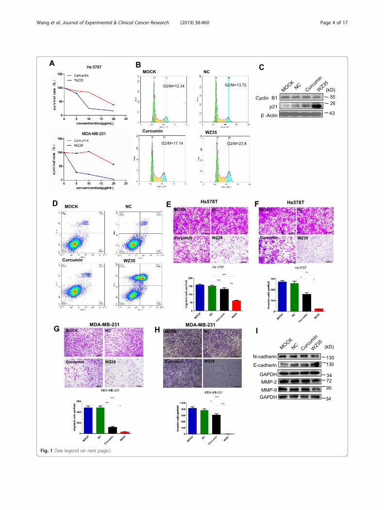
Fig. 1 (See legend on next page.)
Wang et al. Journal of Experimental & Clinical Cancer Research (2019) 38:460 Page 4 of 17
ImmunohistochemistryTissue sections were initially deparaffinized with xylene,rehydrated, and antigen retrieval was performed in 0.01M citrate buffer (PH = 6.0) for 3 min at 95 °C. Followedby incubation with primary antibody at 4 °C overnight,the tissue sections were incubated with secondary anti-body at room temperature for 2 h. Finally, after DABstaining and a neutral gum sealing, immunohistochemi-cal signals were photographed and observed under amicroscope (Olympus Corporation, Tokyo, Japan) withmagnification of 200 × .
Animal experimentsFive-week-old, athymic BALB/c nu/nu female mice (16-19 g, totally n = 24) were purchased from Vital River La-boratories (Beijing, China). The mice were randomlydivided into three groups. MDA-MB-231 cells were sub-cutaneously inoculated into the right flank of mice (1 ×10⁷ cells in 100 ul PBS per mouse). After tumor volumereached 50mm3, the mice were intraperitoneally injectedCastor oil, curcumin and WZ35 (0.2 mL, 25 mg/kg foreach) for 15 days. The tumor size and body weight ofnude mice were measured and recorded once everyother day. The tumor volumes were determined bymeasuring length (L) and width (W) and calculating vol-ume (V = 0.5 × L ×W2) at the indicated time points. Atthe end of experiment, the animals were sacrificed andthe tumors were harvested for use in proteins expressionand histology studies. To investigate the effects of curcu-min or WZ35 on lung metastasis in vivo, 4 × 10⁵ ofMDA-MB-231 cells in 100 ul of PBS were intravenouslyinjected into the mice. After 24 days, 24 nude mice wererandomly divided into four groups (normal saline, castoroil, curcumin, WZ35) and treated mice for 21 days. Thelung metastases were evaluated by microscope after HEstaining.
Statistical analysisAll experiments were conducted in triplicate (n = 3) andthe data are presented as the mean ± SD. All statisticalanalyses were processed with GraphPad Prism 6.0(GraphPad Software, La Jolla, CA, USA). Two-sided stu-dent’s t-test was performed to analyze the differences
between two groups of data. P value< 0.05 means statis-tically significant.
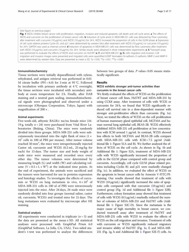
ResultsWZ35 exhibits stronger anti-tumor activities thancurcumin in the breast cancer cellsWe firstly evaluated the effects of WZ35 on the proliferationof breast cancer cell lines, Hs578T and MDA-MB-231 byusing CCK8 assay. After treatment of cells with WZ35 orcurcumin for 24 h, we found that WZ35 significantly re-duced cell survival rate in a dose-dependent manner withstronger anti-proliferative effects than curcumin (Fig. 1a).Next, we tested the effects of WZ35 on the cell proliferationof human mammary gland epithelial cell, MCF10A and hu-man normal lung epithelial cell, BEAS-2B. WZ35 effectivelyinhibited MDA-MB-231 cell proliferation at low concentra-tion with IC50 around 1 μg/ml. In contrast, WZ35 showedless effects to both MCF10A and BEAS-2B cells comparewith MDA-MB-231 cells with IC50 > 7.5 μg/ml (Add-itional file 1: Figure S1A and B). We further analyzed the ef-fects of WZ35 on the cell cycle. As shown in Fig. 1b andAdditional file 1: Figure S2A, treatment of MDA-MB-231cells with WZ35 significantly increased the proportion ofcells in the G2/M phase compared with control group andcurcumin. Accordingly, cell cycle G2/M phase related pro-teins including Cyclin B1 and p21 were markedly changed(Fig. 1c). In addition, we evaluated the effect of WZ35 onthe apoptosis in breast cancer cells by Annexin V-FITC/PI-staining. Our results showed that treatment of cells withWZ35 (10 μg/mL) significantly increased the ratio of apop-totic cells compared with that curcumin (10 μg/mL) andcontrol group (Fig. 1d and Additional file 1: Figure S2B).Furthermore, colony formation assay showed that treatmentof cells with WZ35 (0.5 μg/mL) markedly reduced the num-ber of colonies of MDA-MB-231 and Hs578T cells (Add-itional file 1: Figure S2C-D). Since the metastasis is themajor cause of high mortality in breast cancer, we con-ducted transwell assay after treatment of Hs578T andMDA-MB-231 cells with WZ35 to evaluate the effects ofWZ35 on the cell migration and invasion. Treatment of cellswith curcumin or WZ35 significantly reduced migratoryand invasive ability of Hs578T (Fig. 1e, f) and MDA-MB-231 (Fig. 1g, h and Additional file 1: Figure S2E-F) cells. As
(See figure on previous page.)Fig. 1 WZ35 inhibits breast cancer cells proliferation, migration, invasion and induced apoptotic cell death and cell cycle arrest. a The effects ofWZ35 and curcumin on the proliferation of breast cancer cells. b Induction of cycle arrest in MDA-MB-231 cells was detected by Flow cytometryafter treatment with curcumin (10 μg/mL) and WZ35 (10 μg/mL) for 24 h. WZ53 increased the proportion of cells in the G2/M phase. c Expressionof cell cycle relative proteins Cyclin B1, and p21 were determined by western blot after treatment with WZ35 (10 μg/mL) or curcumin (10 μg/mL)for 24 h. GAPDH was used as internal control. d Induction of apoptosis in MDA-MB-231 cells was determined by flow cytometry after treatmentwith WZ35 (10 μg/mL) and curcumin (10 μg/mL) for 24 h. Similar results were obtained in three independent experiments. e, h Transwell assaywas performed to evaluate the effects of WZ35 and curcumin on Hs578T (e, f) and MDA-MB-231 (g, h) cells migration and invasion. Cellmigration and invasion images were presented and migrated cells were quantified. i EMT biomakers N-cadherin, E-cadherin, MMP-2 and MMP-9were determined by western blot. Data are presented as mean ± SD, *p < 0.05, **p < 0.01, ***p < 0.001
Wang et al. Journal of Experimental & Clinical Cancer Research (2019) 38:460 Page 5 of 17

Fig. 2 (See legend on next page.)
Wang et al. Journal of Experimental & Clinical Cancer Research (2019) 38:460 Page 6 of 17

expected, WZ35 showed stronger anti-migratory and anti-invasive abilities than curcumin. Consistent with thesefindings, we found that the expression of epithelial-mesenchymal transition (EMT) related proteins were alsosignificantly changed. In detail, the expressions of mesenchy-mal markers (N-cadherin, MMP-2 and MMP-9) werereduced; whereas the expression of epithelial marker, E-cadherin was increased (Fig. 1i). All together, these dataindicate that WZ35 plays stronger tumor suppressive rolethan curcumin in breast cancer cells.
WZ35 suppresses MDA-MB-231 xenograft tumor growthand metastasis in vivoBased on the aforementioned results from in vitro exper-iments, we questioned whether WZ35 also has anti-tumor effects in vivo. To confirm our hypothesis, we de-veloped a xenograft mouse model by injecting MDA-MB-231 cells into the right frank of 5 weeks old femalenude mice. Similar with the in vitro experiments, treat-ment of mice with curcumin or WZ35 (0.2 mL, 25 mg/kg for each) showed significantly reduced tumor volumecompared to the control mice. In agreement within vitro analysis, WZ35 showed stronger anti-tumor ac-tivities than curcumin in vivo with no major change ofbody weight suggesting that the WZ35 would not leadto significant cytotoxicity (Fig. 2a). Immunohistochemis-try demonstrated that WZ35 treatment markedly re-duced the expression of Ki-67 in tumor tissuesindicating that WZ35 inhibited cell proliferation in vivo(Fig. 2b). To further determine the anti-metastatic activ-ities of WZ35 in vivo, we intravenously injected MDA-MB-231 cells into the nude mice and evaluated lung me-tastasis foci by HE analysis. As shown in Fig. 2c-e, bothcurcumin and WZ35 significantly reduced the numbersof lung metastasis foci, and WZ35 showed more super-ior anti-metastatic effects than curcumin without majorchange of body weight. Immunofluorescence analysesvalidated the expression of mesenchymal markers, N-cadherin, MMP-2 and MMP-9 were suppressed in micetumor tissues; whereas the expression of epithelialmarker, E-cadherin was increased in mice tumor tissuesafter treatment of mice with WZ35 (Fig. 2f). As ex-pected, WZ35 showed more strong effects than curcu-min. These data further confirmed that the anti-metastatic activities of WZ35 both in vitro and in vivo.
YAP and JNK pathways are involved in WZ35 mediatedbreast cancer cell growth inhibitionWe next set out to determine the potential mechanism ofthe anti-tumor activities of WZ35 in breast cancer cells.Yap has been reported to interact with DNA-bindingprotein TEAD transcription factors to promote the ex-pression of growth-promoting and apoptosis-inhibitinggenes [22]. The Ser/Thr kinase Hippo (Mst1 and Mst2)activates Lats1/2, which phosphorylate YAP and sequesterYAP in cytoplasm to inhibit transcriptional activity ofYAP [23–25]. We found that treatment of MDA-MB-231cells with WZ35 significantly reduced the expression ofMST1/2, Lats1, p-YAP(s397), p-YAP(s127) as well ashippo pathway scaffolding proteins SAV1 and MOB1;whereas increased the expression of YAP with stronger ef-fects than curcumin (Fig. 3a). JNK has been reported toplay an essential role in Hippo signaling induced cell inva-sion [26]. In addition, it is widely known that JNKs areinvolved in cell proliferation, apoptosis, inflammation, dif-ferentiation and migration [27, 28]. The JNK activationcould inhibit anti-apoptotic proteins, BCL-2 and BCL-xlexpression; whereas induces pro-apoptotic protein BAXexpression [29–31]. To further examine the association ofYAP signaling and JNK in the WZ35 mediated breastcancer cells growth inhibition, we performed a series ofknockdown and overexpression analyses. As shown inFig. 3b, treatment of MDA-MB-231 cells with WZ35markedly increased phospho-JNK expression, whereasdecreased phospho-AKT expression. Importantly, knock-down of YAP by siRNA attenuated WZ35 induced p-JNKexpression. YAP knockdown also elevated anti-apoptoticprotein BCL-2 expression, whereas reduced cleavedcaspase-3 expression. Furthermore, treatment of YAPknockdown cells with WZ35 significantly attenuated theeffects of WZ35 on the expression of BCL-2 and cleavedcaspase-3 (Fig. 3c). Accordingly, knockdown of YAP inMDA-MB-231 cells promoted cell proliferation and mi-gration, and co-treatment of cells with YAP siRNA andWZ35 partly reversed tumor suppressive effects of WZ35(Fig. 3d, e). As expected, we observed that the opposite ef-fects in YAP overexpressing cells for p-JNK, BCL-2 andcleaved-CASP3 expression (Fig. 3f). Treatment of YAPoverexpressing cells with WZ35 exhibited stronger anti-proliferative and anti-migratory effects than YAP overex-pression or WZ35 treatment alone (Fig. 3g, h). Taken
(See figure on previous page.)Fig. 2 WZ35 inhibits MDA-MB-231 xenograft tumor growth and metastasis in vivo. a A growth curve analysis of the tumor growth in curcumin,WZ35 or Castor oil treated groups. b Expression of ki67 in tumor tissues was determined by immunohistochemistry. c Mice body weight wasmeasured during the 17-day treatment of WZ35 and curcumin. d Representative microscopic images of lung metastatic lesions at 21 days aftertreatment of mice with curcumin or WZ35 (0.2 mL, 25 mg/kg for each). e The number of lung metastatic tumors was calculated. fImmunofluorescence were used to test the expression level of E-cadherin, N-cadherin, MMP-2 and MMP-9 from curcumin, WZ35, Vehicle andcontrol group. Data are presented as mean ± SD, ***p < 0.001
Wang et al. Journal of Experimental & Clinical Cancer Research (2019) 38:460 Page 7 of 17

Fig. 3 (See legend on next page.)
Wang et al. Journal of Experimental & Clinical Cancer Research (2019) 38:460 Page 8 of 17

together, our data suggest that WZ35 inhibits breast can-cer cell growth and migration through constitutive YAPactivation and subsequent JNK phosphorylation.
WZ35 induced ROS generation is responsible for YAP andJNK activationZou et al. has demonstrated that ROS generation isthe upstream regulator of WZ35-induced apoptosis ingastric cancer [13]. Therefore, we attempt to deter-mine whether the anti-tumor effects of WZ35 inMDA-MB-231 cells were associated with its oxidativestress. As shown in Fig. 4a, treatment of MDA-MB-231 cells with WZ35 increased ROS generation in adose dependent manner. In addition, pretreatment ofMDA-MB-231 cells with N-acetyl cysteine (ROS scav-enger; NAC, 1 mM) significantly reduced WZ35-induced increases in DCF fluorescence as expected(Fig. 4b). Next, we wanted to check whether thenegative growth and migratory signals from WZ35can be attenuated by reduction of ROS levels. Pre-treatment of cells with NAC (1 mM) was able to at-tenuate WZ35 induced anti-proliferative (Fig. 4c) andmigratory (Fig. 4d) ability in MDA-MB-231 cells.Moreover, NAC prevented WZ35 induced alternationsof YAP and p-JNK expression (Fig. 4e) indicating thatWZ35-induced ROS generation is responsible forYAP and JNK activation mediated anti-tumor activ-ities in breast cancer cells. The JNK activation playscritical role in various cancer cell apoptosis. It hasbeen reported that JNK induces apoptosis via phos-phorylating 14–3-3 protein, which in turn releasespro-apoptotic proteins, such as BAX and FOXO tran-scription factors [32–35]. To further demonstrate theimportance of JNK activation in WZ35 mediated anti-tumor activities, MDA-MB-231 cells were pre-treatedwith a JNK inhibitor, SP600125. Our results showedthat SP600125 significantly attenuated anti-proliferative (Fig. 4f) and migratory effects (Fig. 4g) ofWZ35 in MDA-MB-231 cells. Accordingly, we vali-dated that SP600125 inhibited WZ35 induced p-JNKexpression (Fig. 4h). These results indicate that the
WZ35-induced anti-tumor activities in MDA-MB-231cells at least partially mediated by ROS-YAP-JNK sig-naling pathway.
ROS-YAP-JNK pathway is involved in WZ35 inducedmitochondrial dysfunctionMitochondrial dysfunction is a common phenomenon inmultiple human malignancies, such as breast cancer [36],HCC [37], lung cancer [38]. Moreover, mitochondrial fis-sion is important for ROS production [39]. Firstly, to deter-mine the effects of WZ35 in mitochondrial dysfunction, weevaluated mitochondrial DNA transcription, replicationand translation associated proteins including nuclearrespiratory factor 1 (NRF1) and NRF2, DNA polymerasesubunit gamma (POLG) and mitochondrial translation fac-tor EF4 after treatment of cells with WZ35. As shown inAdditional file 1: Figure S3A-B, WZ35 significantly down-regulates the expression of mitochondria-associated pro-teins EF-4, PLOG, NRF1 and NRF2 in MDA-MB-231 cells.Moreover, WZ35 markedly reduced NRF1 and NRF2mRNA levels in MDA-MB-231 cells (Additional file 1: Fig-ure S3C-D). Secondly, to further investigate the effects ofWZ35 on mitochondrial respiration, we evaluated real-timeoxidative phosphorylation by measuring cellular oxygenconsumption rates (OCRs). WZ35 significantly decreasedOCR compared with curcumin and negative control (NC)group in MDA-MB-231 cells suggesting that WZ35 treat-ment led to inhibit mitochondrial respiration (Fig. 5a). Wefurther analyzed major parameters of mitochondria func-tion by evaluating OCR data at various time points. WZ35markedly decreased basal respiration, maximal respiration,spare respiration (Fig. 5b) and ATP production in MDA-MB-231 cells (Additional file 1: Figure S4A). Importantly,co-treatment of MDA-MB-231 cells with ROS inhibitor,NAC and WZ35 significantly attenuated inhibitory effectsof WZ35 on OCR as well as basal respiration, maximalrespiration and spare respiration suggesting that WZ35-induced inhibition of mitochondrial respiration was medi-ated by ROS generation (Fig. 5c, d). To further investigatewhether ROS mediated YAP and JNK activation wereinvolved in the WZ35 induced mitochondrial dysfunction,
(See figure on previous page.)Fig. 3 YAP and JNK pathway are involved in WZ35 mediated breast cancer cell growth inhibition. a Western blot analysis of the Hippo pathwayrelated proteins MST1, MST2, LATS1, MOB1, p-MOB1, p-YAP(s397), p-YAP(s127), YAP/TAZ and SAV1 in MDA-MB-231 cells treated with curcumin(10 μg/mL) or WZ35 (10 μg/mL). GAPDH was used as loading control. b Western blot analysis of p-JNK, AKT and p-AKT expression in MDA-MB-231cells treated with WZ35 (10 μg/mL) or curcumin (10 μg/mL). c MDA-MB-231 cells were transfected with or without YAP siRNA and co-treated withWZ35 (10 μg/mL) for 12 h, the protein expression of YAP, JNK, p-JNK, BCL-2 and cleaved caspase-3 were determined by western blot. d MDA-MB-231 cells were transfected with or without YAP siRNA and co-treated with WZ35 (10 μg/mL) for 24 h, 48 h, 72 h respectively. Then cell viabilitywas detected by CCK8 assay. e Representative images and the numbers of migrated cells were evaluated after treatment of MDA-MB-231 cellswith Si-YAP, WZ35 and Si-YAP+WZ35. f Western blot analysis of YAP, JNK, p-JNK, BCL-2 and Cle-Caspase3 protein levels in MDA-MB-231 cellstreated with YAP-OE, WZ35 and YAP-OE +WZ35. g Representative images and the numbers of migrated cells were evaluated after treatment ofMDA-MB-231 cells with YAP-OE, WZ35 and YAP-OE +WZ35. h MDA-MB-231 cells were transfected with or without YAP-OE and co-treated withWZ35 (10 μg/mL) for 24 h, 48 h, 72 h respectively. The cell viability was detected by CCK8 assay. Data are presented as mean ± SD, *p < 0.05,**p < 0.01, ***p < 0.001
Wang et al. Journal of Experimental & Clinical Cancer Research (2019) 38:460 Page 9 of 17

Fig. 4 (See legend on next page.)
Wang et al. Journal of Experimental & Clinical Cancer Research (2019) 38:460 Page 10 of 17

we performed a series of experiments. First, we overex-pressing or knockdown YAP in MDA-MB-231 cells andtreated cells with WZ35. YAP overexpression significantlyreduced OCR, basal respiration, maximal respiration andspare respiration. Moreover, Treatment of YAP overex-pressing MDA-MB-231 cells with WZ35 exerts strongereffects than either YAP overexpression or WZ35 treatmentalone (Fig. 5e, f). Inversely, knockdown of YAP increasedOCR, basal respiration, maximal respiration and spare res-piration, and treatment of YAP knockdown cells withWZ35 significantly attenuated inhibitory effects of WZ35on basal respiration, maximal respiration and spare respir-ation in MDA-MB-231 cells (Additional file 1: Figure S4B-C). Second, we treated MDA-MB-231 cells with JNKinhibitor (SP600125) and WZ35. As expected, co-treatmentof cells with SP600125 and WZ35 significantly attenuatedinhibitory effects of WZ35 on OCR, basal respiration, max-imal respiration and spare respiration (Fig. 5g, h). Taken alltogether, these data indicate that ROS-YAP-JNK pathway isinvolved in WZ35 induced mitochondrial dysfunction.
YAP expression is down-regulated in breast cancer andcorrelated with the prognosis of certain breast cancerpatientsTreatment of breast cancer cells with WZ35 exhibitedstrong anti-tumor activities through activating YAPmediated JNK signaling suggesting that potentialtumor suppressive role of YAP in breast cancer. Tofurther investigate the functions of YAP in breastcancer tissue specimens, we evaluated YAP mRNAlevels in breast cancer tissues using TCGA publicdatabase. As shown in Fig. 6a, analysis of 226 pairedbreast cancer tissues and normal adjacent tissues(NATs) for which YAP mRNA expression was avail-able in the TCGA dataset, demonstrated YAP mRNAwas significantly down-regulated in breast cancer tis-sues compare to the NATs. We further examinedYAP protein levels in 22 paired fresh breast cancertissues samples and corresponding NATs by westernblot analysis. Among 22 paired patient samples, 19patient samples showed significantly reduced YAPprotein expression levels in breast cancer tissue sam-ples compared with corresponding NATs (Fig. 6b).We also checked mRNA levels of YAP in these 22
pairs of primary breast cancer tissues and their adja-cent tissue counterparts by qRT-PCR. As presented inFig. 6c, YAP mRNA was significantly down regulatedin primary breast cancer tissue specimens comparedwith their adjacent noncancerous tissues. To furtherinvestigate whether downregulated YAP expression inbreast cancer is associated with patient’s survival, weperformed Kaplan-Meier survival analysis by breastcancer subtypes. We found that high expression ofYAP was significantly associated with favorable prog-nosis of ER positive breast cancer patients (N = 762)(Fig. 6d) and PR positive breast cancer patients (N =489) (Fig. 6e). Inversely, high expression of YAP wassignificantly associated with poor prognosis of triplenegative breast cancers (N = 161) (Additional file 1:Figure S5). Altogether, these results imply that YAPplays tumor suppressive functions in breast cancerand associated with the prognosis of certain breastcancer patients by their ER, PR and Her2 status. Fur-ther studies need to validate this result using largenumber of breast cancer patient samples.
DiscussionCurrently, the mainstay of systemic treatment for breastcancer is endocrine therapy. However, chemotherapy stillis indispensable especially for patients they are lackinghormone receptors [3]. Thus, discover novel drugs andtherapeutic targets become a matter of great interest.WZ35, an analog of curcumin, has been demonstrated toexhibit superior anti-tumor effects in gastric cancer andhepatocellular carcinoma [13, 14]. However, anti-tumoreffects of WZ35 in breast cancer and its underlying anti-tumor mechanisms are still unclear. Here, we demon-strated that WZ35 exhibits superior anti-tumor effectscompare to the curcumin on breast cancer cells bothin vitro and in vivo with no evident side effects. Furtheranalysis showed that ROS mediated YAP and JNK activa-tion were involved in WZ35 induced anti-tumor activities.In addition, we found that ROS-YAP-JNK pathway is im-plicated in mitochondria dysfunction in breast cancer cells(Fig. 6f).Of note, curcumin has been reported to stimulate im-
mune systems to exert anti-cancer activity [10, 40]. Inour animal study, curcumin showed less in vitro anti-
(See figure on previous page.)Fig. 4 WZ35 induces cytotoxicity in breast cancer cells is depending on ROS mediated YAP and JNK activation. a Flow cytometry analysis wereperformed to determine the intracellular ROS levels in MDA-MB-231 cells treated with curcumin or WZ35. b Intracellular ROS levels in MDA-MB-231 cells treated with WZ35 or WZ35 + NAC. c Cell proliferation ability was detected by CCK8 assay. d Representative images and the numbers ofmigration cells were evaluated after treatment of MDA-MB-231 cells with NAC, WZ35 and NAC +WZ35. e The expression of YAP, JNK and p-JNKin MDA-MB-231 cells treated with NAC, WZ35 and NAC +WZ35 were detected by western blot. f Cell proliferation ability was detected by CCK8assay. g Representative images and the numbers of migrated cells were evaluated after treatment of MDA-MB-231 cells with SP600125, WZ35and SP600125 +WZ35. h The expression of JNK and p-JNK proteins were determined by western blot. Data are presented as mean ± SD,**p < 0.01, ***p < 0.001
Wang et al. Journal of Experimental & Clinical Cancer Research (2019) 38:460 Page 11 of 17

Fig. 5 (See legend on next page.)
Wang et al. Journal of Experimental & Clinical Cancer Research (2019) 38:460 Page 12 of 17

tumor activities, however, it is quite effective in vivomice xenograft model suggesting possible immunestimulatory effect of curcumin. Although we used im-mune compromised mice to compare anti-tumor effi-cacy of WZ35 and curcumin, considering that NK cellsand macrophages do still exist in these mice, WZ35may have had some immune stimulatory activity whichmight contribute to its anti-tumor activities in vivo al-though further study needs to validate this proposal.The Hippo pathway is associated with cell proliferation,
tissue homeostasis and tumorigenesis [19, 41]. The com-ponents of Hippo pathway (Mst1/2, Lats1/2 etc.) havebeen reported to play tumor suppressive roles in cancers.YAP is a key downstream effector of Hippo pathway,which has been reported to act as either an oncogene ortumor suppressor in breast cancer [42]. Moreover, YAPwas reported to act as a tumor-suppressor in lung SCC viadisruption of intracellular ROS homeostasis [43]. In ourstudy, we demonstrated that YAP exerts tumor suppres-sive role in breast cancer cells. WZ35 induced YAP activa-tion was involved in inhibition of breast cancer cellgrowth and migration. We also found that YAP mRNAand protein levels were markedly down-regulated in breastcancer tissue specimens and reduced YAP expression wasassociated with poor prognosis of certain type of breastcancer patients. Accumulated ROS generation in cancerhas been reported to inhibit cell proliferation [44], induceDNA damage [45], autophagy [46, 47], cellular injury [44],cell death [43, 46] and drug resistance [48, 49]. Inaddition, previous research demonstrated that elevatedROS production could activate JNK signaling pathway,resulting in cell apoptosis [50, 51]. Cancer cells with in-creased oxidative stress are likely to be more vulnerable tothe damage. Therefore, elevated ROS production in can-cer cells that do not cause significant toxicity to normalcells might be a potential therapeutic method. Our studyshowed that WZ35 induced ROS generation in breast can-cer cells and NAC effectively attenuated the anti-tumoractivities of WZ35. We also found for the first time thatWZ35 mediated ROS generation is involved in YAP andJNK activation in breast cancer cells. Several studies havehighlighted that activation of JNK signaling pathway in-duces mitochondria-dependent cell apoptosis via
activating downstream signaling molecules of BCL-2 fam-ily proteins and caspase-3 [52, 53]. We demonstrated thatYAP activation exerts anti-tumor activities via JNK phos-phorylation and activation, and subsequent BCL-2 down-regulation and cle-caspase3 upregulation.Mitochondria plays critical role in the cells through
regulation of energy metabolism, ATP generation, andcalcium homeostasis [54, 55]. Accumulated evidenceshave indicated that mitochondrial metabolism is anattractive target for cancer therapy. Emerging studieshave begun to demonstrate that mitochondria takespart in the activation of signaling pathways includingthe PI3K pathway, and activation of oncogenes suchas MYC and KRAS, which result in promoting cellproliferation [56]. The mitochondrial ATP is mainlygenerated by glycolysis and mitochondrial oxidativephosphorylation. It has been reported that both glyco-lytic and mitochondrial functions were decreased byeither KRAS suppression or ERK inhibition [57]. Inthis study, we further investigated relationship be-tween ROS-YAP-JNK pathway activation and mito-chondrial dysfunction, and found that WZ35 inhibitsmitochondrial oxidative phosphorylation by activatingROS-YAP-JNK pathway. Activation of ROS-YAP-JNKpathway not only induced apoptosis, but also acceler-ated mitochondrial dysfunction by possibly inhibitingATP generation in breast cancer cells resulting in in-hibition of tumor growth. Thus, activating ROS-YAP-JNK pathway might increase the sensitivity of breastcancer cells to anticancer drug. Further study needsto prove this matter.Altogether, our study shows that WZ35 exhibits stron-
ger anti-tumor activities than curcumin by activatingROS-YAP-JNK signaling pathway. The activated ROS-YAP-JNK pathway involved in anti-tumor activities ofbreast cancer cells by inducing mitochondrial dysfunc-tion and apoptosis. These results suggest that noveltherapeutic strategies for breast cancer by targetingROS-YAP-JNK pathway.
ConclusionOur study shows that WZ35 exhibits stronger anti-tumor activities than curcumin by activating ROS-YAP-
(See figure on previous page.)Fig. 5 ROS-YAP-JNK pathway is involved in WZ35 induced mitochondrial dysfunction. a MDA-MB-231 cells were cultured with WZ35 or curcuminfor 12 h, and cellular oxygen consumption rate (OCR) was measured in real time using the Seahorse XF96 Extracellular Flux Analyzer after basalOCR was measured at three time points, followed by sequential injection of oligomycin (1 μM), FCCP (0.5 μM) rotenone (1 μM), and antimycin A(1 μM). The overall OCR curves were plotted as the mean OCR ± SD of three replicates. b Basal respiration, maximal respiration and sparerespiration were assessed, respectively. c, d MDA-MB-231 cells were treated with or without WZ35 and co-treated with NAC. e, f MDA-MB-231cells were overexpressed YAP and treated with or without WZ35. g, h MDA-MB-231 cells were treated with or without WZ35 and co-treated withJNK inhibitor SP600125. The OCR was measured in real time using the Seahorse XF96 Extracellular Flux Analyzer as aforementioned. The Basalrespiration, maximal respiration and spare respiration were also assessed, respectively. Data are presented as the mean ± SD, *p < 0.05,**p < 0.01, ***p < 0.001
Wang et al. Journal of Experimental & Clinical Cancer Research (2019) 38:460 Page 13 of 17

Fig. 6 (See legend on next page.)
Wang et al. Journal of Experimental & Clinical Cancer Research (2019) 38:460 Page 14 of 17

JNK signaling pathway. Our study revealed novel ROS-YAP-JNK pathway which involved in anti-tumor activ-ities of breast cancer cells by inducing mitochondrialdysfunction and apoptosis. These results suggest thatnovel therapeutic strategies for breast cancer by target-ing ROS-YAP-JNK pathway.
Supplementary informationSupplementary information accompanies this paper at https://doi.org/10.1186/s13046-019-1424-4.
Additional file 1: Figure S1. The effects of WZ35 on cell proliferationof normal cells. Figure S2. The effects of WZ35 on cell cycle arrest,apoptosis, migration and invasion ability. Figure S3. W35 significantlydown-regulates the expression of mitochondria-associated proteins. Fig-ure S4. WZ35 induces mitochondrial dysfunction. Figure S5. Kaplan-Meier plot of overall survival of triple negative breast cancer patients ex-pressing high and low levels of YAP.
AbbreviationsATP: Adenosine triphosphate; BCL-2: B cell lymphoma 2; DMEM: Dulbecco’smodified eagle medium; EF4: mitochondrial translation factor;EMT: Epithelial-mesenchymal transition; ER: Estrogen receptor;ERK: Extracellular regulated protein kinases; FBS: Fetal bovine serum;FCCP: Carbonylcyanide-p-trifluorometh oxyphenylhydrazone;HCC: Hepatocellular carcinoma; HRP: Horseradish peroxidase; JNK: c-Jun N-terminal kinase; KRAS: Kirsten rat sarcoma viral onco; LATS1: Large tumoursuppressor 1; MMP-2: Matrix Metalloproteinase-2; MMP-9: MatrixMetalloproteinase-9; MST1: Mammalian STE20-like protein kinase 1;MST2: Mammalian STE20-like protein kinase 2; NAC: N -Acetyl-L-cysteine;NATs: Normal adjacent tissues; NC: Negative control; NRF1: Nuclearrespiratory factor 1; NRF2: Nuclear respiratory factor 2; OCR: Oxygenconsumption rate; OD: Optical density; PBS: Phosphate buffer solution;PI: Propidium Iodide; POLG: DNA polymerase subunit gamma;PR: Progesterone receptor; ROS: Reactive oxygen species; SAV1: Salvadorfamily WW domain-containing protein 1; TCGA: The Cancer Genome Atlas;YAP: Yes-associated protein
AcknowledgementsNone.
Authors’ contributionsTC, RC and GL conceived the idea and designed the research. LW, CW, ZT,LZ, ZZ and WW performed in vitro experiments. LW, CW, ZT, LZ, YH, HC andBZ performed mice xenograft and metastasis experiments. LW, CW, XH andYY analyzed patient samples. TC, RC, GL, LW, ZT and LZ analyzed the data.RC, TC and CW wrote the manuscript. All authors read and approved thefinal manuscript.
FundingThis work was supported by National Natural Science Foundation of China(81672305, 81622043, 81770926), The national key Basic ResearchDevelopment Program of China (2016YFC1101200), The key Basic ResearchDevelopment Program of Zhejiang Province (2018C03G2090634), The NaturalScience Foundation of Zhejiang Province (LQ17H120009).
Availability of data and materialsAll data generated or analyzed in this study are included in this manuscriptand its additional files.
Ethics approval and consent to participateAll animal studies were performed with an approved protocol by theInstitutional Animal Care and Use Committee of Wenzhou MedicalUniversity. Patient’s biopsy samples were obtained from the First AffiliatedHospital of Wenzhou Medical University, based on the Medical ResearchEthics Committee and the Institutional Review Board of Wenzhou MedicalUniversity approved research protocol.
Consent for publicationNot applicable.
Competing interestsThe authors declare that they have no competing interests.
Author details1School of Ophthalmology and Optometry, Eye Hospital, Wenzhou MedicalUniversity, Wenzhou 325000, Zhejiang, China. 2State Key Laboratory ofOphthalmology, Optometry and Visual Science, Wenzhou 325000, Zhejiang,China. 3Affiliated Yueqing Hospital and School of Pharmaceutical Sciences,Wenzhou Medical University, Wenzhou 325035, Zhejiang, China. 4School ofMedicine, Shanghai Jiaotong University, Shanghai 200030, China. 5LaboratoryAnimal Centre, Wenzhou Medical University, Wenzhou, Zhejiang, China.6School of Pharmaceutical Sciences, Wenzhou Medical University, Wenzhou325035, Zhejiang, China.
Received: 28 May 2019 Accepted: 16 September 2019
References1. Anastasiadi Z, Lianos GD, Ignatiadou E, Harissis HV, Mitsis M. Breast cancer in
young women: an overview. Updat Surg. 2017;69:313–7.2. Torre LA, Bray F, Siegel RL, Ferlay J, Lortet-Tieulent J, Jemal A. Global Cancer
statistics, 2012. CA Cancer J Clin. 2015;65:87–108.3. Tang Y, Wang Y, Kiani MF, Wang B. Classification, treatment strategy, and
associated drug resistance in breast Cancer. Clin Breast Cancer. 2016;16:335–43.4. Maughan KL, Lutterbie MA, Ham PS. Treatment of breast Cancer. Am Fam
Physician. 2010;81:1339–46.5. Li H, Sureda A, Devkota HP, Pittala V, Barreca D, Silva AS, Tewari D, Xu S,
Nabavi SM. Curcumin, the golden spice in treating cardiovascular diseases.Biotechnol Adv. 2019;S0734-9750(19):30010-2.
6. Huminiecki L, Horbanczuk J, Atanasov AG. The functional genomic studiesof curcumin. Semin Cancer Biol. 2017;46:107–18.
7. Wang YW, Yu JY, Cui R, Lin JJ, Ding XT. Curcumin in treating breast Cancer:a review. Jala. 2016;21:723–31.
8. Bhattacharyya S, Mandal D, Saha B, Sen GS, Das T, Sa G. Curcumin preventstumor-induced T cell apoptosis through stat-5a-mediated Bcl-2 induction. JBiol Chem. 2007;282:15954–64.
9. Bhattacharyya S, Md Sakib Hossain D, Mohanty S, Sankar Sen G,Chattopadhyay S, Banerjee S, Chakraborty J, Das K, Sarkar D, Das T, Sa G.Curcumin reverses T cell-mediated adaptive immune dysfunctions in tumor-bearing hosts. Cell Mol Immunol. 2010;7:306–15.
10. Mukherjee S, Hussaini R, White R, Atwi D, Fried A, Sampat S, Piao L, Pan Q,Banerjee P. TriCurin, a synergistic formulation of curcumin, resveratrol, andepicatechin gallate, repolarizes tumor-associated macrophages and triggersan immune response to cause suppression of HPV+ tumors. CancerImmunol Immunother. 2018;67:761–74.
(See figure on previous page.)Fig. 6 YAP plays a tumor suppressive functions in breast cancer. a YAP1 mRNA expression in breast cancer tissues and normal adjacent tissuesfrom TCGA RNA-seq data. b The protein levels of YAP in twenty two breast cancer tissues and normal tissues were measured by western blot. cComparison of YAP1 expression in 22 paired breast cancer tissues and normal adjacent tissues by qRT-PCR. d and e Kaplan-Meier plots of overallsurvival of ER positive breast cancer patients (N = 762) (d) and PR positive breast cancer patients (N = 489) (e) expressing high and low levels ofYAP. Data obtained from the Kaplan-Meier plotter database (kmplot.com/analysis). f Schematic illustration for the underlying anti-cancermechanism of WZ35 in breast cancer. Data are presented as the mean ± SD, ***p < 0.001, ****p < 0.0001
Wang et al. Journal of Experimental & Clinical Cancer Research (2019) 38:460 Page 15 of 17

11. Anand P, Kunnumakkara AB, Newman RA, Aggarwal BB. Bioavailability ofcurcumin: problems and promises. Mol Pharm. 2007;4:807–18.
12. Shehzad A, Khan S, Shehzad O, Lee YS. Curcumin therapeuticpromises and bioavailability in colorectal cancer. Drugs Today (Barc).2010;46:523–32.
13. Zou P, Zhang JR, Xia YQ, Kanchana K, Guo GL, Chen WB, Huang Y, Wang Z,Yang SL, Liang G. ROS generation mediates the anti-cancer effects of WZ35via activating JNK and ER stress apoptotic pathways in gastric cancer.Oncotarget. 2015;6:5860–76.
14. Wang LH, Han LJ, Tao ZY, Zhu Z, Han L, Yang ZF, Wang H, Dai DD, WuL, Yuan ZZ, Chen TK. The curcumin derivative WZ35 activates ROS-dependent JNK to suppress hepatocellular carcinoma metastasis. FoodFunct. 2018;9:2970–8.
15. Zhang J, Feng Z, Wang C, Zhou H, Liu W, Kanchana K, Dai X, Zou P, Gu J,Cai L, Liang G. Curcumin derivative WZ35 efficiently suppresses coloncancer progression through inducing ROS production and ER stress-dependent apoptosis. Am J Cancer Res. 2017;7:275–88.
16. Ardestani A, Maedler K. The hippo signaling pathway in pancreatic β-cells:functions and regulations. Endocr Rev. 2018;39:21–35.
17. Harvey KF, Zhang X, Thomas DM. The hippo pathway and human cancer.Nat Rev Cancer. 2013;13:246–57.
18. Plouffe SW, Hong AW, Guan KL. Disease implications of the hippo/YAPpathway. Trends Mol Med. 2015;21:212–22.
19. Yu FX, Zhao B, Guan KL. Hippo pathway in organ size control, tissuehomeostasis, and Cancer. Cell. 2015;163:811–28.
20. Elster D, Tollot M, Schlegelmilch K, Ori A, Rosenwald A, Sahai E, von Eyss B.Author correction: TRPS1 shapes YAP/TEAD-dependent transcription inbreast cancer cells. Nat Commun. 2018;9:3781.
21. Cheng GZ, Chan J, Wang Q, Zhang W, Sun CD, Wang LH. Twisttranscriptionally up-regulates AKT2 in breast cancer cells leading toincreased migration, invasion, and resistance to paclitaxel. Cancer Res. 2007;67:1979–87.
22. Zhao B, Li L, Tumaneng K, Wang CY, Guan KL. A coordinatedphosphorylation by Lats and CK1 regulates YAP stability through SCF (beta-TRCP). Genes Dev. 2010;24:72–85.
23. Yagi R, Chen LF, Shigesada K, Murakami Y, Ito Y. A WW domain-containingyes-associated protein (YAP) is a novel transcriptional co-activator. EMBO J.1999;18:2551–62.
24. Basu S, Totty NF, Irwin MS, Sudol M, Downward J. Akt phosphorylates theyes-associated protein, YAP, to induce interaction with 14-3-3 andattenuation of p73-mediated apoptosis. Mol Cell. 2003;11:11–23.
25. Komuro A, Nagai M, Navin NE, Sudol M. WW domain-containing proteinYAP associates with ErbB-4 and acts as a co-transcriptional activator for thecarboxyl-terminal fragment of ErbB-4 that translocates to the nucleus. J BiolChem. 2003;278:33334–41.
26. Ma XJ, Wang HX, Ji JS, Xu WY, Sun YH, Li WZ, Zhang XP, Chen JX, Xue L.Hippo signaling promotes JNK-dependent cell migration. Proc Natl Acad SciU S A. 2017;114:1934–9.
27. Bubici C, Papa S. JNK signalling in cancer: in need of new, smartertherapeutic targets. Br J Pharmacol. 2014;171:24–37.
28. Kim EK, Choi EJ. Pathological roles of MAPK signaling pathways in humandiseases. Biochim Biophys Acta. 1802;2010:396–405.
29. Shajahan AN, Dobbin ZC, Hickman FE, Dakshanamurthy S, Clarke R.Tyrosine-phosphorylated caveolin-1 (Tyr-14) increases sensitivity to paclitaxelby inhibiting BCL2 and BCLxL proteins via c-Jun N-terminal kinase (JNK). JBiol Chem. 2012;287:17682–92.
30. Yamamoto K, Ichijo H, Korsmeyer SJ. BCL-2 is phosphorylated andinactivated by an ASK1/Jun N-terminal protein kinase pathway normallyactivated at G (2)/M. Mol Cell Biol. 1999;19:8469–78.
31. Kim BJ, Ryu SW, Song BJ. JNK- and p38 kinase-mediated phosphorylation ofBax leads to its activation and mitochondrial translocation and to apoptosisof human hepatoma HepG2 cells. J Biol Chem. 2006;281:21256–65.
32. Nikolaou K, Tsagaratou A, Eftychi C, Kollias G, Mosialos G, Talianidis I.Inactivation of the deubiquitinase CYLD in hepatocytes causesapoptosis, inflammation, fibrosis, and cancer. Cancer Cell. 2012;21:738–50.
33. Song IS, Jun SY, Na HJ, Kim HT, Jung SY, Ha GH, Park YH, Long LZ,Yu DY, Kim JM, Kim JH, Ko JH, et al. Inhibition of MKK7-JNK by theTOR signaling pathway regulator-like protein contributes toresistance of HCC cells to TRAIL-induced apoptosis. Gastroenterology.2012;143:1341–51.
34. Wang L, Zhang S, Cheng H, Lv H, Cheng G, Ci X. Nrf2-mediated liverprotection by esculentoside a against acetaminophen toxicitythrough the AMPK/Akt/GSK3beta pathway. Free Radic Biol Med.2016;101:401–12.
35. Weston CR, Davis RJ. The JNK signal transduction pathway. Curr Opin CellBiol. 2007;19:142–9.
36. Zhao J, Zhang J, Yu M, Xie Y, Huang Y, Wolff DW, Abel PW, Tu Y.Mitochondrial dynamics regulates migration and invasion of breast cancercells. Oncogene. 2013;32:4814–24.
37. Zhan L, Cao HY, Wang G, Lyu YH, Sun XC, An JZ, Wu ZB, Huang QC, Liu BR,Xing JL. Drp1-mediated mitochondrial fission promotes cell proliferationthrough crosstalk of p53 and NF-kappa B pathways in hepatocellularcarcinoma. Oncotarget. 2016;7:65001–11.
38. Rehman J, Zhang HJ, Toth PT, Zhang Y, Marsboom G, Hong Z, Salgia R,Husain AN, Wietholt C, Archer SL. Inhibition of mitochondrial fissionprevents cell cycle progression in lung cancer. FASEB J. 2012;26:2175–86.
39. Jheng HF, Tsal PJ, Guo SM, Rua LH, Chang CS, Su IJ, Chang CR, Tsai YS.Mitochondrial fission contributes to mitochondrial dysfunction and insulinresistance in skeletal muscle. Mol Cell Biol. 2012;32:309–19.
40. Mukherjee S, Fried A, Hussaini R, White R, Baidoo J, Yalamanchi S, BanerjeeP. Phytosomal curcumin causes natural killer cell-dependent repolarizationof glioblastoma (GBM) tumor-associated microglia/macrophages andelimination of GBM and GBM stem cells. J Exp Clin Cancer Res. 2018;37:168.
41. Moroishi T, Hansen CG, Guan KL. The emerging roles of YAP and TAZ incancer. Nat Rev Cancer. 2015;15:73–9.
42. Johnson R, Halder G. The two faces of hippo: targeting the hippo pathway forregenerative medicine and cancer treatment. Nat Rev Drug Discov. 2014;13:63–79.
43. Huang H, Zhang W, Pan Y, Gao Y, Deng L, Li F, Li F, Ma X, Hou S, Xu J, Li P,Li X, et al. YAP suppresses lung squamous cell carcinoma progression viaderegulation of the DNp63-GPX2 Axis and ROS accumulation. Cancer Res.2017;77:5769–81.
44. Moloney JN, Cotter TG. ROS signalling in the biology of cancer. Semin CellDev Biol. 2018;80:50–64.
45. Evans MD, Dizdaroglu M, Cooke MS. Oxidative DNA damage and disease:induction, repair and significance. Mutat Res. 2004;567:1–61.
46. Tang JY, Farooqi AA, Ou-Yang F, Hou MF, Huang HW, Wang HR, Li KT,Fayyaz S, Shu CW, Chang HW. Oxidative stress-modulating drugs havepreferential anticancer effects - involving the regulation of apoptosis, DNAdamage, endoplasmic reticulum stress, autophagy, metabolism, andmigration. Semin Cancer Biol. 2018;58:109-17.
47. Vera-Ramirez L, Vodnala SK, Nini R, Hunter KW, Green JE. Autophagypromotes the survival of dormant breast cancer cells and metastatic tumourrecurrence. Nat Commun. 2018;9:1944.
48. Ge W, Zhao K, Wang X, Li H, Yu M, He M, Xue X, Zhu Y, Zhang C, Cheng Y,Jiang S, Hu Y. iASPP Is an Antioxidative Factor and Drives Cancer Growthand Drug Resistance by Competing with Nrf2 for Keap1 Binding. CancerCell. 2017;32:561–73 e566.
49. Wang L, Leite de Oliveira R, Huijberts S, Bosdriesz E, Pencheva N, Brunen D,Bosma A, Song JY, Zevenhoven J, Los-de Vries GT, Horlings H, Nuijen B,et al. An Acquired Vulnerability of Drug-Resistant Melanoma withTherapeutic Potential. Cell. 2018;173:1413–25 e1414.
50. Seki E, Brenner DA, Karin M. A liver full of JNK: signaling in regulation of cellfunction and disease pathogenesis, and clinical approaches.Gastroenterology. 2012;143:307–20.
51. Yuan D, Huang S, Berger E, Liu L, Gross N, Heinzmann F, Ringelhan M, ConnorTO, Stadler M, Meister M, Weber J, Ollinger R, et al. Kupffer Cell-Derived TnfTriggers Cholangiocellular Tumorigenesis through JNK due to ChronicMitochondrial Dysfunction and ROS. Cancer Cell. 2017;31:771–89 e776.
52. Dai Y, Rahmani M, Pei XY, Khanna P, Han SI, Mitchell C, Dent P, Grant S.Farnesyltransferase inhibitors interact synergistically with the Chk1 inhibitor UCN-01to induce apoptosis in human leukemia cells through interruption of both Akt andMEK/ERK pathways and activation of SEK1/JNK. Blood. 2005;105:1706–16.
53. Zhang R, Al-Lamki R, Bai L, Streb JW, Miano JM, Bradley J, Min W.Thioredoxin-2 inhibits mitochondria-located ASK1-mediated apoptosis in aJNK-independent manner. Circ Res. 2004;94:1483–91.
54. von Jagow G, Engel WD. Structure and function of the energy-convertingsystem of mitochondria. Angew Chem Int Ed Engl. 1980;19:659–75.
55. Szabadkai G, Duchen MR. Mitochondria: the hub of cellular Ca2+ signaling.Physiology (Bethesda). 2008;23:84–94.
56. Lunt SY, Vander Heiden MG. Aerobic glycolysis: meeting the metabolicrequirements of cell proliferation. Annu Rev Cell Dev Biol. 2011;27:441–64.
Wang et al. Journal of Experimental & Clinical Cancer Research (2019) 38:460 Page 16 of 17

57. Bryant KL, Stalnecker CA, Zeitouni D, Klomp JE, Peng S, Tikunov AP, GundaV, Pierobon M, Waters AM, George SD, Tomar G, Papke B, et al.Combination of ERK and autophagy inhibition as a treatment approach forpancreatic cancer. Nat Med. 2019;25(4):628-64.
Publisher’s NoteSpringer Nature remains neutral with regard to jurisdictional claims inpublished maps and institutional affiliations.
Wang et al. Journal of Experimental & Clinical Cancer Research (2019) 38:460 Page 17 of 17