Investigations on apocynin, a potent NADPH oxidase ... - DSpace
1 23
Pharmaceutical ResearchAn Official Journal of theAmerican Association ofPharmaceutical Scientists ISSN 0724-8741Volume 28Number 4 Pharm Res (2011) 28:827-838DOI 10.1007/s11095-010-0336-y
Difluorinated-Curcumin (CDF): A NovelCurcumin Analog is a Potent Inhibitor ofColon Cancer Stem-Like Cells

1 23
Your article is protected by copyright and
all rights are held exclusively by Springer
Science+Business Media, LLC. This e-offprint
is for personal use only and shall not be self-
archived in electronic repositories. If you
wish to self-archive your work, please use the
accepted author’s version for posting to your
own website or your institution’s repository.
You may further deposit the accepted author’s
version on a funder’s repository at a funder’s
request, provided it is not made publicly
available until 12 months after publication.
RESEARCH PAPER
Difluorinated-Curcumin (CDF): A Novel Curcumin Analogis a Potent Inhibitor of Colon Cancer Stem-Like Cells
Shailender Singh Kanwar & Yingjie Yu & Jyoti Nautiyal & Bhaumik B. Patel & Subhash Padhye & Fazlul H. Sarkar & Adhip P. N. Majumdar
Received: 26 August 2010 /Accepted: 23 November 2010 /Published online: 14 December 2010# Springer Science+Business Media, LLC 2010
ABSTRACTPurpose Recurrence of colon cancer, which affects nearly50% of patients treated by conventional therapeutics, isthought to be due to re-emergence of chemotherapy-resistant cancer stem/stem-like cells (CSCs). Therefore, devel-opment of therapeutic strategies for targeted elimination ofCSCs would be a novel strategy. The current study examineswhether diflourinated-curcumin (CDF), a novel analog of thedietary ingredient of curcumin, in combination with 5-fluorouraciland oxaliplatin (5-FU + Ox), the mainstay of colon cancerchemotherapeutic, would be effective in eliminating colon CSCs.Methods Multiple methodologies that include real-time RT-PCR, Western blot, MTT assay, caspase-3 activity, colono-sphere formation, Hoechst-33342 dye exclusion and NF-κB-ELISA were used.Results We observed that CDF together with 5-FU + Oxwere more potent than curcumin in reducing CD44 andCD166 in chemo-resistant colon cancer cells, accompanied byinhibition of growth, induction of apoptosis and disintegrationof colonospheres. These changes were associated with down-regulation of the membrane transporter ABCG2 and attenu-ation of EGFR, IGF-1R, and NF-κB signaling consistent with
inactivation of β-catenin, COX-2, c-Myc and Bcl-xL andactivation of the pro-apoptotic Bax.Conclusions Our results suggest that CDF together with theconventional chemotherapeutics could be an effective treat-ment strategy for preventing the emergence of chemo-resistant colon cancer cells by eliminating CSCs.
KEY WORDS β-catenin . chemo-resistance . NF-κB .oxaliplatin . 5-fluorouracil
INTRODUCTION
Despite recent advances, nearly 50% of patients withcolorectal carcinoma (CRC), the third deadliest cancer inthe US, will develop recurrent disease, underscoring theneed for improved therapies (1). Response to 5-Fluorouracil(5-FU) or 5-FU plus Oxaliplatin (5-FU + Ox) or FOLFOX,the current backbone of colorectal cancer chemotherapeu-tics, is often incomplete, resulting in cancer recurrencecontributing to overall poor survival of patients diagnosedwith CRC (2–4). Although the reasons for this recurrence
S. S. Kanwar : Y. Yu : J. Nautiyal : B. B. Patel : A. P. N. MajumdarVeterans Affairs Medical Center, Wayne State UniversityDetroit, Michigan 48201, USA
S. S. Kanwar : Y. Yu : J. Nautiyal : B. B. Patel : F. H. Sarkar :A. P. N. MajumdarDepartment of Internal Medicine, Wayne State UniversityDetroit, Michigan 48201, USA
J. Nautiyal : F. H. SarkarDepartment of PathologyDetroit, Michigan 48201, USA
B. B. Patel : F. H. Sarkar : A. P. N. MajumdarKarmanos Cancer Institute, Wayne State UniversityDetroit, Michigan 48201, USA
S. PadhyeDr. D.Y. Patil UniversityPune 411018, India
A. P. N. Majumdar (*)VA Medical Center, Research Service4646 John R; Room-B4238Detroit, Michigan 48201, USAe-mail: [email protected]
Pharm Res (2011) 28:827–838DOI 10.1007/s11095-010-0336-y
Author's personal copy

are not fully understood, a growing body of evidencesuggests that this could in part be due to enrichment ofchemotherapy-resistant cancer stem-like cells (CSCs) thatretain the limitless potential to regenerate (5). Developmentof therapeutic strategies that specifically target CSCs is,therefore, warranted. Furthermore, the continued use ofchemotherapy can lead to additional toxicities, some ofwhich may be fatal. Therefore, validation of a non-toxicagent(s) that could improve upon the current chemothera-peutic regimen(s) would be highly desirable.
Curcumin [diferuloylmethane; I,7-bis-(4-hydroxy-3-methoxyphenyl)-1 ,6-heptadiene-3,5-dione], the major pig-ment in turmeric powder that possesses anti-inflammatoryand anti-oxidant properties (6) with no discernable toxicity,has been shown to inhibit the growth of transformed cellsand colon carcinogenesis at the initiation, promotion andprogression stages in carcinogen-induced rodent models(7–9). Recently, we reported that curcumin synergizes withthe combination treatment of 5-fluorouracil (5-FU) andoxaliplatin (hereafter referred to as 5-FU + Ox), themainstay of colon cancer chemotherapy, to inhibit thegrowth of colon cancer cells in vitro (10). Curcumin has alsobeen found to synergize with dasatanib, a specific inhibitorof c-Src tyrosine kinases, to inhibit the growth of coloncancer cells in vitro and also caused regression of intestinaladenomas in APCmin-/+ mice (11). Various independentstudies have shown that the combination treatment ofcurcumin with a variety of chemotherapy drugs (i.e.,cisplatin, danorubicin, doxorubicin, and vinscristine)enhances the cellular accumulation of these drugs, therebyincreasing the cells’ sensitivity to the chemotherapeutics(12). These findings strongly indicate that curcumin or itsderivative holds a great promise as an anti-cancer agent,and, given a strong link between the CSCs and chemo-resistance, this could be utilized to target CSCs. Indeed, wehave recently observed that curcumin either alone or incombination with 5-FU + Ox is highly effective in reducingcolon CSCs in vitro (13).
However, the use of curcumin as a therapeutic agent hasmet with considerable skepticism because of its poorbioavailability. Since as much as 75% of curcumin isexcreted in the feces (14) and also undergoes rapidinactivation due to glucuronidation (15), several strategieshave been developed to improve the biological activity ofcurcumin (16–19), but none had proved to be successful.This issue has been addressed by slowing down the rapidmetabolism of curcumin by preparing its Knoevenagelcondensates and their metal complexes and, further,generating the fluoro- analog of curcumin termedDiflourinated-Curcumin (referred to as CDF) that exhibitsincreased metabolic stability (20,21). The CDF has alsobeen found to exhibit superior growth-inhibitory propertiesto the parental compound curcumin (20,21). In view of our
recent observation that curcumin either alone or togetherwith 5-FU and oxaliplatin reduced colon CSCs (13), thecurrent investigation was undertaken to compare theeffectiveness of CDF with curcumin in inhibiting the growthof 5-FU + Ox-resistant colon cancer cells (hereafter referredto as chemo-resistant cells) with particular reference toformation and disintegration of colonospheres that are highlyenriched in colon cancer stem-like cells (CSCs) (22). Inaddition, regulatory mechanisms for CDF-induced inhibitionof colon cancer chemo-resistant cells were examined byanalyzing the events of β-catenin and NF-κB signaling.
MATERIALS AND METHODS
Drugs and Reagents
Curcumin, protease inhibitor cocktail, 3-(4,5-dimethylthiazol-2-yl)-2,5-diphenyltetrazolium bromide (MTT), and all otherchemicals were obtained from Sigma (St. Louis, MO).Rabbit anti-p-IGF-1R (Tyr 1161), mouse anti-Bcl-xL,rabbit anti-Bax, mouse anti-β-catenin and rabbit anti-ABCG2 antibodies were obtained from Santa CruzBiotechnology Inc., Santa Cruz, CA. Rabbit anti-p-EGF-Receptor (Tyr 1173), rabbit anti-c-Myc, rabbitanti-phospho-β-catenin and rabbit anti-cleaved caspase-3antibodies were the products of Cell Signaling, Danvers,MA, and the mouse anti-β-actin antibodies were purchasedfrom Chemicon International, Billerica, MA. EnhancedChemiluminescence (ECL) kit for detection of proteins wasobtained from Amersham Biosciences/Amersham PharmaciaBiotech (Piscataway, NJ). Dulbecco’s modified Eagle medium(DMEM), fetal bovine serum (FBS), phosphate saline buffer(PBS), Hanks’ balanced salt solution (HBSS) and antibiotic/antimycotic reagents were obtained from GIBCO-Invitrogen(Carlsbad, California). Human colon cancer HCT-116 andHT-29 cells were obtained from American Type CultureCollection (ATCC, Manassas, VA).
Cell Culture and Generation of 5-FU +Oxaliplatin-Resistant Colon Cancer Cells
The cells were maintained in tissue culture flasks in ahumidified incubator at 37°C in an atmosphere of 95% airand 5% CO2. Medium was supplemented with 10% FBSand 1% antibiotic/antimycotic agents. Medium waschanged three times a week, and cells were passaged using0.5% trypsin/EDTA.
Unless otherwise stated, 5-FU + Ox-resistant (referred toas chemo-resistant cells) were generated by exposing HCT-116 and HT-29 cells to the combination of 5-Fluorouracil(5-FU) and oxaliplatin (Ox) at clinically relevant doses andschedules as described previously (13). Initially, HCT-116
828 Kanwar et al.
Author's personal copy
or HT-29 cells were incubated with the combination of25 μM 5-FU and 0.625 μM oxaliplatin for one week. Theadherent cells, which survived the 5-FU + Ox insult, weresubjected to trypsin/EDTA treatment and allowed to growin normal DMEM for two weeks. The surviving cells werethen split and gradually exposed to incremental doses of5-FU and Ox to maximal concentrations of 250 μM 5-FUand 6.25 μM Ox for two to three weeks for each treatmentperiod. The 5-FU + Ox-resistant (chemo-resistant cells)cells were maintained in normal culture medium containing50 μM 5-FU + 1.25 μM oxaliplatin. The medium waschanged three times a week, and the cells were passagedusing trypsin/EDTA.
Cell Growth Inhibition Assay
The growth of chemo-resistant HCT-116 and HT-29 cellswas assessed by 3-(4,5-dimethylthiazol-2yl)-2, 5-diphenyltetrazolium bromide (MTT) assay as describedpreviously (22). Briefly, the cells were dispersed by trypsin-EDTA , subsequently re-suspended in DMEM containing10% FBS and seeded into 96-well culture plates with six toeight replicates. After 24 h of plating, they were incubatedfor 48 h in the absence (control) or presence of testingagents, described in the legends to figures. The reaction wasterminated by adding 20 μl of 5-mg/ml stock of MTT intoeach well. After 3 to 4 h at 37°C,the medium was removedand formazon crystals dissolved by adding 0.1 ml ofdimethyl sulfoxide (DMSO). The intensity of the colorwas measured at 570 nm. All values were compared to thecorresponding controls, and the experiments were repeatedmultiple times.
Colonosphere Formation and Disintegration Assay
The ability of chemo-resistant colon cancer cells to formspheres in suspension was evaluated as described by Liu etal. (23), with slight modifications (22). Briefly, primarycolonospheres were generated by incubating the limitednumber of chemo-resistant HCT-116 and HT-29 cells at aconcentration of 100 cells per 200 μL in serum-free stemcell medium (SCM) containing DMEM/F12 (1:1) supple-mented with B27 (Life Technologies, Gaithersburg, MD),20 ng/ml EGF (Sigma, St Louis, MO), 10 ng/ml fibroblastgrowth factor (Sigma), and antibiotic-anti-mycotic agent in24-well ultra low-attachment plates (Corning Inc, Lowell,MA) for five days. The average number of colonospheresformed in each well after five days was calculated by countingthem using light microscopy (20X objective). In all experi-ments the results were expressed as percent colonospheresformed with respect to untreated solvent controls.
For colonospheres disintegration assay, the agents wereadded into the wells after five days of the formation of
spheroids. The spheroids were followed for next five days toanalyze for disintegration. The extent of disintegration wasphotographed.
Secondary Sphere Formation Assay
Self-renewing/regeneration abilities of the chemo-resistantsphere-derived cells were analyzed for secondary colono-sphere formation in the absence of any agent. Primarycolonospheres formed over a period of five days in SCMcontaining 50 μM 5-FU and 1.25 μM Ox with or withoutany of the testing agent were collected by centrifugation,dissociated with 0.05% trypsin/EDTA, and subsequentlypassed through a 40 μM sieve to obtain single cellsuspensions (24). An equal number of cells obtained fromprimary colonosphere culture were plated (100 cells/200 μL in SCM) into each of the 96 ultra low-attachmentwells. The secondary colonospheres formed after five dayswere recorded for their number and size by lightmicroscopy.
Bright Field Microscopy
Colon cancer cells were seeded in six-well plates at a densityof 4×10⁴ cells per well and incubated overnight to allowthe cells to adhere. The cells were treated with the listedagents and incubated for the periods as described above.The cells were washed in PBS and photographed usingbright field microscopy (10 x magnification). Images weretaken with Olympus DP72 camera on a computerequipped with DP2-BSW microscope digital-imaging soft-ware. Three images of each treatment were taken fromrandomly chosen fields, and a representative image wasselected for display in the figure.
Extreme Limiting Dilution Analysis
Extreme limiting dilution analysis (ELDA) was performed asdescribed by Hu and Smyth (25). Briefly, single cell suspensionobtained from the chemo-resistant cells was plated at aconcentration of 1000, 100, 10 and 1 cell(s) per 200 μl SCM(24 well for each dilution) in 96-well ultra-low attachmentand incubated for five days. At the end of five days, thenumber of wells showing formation of colonospheres wascounted. The frequency of sphere-forming cells in aparticular cell type was determined using ELDA web-toolavailable at http://bioinf.wehi.edu.ac/software/elda.
Hoechst 33342 Dye Exclusion Assay
Single cell suspension obtained from parental and chemo-resistant cells were washed with PBS (three times) andstained with Hoechst 33342 or H342 (5 μg/ml, Sigma-
CDF Eliminates Colon Cancer Stem-Like Cells 829
Author's personal copy

Aldrich Inc., St. Louis, MO) for 45 min at 37°C in HBSSbuffer (GIBCO-Invitrogen Corp., Carlsbad, California),vortexing gently every 15 min. The controls were treatedwith an inhibitor of membrane ABCG2 transporterprotein, verapamil (Sigma, 50 μM) for ten minutes at roomtemperature prior to the addition of H342. The stainedcells were collected, washed with PBS and re-suspended in3 ml of PBS containing 2 μg/ml of propidium iodide, andsubsequently analyzed by flow cytometer- FACS VantageSE/DiVa SORP (BD Biosciences, San Jose, CA) with all-digital electronics and octagon- and trigon-shaped detectorarrays. Excitation of 100 mW at 488 nm was provided by a177-G argon ion laser (Spectra-Physics, Mountain View,CA), and 200 mW of all-lines UV (351–365 nm) wasprovided by an Innova 90-5 argon ion laser (Coherent,Palo Alto, CA). Forward and side laser scatter weredetected from 488 nm excitation. H342 and propidiumiodide fluorescence from UV excitation was split into“blue” and “red” wavelengths by a 505 nm long passdichroic with a 450/50 bandpass (425–475 nm) filter infront of the “blue” detector and a 630 nm long pass filter infront of the “red” detector. Cell population showing H342Bright (H342Br) and H342 Low (H342Lo) was determined,and the ratio of H342Lo/H3342Br was calculated toevaluate the dye-efflux capacities of the cells. The gatingof H342Lo and H342Br cells was based on a verapamilcontrol. Dead cells were gated out based on positivestaining with propidium iodide (22).
Western Blot Analysis
Western blot analysis was performed essentially accordingto our standard protocol (13,22). Briefly, the cells weresolubilized in lysis buffer (50 mM Tris, 100 mM NaCl,2.5 mM EDTA, 1% Triton X-100, 1% Nonidet P-40,2.5 mM Na3VO4, 25 μg/ml aprotinin, 25 μg/ml leupep-tin, 25 μg/ml pepstatin A, and 1 mM phenylmethylsulfonylfluoride). After clarification at 10,000 g for 15 min, thesupernatant was used for Western blot analysis. In allanalyses, protein concentration, determined by the Bio-RadProtein Assay kit (Bio-Rad, Hercules, CA), was standard-ized among the samples. Aliquots of cell lysates containing50 μg of protein were separated by sodium dodecyl sulfate-polyacrylamide gel electrophoresis. After electrophoresis,proteins were transferred electrophoretically onto sup-ported polyvinylidene difluoride membrane (MilliporeCorp, Bedford, MA), incubated for 1 h at room temper-ature with blocking buffer, TBS-T (20 mM Tris, pH7.6,100 μM NaCl, 0.1% Tween-20) and 5% nonfat dry milkwith gentle agitation. After washing the membranes withTBS-T, they were incubated overnight with primaryantibodies (1:1,000 dilutions) in TBS-T buffer containing5% milk at 4°C. The membranes were washed with TBS-T,
subsequently incubated with appropriate secondary anti-bodies (1:5,000 dilutions) in TBS-T/ 5% milk for 1–2 h atroom temperature. The membranes were washed againwith TBS-T, and the protein bands were visualized byenhanced chemiluminescence (ECL) detection system(Amersham, Piscataway, NJ). The membranes wereexposed to Amersham Hyper Film ECL (GE Healthcare,Buckinghamshire, UK). The membranes were stripped(2×for 15 min at 55°C) in stripping buffer containing100 mM 2-mercaptoethanol, 2% sodium dodecyl sulfate,and 62.5 mM Tris-HCl pH 6.7, and re-probed for β-actin. All Western blots were performed at least threetimes for each experiment.
Isolation of RNA and Quantitative Polymerase ChainReaction Analysis
Total RNA was extracted from the parental and chemo-resistant HCT-116 cells using RNA-STAT solution (TelTest, Friendswood, TX) according to the manufacturer’sinstruction. RNA was treated with DNase I to removecontaminating genomic DNA and subsequently purifiedusing RNAeasy Mini Kit (Qiagen, Valencia, CA). RNAconcentration was measured spectrophotometrically at anoptical density of 260 nm.
Quantitative reverse transcription-polymerase chainreaction (qRT-PCR) was performed using the GeneAmpRNA PCR Kit (Applied Biosystems, Foster City, CA).Briefly, 1 μg of purified RNA was reverse-transcribed inthe presence of 2.5 mM MgCl2, 1×RT-PCR buffer,1 mM dNTPs, 10 mM dithiothreitol, 10 U of RNaseinhibitor, 1.25 μM random hexamers, and 15 U ofMultiScribe Reverse Transcriptase in a final reactionvolume of 20 μl. After mixing the components, themixture was briefly centrifuged and incubated at 25°Cfor 10 min for hybridization. The reactions were carriedout at 42°C for 15 min in a GeneAmp PCR System 9600(Perkin-Elmer) and then by cooling to 4°C. The RTreactions were subjected to quantitative PCR amplifica-tion. Five microliters of complementary DNA productswere amplified with SYBR Green Quantitative PCRMaster Mix (Applied Biosystems). PCR primers were usedas follows: CD44 forward: 5′- aaggtggagcaaacacaacc-3′and reverse: 5′-aactgcaatgcaaactgcaag-3’, CD166 forward:5′-tagcaggaatgcaactgtgg-3′ and reverse: cgcagacatagtttccagca- 3′, and β-actin forward: 5′-cccagcacaatgaagaatcaa-3′ and reverse 5′-acatctgctggaaggtggtggac-3′. Reac-tions were carried out in Applied Biosystems 7500 Real-Time PCR System. The conditions for RT-PCR runningwere as follows: for activating the DNA polymerase, hotstart was performed for 10 min at 95°C, and then cyclingat 95°C for 15 s and 60°C for 1 min for a total of 40 cycles(13).
830 Kanwar et al.
Author's personal copy

Nuclear Factor Kappa B (NF-κB) Binding Assay
Nuclear extract was prepared from the parental andchemo-resistant HCT-116 cells using NE-PER nuclearand cytoplasmic extraction reagents (Thermo Scientific,Rockford, Illinois, USA), and the protein concentrationswere determined spectrophotometrically. NF-κB p65DNA binding activity was assessed in the nuclear extractsusing Trans-AM NF-κB p65 transcription factor assay kit(Active Motif North America, Carlsbad, California, USA)according to the manufacturer’s instructions. Ten micro-grams of nuclear extracts were added to 96-well platescoated with an oligonucleotide containing the nuclearfactors consensus site. The binding of NF-κB to DNAwas visualized by means of anti-p65 antibody, whichspecifically recognizes activated NF-κB. Antibody bindingwas measured spectrophotometrically. The specificity ofNF-κB activation was determined by competition experi-ments using wild-type and mutant consensus oligonucleo-tides provided with the kit. The data represent theaverage ±SD of three separate binding assays and areexpressed as percent of NF-κB activation relative totreatment controls.
Assessment of Apoptosis and Caspase-3 Activity
Apoptosis was quantitatively evaluated by acridineorange-ethidium bromide (AO-EtBr) staining asdescribed previously (26). Briefly, the cells were washedonce with cold 1× PBS and resuspended in 1× PBS (0.5×10⁶ to 1×10⁶ cells/mL). Cell suspension (10 μL) wasstained with 10 μL of acridine orange/ethidium bromidemixture according to the manufacturer’s instructions.Within 5 min of addition of the AO-EtBr mixture, aliquotscontaining 300 to 500 cells were counted under afluorescent microscope with blue-green filter. The per-centage of apoptotic cells, as defined by cytoplasmic andnuclear shrinkage and chromatin condensation or frag-mentation, was determined.
The activity of caspase-3, an enzyme that initiatesapoptosis, was determined in the cell lysates using thecolorimetric assay kit from the BioVision Research Prod-ucts (Mountain View, CA) according to the manufacturer’sinstruction. The assay is based on spectrophotometricdetection of the chromophore p-nitroanilide (pNA) aftercleavage from the labeled substrate DEVD-pNA (27).
Statistical Analysis
Unless otherwise stated, data are expressed as mean ±SD ofsix observations. Where applicable, the results wereanalyzed using paired t-test. p<0.05 was designated as thelevel of significance.
RESULTS
Recent evidence suggests that the cancer cells that areresistant to chemotherapy demonstrate characteristics sim-ilar to those of CSCs (28,29). Previously, we reported thepredominance of CSCs in the chemo-resistant (5-FU +Ox-resistant) colon cancer cells that displayed elevatedlevels of various CSC markers (13). Consistent with ourprevious findings, we observed that the chemo-resistantHCT-116 cells displayed 3–5-fold higher mRNA levels ofCD44 and CD166, compared to the correspondingparental cells (Fig. 1a). When the drug efflux abilitybetween the chemo-resistant and parental HCT-116 cellswas determined by examining the exclusion of H342 dye, itwas found to be 2.5-fold higher in chemo-resistant cellscompared to parental cells (Fig. 1b). This was accompaniedby a 3-fold increase in ABCG2 expression (Fig. 1c), amember of the superfamily of ATP-binding cassette (ABC)transporters whose primary function is to transport variousmolecules across the intra- and extra-cellular membranes(30). Further, these changes were associated with a 1.5-foldincrease in the activity of NF-κB (Fig. 1d).
Functional CSCs possess sphere-forming ability whenpresent in limited numbers under serum-free conditionsand are known to exhibit higher drug efflux capacity. Usingthe in vitro sphere formation as a surrogate for tumorformation, functional CSCs in chemo-resistant cells werevalidated by employing extreme limiting dilution analysis(ELDA) and statistically analyzing the frequency of sphereforming cells (Table 1). Results revealed a 2-fold greaterfrequency of sphere-forming cells of the chemo-resistantcells, compared to the parental cancer cells (Table 1).
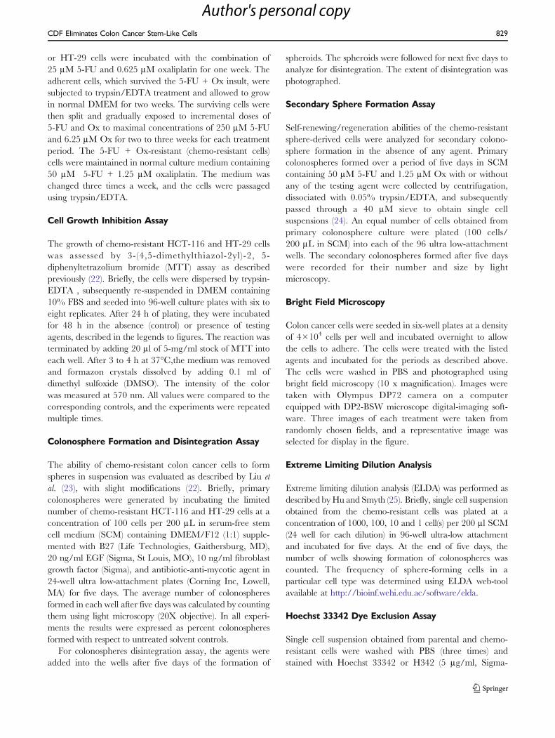
One of the objectives of our current investigation was tocompare the efficacy of curcumin with CDF on growthinhibition of chemo-resistant colon cancer cells whencombined with chemotherapy of 5-FU + Ox. The chemo-resistant HCT-116 and HT-29 cells were analyzed forcellular growth following incubation with 5-FU + Ox aloneor in combination with increasing concentrations (up to8 μM) of either CDF or curcumin. A significant inhibitionof 40–70% in cellular growth of chemo-resistant cells wasobserved when the chemo-resistant HCT-116 or HT-29cells were incubated for 48 h with the combination of 4 or8 μM CDF and 5-FU + Ox, whereas the same dose ofcurcumin caused ≤20% inhibition (Fig. 2a). In contrast, thecombination therapy caused only a 25–30% inhibition ofgrowth of non-transformed intestinal epithelial cells (IEC-6)cells which are derived from weanling rats (data not shown).It should also be stated here that a greater inhibition ofcellular growth of chemo-resistant HCT-116 cells by CDFcompared to curcumin was associated with a correspondingloss of adherent cells (Fig. 2b). Interestingly, furthertreatment of the chemo-resistant HCT-116 cells with the
CDF Eliminates Colon Cancer Stem-Like Cells 831
Author's personal copy

combination of 5-FU and Ox caused a slight increase inadherent cells, compared with the controls (Fig. 2b).
In the next set of experiments, changes in the expressionof CSC markers, CD44 and CD166 in chemo-resistantHCT-116 cells in response to CDF or curcumin togetherwith 5-FU + Ox were examined. Quantitative Real Time-PCR (qRT-PCR) analysis was employed. We observed thatwhile 4 μM curcumin together with 5-FU + Ox caused noappreciable change in mRNA levels of either CD44 orCD166, the same combination treatment with 4 μM CDFcaused over 70% inhibition of CD44 and CD166 expres-sion (Fig. 3, left panel). As has been observed for CD44 andCD166, the expression of ABC transporter protein ABCG2in chemo-resistant HCT-116 cells was inhibited by more
than 50% in response to the combination of 4 μM CDFand 5-FU + Ox, compared to the 5-FU + Ox-treatedcontrols (Fig. 3, right panel). Curcumin was less effective inthis setting.
Earlier, we reported a marked activation of EGFR andIGF-1R, accompanied by elevated expression of CD44 andCD166 in HCT-116 cells that survived the 48 h treatmentof 5-FU + Ox (referred to as FOLFOX) (31), indicating thepresence of the growth factor receptors in chemo-survivingcells that are enriched in colon CSCs (13). To determinewhether the current treatment strategy would also affect thefunctioning of the growth factor receptors, the levels ofphosphorylated (activated) form of EGFR and IGF-1Rwere examined. Our results demonstrated that although
Number of cells/well Number of wells plated Number of wells showing colonospheres
HCT-116 Parental HCT-116 Chemoresistant
1,000 24 24 24
100 24 24 24
10 24 24 24
1 24 5 13
Sphere forming frequency (95% CI) 1/4 (1/6–1/3) 1/2 (1/3–1/2)
p value < 0.05
Table 1 Extreme LimitingDilution Analysis ofColonospheres FormingFrequency of Parental andChemo-Resistant HCT-116 Cells
Data are pooled from three inde-pendent experiments for each.
CI confidence interval
Fig. 1 Chemo-resistant colon cancer HCT-116 cells display cancer stem cell properties. (A) Real time-quantitative RT-PCR (qRT-PCR) analysis showingincreased levels of mRNAs of colon CSCs markers- CD44 and CD166 in chemo-resistant cells as compared to parental control cells. (B) The quantitationof flow cytometry analysis of H342 dye exclusion showing higher exclusion by chemo-resistant HCT-116 cells, compared to parental cells. (C) Westernblots showing increased expression of ABCG2 in chemo-resistant HCT-116 cells, compared to the corresponding parental cells. (D) The NF-κB DNAbinding activity in parental and chemo-resistant cells. Results are average of three experiments with standard deviation. The values are normalized toparental cells. *p<0.001, compared to the corresponding treatment controls.
832 Kanwar et al.
Author's personal copy
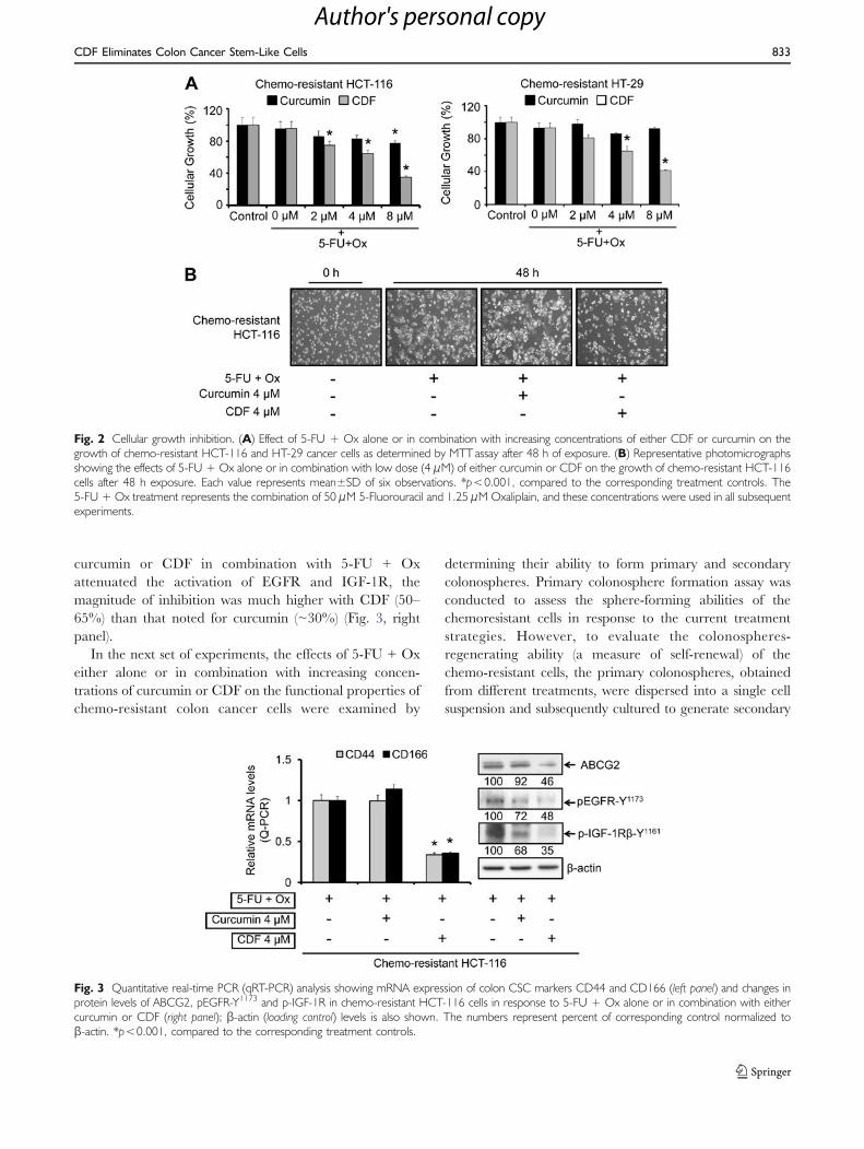
curcumin or CDF in combination with 5-FU + Oxattenuated the activation of EGFR and IGF-1R, themagnitude of inhibition was much higher with CDF (50–65%) than that noted for curcumin (~30%) (Fig. 3, rightpanel).
In the next set of experiments, the effects of 5-FU + Oxeither alone or in combination with increasing concen-trations of curcumin or CDF on the functional properties ofchemo-resistant colon cancer cells were examined by
determining their ability to form primary and secondarycolonospheres. Primary colonosphere formation assay wasconducted to assess the sphere-forming abilities of thechemoresistant cells in response to the current treatmentstrategies. However, to evaluate the colonospheres-regenerating ability (a measure of self-renewal) of thechemo-resistant cells, the primary colonospheres, obtainedfrom different treatments, were dispersed into a single cellsuspension and subsequently cultured to generate secondary
Fig. 3 Quantitative real-time PCR (qRT-PCR) analysis showing mRNA expression of colon CSC markers CD44 and CD166 (left panel) and changes inprotein levels of ABCG2, pEGFR-Y1173 and p-IGF-1R in chemo-resistant HCT-116 cells in response to 5-FU + Ox alone or in combination with eithercurcumin or CDF (right panel); β-actin (loading control) levels is also shown. The numbers represent percent of corresponding control normalized toβ-actin. *p<0.001, compared to the corresponding treatment controls.
Fig. 2 Cellular growth inhibition. (A) Effect of 5-FU + Ox alone or in combination with increasing concentrations of either CDF or curcumin on thegrowth of chemo-resistant HCT-116 and HT-29 cancer cells as determined by MTT assay after 48 h of exposure. (B) Representative photomicrographsshowing the effects of 5-FU + Ox alone or in combination with low dose (4 μM) of either curcumin or CDF on the growth of chemo-resistant HCT-116cells after 48 h exposure. Each value represents mean±SD of six observations. *p<0.001, compared to the corresponding treatment controls. The5-FU + Ox treatment represents the combination of 50 μM 5-Fluorouracil and 1.25 μM Oxaliplain, and these concentrations were used in all subsequentexperiments.
CDF Eliminates Colon Cancer Stem-Like Cells 833
Author's personal copy

colonospheres in the absence of any treatment. Resultsrevealed that CDF in combination with 5-FU + Ox wassuperior to curcumin in inhibiting the growth of primary aswell as secondary colonospheres (Fig. 4a). These resultsindicate that the treatment strategy with CDF not onlypossesses a superior inhibitory effect on the growth of sphere-forming cells but also shows a better post-treatmentinhibition of the self-renewing capacities of the sphere
derived from chemo-resistant cells (Fig. 4a). A similarphenomenon was also observed with respect to disintegrationof colonospheres, where we found CDF together with 5-FU +Ox caused a much greater disintegration than that caused by5-FU + Ox alone or the combination of 5-FU + Ox andcurcumin (Fig. 4b).
To determine the underlying mechanisms for changes inviability of chemo-resistant colon cancer cells in response to
Fig. 4 Formation of primary and secondary colonospheres by the chemo-resistant HCT-116 cells is inhibited by CDF. (A) Effect of 5-FU + Ox alone orin combination with increasing concentrations of either CDF or curcumin on the formation primary colonospheres by chemo-resistant HCT-116 cells(upper panel). The primary colonospheres obtained from respective treatments were then dissociated into single cell suspension, subsequently subjectedto secondary sphere formation in the absence of any treatment. To assess post-treatment self-renewing abilities of the colonosphere-derived cells, anequal number of these cells, diluted in stem cell media, were plated in 96-well low attachment plates for secondary colonosphere generation in theabsence of any agent (lower panel). (B) Representative photomicrographs showing the extent of colonosphere disintegration in chemo-resistant HCT-116cells in the absence (control-untreated) or presence of 5-FU + Ox alone or in combination with low dose of either curcumin or CDF. *p<0.001,compared to the corresponding treatment controls.
Fig. 5 (A) Western blot showing changes in the levels of pro-apoptotic Bax, and anti-apoptotic Bcl-xL in untreated parental as compared to chemo-resistant HCT-116 cells in response to the absence or presence of 5-Fu + Ox alone or in combination with either CDF or curcumin. The numbersrepresent percent of corresponding control normalized to β-actin. (B) Induction of early apoptosis as determined by acridine-orange/ethidium-bromidestaining in chemo-resistant HCT-116 and HT-29 cells after incubation of cells in the absence or presence of 5-Fu + Ox alone or in combination witheither CDF or curcumin. Values are mean±SD of 4 experiments. *p<0.001, compared to the corresponding treatment control.
834 Kanwar et al.
Author's personal copy

the current treatment strategies, we examined the expres-sion of anti-apoptotic Bcl-xL and pro-apoptotic Baxproteins. The chemo-resistant HCT-116 cells showed asignificant 30–40% reduction in the levels of pro-apoptoticBax protein, whereas the levels of anti-apoptotic Bcl-xLwere increased by about 400% when compared with thecorresponding values of the parental cells (Fig. 5a). How-ever, in response to the combination of 4 μM CDF and 5-FU + Ox, the expression of pro-apoptotic protein Bax inthe chemo-resistant HCT-116 cells was increased by about100%, whereas the same treatment with curcumin caused amere 27% increase in Bax protein when compared with thecorresponding control (Fig. 5a). On the other hand, theexpression of the anti-apoptotic Bcl-xL was decreased by150–250% in response to the combination of CDF orcurcumin and 5-FU + OX when compared with thecorresponding values of the chemo-resistant cells treatedwith only 5-FU + Ox (Fig. 5a). Further, the combinationtreatment of CDF and 5-FU + OX caused a staggering5–6-fold increase in apoptosis of both HCT-116 and HT-29chemo-resistant cells (Fig. 5b).
It is known that following induction of apoptosis,proteolytic cleavage of procaspase-3 occurs to generate anactive caspase-3 fragment, which targets key modulators ofthe apoptotic pathway. To corroborate our observation ofinduction of apoptosis, we examined the levels of cleavedcaspase-3 in chemo-resistant HCT-116 cells by Westernblot and the activity of caspase-3 in chemo-resistant HT-29cells in response to the current treatment strategies. Ourresults revealed a 1.7–2.5-fold increase in the levels ofcleaved caspase fragments (19 kDa and 17 kDa, Fig. 6a)and also a similar induction (~ 1.7-fold increase) in caspase-3 activity following the combination of CDF and 5-FU +Ox (Fig. 6b). On the other hand, the combination ofcurcumin and 5-Fu + Ox was ineffective in inducingcaspase 3 activity (Fig. 6b).
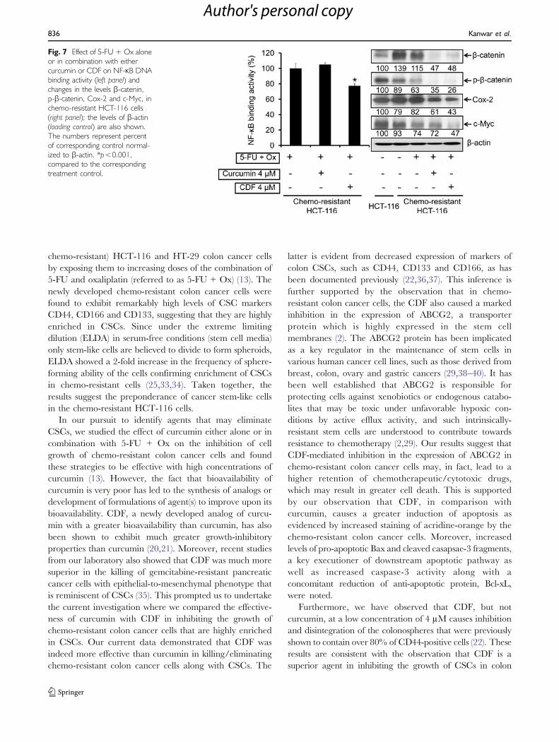
Further regulatory events were evaluated by examiningthe changes in the activation of NF-κB and β-catenin alongwith the expression of the downstream effectors Cox-2 andc-Myc. The results revealed that 4 μM CDF, but notcurcumin in combination with 5-FU + Ox, caused a 30%reduction in NF-κB activity in chemo-resistant HCT-116cells, compared with the controls (Fig. 7, left panel). Inaddition, we observed a 40% increase in the levels of β-catenin in chemo-resistant HCT-116 cells, compared to theparental cells (Fig. 7, right panel). In contrast, a significant60% reduction of β-catenin was noted following treatmentwith the combination of CDF and 5-FU + Ox (Fig. 7,right panel). Further, studies revealed that both curcuminand CDF treatments were alike in inhibiting the activationof β-catenin, as evidenced by a complete loss of phosphor-ylated form of the protein (Fig. 7, right panel). However,CDF was little more effective in inhibiting the expression of
Cox-2 and c-Myc when compared with correspondinglevels in 5-FU + Ox-treated controls (Fig. 7, right panel).
DISCUSSION
There is an emerging body of evidence suggesting thattumor cells that are resistant to chemotherapy represent asubpopulation of cells of the original tumor. These chemo-resistant cells, which are molecularly and phenotypicallydistinct, are also referred to as tumor-initiating cells, tumor-promoting cells or, more commonly, cancer stem cells orcancer stem-like cells (CSC) (Ref (32)). To study the role ofCSCs and emergence of chemo-resistance in colon cancer,we have generated 5-FU + Ox-resistant (referred to as
Fig. 6 (A) Western blot analysis showing changes in the proteinexpression of cleaved caspase-3 in chemo-resistant HCT-116 cells after48 h incubation of cells in the absence or presence of 5-Fu + Ox alone orin combination with either CDF or curcumin, β-actin (loading control)levels are also shown (upper panel), and changes in the levels of 19 kDaand 17 kDa fragments of cleaved caspase-3 relative to control normalizedto β-actin, as determined by densitometry analysis which, are depicted inthe histogram (lower panel). (B) Changes in the caspase-3 activity in thechemo-resistant HT-29 cells. Values are mean±SD of 4 experiments. p<0.001, compared to the corresponding treatment control.
CDF Eliminates Colon Cancer Stem-Like Cells 835
Author's personal copy
chemo-resistant) HCT-116 and HT-29 colon cancer cellsby exposing them to increasing doses of the combination of5-FU and oxaliplatin (referred to as 5-FU + Ox) (13). Thenewly developed chemo-resistant colon cancer cells werefound to exhibit remarkably high levels of CSC markersCD44, CD166 and CD133, suggesting that they are highlyenriched in CSCs. Since under the extreme limitingdilution (ELDA) in serum-free conditions (stem cell media)only stem-like cells are believed to divide to form spheroids,ELDA showed a 2-fold increase in the frequency of sphere-forming ability of the cells confirming enrichment of CSCsin chemo-resistant cells (25,33,34). Taken together, theresults suggest the preponderance of cancer stem-like cellsin the chemo-resistant HCT-116 cells.
In our pursuit to identify agents that may eliminateCSCs, we studied the effect of curcumin either alone or incombination with 5-FU + Ox on the inhibition of cellgrowth of chemo-resistant colon cancer cells and foundthese strategies to be effective with high concentrations ofcurcumin (13). However, the fact that bioavailability ofcurcumin is very poor has led to the synthesis of analogs ordevelopment of formulations of agent(s) to improve upon itsbioavailability. CDF, a newly developed analog of curcu-min with a greater bioavailability than curcumin, has alsobeen shown to exhibit much greater growth-inhibitoryproperties than curcumin (20,21). Moreover, recent studiesfrom our laboratory also showed that CDF was much moresuperior in the killing of gemcitabine-resistant pancreaticcancer cells with epithelial-to-mesenchymal phenotype thatis reminiscent of CSCs (35). This prompted us to undertakethe current investigation where we compared the effective-ness of curcumin with CDF in inhibiting the growth ofchemo-resistant colon cancer cells that are highly enrichedin CSCs. Our current data demonstrated that CDF wasindeed more effective than curcumin in killing/eliminatingchemo-resistant colon cancer cells along with CSCs. The
latter is evident from decreased expression of markers ofcolon CSCs, such as CD44, CD133 and CD166, as hasbeen documented previously (22,36,37). This inference isfurther supported by the observation that in chemo-resistant colon cancer cells, the CDF also caused a markedinhibition in the expression of ABCG2, a transporterprotein which is highly expressed in the stem cellmembranes (2). The ABCG2 protein has been implicatedas a key regulator in the maintenance of stem cells invarious human cancer cell lines, such as those derived frombreast, colon, ovary and gastric cancers (29,38–40). It hasbeen well established that ABCG2 is responsible forprotecting cells against xenobiotics or endogenous catabo-lites that may be toxic under unfavorable hypoxic con-ditions by active efflux activity, and such intrinsically-resistant stem cells are understood to contribute towardsresistance to chemotherapy (2,29). Our results suggest thatCDF-mediated inhibition in the expression of ABCG2 inchemo-resistant colon cancer cells may, in fact, lead to ahigher retention of chemotherapeutic/cytotoxic drugs,which may result in greater cell death. This is supportedby our observation that CDF, in comparison withcurcumin, causes a greater induction of apoptosis asevidenced by increased staining of acridine-orange by thechemo-resistant colon cancer cells. Moreover, increasedlevels of pro-apoptotic Bax and cleaved casapsae-3 fragments,a key executioner of downstream apoptotic pathway aswell as increased caspase-3 activity along with aconcomitant reduction of anti-apoptotic protein, Bcl-xL,were noted.
Furthermore, we have observed that CDF, but notcurcumin, at a low concentration of 4 μM causes inhibitionand disintegration of the colonospheres that were previouslyshown to contain over 80% of CD44-positive cells (22). Theseresults are consistent with the observation that CDF is asuperior agent in inhibiting the growth of CSCs in colon
Fig. 7 Effect of 5-FU + Ox aloneor in combination with eithercurcumin or CDF on NF-κB DNAbinding activity (left panel) andchanges in the levels β-catenin,p-β-catenin, Cox-2 and c-Myc, inchemo-resistant HCT-116 cells(right panel); the levels of β-actin(loading control) are also shown.The numbers represent percentof corresponding control normal-ized to β-actin. *p<0.001,compared to the correspondingtreatment control.
836 Kanwar et al.
Author's personal copy
cancer, a phenomenon similar to that observed with drug-resistant pancreatic cancer (35). Additional support comesfrom the observation that secondary sphere formation byCDF-treated colonosphere-derived cells is greatly inhibitedin the absence of further treatment with CDF. In view of thefact that secondary sphere formation represents self-renewingability of the CSCs (22,41), our observation of decreasedsecondary formation by CDF-treated primary colonosphere-derived cells suggests that CDF in combination with 5-FU +Ox could be an effective therapeutic strategy for eliminationof colon cancer recurrence.
Although, the precise regulatory mechanisms in supportof the CDF-mediated inhibition of growth of chemo-resistant colon cancer cells are not fully understood, ourobservation showed that CDF greatly inhibits the activationof EGFR and IGF-1R, which we have shown to beimplicated in the development of chemo-resistance, sug-gesting that CDF may act through the inactivation ofEGFR and IGF-1R signaling pathways (13,31,42). Thisinference is supported by the observation that CDF inhibitsthe activation of β-catenin as well as NF-κB, both of whichare downstream of both EGFR and IGF-1R signaling (43–45). The fact that CDF but not curcumin at 4 μMconcentration inhibits the expression of c-Myc and Cox-2,which are downstream effectors of β-catenin and NF-κBsignaling, respectively, further indicates a superior efficacyof CDF than curcumin in inhibiting the growth of CSC-enriched chemo-resistant colon cancer cells.
In conclusion, our observations demonstrate that thechemo-resistant colon cancer cells display increased activityof β-catenin and NF-κB, along with higher expression of c-Myc and Cox-2, and ABCG2, which is involved in drugexclusion. We have also demonstrated that chemo-resistantcolon cancer cells are highly enriched in CSCs, asevidenced by the increased expression of CD44 andCD166 and their ability to form spheres under ELDA.Our data also suggest that CDF in combination with 5-FU+ Ox is more effective than curcumin in inhibiting thegrowth of chemo-resistant colon cancer cells that areenriched in CSCs. Taken together, our results suggest apotential therapeutic role for CDF in preventing theemergence of chemo-resistant colon cancer cells by reduc-ing/eliminating the CSCs, which could become a novelstrategy for improving the overall survival of patientsdiagnosed with colon cancer.
ACKNOWLEDGMENTS
This work was supported by grants from the NationalInstitutes of Health/National Institute on Aging (5RO1AG014343) and the Department of Veterans Affairs (A.P.N.M.) and from the National Cancer Institute, NIH (3RO1CA131151-02) (F.H.S).
REFERENCES
1. Jemal A, Siegel R, Ward E, Hao Y, Xu J, Thun MJ. CancerStatistics, 2009. CA Cancer J Clin. 2009;59:225–49.
2. Dean M, Fojo T, Bates S. Tumour stem cells and drug resistance.Nat Rev Cancer. 2005;5:275–84.
3. Andre T, Boni C, Mounedji-Boudiaf L, Navarro M, Tabernero J,Hickish T, et al. Oxaliplatin, fluorouracil, and leucovorin asadjuvant treatment for colon cancer. N Engl J Med.2004;350:2343–51.
4. Neugut AI, Lautenbach E, Abi-Rached B, Forde KA. Incidenceof adenomas after curative resection for colorectal cancer. Am JGastroenterol. 1996;91:2096–8.
5. Odoux C, Fohrer H, Hoppo T, Guzik L, Stolz DB, Lewis DW,et al. A stochastic model for cancer stem cell origin in metastaticcolon cancer. Cancer Res. 2008;68:6932–41.
6. Toda S, Miyase T, Arichi H, Tanizawa H, Takino Y. Naturalantioxidants. III. Antioxidative components isolated fromrhizome of Curcuma longa L. Chem Pharm Bull (Tokyo).1985;33:1725–8.
7. Huang MT, Wang ZY, Georgiadis CA, Laskin JD, Conney AH.Inhibitory effects of curcumin on tumor initiation by benzo[a]pyrene and 7, 12-dimethylbenz[a]anthracene. Carcinogenesis.1992;13:2183–6.
8. Rao CV, Rivenson A, Simi B, Reddy BS. Chemoprevention ofcolon carcinogenesis by dietary curcumin, a naturally occurringplant phenolic compound. Cancer Res. 1995;55:259–66.
9. Rao CV, Simi B, Reddy BS. Inhibition by dietary curcumin ofazoxymethane-induced ornithine decarboxylase, tyrosine proteinkinase, arachidonic acid metabolism and aberrant crypt fociformation in the rat colon. Carcinogenesis. 1993;14:2219–25.
10. Patel BB, Sengupta R, Qazi S, Vachhani H, Yu Y, Rishi AK, et al.Curcumin enhances the effects of 5-fluorouracil and oxaliplatin inmediating growth inhibition of colon cancer cells by modulatingEGFR and IGF-1R. Int J Cancer. 2008;122:267–73.
11. Nautiyal J, Banerjee S, Kanwar SS, Yu Y, Patel BB, Sarkar FH,et al. Curcumin enhances dasatinib-induced inhibition of growthand transformation of colon cancer cells. Int J Cancer. 2010.
12. Pateland BB, Majumdar AP. Synergistic role of curcumin withcurrent therapeutics in colorectal cancer: minireview. NutrCancer. 2009;61:842–6.
13. Yu Y, Kanwar SS, Patel BB, Nautiyal J, Sarkar FH, MajumdarAPN. Elimination of colon cancer stem–like cells by thecombination of curcumin and FOLFOX. Transl Oncol.2009;2:321–8.
14. Wahlstromand B, Blennow G. A study on the fate of curcumin inthe rat. Acta Pharmacol Toxicol (Copenh). 1978;43:86–92.
15. Anand P, Kunnumakkara AB, Newman RA, Aggarwal BB.Bioavailability of curcumin: problems and promises. Mol Pharm.2007;4:807–18.
16. Labbozzetta M, Baruchello R, Marchetti P, Gueli MC, Poma P,Notarbartolo M, et al. Lack of nucleophilic addition in theisoxazole and pyrazole diketone modified analogs of curcumin;implications for their antitumor and chemosensitizing activities.Chem Biol Interact. 2009;181:29–36.
17. Bhutani MK, Bishnoi M, Kulkarni SK. Anti-depressant like effectof curcumin and its combination with piperine in unpredictablechronic stress-induced behavioral, biochemical and neurochemi-cal changes. Pharmacol Biochem Behav. 2009;92:39–43.
18. Mosley CA, Liotta DC, Snyder JP. Highly active anticancercurcumin analogues. Adv Exp Med Biol. 2007;595:77–103.
19. Zambre AP, Kulkarni VM, Padhye S, Sandur SK, Aggarwal BB.Novel curcumin analogs targeting TNF-induced NF-kappaBactivation and proliferation in human leukemic KBM-5 cells.Bioorg Med Chem. 2006;14:7196–204.
CDF Eliminates Colon Cancer Stem-Like Cells 837
Author's personal copy

20. Padhye S, Banerjee S, Chavan D, Pandye S, Swamy KV, Ali S,et al. Fluorocurcumins as cyclooxygenase-2 inhibitor: moleculardocking. Pharmacokinetics and tissue distribution in mice. PharmRes-Dord. 2009;26:2438–45.
21. Padhye S, Yang H, Jamadar A, Cui QC, Chavan D, Dominiak K,et al. New difluoro Knoevenagel condensates of curcumin, theirSchiff bases and copper complexes as proteasome inhibitors andapoptosis inducers in cancer cells. Pharm Res. 2009;26:1874–80.
22. Kanwar SS, Yu Y, Nautiyal J, Patel BB, Majumdar AP. TheWnt/beta-catenin pathway regulates growth and maintenance ofcolonospheres. Mol Cancer. 2010;9:212.
23. Liu S, Dontu G, Mantle ID, Patel S, Ahn NS, Jackson KW, et al.Hedgehog signaling and Bmi-1 regulate self-renewal of normaland malignant human mammary stem cells. Cancer Res.2006;66:6063–71.
24. Kakarala M, Brenner DE, Korkaya H, Cheng C, Tazi K,Ginestier C, et al. Targeting breast stem cells with the cancerpreventive compounds curcumin and piperine. Breast Cancer ResTreat. 2009.
25. Huand Y, Smyth GK. ELDA: extreme limiting dilution analysisfor comparing depleted and enriched populations in stem cell andother assays. J Immunol Methods. 2009;347:70–8.
26. Nautiyal J, Yu Y, Aboukameel A, Kanwar SS, Das JK, Du J, et al.ErbB-inhibitory protein: a modified ectodomain of epidermalgrowth factor receptor synergizes with dasatinib to inhibit growthof breast cancer cells. Mol Cancer Ther. 2010;9:1503–14.
27. Majumdarand AP, Du J. Phosphatidylinositol 3-kinase/Aktsignaling stimulates colonic mucosal cell survival during aging.Am J Physiol Gastrointest Liver Physiol. 2006;290:G49–55.
28. Richand JN, Bao S. Chemotherapy and cancer stem cells. CellStem Cell. 2007;1:353–5.
29. Anand Y, Ongkeko WM. ABCG2: the key to chemoresistance incancer stem cells? Expert Opin Drug Metab Toxicol.2009;5:1529–42.
30. Fletcher JI, Haber M, Henderson MJ, Norris MD. ABC trans-porters in cancer: more than just drug efflux pumps. Nat RevCancer. 2010;10:147–56.
31. Patel BB, Gupta D, Elliott AA, Sengupta V, Yu Y, Majumdar AP.Curcumin targets FOLFOX-surviving colon cancer cells viainhibition of EGFRs and IGF-1R. Anticancer Res. 2010;30:319–25.
32. Clevers H. Wnt/beta-catenin signaling in development anddisease. Cell. 2006;127:469–80.
33. Ricci-Vitiani L, Lombardi DG, Pilozzi E, Biffoni M, Todaro M,Peschle C, et al. Identification and expansion of human colon-cancer-initiating cells. Nature. 2007;445:111–5.
34. Vermeulen L, Todaro M, de Sousa Mello F, Sprick MR, KemperK, Perez Alea M, et al. Single-cell cloning of colon cancer stemcells reveals a multi-lineage differentiation capacity. Proc NatlAcad Sci USA. 2008;105:13427–32.
35. Ali S, Ahmad A, Banerjee S, Padhye S, Dominiak K, SchaffertJM, et al. Gemcitabine sensitivity can be induced in pancreaticcancer cells through modulation of miR-200 and miR-21expression by curcumin or its analogue CDF. Cancer Res.2010;70:3606–17.
36. Yeung TM, Gandhi SC, Wilding JL, Muschel R, Bodmer WF.Cancer stem cells from colorectal cancer-derived cell lines. ProcNatl Acad Sci USA. 2010;107:3722–7.
37. Sukachand A, Ivanov E. Formation of spherical colonies as aproperty of stem cells. Cell Tissue Biol. 2007;1:476–81.
38. Allenand JD, Schinkel AH. Multidrug resistance and pharmaco-logical protection mediated by the breast cancer resistance protein(BCRP/ABCG2). Mol Cancer Ther. 2002;1:427–34.
39. Katayama R, Koike S, Sato S, Sugimoto Y, Tsuruo T, Fujita N.Dofequidar fumarate sensitizes cancer stem-like side populationcells to chemotherapeutic drugs by inhibiting ABCG2/BCRP-mediated drug export. Cancer Sci. 2009.
40. Meissner K, Heydrich B, Jedlitschky G, Meyer zu SchwabedissenH, Mosyagin I, Dazert P, et al. The ATP-binding cassettetransporter ABCG2 (BCRP), a marker for side population stemcells, is expressed in human heart. J Histochem Cytochem.2006;54:215–21.
41. Kakarala M, Brenner D, Korkaya H, Cheng C, Tazi K, GinestierC, et al. Targeting breast stem cells with the cancer preventivecompounds curcumin and piperine. Breast Cancer Res Treat.2010;122:777–85.
42. Yu D, Jing T, Liu B, Yao J, Tan M, McDonnell TJ, et al.Overexpression of ErbB2 blocks taxol-induced apoptosis byupregulation of p21Cip1, which inhibits p34Cdc2 kinase. MolCell. 1998;2:581–91.
43. Vanamala J, Reddivari L, Radhakrishnan S, Tarver C.Resveratrol suppresses IGF-1 induced human colon cancer cellproliferation and elevates apoptosis via suppression of IGF-1R/Wnt and activation of p53 signaling pathways. BMC Cancer.2010;10:238.
44. Dihlmannand S, Doeberitz MVK. Wnt/beta-catenin-pathway asa molecular target for future anti-cancer therapeutics. Int JCancer. 2005;113:515–24.
45. Ali S, Banerjee S, Schaffert JM, El-Rayes BF, Philip PA, SarkarFH. Concurrent inhibition of NF-kappa B, cyclooxygenase-2, andepidermal growth factor receptor leads to greater anti-tumoractivity in pancreatic cancer. J Cell Biochem. 2010;110:171–81.
838 Kanwar et al.
Author's personal copy
Copyright © 2022 FDOKUMEN