Curcumin ameliorated low dose‑Bisphenol A induced gastric ...
Upload
independentCategory
view
6download
0
This content has been downloaded from IOPscience. Please scroll down to see the full text.
Download details:
IP Address: 110.4.24.170
This content was downloaded on 09/10/2013 at 12:41
Please note that terms and conditions apply.
Preparation and anti-cancer activity of polymer-encapsulated curcumin nanoparticles
View the table of contents for this issue, or go to the journal homepage for more
2012 Adv. Nat. Sci: Nanosci. Nanotechnol. 3 035002
(http://iopscience.iop.org/2043-6262/3/3/035002)
Home Search Collections Journals About Contact us My IOPscience
IOP PUBLISHING ADVANCES IN NATURAL SCIENCES: NANOSCIENCE AND NANOTECHNOLOGY
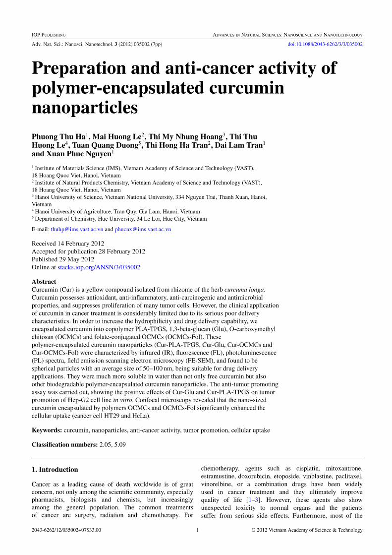
Adv. Nat. Sci.: Nanosci. Nanotechnol. 3 (2012) 035002 (7pp) doi:10.1088/2043-6262/3/3/035002
Preparation and anti-cancer activity ofpolymer-encapsulated curcuminnanoparticlesPhuong Thu Ha1, Mai Huong Le2, Thi My Nhung Hoang3, Thi ThuHuong Le4, Tuan Quang Duong5, Thi Hong Ha Tran2, Dai Lam Tran1
and Xuan Phuc Nguyen1
1 Institute of Materials Science (IMS), Vietnam Academy of Science and Technology (VAST),18 Hoang Quoc Viet, Hanoi, Vietnam2 Institute of Natural Products Chemistry, Vietnam Academy of Science and Technology (VAST),18 Hoang Quoc Viet, Hanoi, Vietnam3 Hanoi University of Science, Vietnam National University, 334 Nguyen Trai, Thanh Xuan, Hanoi,Vietnam4 Hanoi University of Agriculture, Trau Quy, Gia Lam, Hanoi, Vietnam5 Department of Chemistry, Hue University, 34 Le Loi, Hue City, Vietnam
E-mail: [email protected] and [email protected]
Received 14 February 2012Accepted for publication 28 February 2012Published 29 May 2012Online at stacks.iop.org/ANSN/3/035002
AbstractCurcumin (Cur) is a yellow compound isolated from rhizome of the herb curcuma longa.Curcumin possesses antioxidant, anti-inflammatory, anti-carcinogenic and antimicrobialproperties, and suppresses proliferation of many tumor cells. However, the clinical applicationof curcumin in cancer treatment is considerably limited due to its serious poor deliverycharacteristics. In order to increase the hydrophilicity and drug delivery capability, weencapsulated curcumin into copolymer PLA-TPGS, 1,3-beta-glucan (Glu), O-carboxymethylchitosan (OCMCs) and folate-conjugated OCMCs (OCMCs-Fol). Thesepolymer-encapsulated curcumin nanoparticles (Cur-PLA-TPGS, Cur-Glu, Cur-OCMCs andCur-OCMCs-Fol) were characterized by infrared (IR), fluorescence (FL), photoluminescence(PL) spectra, field emission scanning electron microscopy (FE-SEM), and found to bespherical particles with an average size of 50–100 nm, being suitable for drug deliveryapplications. They were much more soluble in water than not only free curcumin but alsoother biodegradable polymer-encapsulated curcumin nanoparticles. The anti-tumor promotingassay was carried out, showing the positive effects of Cur-Glu and Cur-PLA-TPGS on tumorpromotion of Hep-G2 cell line in vitro. Confocal microscopy revealed that the nano-sizedcurcumin encapsulated by polymers OCMCs and OCMCs-Fol significantly enhanced thecellular uptake (cancer cell HT29 and HeLa).
Keywords: curcumin, nanoparticles, anti-cancer activity, tumor promotion, cellular uptake
Classification numbers: 2.05, 5.09
1. Introduction
Cancer as a leading cause of death worldwide is of greatconcern, not only among the scientific community, especiallypharmacists, biologists and chemists, but increasinglyamong the general population. The common treatmentsof cancer are surgery, radiation and chemotherapy. For
chemotherapy, agents such as cisplatin, mitoxantrone,estramustine, doxorubicin, etoposide, vinblastine, paclitaxel,vinorelbine, or a combination drugs have been widelyused in cancer treatment and they ultimately improvequality of life [1–3]. However, these agents also showunexpected toxicity to normal organs and the patientssuffer from serious side effects. Furthermore, most of the
2043-6262/12/035002+07$33.00 1 © 2012 Vietnam Academy of Science & Technology

Adv. Nat. Sci.: Nanosci. Nanotechnol. 3 (2012) 035002 P T Ha et al
chemotherapeutic agents may not kill all cancer cells and theirrepeated administration develops drug resistance or androgenrefractory stage which is most difficult to cure [4].
Therefore, there is an urgent need to develop therapeuticmodalities with no or minimal side effects to normal organs.In this regard, a variety of natural dietary compoundshave been investigated. As a potential candidate, curcumin[1,7-Bis(4-hydroxy-3-medithoxyphenyl)-1,6-heptadiene-3,5-dione], a yellow compound isolated from rhizome of the herbcurcuma longa, has been receiving considerable attentionbecause of its putative cancer prevention and anti-canceractivities which are mediated through influencing multiplesignaling pathways [5, 6].
Although curcumin proves to be remarkably non-toxicand has promising anti-cancer activities, its application inanti-cancer therapies is limited due to its low aqueoussolubility and poor bioavailability. To deal with this obstacle,a variety of methods including the incorporation of curcumininto liposomes and into phospholipid vesicles are beingstudied [7, 8]. More recently, the approach of biodegradablepolymer nanoparticles has been developed [9–11]. Thisoffers promising therapeutic performance of anti-cancer drugsby increasing their bioavailability, solubility and retentiontime [12]. These drug formulations are superior to traditionalmedicines with respect to control release, targeted deliveryand therapeutic impact.
Polymeric nanoparticles act as nanocarriers with manyadvantages, such as low toxicity and high stability.Several drugs formulated in polymeric micelles are usedin clinical trial development for the treatment of variouscancers [13]. As indicated in [14], nanocurcumin particlesless than 100 nm in size could be synthesized usinga cross-linked and random copolymer of N-isopropylcrylamid (NIPAAM) with N-vinyl-2-pyrrolidone (VP) andpoly(ethyleneglicol)monoacrylate (PEG-A), which demon-strate superior efficacy compared to free (bulk) curcumin inhuman cancer cell line models. Polymeric nanoparticles haveattracted significant attention in the study of drug deliverysystems as they offer a means for localized or targeted deliverysystems of a drug to specific tissue/organ sites of interest withan optimal release rate [15].
The above-mentioned drug delivery systems are usuallyrestricted by the poor biocompatibility of the polymericmatrix material and the surfactant used in the formulationprocess. For formalization of curcumin nanosystem weconsider in our study three polymer materials derivedfrom natural product. Firstly, we aimed at synthesis ofan amphiphilic copolymer, which comprises polylactide(PLA)—often used in studies of drug delivery due to itsvery low toxicity and D-α-tocopheryl polyethylene glycolsuccinate (TPGS) —a safe and effective form of vitamin E dueto its good oral bioavailability. The PLA–TPGS copolymerhas many other potential applications, such as solubilizer,absorption enhancer and as a vehicle for lipid-based drugdelivery formulations as well as enhancement of cytotoxicityof anticancer agents such as doxorubicin, vinblastine,paclitaxel and curcumin [16]. Secondly, Hericium erinaceus,a traditional edible mushroom, was chosen for investigationbecause of its biological activities [17]. Hericium erinaceuswas also reported to have cytotoxic effects on cancer cell
lines thanks to its polysaccharide 1,3-β-glucan [18]. Thethird polymer was O-carboxymethyl chitosan (OCMCs)—anamphiprotic ether exhibiting non-toxicity, biodegradability,biocompatibility and strong bioactivity. It has thereforestimulated increasing interest in biomedical applications.More interestingly, OCMCs can load hydrophobic anticancerdrugs effectively [19–21] and also immobilize a targetingagent such as folic acid (Fol). Several studies have recentlyreported that OCMCs-Fol is a potential targeted drug deliverysystem [22–25].
In this paper, we not only present the procedures forthe encapsulation of curcumin by copolymer PLA–TPGS,polysaccharide Glu, OCMCs and OCMCs-Fol, but alsoindicate the improvements of the solubility and anti-canceractivity of the fabricated nanosystems.
2. Experimental
2.1. Materials
Lactide (3,6-dimethyl-1,4-dioxane, C6H8O4), stannousoctoate (Sn (OOCC7H15)2), O-carboxymethyl chitosanand folic acid, ethanol (>99.5%), chloroform (>99.5%),dimethylsulfoxide (DMSO) (>99.9%), triethylamine (TEA),N-hydroxysuccinimide (NHS) and 1-[3-dimethylamino)propyl]-3-ethylcarbodiimide hydrochloride (EDC), trisbase, trichloroacetic (TCA), sulforhodamine B (SRB),acetic acid, fetal bovine serum (FBS), fetal bovine serumminimum essential medium (FBS-MEM), phosphatebuffered saline (PBS), agar, agarose, cell culture medialike Dulbecco’s modified eagle medium, Roswell ParkMemorial Institute (RPMI) 1640 medium, and tumorinitiator N-methyl-N′-nitro-N-nitrosoguanidine (MNNG)were purchased from Sigma-Aldrich. Vitamin E TPGS(d-α-tocopheryl polyethylene glycol 1000 succinate) andC33O5H54(CH2CH2O)23 were from Merck. Curcumin(>95% purity, (E,E)-1,7-bis(4-hydroxy-3-methoxyphenyl)-1,6-heptadiene-3,5-dione) was purchased from Mumbai,India. 1,3-β-Glucan was isolated from medicinal mushroomHericium erinaceus SH. Anti-tumor promotion assay in vitroon human hepatocellular carcinoma cell line (HepG2) (the cellline obtained from National Institute of Hygienic Epidemy—NIHE) has been performed at Experimental BiologyLab—Institute of Natural Products Chemistry, VietnamAcademy of Science and Technology. Human hepatocellularcarcinoma cell lines HT29, HeLa were obtained fromDepartment of Biology, Hanoi University of Science. Allchemicals were used as received without further purification.
2.2. Preparation of polymers
PLA-TPGS copolymer was synthesized by ring-openingbulk polymerization of lactide monomer (3,6-dimethyl-1,4-dioxane, C6H8O4) with vitamin E TPGS in the presenceof stannous octoate as catalyst [26].
1,3-β-glucan with short chain and molecular weight of990 was obtained from polysaccharides isolated from themushroom Hericium erinaceus. Amylase enzyme was usedto break down the long chain of the polysaccharides andeliminate 1,4-α-glucan [27].
2

Adv. Nat. Sci.: Nanosci. Nanotechnol. 3 (2012) 035002 P T Ha et al
Figure 1. Solubility of (a) Cur nanoparticles and (b) Cur in water.
2.3. Encapsulation of curcumin
Nanoprecipitation technique was used to prepare thepolymer-encapsulated curcumin. Polymers were firstdissolved in double distilled water. Curcumin dissolvedin absolute ethanol was added into solutions of polymer.The resulting solutions were then stirred or ultrasonicallyvibrated for hours. This dispersion of nanoparticles wasvacuum evaporated to eliminate the organic solventcompletely. Larger aggregates and free polymers wereremoved by centrifugation at 5000 rpm for 15 min. Thesupernatant containing curcumin-encapsulated nanoparticleswas recovered by ultra-centrifugation at 30 000 rpm.
Folate was attached to the surface amino groups ofOCMCs via a carbodiimide reaction [22,23]. Briefly, folicacid was dissolved into a mixture of anhydrous DMSO, TEAand activated by equal amounts of EDC and NHS undernitrogen anhydrous conditions for 2 h at room temperature.The OCMCs were dissolved in distilled water, and stirred untilthe solutions were optically transparent. Then activated folicacid was added dropwise to OCMCs solution. The resultingmixture was stirred at room temperature for about 24 h undernitrogen atmosphere to let folic acid conjugate onto OCMCsmolecules, and then titrated to pH 9.0 with 0.1 M NH3
solution to terminate the reaction. The solution was dialysedfirst against phosphate buffer saline (PBS, pH = 7.4) for 3days to remove excess of unreacted substrates and then againstdistilled water for 3 days to obtain OCMCs-Fol solution.
Curcumin was then encapsulated to OCMCs-Fol solutionto form Cur-OCMCs-Fol in a similar way of preparation toCur-OCMCs.
2.4. Characterization
Infrared spectra were recorded with a Fourier transforminfrared (FTIR) spectrometer SHIMADZU, using KBr pellets,in the region of 400–4000 cm−1. Field emission scanningelectron microscope (FE-SEM) images were taken by aHitachi S-4800. Fluorescence spectra were recorded by usinga Jobin-Yvon FL3-22. Photoluminsescence spectra weretaken with a 442 nm excitation line. Encapsulated curcuminwere estimated using the calibration curve of curcuminsolution in acetone or ethanol.
Figure 2. FTIR spectra of Cur, Cur-OCMCs and Cur-OCMCs-Fol.
Figure 3. Fluorescence spectra of Cur, Cur-OCMCs andCur-OCMCs-Fol.
3. Results and discussion
3.1. Encapsulation efficiency
The polymer-encapsulated curcumin nanoparticles showenormous improvements in aqueous solubility characteristics.While free curcumin immediately precipitates in aqueousmedium due to very low solubility (∼20 µg ml−1), theabsolute concentration of curcumin in filtered 1,3-β-glucansolution was found to be 4 mg ml−1, which is 220-foldscompared with the solubility of curcumin encapsulated byhydrophobically modified starch (HMS) [28]. The aqueoussolubility of Cur-Glu was 2-folds compared with that ofCur-PLA-TPGS (2 mg ml−1) and 4-folds compared with thatof Cur-OCMCs or Cur-OCMCs-Fol (1 mg ml−1). The highersolubility characteristics of Cur-Glu may result from bettercompatibility to curcumin of 1,3-β-Glucan due to its shortchain.
Lyophilized Cur-Glu, Cur-PLA-TPGS, Cur-OCMCs andCur-OCMCs-Fol powder were reconstituted with water. Thesepowders dissolved back into clear solution very quickly andeasily, with no noticeable curcumin precipitates (figure 1).The results suggested that curcumin was indeed trapped in themicelles and the complex of polymers and curcumin couldresist against freeze-drying.
3

Adv. Nat. Sci.: Nanosci. Nanotechnol. 3 (2012) 035002 P T Ha et al
(a) (b)
(c) (d)
Figure 4. FE-SEM images of Cur-PLA-TPGS (a), Cur-Glu (b), Cur-OCMCs (c) and Cur-OCMCs-Fol (d).
3.2. FTIR spectra
All FTIR spectra of Cur-Glu, Cur-PLA-TPGS, Cur-OCMCsand Cur-OCMCs-Fol have several shifts as compared to thoseof free curcumin or polymers. This indicates the formation ofpolymer-encapsulated curcumin nanoparticles. For example,compared with that of pure 1,3-β-Glucan, the IR spectrumof Cur-Glu showed a band shift from 3400 to 3417 cm−1,which is probably due to the hydrogen bonding between–OHgroups in curcumin and 1,3-β-Glucan (spectra omitted forbrevity). The FTIR spectrum of OCMCs showed broad bankat 3420 cm−1 due to the stretching vibration of hydroxylgroup. A peak at 1634 cm−1 corresponds to the stretchingvibrations of carbonyl. Comparing OCMCs and Cur-OCMCs,peak shifts were observed from 3420 to 3261 cm−1 and1634 to 1625 cm−1. The result confirmed the presence ofcurcumin in the Cur-OCMCs. The characteristic absorptionband of OCMCs that appeared at 1598 cm−1 was assignedto the N–H banding vibration of the primary amine. In thecase of Cur-OCMCs-Fol this peak is shifted to 1635 cm−1.The increased absorption of amide band may be due to theformation of the amide linkage between the amino acid groupon the OCMCs and the carboxyl group of folic acid (figure 2).
3.3. Fluorescence spectra
The fluorescence spectra of curcumin and polymer-encapsulated curcumin are shown in figure 3. Curcuminin ethanolic solution exhibits an absorption peak at 540 nm,while the solutions of Cur-Glu, Cur-PLA-TPGS, Cur-OCMCsand Cur-OCMCs-Fol show peaks at 529, 530, 491 and525 nm, respectively. The blue-shifts in the fluorescenceare likely due to the intermolecular hydrogen bondingbetween curcumin and polymers. Especially, the appearanceof a weak peak at 435 nm in the fluorescence spectrum ofCur-OCMCs-Fol might be explained by the presence of folateon the nanoparticles.
While the fluorescence intensity of Cur-OCMCs-Folis slightly lower than that of Curcumin itself, the muchhigher fluorescence intensity of Cur-PLA-TPGS, Cur-Glu,Cur-OCMCs suggests that curcumin is encapsulated in thehydrophobic core of micelle of PLA-TPGS and curcumin ispresent in the Cur-Glu and Cur-OCMCs.
3.4. Surface morphology
The size and morphology of the polymer-encapsulatedcurcumin nanoparticles were confirmed by FE-SEM imaging.Figure 4 shows FE-SEM images of the curcumin encapsulated
4

Adv. Nat. Sci.: Nanosci. Nanotechnol. 3 (2012) 035002 P T Ha et al
by PLA-TPGS (a), Glu (b), OCMCs (c) and OCMCs-Fol(d). It is shown that the particles have an average size of50–100 nm, which lies in the optimal size range (below200 nm) suitable for drug delivery applications. There isa significant decrease in size of curcumin nanoparticlescompared to that of curcumin. This is probably because thehydrophilic polymers prevent the aggregation of hydrophobiccurcumin.
A PL image of free curcumin dispersed in ethanolicsolution is shown in figure 5(a). The spherical shape of theparticles is seen with a size of 1–10 µm. Figures 5(b) and (c)show PL images of the polymer-encapsulated curcumin witha large range of sizes similar to that of curcumin. Curcuminin the form of nanoparticles is a strong PL substance, thuswhen used to treat cancer it could also act as a labelingmaterial. Hence we can determine the efficiency of thedrug transport process in different conditions. PL images ofCur-PLA-TPGS (figure 5(b)) and Cur-Glu (figure 5(c)) showthat both nanoparticles are highly fluorescent, implying thatthese materials can be used not only for cancer treatment butalso for biolabeling.
3.5. Colony assays in soft agar
Cell survival cytotoxicity experiments using sulforhodamineB method were performed in order to determine the maximaldoses of test materials for anti-tumor-promoting activityassays. Soft agar colony assay anti-tumor-promoting activitywas estimated based on the inhibition of soft agar colonyinduction in the Hep-G2 cell line. The cells were culturedin 10% FBS-MEM medium at 36.5 ◦C in an incubator with5% CO2 and 95% air. Cells growing logarithmically in amonolayer culture were trypsinized and suspended in 0.33%agar medium containing 10% FBS with or without samples atthe concentrations of 25 µg ml−1. For anti-tumor promotingassay, in duplicate 6-well plate, 500 µl of the suspension (1 ×
104 cells) was poured onto an agar layer containing the sameconcentration of sample (10 µg ml−1) in 5% DMSO. Soft agarcolonies of cells were investigated after 2 weeks’ incubationunder an inverted microscope with camera to compare thevisual cell in their tumor formation, the tumor size andmorphology. The inhibitory activities were the average of twoindependent experiments and expressed as a percentage of thatof the control.
The results showed that there were no distinct differencesof cell survival in cytotoxicity assay, and the ratio of tumorpromotion in anti-tumor promoting assay with the Cur,Glu, PLA-TPGS alone was comparable to the control, butthere were clear changes in size and morphology of tumorbetween the control and all the tested samples, especiallycurcumin encapsulated with glucan copolymer. In the controlwells, the tumor size was much larger and their surface wasvery rough in comparison to the tumor on the tested wells(figure 6). It was obvious that encapsulated curcumin hadpositive effects on tumor promotion of Hep-G2 cell linein vitro.
3.6. Intracellular uptake of nanoparticles
To study the uptake of the nanoparticles Cur-OCMCsand OCMCs-Cur-Fol, confocal imaging was performed on
(a)
(b)
(c)
Figure 5. Fluorescent images of Cur (a), Cur-PLA-TPGS (b) andCur-Glu (c).
Hela and HT29 cells at 4 and 12 h. Hela, HT29 cellswere maintained 24 h and then incubated with Cur-OCMCsand Cur-OCMCs-Fol within 4 and 12 h. Immunofluorescentstains were processed and cells were visualized in confocallaser scanning microscopy LSM-510. Fluorescence intensitiesin specimens were compared to evaluate the quantity ofcurcumin within cancer cells.
5

Adv. Nat. Sci.: Nanosci. Nanotechnol. 3 (2012) 035002 P T Ha et al
(a)
(b)
(c)
Figure 6. Anti-tumor-promoting effects of the curcuminencapsulated by copolymer in Hep-G2 cell lines after two weeksof cell growth on agar: control (a), Cur (b) and Cur-PLA-TPGS(c) under inverted microscope ×100.
From the confocal microscope images (figure 7) thefolate-conjugated nanoparticles were found to be distributedin the zone of nucleus, indicating cellular uptake insteadof adhesion to the surface, and that the nanoparticlespreferentially targeted the cancer cells and were internalized.This internalization might be due to the folate receptormediated endocytosis [23]. This observation clearly infers
(a)
(b)
(c)
Figure 7. Fluorescent Image of HT29 after 4 h incubating withcontrol (a), Cur-OCMCs (b) and Cur-OCMCs-Fol (c).
that folate-conjugated carboxymethyl chitosan may be veryeffective carrier to use as delivery system for targetedanticancer drug.
The rate of Cur-OCMCs and Cur-OCMCS-Fol inHT29 and HeLa indicates a different level of uptake ofcurcumin. This is consistent with the level of expressionof folate receptor on cell surface: HT29-overexpression andHela-mediated expression.
Fluorescence intensity of Cur-OCMCs-Fol insidethe cell at 12 h (figure 8(b)) shows a lower content thanthat at 4 h (figure 8(a)). This can be explained by thedegradation of curcumin to form smaller molecules such astrans-6-(4-hydroxy-3-methoxyphenul)-2, 4-dioxo-5-hexenal,vaniline, ferulic acid, feruloy methane which can no longerremain auto-fluorescence like curumin [29]. However,fluorescence intensity of Cur-OCMCs at 12 h shows a highercontent than that at 4 h.
6

Adv. Nat. Sci.: Nanosci. Nanotechnol. 3 (2012) 035002 P T Ha et al
(a)
(b)
Figure 8. Comparison of cellular uptake between Cur-OCMCS andCur-OCMCS-Fol on HT29 and Hela cell lines at 4 h (a) and 12 h (b).
The key difference may come from the presence of folicacid, which actively leads the nanosystem to the cancer cellswith expression of folate receptor on its surface. In that casecurcumin can be transferred to the cancer cells more quicklyand efficiently.
4. Conclusion
In the present studies copolymer PLA-TPGS, 1,3-β-Glucan, O-carboxymethyl chitosan and folate-conjugatedO-carboxymethyl chitosan-encapsulated curcumin nano-particles were prepared successfully by nanoprecipitationtechnique. It was found that these particles have a goodsolubility in water. As spherical particles with an averagesize from 50 to 100 nm, they are also believed to besuitable for drug delivery applications. Confocal microscopyrevealed that folate enhances the uptake of curcumin intocancer cells expressing folate receptor. Besides, the anti-tumorpromoting assay also shows strong positive effects ofCur-PLA-TPGS and Cur-Glu on tumor promotion of Hep-G2cell line in vitro. With all these good features that havebeen found, Cur-PLA-TPGS, Cur-Glu, Cur-OCMCs andCur-OCMCS-Fol, could be used toward cancer therapy.
Acknowledgments
This work was financially supported by an IMSresearch grant, the National Foundation for Science andTechnology Development of Vietnam NAFOSTED grantNo 106.99-2010.42 (HPT), No 106.03.84.09 (MHL) andthe Ministry of Science and Technology grant (No 04/ 02/742/2009/HD-DTDL). The authors are thankful to Professor,Academician Nguyen Van Hieu for his encouragementand interest in this research. The authors would like toacknowledge all members of IMS-VAST Key Laboratory forproviding laboratory facilities.
References
[1] Berry W R 2005 Urology 65 2[2] Oh W K, Tay M H and Huang J 2007 Cancer 109 477[3] Petrylak D P 2005 Urology 65 8[4] Urakami S, Shiina H, Sumura M, Honda S, Wake K, Hiraoka
T, Inoue S, Ishikawa N and Igawa M 2008 Int. Urol.Nephrol. 40 365
[5] Karmakar S, Banik N L, Patel S J and Ray S K 2006 Neurosci.Lett. 407 53
[6] Anand P, Sundaram C, Jhurani S, Kunnumakkara A B andAggarwal B B 2008 Cancer Lett. 267 133
[7] Li L, Braiteh F S and Kurzrock R 2005 Cancer 104 1322[8] Sou K, Inenaga S, Takeoka S and Tsuchida E 2008 Int. J.
Pharm. 325 287[9] Yallapu M M, Jaggi M and Chauhan S C 2010 Colloids Surf. B
79 113[10] Anand P, Nair H B, Sung B, Kunnumakkara A B, Yadav V R,
Tekmal R R and Aggarwal B B 2010 Biochem. Pharmacol.79 330
[11] Tran D L et al 2010 Colloids Surf. A 371 104[12] Shenoy D B and Amiji M M 2005 Int. J. Pharm 293 261[13] Sahu A, Bora U, Kasoju N and Goswami P 2008 Acta
Biomater. 4 1752[14] Bisht S, Feldmann G, Soni S, Ravi R, Karikar C, Amornath M
and Aniirban M 2007 J. Nanobiotechnol. 5 3[15] Mu L and Seow P H 2006 Colloids Surf. B 47 90[16] Pan J and Feng S S 2008 Biomaterials 29 2663[17] Nakatsugawa M, Takahashi H, Takezawa C, Nakajima K,
Harada K, Sugawara Y, Kobayashi S, Kondo T and Abe S2003 Int. Med. 42 1219
[18] Mizuno T, Wasa T, Ito H, Suzuki C and Ukai N 1992 Biosci.Biotechnol. Biochem. 56 347
[19] Zhu A P, Liu J H and Ye W H 2006 Carbohydr. Polym.63 89
[20] Ha P T et al 2011 Chem. Lett. 40 1264[21] Anitha A, Maya S, Deepa N, Chennazhi K P, Nair S V, Tamura
H and Jayakumar R 2011 Carbohydr. Polym. 83 452[22] Wang F, Zhang D, Duan C, Jia L, Feng F, Liu Y, Wang Y, Hao
L and Zhang Q 2011 Carbohydr. Polym. 84 1192[23] Sahu S K, Mallick S K, Santra S, Maiti T K, Ghosh S K and
Pramanik P 2010 J. Mater. Sci.: Mater. Med. 21 1587[24] Yang S J, Lin F H, Tsai H M, Lin C F, Chin H C, Wong J M
and Shieh M J 2011 Biomaterials 32 2174[25] Low P S and Kularatne S A 2009 Curr. Opin. Chem. Biol.
13 256[26] Ha P T, Tran T M N, Pham H D, Nguyen Q H and Nguyen X P
2010 Adv. Nature Sci.: Nanosci. Nanotechnol. 1 015012[27] Le M H, Ha P T, Nguyen T B T, Tran T H H, Ha T M T, Mai
T T, Tran T N H, Do H N, Nguyen X P and Duong T Q2011 Chem. Lett. 40 846
[28] Yu H and Huang Q 2010 Food Chem. 11 669[29] Aggarwal B B, Surh Y J and Shishodia S 2007 Adv. Exp. Med.
Biol. 595 269
7
Copyright © 2022 FDOKUMEN