efficiency of curcumin and chitosan nanoparticles against ...
Upload
khangminh22Category
view
3download
0
THE EFFECT OF CURCUMIN AND TETRAHYDROCURCUMIN IN COMBINATION WITH 5-FLUOROURACIL ON ESOPHAGEAL CANCER CELL LINES
Emily G. Pendleton
A Thesis
Submitted to the Graduate College of Bowling Green State University in partial fulfillment of
the requirements for the degree of
MASTER OF SCIENCE
August 2015
Committee:
Roudabeh J. Jamasbi, Advisor
Michael E. Geusz
Carol A. Heckman

© 2015
Emily Pendleton
All Rights Reserved

iii
ABSTRACT
Roudabeh J. Jamasbi, Advisor
Despite our constant efforts to improve upon our current treatments, esophageal cancer
does not respond well to traditional therapy. In an effort to better treat esophageal cancer,
different remedies are being implemented to complement current remedies. Curcumin is an
active phytochemical found in turmeric that has received attention as a promising cancer
treatment because it has been shown to interact with multiple cancer targets. However,
curcumin’s poor bioavailability remains one of the biggest obstacles in clinical use.
Tetrahydrocurcumin, one of curcumin’s metabolites, has a higher bioavailability than curcumin,
and thus has gained more attention as a possible contributor to curcumin’s pharmacological
activity.
The present study used a previously determined effective dose of curcumin, 40 µM, on
three esophageal squamous cell carcinoma (ESCC) cell lines, KY-5, TE-1, and TE-8. We then
used 40 µM of tetrahydrocurcumin to determine whether it was more or less effective at
inhibiting cell proliferation in the three ESCC cell lines in vitro. Specifically, I identified a low
therapeutic dose of 5-fluorouracil for these three lines and evaluated the effect of
tetrahydrocurcumin at the same dose as the curcumin. I then tested pairwise combinations of
these three drugs to evaluate and compare complementary therapies.
Preliminary data to determine an effective dosage for 5-fluorouracil was performed using
Trypan Blue to count the number of dead and live cells in the KY-5 cell line. From there, all
three ESCC cell lines were exposed to 40 µM 5-FU at different combinations of seeding
densities and incubation times and cell proliferation and viability was measured with a

iv
PrestoBlue fluorescence assay. The results of these experiments lead to the selection of 104
cells/well and 48-hour incubation with all drugs for the subsequent experiments. The three
ESCC cell lines were then exposed to curcumin, THC, THC with curcumin, THC with 5-FU, and
curcumin with 5-FU for the same time period. The results of this experiment indicate that at the
same effective dose as curcumin, THC has no impact on the cell viability of these three lines.
When both 5-FU and curcumin are used alone at the same dosage, curcumin is more effective at
inhibiting cell viability in these ESCC lines. Our study supports previously reported findings
that curcumin is significantly effective in growth inhibition of ESCC lines. Our findings further
corroborate past studies by suggesting that curcumin may be a better anticancer agent in ESCC
than traditional chemotherapeutic agents such as 5-FU because curcumin can be better at
inhibiting ESCC cell proliferation and has far less impact on normal cells.

v
I dedicate this work to my parents, Mark and Tami Pendleton, for their constant love and encouragement.

vi
ACKNOWLEDGMENTS
A thank you should first be extended to my parents. They have instilled drive and
passion within me and have been a constant source of guidance and support while completing
this work. My little brother has been supportive of this work as well. His personal motivation
has inspired me strive to do and be better. My fiancé, Tyler, has been instrumental in my
completion of this work. Thank you for your love and sacrifices made in order for this to
happen.
I am thankful for the instruction and advice of my committee. They have taught me how
to properly conduct science, think like a scientist, and have been instrumental in my professional
growth. I have great gratitude for Dr. Roudabeh Jamasbi, my advisor. Her friendship, advice,
and support throughout the process has been invaluable. I am indebted to Dr. Michael Geusz
and Dr. Carol Heckman for their mentoring, dedication, and willingness to help in my studies.
Appreciation of the input of my lab member, Astha Malik, should be recognized; her
dedication to helping others is incredible.
Lastly, I would like to extend a thank you for the help given by those that surround me on
a daily basis and their efforts in finishing this project.

vii
TABLE.OF CONTENTS
Page
INTRODUCTION………………………………………………………………………..... 1
Esophagus…………………………………………. ................................................. 1
Esophageal Cancer…………………………………………. .................................... 1
Cancer Stem Cells…………………………………………. ..................................... 3
Current Treatments for ESCC…………………………………………. ................... 4
5-fluorouracil………………………………………. ................................................ 7
Curcumin………………………………………….................................................... 9
Tetrahydrocurcumin…………………………………………. .................................. 12
Hypotheses…………………………………………. ................................................ 14
MATERIALS AND METHODS………………………………………………………….. . 15
Cell Lines Culture and Conditions…………………………………………. ............ 15
Determining 5-fluorouracil Concentration………………………………………. ... 16
Trypan Blue…………………………………………. .............................................. 17
PrestoBlue………………………………………. ..................................................... 18
Phenol Red…………………………………………. ................................................ 18
Cell Density Curve Calibration……………………………………. ......................... 19
Seeding Density and Incubation Time Calibration………………………………… 19
Cytotoxic Effects of 5-fluorouracil, Curcumin, and Tetrahydrocurcumin on ESCC
Lines…………………………………………. .......................................................... 20
RESULTS………………………. ......................................................................................... 21
5-fluorouracil Concentration……………………………………………………….. 21

viii
Phenol Red Fluorescence………………………………………………………… ... 23
Cell Density Curve……………………………………………………….. ............... 24
Calibration of Seeding Density and Incubation Time……………………………… 26
Cytotoxic Effects of 5-fluorouracil, Curcumin, and Tetrahydrocurcumin on ESCC
Lines…………………………………….. ................................................................. 30
DISCUSSION……………………………. ........................................................................... 35
Cell Density Curve……………………………………………………………….. ... 35
Normalized Seeding Density and Incubation Time……………………………… ... 36
Incubation Period……………………………………………………………….. .... 36
5-fluorouracil Toxicity…………………………………………………………… ... 37
Tetrahydrocurcumin Toxicity…………………………………………………….. .. 38
Curcumin Toxicity…………………………………………………………… ......... 39
Combination Toxicity…………………………………………………………… .... 41
CONCLUSION…………………………………………………………… .......................... 44
REFERENCES……………………………………………………………………………… 45

ix
LIST OF FIGURES
Figure Page
1 Chemical structures of curcuminoids, curcumin analogs, and curcumin metabolites 10
2 Metabolic pathway of curcumin that leads to the formation of tetrahydrocurcumin 12
3 Trypan Blue experiments ........................................................................................... 22
4 Effect of Phenol Red .................................................................................................. 23
5 Cell density curve ...................................................................................................... 25
6 Seeding density and incubation time ......................................................................... 27
7 Normalized seeding density and incubation time ...................................................... 29
8 Cytotoxic effects of THC, curcumin, 5-FU and their combinations.......................... 34

x
LIST OF TABLES
Table Page
1 Characteristics of cell lines ....................................................................................... 16
2 Significant differences among the treatment groups of each cell line ...................... 32

1
INTRODUCTION
Esophagus
The esophagus is a muscular tube measuring 25-30 cm in length. It is part of the
digestive system which, as a whole, is responsible for the processing of food, extraction of
nutrients from food, and elimination of waste. The esophagus begins at vertebra C6, next to the
cricoid cartilage, and ends at the stomach near vertebra T7 (Saladin, 2015). The esophagus is
composed of three layers of tissue: mucosa, submucosa, and muscularis propia. The mucosa in
the esophagus contains stratified squamous epithelium. The mucosa near the lower esophageal
sphincter, the sphincter that separates the esophagus and the stomach, has both stratified
squamous and columnar epithelial specializations. Connective tissue, lymphocytes, and nerve
cells constitute the submucosa. The outer layer of the esophagus, the muscularis propia, consists
of inner circular and outer longitudinal muscle layers (Thompson & Shaffer, 2012).
Esophageal Cancer
Esophageal cancer is the sixth most lethal cancer in the world with a five-year survival
rate of 14% (Mohamed et al., 2014). There are two classifications of esophageal cancer:
esophageal squamous cell carcinoma (ESCC) and adenocarcinoma (EAC). These two cancers
differ based on the type of cells that become cancerous.
Cancer of the epithelial layer of the mucosa, consisting of squamous cells of the
esophagus, gives rise to ESCC (Kuwano et al., 2005). Well established contributing factors of
ESCC are elevated consumption of alcohol, smoking, and nutrient poor diets (Brown et al.,

2
2001). More recent studies conclude that intake of hot beverages such as Maté or other teas can
induce ESCC in South America and Asia. The greatest risk factors of ESCC are smoking and
drinking, increasing an individual’s risk by 3-7 and 3-5 fold, respectively (Kamangar et al.,
2009). These risk factors cause chronic irritation and inflammation leading to dysplasia of the
esophagus (Mohamed et al., 2014; Stoner & Gupta, 2001). These atypical cells present with
hyperchromasia, loss of polarity, and irregularly clumped chromatin. ESCC progresses from
dysplasia to carcinoma in situ, then invasive metastatic cancer (Stoner & Gupta, 2001). ESCC
most often metastasizes to the lungs and liver and less often to the brain and bones. The
probability of metastasis to lymph nodes when ESCC remains localized in the mucosa is 5%.
The risk of metastasis increases to 30% when ESCC occupies the submucosa (Gabbert et al.,
2000) .
While the squamous epithelial cells become cancerous in ESCC, EAC occurs when the
glandular cells are malignantly altered. EAC arises in the lower third of the esophagus after it
endures intestinal metaplasia, when glandular columnar cells replace the squamous cells of the
mucosa. Intestinal metaplasia is a result of repetitive injury to the esophagus, typically in the
form of gastro-esophageal reflux disease (GERD), which often leads to Barrett’s esophagus
(Gabbert et al., 2000; Castell et al. 2004). Barrett’s esophagus is the result of continuous reflux
of gastric acid into the esophagus (Castell et al., 2004). When the esophagus experiences
prolonged exposures to stomach fluids, the epithelial cells become damaged and are replaced
with glandular columnar cells. Thus, Barrett’s esophagus is the biggest risk factor for EAC
(Taniere et al., 2001). The progression of inflammation, metaplasia, dysplasia, and carcinoma in
situ gives rise to invasive EAC (Chen & Yang, 2006).

3
Cancer Stem Cells
Stem cells are the cells that are able to propagate themselves and create mature,
specialized cells through the process of differentiation (Reya et al., 2001). Stem cells have the
ability to self-renew which allows the parent stem cell to create daughter stem cells with the
same developmental and replication abilities (Lobo et al., 2007). Stem cells have the ability to
self-renew, allowing them to persist over the lifetime of the animal, and are able to maintain
undifferentiated cells that are tissue-specific (Reya et al., 2001; Lobo et al., 2007). The second
characteristic of stem cells is the ability to differentiate, by which stem cells produce a daughter
cell that has the ability to become a tissue-specific specialized cell. This is achieved when
progenitor cells activate various genetic and epigenetic mechanisms to transform the daughter
cell into a mature cell (Lobo et al., 2007). These two characteristics of stem cells create the
assortment of cells that make up tissues and organs.
In conjunction with these traits, stem cells also function to maintain homeostasis in
normal tissue by repairing injured tissues. Stem cells also have the ability to switch from their
normal quiescent state to a state of high proliferation to replace differentiated cells during a time
of high tissue turnover (Efferth, 2012). Because both stem and progenitor cells have a lifespan
longer than differentiated cells, they are more likely to acquire tumor-initiating mutations
(Efferth, 2012).
The cancer stem cell (CSC) theory states that the heterogeneity of tumors is the result of a
small subset of cancer cells that have the sole responsibility of tumorigenesis and creation of the
bulk of differentiated cancer cells (Fabian et al., 2013; Lobo et al., 2007). Thus, this theory
holds that CSCs share the same defining characteristics with somatic stem cells. Like somatic

4
stem cells, CSCs have the ability to self-renew and to differentiate (Fabian et al., 2013). There
are two theories for the development of CSCs. One model suggests that mutations in normal
stem cells allow for the growth of CSCs while a second model suggests that CSCs are the result
of mutations in progenitor cells that can dedifferentiate into stem cells and then self-renew (Lobo
et al., 2007). Regardless of their origins, CSCs have the ability of self-renewal and
differentiation, allowing them to form, grow, and maintain tumors (Fabian et al., 2013). This
parallel between CSCs and normal stem cells is confirmed by the presence of signaling pathways
needed for self-renewal and differentiation in both types of stem cells (Almanaa, 2013).
Current cancer treatment is able to kill the differentiated cancer cells that make up the
majority of the tumor volume (Almanaa, 2013). It is believed that CSCs are responsible for
therapy failure, resistant subpopulations, relapse, and metastasis (Fabian et al., 2013). Therefore,
identifying effective therapeutic agents that can work in conjunction with current treatments is
essential for more potent treatment of both cancer cells and CSCs.
Current Treatments for ESCC
There are various treatments for ESCC, but the results of the treatment are disappointing.
ESCC can extend into surrounding areas, including through the esophageal muscle and into
surrounding lymphatic tissue. Patients typically display symptoms after the cancer has become
advanced (Thompson & Shaffer, 2012). These symptoms are predominantly dysphagia,
retrosternal and/or epigastric pain, weight loss, and regurgitation (Gabbert et al., 2000). Due to
the delay in the onset of symptoms and therefore diagnosis, only 5% of ESCC can be treated
with a surgical procedure (Thomson & Shaffer, 2012).

5
Surgery is a viable option for patients with ESCC at an early stage. Endoscopic
resectioning of the mucosal layer can rid the body of the carcinoma. In Japan, improved survival
rates are attributed to the treatment of advanced esophageal cancer with three-field lymph node
dissection. Such invasive procedures, however, do come with a high morbidity and mortality
rate. Despite its promise, surgical resectioning of the esophagus is a poor treatment option due to
the limitations of the therapeutic results (Nakajima & Kato, 2013).
ESCC tumors are radiosensitive. Radiotherapy is a viable alternative for patients who are
poor surgical candidates due to advanced tumors or other health issues. When radiotherapy was
used as the primary form of therapy in patients who could tolerate surgery, the five-year survival
rate was 17%; considerably higher than the 5-10% survival rate of surgery alone (Thompson &
Shaffer, 2012).
Chemotherapy is often used to reduce tumor size. Chemotherapeutic drugs are used with
the hope of eliminating hematogenous and/or lymphogenous micro-metastases, therefore limiting
postoperative disease recurrence (Baba et al., 2014). There are many chemotherapeutic drugs to
treat ESCC, and they are often used in combination with one another. According to the
American Cancer Society, chemotherapeutic agents that are used to treat ESCC include cisplatin,
carboplatin, 5-fluorouracil, epirubicin, and docetaxel (American Cancer Society, 2014).
Neoadjuvant therapy, treatment given prior to surgery, is often used to combat ESCC.
The forms of neoadjuvant therapy used to diminish ESCC are radiotherapy (RT), chemotherapy
(CT), and chemoradiotherapy (CRT) (Nakajima & Kato, 2013). By using these therapies prior to
surgery, tumor size is reduced and undamaged blood vessels may allow for better drug delivery.

6
Patient response to neoadjuvant therapy is not universal. If a patient does not respond to the
neoadjuvant therapy, they may progress to further metastasis (Baba et al., 2014).
Neoadjuvant RT is not used in most cases of ESCC (Nakajima & Kato, 2013). When it is
performed, the goal is to down-size or eradicate tumors in the regional lymph nodes (Baba et al.,
2014). In five randomized studies of neoadjuvant RT in patients with squamous cell carcinoma,
only one showed significant survival benefit (Nakajima & Kato, 2013). Currently, neoadjuvant
RT is not suggested for ESCC patients (Baba et al., 2014).
Neoadjuvant CT is used with ESCC patients in stages I, II, and III. By adding
chemotherapy to the treatment regimen, there is an increased risk of morbidity and mortality due
to the toxicity of the drugs, disease progression due to selection of chemoresistant cells, and the
delay in surgical management (Nakajima & Kato, 2013).
Neoadjuvant CRT is standard therapy for ESCC patients who cannot undergo surgery,
and is also an option for those for whom surgery is recommended. Concurrent CRT therapy
gives an 8% improvement in survival over two years when compared to successive CT and RT.
In a study that followed patients that received either RT or CRT, the five-year survival rate was
0% in the RT group and 26% in the CRT group (Nakajima & Kato, 2013). Furthermore, when
neoadjuvant CRT was administered, postoperative morbidity and mortality rates of ESCC
patients did not change (Baba et al., 2014).
Most cases of ESCC are treated with palliative care. Palliation for ESCC comes in the
form of radiotherapy and surgery. Peroral dilation can alleviate dysphagia. Prosthetics are
placed in the esophagus to maintain the lumen in later stages of cancer. Photodynamic therapy

7
and radiofrequency ablation both target cancer cells and are newer forms of palliative care in
ESCC patients (Thompson & Shaffer, 2012).
5-fluorouracil
One of the most commonly used chemotherapeutic agents to treat cancers is 5-
fluorouracil (5-FU) (Okamoto et al., 2014). An analogue of uracil, 5-FU has replaced the
hydrogen atom on carbon-5 with a fluorine atom. C4H3FN2O2 is the molecular formula for 5-FU
with a molecular weight of 130.8 g/mol (National Center for Biotechnology Information, 2005a).
After administration, 5-FU is transported into the cell via the same facilitated transport
mechanism as uracil. Once inside of the cell, 5-FU is changed into different metabolites
(Longley et al., 2003). 5-FU can be converted to form 5-fluorodeoxyuridine monophosphate
(FdUMP) which inhibits thymidylate synthase. High levels of thymidylate synthase have been
associated with chemoresistance to 5-FU (Ratain & Plunkett, 2003). FdUMP can be further
transformed into fluorodeoxyuridine triphosphate (FdUTP). Similarly, 5-FU can be converted
into fluorouridine triphosphate (FUTP) (Longley et al., 2003). These three cytotoxic metabolites
produced by 5-FU are incorporated into RNA and DNA resulting in cell cycle arrest and
apoptosis (Okamoto et al., 2014). It has been found that 5-FU works to induce cell death at the
S/G2 phase of the cell cycle in MCF-7 breast cancer cells (Yano et al., 2014).
As one of the first chemotherapeutic agents, 5-FU has been used for more than 40 years
in the treatment of cancers (Okamoto et al., 2014). During this time, various delivery methods
have been developed. The potency of 5-FU can vary based on the avenue of administration:
bolus injection, intravenous infusion, or oral administration (de Bono & Twelves, 2001).

8
The concentration of 5-FU in plasma is the result of the dose and rate of administration.
When the phramacokinetics of 5-FU are saturated, it allows for slower clearance and hepatic
metabolism of the drug. When 500-600 mg of 5-FU is dispensed in bolus injection form, its
half-life is 8-14 minutes. The half-life increases as more drug is allotted. However, the area
under the curve (AUC) increases disproportionally at higher doses of 5-FU. This makes dosage
adjustments in bolus administration difficult as drug concentration in the blood is hard to gauge
(de Bono & Twelves, 2001). In comparison to the bolus injection, 5-FU has a shorter plasma
half-life when administered through intravenous infusion because it is cleared from the body
more rapidly (Ahmad et al., 2012). Milano et. al. (1994) were able to demonstrate the benefits of
intravenous administration. They treated 186 head and neck cancer patients with intravenous
administration of 5-FU. Their findings show that higher AUC values lead to greater tumor
response. There was no correlation between total dosage of 5-FU and survival rate. This
suggests that achieving optimal AUC through constant exposure of 5-FU gives the highest
survival rate and lower levels of toxicity (Milano et al., 1994). The most cumbersome problem
with intravenous administration of 5-FU is the inconvenience of moveable pumps or
administration of the drug in the hospital for 24 hours at a time for ten weeks or more. The time
required to administer full doses of 5-FU hinder the patient’s quality of life and leave them
susceptible to infections and vascular complications (Ahmad et al., 2012; de Bono & Twelves,
2001).
When treatment regimens use a higher dose of 5-FU, severe side effects such as
myelosuppression, diarrhea, and dermatitis can arise (Okamoto et al., 2014). However, an
increased dosage of 5-FU may be necessary because more than 80% of 5-FU is catabolized by

9
dihydropyrimidine dehydrogenase (DPD), most often in the liver, rendering it inactive (Longley
et al., 2003).
Curcumin
Curcumin is an active phytochemical from turmeric (Curcuma longa), a spice often used
in the preparation of curry (Li & Zhang, 2014). Curcumin was isolated from the turmeric
rhizome almost two hundred years ago by Vogel and Pelletier of Harvard College (Prasad et al.,
2014). Historically, curcumin has been used to treat a variety of diseases, including acne,
psoriasis, and diaper rash. In 1949, curcumin was found to have antibacterial properties (Gupta
et al., 2013). Further studies have shown that curcumin has antioxidant, antiproliferative, anti-
inflammatory, and antiangiogenic properties and has been tested in clinical trials as a possible
treatment for diseases such as Alzheimer’s disease and arthritis (Teiten et al., 2013; Mimeault &
Batra, 2011) .
Curcumin has been identified as 1,6-heptadiene-3, 5-dione-1, 7-bis (4-hydroxy-3-
methoxyphenyl)-(1E, 6E), also called diferuloylmethane (Strimpakos & Sharma, 2008).
Curcumin has a molecular weight of 368.38 g/mol, with the molecular formula C21H20O6
(Hatcher et al., 2008). Three main curcuminoids found in C. longa are curcumin,
demethoxycurcumin (DMC), and bisdemethoxycurcumin (BDMC). The natural ratio of
combined curcuminoids sold for research and clinical use is about 77% pure curcumin, 17%
DMC, and 3% BDMC (Yodkeeree et al., 2007) There are synthetic analogs of curcumin,
including tetramethyl curcumin (TMC) and dimethylcyclohexyl curcumin (DMCHC). Curcumin
can be metabolized in the body to form other curcuminoids, including tetrahydrocurcumin

10
(THC), curcumin O-glucuronide (COG), and curcumin O-sulfate (COS) (Cao et al., 2014)
(Figure 1).
Curcumin has the ability to interact with multiple targets that are involved in cancer.
Cancer is a disease of abnormal cell proliferation which progresses as genetic mutations
accumulate in a cell (Shehzad et al., 2013). The genetic alterations are found in oncogenes,
tumor suppressor genes, growth factors, cell signaling pathways, transcription factors, cytokines,
enzymes, and apoptosis (Shehzad et al., 2013; Altieri et al., 2013).
Numerous studies show that curcumin has the ability to modulate molecules involved in
nearly all stages of cancer (Prasad et al., 2014). Curcumin blocks molecules specifically
involved in the cell cycle (cyclin D1 and cyclin E), transcription (STAT), metastasis (CXCR-4),
inflammation (NF-κB and COX-2) and growth factors (HGF and EGF) (Anand, Sundaram et al.,
2008a; Prasad et al., 2014). Curcumin has the ability to down regulate the transcription factor
Figure 1. Chemical structures of curcuminoids, curcumin analogs, and curcumin metabolites. (adapted from Cao, et. al., 2014).

11
NF-κB, an over-expressed molecule in ESCC (Shehzad et al., 2013). Genes involved in
inflammation, survival, metastasis, and chemo-radio-resistance are regulated by NF-κB (Garg &
Aggarwal, 2002). When the impact of curcumin on the cell cycle was examined in Her2-positive
cells, it was found that it induces cell cycle arrest in cancer cells in the G1 phase (Sun et al.,
2012). ESCC is often resistant to traditional chemotherapeutic agents, such as 5-FU, and
curcumin is a promising treatment for chemo-resistant cells (O'Sullivan-Coyne et al., 2009).
Curcumin has a potential treatment capability for ESCC because it comes in contact with the
squamous cells of the esophageal tissue during ingestion (Almanaa, 2013). Curcumin’s ability to
impact multiple molecular targets makes it a promising agent for the suppression of cancer.
One of the major problems with curcumin is its low bioavailability. A substance is
considered to have poor bioavailability if it has low intrinsic activity in the body. For example, it
may show little absorption, be metabolized quickly, have inactive metabolites, or be eliminated
quickly from the body (Anand et al., 2007). Curcumin is identified as a compound with low
bioavailability because it is not readily solubilized, poorly absorbed in the gut, quickly
metabolized, and quickly excreted from the body (Jӓger et al., 2014). Undetectable or low levels
of curcumin in human blood serum were found when 2 g of curcumin was administered to
healthy volunteers (Shoba et al., 1998). A more recent study showed that 3.6 g of curcumin
administered orally gave rise to 0.0111 µM concentration of curcumin in human plasma after one
hour (Sharma et al., 2004).
In conjunction with low bioavailability of curcumin in blood plasma, curcumin is not
well distributed into tissue. Anand et al. (2007) reported past studies have shown that after oral
administration of 400 mg of curcumin to rats, trace levels of curcumin were found in the kidney
and liver and only 1% of the curcumin was left in the stomach and small intestine after 24 hours.

12
Furthermore, a study conducted on humans with colorectal cancer that had metastasized to the
liver showed no curcumin in the liver after oral administration of 450-3600 mg of curcumin each
day for a week (Anand et al., 2007). Curcumin’s poor bioavailabillity forces investigators to
look for similar anticancer drugs with higher bioavailability.
Tetrahydrocurcumin
Tetrahydrocurcumin (THC) is a major curcumin metabolite produced when curcumin is
partially reduced. Reduction of curcumin to THC occurs in two steps. First, curcumin is
reduced to dihydrocurcumin through NADPH-dependent reduction with the enzyme NADPH-
dependent curcumin/dihydrocurcumin reductase by intestinal E.Coli (CurA). In the second step,
dihydrocurcumin is further reduced to THC via the same enzyme (Figure 2) (Helson et al.,
2012). The THC molecule is made up of a β-diketone group and a phenol (Wu et al., 2014). The
molecular weight of THC is 372.41 g/mol and the molecular formula is C21H24O6 (National
Center for Biotechnology Information, 2005b). THC is colorless and more hydrophilic than
curcumin due to the addition of hydrogen molecules (Anand et al., 2008b).
Figure 2. Metabolic pathway of curcumin that leads to the formation of tetrahydrocurcumin. (Adapted from Hassainasab, et. al., 2011)

13
Less is known about THC when compared to curcumin, but current studies on THC
conclude that this metabolite shows promise. THC exhibits pharmacological and physiological
properties similar to curcumin (Yoysungmoen et al., 2008). The best antioxidant among
curcuminoids is THC (Pari & Murugan, 2004) . When compared to curcumin, THC is a better
anti-inflammatory and is better at scavenging free-radicals (Hassainasab et al., 2011; Anand et
al., 2008b). At the conclusion of a two-week study in which both THC and curcumin were
incubated in human plasma, THC was found to be more stable than curcumin with respective
recovery rates of 67-77% and 35-45%. This quality of THC is attributed to higher saturation,
making it more water soluble, when compared to curcumin (Cao et al., 2014). Furthermore, in
vitro methods show that curcumin is less stable in culture medium than THC, with half-lives of
186 and 813 minutes, respectively (Saradhi et al., 2010). THC is also more readily absorbed in
the gastrointestinal tract (Wu et al., 2014). For these reasons, it is suggested that THC is
responsible for curcumin’s pharmacological affects in vivo (Kang et al., 2014).
Several molecular targets of THC have been identified, including targets that make it
effective against cancers. THC induces the antioxidant enzymes NADPH:quinone reductase,
glutathione peroxidase, and glutathione S-transferase (Prasad et al., 2014). Kang et al. (2014)
performed an in vitro experiment by treating MCF-7 breast cancer cells with 15-112.5 µM THC
and found that these cells exhibit mitochondrial apoptosis and G2/M phase arrest when treated in
a dose-dependent manner. This effect is the result of THC increasing the protein expression of
Bax and caspases-3 and -9 (Kang et al., 2014). Furthermore, THC displays chemopreventative
abilities by retarding the development of aberrant crypt foci in the colon (Kim et al., 1998). THC
is also a better alternative to curcumin at inhibiting colon carcinogenesis because it decreases
cancer cell proliferation and inhibiting phosphorylation of ERK1/2 (Lai et al., 2011). When

14
HepG2 cells were treated with both curcumin and THC, curcumin was found to have stronger
anti-proliferation properties while THC was a stronger anti-angiogenic agent (Yoysungmoen et
al., 2008). THC is effective in increasing leukemic cell death through autophagy. Wu et al.
(2014) states that THC inhibits the metastasis of fibrosarcoma by lowering levels of MMP-2 and
MMP-9 in HT1080 cells.
Hypotheses
My research poses to answer two questions. One, when using a common dosage, is THC
more effective than curcumin at limiting cell viability? Two, is there an additive or synergistic
effect when THC, curcumin, ad 5-FU are used in pairwise combinations? Based on previous
research, THC has been shown to be effective against some cancers. THC is more stable in cell
medium and is a better antioxidant than curcumin. Because of these previous results, I
hypothesize that THC will be more effective than curcumin in limiting cell viability.
Furthermore, because these drugs have been shown to impact cancer cells at different points of
the cell cycle, I predict that there will be a synergistic effect.
15
MATERIALS AND METHODS
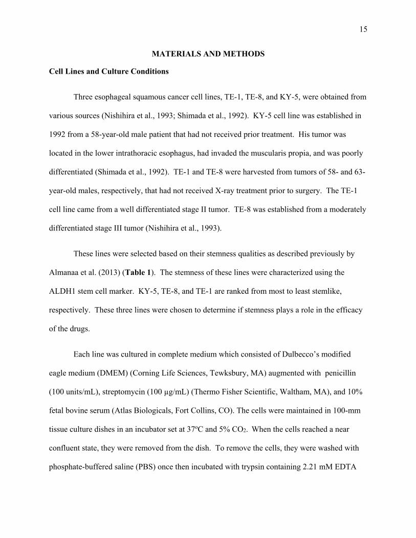
Cell Lines and Culture Conditions
Three esophageal squamous cancer cell lines, TE-1, TE-8, and KY-5, were obtained from
various sources (Nishihira et al., 1993; Shimada et al., 1992). KY-5 cell line was established in
1992 from a 58-year-old male patient that had not received prior treatment. His tumor was
located in the lower intrathoracic esophagus, had invaded the muscularis propia, and was poorly
differentiated (Shimada et al., 1992). TE-1 and TE-8 were harvested from tumors of 58- and 63-
year-old males, respectively, that had not received X-ray treatment prior to surgery. The TE-1
cell line came from a well differentiated stage II tumor. TE-8 was established from a moderately
differentiated stage III tumor (Nishihira et al., 1993).
These lines were selected based on their stemness qualities as described previously by
Almanaa et al. (2013) (Table 1). The stemness of these lines were characterized using the
ALDH1 stem cell marker. KY-5, TE-8, and TE-1 are ranked from most to least stemlike,
respectively. These three lines were chosen to determine if stemness plays a role in the efficacy
of the drugs.
Each line was cultured in complete medium which consisted of Dulbecco’s modified
eagle medium (DMEM) (Corning Life Sciences, Tewksbury, MA) augmented with penicillin
(100 units/mL), streptomycin (100 µg/mL) (Thermo Fisher Scientific, Waltham, MA), and 10%
fetal bovine serum (Atlas Biologicals, Fort Collins, CO). The cells were maintained in 100-mm
tissue culture dishes in an incubator set at 37oC and 5% CO2. When the cells reached a near
confluent state, they were removed from the dish. To remove the cells, they were washed with
phosphate-buffered saline (PBS) once then incubated with trypsin containing 2.21 mM EDTA

16
(Corning Life Sciences, Tweksbury, MA) for two minutes. The cells were then washed with
complete medium to inactivate the trypsin. The cells were collected and centrifuged at 800 x g
and the supernatant was removed. The cell pellets were agitated and resuspended in complete
medium and then plated in tissue culture dishes.
Table 1. Characteristics of cell lines. All three lines come from male patients. The quality of tumorigenicity for each cell line was determined by subcutaneous injection into nude mice. (Adapted from Almanaa, 2013).
Determining 5-Fluorouracil Concentration
To analyze the effect of 5-fluorouracil, KY-5 was plated in a 24-well plate (Corning) at a
density of 5 x 10⁴ cells/well in 1 mL of complete medium. The cells were incubated for 24 hours
in an incubator set at 37oC and 5% CO2 and allowed to attach. After attaching, the cells were
rinsed with PBS and were either exposed to1 mL of a 40 μM dose of 5-FU (4 wells/treatment) or
received complete media with 0.2% DMSO and incubated for 30 hours. The incubation time of
30 hours was used based on previous results (Almanaa et al., 2012).

17
Similarly, KY-5 was seeded in two 60-mm dishes at a density of 5 x10⁴ cells/dish. After
24 hours of incubation, one dish was treated with 3 mL of 0.2% DMSO to serve as a control and
the other dish received 3 mL of 40 μM 5-FU and allowed to incubate for 72 hours. This
experiment was allowed to incubate for 72 hours because it is one of the longest times published
for incubation of this drug according to the NCBI’s PubChem database (2015). All preliminary
data was measured by a typical Trypan Blue experiment.
Trypan Blue
The cell death rate in response to 5-FU was measured by Trypan Blue for all preliminary
data. A trypan blue assay was done by removing media that contained 5-FU from each well and
moving the media into individual test tubes, one well per test tube. In 24-well plates, the cells
were rinsed with PBS once and incubated with trypsin for 2 minutes. Fresh complete media was
added to the wells to inactivate the trypsin. The trypsin, fresh media, and cells were then added
to the corresponding test tubes with the original media. The cells were centrifuged at 800 x g
and the supernatant was removed. The cell pellet was agitated and resuspended in 1 mL of
complete media. To create a 1:4 dilution of cells, 10 μL of the cell suspension was added to a
culture tube along with 10 μL of Trypan Blue and 30 μL of PBS. The live and dead cells were
then counted using a hemocytometer. Dead cells absorbed Trypan Blue and appear blue.

18
PrestoBlue
The percentage of change in cell viability was measured by a standard PrestoBlue (Life
Technologies, Carlsbad, CA) assay. This was achieved by removing media from each well and
rinsing the cells once with PBS. Fresh complete media with 10% PrestoBlue was added to each
well in ½ mL aliquots. The plate was then incubated for 90 minutes. Immediately following the
incubation, the fluorescence of each well was read at 615 nm using a Wallac Victor 1420
Multilabel plate reader (Perkin, Elmer, Walthan, MA). The lid was removed prior to reading to
eliminate noise created by condensation. Background readings from the media and PrestoBlue
alone were subtracted from each reading and the percent change in cell viability was calculated.
OriginPro 7.5 was used for all statistical analysis. We evaluated a single outlier with the Grubbs’
test and the Modified Thompson Tau test. Once identified, statistical outliers were then removed
from the group.
Phenol Red
Our complete medium contains phenol red. Therefore, the fluorescence of phenol red
needed to be determined. Four wells of a 24-well plate received ½ mL of complete medium and
allowed to incubate for 90 minutes. The fluorescence was then measured at 615 nm. The plate
was allowed to sit in ambient room conditions for twenty minutes to alter the pH, and thus color,
of the medium; the plate was then read for a second time. This was done in order to determine if
the phenol red fluoresced differently at different pH levels.

19
Cell Density Curve Calibration
A standard cell density curve for each cell line was determined to estimate the number of
viable cells remaining after each treatment. A cell density curve was created for each line due to
different growth rates and biomass. For this purpose, each line was seeded at 0, 1,000, 10,000,
20,000, 40,000, 50,000 cells per well in 1 mL of complete medium in a 24-well plate. There
were four replicates of each seeding density. Routinely, cells have attached but have not begun
to spread and divide 6 hours after seeding. At this time point, the cell viability was measured by
a standard PrestoBlue assay as explained above.
Seeding Density and Incubation Time Calibration
Optimization of cell density and incubation time for each treatment group was performed.
A set of four experiments were designed in order to determine the best seeding density over two
periods of time. Each of the three cell lines was seeded in four 24-well plates. Each cell line
was allotted two columns per plate, one column served as a control and was treated with 0.2%
DMSO while the other column was administered 40 μM 5-FU in media containing DMSO. Two
of the plates were seeded at a density of 1 x 10⁴ cells/well and the two other plates seeded at 2 x
104 cells/well. Cells were allowed to grow for 24 hours. The medium was removed, rinsed with
PBS once, and was given either medium alone or medium containing 5-FU. One of the plates
seeded with 1 x 10⁴ cells/well and a plate seeded with 2 x 10⁴ cells/well were allowed to
incubate for 48 hours. The other two plates were allowed to incubate for 72 hours. After
incubation, cell viability was measured using a standard PrestoBlue assay.

20
Cytotoxic Effects of 5-fluorouracil, Curcumin, and Tetrahydrocurcumin on the ESCC
Lines
Curcumin (Sigma Aldrich, St. Louis, MO), THC (Chromadex, Irvine, CA), and 5-FU
(Acros Organics, Fair Lawn, NJ) were dissolved in DMSO to make a stock solution of 10 mM.
The stock solution was aliquoted and frozen at -80 oC. For each experiment, the stock solution
was thawed and diluted in complete media to the final working concentration of 40 μM.
Medium containing 0.2% DMSO was used as a control for all treatment groups.
To analyze the effect of curcumin, THC, and 5-FU, each cell line was plated in a 24-well
plate (Corning) at a density of 1 x 10⁴ cells/well in 1 mL of complete medium. The cells were
incubated and allowed to grow for 24 hours in 37oC and 5% CO2. After this period, the cells
were rinsed with PBS and were exposed to 1 mL of 40 μM curcumin, THC, or 5-FU (4
wells/treatment) and incubated for 48 hours. Cocktails of these three drugs— curcumin with
THC or 5-FU, and THC with 5-FU—were administered and handled in the same manner. Four
wells of each cell line served as controls received complete media containing 0.2% DMSO. The
resulting cell density was determined by a standard PrestoBlue assay.
21
RESULTS
5-fluorouracil Concentration
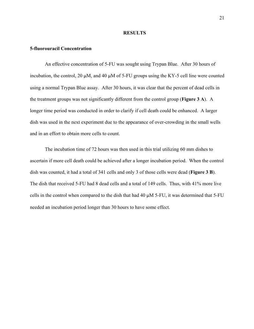
An effective concentration of 5-FU was sought using Trypan Blue. After 30 hours of
incubation, the control, 20 μM, and 40 μM of 5-FU groups using the KY-5 cell line were counted
using a normal Trypan Blue assay. After 30 hours, it was clear that the percent of dead cells in
the treatment groups was not significantly different from the control group (Figure 3 A). A
longer time period was conducted in order to clarify if cell death could be enhanced. A larger
dish was used in the next experiment due to the appearance of over-crowding in the small wells
and in an effort to obtain more cells to count.
The incubation time of 72 hours was then used in this trial utilizing 60 mm dishes to
ascertain if more cell death could be achieved after a longer incubation period. When the control
dish was counted, it had a total of 341 cells and only 3 of those cells were dead (Figure 3 B).
The dish that received 5-FU had 8 dead cells and a total of 149 cells. Thus, with 41% more live
cells in the control when compared to the dish that had 40 μM 5-FU, it was determined that 5-FU
needed an incubation period longer than 30 hours to have some effect.

22
Figure 3. Trypan Blue experiments. KY-5 cells were exposed to either 20 or 40 μM 5-FU for different times. The average live, dead, and total cell count of each experimental group is represented. A. 30 hours of 5-FU exposure. B. 72 hours of 5-FU exposure.
0
10
20
30
40
50
60
Control 20 μM 40 μM
Num
ber o
f Cel
ls
5-Flourouracil
A.
Dead
Live
0
50
100
150
200
250
300
350
Control 40 μM
Num
ber o
f Cel
ls
5-Fluorouracil
B.
Dead
Live
0
10
20
30
40
50
60
Control 20 μM 40 μM
Num
ber o
f Cel
ls
5-Fluorouracil
A.
Dead
Live
23
Phenol Red Fluorescence
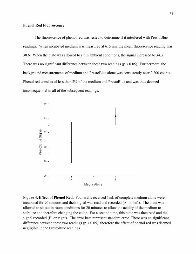
The fluorescence of phenol red was tested to determine if it interfered with PrestoBlue
readings. When incubated medium was measured at 615 nm, the mean fluorescence reading was
30.6. When the plate was allowed to sit in ambient conditions, the signal increased to 34.3.
There was no significant difference between these two readings (p < 0.05). Furthermore, the
background measurements of medium and PrestoBlue alone was consistently near 2,200 counts.
Phenol red consists of less than 2% of the medium and PrestoBlue and was thus deemed
inconsequential in all of the subsequent readings.
Figure 4. Effect of Phenol Red. Four wells received 1mL of complete medium alone were incubated for 90 minutes and their signal was read and recorded (A, on left). The plate was allowed to sit out in room conditions for 20 minutes to allow the acidity of the medium to stabilize and therefore changing the color. For a second time, this plate was then read and the signal recorded (B, on right). The error bars represent standard error. There was no significant difference between these two readings (p < 0.05), therefore the effect of phenol red was deemed negligible in the PrestoBlue readings.

24
Cell Density Curve
In order to appropriately estimate the number of viable cells remaining after each
experiment, a standard cell density curve for each cell line was dtermined. Six hours after
seeding at various densities, the viable cell density was measured. Each line was graphed to
show the mean fluorescence against the seeding density and a sigmoidal curve of best fit was
assigned. All three cell lines showed similar PrestoBlue signals at the 1,000 cells/well seeding
density. The TE-1 cell line exhibited a greater change in signal between 10,000 and 20,000
cells/well. Therefore, the seeding density of 20,000 cells/well was repeated a total of three
times. At this seeding density, TE-1 had a total number of twelve replicates which were then
averaged. TE-8 and KY-5 followed a gradual slope within the range of densities seeded. All
three lines converged near the 20,000 PrestoBlue signal when seeded at 50,000 cells/well. This
is the upper limit of PrestoBlue as stated by Life Technologies.

25
Figure 5. Cell density curve. Each cell line was seeded in a 24-well dish at densities from 1,000-50,000 cells/well. By 6 hours after seeding, the cells had attached and had not yet begun to spread. At this time, the plates were treated with a standard PrestoBlue assay. The average signal standard deviations were plotted and the best sigmoidal fit was applied for each cell line.
26
Calibration of Seeding Density and Incubation Time
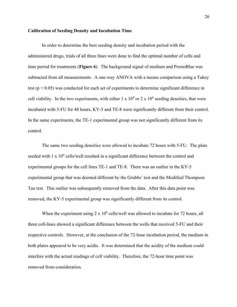
In order to determine the best seeding density and incubation period with the
administered drugs, trials of all three lines were done to find the optimal number of cells and
time period for treatments (Figure 6). The background signal of medium and PrestoBlue was
subtracted from all measurements. A one-way ANOVA with a means comparison using a Tukey
test (p < 0.05) was conducted for each set of experiments to determine significant difference in
cell viability. In the two experiments, with either 1 x 10⁴ or 2 x 10⁴ seeding densities, that were
incubated with 5-FU for 48 hours, KY-5 and TE-8 were significantly different from their control.
In the same experiments, the TE-1 experimental group was not significantly different from its
control.
The same two seeding densities were allowed to incubate 72 hours with 5-FU. The plate
seeded with 1 x 10⁴ cells/well resulted in a significant difference between the control and
experimental groups for the cell lines TE-1 and TE-8. There was an outlier in the KY-5
experimental group that was deemed different by the Grubbs’ test and the Modified Thompson
Tau test. This outlier was subsequently removed from the data. After this data point was
removed, the KY-5 experimental group was significantly different from its control.
When the experiment using 2 x 10⁴ cells/well was allowed to incubate for 72 hours, all
three cell-lines showed a significant difference between the wells that received 5-FU and their
respective controls. However, at the conclusion of the 72-hour incubation period, the medium in
both plates appeared to be very acidic. It was determined that the acidity of the medium could
interfere with the actual readings of cell viability. Therefore, the 72-hour time point was
removed from consideration.

27
Thus, the results of these four experiments and the cell density curve lead to the selection
of 1 x 10⁴ cells/well and 48 hours as the seeding density and incubation time for all future
experiments. This density was selected over 2 x 10⁴ cells/well in order to accommodate the
PrestoBlue readings which saturate between 4 x 10⁴ – 5 x 10⁴ cells/well.
Figure 6. Seeding Density and Incubation Time. The three ESCC lines were seeded at either 1 x 10⁴ or 2 x 10⁴ cells/well and were incubated with 5-FU for either 48 or 72 hours. Cell viability was measured with a standard PrestoBlue. The experimental and control group of each cell line was then compared within the same seeding density and time period. Significant difference (p < 0.05) was found in both the KY-5 and TE-8 cell lines at all combinations of seeding densities and incubation times. The TE-1 cell line showed significant difference in cell viability between the control and experimental groups in both experiments incubated for 72 hours. However, the media appeared very acidic in all cell lines regardless of seeding density after 72 hours. Based on this data and the cell density curve, 10,000 cells/well and 48 hours of incubation was chosen for all successive experiments.

28
The present data was normalized to their respective controls in order to determine if the
seeding density and incubation time significantly impacted the cell viability of each line. In all
three lines, there was significant difference between the controls and experimental groups when
cells were seeded at either density and allowed to incubate for 72 hours. When either seeding
density was used and allowed to incubate with 5-FU for 48 hours, only KY-5 and TE-8 showed
significant difference between their control and experimental groups.
From there, the experimental results of each seeding density and incubation time were
compared to one another within each cell line. The pairing of 1 x 10⁴ cells/well with 72 hour
incubation time had significantly fewer cells than the combinations of both seeding densities at
48 hours in all three cell lines. There is also a significant difference between the two seeding
densities incubated for 72 hours for the TE-1 and KY-5 cell lines.

29
Figure 7. Normalized seeding density and incubation time. After the cell viability of each combination was measured with a PrestoBlue assay, each data set was normalized to its control. The experimental groups within each cell line were then compared to one another. Within both the KY-5 and TE-8 cell lines, the experimental group that was seeded at 1 x 10⁴ cells/well and incubated for 72 hours was significantly different (p < 0.05) from all other seeding density and incubation period combinations. The TE-1 cell line group that was seeded with 1 x 10⁴ cells/well and incubated for 72 hours showed significant difference from the both seeding densities that were incubated for 48 hours.

30
Cytotoxic effects of 5-fluorouracil, curcumin, and tetrahydrocurcumin on the ESCC lines
The three cell lines were also treated with curcumin, THC, and the combination pairings
of curcumin, THC, and 5-FU. This was done to determine the cytotoxic effects of these drugs
individually and in combination on the cell lines. After each cell line was seeded at a density of
1 x 10⁴ cells/well in a 24-well plate, each well received 1 mL of 40 μM curcumin, THC, 5-FU, or
combinations of these drugs. The viability of the remaining cells was determined by a
PrestoBlue 48 hours after drug delivery.
There were significant differences within each cell line among the treatment groups
(Table 1 and Figure 7). In order to compare these results with the 5-FU results obtained from
the seeding density and incubation time calibration experiments, the controls were averaged
together and all groups were normalized to the controls. Again, a means comparison using a
Tukey test (p< 0.05) with a one-way ANOVA was performed on each set of experiments to find
significant difference in cell viability.
The TE-1 cell line was first examined. The TE-1 control and THC groups showed
similar cell viability. There were significantly more viable cells in the control wells than the
curcumin, THC with 5-FU, curcumin with 5-FU, and THC with curcumin treatment groups. The
THC experimental group had significantly more cells than curcumin, curcumin with 5-FU and
THC with curcumin. There were also significantly more viable cells in the 5-FU group than the
curcumin with 5-FU as well as the THC with curcumin groups. A similarity found in all three
cell lines is the THC with 5-FU treatment had significantly more viable cells than the wells that
were given curcumin with 5-FU and THC with curcumin.

31
The TE-8 cell line was the next to be examined. All experimental groups except for THC
were significantly different from the control. Again, the groups given THC did not respond well
to the drug, and had significantly more viable cells than curcumin, curcumin with 5-FU, and
THC with curcumin. There were significantly fewer viable TE-8 cells in the groups that
received curcumin, curcumin with 5-FU, and THC with curcumin when compared to the group
that was given 5-FU alone. TE-8 had more viable cells in the THC with 5-FU group than the
curcumin alone treatment.
The final cell line in this experiment is KY-5. Like TE-8, all treatment groups except for
THC were significantly different from the control group. The wells that received THC exhibited
a signal similar to the control wells and were significantly different from the same experimental
groups. When KY-5 was given 5-FU alone, it showed significantly more viable cells than the
treatment groups given curcumin alone, curcumin with 5-FU, and THC with curcumin.
Curcumin treated calls also had fewer viable cells than THC with 5-FU.
The determination of significant difference among the cell lines within each experimental
group was analyzed. In this analysis, an ANOVA was done in two ways: one included all of the
experimental groups done in one comparison; the other resulted from multiple ANOVAs with
only the three cell lines within each experimental group. When the ANOVA was done as first
described, the only meaningful change in response to the treatment was between KY-5 and TE-1
after being treated with curcumin. When only the cell lines involved in each trial were compared
with an ANOVA, TE-1 was found to be significantly different from both KY-5 and TE-8 in both
the curcumin and 5-FU treatment groups. In addition, in the curcumin with 5-FU experimental
group TE-1 was again significantly different from KY-5 and TE-8. All other comparisons in
both applications of an ANOVA were found to be insignificant.

32
Table 2. Significant differences among the treatment groups of each cell line. The normalized results of each drug and the combinations of drugs within each cell line were compared to one another. In the tables above, each experimental group was compared to the
Is there significant difference among the TE-1 treatment groups?THC 5-FU Curcumin THC &
5-FU Curcumin
& 5-FU THC &
Curcumin
Controls No No Yes* Yes* Yes* Yes*
THC No Yes* No Yes* Yes*
5-FU Yes* No Yes* Yes*
Curcumin No No No
THC & 5-FU Yes* Yes*
Curcumin & 5-FU No
Is there significant difference among the TE-8 treatment groups?THC 5-FU Curcumin THC &
5-FU Curcumin
& 5-FU THC &
Curcumin
Controls No Yes* Yes* Yes* Yes* Yes*
THC No Yes* No Yes* Yes*
5-FU Yes* No Yes* Yes*
Curcumin Yes* No No
THC & 5-FU Yes* Yes*
Curcumin & 5-FU No
Is there significant difference among the KY-5 treatment groups? THC 5-FU Curcumin THC &
5-FU Curcumin
& 5-FU THC &
Curcumin
Controls No Yes* Yes* Yes* Yes* Yes*
THC Yes* Yes* Yes* Yes* Yes*
5-FU Yes* No Yes* Yes*
Curcumin Yes* No No
THC & 5-FU Yes* Yes*
Curcumin & 5-FU No

33
other groups within its own cell line. At the intersections of two groups in the table, one on the x-axis and the other on the y-axis, “Yes*” indicates significant difference between the two groups (p < 0.05).

34
Figure 8. Cytotoxic effects of THC, curcumin, 5-FU, and their combinations. All data was normalized and then compared to one another using a one-way ANOVA with a Tukey test. The KY-5 cell line showed significant difference (p < 0.05) from the control in the curcumin, THC with curcumin, curcumin with 5-FU and 5-FU alone experimental groups. The cell line TE-1 exhibited significantly less viable cells in the curcumin, THC and curcumin, as well as the curcumin with 5-FU groups than the control groups. The TE-8 group was significantly different in the same groups as TE-1. In all lines, THC alone had little impact on the cell viability. In fact, in the KY-5 cell line, the PrestoBlue signal was slightly higher in the THC group than the control, indicating that there were more viable cells in the experimental group. When used alone, curcumin displayed better reduction of viable cells than when 5-FU and THC were used alone. All data was normalized in order to compare percent cell viability. In all experiments, “Control A” was done as the same time as the THC, curcumin, THC and curcumin, THC and 5-FU, as well as the curcumin and 5-FU experiments. “Control B” was done at the same time as the 5-FU experiments during the seeding density and incubation time calibration.

35
DISCUSSION
Cell Density Curve
A cell density curve was estabished in order to determine the saturation of the PrestoBlue
assay specific for each cell line. This curve measured the enzymatic activity that interacts with
PrestoBlue of each cell line. The rate of change in the enzymes of TE-8 and KY-5 behave
similarly to one another. The KY-5 cell line has a higher enzymatic activity than TE-8 as
indicated by the curves. KY-5 is more enzymatically sensitive than TE-8 at 1 x 10⁴ through 5 x
104 cells/well. Thus, more PrestoBlue signal is achieved by KY-5 than TE-8 cells when seeded
at the same density.
The enzymes of TE-1 behave differently from the other two lines. The steep slope of the
sigmoidal curve of the TE-1 cell line suggests that the enzymatic rate of TE-1 increases faster
than the other two lines at the seeding density nears 2 x 10⁴ cells/well. The rate of change in the
enzymatic activity of TE-1 cells between 2 x 10⁴ and 4 x 10⁴ cells/well was slower, suggesting
that the enzymatic activity begins to saturate at this point.
All cell lines converged near the 5 x 10⁴ cells/well seeding density around the 20,000
PrestoBlue signal. Based on this curve, it was decided to use a low seeding density to in order to
avoid the ceiling effect in which the number of cells present would no longer have an impact on
the PrestoBlue signal.
36
Normalized Seeding Density and Incubation Time
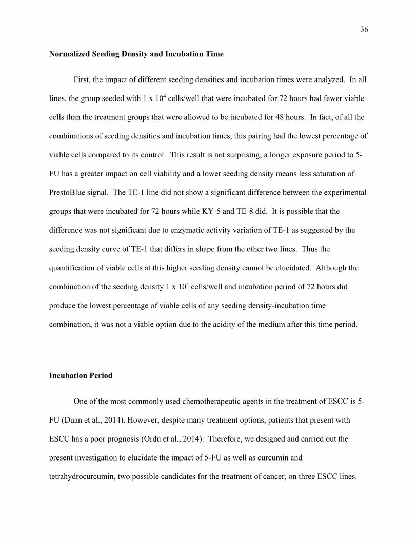
First, the impact of different seeding densities and incubation times were analyzed. In all
lines, the group seeded with 1 x 10⁴ cells/well that were incubated for 72 hours had fewer viable
cells than the treatment groups that were allowed to be incubated for 48 hours. In fact, of all the
combinations of seeding densities and incubation times, this pairing had the lowest percentage of
viable cells compared to its control. This result is not surprising; a longer exposure period to 5-
FU has a greater impact on cell viability and a lower seeding density means less saturation of
PrestoBlue signal. The TE-1 line did not show a significant difference between the experimental
groups that were incubated for 72 hours while KY-5 and TE-8 did. It is possible that the
difference was not significant due to enzymatic activity variation of TE-1 as suggested by the
seeding density curve of TE-1 that differs in shape from the other two lines. Thus the
quantification of viable cells at this higher seeding density cannot be elucidated. Although the
combination of the seeding density 1 x 10⁴ cells/well and incubation period of 72 hours did
produce the lowest percentage of viable cells of any seeding density-incubation time
combination, it was not a viable option due to the acidity of the medium after this time period.
Incubation Period
One of the most commonly used chemotherapeutic agents in the treatment of ESCC is 5-
FU (Duan et al., 2014). However, despite many treatment options, patients that present with
ESCC has a poor prognosis (Ordu et al., 2014). Therefore, we designed and carried out the
present investigation to elucidate the impact of 5-FU as well as curcumin and
tetrahydrocurcumin, two possible candidates for the treatment of cancer, on three ESCC lines.

37
Our first attempt to treat cells with 5-FU was done at thirty hours of incubation. This
time period was chosen based on previous published results in our lab (Almanaa et al., 2012). At
this time point, 5-FU did not have any impact on the cell viability or death. 5-FU did not have an
impact on the cells until they were incubated with 5-FU for a minimum of 48 hours. Thus, we
were able to show that curcumin is able to cause cell death at a shorter time period than 5-FU can
impede cell growth.
Curcumin is able to induce more cell death in a shorter time period than 5-FU. This is of
clinical importance because of the contrast in the toxicity of these two drugs. 5-FU is cytotoxic
to normal and cancerous cells alike whereas curcumin is only toxic to cancer cells. When
patients endure prolonged exposure to toxic doses of 5-FU they exhibit side effects such as
myelosuppression, mucositis, diarrhea, and cutaneous erythema (Diaz et al., 2003). In
comparison, a phase I clinical study that followed healthy volunteers that consumed turmeric oil
over three months found no side effects (Prasad & Aggarwal, 2011). The results of this study
show curcumin, a nontoxic herbal remedy, acts more quickly on these ESCC lines and should be
further examined for its applicability for cancer treatments.
5-fluorouracil Toxicity
5-FU is a standard chemotherapeutic agent for ESCC treatment and often times used in
conjunction with other drugs such as docetaxel and cisplatin. Although neoadjuvant therapy in
patients with EAC leads to a significantly better postoperative survival, it is not an approach that
is currently used for ESCC patients (Nakajima & Kato, 2013).

38
The effects of 5-FU were examined after the determination of the optimal incubation time
period of 48 hours. After 48 hours of treatment, the KY-5 and TE-8 cell lines showed
significantly fewer cells in the 5-FU treated group than that of the control. The cell lines KY-5
and TE-8 were nearly identical in their response to 5-FU. Under the same 5-FU treatment
conditions, TE-1 showed more viable cells than the other two lines. The rank order of stemness
from most to least stemlike is KY-5, TE-8, and TE-1; while the rank order of most to least viable
cells after 5-FU treatment is TE-1, TE-8, and KY-5. It appears that their stemness properties did
not indicate which cell line would be most susceptible to the effects of 5-FU. It is possible that
there are other factors in these lines that make them resistant to 5-FU. Such factors could include
an overexpression of the Notch-1 signaling pathway which has been shown to make ESCC cell
lines more resistant to the effects of 5-FU (Liu et al., 2013).
Tetrahydrocurcumin Toxicity
Tetrahydrocurcumin has gained attention in recent years. Some studies suggest that THC
could be a better anticancer agent than curcumin (Kang et al., 2014). There is a discrepancy
about whether THC is more or less active than curcumin itself (Anand et al., 2007). It was
therefore decided to include THC in the present study.
All three ESCC lines used in this study showed little impact from the THC treatment.
THC had marginal deleterious effects on viability in all three cell lines. Since the cell viability
of these treatment groups was similar to those of the controls, it can be concluded that THC is
not an effective treatment for these ESCC cell lines. Despite the fact that THC has more
antioxidant potential and better bioavailability than curcumin, it appears that curcumin may be a

39
better therapeutic agent in the treatment of ESCC. It is possible that THC is not effective against
ESCC because of the over expression of NF-κB in these lines which is not targeted by this drug
(Anand et al., 2008b).
This study indicates that the in vivo effects of THC seen in other studies cannot be
realized in vitro. Future in vitro studies involving THC and ESCC should elucidate whether
THC is able to prevent metastasis, invasion, and migration. THC was suggested to have the
ability to prevent metastasis by Wu et al. (2014) in their study of HT1080 cells that showed
lower levels of MMP-2 and MMP-9 in cells treated with THC. Based on the in vitro results of
this study, the growth of an ESCC tumor would probably not be affected by THC. However, an
in vivo study examining inhibition of colon carcinogenesis found a decrease in cancer cell
proliferation when mice were injected with THC. The same study claimed THC was better than
curcumin at inhibiting the development of aberrant crypt foci (Kim et al., 1998). The present
study was not designed to address this issue. When removed from the consequences of the rest
of the body, the effect on the ESCC cells in response to THC treatments were not observed.
Therefore, it seems unlikely that THC has a direct effect on these cells. While these results
could not be seen in vitro when these ESCC cell lines were treated with THC, an in vivo study
should be designed with ESCC and THC to further corroborate or refute these claims.
Curcumin Toxicity
Curcumin has therapeutic abilities against a variety of different cancer cell lines, and
thus, is a promising anticancer agent (Anand et al., 2008a). Although curcumin is effective in
inducing cell death in cancer cells, it is has not been examined in comparison to THC alone nor

40
in combination with 5-FU or THC. This study was performed to elucidate the abilities of these
drugs for treatments of ESCC.
After 48 hours of incubation, all three ESCC cell lines showed a significant difference
between their control and curcumin treated groups. The three lines were impacted as expected
based on their stemness, with KY-5 being the most stem-like and showing the most inhibitory
effects of curcumin and TE-1 showing the least stem-like characteristics and displaying the least
impacts by curcumin. The difference in stemness among these three lines could be responsible
for TE-1’s significantly different response to curcumin when compared to KY-5 and TE-8.
In fact, of the three drug treatments, the curcumin treatment groups had the lowest
number of viable cells across all three lines. In all three lines, there were significantly fewer
cells in the curcumin treatment group than the 5-FU groups. Therefore, not only does this
experiment support previous studies where curcumin was found to be effective against ESCC
cell lines, but it also indicates curcumin could be a better, more specific therapeutic agent for
ESCC than the other treatment options.
While the present rank order of susceptibility to the effects of curcumin matches with the
stemness sequence done by Almanaa (2013), it does not match her rank order of these cell lines
to the susceptibility to curcumin. This indicates that the stemness of the cell lines may only be
indicative of curcumin effects if the lines are exposed to curcumin and its cellular metabolites for
a longer period of time, 30 hours vs. 48 hours. Curcumin degrades quickly with a half-life of
about 186 minutes in medium (Saradhi et al., 2010). There have not been many studies done on
these curcumin degradation products in vivo, but one such study showed the presence of two
curcumin degradation products, dihydroferulic acid and ferulic acid, in the bile of rats that had

41
been orally administered curcumin (Shen & Ji, 2012). Based on this knowledge and the present
study, it is possible that curcumin’s degradation products continue to effectively inhibit the
growth of cancer cells. These degradation products might also better target CSCs than typical
chemotherapeutic agents thus explaining the difference in rank order after a longer period of
time. This hypothesis should be tested in future studies.
Combination toxicity
As a whole, there are no definitive synergistic nor antagonistic effects between any
pairing of the drugs; however, it is possible some of the drug pairings could have an additive
effect.
The more interesting curcumin results were found when curcumin was used in
combination with THC or 5-FU. These two combinations inhibited cell viability similarly to one
another across the cell lines. When curcumin was used with THC, there were insignificantly less
viable cells in the combination treatment group than there were in the curcumin alone. However,
the percentage of inhibited cell viability was greater than the sum of the inhibited cell viability of
the curcumin and THC treatment groups. More replicates and possible changes in concentrations
of THC would need to be done in order to determine if this is an additive or synergistic effect. A
significantly higher therapeutic dose of THC would work best to elucidate this. There are a
limited number of studies that compare these two drugs side by side and this is the first paper to
explore a possibility of a therapeutic combinational effect of these two curcuminoids. An in vivo
study would be ideal for studying the total effects on ESCC between these two drugs in order to

42
compare cellular proliferation, angiogenesis, and tumorogenesis while in the context of plasma
concentration of each drug.
When curcumin was combined with 5-FU, there were fewer surviving cells than in the
individual treatment groups. The TE-1 cell line responded significantly different from the TE-8
and KY-5 cell lines in this treatment group. The same significant differences among the cell
lines arose when curcumin was used alone, so the stemness of each line may have contributed to
these significant differences.
The combination effects of curcumin and 5-FU were neither additive nor synergistic.
These results imply that both drugs may have overlapping molecular targets in ESCC. Even
though this combination does not show additive or synergistic effects, these two drugs do not
have antagonistic impacts on one another. This is important when treating a patient with
recurrent or metastatic ESCC which is thought to be the result of CSCs. Curcumin could be
safely added to 5-FU treatment in order to better target this subpopulation of cancer cells. Future
studies should include other traditional chemotherapeutic agents with curcumin. Such studies
have been carried out in studying laryngeal cancer using curcumin and cisplatin. This study
showed enhanced apoptosis when the drugs were used in combination (Zhang et al., 2013).
The combination group receiving THC with 5-FU acted similarly within the cell lines.
All three cell lines showed no significant difference between the 5-FU alone and 5-FU with THC
treatment groups. This result is not surprising because THC alone caused a minimal change in
cell proliferation. However, there is not a significant difference between the 5-FU alone and
THC with 5-FU experimental groups in these ESCC lines. Future studies, similar in nature to the
studies previously proposed, would need to be done with varying dosages of 5-FU and THC in

43
order to determine if they have an additive or synergistic effects on the KY-5 and TE-1 cell lines.
It is clear that curcumin is a better antiproliferative agent than both THC and 5-FU against ESCC
at 40 µM, but it is yet to be discovered if curcumin can be used with other chemotherapeutic
agents in order to create a synergistic effect against this cancer.

44
CONCLUSION
The present study demonstrates that curcumin is the best anti-proliferation agent in ESCC
when compared with 5-FU and THC. By comparing three ESCC lines we showed different
responses to 5-FU and curcumin along with their combination with each other and THC. KY-5,
the most stem-like line, responded best to curcumin. This study showed that there is no
definitive synergism among these drugs; at best, their effects are additive. This study concludes
that at the same dose that makes curcumin effective, THC has no impact on these ESCC lines.
Thus, in the treatment of ESCC, a modified curcumin that increases its bioavailability may be the
most effective therapy. By incorporating plant-based drugs such as curcumin into treatment
regimens, a patient may experience fewer side effects of treatment, and thus a better quality of
life. A tumor that arises after curcumin treatment may be more easily treated because curcumin
is able to target CSCs better than traditional chemotherapy. This conclusion was drawn since the
CSCs that are believed to be more aggressive and metastatic would be fewer in number.

45
REFERENCES
Ahmad, M., Ghumman, S. A., Sher, A., Sharif, T., Sher, M., & Abbas, T. (2012). Effect of
skimmed milk on the absorption and metabolism of 5-fluorouracil (5-FU) in
animals. Iranian Journal of Pharmaceutical Research,11(1), 69-75.
Almanaa, T. N. (2013). Targeting cancer stem-like cells in human esophageal squamous cell
lines by curcumin. (Doctor of Philosophy, Bowling Green State University).
Almanaa, T. N., Guesz, M. E., & Jamasbi, R. J. (2012). Effects of curcumin on stem-like cells in
human esophageal squamous carcinoma cell lines. BMC Complementary and Alternative
Medicine, 12(195).
Almanaa, T. N., Guesz, M. E., & Jamasbi, R. J. (2013). A new method for identifying stem-like
cells in esophageal cancer cell lines. Journal of Cancer, 4(7), 536-548.
Altieri, F., Arcari, P., & Rippa, E. (2013). Gastric cancer: Molecular pathology state. In G.
Mozsik (Ed.), Current topics in gastritis. InTech.
American Cancer Society. (2014). Chemotherapy for cancer of the esophagus. Retrieved 10/5,
2014, from http://www.cancer.org/cancer/esophaguscancer/detailedguide/esophagus-cancer-
treating-chemotherapy
Anand, P., Kunnumakkara, A. B., Newman, R. A., & Aggarwal, B. B. (2007). Bioavailability of
curcumin: Problems and promises. Molecular Pharmaceutics, 4(6), 807-818.

46
Anand, P., Sundaram, C., Jhurani, S., Kunnumakkara, A. B., & Aggarwal, B. B. (2008a).
Curcumin and cancer: An "old-age" disease with an "age-old" solution. Cancer
Letters, 267(1), 133-164.
Anand, P., Thomas, S. G., Kunnumakkara, A. B., Sundaram, C., Harikumar, K. B., Sung, B., et
al. (2008b). Biological activities of curcumin and its analogues (congeners) made by man
and mother nature. Biochemical Pharmacology, 76, 1590-1611.
Baba, Y., Watanabe, M., Yoshida, N., & Baba, H. (2014). Neoadjuvant treatment for esophageal
squamous cell carcinoma. World Journal of Gastrointestinal Oncology, 6(5), 121-128.
Brown, L. M., Hoover, R., Silverman, D., Baris, D., Hayes, R., Swanson, M., et al. (2001).
Excess incidence of squamous cell esophageal cancer among US black men: Role of social
class and other risk factors. American Journal of Epidemiology, 153(2), 114-122.
Cao, Y., Xu, R. H., & Liu, Z. (2014). A high-throughput quantification method of curcuminoids
and curcumin metabolites in human plasma via high-performance liquid
chromatography/tadem mass spectrometry. Journal of Chromatography B, 949-950, 70-78.
Castell, D. O., Murray, J. A., Tutuian, R., Orland, R. C., & Arnold, R. (2004). Review article:
The pathophysiology of gastro-oesophageal reflux disease- oesophageal
manifestations. Alimentary Pharmacology and Therapeutics, 20(Suppl 9), 14-25.
Chen, X., & Yang, C. S. (2006). Esophageal adenocarcinoma: A review and perspectives on the
mechanism of carcinogenesis and chemoprevention. Oncogene, 25(2), 278-287.

47
de Bono, J. S., & Twelves, C. J. (2001). The oral fluorinated pyrimidines. Investigational New
Drugs, 19(1), 41-59.
Diaz, E. M. J., Sturgis, E. M., Laramore, G. E., Sabichi, A. L., Lippman, S. M., & Clayman, G.
(2003). Treatment. In D. W. Kufe, et al. (Eds.), Holland-frei cancer medicine (6th ed.).
Hamilton (ON): BC Decker.
Duan, X. F., Tang, P., & Yu, Z. T. (2014). Neoadjuvant chemotherapy for resectable esophageal
canger: An in-depth study of randomized controlled trials and literature review. Cancer
Biology and Medicine, 11(3), 191-201.
Efferth, T. (2012). Stem cells, cancer stem-like cells, and natural products. Planta
Medica, 78(10), 935-942.
Fabian, A., Vereb, G., & Szollosi, J. (2013). The hitchhikers guide to cancer stem cell theory:
Markers, pathways and therapy. Cytometry A, 83(1), 62-71.
Gabbert, H. E., Shimoda, T., Hainaut, P., Nakamura, Y., Field, J. K., & Inoue, H. (2000).
squamous cell carcinoma of the oesophagus. In S. R. Hamilton, & L. A. Aaltonen (Eds.),
World health organization classification of tumours. pathology and genetics
of tumours of the digestive system (pp. 10-19). Lyon: IARC Press.
Garg, A., & Aggarwal, B. B. (2002). Nuclear transformation factor-kappaB as a target for cancer
drug development. Leukemia, 16(6), 1053-1068.

48
Gupta, S. C., Patchva, S., & Aggarwal, B. B. (2013). Therapeutic roles of curcumin: Lessons
learned from clinical trials. The American Association of Pharmaceutical Scientists
Journal, 15(1), 195-218.
Hassainasab, A., Hashimoto, Y., Tomita-Yokotani, K., & Kobayashi, M. (2011). Discovery of
the curcumin metabolic pathway involving a unique enzyme in an intestinal
microorganism. Proceedings of the National Academy of Sciences of the United States of
America, 108(16), 6615-6620.
Hatcher, H., Planalp, R., Cho, J., Torti, F. M., & Torti, S. V. (2008). Curcumin: From ancient
medicine to current clinical trials. Cellular and Molecular Life Sciences, 65(11), 1631-1652.
Helson, L., Bolger, G., Majeed, M., Vcelar, B., Pucaj, K., & Matabudul, D. (2012). Infusion of
pharacokinetics of lipocurc (liposomal curcumin) and its metabolite tetrahydrocurcumin in
beagle dogs. Anticancer Research, 32, 4365-4370.
Jӓger, R., Lowery, R. P., Calvanese, A. V., Joy, J. M., Purpura, M., & Wilson, J. M. (2014).
Comparative absorption of curcumin formulations. Nutrition Journal, 13(11).
Kamangar, F., Chow, W. H., Abnet, C., & Dawsey, S. (2009). Environmental causes of
esophageal cancer. Gastroenterology Clinics of North America, 38(1), 27-57.
Kang, N., Wang, M. M., Wang, Y. H., Zhang, Z. N., Cao, H. R., Lv, Y. H., et al. (2014).
Tetrahydrocurcumin induces G2/M cell cycle arrest and apoptosis involving p38 MAPK
activation in human breast cancer cells. Food and Chemical Toxicology, 67, 193-200.

49
Kim, J. M., Araki, S., Kim, D. J., Park, C. B., Takasuka, N., Baba-Toriyama, H., et al. (1998).
Chemopreventative effects of carotenoids and curcumins on mouse colon carcinogenesis
after 1,2-dimethylhydrazine initiation.Carcinogenesis, 19(1), 81-85.
Kuwano, H., Kato, H., Miyazaki, T., Fukuchi, M., Masuda, N., Nakajima, M., et al. (2005).
genetic alterations in esophageal cancer. Surgery Today, 35(1), 7-18.
Lai, C. S., Wu, J. C., Yu, S. F., Badmaev, V., Nagabhushanam, K., Ho, C. T., et al. (2011).
Tetrahydrocurcumin is more effective than curcumin in preventing azoxymethane-induced
colon carcinogenesis. Molecular Food and Nutrition Research, 55, 1819-1828.
Li, Y., & Zhang, T. (2014). Targeting cancer stem cells by curcumin and clinical
applications. Cancer Letters, 346(2), 197-205.
Liu, J., Fan, H., Man, Y., Liang, D., Huang, R., Wang, J., et al. (2013). Notch1 is a 5-fluorouracil
resistant and poor survival marker in human esophagus squamous cell carcinomas. PLOS
One, 8(2), e56141.
Lobo, N. A., Shimono, Y., Qian, D., & Clarke, M. F. (2007). The biology of cancer stem
cells. Annual Review of Cell and Developmental Biology, 23, 675-699.
Longley, D. B., Harkin, D. P., & Johnston, P. G. (2003). 5-fluorouracil: Mechanisms of action
and clinical strategies. Nature Reviews Cancer, 3(5), 330-338.
Milano, G., Etienne, M. C., Renée, N., Thyss, A., Schneider, M., Ramaioli, A., et al. (1994).
Relationship between fluorouracil systemic exposure and tumor response and patient
survival. Journal of Clinical Oncology, 12(6), 1291-1295.

50
Mimeault, M., & Batra, S. K. (2011). Potential applications of curcumin and its novel synthetic
analogs and nanotechnology-based formulations in cancer prevention and therapy. Chinese
Medicine, 6(31).
Mohamed, A., El-Rayes, B., Khuri, F. R., & Saba, N. F. (2014). Targeted therapies in metastatic
esophageal cancer: Advances over the past decade. Critical Reviews in Oncology
Hematology, 91(2), 186-196.
Nakajima, M., & Kato, H. (2013). Treatment options for esophageal squamous cell
carcinoma. Expert Opinion on Pharmacotherapy, 14(10), 1345-1354.
National Center for Biotechnology Information. (2005a). PubChem BioAssay database; CID=
3385. Retrieved 9/22, 2014,
from http://pubchem.ncbi.nlm.nih.gov/summary/summary.cgi?cid=3385&loc=ec_rcs
National Center for Biotechnology Information. (2005b). PubChem BioAssay database;
CID=124072. Retrieved 8/29, 2014,
from http://pubchem.ncbi.nlm.nih.gov/summary/summary.cgi?cid=124072&loc=ec_rcs
Nishihira, T., Hashimoto, Y., Katayama, M., Mori, S., & Kuroki, T. (1993). Molecular and
cellular features of esophageal cancer cells. Journal of Cancer Research and Clinical
Oncology, 119(8), 441-449.
Okamoto, M., Fujiwara, M., Hori, M., Okada, K., Yazama, F., Konishi, H., et al. (2014). tRNA
modifying enzymes, NSUM2 and METTL1, determines sensitivity to 5-fluorouracil in
HeLa cells. PLOS Genetics, 10(9).

51
Ordu, A. D., Nieder, C., Geinitz, H., Kup, P. G., Deymann, L. F., Scherer, V., et al. (2014).
Radio(chemo)therapy for locally advanced squamouse cel lcarcinoma of the esophagus:
Long-term outcome. Strahlentherapie Und Onkologie, 191(2), 153-160.
Osawa, T., Sugiyama, Y., Inayoshi, M., & Kawakishi, S. (1995). Antioxidative activity of
tetrahydrocurcuminoids. Bioscience, Biotechnology and Biochemistry, 59(9), 1609-1612.
O'Sullivan-Coyne, G., O'Sullivan, G. C., O'Donovan, T. R., Piwocka, K., & McKenna, S. L.
(2009). Curcumin induces apoptosis-independent death in oesophageal cancer cells. British
Journal of Cancer, 101(9), 1586-1595.
Pari, L., & Murugan, P. (2004). Protective role of tetrahydrocurcumin against erythromycin
estolate-induced hepatotoxicity. Pharacological Research, 49(5), 481-486.
Prasad, S., Gupta, S. C., Tyagi, A., & Aggarwal, B. B. (2014). Curcumin, a component of golden
spice: From bedside to bench and back. Biotechnology Advances, 32(6), 1053-1064.
Ratain, M. J., & Plunkett, W. K. J. (2003). General mechanisms of drug action. In D. W. Kufe,
R. E. Pollock, R. R. Weichselbaum & et. al. (Eds.), Holland-frei cancer medicine (6th ed.).
BC Decker: Hamilton.
Reya, T., Morrison, S. J., Clarke, M. F., & Weissman, I. L. (2001). Stem cells, cancer, and
cancer stem cells. Nature, 414(6859), 105-111.
Saladin, K. S. (2015). The digestive system. In S. J. Sullivan, & C. A. Gan (Eds.), Anatomy and
physiology: The unity of form and function (7th ed., pp. 947-993). New York: McGraw Hill.

52
Saradhi, U. V. R. V., Ling, Y., Wang, J., Chiu, M., Schwartz, E. B., Fuchs, J. R., et al. (2010). A
liquid chromatography-tandem mass spectrometric method for quantification of
curcuminoids in cell medium and mouse plasma.Journal of Chromatography B, 878, 3045-
3051.
Sharma, R. A., Euden, S. A., Platton, S. L., Cooke, D. N., Shafayat, A., Hewitt, H. R., et al.
(2004). Phase I clinical trial of oral curcumin: Biomarkers of systemic activity and
compliance. Clinical Cancer Research, 10(20), 6847-6854.
Shehzad, A., Lee, J., & Lee, Y. S. (2013). Curcumin in various cancers. Biofactors, 39(1), 56-68.
Shen, L., & Ji, H. F. (2012). The pharmacology of curcumin: Is it the degredation
products? Trends in Molecular Medicine, 18(3), 138-144.
Shimada, Y., Imamura, M., Wagata, T., Yamaguchi, N., & Tobe, T. (1992). Characterization of
21 newly established esophageal cancer cell lines. Cancer, 69(2), 277-284.
Shoba, G., Joy, D., Joseph, T., Majeed, M., Rajendran, R., & Srinivas, P. S. (1998). Influence of
piperine on the pharmacokinetics of curcumin in animals and human volunteers. Planta
Medica, 64(4), 353-356.
Stoner, G. D., & Gupta, A. (2001). Etiology and chemoprevention of esophageal squamous cell
carcinoma. Carcinogenesis, 22(11), 1737-1746.
Strimpakos, A. S., & Sharma, R. A. (2008). Curcumin: Preventative and therapeutic properties in
the laboratory studies and clinical trials. Antioxidants and Redox Signaling, 10(3), 511-545.

53
Sun, S. H., Huang, H. C., Huang, C., & Lin, J. K. (2012). Cycle arrest and apoptosis in MDA-
MB/Her2 cells induced by curcumin. European Journal of Pharmacology, 690(1-3), 22-30.
Taniere, P., Martel-Planche, G., Maurici, D., Lombard-Bohas, C., Scoazec, J. Y., Montesano, R.,
et al. (2001). Molecular and clinical differences between adenocarcinomas of the esophagus
and of the gastric cardia. The American Journal of Pathology, 158(1), 33-40.
Teiten, M., Dicato, M., & Diederich, M. (2013). Curcumin as a regulator of epigenetic
events. Molecular Food and Nutrition Research, 57, 1619-1629.
Thomson, A. B. R., & Shaffer, E. A. (Eds.). (2012). First principles of gastroenterology and
hepatology (6th ed.). Canada: CreateSpace Independent Publishing Platform.
Wu, J. C., Tsai, M. L., Lai, C. S., Wang, Y. J., Ho, C. T., & Pan, M. H. (2014).
Chemopreventative effects of tetrahydrocurcumin on human diseases. Food and
Function, 5(1), 12-17.
Yano, S., Li, S., Qinghong, H., Tan, Y., Bouvet, M., Fujiwara, T., et al. (2014). Selective
methioninase-induced trap of cancer cells in S/G2 phase visualized by FUCCI imaging
confers chemosensitivity. Oncotarget, 5(18), 8729-8736.
Yodkeeree, S., Chaiwangyen, W., Garbisa, S., & Limtrakul, P. (2007). Curcumin,
demethoxycurcumin, and bisdemethoxycurcumin differentially inhibit cancer cell invasion
through the down-regulation of MMPs and uPA.Journal of Nutritional Biochemistry, 20,
87-95.

54
Yoysungmoen, P., Wirachwong, P., Changtam, C., Suksamrarn, A., & Patumraj, S. (2008). Anti-
cancer and anti-angiogenic effects of curcumin and tetrahydrocurcumin on implanted
hepatocellular carcinoma in nude mice.World Journal of Gastroenterology, 14(13), 2003-
2009.
Zhang, H., Yu, T., Wen, L., Wang, H., Fei, D., & Jin, C. (2013). Curcumin enhances the
effectiveness of cisplatin by suppressing CD133+ cancer stem cells in laryngeal carcinoma
treatment. Experimental and Therapeutic Medicine, 6, 1317-1321.
Copyright © 2022 FDOKUMEN