THE EFFECT OF WONDERFUL COLA ON LIVER HISTOLOGY IN ALLOXAN INDUCED DIABETIC WISTAR RATS
Upload
independentCategory
view
5download
0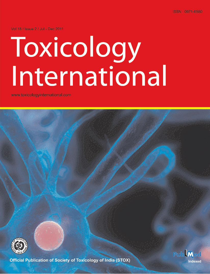

Toxicology International Jul-Dec 2011 / Vol-18 / Issue-2 124
Hepatoprotective Effect of Curcumin on Lindane-induced Oxidative Stress in Male Wistar Rats
Rambir Singh, Poonam Sharma1
Departments of Biomedical Sciences and 1Zoology, Bundelkhand University, Jhansi, Uttar Pradesh, India
ABSTRACT
Lindane, an organochlorine pesticide, is recognized as a major public health concern because of its potential toxic effects on human health. Its persistence in the body fluids may lead to continuous blood circulation, liver exposure and hepatotoxicity. The present study was undertaken to evaluate the possible protective role of curcumin on lindane-induced hepatotoxicity. Forty-two healthy adult male Wistar rats were divided into seven groups of six rats each. Group I was given dimethylsulfoxide. A single dose of lindane (60 mg/kg bw) was given to group II. Lindane (30 mg/kg bw) was given daily to group III for 14 days. Treatment with curcumin (100 and 200 mg/kg) was given to groups IV and V before (pretreatment) and to groups VI and VII after (post-treatment) 14 days exposure of lindane. Oxidative stress parameters and antioxidative enzymes were investigated in the liver of exposed and treated rats. A significant increase in lipid peroxidation, and decrease in glutathione level, Superoxide dismutase catalase, glutathione-S-transferase, glutathione peroxidase, glutathione reductase and NADPH quinine reductase activities was observed in liver of rats exposed to lindane. Curcumin (Pre- and post-treatment) nearly normalized all these parameters. Histological alterations were also observed in the liver tissue after lindane exposure. Treatment with curcumin significantly prevented the lindane-induced histological alterations. In conclusion, curcumin has protective effect over lindane-induced oxidative damage in rat liver.
Key words: Curcumin, hepatotoxicity, lindane, Wistar rat
INTRODUCTION
Lindane is an organochlorine pesticide that persists in the environment, bioaccumulates through food chain and has a risk of causing adverse effects on human health and environment. Although banned currently, lindane had been widely used to control arthropod pests on food crops, timber, farm animals, and humans. It was earlier used as
Original Article
an ingredient of shampoos for treatment of scabies and head lice in humans. According to an estimate, around 4.8 million tons of lindane is still present in the atmosphere as residue.[1] Lindane is absorbed through respiratory, digestive or cutaneous routes and accumulates in fatty tissues in the body. It is reported to damage human liver, kidney, neural and immune systems, and induces birth defects, cancer and death. Lindane exposure is reported to cause neurotoxicity,[2] reproductive toxicity[3] and hepatotoxicity.[4] Toxic chemicals like lindane largely disturb the xenobiotic metabolizing system of hepatotocytes. Acute lindane intoxication causes altered gene expression in rat liver.[5,6] There are reports that certain exogenous antioxidants strengthen the cellular xenobiotic metabolizing system, thereby protecting cells from oxidative stress.[7] In search for these new chemical entities as modulators of xenobiotic metabolism, we reviewed literature on Ayurvedic medicinal
Address for correspondence: Dr. Poonam Sharma, Department of Zoology, Institute of Basic Sciences, Bundelkhand University, Jhansi, Uttar Pradesh, India. E-mail: [email protected]
Access this article onlineQuick Response Code: Website:
www.toxicologyinternational.com
DOI:
10.4103/0971-6580.84264
Toxicology International Jul-Dec 2011 / Vol-18 / Issue-2125
plants. The use of several medicinal plants or their active principle for reducing the toxicity of xenobiotics is reported in traditional system of medicine from different ethnic societies. Ameliorating effect of ajwain[7] and ginger[8] is reported in lindane-induced toxicity. Curcumin, the main coloring agent of turmeric (Curcuma longa) rhizome, used as a spice in India,[9] possesses a number of pharmacological activities.[10] The antioxidant and free radical scavenging activity of curcumin has been reported by Ak and Gu¨lcin.[11] Recently, curcumin has been reported to ameliorate cypermethrin-induced oxidative stress in liver, kidney and brain of Wistar rats.[12] Curcumin is considered nontoxic in animal and human studies.[13,14]
Curcumin has been granted an acceptable daily intake of up to 3 mg/kg based on genotoxic studies in F1 and F2 generations of Wistar rats, with parental intake of 250–320 mg/kg bw as no observed adverse effect level (NOAEL) by the joint FAO and WHO expert committee on food additives, 1996.[15]
MATERIALS AND METHODS
AnimalsMale rats of Wistar strain, weighing about 150–200 g, were used in this experiment. Animals were obtained from Central Drug Research Institute (CSIR), Lucknow, and were kept for acclimatization in the animal house at an ambient temperature of 25°C and a relative humidity of 45–55%, with 12 hours each of dark and light cycles. Animals were fed pelleted diet and water ad libitum. Principles of Laboratory Animal Care (NIH publication no. 85-23, revised 1985) was followed for animal care during the course of experimentation. Approval for all animal experiments was obtained from the Institutional Animal Ethical Committee.
ChemicalsLindane and curcumin were purchased from Sigma Aldrich (St. Louis, MO, USA). All other chemicals required for biochemical and histopathologic estimations were of high purity and purchased locally. Curcumin and lindane were dissolved in dimethylsulfoxide (DMSO) and given orally by gavaging.[16]
Treatment scheduleThe animals were divided into the following groups during the course of study.Group I Vehicular control Normal diet and DMSO for 14 daysGroup II Acute exposure Lindane 60 mg/kg bw for 24 hoursGroup III Sub-acute exposure Lindane 30 mg/kg bw for 14 days
Singh and Sharma: Hepatoprotective role of curcumin in lindane induced toxicity
Group IV Pretreatment A Curcumin 100 mg/kg bw for 14 days followed by lindane 30 mg/kg bw for 14 daysGroup V Pretreatment B Curcumin 200 mg/kg bw for 14 days followed by lindane 30 mg/kg bw for 14 days Group VI Post-treatment A Lindane 30 mg/kg bw for 14 days followed by curcumin 100 mg/kg bw for 14 daysGroup VII Post-treatment B Lindane 30 mg/kg bw for 14 days followed by curcumin 200 mg/kg bw for 14 days
At the end of the study, animals were sacrificed, liver was dissected out and washed with 0.9% NaCl and stored at −40°C until use. Part of liver was used for biochemical estimation and part was used for histological examination.
Tissue homogenate preparationThe liver tissues were homogenized in 10% (w/v) ice-cold 0.1 M phosphate buffered saline (PBS; pH 7.4). A part of the crude homogenate was used for the estimation of lipid peroxidation (LPO) and reduced glutathione (GSH) and the rest of the homogenate was centrifuged at 12,000 rpm for 20 min to obtain the supernatant (S) and this supernatant was used for other enzymatic estimations.
Lipid peroxidationLPO was estimated as described by Ohkawa et al[17] One milliliter of 10% homogenate was incubated at 37°C for 10 min. One milliliter of 10% chilled trichloroacetic acid (TCA) (w/v) was added to it and centrifuged at 2500 rpm for 15 min at room temperature. 1 ml of 0.67% thiobarbituric acid (TBA) was added to 1 ml of supernatant and kept in boiling water bath for 10–15 min and cooled under tap water. After cooling, 1 ml of distilled water was added to it and absorbance was measured at 530 nm. The results were expressed as nmoles MDA/hour/g tissue.
Reduced glutathioneGSH was estimated by the method described by Ellman.[18] One milliliter of 10% of liver homogenate was taken and 1 ml of 5% TCA (w/v) was added. After 30 min, the mixture was centrifuged at 2500 rpm for 15 min. Also, 0.5 ml of supernatant was taken and 2.5 ml of 5,5’dithiobis (2-nitobanzoic acid)(DTNB) was added, mixed thoroughly and absorbance was recorded at 412 nm. The results were expressed as μmols/g tissue.
Superoxide dismutaseSOD was estimated by the method described by Misra and Fridovich.[19] 0.25 ml of ice-cold chloroform was added to 0.1 ml of supernatant (S), followed by addition of 0.15 ml of ice-cold ethanol. The mixture was centrifuged at

Toxicology International Jul-Dec 2011 / Vol-18 / Issue-2 126
Glutathione reductaseGR was estimated by the method given by Carlberg and Mannervik.[23] The reaction mixture consisting of 2.5 ml buffer, 0.2 ml NADPH, 0.2 ml glutathione disulfide (GSSG) and 0.1 ml supernatant was allowed to stand for 30 sec and absorbance was recorded at 340 nm for 3 min at 30 sec intervals. GR activity was represented in terms of nmol/min/mg protein.
NADPH quinone reductaseQR was estimated by the method of Benson et al.[24] OD was measured at 600 nm for 3 min at 30 sec intervals and NADPH QR activity was represented as nmol/min/mg protein.
Liver histologyA part of the liver was fixed in a 10% neutral buffered formalin solution and embedded in paraffin for cutting sections for histological examinations.
Statistical analysis Mean and standard error values were determined for all the parameters and the results were expressed as a Mean±SEM. The data were analyzed employing analysis of variance (ANOVA) using statistical software Graph Pad In Stat Software Inc., v. 3.06 (San Diego, CA USA). Dunnett test for multiple comparisons of groups against control was performed to determine the significant differences among the groups.
RESULTS
Single dose toxicityA significant increase (P<0.05) in the oxidative stress status, characterized by an increase in thiobarbituric acid reactive species (TBARS) was observed [Table 1]. A nonsignificant (P>0.05) decrease in activity of SOD, GST, GPx, and QR, and a significant reduction in CAT, GSH, and GR activity were observed after single dose exposure of lindane [Table 1]. Histological changes revealed the incidences
Singh and Sharma: Hepatoprotective role of curcumin in lindane induced toxicity
3000 rpm for 10 min at 4°C. 0.2 ml supernatant was taken and 1.3 ml buffer, 0.5 ml ethylenediaminetetraacetic acid (EDTA) and 0.8 ml water were added. Reaction was started by adding 0.2 ml epinephrine. Change in absorbance, ∆OD/min, at 480 nm was read for 3 min. The results were expressed as nmol/min/mg protein.
CatalaseCAT was estimated by the method described by Sinha.[20] One milliliter of phosphate buffer and 0.4 ml of water were added to 0.1 ml of supernatant (S). Reaction was started by adding 0.5 ml H2O2 and the mixture was incubated at 37°C for 1 min. Reaction was stopped by adding 2 ml of dichromate and acetic acid reagent and kept in boiling water bath for 15 min. The mixture was cooled and absorbance was read at 570 nm. CAT activity was calculated in terms of μmols/min/mg protein.
Glutathione-S-transferaseGST was estimated by the method of Habig et al.[21] The reaction mixture consisting of 1.425 ml phosphate buffer (0.1 M, pH 6.5), 0.2 ml reduced glutathione (1.0 mM), 0.25 ml of 1-chloro-2,4-dinitrobenzene (CDNB) (1 mM) and 0.30 ml post mitochondrial supernatant PMS was mixed to give a total volume of 2.0 ml. Absorbance was recorded at 340 nm and the enzyme activity was calculated as nanomoles of CDNB conjugate formed per minute per milligram protein, using a molar extinction coefficient of 9.6 × 10³/M/cm.
Glutathione peroxidaseGPx was estimated by the method of Rotruck et al.[22] The reaction mixture consisting of 0.4 ml tris HCl buffer, 0.2 ml GSH, 0.1 ml sodium azide, 0.1 ml water, 0.1 ml H2O2 and 0.1 ml homogenate was incubated at 37ºC for 15 min, 0.5 ml TCA was added and centrifuged. 0.5 ml of supernatant was taken and 2 ml Na2HPO4.2H2O and 0.5 ml Ellman’s Reagent were added. Absorbance was read at 420 nm. The results were expressed as μmol/min/mg protein.
Table 1: Effect of lindane and curcumin on LPO, GSH and antioxidant enzyme estimations in different groups in liver of Wistar ratsParameter Control G-I Single exposure
lindane (60 mg/kg bw)G-II
Exposure lindane (30 mg/kg bw)
G-III
Pretreatment A (100 mg/kg)
G-IV
Pretreatment B (200 mg/kg) G-V
Post-treatment A (100 mg/kg) G-VI
Post-treatment B (200 mg/kg) G-VII
LPO 4.234±0.023 5.356±0.144* 6.896±0.440)** 5.031±0.511• 4.688±0.871• 5.235±0.724• 4.946±0.275•
GSH 2.111±0.229 1.563±0.127* 1.304±0.083** 1.808±0.039• 2.039±0.194• 1.651±0.050• 1.918±0.182•
SOD 68.392±5.662 63.627±6.063• 36.125±0.827* 65.542±3.597• 67.365±12.086• 64.727±5.573• 66.716±13.009•
CAT 29.602±1.206 24.732±1.663* 16.619±0.528** 26.842±0.029• 28.676±2.628• 26.573±6.837• 27.735±2.363•
GST 661.85±58.856 656.958±58.771• 407.09±40.646* 534.82±19.484• 614.77±85.184• 521.659±22.194• 570.22±69.585•
GPx 22.894±1.964 18.349±1.669• 14.469±0.581* 20.139±0. 414• 21.905±1.347• 19.911±4.311• 21.092±0.620•
GR 38.306±4.397 28.40±0.494* 22.399±0.660** 34.582±0. 907• 36.205±2.312• 33.397±7.499• 35.275±2.630•
NADPH QR 81.986±11.711 77.400±11.865• 47.713±4.056* 74.058±0.824• 78.702±15.391• 73.241±3.049• 75.050±2.912•
The values represent the Mean±SEM; **P<0.01; *P<0.05;•P>0.05, as compared with control values
Toxicology International Jul-Dec 2011 / Vol-18 / Issue-2127
anion. It is the major antioxidative enzyme in the liver. This enzyme detoxifies the superoxide anion, thus converting it into H2O2 and water. It is a metalloprotein widely distributed in all cells and plays an important role against oxidative damage induced by ROS. Reduced SOD level may be due to generation of more superoxide anion.
Singh and Sharma: Hepatoprotective role of curcumin in lindane induced toxicity
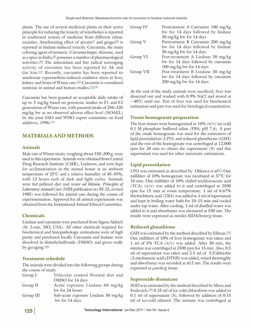
of marked necrosis in centrilobular area and enlargement of hepatocytes’ cell size [Figure 1] as compared to liver histology of control [Figure 2]
Sub-acute toxicityLPO level in liver was significantly (P<0.01) increased in group III as compared to group I [Table 1]. Lindane significantly (P<0.01) depleted liver GSH level in groups II and III as compared to control group I [Table 1]. The activities of SOD, CAT, GST, GPx, GR and NADPH QR were significantly (P<0.05) reduced in group III, as compared to control group I [Table 1]. CAT and GR were significantly (P<0.01) reduced in group III, while a significant (P<0.05) decrease in GST, GPx and NADPH QR was found in group III. In pre- and post-curcumin treatment groups (groups IV, V, VI and VII), changes in LPO, GSH level and SOD, CAT, GST, GPx, GR and NADPH QR enzyme activity were nonsignificant (P>0.05) as compared to control.
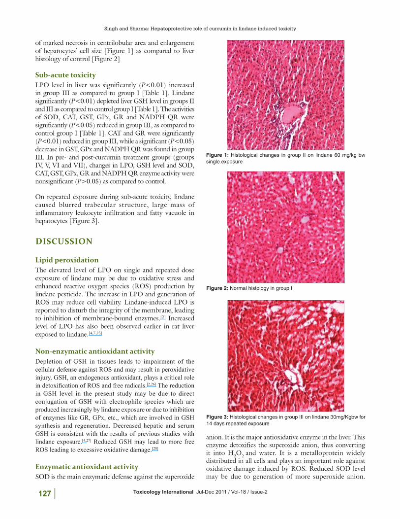
On repeated exposure during sub-acute toxicity, lindane caused blurred trabecular structure, large mass of inflammatory leukocyte infiltration and fatty vacuole in hepatocytes [Figure 3].
DISCUSSION
Lipid peroxidationThe elevated level of LPO on single and repeated dose exposure of lindane may be due to oxidative stress and enhanced reactive oxygen species (ROS) production by lindane pesticide. The increase in LPO and generation of ROS may reduce cell viability. Lindane-induced LPO is reported to disturb the integrity of the membrane, leading to inhibition of membrane-bound enzymes.[2] Increased level of LPO has also been observed earlier in rat liver exposed to lindane.[4,7,25]
Non-enzymatic antioxidant activityDepletion of GSH in tissues leads to impairment of the cellular defense against ROS and may result in peroxidative injury. GSH, an endogenous antioxidant, plays a critical role in detoxification of ROS and free radicals.[2,26] The reduction in GSH level in the present study may be due to direct conjugation of GSH with electrophile species which are produced increasingly by lindane exposure or due to inhibition of enzymes like GR, GPx, etc., which are involved in GSH synthesis and regeneration. Decreased hepatic and serum GSH is consistent with the results of previous studies with lindane exposure.[4,27] Reduced GSH may lead to more free ROS leading to excessive oxidative damage.[28]
Enzymatic antioxidant activitySOD is the main enzymatic defense against the superoxide
Figure 1: Histological changes in group II on lindane 60 mg/kg bw single exposure
Figure 3: Histological changes in group III on lindane 30mg/Kgbw for 14 days repeated exposure
Figure 2: Normal histology in group I

Toxicology International Jul-Dec 2011 / Vol-18 / Issue-2 128
Singh and Sharma: Hepatoprotective role of curcumin in lindane induced toxicity
Decrease in SOD activity will favor an increase in the production of ROS, which in turn will inactivate CAT.[29] Similar results were shown earlier in rat liver[4] exposed to lindane. GPx is the enzyme which protects the cells from oxidative damage. GPx is an important GSH using enzyme and plays an important role in maintaining GSH homeostasis and tissue detoxification. Reduced GPx level might be due to the depleted level of GSH because GPx uses GSH as a substrate. GST catalyzes the conjugation of GSH via a sulfhydryl group to electrophilic centers on a wide variety of substrates. This activity detoxifies endogenous compounds such as peroxidized lipids, as well as breakdown products of xenobiotics. GST may also bind to toxins and function as transport proteins. The decrease in GST activity might be responsible for lindane accumulation in the liver of rat, resulting in neurotoxicity and hepatotoxicity. GR is an enzyme that reduces GSSG (oxidized glutathione) to the sulfhydryl form of GSH, which is an important cellular antioxidant. In the present investigation, we observed decrease in GR as well as GSH level. QR activity was decreased in rats exposed to lindane. Reduction in QR activity in the kidney of rats exposed to ferric nitrilotriacetic acid has already been reported.[30]
Histological studiesHistological changes revealed the incidences of marked necrosis in centrilobular area and enlargement of hepatocytes a f ter s ing le exposure of l indane [Figure 2], as compared to liver histology of control animals [Figure 1]. The histological disturbances may be due to excessive accumulation of lindane in the liver.
On repeated exposure during sub-acute toxicity, lindane also caused blurred trabecular structure, large mass of inflammatory leukocyte infiltration and fatty vacuole in hepatocytes [Figure 3]. The repeated dose exposure causes excessive accumulation of lindane and disturbing drug metabolizing system of hepatocytes, leading to further damage evident from marked histological disturbances compared to control as well as single dose exposure.
Role of curcuminIn pre- and post-treatment groups (groups IV, V, VI and VII), we studied the role of curcumin against lindane in the liver of Wistar rats. Curcumin showed protective role by decreasing the LPO level in animals exposed to lindane.
Figure 7: Histological changes in group VII on lindane exposure and 200mg/kgbw curcumin posttreatment
Figure 4: Histological changes in group IV on lindane exposure and 100mg/kgbw curcumin pretreatment
Figure 5: Histological changes in group V on lindane exposure and 200mg/kgbw curcumin pretreatment
Figure 6: Histological changes in group VI on lindane exposure and 100mg/kgbw curcumin posttreatment
Toxicology International Jul-Dec 2011 / Vol-18 / Issue-2129
Singh and Sharma: Hepatoprotective role of curcumin in lindane induced toxicity
Curcumin worked as an antioxidant and increased the levels of non-enzymatic antioxidant GSH, enzymatic antioxidants CAT, SOD, GPx, GST, GR and QR in animals exposed to lindane [Table 1]. Curcumin reduced oxidative stress in the test animals by scavenging ROS, protecting the antioxidant enzymes from being denatured and reducing the oxidative stress marker LPO. The protective effect of curcumin was dose dependent and 200 mg/kg bw dose produced better ameliorating effect as compared to 100 mg/kg bw. Pre- and post-treatment of curcumin showed restoration of normal arrangement of hepatocytes or improvement of hepatic tissues [Figures 4–7].
REFERENCES
1. Shen L, Wania F, Lei YD, Teixeira C, Muir DC, Bidleman TF. Hexachlorocyclohexanes in the North American atmosphere. Environ Sci Technol 2004;38:965-75.
2. Bano M, Bhatt DK. Ameliorative effect of a combination of vitamin-E, Vitamin-C α-lipoic acid and stilbene resveratrol on lindane induced toxicity in mice olfactory lobe and cerebrum. Indian J Exp Biol 2010;48:150-8.
3. Sahoo DK, Roy A, Chainy GB. Protective effect of vitamin E and curcumin on L- thyroxine-induced rat testicular oxidative stress. Chem Biol Interact 2008;176:121-8.
4. Vijaya Padma V, Sowmya P, Arun FT, Baskaran R, Poornima P. Protective effect of gallic acid against lindane induced toxicity in experimental rats. Food Chem Toxicol 2011;49:991-8.
5. Sumida K, Saito K, Oeda K, Yakabe Y, Otsuka M, Matsumoto H, et al. A comparative study of gene expression profiles in rat liver after administration of alpha-hexachlorocyclohexane and lindane. J Toxicol Sci.2007;32:261-88.
6. Videla LA, Tapia G, Varela P, Cornejo P, Guerrero J, Israel Y, et al. Effects of acute gamma-hexachlorocyclohexane intoxication in relation to the redox regulation of nuclear factor-kappaB, cytokine gene expression, and liver injury in the rat. Antioxid Redox Signal 2004;6:471-80.
7. Anilakumar KR, Saritha V, Khanum F, Bawa AS. Ameliorative effect of ajwain extract on hexachlorocyclohexane-induced lipid peroxidation in rat liver. Food Chem Toxicol 2009;47:279-82.
8. Ahmed RS, Suke SG, Seth V, Chakraborti A, Tripathi AK, Banerjee BD. Protective effects of dietary ginger (Zingiber officinales Rosc.) on lindane-induced oxidative stress in rats. Phytother Res 2008;22:902-6.
9. Padmaja, S, Raju, TN. Antioxidant effect of curcumin in selenium induced cataract of Wistar rats. Indian J Exp Biol 2004;42:601-3.
10. Luthra PM, Singh R, Chandra R Therapeutic Uses of Curcuma longa. Ind J Clin Biochem 2001;16:153-60.
11. Ak T, Gülçin I. Antioxidant and radical scavenging properties of curcumin. Chem Biol interact 2008;174:27-37.
12. Sankar P, Telang AG, Manimaran A. Protective effect of curcumin on cypermethrin-induced oxidative stress in Wistar rats. Exp Toxicol Pathol 2010.
13. Qureshi S, Shah AH, Ageel AM. Toxicity studies on Alpinia galangal and Curcuma longa. Planta Med 1992;58:124-7.
14. Lao CD, Ruffin MT 4th, Normolle D, Heath DD, Murray SI, Bailey JM, et al. Dose escalation of a curcuminoid formulation. BMC Complement Altern Med 2006;6:10-3.
15. NCI. Clinical development plan: curcumin. J Cell Biochem Suppl 1996;26:72-85.
16. Mladenović D, Hrncić D, Vucević D, Radosavljević T, Loncar-Stevanović H, Petrović J, et al. Ethanol suppressed seizures in lindane-treated rats Electroencephalographic and behavioral studies. J Physio Pharmacol 2007;58:641-56.
17. Ohkawa H, Ohishi Ν, Yagi Κ. Assay for lipid peroxides in animal tissues by thiobarbituric acid reaction. Anal Biochem 1979;95:351-8.
18. Ellman GL, Tissue sulphydryl groups. Arch Biochem Biophys 1959;82:70-7.
19. Misra HP, Fridovich I. The role of superoxide anion in the autoxidation of epinephrine and a simple assay for superoxide dismutase. J Biol Chem 1972;247:3170-5.
20. Sinha AK. Colorimetric assay of catalane. Anal Biochem 1972;47:389-94.
21. Habig WJ, Pabst MJ, Jakoby WB. Glutathion-s-transferase:The first enzymatic step in mercapturic acid formation. J Biol Chem 1974;249:7130-9.
22. Rotruck JT, Pope AL, Ganther HE, Hafeman DG, Hoekstra WG. Selenium: Biochemical role as a component of glutathione peroxidase. Science 1973;179:588-90.
23. Carlberg I, Mannervik B. Glutathione reductase. Methods Enzymol 1985;113:484-90.
24. Benson AM, Hunkeler MJ, Talaly P. Increase of NAD(P)H quinone reductase by dietary antioxidants: possible role in protection against carcinogenesis and toxicity. Proc Natl Acad Sci U S A 1980;77:5216-20.
25. Radosavljević T, Mladenović D, Jakovljević V, Vucvić D, Rasć-Marković A, Hrncić D, et al. Oxidative stress in liver and red blood cells in acute lindane toxicity in rats. Hum Exp Toxicol 2009;28:747-57.
26. Mediratta PK, Tanwar K, Reeta KH, Mathur R, Benerjee BD, Singh S, et al. Attenuation of the effect of lindane on immune responses and oxidative stress by ocimum sancatum seed oil (OSSO) in rats. Indian J Physiol Pharmocol 2008;52:171-7.
27. Ahmed RS, Suke SG, Seth V, Chakraborti A, Tripathi AK, Banerjee BD. Protective effects of dietary ginger (Zingiber officinales Rosc) on lindane induced oxidative stress in rats. Phytother Res 2008;22:902-6.
28. Videla LA, Simizu K, Barros SB, Junqueira VB. Mechanisms of lindane-induced hepatotoxicity: alterations of respiratory activity and sinusoidal glutathione efflux in the isolated perfused rat liver. Xenobiotica 1991;21:1023-32.
29. Kono Y, Fridovich I. Superoxide radical inhibits catalase. J Biol Chem 1982;257:5751-4.
30. Sultana S, Prasad L, Jahangir T. Luteolin ameliorates ferric nitrilotriacetic acid induced renal toxicity and tumor promotional response in rats. Indian J Exp Biol 2009;47:355-60.
How to cite this article: Singh R, Sharma P. Hepatoprotective effect of curcumin on lindane-induced oxidative stress in male wistar rats. Toxicol Int 2011;18:124-9.Source of Support: Nil. Conflict of Interest: None declared.
Copyright © 2022 FDOKUMEN