ANTIDIABETIC, ANTIATHEROSCLEROTIC AND HEPATOPROTECTIVE PROPERTIES OF DECALEPIS HAMILTONII IN...
18
ANTIDIABETIC, ANTIATHEROSCLEROTIC AND HEPATOPROTECTIVE PROPERTIES OF DECALEPIS HAMILTONII IN STREPTOZOTOCIN-INDUCED DIABETIC RATS S. NAVEEN and F. KHANUM 1 Department of Biochemistry and Nutrition Defence Food Research Laboratory Mysore-570011. Karnataka, India Accepted for Publication February 15, 2009 ABSTRACT Decalepis root is consumed as ingredient in pickles and is a common ingredient in many of the ayurvedic preparation. Streptozotocin (STZ) is a potential source of oxidative stress that induces diabetes. In the present study, we have investigated the effect of Decalepis root extract (DRE) on antioxidant defence system in the body and other diabetic complications in rats. Rats were prefed with DRE (0.05 and 0.1%) and then injected with STZ (50 mg/kg bodyweight, i.p.). The results demonstrated that STZ injection lead to increased oxidation of tissue lipids apart from diabetic changes, decreased activities of the antioxidant enzymes causing liver and DNA damage. In rats, which were prefed with DRE, peroxidation of lipids was prevented to a sig- nificant level (heart 19%, kidney 42% and liver 88%). DRE protected the rats against STZ induced oxidative stress, reduced the risk of oxidative stress, DNA damage and ameliorated liver damage and diabetic condition. PRACTICAL APPLICATIONS The results obtained in this study clearly demonstrated that Decalepis root extract is plant-derived and nontoxic, it could be introduced in the cancer therapy regime in combination with conventional therapeutic agents, after some more detailed investigation. This extract could be used not only as food preservative (to replace for the toxic butylated hydroxyanisole and butylated hydroxytoluene currently under use) but also can be used in the preparation of nutraceutical and pharmaceutical products. Reactive oxygen species-mediated oxidative stress is now regarded as a major factor leading to degenerative 1 Correspondence author. TEL: +91-821-2470364; FAX: +91-821-2473468; EMAIL: [email protected] DOI: 10.1111/j.1745-4514.2010.00361.x Journal of Food Biochemistry 34 (2010) 1231–1248. © 2010, Wiley Periodicals, Inc. 1231
Transcript of ANTIDIABETIC, ANTIATHEROSCLEROTIC AND HEPATOPROTECTIVE PROPERTIES OF DECALEPIS HAMILTONII IN...
jfbc_361 1231..1248
ANTIDIABETIC, ANTIATHEROSCLEROTIC ANDHEPATOPROTECTIVE PROPERTIES OF DECALEPIS
HAMILTONII IN STREPTOZOTOCIN-INDUCED DIABETIC RATS
S. NAVEEN and F. KHANUM1
Department of Biochemistry and NutritionDefence Food Research LaboratoryMysore-570011. Karnataka, India
Accepted for Publication February 15, 2009
ABSTRACT
Decalepis root is consumed as ingredient in pickles and is a commoningredient in many of the ayurvedic preparation. Streptozotocin (STZ) is apotential source of oxidative stress that induces diabetes. In the present study,we have investigated the effect of Decalepis root extract (DRE) on antioxidantdefence system in the body and other diabetic complications in rats. Rats wereprefed with DRE (0.05 and 0.1%) and then injected with STZ (50 mg/kgbodyweight, i.p.). The results demonstrated that STZ injection lead toincreased oxidation of tissue lipids apart from diabetic changes, decreasedactivities of the antioxidant enzymes causing liver and DNA damage. In rats,which were prefed with DRE, peroxidation of lipids was prevented to a sig-nificant level (heart 19%, kidney 42% and liver 88%). DRE protected the ratsagainst STZ induced oxidative stress, reduced the risk of oxidative stress, DNAdamage and ameliorated liver damage and diabetic condition.
PRACTICAL APPLICATIONS
The results obtained in this study clearly demonstrated that Decalepisroot extract is plant-derived and nontoxic, it could be introduced in the cancertherapy regime in combination with conventional therapeutic agents, aftersome more detailed investigation. This extract could be used not only as foodpreservative (to replace for the toxic butylated hydroxyanisole and butylatedhydroxytoluene currently under use) but also can be used in the preparation ofnutraceutical and pharmaceutical products. Reactive oxygen species-mediatedoxidative stress is now regarded as a major factor leading to degenerative
1 Correspondence author. TEL: +91-821-2470364; FAX: +91-821-2473468; EMAIL:[email protected]
DOI: 10.1111/j.1745-4514.2010.00361.x
Journal of Food Biochemistry 34 (2010) 1231–1248.© 2010, Wiley Periodicals, Inc. 1231
diseases such as ageing, cardiovascular disease, cancer, diabetes, stroke,osteoporosis, diseases of the brain and nervous system and eye diseases suchas cataract and age-related macular degeneration; a suitable antioxidant thera-pies with the extract could be one of the medicine to control these degenerativediseases. This study presents cost effective source of highly potent antioxi-dants that is safe and natural with antidiabetic, anti-atherosclerotic and hepato-protective properties.
INTRODUCTION
Streptozotocin (STZ) is a commonly employed compound for the induc-tion of type-1 diabetes in rats (Tao and Qin 2007). STZ causes diabetes by therapid depletion of b-cells, which leads to a reduction in the insulin release. Aninsufficient release of insulin causes high blood glucose (hyperglycemia). STZhas also been shown to increase generation of reactive nitrogen species andreactive oxygen species (ROS; Marianna et al. 2006) leading to oxidativestress and the development of diabetic complications (Donnini et al. 1996;Ceriello 2000; Vallabhji et al. 2001). Oxidative stress results in the depletionof antioxidant defenses and leads to disruption of cellular structure and func-tions. Diabetic experimental animal models have shown that the antioxidantdefense system including the antioxidant enzymes are damaged after STZ-treatment promoting free radicals generation (Kakkar et al. 1997; Bhor et al.2004). Increased free radical generation leads to oxidation of low-densitylipoproteins and in turn causes atherosclerosis. STZ is also a potent alkylatingagent known to directly methylate DNA (Tjälve 1983; Bennett and Pegg1981). Decalepis hamiltonii (family Asclepiadaceae; common name: swollenroot), a climbing shrub, grows in the forests of peninsular India. Its tuberousroots are consumed as pickles and juice for its health promoting properties.The roots of D. hamiltonii are also used in folk medicine and in ayurvedicpreparations (Nayar et al. 1978). The roots possess potent antioxidant proper-ties, which could be responsible for their health benefits (Srivastava et al.2007). In the present study, we intend to investigate whether those roots couldbe exploited to prevent diabetes and its complications. Experiments were alsodesigned to study effect of Decalepis root extract (DRE) on oxidative stressinduced DNA damage in STZ-treated rats.
MATERIALS AND METHODS
Animals
Adult male Wistar rats weighing 100–130 g were used (Institute AnimalEthics Committee permission obtained). The animals were maintained in a
1232 S. NAVEEN and F. KHANUM
controlled environment under standard conditions of temperature and humid-ity, with an alternating light and dark cycle.
Diet
The rats were fed with the diet prepared as per Table 1.
DRE
Decalepis hamiltonii root powder were extracted using ethanol so as toextract both non-polar and polar antioxidants. About 100 g of root powder wasextracted in 1 L of the solvent in a conical flask on a shaker for 10–12 h atroom temperature. The extract was filtered and dried using rotary evaporator.The resulted residue was redissolved in ethanol and was filtered and com-pletely dried using rotary evaporator at room temperature to get DRE. Fla-vonoid content was found to be 200 mg of catechin equivalents andpolyphenols content was found to be 375 mg of galic acid equivalents/g DRE.Flavonoid assay was carried out by Eberhardt et al. 2000 method in brief.Sample was treated with of 5% NaNO2 and incubated at room temperature for5 min, after incubation 1 N NaOH was added and made up to known volumeand absorbance was read at 510 nm. Total phenols by Singleton et al. 1965method in brief. Sample was treated with Folin ciocalteu reagent and wasincubated at room temperature for 3 min after which 20% Na2CO3 solutionwas added, shook well and kept for boiling for 1 min, then cooled and absor-bance was read at 650 nm.
TABLE 1.COMPOSITION OF CONTROL DIET (g/kg DIET)§
Constituents Quantity (g/kg)
Casein 240Methionine 2Vitamin mixture† 10Mineral mixture USP XIV‡ 40Ground nut oil 40Cod liver oil¶ 10Corn starch 658
† Prepared as per Indian standards I S 7481 (1975).‡ Purchased from M/s Sisco Research Laboratories Pvt. Ltd.,
Mumbai, India.§ 0.01g a-tocopherol acetate per kg diet was added.¶ Provides 1500 IU vitamin A and 100 IU vitamin D per gram.
1233DECALEPIS HAMILTONII
Experimental Design
Rats were segregated randomly into five groups of eight rats each. Groups1 and 2 were fed with the control diet; group 4 with 0.05% Decalepis hamil-tonii root extract (DRE) incorporated diet whereas group 3 and 5 received0.1% DRE incorporated diet (0.1 g extract in 100 g dry diet) for 5 weeks, allthe rats were fed ad libitum with free access to water. Food intake (on dryweight basis) and weight gain were monitored weekly. One week prior tosacrifice, groups 2, 4 and 5 were given a single dose of STZ (50 mg/kg bodyweight, i.p.). Rats were sacrificed under mild anesthesia and organs/tissueswere quickly exercised and stored in liquid nitrogen until analyses (completedwithin a week). Food was cooked using boiling water in the ratio of 20 gcontrol diet + 20 mL boiling water, before feeding.
Biochemical Assays
Lipid peroxidation in liver homogenate was assessed by analyzing theformation of thiobarbituric acid reactive substances (TBARS) spectrophoto-metricaly at 535 nm according to the modified method of Nichans and Sam-uelson 1968). Liver catalase activity was assayed according to the method ofCohen et al. 1970; in brief, liver (0.5 g) was homogenized in phosphate buffer(5 M, pH 7.4) and the homogenates were centrifuged at 700¥ g. The superna-tant was used for the assay with hydrogen peroxide as a substrate. Glutathioneperoxidase (GSHPx) activity was determined by the method of Weiss et al.1980 in the supernatant of liver homogenate prepared in phosphate buffer(0.5 M, pH 7.0) using H2O2 and NADPH as substrates. Superoxoide dismutase(SOD) was measured by monitoring the inhibition of cytochrome C reductionmediated via superoxide anions generated by xanthine–xanthine oxidase(Samanta and Chainy 1995). One unit of SOD is defined, as the amount ofenzyme required to inhibit the reduction of cytochrome C by 50%. Protein intissues was measured according to Lowry et al. 1951. Heparinized plasmaVitamin C and glutathione (GSH) were estimated by the methods of Omayeet al. 1997. Plasma a-tocopherol was determined according to Desai 1984.Fasting blood glucose concentration (mg/dL) was determined using aglucometer-elite commercial test (Bayer); based on the glucose oxidasemethod (Paola et al. 2003). For glycogen content (Kalyanasundaram 2003),the tissue was digested in hot concentrated 30% KOH, precipitated withethanol, hydrolyzed and finally determined as reducing sugar in the hydrolyz-ate. Total cholesterol and triglyceride in 12 h fasting serum was determinedusing the biochemical kits from M/s Crest Biosystem, Mumbai, India (Bok-Kyeong-Cha 2004). Serum alanine aminotransferase (ALT) and aspartate ami-notransferase (AST) activities were also determined using a quantitative,
1234 S. NAVEEN and F. KHANUM
colorimetric end-point assay kit that used a-ketoglutaric acid as the substrateand that detected production of pyruvic acid. Results were expressed as IU/dL.(Hee-Kyoung-Jung et al. 1998).
Single-Cell Gel Electrophoresis (SCGE) Assay
SCG assay was carried out according to Tice et al. 2000. Seventy-fivemilliliters of 1% low melting point agarose containing 5 mL blood was imme-diately pipetted onto agarose coated slide. The slide was immersed in ice coldlysing solution (2.5 M NaCl, 100 mM ethylenediaminetetraacetic acid[EDTA], 10 mM Tris, 1% sarkosyl, 5% dimethyl sulfoxide and 1% TritonX-100, pH 10.0) for 60 min. the slide was placed on an electrophoretic traywith an alkaline buffer (300 mM NaOH and 1 mM EDTA, pH > 13) for10 min. Electrophoresis was then performed at 24 volts (~1 V/cm) and adjustthe current to 300 milliamperes by raising or lowering the buffer level for15 min in the same alkaline buffer maintained at 4C. Following electrophore-sis, the slides were neutralized with Tris HCl buffer (pH 7.4) for 10 min andrinsed with distilled water prior to staining with ethidium bromide. The DNAwas visualized under an Olympus florescence microscope (Olympus America,Inc., Center Valley, PA).
Statistical Analysis
Results were analysed by one-way analysis of variance using SPSSversion 7.5 (SPSS, Cary, NC). Statistical significance of the results was ana-lyzed by comparing all groups with control and STZ group.
RESULTS AND DISCUSSION
Anti-Diabetogenic Potential
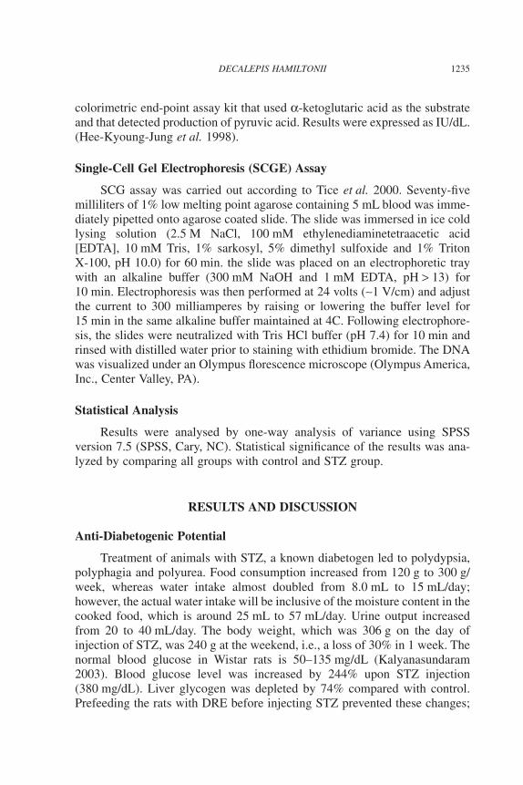
Treatment of animals with STZ, a known diabetogen led to polydypsia,polyphagia and polyurea. Food consumption increased from 120 g to 300 g/week, whereas water intake almost doubled from 8.0 mL to 15 mL/day;however, the actual water intake will be inclusive of the moisture content in thecooked food, which is around 25 mL to 57 mL/day. Urine output increasedfrom 20 to 40 mL/day. The body weight, which was 306 g on the day ofinjection of STZ, was 240 g at the weekend, i.e., a loss of 30% in 1 week. Thenormal blood glucose in Wistar rats is 50–135 mg/dL (Kalyanasundaram2003). Blood glucose level was increased by 244% upon STZ injection(380 mg/dL). Liver glycogen was depleted by 74% compared with control.Prefeeding the rats with DRE before injecting STZ prevented these changes;
1235DECALEPIS HAMILTONII
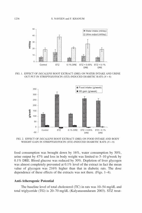
food consumption was brought down by 16%, water consumption by 50%,urine output by 47% and loss in body weight was limited to 5–10 g/week by0.1% DRE. Blood glucose was reduced by 30%. Depletion of liver glycogenwas almost completely prevented at 0.1% level of the extract in fact the meanvalue of glycogen was 216% higher than that in diabetic rats. The dosedependence of these effects of the extracts was not there. (Figs. 1–4).
Anti-Atherogenic Potential
The baseline level of total cholesterol (TC) in rats was 10–54 mg/dL andtotal triglyceride (TG) is 20–70 mg/dL (Kalyanasundaram 2003). STZ treat-
0
10
20
30
40
50
Control STZ 0.1% DRE STZ + 0.05%DRE
STZ + 0.1%DRE
ml/d
ayWater intake (ml/day)
Urine output (ml/day)
FIG. 1. EFFECT OF DECALEPIS ROOT EXTRACT (DRE) ON WATER INTAKE AND URINEOUT PUT IN STREPTOZOTOCIN (STZ)-INDUCED DIABETIC RATS (N = 8)
-50
0
50
100
150
200
250
300
350
Control STZ 0.1% DRE STZ + 0.05%
DRE
STZ + 0.1%
DRE
g/w
eek
Food intake (g/week)
Wt gain (g/week)
FIG. 2. EFFECT OF DECALEPIS ROOT EXTRACT (DRE) ON FOOD INTAKE AND BODYWEIGHT GAIN IN STREPTOZOTOCIN (STZ)-INDUCED DIABETIC RATS (N = 8)
1236 S. NAVEEN and F. KHANUM

ment increased serum TC and TG levels to 67 and 125%, respectively, com-pared with the untreated control. However, in the rats Pre-fed with 0.1% DRE,STZ injection could only increase the TC and TG levels by 29 and 37%,respectively (Table 2).
Antioxidant Potential
Lipid peroxidation, a measure of oxidative stress was estimated in liver,heart and kidney. The level of lipid peroxidation as monitored by the formation
0
100
200
300
400
500
Control STZ 0.1% DRE STZ + 0.05%
DRE
STZ + 0.1%
DRE
mg
/dl
Initial
Final
FIG. 3. EFFECT OF DECALEPIS ROOT EXTRACT (DRE) ON BLOOD GLUCOSE LEVELS INSTREPTOZOTOCIN (STZ)-INDUCED DIABETIC RATS (N = 8)
0
1
2
3
4
5
6
7
Control STZ 0.1% DRE STZ + 0.05%
DRE
STZ + 0.1%
DRE
mg
/g l
iver
FIG. 4. EFFECT OF DECALEPIS ROOT EXTRACT (DRE) ON LIVER GLYCOGEN LEVELSOF STREPTOZOTOCIN (STZ)-INDUCED DIABETIC RATS (N = 8)
1237DECALEPIS HAMILTONII
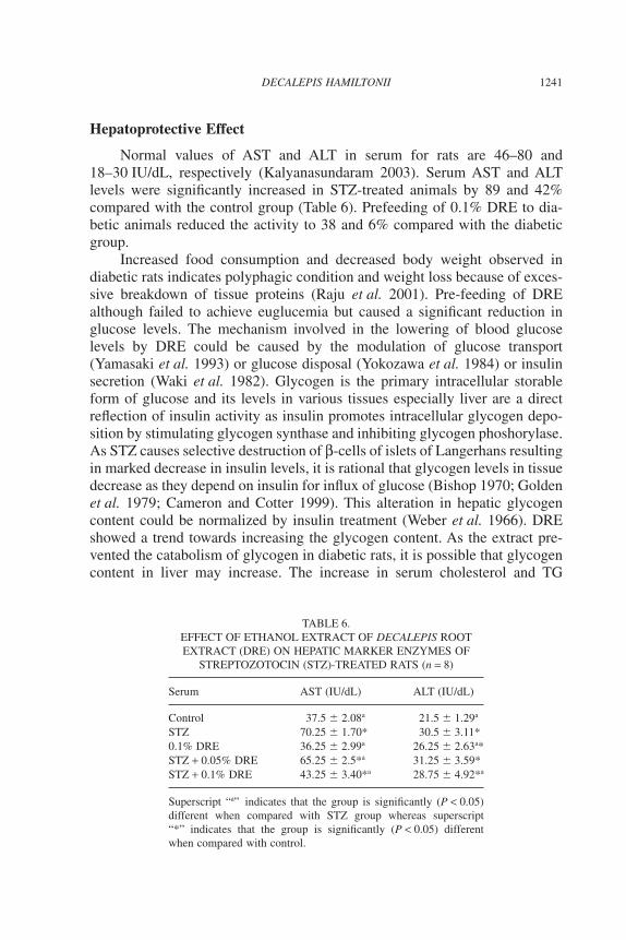
of malondialdehyde was increased in all the tissues upon injection of STZ(Table 3). In liver the increase was 868% whereas in kidney and heart it was103 and 36%, respectively, compared with the normal or control group. Per-oxidation was reduced significantly in rats prefed with 0.1% DRE to 19% inliver, 42% in heart and 88% in kidney compared with the diabetic group.
STZ administration resulted in significant increase in vitamin E content(141%) and decrease in GSH levels to 45% compared with controls. Prefeed-ing with DRE however, did not alter the levels of antioxidants in serum(Table 4).
Activities of SOD and catalase enzymes were significantly decreased inkidney as compared with control (by 48 and 67%, respectively, Table 5).Prefeeding with 0.1% DRE the activity regained by 61% compared with thediabetic group. On the other hand, STZ administration resulted in significant
TABLE 2.EFFECT OF DECALEPIS ROOT EXTRACT (DRE) ON LIPID PROFILE OF
STREPTOZOTOCIN (STZ)-INDUCED DIABETIC RATS (n = 8).
Total cholesterol (mg/dL) Triglyceride (mg/dL)
Control 70 � 3.56a 58.25 � 3.09a
STZ 117.25 � 5.31* 131.25 � 4.11*0.1% DRE 69.75 � 3.30a 56.75 � 2.75a
STZ + 0.05% DRE 83.75 � 1.71*a 74.25 � 2.99*a
STZ + 0.1% DRE 72.75 � 4.64a 65 � 2.58*a
Superscript “a” indicates that the group is significantly (P < 0.05) different when compared with STZgroup whereas superscript “*” indicates that the group is significantly (P < 0.05) different whencompared with control.
TABLE 3.EFFECT OF ETHANOL EXTRACT OF DECALEPIS ROOT EXTRACT (DRE) ON
MALONDIALDEHYDE (MDA) LEVELS IN LIVER OF STREPTOZOTOCIN (STZ)TREATMENT (n = 8)
MDA¥ 10-10 moles/g
Liver Heart Kidney
Control 0.74 � 0.06a 8.55 � 0.73a 21.36 � 1.37a
STZ 7.17 � 0.46* 11.69 � 1.70* 43.54 � 1.27*0.1% DRE 0.60 � 0.06a 9.18 � 0.59a 19.33 � 0.64a
STZ + 0.05% DRE 1.72 � 0.03*a 9.86 � 0.63a 28.00 � 2.94*a
STZ + 0.1% DRE 0.82 � 0.21a 9.48 � 0.74a 25.25 � 3.78a
Superscript “a” indicates that the group is significantly (P < 0.05) different when compared with STZgroup whereas superscript “*” indicates that the group is significantly (P < 0.05) different whencompared with control.
1238 S. NAVEEN and F. KHANUM

TABLE 4.EFFECT OF DECALEPIS ROOT EXTRACT (DRE) ON PLASMA ANTIOXIDANT LEVELS IN
STREPTOZOTOCIN (STZ)-TREATED RATS (n = 8)
Plasma (mg/dL) Vitamin E Vitamin C GSH
Control 1.47 � 0.05a 1.43 � 0.29a 24.58 � 0.51a
STZ 3.55 � 0.13* 1.05 � 0.1* 13.58 � 0.40*0.1% DRE 2.13 � 0.66a 1.67 � 0.08a 24.00 � 0.82a
STZ + 0.05% DRE 2.90 � 0.14* 1.20 � 0.08* 15.75 � 1.26*STZ + 0.1% DRE 2.75 � 0.06* 1.30 � 0.08* 18.00 � 0.82*
Superscript “a” indicates that the group is significantly (P < 0.05) different when compared with STZgroup whereas superscript “*” indicates that the group is significantly (P < 0.05) different whencompared with control.GSH, glutathione.
TABLE 5.EFFECT OF DECALEPIS ROOT EXTRACT (DRE) ON ANTIOXIDANT STATUS IN
STREPTOZOTOCIN (STZ)-TREATED RATS (n = 8)
Kidney CAT¥ 104† SOD¥ 102‡
Control 4.28 � 0.33a 0.21 � 0.03a
STZ 1.43 � 0.17* 0.11 � 0.01*0.05% DRE 4.85 � 0.13a 0.22 � 0.02a
STZ + 0.05% DRE 2.71 � 0.27*a 0.25 � 0.02a
STZ + 0.1% DRE 2.31 � 0.30*a 0.20 � 0.012a
Heart CAT¥ 104† SOD ¥ 102‡
Control 2.00 � 0.08a 0.15 � 0.01a
STZ 3.48 � 0.29* 0.20 � 0.02*0.1% DRE 2.22 � 0.21a 0.16 � 0.01STZ + 0.05% DRE 2.38 � 0.99 0.16 � 0.01STZ + 0.1% DRE 2.10 � 0.20a 0.17 � 0.03
Liver CAT¥ 104† SOD¥ 102‡ GSHP¥§
Control 1.24 � 0.24a 2.45 � 0.13a 4.8 � 0.70a
STZ 2.73 � 0.21* 3.13 � 0.21* 2.31 � 0.20*0.1% DRE 1.31 � 0.11a 2.38 � 0.18a 5.67 � 0.29*a
STZ + 0.05% DRE 1.51 � 0.08a 2.52 � 0.28a 3.69 � 0.41*a
STZ + 0.1% DRE 1.46 � 0.24a 2.24 � 0.13a 4.27 � 0.28a
Superscript “a” indicates that the group is significantly (P < 0.05) different when compared with STZgroup whereas superscript “*” indicates that the group is significantly (P < 0.05) different whencompared with control.† DA of 0.1 unit/min/mg protein.‡ Units/min/mg protein.§ m moles NADP+ produced/min/mg protein.GSHP, glutathione peroxidase; SOD, superoxoide dismutase.
1239DECALEPIS HAMILTONII

elevation of cardiac and hepatic antioxidant enzymes SOD to 28 and 33% andcatalase to 120 and 74% compared with control group, respectively, withprefeeding of 0.1% DRE the enzyme activity significantly reduced by 28 and15%, and 47 and 40%, respectively, compared with the diabetic group(Table 5). Hepatic GSHPx activity was reduced by 52% on STZ treatment(Table 5), which on prefeeding with 0.1% DRE the activity increased to 84%compared with the diabetic group.
DNA Damage
Electrophoresis of lyzed cells under an alkaline condition provides con-ditions for the damaged DNA to unwind and spread giving the whole cells anappearance of a comet. The tail of the comet comprises the damage DNAstrands. Thus length of the tail gives an index of their DNA damage. Thedistance between edge of the head and end of the tail is taken as the length ofthe tail and was measured using Image Pro Plus software (M/s OlympusAmerica, Inc.). SCGE of leukocytes from control rats, as well as the extract-treated rats was carried out (Fig. 5), there was a considerable amount of DNAdamage in STZ-treated leukocytes, the comet tail length was 200 m. The taillength was reduced to 55 m in the diabetic rats prefed with DRE, clearlyindicating the DNA protective effect.
Control leukocyte: Tail length: 2 micrometer
STZ treated leukocyte: Tail length: 200 micrometer
0.1% DRE fed diabetic leukocyte:Tail length: 55 micrometer
FIG. 5. SINGLE-CELL GEL ELECTROPHORESIS OF LEUKOCYTES OFSTREPTOZOTOCIN-TREATED RATS (N = 8)
DRE, Decalepis root extract.
1240 S. NAVEEN and F. KHANUM
Hepatoprotective Effect
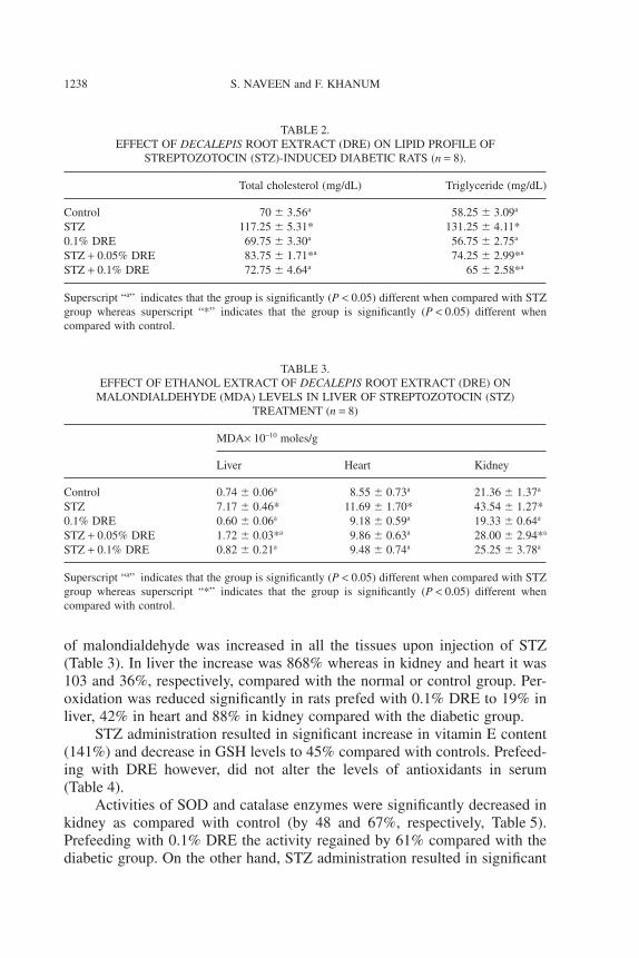
Normal values of AST and ALT in serum for rats are 46–80 and18–30 IU/dL, respectively (Kalyanasundaram 2003). Serum AST and ALTlevels were significantly increased in STZ-treated animals by 89 and 42%compared with the control group (Table 6). Prefeeding of 0.1% DRE to dia-betic animals reduced the activity to 38 and 6% compared with the diabeticgroup.
Increased food consumption and decreased body weight observed indiabetic rats indicates polyphagic condition and weight loss because of exces-sive breakdown of tissue proteins (Raju et al. 2001). Pre-feeding of DREalthough failed to achieve euglucemia but caused a significant reduction inglucose levels. The mechanism involved in the lowering of blood glucoselevels by DRE could be caused by the modulation of glucose transport(Yamasaki et al. 1993) or glucose disposal (Yokozawa et al. 1984) or insulinsecretion (Waki et al. 1982). Glycogen is the primary intracellular storableform of glucose and its levels in various tissues especially liver are a directreflection of insulin activity as insulin promotes intracellular glycogen depo-sition by stimulating glycogen synthase and inhibiting glycogen phoshorylase.As STZ causes selective destruction of b-cells of islets of Langerhans resultingin marked decrease in insulin levels, it is rational that glycogen levels in tissuedecrease as they depend on insulin for influx of glucose (Bishop 1970; Goldenet al. 1979; Cameron and Cotter 1999). This alteration in hepatic glycogencontent could be normalized by insulin treatment (Weber et al. 1966). DREshowed a trend towards increasing the glycogen content. As the extract pre-vented the catabolism of glycogen in diabetic rats, it is possible that glycogencontent in liver may increase. The increase in serum cholesterol and TG
TABLE 6.EFFECT OF ETHANOL EXTRACT OF DECALEPIS ROOTEXTRACT (DRE) ON HEPATIC MARKER ENZYMES OF
STREPTOZOTOCIN (STZ)-TREATED RATS (n = 8)
Serum AST (IU/dL) ALT (IU/dL)
Control 37.5 � 2.08a 21.5 � 1.29a
STZ 70.25 � 1.70* 30.5 � 3.11*0.1% DRE 36.25 � 2.99a 26.25 � 2.63a*STZ + 0.05% DRE 65.25 � 2.5*a 31.25 � 3.59*STZ + 0.1% DRE 43.25 � 3.40*a 28.75 � 4.92*a
Superscript “a” indicates that the group is significantly (P < 0.05)different when compared with STZ group whereas superscript“*” indicates that the group is significantly (P < 0.05) differentwhen compared with control.
1241DECALEPIS HAMILTONII

observed in this study in STZ-treated rats are confirmed by the study where indyslipidemia induced by STZ is reported (Reaven 1988; Uma et al. 2005).DRE could produce significant fall in serum total cholesterol and triglyceridelevels indicating a strong lipid lowering activity.
Vitamin E is the most important lipid soluble, radical scavenging anti-oxidant. It effectively reduces lipid hydroperoxides generated during the pro-cess of peroxidation and protects the cell structure against damage (Kinalskiet al. 2000). Increased in the vitamin E level observed in diabetic rats could bea protective mechanism against increased peroxidation in diabetes (Stanelyet al. 1998). GSH another efficient antioxidant molecule in the body protectsthe tissue from free radical damage by inhibiting free radical mediated lipidperoxidation (Meistor and Anderson 1983). Decreased levels of plasma GSHin diabetic rats could be caused by the utilization of GSH as a result ofincreased ROS generation by increased levels of glucose (Stanely et al. 1998)in STZ-treated animals. There was a significant improvement in the plasmaantioxidant levels of diabetic rats prefed with DRE in our studies.
A marked rise in the extent of lipid peroxidation in diabetes is known tocause decreased cell survival, altered membrane lipid asymmetry, hypercoagu-lability and increased adhesivity to the endothelium (Jain et al. 1990). Tre-mendous increase in lipid peroxidation in different tissues observed in diabeticrats is attributed to chronic hyperglycemia, which in turn causes increasedproduction of ROS and reactive nitrogen species (RNS) because of the autoxi-dation of monosaccharides that leads to the production of superoxide andhydroxyl radicals (Wolff and Dean 1987; Marianna et al. 2006), which causetissue damage by reacting with polyunsaturated fatty acids, proteins and DNA(Das et al. 2000). As high blood glucose is susceptible to oxidation, hyper-glycemia causes high ROS production and, in turn, leads to high TBARS intissues (Das et al. 2000). The experimental results indicate that lipid peroxi-dation played a role in tissue injury in STZ induced diabetic rats. DRE reducedthe lipid peroxidation in heart, kidney and liver, thus protecting cell functionsand structure.
SOD and CAT are considered primary antioxidant enzymes, as they areinvolved in direct elimination of ROS (Halliwell and Cutteridge 1985). Effectof STZ on CAT and SOD activities were found to be tissue dependent.Increased cardiac CAT and SOD activities in diabetic rats, suggests that inorder to overcome the oxidative stress in heart, some other compensatorymechanisms may exist in heart in addition to antioxidant enzymes (Guptaet al. 2004). Pretreatment of DRE stimulated SOD and CAT to reverse oxida-tive damage. The study also revealed that CAT and SOD activities weresignificantly inhibited in kidney of STZ-treated diabetic animals, which werereversed by DRE pretreatment. Significant increase in lipid peroxidation, SODand CAT in pancreas, heart and blood and increase in GSHPx in kidney and
1242 S. NAVEEN and F. KHANUM
pancreas in diabetes has been reported by Kakar et al. 1995). The resultsindicate that DRE showed significant protection against the oxidative damageinduced by STZ in heart, kidney and liver.
ROS and RNS may play a major role as endogenous initiators andpromoters of DNA damage and mutations that contribute to cancer, diabetes,heart disease and other age-related diseases (Wiseman and Halliwell 1996;Park et al. 2001). STZ has reported to induce DNA strand breaks and alkali-labile sites (Erickson et al. 1978; Petzold and Swenberg 1978). In the presentstudy, DNA in leukocytes was significantly damaged by STZ injection, andthis damage was found to be significantly less in DRE-fed rats.
Serum AST and ALT are sensitive indicators, of liver cell injury and aremost helpful in recognizing hepatic problems. High levels of AST and ALTindicate that there may be liver inflammation and/or damage. STZ was foundto significantly increase the levels of serum AST and ALT (Angulo and Lindor2002; Imaeda et al. 2002), which were effectively brought under control byDRE.
CONCLUSION
In this study, we examined oxidative stress markers in rats with STZ-induced diabetes and its effect on pre-feeding of D. hamiltonii root extract.STZ is a generally employed compound for the induction of type-I diabetes inrats (Tomlinson et al. 1992). Autoxidation and glycation reactions of glucoseand metabolites, catalyzed by transition metal ions, become an importantsource of free radicals in diabetes, which leads to further complications(Cameron and Cotter 1999). STZ treatment of rats led to hyperglycemia, adiabetic condition and also its associated symptoms like increased thirst,increased hunger, increased urine output, etc. These findings correlate withthose reported by others (Bishop 1970; Golden et al. 1979; Cameron andCotter 1999). STZ injection also led to increased cholesterol and triglyceridelevels, increased oxidative stress and DNA damage apart from inducing liverdamage. The extract of D. hamiltonii root, prefed to rats for 4 weeks beforeand 1 week after injection with STZ was found to provide significant protec-tion from all the serious effects of STZ. The plant root is known to containphytochemicals such as vanillin, salicylaldehyde, p-anisaldehyde, 2-hydroxy-4-methoxybenzaldehyde, bis-2, 3, 4, 6-galloyl-a/b-D-glucopyranoside,borneol, inositol, saponins, ketonic substances, sterols, amyrins and lupeols(Nagarajan et al. 2001; Thangadurai et al. 2002; Nagarajan and Rao 2003;Harish et al. 2005; Srivastava et al. 2006a,b), which may be responsible for theantioxidant property of root.
The results mentioned earlier indicate that one single food material/spice could have multifaceted benefits. However, the study brings forth many
1243DECALEPIS HAMILTONII

questions, which need further through investigation, such as the mechanismof action of the root extract, is the extract inactivating the STZ in the blooditself, or prevents its transport across into pancreatic and hepatic cells?Whether the extract simply mops off the ROS or is it a chain-breaking anti-oxidant? Whether the prevention of the liver damage seen with DRE is theresult of its antioxidant property or does it induce the activity of liverenzymes? Its effects on DNA damage are also very fascinating. STZ isknown to alkylate DNA, thus damaging its structure. Is decalypis capable ofpreventing only alkylation of DNA or can it protect other types of damagesas well. The plant seems to be worth investing further and could be anexcellent source for nutraceuticals.
REFERENCES
ANGULO, P. and LINDOR, K.D. 2002. Non-alcoholic fatty liver disease. J.Gastroenterol. Hepatol. 17(Suppl), S186–S190.
BENNETT, R.A. and PEGG, A.E. 1981. Alkylation of DNA in rat tissuesfollowing administration of Streptozotocin. Cancer Res. 41, 2786–2790.
BHOR, V.M., RAGHURAM, N. and SIVAKAMI, S. 2004. Oxidative damageand altered antioxidant enzyme activities in the small intestine ofstreptozotocin-induced diabetic rats. Int. J. Biochem. Cell Biol. 36,89–97.
BISHOP, J.S. 1970. Inability of insulin to activate liver glycogen transferase Dphosphatase in the diabetic pancreatectomized dog. Biochim. Biophys.Acta 208, 208–218.
BOK-KYEONG-CHA 2004. A study of serum lipid, blood sugar, blood pres-sure of Buddhist nuns in vegetarians and non-vegetarians (III). Based onage. J. Korean Soc. Food Sci. Nutr. 33, 1310–1319.
CAMERON, N.E. and COTTER, M.A. 1999. Effects of the iron chelator,hydroxy-ethyl starch deferoxamine, on nerve conduction and blood flowin diabetic rats. Diabetes 48(Suppl. 1), A54.
CERIELLO, A. 2000. Oxidative stress and glycemic regulation. Metabolism49, 27–29.
COHEN, G., DEMBIEC, C. and MARENS, J. 1970. Measurement of catalaseactivity in tissue extracts. Anal. Biochem. 34, 30–38.
DAS, S., VASISHT, S., SNEHALATA, DAS, N. and SRIVASTAVA, M. 2000.Correlation between total antioxidant status and lipid peroxidation inhypercholesterolemia. Curr. Sci. 78, 486.
DESAI, D.I. 1984. Vitamin E analysis methods for animal tissues. MethodsEnzymol. 105, 138–147.
1244 S. NAVEEN and F. KHANUM

DONNINI, D., ZAMBITO, A.M., PERELLA, G., AMBESI-IMPIOMBAT, O.and CURCIO, F. 1996. Glucose may induce cell death through a freeradical mediated mechanism. Biochem. Biophys. Res. Commun. 219,412–417.
EBERHARDT, M.V., CHANG, Y. and LEE, R.-H.-L. 2000. Antioxidantactivity of fresh apples. Nature, UK 405(6789), 903–904.
ERICKSON, L.C., BRADLEY, M.O. and KOHN, K.W. 1978. Measurementsof DNA damage in Chinese hamster cells treated with equitoxic andequimutagenic doses of nitrosoureas. Cancer Res. 38, 3379–3384.
GOLDEN, S., WALS, P.A. and OKAJIMA, F. 1979. Glycogen synthesis byhepatocytes from diabetic rats. Biochem. J. 182, 727–734.
GUPTA, S., KATARIA, M., GUPTA, P.K., MURGANANDAN, S. andYASHROY, R.C. 2004. Protective role of extracts of neem seeds indiabetes caused by streptozotocin in rats. J. Ethnopharmacol. 90, 186–189.
HALLIWELL, B. and CUTTERIDGE, J.M.C. 1985. Free Radical in Biologyand Medicine, Clarendon, Oxford, England.
HARISH, R., DIVAKAR, S., SRIVASTAVA, A. and SHIVANANDAPPA, T.2005. Isolation of antioxidant compounds from the methanolic extract ofthe roots of Decalepis hamiltonii (Wight & Arn.). J. Agric. Food Chem.53, 7709–7714.
HEE, K.-J., PYUNG, S.-P., NAM, C.-H., SUNG, O.-K., KYUNG, S.-K. andMYUNG, Y.-L. 1998. Inhibitory effect of Angelica keiskei Koidz greenjuice on the liver damage in CCl4-treated rats. J. Korean Soc. Food Sci.Nutr. 27, 531–536.
IMAEDA, A.T., KANEKO, T., AOKI, Y. and KONDO, H. 2002. NagaseDNA damage and the effect of antioxidants in streptozotocin-treatedmice. Food Chem. Toxicol. 40, 979–987.
JAIN, S.K., LEVINE, S.N., DEUTT, J. and HOLLIER, B. 1990. Elevated lipidperoxidation levels in red blood cells of streptozotocin-treated diabeticrats. Metabolism 39, 971–975.
KAKAR, R., KALRA, J., MANTHA, S.V. and PRASAD, K. 1995. Lipidperoxidation and activity of antioxidant enzymes in diabetic rats. Mol.Cell. Biochem. 151, 113–119.
KAKKAR, R., MANTHA, S.V., RADHI, J., PRASAD, K. and KALRA, J.1997. Antioxidant defense system in diabetic kidney: A time coursestudy. Life Sci. 60, 667–679.
KALYANASUNDARAM 2003. Laboratory Manual, National Institute ofNutrition, Hyderabad, India.
KINALSKI, M., SLEDZIEWSKI, A., TELEJKO, B., ZARZYCKI, W. andKINALSKA, I. 2000. Lipid peroxidation and scavenging enzyme activityin streptozotocin induced diabetes. Acta Diabetol. 37, 179–183.
1245DECALEPIS HAMILTONII

LOWRY, O.H., ROSEBROUGH, N.J., FARO, A.L. and RANDALL, R.J.1951. Protein measurement with the Follin phenol reagent. J. Biol. Chem.193, 265–275.
MARIANNA, M., CHAONAN, D., QINGPING, H.E., YANLING, L. andPING, A.L. 2006. Streptozotocin-induced diabetes causes astrocyte deathafter ischemia and reperfusion injury. Diabetes 55, 349–355.
MEISTOR, A. and ANDERSON, M.E. 1983. Glutathione. Annu. Rev.Biochem. 52, 711–760.
NAGARAJAN, S. and RAO, L.J. 2003. Determination of 2-hydroxy-4-methoxybenzaldehyde in roots of Decalepis hamiltonii (Wight & Arn.)and Hemidesmus indicus R. Br. J. AOAC Int. 86, 564–567.
NAGARAJAN, S., RAO, L.J. and GURUDUTT, K.N. 2001. Chemical com-position of the volatiles of Decalepis hamiltonii (Wight & Arn). FlavourFragr. J. 16, 27–29.
NAYAR, R.C., SHETTY, J.K.P., MARY, Z. and YOGANARSHIMHAN, S.N.1978. Pharmacognostical studies on the root of Decalepis hamiltonii Wt.and Arn. And comparison with Hemidesmus indicus (L.) R.Br. Proc.Indian Acad. Sci. 87, 37–48.
NICHANS, W.G., JR and SAMUELSON, B. 1968. Formation of MDA fromphospholipids arachidonate during microsomal lipid per oxidation. Eur. J.Biochem. 6, 21–26.
OMAYE, S.T., TURNBULL, J.D. and SAUBERLICHH, E. 1997. MethodsEnzymol. 62, 3–8.
PAOLA, R.-D.-D.I., ASIS, R. and ALDAO, M.-A.-J. 2003. Evaluation of thedegree of starch gelatinization by a new enzymatic method. Starch/Staerke 55, 403–409.
PARK, K.S., KIM, J.H., KIM, M.S., KIM, J.M., KIM, S.K., CHOI, J.Y.,CHUNG, M.H., HAN, B., KIM, S.Y. and LEE, H.K. 2001. Effects ofinsulin and antioxidant on plasma 8-hydroxyguanine and tissue8-hydroxydeoxyguanosine in streptozotocin- induced diabetic rats. Dia-betes 50, 2837–2841.
PETZOLD, G.L. and SWENBERG, J.A. 1978. Detection of DNA damageinduced in vivo following exposure of rats to carcinogens. Cancer Res.38, 1589–1594.
RAJU, J., GUPTA, D. and RAO, A.R. 2001. Trigonellafoenum graecum(fenugreek) seed powder improves glucose homeostasis in allaxan dia-betic rat tissues by revering the altered glycolytic, glucogenic and lipo-genic enzymes. Mol. Cell. Biochem. 224, 45–51.
REAVEN, G.M. 1988. Role of insulin resistance in human disease. Diabetes37, 1595–1607.
SAMANTA, L. and CHAINY, G.B.N. 1995. Hexachlorocyclohexane inducedchanges in lipid peroxidation, superoxide dismutase and catalase
1246 S. NAVEEN and F. KHANUM
activities and glutathione content in chick liver. Indian J. Exp. Biol. 33,131–133.
SINGLETON, V.L. and ROSSI, J.A. JR. 1965. Colorimetry of totalphenolics with phosphomolybdic acid reagent. Am. J Enol. Vitic. 16,144–158.
SRIVASTAVA, A., HARISH, R. and SHIVANANDAPPA, T. 2006a. Novelantioxidant compounds from the aqueous extract of the roots of Decal-epis hamiltonii (Wight and Arn.) and their inhibitory effect on low-density lipoprotein oxidation. J. Agric. Food Chem. 54, 790–795.
SRIVASTAVA, A., SHEREEN, HARISH, R. and SHIVANANDAPPA, T.2006b. Antioxidant activity of the roots of Decalepis hamiltonii (Wight &Arn). Lebensm. Wiss. Technol. 39, 1059–1065.
SRIVASTAVA, A., JAGAN, M., RAO, L. and SHIVANANDAPPA, T. 2007.Isolation of ellagic acid from the aqueous extract of the roots of Decalepishamiltonii: Antioxidant activity and cytoprotective effect. Food Chem.103, 224–233.
STANELY, M., PRINCE, P. and MENON, V.P. 1998. Effect of Syzigiumcumini in plasma antioxidants on aloxan-induced diabetes in rats. J. Clin.Biochem. Nutr. 25, 81–86.
TAO, X. and QIN, W. 2007. D-Chiro-Inositol found in momordica charantiafruit extract plays a role in reducing blood glucose in streptozotocin-diabetic rats. J. Food Biochem 31, 551–562.
THANGADURAI, D., ANITHA, S., PULLAIAH, T., REDDY, P.N. andRAMACHANDRAIAH, O.S. 2002. Essential oil constituents and invitro antimicrobial activity of Decalepis hamiltonii roots against foodborne pathogens. J. Agric. Chem. 50, 3147–3149.
TICE, R.R., AGURELL, E., ANDERSON, D., BURLINSON, B., HART-MANN, A., KOBAYASHI, H., MIYAMAE, Y., ROJAS, E., RYU, J.C.and SASAKI, Y.F. 2000. Single cell gel comet assay: Guidelines forin-vitro and in-vivo genetic toxicology testing. Environ. Mol. Mutagen.35, 206–221.
TJÄLVE, H. 1983. Streptozotocin: Distribution, metabolism and mechanismsof action. Uppsala J. Med. Sci. (Suppl. 39), 145–157.
TOMLINSON, K.C., GARDINER, S.M., HEBDEN, R.A. and BENNETT, T.1992. Functional consequences of Streptozotocin induce diabetes melli-tus with paticular refrence to the cardiovascular system. Pharmacol. Rev.44, 105–150.
UMA, B., RAMAN, K. and PILLAI, K.K. 2005. Effect of ethanolic extract ofZingiber officinale on dyslipidaemia in diabetic rats. J. Ethnopharmacol.97, 227–230.
VALLABHJI, J., MCCOLL, A.J., RICHMOND, W., SCHACHTER, M.,RUBENS, M.B. and ELKELES, R.S. 2001. Total antioxidant status and
1247DECALEPIS HAMILTONII
coronary artery calcification in Type 1 diabetes. Diabetes Care 24, 1608–1613.
WAKI, I., KYO, H., YASUDA, M. and KIMURA, M. 1982. Effects of ahypoglycemic component of ginseng radix on insulin biosynthesis innormal and diabetic animals. J. Pharmacobiodyn. 5, 547–554.
WEBER, G., LEA, M.A. and FISHER, E.A. 1966. Regulatory pattern of livercarbohydrate metabolizing enzymes; insulin as an inducer of key glyco-lytic enzymes. Enzymol. Biol. Clin. (Basel) 7, 11–24.
WEISS, C., MAKER, H.S. and LEHRER, G.M. 1980. Sensitive fluorometricassays for glutathione peroxidase and reductase. Anal. Biochem. 106,512–516.
WISEMAN, H. and HALLIWELL, B. 1996. Damage to DNA by reactiveoxygen and nitrogen species: Role in inflammatory disease and progres-sion to cancer. Biochem. J. 313, 17–29.
WOLFF, S.P. and DEAN, R.T. 1987. Glucose autoxidation and protein modi-fication. Biochem. J. 245, 243–250.
YAMASAKI, K., MURAKAMI, C., OHTANI, K., KASAI, R.,KUROKAWA, T. and ISHIBASHI, S. 1993. Effects of standardizedPanax ginseng extract G115 on the d-glucose transport by Ehrlich ascitestumour cells. Phytother. Res. 7, 200–202.
YOKOZAWA, T., KOBAYASHI, T., KAWAI, A., OURA, H. andKAWASHIMA, Y. 1984. Stimulation of lipid and sugar metabolism inginsenoside- Rb2 treated rats. Chem. Pharm. Bull. 32, 2766–2772.
1248 S. NAVEEN and F. KHANUM