Design, synthesis and pharmacological evaluation of novel vanadium-containing complexes as...
11
Design, Synthesis and Pharmacological Evaluation of Novel Vanadium-Containing Complexes as Antidiabetic Agents Elena V. Fedorova 1 *, Anna V. Buryakina 1 , Alexey V. Zakharov 2,3 , Dmitry A. Filimonov 3 , Alexey A. Lagunin 3 , Vladimir V. Poroikov 3 1 Saint-Petersburg State Chemical Pharmaceutical Academy, Ministry of Healthcare and Social Development of Russian Federation, Saint-Petersburg, Russian Federation, 2 National Cancer Institute, National Institutes of Health, Frederick, Maryland, United States of America, 3 Orekhovich Institute of Biomedical Chemistry of Russian Academy of Medical Sciences, Moscow, Russian Federation Abstract Based on the data about structure and antidiabetic activity of twenty seven vanadium and zinc coordination complexes collected from literature we developed QSAR models using the GUSAR program. These QSAR models were applied to 10 novel vanadium coordination complexes designed in silico in order to predict their hypoglycemic action. The five most promising substances with predicted potent hypoglycemic action were selected for chemical synthesis and pharmacological evaluation. The selected coordination vanadium complexes were synthesized and tested in vitro and in vivo for their hypoglycemic activities and acute rat toxicity. Estimation of acute rat toxicity of these five vanadium complexes was performed using a freely available web-resource (http://way2drug.com/GUSAR/acutoxpredict.html). It has shown that the selected compounds belong to the class of moderate toxic pharmaceutical agents, according to the scale of Hodge and Sterner. Comparison with the predicted data has demonstrated a reasonable correspondence between the experimental and predicted values of hypoglycemic activity and toxicity. Bis{tert-butyl[amino(imino)methyl]carbamato}ox- ovanadium (IV) and sodium(2,29-Bipyridyl)oxo-diperoxovanadate(V) octahydrate were identified as the most potent hypoglycemic agents among the synthesized compounds. Citation: Fedorova EV, Buryakina AV, Zakharov AV, Filimonov DA, Lagunin AA, et al. (2014) Design, Synthesis and Pharmacological Evaluation of Novel Vanadium- Containing Complexes as Antidiabetic Agents. PLoS ONE 9(7): e100386. doi:10.1371/journal.pone.0100386 Editor: Joseph J. Barchi, National Cancer Institute at Frederick, United States of America Received January 19, 2014; Accepted May 27, 2014; Published July 24, 2014 Copyright: ß 2014 Fedorova et al. This is an open-access article distributed under the terms of the Creative Commons Attribution License, which permits unrestricted use, distribution, and reproduction in any medium, provided the original author and source are credited. Funding: This work was supported by Ministry of Education and Science of the Russian Federation on the main academic programs of higher professional education, program 1.2.1. (fcpk.ru). We also acknowledge the support of the Russian Foundation for Basic Research (http://www.rfbr.ru/rffi/ru/)in the payment of the license for using the Cambridge Structural Database, project no. 02-07-90322. The funders had no role in study design, data collection and analysis, decision to publish, or preparation of the manuscript. Competing Interests: The authors have declared that no competing interests exist. * Email: [email protected] Introduction Vanadium is a biologically important element involved in numerous physiological processes. The presence of transition valence determines wide distribution of vanadium compounds in nature that are found only in chemically combined form. Vanadium compounds, in particular organic derivatives, are effective oral insulinomimetics, which inhibit lipolysis, decrease blood glucose levels (BGL) in animals and in clinical trials, and stimulate insulin secretion in experimental models of Diabetes Mellitus (DM) [1–5]. Although mechanisms of the insulin mimetic effect of vanadium complexes still have to be clarified, their ability to sensitize peripheral tissues to insulin and to reduce insulin resistance attracts significant attention in the context of their potential use for the treatment of Diabetes Type 1 (DM1), Diabetes Type 2 (DM2) and obesity [6,7]. According to the Integrity database (http://integrity.thomson- pharma.com), vanadium compounds have been developed as drugs for treatment of Diabetes, Hypertension, Ischemia and Myocardial Stroke. The major problem limiting further clinical applications of inorganic vanadium compounds is a low oral absorption (1–10%). However, it was shown that the oral bioavailability of vanadium complexes with organic ligands exceeded 20–40%, which can be considered as acceptable level of bioavailability [8]. Currently QSAR modeling is widely applied in rational drug design [9]. Despite the many examples of successful application of QSAR for finding and optimization of new lead compounds, only few QSAR studies on biological activities of vanadium complexes have been reported (see, e.g., [10]). Limited number of QSAR applications to the metal-complexes may be explained by the difficulties with the development of chemical descriptors, which reflect well the quantitative structure-activity relationships [11]. Recently a novel QSAR approach based on Quantitative Neighborhoods of Atoms (QNA) descriptors and self-consisted regression has been proposed; and its superiority in comparison with many other popular methods was shown [12]. This approach was realized in GUSAR program: General Unrestricted Structure- Activity Relationships. Since GUSAR was successfully applied for compounds from different chemical series and various activity/ property endpoints [12], it was interesting to investigate if it would provide the accurate prediction results for vanadium complexes with antidiabetic activity. Moreover, GUSAR can be used not PLOS ONE | www.plosone.org 1 July 2014 | Volume 9 | Issue 7 | e100386
-
Upload
independent -
Category
Documents
-
view
0 -
download
0
Transcript of Design, synthesis and pharmacological evaluation of novel vanadium-containing complexes as...

Design, Synthesis and Pharmacological Evaluation ofNovel Vanadium-Containing Complexes as AntidiabeticAgentsElena V. Fedorova1*, Anna V. Buryakina1, Alexey V. Zakharov2,3, Dmitry A. Filimonov3,
Alexey A. Lagunin3, Vladimir V. Poroikov3
1 Saint-Petersburg State Chemical Pharmaceutical Academy, Ministry of Healthcare and Social Development of Russian Federation, Saint-Petersburg, Russian Federation,
2 National Cancer Institute, National Institutes of Health, Frederick, Maryland, United States of America, 3 Orekhovich Institute of Biomedical Chemistry of Russian
Academy of Medical Sciences, Moscow, Russian Federation
Abstract
Based on the data about structure and antidiabetic activity of twenty seven vanadium and zinc coordination complexescollected from literature we developed QSAR models using the GUSAR program. These QSAR models were applied to 10novel vanadium coordination complexes designed in silico in order to predict their hypoglycemic action. The five mostpromising substances with predicted potent hypoglycemic action were selected for chemical synthesis andpharmacological evaluation. The selected coordination vanadium complexes were synthesized and tested in vitro andin vivo for their hypoglycemic activities and acute rat toxicity. Estimation of acute rat toxicity of these five vanadiumcomplexes was performed using a freely available web-resource (http://way2drug.com/GUSAR/acutoxpredict.html). It hasshown that the selected compounds belong to the class of moderate toxic pharmaceutical agents, according to the scale ofHodge and Sterner. Comparison with the predicted data has demonstrated a reasonable correspondence between theexperimental and predicted values of hypoglycemic activity and toxicity. Bis{tert-butyl[amino(imino)methyl]carbamato}ox-ovanadium (IV) and sodium(2,29-Bipyridyl)oxo-diperoxovanadate(V) octahydrate were identified as the most potenthypoglycemic agents among the synthesized compounds.
Citation: Fedorova EV, Buryakina AV, Zakharov AV, Filimonov DA, Lagunin AA, et al. (2014) Design, Synthesis and Pharmacological Evaluation of Novel Vanadium-Containing Complexes as Antidiabetic Agents. PLoS ONE 9(7): e100386. doi:10.1371/journal.pone.0100386
Editor: Joseph J. Barchi, National Cancer Institute at Frederick, United States of America
Received January 19, 2014; Accepted May 27, 2014; Published July 24, 2014
Copyright: � 2014 Fedorova et al. This is an open-access article distributed under the terms of the Creative Commons Attribution License, which permitsunrestricted use, distribution, and reproduction in any medium, provided the original author and source are credited.
Funding: This work was supported by Ministry of Education and Science of the Russian Federation on the main academic programs of higher professionaleducation, program 1.2.1. (fcpk.ru). We also acknowledge the support of the Russian Foundation for Basic Research (http://www.rfbr.ru/rffi/ru/)in the payment ofthe license for using the Cambridge Structural Database, project no. 02-07-90322. The funders had no role in study design, data collection and analysis, decisionto publish, or preparation of the manuscript.
Competing Interests: The authors have declared that no competing interests exist.
* Email: [email protected]
Introduction
Vanadium is a biologically important element involved in
numerous physiological processes. The presence of transition
valence determines wide distribution of vanadium compounds in
nature that are found only in chemically combined form.
Vanadium compounds, in particular organic derivatives, are
effective oral insulinomimetics, which inhibit lipolysis, decrease
blood glucose levels (BGL) in animals and in clinical trials, and
stimulate insulin secretion in experimental models of Diabetes
Mellitus (DM) [1–5]. Although mechanisms of the insulin mimetic
effect of vanadium complexes still have to be clarified, their ability
to sensitize peripheral tissues to insulin and to reduce insulin
resistance attracts significant attention in the context of their
potential use for the treatment of Diabetes Type 1 (DM1),
Diabetes Type 2 (DM2) and obesity [6,7].
According to the Integrity database (http://integrity.thomson-
pharma.com), vanadium compounds have been developed as
drugs for treatment of Diabetes, Hypertension, Ischemia and
Myocardial Stroke.
The major problem limiting further clinical applications of
inorganic vanadium compounds is a low oral absorption (1–10%).
However, it was shown that the oral bioavailability of vanadium
complexes with organic ligands exceeded 20–40%, which can be
considered as acceptable level of bioavailability [8].
Currently QSAR modeling is widely applied in rational drug
design [9]. Despite the many examples of successful application of
QSAR for finding and optimization of new lead compounds, only
few QSAR studies on biological activities of vanadium complexes
have been reported (see, e.g., [10]). Limited number of QSAR
applications to the metal-complexes may be explained by the
difficulties with the development of chemical descriptors, which
reflect well the quantitative structure-activity relationships [11].
Recently a novel QSAR approach based on Quantitative
Neighborhoods of Atoms (QNA) descriptors and self-consisted
regression has been proposed; and its superiority in comparison
with many other popular methods was shown [12]. This approach
was realized in GUSAR program: General Unrestricted Structure-
Activity Relationships. Since GUSAR was successfully applied for
compounds from different chemical series and various activity/
property endpoints [12], it was interesting to investigate if it would
provide the accurate prediction results for vanadium complexes
with antidiabetic activity. Moreover, GUSAR can be used not
PLOS ONE | www.plosone.org 1 July 2014 | Volume 9 | Issue 7 | e100386

only as a tool for creation of QSAR models for the series of
compounds with known activity values but also for estimating the
acute rats toxicity (LD50 values) for compounds, according to
different route of administration (oral, intravenous, intraperitoneal
and subcutaneous) [13].
In this study we apply the computer program GUSAR to
analyze the structure-activity relationships of vanadium complexes
with antidiabetic action, predict antidiabetic activity and toxicity
of novel designed compounds, select the most promising of them
for synthesis and biological testing, and verify the computational
predictions by the experiment.
Results
Quantitative structure-activity relationships (QSAR)We collected information about structures of the twenty seven
zinc and vanadium complexes with (O4), (N2O2), (N2S2), (O2S2),
and (S4) coordination modes and their hypoglycemic activity from
the available sources (File S1). They have been reported to display
considerable insulinomimetic effects in in vitro experiments on
enzyme inhibitions and blood glucose-lowering effects. The
hypoglycemic activity was presented as 50 percent inhibitory
concentration (IC50). Both zinc and vanadium complexes were
used for QSAR analysis. The collected data was randomly divided
twenty times onto the training and test sets with a ratio of 70% and
30%, respectively. Here we call this multiple splitting procedure a
leave-30%-out cross-validation (L30%CV). This procedure is
similar to n-fold external validation, but there is only one
difference. During the n-fold validation each compound may be
used as a test compound only once, but during the multiple
splitting validation each compound may be used as a test
compound several times, which depends from splitting. The
training sets were used for creation of QSAR models and test sets
for the assessment of external predictive accuracy. In addition to
L30%CV we have used the procedure of selection of the test set
compounds taking into account the activity distribution (Act.Dis.)
using the same proportion as mentioned above: 70% for the
training set and 30% for the test set. QSAR models were
developed using GUSAR program (version 2011), which based on
Multilevel and Quantitative Neighborhoods of Atoms (MNA,
QNA) descriptors [14,15] and self-consistent regression (SCR)
algorithm [12,13].
Two hundred QNA and MNA based models were generated for
each training set. Models that satisfied the following criteria were
considered as acceptable for prediction: I) R2test.0.5, II) Q2.0.5.
As a result, 14 acceptable models were selected. Characteristics of
the obtained models as well as the results of external predictivity of
each model calculated by multiple splitting procedure (L30%CV)
and by activity distribution procedure (L30%Act.Dis) are present-
ed in Table 1. According to the results described in Table 1 the
multiple splitting procedure provides more reliable assessment of
the predictivity of models compared to the activity distribution
procedure, which is overoptimistic. Thus, selection of models had
been done by L30%CV. Only QNA based models were satisfied
the described criteria. Thus, QNA descriptors are more suitable
for modeling of complex compounds compared to MNA
descriptors.
Selected models were used for consensus prediction of 10 novel
vanadium coordination complexes designed in silico, to estimate
their hypoglycemic activity. Prediction results obtained using the
consensus model for 10 novel compounds are presented in Table 2.
Five most active compounds according to the prediction results
described in Table 2 were selected for synthesis and hypoglycemic
activity testing.
SynthesisBased on prediction, we have selected for future synthesis five
vanadium complexes that are expected to be more effective in
lowering the glucose concentration in blood serum than VOSO4
that is usually used as a reference substance [16]. Among the
selected structures the vanadium complexes in various oxidation
states (both neutral and anionic forms) were found, which contain
a variety of coordination modes: oxocomplexes II, III and IV of
vanadium(IV) – the general formula VOL2; oxocomplexes V of
vanadium(V) with tridentate ligand, the general formula
[VO(H2O)(L)]; and oxodiperoxo complexes I of vanadium(V) - the
general formula [VO(O2) 2(L)2]n-, where n = 1.
Complex I was formed by the interaction of sodium
metavanadate with bidentate chelating ligand 2,29-Bipyridine
(C10H8N2) and hydrogen peroxide:
NaVO3zC10H8N2zH2O2
?Na VO O2ð Þ2 C10H8N2ð Þ� �
zH2O
Formation of complex II is a two-step reaction. First, V2O5 is
reduced to vanadyl ion [VO]2+ which is found as vanadyl sulphate
([V(O)(H2O)4]SO4):
V2O5z7H2OzSO2zH2SO4
?2 V Oð Þ H2Oð Þ4� �
SO4
Then, vanadyl sulphate reacts with acetyl acetone in basic
medium:
V Oð Þ H2Oð Þ4� �
SO4zC5H8O2zNa2CO3
?½V Oð Þ C5H7O2ð Þ2zNa2SO4z4H2OzCO2
and the overall reaction is:
2V2O5z9C5H8O2
?4VO C5H7O2ð Þ2z CH3COð Þ2COz5H2O
Complex IV was prepared by reacting of tartaric acid with
sodium vanadium oxide (V) under heating and further neutrali-
zation with potassium hydroxide solution:
V2O5z2C4H6O6z4KOHzH2O
?K4½(VO)2(C4H4O6)2�:2H2Oz6OH{
Complex III and complex V were formed according to Fig. 1
and Fig. 2 respectively.
The information about structure of complexes is summarized in
Table 3. The chemical structures of these compounds were
characterized by 1H-, 51V-NMR spectra for vanadium(V)
complexes (Fig. 3A–C), IR and Raman spectra for vanadium(IV)
and (V) complexes (Fig. 3D, E). The crystal structures of the
vanadium complexes I, II, IV and V were confirmed by X-ray
diffraction analysis (Fig. 4 A–E).
Design of Novel Vanadium Complexes by QSAR Method
PLOS ONE | www.plosone.org 2 July 2014 | Volume 9 | Issue 7 | e100386

Ta
ble
1.
Ch
arac
teri
stic
so
fQ
SAR
mo
de
ls.
Mo
de
lN
um
be
rD
esc
rip
tors
Co
mp
ou
nd
’sN
um
be
rR
2Q
2F
ish
er
SD
Nu
mb
er
of
Va
ria
ble
sR
2te
st,
L3
0%
Ou
tR
2te
st,
L3
0%
Act
.Dis
Mo
de
l1
QN
A,V
27
0.7
82
0.5
76
12
.76
60
.18
66
0.5
24
0.6
04
Mo
de
l2
QN
A,L
ip2
70
.71
50
.56
17
.45
20
.20
97
0.7
11
0.8
01
Mo
de
l3
QN
A,L
ip2
70
.71
10
.54
87
.36
30
.21
70
.58
80
.65
8
Mo
de
l4
QN
A,L
ip2
70
.78
20
.61
29
.19
80
.19
88
0.6
24
0.7
24
Mo
de
l5
QN
A,L
ip2
70
.79
70
.58
61
1.5
74
0.1
86
70
.54
30
.62
3
Mo
de
l6
QN
A2
70
.72
50
.51
58
.03
90
.21
70
.63
50
.70
5
Mo
de
l7
QN
A2
70
.69
60
.52
88
.50
40
.21
56
0.6
22
0.7
12
Mo
de
l8
QN
A2
70
.79
90
.68
21
3.9
89
0.1
77
60
.58
20
.67
2
Mo
de
l9
QN
A,L
ip2
70
.80
80
.62
81
2.3
78
0.1
81
70
.67
70
.74
7
Mo
de
l1
0Q
NA
,Lip
27
0.7
48
0.5
77
7.4
20
.20
48
0.6
27
0.7
27
Mo
de
l1
1Q
NA
,Lip
27
0.8
09
0.6
28
12
.38
30
.18
17
0.6
46
0.7
26
Mo
de
l1
2Q
NA
27
0.7
40
.52
78
.61
80
.20
67
0.6
20
.72
Mo
de
l1
3Q
NA
27
0.7
01
0.5
01
6.1
40
.22
18
0.8
27
0.9
07
Mo
de
l1
4Q
NA
27
0.7
74
0.5
88
8.7
66
0.2
01
80
.50
10
.59
1
V–
Vo
lum
ed
esc
rip
tor,
Lip
–lo
gP
de
scri
pto
r,L3
0%
Ou
t–
leav
e-3
0%
-ou
tcr
oss
-val
idat
ion
pro
ced
ure
,L3
0%
Act
.Dis
–le
ave
-30
%-o
ut
split
tin
gp
roce
du
reb
yac
tivi
tyd
istr
ibu
tio
n.
do
i:10
.13
71
/jo
urn
al.p
on
e.0
10
03
86
.t0
01
Design of Novel Vanadium Complexes by QSAR Method
PLOS ONE | www.plosone.org 3 July 2014 | Volume 9 | Issue 7 | e100386

Discussion
The developed QSAR models were applied to 10 novel
vanadium coordination complexes designed in silico to predict
their hypoglycemic activity. The organic molecules with donor
atoms, preferably N and O, were used as ligands since the cations
of vanadium (IV) and vanadium (V) form a stronger bond with
oxygen than to nitrogen.
It is well known that both V(IV) and V(V) complexes show
insulin mimetic effects [6], however it is generally considered that
the most effective insulin-mimetic vanadium complexes are neutral
V(IV) compounds of the general formula VO[L2]0. Several such
VOL2 type complexes with hypoglycemic activity have already
been patented. The bis(maltolato)oxovanadium(IV) BMOV was
introduced into clinical tests several years ago [17]. Also, an ethyl-
derivative of BMOV has been recently entered into clinical trials
[18].
On the other hand, vanadium complexes with oxidation state +V electronically and structurally imitate phosphate. Recent studies
have also demonstrated that some of pentavalent vanadium
complexes with organic ligands are much less toxic than caffeine
(LD50 is 192 mg/kg) and oxocompex - BMOV (LD50 is 220 mg/
kg) and, in addition, they are more soluble and bioavailable [17].
Therefore, predictions were made for various vanadium
structures such as well known VOL2 type and for the vanadium
(V) complexes that, in our opinion, could be also very efficient as
insulin mimetic agents. All compounds match to the applicability
domain of the obtained QSAR models. Five most promising
substances with predicted potent hypoglycemic action were
selected for chemical synthesis.
Various approaches were applied for the synthesis of vanadium
complexes I–V (see Methods), using as a starting material both
inorganic vanadium(IV) and vanadium(V) compounds.
To analyze the behavior in physiological solutions and to
determine the structure of vanadium complexes we used various
physico-chemical methods (IR, NMR, X-Ray). Vanadium - 51
(nuclear spin = 7/2) is about 40% as sensitive as protons toward
NMR observation, and therefore spectra are generally easily
obtained (Fig. 3A). Method 51V-NMR accurately recognizes the
specific behavior of compounds V (V) in solutions. During
registration of 51V-NMR spectra as the external standard VOCl3was used.
We used the method of 51V-NMR spectroscopy for the study of
diamagnetic vanadium complexes I and V. The 51V-NMR
spectrum of complex I is represented by a singlet at –662 ppm
(Fig. 3B) typical of oxodiperoxovanadium complexes. The 51V-
NMR spectra of complex V shows two signals in the range from –
540 to –565 ppm (Fig. 3C) corresponding to oxovanadium
complexes. The presence of two signals in the spectra of complex
V can be evidence of the existence of two diastereomeric endo and
exo forms of that complex (Fig. 5).
Infrared spectroscopy is used mainly to prove the presence of
functional groups in the molecule. Thus, the stretching vibrations
of the V = O in oxocomplexes of tetravalent vanadium is usually
found at higher frequencies compared to the oxocomplexes of
pentavalent vanadium. As an example in the IR spectra of
complexes II the strongest bands at 980 cm21 correspond to the
V = O stretching vibrations (Fig. 3D). In the Raman spectrum of
complex II, the most intense lines observed at frequencies of 993
and 466 cm–1 correspond to the stretching vibrations of the V = O
bonds and to the totally symmetric vibrations of the V–O bonds,
Table 2. Prediction results achieved by consensus model for 10 novel compounds.
Compound Organic ligands IC50 Predicted Name of the ligand
1 C10N2 0.5 Bipyridine
2 C5H7O2 0.53 Acetyl acetone
3 C6H12O2N3 0.75 Tert-butyl[amino(imino)methyl]carbamate
4 C10H11NO3 0.87 2-(N-salicylidene)aminopropionate
5 C4H6O6 0.93 Tartaric acid
VOSO4 as reference compound inorganicvanadium salt IC50 = 1.0060.09
none 1.00 none
6 C8H4Cl2N2OS 1.12 1,3,4-oxadiazole-2-thiol, 5-(2,4-dichlorophenyl)-
7 C8H5ClN2OS 1.13 1,3,4-oxadiazole-2-thiol, 5-(4-chlorophenyl)-
8 C8H6N2O2S 1.16 3-(5-mercapto-1,3,4-oxadiazol-2-yl)-
9 C8H5BrN2OS 1.16 5-(3-bromophenyl)-1,3,4-oxadiazole-2-thiol
10 C8H6N2OS 1.21 1,3,4-oxadiazole-2-thiol, 5-phenyl-
doi:10.1371/journal.pone.0100386.t002
Figure 1. Synthesis of oxovanadium complexes with tert-butyl [amino(imino)methyl] carbamate ligand.doi:10.1371/journal.pone.0100386.g001
Design of Novel Vanadium Complexes by QSAR Method
PLOS ONE | www.plosone.org 4 July 2014 | Volume 9 | Issue 7 | e100386

respectively. The medium-intensity line at 610 cm–1 and an
intense line at 486 cm–1 are associated with the two other among
the three remaining of VO4 vibrations (Fig. 3E).
The most intense bands in the IR spectrum of the synthesized
complex I correspond to vibrations of crystal water molecules
(3450–3200 cm–1) and to the stretching vibrations of the
oxodiperoxovanadium moiety: the band at 926 cm–1 is due to
the V = O vibrations, the bands at 879 and 858 cm–1 correspond
to O–O bond vibrations. The broad band at 586 cm–1 and the
medium-intensity band at 624 cm–1 are produced by the
symmetric and antisymmetric vibrations of the VO2 fragment,
respectively. The positions of these bands coincide with published
data to within 4 cm–1 [19]. The band at 766 cm–1, which virtually
retains its position in the spectrum of crystalline 2, 29-Bipy, and a
number of weak bands in the region of 1200–950 cm–1 can be
attributed to ligand vibrations. The positions and intensities of
most of the 2, 29-Bipy bands in the IR spectrum of complex I,
primarily in the region of 1700–1300 cm–1, are markedly different
from those observed for ligand-free form 2, 29-Bipy (seeabbreviation in Table 3), which is due to the conformational
change that takes place in the 2, 29-Bipy molecule upon
complexation: the trans-arrangement of the pyridine rings in the
free ligand is changed for the cis-arrangement in the complex.
In the IR spectra of complex V, the most intense bands
correspond to the vibrations of crystallization water molecules
(3450–3200 cm–1) and to the stretching vibrations of the V = O
bonds (at 980 cm–1). In the IR spectrum of this complex, the
ligand vibrations are primarily responsible for the band at a
frequency of 1625 cm–1, which is associated with the stretching
vibrations of the CH = N bonds of salen ligand (see abbrevia-tion in Table 3).
The crystal structures of vanadium compounds are also
determined by using single-crystal X-ray diffraction. The crystal
data and refinement parameters for the structures of compounds
are presented in the Methods. The crystal data for structures
(CIFfiles) of complexes I, II and V have been additionally
deposited with the Cambridge Structural Database.
The observed values of hypoglycemic activity for vanadium
complexes reasonably correspond to those calculated on the basis
of QSAR models (Table 4). It was interesting that predictive value
of IC50 (FFAs) for anionic V(V) complex I was better than the
predictive values of IC50 (FFAs) for all neutral V(IV) structures (see
Table 4) contrary to the statement mentioned above.
In vitro study of the free fatty acids decrease amount released in
adipocytes shows that FFAs level goes down in the presence of
0.5–1 mM of vanadium complexes (see Table 4) and all of them
were more effective than VOSO4. The most effective complexes
according to in vitro experiment were neutral complex III which
belongs to VOL2 type and anionic complex I which belongs to
[VO(O2)2L]n- type vanadium compounds.
Recently it was shown that there is a correlation between
glucose level and FFAs in normal and streptozotocin-diabetic rat
(STZ-Rats) [20,21]. It was observed that correlation coefficient of
the liner regression (y = 177.5x+97.3) was 0.84 for a total of 64 for
normal and STZ-Rats [20,21]. This indicates that the serum FFAs
level along with the serum glucose level is a good index of the
degree of diabetes mellitus.
In order to test whether the correlation between glucose level
(experimental data) and FFAs (predicted values) we carried out
Figure 2. Synthesis of 2-(N-salicylidene)aminopropionate and their vanadium oxocomplex.doi:10.1371/journal.pone.0100386.g002
Table 3. Structures of synthesized vanadium complexes I–V.
Complexes Formula Name Abbreviation in article
Complex I (anionic) Na[VO(O2)2(C10H8N2)]?8H2O Sodium(2,29-Bipyridyl) oxo-diperoxovanadate(V) octahydrate 2,29-Bipy
Complex II (neutral) [VO(C5H7O2)2] Bis(acetylacetonato)oxo-vanadium(IV) acac
Complex III (neutral) [VO(C6H12O2N3)2] Bis{tert-butyl[amino(imino) methyl] carbamato} oxovanadium (IV) bamc
Complex IV (anionic) K4[V2O2(C4H4O6)2]?2H2O Potassium bis((m2-d,d-tartrato-O,O9,O99,O999)-oxo-vanadium(IV)) dihydrate tartrate
Complex V (neutral) [VO(C10H11NO3)(H2O)] (S)-[2-(N-salicylidene) amino-propionate] oxovanadium(IV) monohydrate salen
doi:10.1371/journal.pone.0100386.t003
Design of Novel Vanadium Complexes by QSAR Method
PLOS ONE | www.plosone.org 5 July 2014 | Volume 9 | Issue 7 | e100386

in vivo study of hypoglycemic activity of our complexes I–V. Invivo hypoglycemic activity was determined in the model of
adrenaline hyperglycemia caused by subcutaneous administration
of 0.1% epinephrine hydrochloride. The results of decreasing
blood glucose level (BGL) by vanadium compounds which were
expressed in the percentage are summarized in Table 4. The good
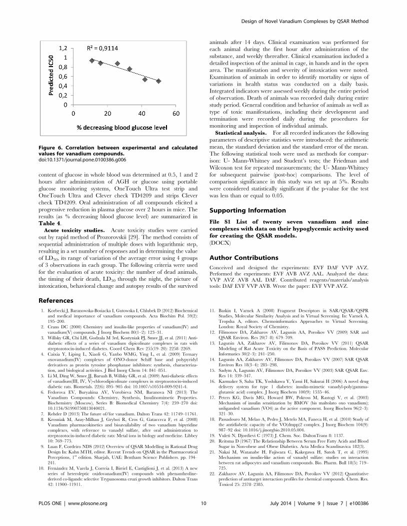
correlation (R2 = 0.91) between experimental and calculated
values was observed (Fig. 6).
Estimation of rodent acute toxicity is an important task in drug
design and risk assessment of chemicals. LD50 value is one of the
important characteristics of acute toxicity that corresponds to the
dose causing 50% mortality within 24 hours of administration. In
order to evaluate the toxicity of selected complexes, we used
‘‘GUSAR online’’ web service which allows predicting the acute
rat toxicity (LD50 values) with oral administration for compounds
based on their structures. Obtained values of LD50 for selected
complexes and experimental data are shown in Table 5. It should
be noted that the class of toxicity and values of LD50 for all tested
complexes were predicted accurately.
Conclusions
Five coordination complexes of vanadium were synthesized and
tested in vivo for their hypoglycemic activities and toxicity and
were compared with the predictive data. Reasonable correspon-
dence between the experimental and predicted values of toxicity
and hypoglycemic activity for vanadium compounds indicates that
GUSAR software may be successfully applied to explore the
structure-activity relationships of metal complexes. Bis{tert-
butyl[amino(imino)methyl]carbamato}oxovanadium(IV) which
IC50 value was almost twice less than those obtained by vanadyl
sulfate was identified as the most potent hypoglycemic agent
among the synthesized compounds. We have shown that it may be
possible to develop some vanadium compounds which have
stronger selectivity against FFAs.
Methods
QSAR MethodGUSAR (version 2011) is based on Multilevel and Quantitative
Neighborhoods of Atoms (MNA, QNA) descriptors [13,14]. The
calculation of QNA descriptors is based on the connectivity matrix
(C), and also, on the standard values of ionization potential (IP)
and electron affinity (EA) of atoms in a molecule [11,12].
For any given atom i, the QNA descriptors are calculated as
follows:
Pi~Bi
X
k
(Exp({1
2C))ikBk,
Qi~Bi
X
k
(Exp({1
2C))
ikBkAk,
with Ak~1
2(IPkzEAk), Bk~(IPk{EAk){
12.
Figure 3. 51V-NMR spectra for vanadium (V). The wide spectral dispertion of the signals as characteristic of vanadium NMR spectra (A) complexI (B); complex V (C). IR (D) and Raman (E) spectra for vanadium complex II.doi:10.1371/journal.pone.0100386.g003
Design of Novel Vanadium Complexes by QSAR Method
PLOS ONE | www.plosone.org 6 July 2014 | Volume 9 | Issue 7 | e100386

The P and Q values are calculated for all atoms of the molecule.
Two-dimensional Chebyshev polynomials are used for approxi-
mating the functions P and Q. Thus, the independent regression
variables are calculated as average values of particular two-
dimensional Chebyshev polynomials of P and Q values for the
atoms in a molecule.
In addition, GUSAR allows the creation of QSAR models
based on predicted biological activity profiles of compounds. This
is done by using the PASS algorithm on each compound’s
representation as a list of MNA descriptors for the prediction of
the compound’s biological activity profile [22,23]. The PASS
program version of 10.1 predicts 4130 types of biological activity
with a mean prediction accuracy of about 95%. The list of
predicted biological activities includes pharmacotherapeutic ef-
fects, mechanisms of action, adverse and toxic effects, metabolic
terms, susceptibility to transporter proteins and activities related to
gene expression. The results of the PASS procedure are output as
a list of the difference between the probability, for each biological
Figure 4. Molecular structure and the atomic numbering for complex I(C), complex II(A), complex IV(D, E) and complex V(B). Theellipsoids of thermal vibrations are shown at the 50% probability level.doi:10.1371/journal.pone.0100386.g004
Design of Novel Vanadium Complexes by QSAR Method
PLOS ONE | www.plosone.org 7 July 2014 | Volume 9 | Issue 7 | e100386
activity, of the compound to be active (Pa) or to be inactive (Pi). For
obtaining the different QSAR models in GUSAR, subsets of these
Pa-Pi values were randomly selected from the total list of predicted
biological activities as input independent variables for the
regression analysis. The size of these subsets depends on the
number of compounds in the training set. If the number of
compounds in the training set falls between 10 and 30, then the
number of initial variables is closer to the number of compounds in
the training set.
GUSAR (version 2011) also allows the calculation of whole-
molecule descriptors: topological length, topological volume and
lipophilicity. These descriptors are used in combination with the
QNA and MNA descriptors.
For generation of the QSAR models, GUSAR uses a self-
consistent regression (SCR) algorithm. SCR is based on the
regularized least-squares method. Unlike stepwise regression and
other methods of combinatorial search, the initial SCR model
includes all regressors. The basic purpose of the SCR method is to
remove the variables that poorly describe the modeled value but to
retain the set of variables correctly representing the existing
relationship. The number of final variables in the QSAR equation
selected after the SCR procedure is typically significantly lower
than the number of the initial variables. The details of the
algorithms for descriptor calculation and the self-consistent
regression methods have been described previously [22,23].
For assessment of the applicability domain of the obtained
models GUSAR uses the three different approaches: similarity,
leverage, and accuracy assessment. The detailed description of
these approaches is presented here [24].
The QSAR models obtained using both MNA and QNA
descriptors are integrated into the consensus model which is
further used for prediction. The final predicted value is estimated
by taking into account a weighted average of the predicted values
from each obtained QSAR model (QSAR models provide the
predictions that are within the respective applicability domains).
The predicted value obtained from each developed model is
weighted on similarity value calculated during the evaluation of
applicability domain.
Chemistry MethodGeneral methods for preparation of the vanadium
derivatives. All reagents and solvents were used without further
purification or drying. All the reagents (including reference
material VOSO4) were purchased from Vekton (Russia). All
reactions were proceeded in thermostat reactor under argon
atmosphere. Beforehand, the solvents were dried and distilled
under nitrogen using standard methods. Elementary analyses of H,
C and N were performed with a Carlo Erba microanalyzer. 1H-
NMR spectra at 400MHz were recorded on Varian XR-400
instrument, 51V-NMR spectra at 52.6MHz were recorded on
Tesia Bruker AL-200 instrument, and mass spectra were obtained
on a Finnigan MAT 112S spectrometer. IR spectra were obtained
on a Specord M80 (Carl Zeiss) spectrophotometer within the
spectral range of 400–4000 cm21 under resolution of 2.5–4 cm21.
Spectra of the solutions in CH2Cl2 or CHCl3 were shown to
represent pure solvents; the films were obtained on a KBr
substrate by evaporation of the solvent. X-Ray diffraction study of
the single crystal was performed on an Enraf-Nonius CAD4
automated diffractometer. The unit cell parameters were deter-
mined and refined using 25 reflections in the range of 15u#h#
16u(lMoKa radiation graphite monochromator).
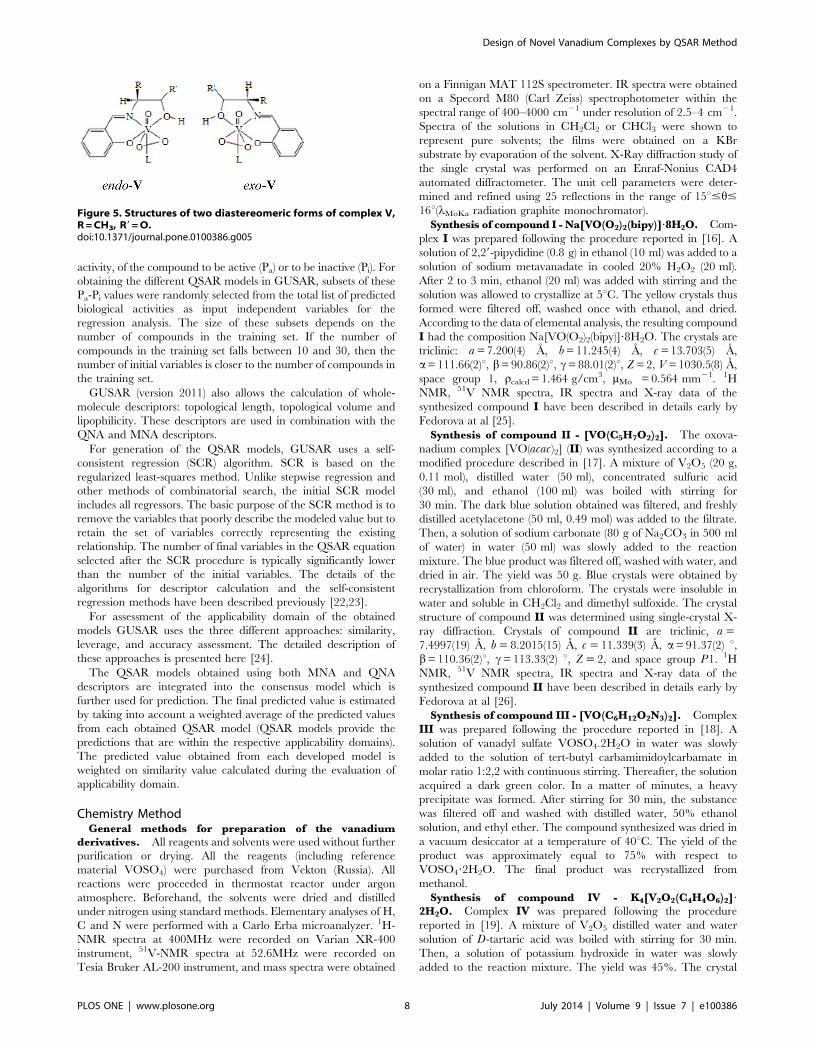
Synthesis of compound I - Na[VO(O2)2(bipy)]?8H2O. Com-
plex I was prepared following the procedure reported in [16]. A
solution of 2,29-pipydidine (0.8 g) in ethanol (10 ml) was added to a
solution of sodium metavanadate in cooled 20% H2O2 (20 ml).
After 2 to 3 min, ethanol (20 ml) was added with stirring and the
solution was allowed to crystallize at 5uC. The yellow crystals thus
formed were filtered off, washed once with ethanol, and dried.
According to the data of elemental analysis, the resulting compound
I had the composition Na[VO(O2)2(bipy)]?8H2O. The crystals are
triclinic: a = 7.200(4) A, b = 11.245(4) A, c = 13.703(5) A,
a= 111.66(2)u, b= 90.86(2)u, c= 88.01(2)u, Z = 2, V = 1030.5(8) A,
space group 1, rcalcd = 1.464 g/cm3, mMo = 0.564 mm21. 1H
NMR, 51V NMR spectra, IR spectra and X-ray data of the
synthesized compound I have been described in details early by
Fedorova at al [25].
Synthesis of compound II - [VO(C5H7O2)2]. The oxova-
nadium complex [VO(acac)2] (II) was synthesized according to a
modified procedure described in [17]. A mixture of V2O5 (20 g,
0.11 mol), distilled water (50 ml), concentrated sulfuric acid
(30 ml), and ethanol (100 ml) was boiled with stirring for
30 min. The dark blue solution obtained was filtered, and freshly
distilled acetylacetone (50 ml, 0.49 mol) was added to the filtrate.
Then, a solution of sodium carbonate (80 g of Na2CO3 in 500 ml
of water) in water (50 ml) was slowly added to the reaction
mixture. The blue product was filtered off, washed with water, and
dried in air. The yield was 50 g. Blue crystals were obtained by
recrystallization from chloroform. The crystals were insoluble in
water and soluble in CH2Cl2 and dimethyl sulfoxide. The crystal
structure of compound II was determined using single-crystal X-
ray diffraction. Crystals of compound II are triclinic, a =7.4997(19) A, b = 8.2015(15) A, c = 11.339(3) A, a= 91.37(2) u,b= 110.36(2)u, c= 113.33(2) u, Z = 2, and space group P1. 1H
NMR, 51V NMR spectra, IR spectra and X-ray data of the
synthesized compound II have been described in details early by
Fedorova at al [26].
Synthesis of compound III - [VO(C6H12O2N3)2]. Complex
III was prepared following the procedure reported in [18]. A
solution of vanadyl sulfate VOSO4?2H2O in water was slowly
added to the solution of tert-butyl carbamimidoylcarbamate in
molar ratio 1:2,2 with continuous stirring. Thereafter, the solution
acquired a dark green color. In a matter of minutes, a heavy
precipitate was formed. After stirring for 30 min, the substance
was filtered off and washed with distilled water, 50% ethanol
solution, and ethyl ether. The compound synthesized was dried in
a vacuum desiccator at a temperature of 40uC. The yield of the
product was approximately equal to 75% with respect to
VOSO4?2H2O. The final product was recrystallized from
methanol.
Synthesis of compound IV - K4[V2O2(C4H4O6)2]?
2H2O. Complex IV was prepared following the procedure
reported in [19]. A mixture of V2O5 distilled water and water
solution of D-tartaric acid was boiled with stirring for 30 min.
Then, a solution of potassium hydroxide in water was slowly
added to the reaction mixture. The yield was 45%. The crystal
Figure 5. Structures of two diastereomeric forms of complex V,R = CH3, R9 = O.doi:10.1371/journal.pone.0100386.g005
Design of Novel Vanadium Complexes by QSAR Method
PLOS ONE | www.plosone.org 8 July 2014 | Volume 9 | Issue 7 | e100386

structures of compound IV was determined using single-crystal X-
ray diffraction. Complex IV with square–pyramidal coordination
geometry, space group P4(3)22, a = 7.9345 A, b = 7.9345 A,
c = 30.5035 A, V = 1920.39(22) A3, Z = 4, Rw = 3.11%. 1H
NMR, 51V NMR spectra, IR spectra and X-ray data of the
synthesized compound IV have been described in details earlier by
Fedorova at al [27].
Synthesis of compound V - [VO(C10H7O3)(H2O)]. Oxo-
vanadium complex V was synthesized according to a modified
procedure described earlier in [17]. The ligand in the complex is a
bivalent tridentate Schiff’s base, which was produced by the
reaction of salicylaldehyde (Sal) with a-amino acid. L-alanine
amino acid (0.1 mol) and sodium acetate (0.2 mol) were dissolved
in distilled water (200 ml). In order to dissolve the amino acid
completely, the solution was heated and filtered. A solution of
salicylaldehyde (0.1 mol) in ethanol (250 ml) was added to the
filtrate. A solution of vanadyl sulfate VOSO4?2H2O (0.085 mol) in
water (80 ml) was slowly added to the above solution with
continuous stirring. Thereafter, the solution acquired a dark
brown color. In a matter of minutes, a heavy precipitate was
formed. After stirring for 30 min, the substance was filtered off and
washed with distilled water, 50% ethanol solution, and ethyl ether.
The compound synthesized was dried in a vacuum desiccator at a
temperature of 40uC. The yield of the product was approximately
equal to 80% with respect to VOSO4?2H2O. The final product
was recrystallized from metha similarity nol. The blue crystals thus
prepared were insoluble in water, acetone, ether, and benzene and
soluble in methanol, pyridine, methylene chloride, and chloro-
form. The melting temperature of the crystals was estimated as Tm
, 250uC. Crystals of compound V are monoclinic, a = 8.5106(16)
A, b = 7.373(2) A, c = 9.1941(16) A, b= 101.88(1), Z = 2, and
space group P21. 1H NMR, 51V NMR spectra, IR spectra and X-
ray data of the synthesized compound V have been described in
details early by Fedorova at al [26].
Pharmacology MethodAll procedures were performed according to institutional
guidelines for animal experimentation; protocol was submitted
and approved by the Institutional Committee for Laboratory
Animal Use and Care of the Saint-Petersburg Chemical Pharma-
ceutical Academy. Adult male Wistar rats and mongrel mice
obtained from the ‘Rappolovo’ lab animals breeding farm near St.
Petersburg, belonged to Russian Academy of Medical Science
were used. All experiments were approved by the Animal Ethics
Committee of the Saint-Petersburg Chemical Pharmaceutical
Academy.Insulin-mimetic action of vanadium complexes. All rats
(250–300 g) and mice (22–25 g) were housed in a temperature
controlled room (24uC61uC) and they had received standard food
pellets and tap water until they weighed. At the end of the study
(24 h after CIN induction), all animal test subjects were
euthanized by inhalation of ether anesthesia. Rat adipocytes were
prepared from the fat pads of male Wistar rats by collagenase
digestion according to the method of Rodbell [28]. Freshly
prepared adipocytes were incubated (37uC, 30 min) with solutions
of the vanadium compounds at three concentrations (0.1, 0.5 and
1 mM in isotonic saline and 5 mM glucose), followed by 3 hours
of incubation with epinephrine. The inhibitory effect was
determined in terms of the decrease of the amount free fatty
acids released in adipocytes. All assays were performed in
duplicate or triplicate. Table 4 summarizes the IC50 values,
which are defined as the concentrations c(V) at which 50%
inhibition of FFA release takes place. Additionally, five coordina-
tion complexes of vanadium and reference material VOSO4 have
been tested in vivo for their hypoglycemic activities. Investigations
were carried out on mongrel white mice.Hyperglycemic activity. Determination of hypoglycemic
activity of 5 complexes was performed on 75 mice. Adrenaline
hyperglycemia was caused by subcutaneous administration of
0.1% epinephrine hydrochloride (AGH) at a dose of 1 mg/kg.
With a single run all the compounds were administered orally 1
hour prior to the AGH, at 1/10 of the LD50. In each experimental
group there were 3 animals and 3 replicates were performed. The
Table 4. Predictive values of IC50 in mM (FFAs) for vanadium compounds and in vivo observations IC50 in mM (FFAs) anddecreasing blood glucose level.
Compounds VOSO4 Complex I Complex II Complex III Complex IV Complex V
IC50 predicted – 0.50 0.75 0.53 0.93 0.87
IC50 exp (6S.D.) 1.060.09 0.7260.09 0.9560.09 0.5460.07 0.9560.10 0.8160.06
decreasing blood glucose level 16% 50% 33% 42% 11% 26%
doi:10.1371/journal.pone.0100386.t004
Table 5. Predicted and observed values LD50 of vanadium compounds.
# Compounds LD50pred/Class in AD LD50exp/Class in AD
1 VOSO4 518.6/3 448(356–540)/3
2 Complex I 279.0/3 108(95–122)/3
3 Complex II 251.0/3 250(210–280)/3
4 Complex III 211.0/3 190(170–210)/3
5 Complex IV 357.1/3 261(210–312)/3
6 Complex V 265.1/3 245(210–280)/3
doi:10.1371/journal.pone.0100386.t005
Design of Novel Vanadium Complexes by QSAR Method
PLOS ONE | www.plosone.org 9 July 2014 | Volume 9 | Issue 7 | e100386
content of glucose in whole blood was determined at 0.5, 1 and 2
hours after administration of AGH or glucose using portable
glucose monitoring systems, OneTouch Ultra test strip and
OneTouch Ultra and Clever check TD4209 and strips Clever
check TD4209. Oral administration of all compounds elicited a
progressive reduction in plasma glucose over 2 hours in mice. The
results (as % decreasing blood glucose level) are summarized in
Table 4.Acute toxicity studies. Acute toxicity studies were carried
out by rapid method of Prozorovskii [29]. The method consists of
sequential administration of multiple doses with logarithmic step,
resulting in a set number of responses and in determining the value
of LD50, its range of variation of the average error using 4 groups
of 3 observations in each group. The following criteria were used
for the evaluation of acute toxicity: the number of dead animals,
the timing of their death, LD50 through the night, the picture of
intoxication, behavioral change and autopsy results of the survived
animals after 14 days. Clinical examination was performed for
each animal during the first hour after administration of the
substance, and weekly thereafter. Clinical examination included a
detailed inspection of the animal in cage, in hands and in the open
area. The manifestation and severity of intoxication were noted.
Examination of animals in order to identify mortality or signs of
variations in health status was conducted on a daily basis.
Integrated indicators were assessed weekly during the entire period
of observation. Death of animals was recorded daily during entire
study period. General condition and behavior of animals as well as
type of toxic manifestations, including their development and
termination were recorded daily during the procedures for
monitoring and inspection of individual animals.
Statistical analysis. For all recorded indicators the following
parameters of descriptive statistics were introduced: the arithmetic
mean, the standard deviation and the standard error of the mean.
The following statistical tools were used as methods for compar-
ison: U- Mann-Whitney and Student’s tests; the Friedman and
Wilcoxon test for repeated measurements; the U- Mann-Whitney
for subsequent pairwise (post-hoc) comparisons. The level of
comparison significance in this study was set up at 5%. Results
were considered statistically significant if the p-value for the test
was less than or equal to 0.05.
Supporting Information
File S1 List of twenty seven vanadium and zinccomplexes with data on their hypoglycemic activity usedfor creating the QSAR models.
(DOCX)
Author Contributions
Conceived and designed the experiments: EVF DAF VVP AVZ.
Performed the experiments: EVF AVB AVZ AAL. Analyzed the data:
VVP AVZ AVB AAL DAF. Contributed reagents/materials/analysis
tools: DAF EVF VVP AVB. Wrote the paper: EVF VVP AVZ.
References
1. Korbecki J, Baranowska-Bosiacka I, Gutowska I, Chlubek D (2012) Biochemical
and medical importance of vanadium compounds. Acta Biochim Pol. 59(2):
195–200.
2. Crans DC (2000) Chemistry and insulin-like properties of vanadium(IV) and
vanadium(V) compounds. J Inorg Biochem 80(1–2): 123–31.
3. Willsky GR, Chi LH, Godzala M 3rd, Kostyniak PJ, Smee JJ, et al. (2011) Anti-
diabetic effects of a series of vanadium dipicolinate complexes in rats with
streptozotocin-induced diabetes. Coord Chem Rev 255(19–20): 2258–2269.
4. Caixia Y, Liping L, Xiaoli G, Yanbo WMG, Ying L, et al. (2009) Ternary
oxovanadium(IV) complexes of ONO-donor Schiff base and polypyridyl
derivatives as protein tyrosine phosphatase inhibitors: synthesis, characteriza-
tion, and biological activities. J Biol Inorg Chem 14: 841–851.
5. Li M, Ding W, Smee JJ, Baruah B, Willsky GR, et al. (2009) Anti-diabetic effects
of vanadium(III, IV, V)-chlorodipicolinate complexes in streptozotocin-induced
diabetic rats. Biometals. 22(6): 895–905 doi: 10.1007/s10534-009-9241-4.
6. Fedorova EV, Buryakina AV, Vorobieva NM, Baranova NI (2013) The
Vanadium Compounds: Chemistry, Synthesis, Insulinomimetic Properties.
Biochemistry (Moscow), Series B: Biomedical Chemistry 7(4): 259–270 doi:
10.1134/S1990750813040021.
7. Rehder D (2013) The future of/for vanadium. Dalton Trans 42: 11749–11761.
8. Krosniak M, Azay-Milhau J, Grybos R, Cros G, Gatacceca F, et al. (2008)
Vanadium pharmacokinetics and bioavailability of two vanadium bipyridine
complexes, with reference to vanadyl sulfate, after oral administration to
streptozotocin-induced diabetic rats: Metal ions in biology and medicine. Libbey
10: 769–775.
9. Luan F, Cordeiro NDS (2012) Overview of QSAR Modelling in Rational Drug
Design In: Kahn MTH, editor. Recent Trends on QSAR in the Pharmaceutical
Perceptions, 1st edition. Sharjah, UAE: Bentham Science Publishers. pp. 194–
241.
10. Fernandez M, Varela J, Correia I, Birriel E, Castiglioni J, et al. (2013) A new
series of heteroleptic oxidovanadium(IV) compounds with phenanthroline-
derived co-ligands: selective Trypanosoma cruzi growth inhibitors. Dalton Trans
42: 11900–11911.
11. Baskin I, Varnek A (2008) Fragment Descriptors in SAR/QSAR/QSPR
Studies, Molecular Similarity Analysis and in Virtual Screening. In: Varnek A,
Tropsha A, editors. Chemoinformatics Approaches to Virtual Screening.
London: Royal Society of Chemistry.
12. Filimonov DA, Zakharov AV, Lagunin AA, Poroikov VV (2009) SAR and
QSAR Environ. Res 20(7–8): 679–709.
13. Lagunin AA, Zakharov AV, Filimonov DA, Poroikov VV (2011) QSAR
Modeling of Rat Acute Toxicity on the Basis of PASS Prediction. Molecular
Informatics 30(2–3): 241–250.
14. Lagunin AA, Zakharov AV, Filimonov DA, Poroikov VV (2007) SAR QSAR
Environ Res 18(3–4): 285–298.
15. Sadym A, Lagunin AV, Filimonov DA, Poroikov VV (2003) SAR QSAR Env.
Res 14: 339–347.
16. Karmaker S, Saha TK, Yoshikawa Y, Yasui H, Sakurai H (2006) A novel drug
delivery system for type 1 diabetes: insulin-mimetic vanadyl-poly(gamma-
glutamic acid) complex. J Inorg Biochem 100(9): 1535–46.
17. Peters KG, Davis MG, Howard BW, Pokross M, Rastogi V, et al. (2003)
Mechanism of insulin sensitization by BMOV (bis maltolato oxo vanadium);
unliganded vanadium (VO4) as the active component. Inorg Biochem 96(2–3):
321–30.
18. Passadouro M, Melao A, Pedro J, Metelo MA, Faneca H, et al. (2010) Study of
the antidiabetic capacity of the VO(dmpp)2 complex. J Inorg Biochem 104(9):
987–92 doi: 10.1016/j.jinorgbio.2010.05.004.
19. Vuleti N, Djordievi C (1973) J. Chem. Soc. DaltonTrans 8: 1137.
20. Reitsma D (1967) The Relationship Between Serum Free Fatty Acids and Blood
Sugar in Non-obese and Obese Diabetics. Acta Medica Scandinavica 182(3).
21. Nakai M, Watanabe H, Fujiwara C, Kakegawa H, Satoh T, et al. (1995)
Mechanism on insulin-like action of vanadyl sulfate: studies on interaction
between rat adipocytes and vanadium compounds. Bio. Pharm. Bull 18(5): 719–
725.
22. Zakharov AV, Lagunin AA, Filimonov DA, Poroikov VV (2012) Quantitative
prediction of antitarget interaction profiles for chemical compounds. Chem. Res.
Toxicol 25: 2378–2385.
Figure 6. Correlation between experimental and calculatedvalues for vanadium compounds.doi:10.1371/journal.pone.0100386.g006
Design of Novel Vanadium Complexes by QSAR Method
PLOS ONE | www.plosone.org 10 July 2014 | Volume 9 | Issue 7 | e100386

23. Kokurkina GV, Dutov MD, Shevelev SA, Popkov SV, Zakharov AV, et al.
(2011) Synthesis, antifungal activity and QSAR study of 2-arylhydroxynitroin-
doles. Eur. J. Med. Chem 46: 4374–4382.
24. Zakharov AV, Peach ML, Sitzmann M, Filippov IV, McCartney HJ, et al.
(2012) Computational tools and resources for metabolism-related property
predictions. 2. Application to prediction of half-life time in human liver
microsomes. Future Med. Chem 4: 1933–1944.
25. Fedorova EV, Rybakov VB, Senyavin VM, Aslanov LA, Anisimov AV (2002)
Russian Journal of Coordination Chemistry 28 (7): 483–486.
26. Fedorova EV, Rybakov VB, Senyavin VM, Aslanov LA, Anisimov AV (2005)
Crystallography Reports 50 (2): 224–229.27. Fedorova EV, Vorobyeva NM (2011) II International Scientifically-Practical
Conference ‘‘The High Technologies, Fundamental and Applied Researches in
Physiology and Medicine 3: 125.28. Rodbell M (1964) Metabolism of Isolated Fat Cells: I. Effects of hormones on
glucose metabolism and lipolysis. J. Biol. Chem 239: 375–380.29. Prozorovskii VB, Prozorovskaya MP, Demchenko VM (1978) Express method of
determining the median effective dose and its error. Pharmacology and
Toxicology 4: 497–500.
Design of Novel Vanadium Complexes by QSAR Method
PLOS ONE | www.plosone.org 11 July 2014 | Volume 9 | Issue 7 | e100386