Histological Liver Changes in Streptozotocin induced Diabetic Mice
11
Int. Med J Vol. 8 No 1 June 2009 Histological Liver Changes in Streptozotocin induced Diabetic Mice Imad M. Al-Ani a ; Noman D. Salih Al-Mishadani b ; Raad K. Muslih a and Salim R.Hamoodi c a Dept. of Basic Medical Sciences, Kulliyyah of Medicine, International Islamic University Malaysia, Kuantan, Malaysia; b Dept. of Biology, College of Science, Almustansiriah Univ. Baghdad Iraq; c Dept of pathology, College of Medicine, Baghdad Univ. Baghdad Iraq. ABSTRACT: Streptozotocin (STZ) is selectively toxic to cells in the pancreatic islets. It is well known that STZ causes specific death of B cells and induces diabetes mellitus. This study has been conducted to detect the histological changes that could be induced in liver of adult diabetic mice. The experiments were performed using a single dose of STZ (150mg/Kg. I.P.). Animals were killed two, four and six weeks after injection. The relative percentage of liver weight and liver histology were studied. The results showed several morphological and histological alterations in liver tissues, indicated by increase in the liver percentage of liver weight, glycogen reduction, associated with lipid deposition, inflammatory cells infiltration and Kupffer cells hyperplasia. Furthermore, this study illustrated the worsening of liver histology over a short time in STZ diabetic mice. INTRODUCTION Streptozotocin (STZ) is an N-nitros0-n-methylurea derivative of 2-deoxy-d- glucose, produced by Streptomyces acromogenes, and has been shown in animal’s model to induce a chronic diabetic state by destruction of B bells in the pancreatic islet tissue 1 . Streptozotocin induced diabetes mellitus in many animal species has been reported to resemble human hyperglycemic diabetes mellitus 2 . It has been reported that STZ selectively damages pancreatic B cells and produces much less toxic side effect than other chemical diabetogenic agents 3 and induces diabetes similar to poorly treated human diabetes, it develops many features seen in human patients 4 . Diabetes mellitus is a commonly occurring
-
Upload
independent -
Category
Documents
-
view
0 -
download
0
Transcript of Histological Liver Changes in Streptozotocin induced Diabetic Mice
Int. Med J Vol. 8 No 1 June 2009
Histological Liver Changes in Streptozotocin induced Diabetic Mice
Imad M. Al-Ania; Noman D. Salih Al-Mishadanib; Raad K. Musliha and Salim
R.Hamoodi c
aDept. of Basic Medical Sciences, Kulliyyah of Medicine, International Islamic University Malaysia, Kuantan, Malaysia; bDept. of Biology, College of Science, Almustansiriah Univ. Baghdad Iraq; cDept of pathology, College of Medicine, Baghdad Univ. Baghdad Iraq.
ABSTRACT:
Streptozotocin (STZ) is selectively toxic to cells in the pancreatic islets. It is well
known that STZ causes specific death of B cells and induces diabetes mellitus.
This study has been conducted to detect the histological changes that could be
induced in liver of adult diabetic mice. The experiments were performed using a
single dose of STZ (150mg/Kg. I.P.). Animals were killed two, four and six weeks
after injection. The relative percentage of liver weight and liver histology were
studied. The results showed several morphological and histological alterations in
liver tissues, indicated by increase in the liver percentage of liver weight,
glycogen reduction, associated with lipid deposition, inflammatory cells infiltration
and Kupffer cells hyperplasia. Furthermore, this study illustrated the worsening of
liver histology over a short time in STZ diabetic mice.
INTRODUCTION
Streptozotocin (STZ) is an N-nitros0-n-methylurea derivative of 2-deoxy-d-
glucose, produced by Streptomyces acromogenes, and has been shown in
animal’s model to induce a chronic diabetic state by destruction of B bells in the
pancreatic islet tissue 1. Streptozotocin induced diabetes mellitus in many animal
species has been reported to resemble human hyperglycemic diabetes mellitus
2. It has been reported that STZ selectively damages pancreatic B cells and
produces much less toxic side effect than other chemical diabetogenic agents 3
and induces diabetes similar to poorly treated human diabetes, it develops many
features seen in human patients 4. Diabetes mellitus is a commonly occurring

disease, characterized by elevated plasma glucose concentrations resulting from
insufficient insulin, insulin resistance, or both 5, and it affects carbohydrate, fat
and protein metabolism. Diabetes is a complex and multifarious group of
disorders characterized by hyperglycemia that has reached epidemic proportions
in the present century 6 Diabetes is known to produce substantial changes in
intracellular metabolism in most tissues, including liver 7, 8.
Because of the importance of the liver in carbohydrate metabolism, the present
study is to investigate liver histopathological changes possibly occurring in
diabetic mice at different periods.
MATERIALS & METHODS
The experiments were performed on 200 male Swiss albino mice of the balb/c,
weighing 20-25 grams, in good health housed in plastic cages with free access to
water and food. They were bred in the animal house (Almustansiriah College of
Science), kept at room temperature and fed standard chew of pellets (Al-
Autiafiah Company) and water was provided adlibitum.
The animals were randomly divided into four groups of fifty; the first group was
the control, they were given saline-citrate buffer; the second, third, and fourth
groups were the diabetic groups, after fasting over night, injected with a single
dose of STZ (150mg/Kg, Sigma Chem. Co., USA) dissolved in citrate buffer PH
5.4 In order to prevent hypoglycemia, STZ-treated animals received a solution of
10% glucose instead of normal drinking water over the 24 hours following the
treatment.
The animals were sacrificed at the second, fourth and sixth week. After
sacrificed, the abdominal cavity was opened, the liver was removed to obtain its
weight, and small specimens of the liver were taken for histological examination.
Liver specimens were fixed in Boun’s solution for 12-16 hours. After fixation, they
were washed using many changes of 50%, 70% ethanol to remove the yellow
colour of picric acid, then they dehydrated through graded alcohols and cleared

using two changes of xylene and embedded in paraffin wax. Serial transverse
sections of 4-5 micron thickness were prepared using the microtome. The
sections then floated in a water bath at 50 C, and mounted on slides. The
sections were stained by Harris’s hematoxylin and eosin, others stained by
periodic Acid-Schiff stain (PAS).
RESULTS
A significant increase in the relative percentage of liver weight has been shown in
this study (Fig. 1).
Liver biopsy of the animal of the control group didn’t show any histological
changes during the period of the experiments, the hepatic lobules were seen
normally with polygonal hepatocytes having regular nucleus and cytoplasm (plate
1).
In the diabetic animals, two weeks after STZ-injection, the liver showed several
alterations including mild degree of fatty changes, cloudy swelling, mild infiltration
of lymphocytes with hemorrhage (plate.2).after weeks, STZ-diabetic animals
showed more progressive changes, sever congestion, necrotic foci, hydropic
changes, aggregation of lymphocytes between the hepatocytes (plate 3).
In the last group, mononuclear inflammatory cells infiltration, sever hydropic
degeneration changes and kupffer cell hyperplasia can be recognized (plate. 4).
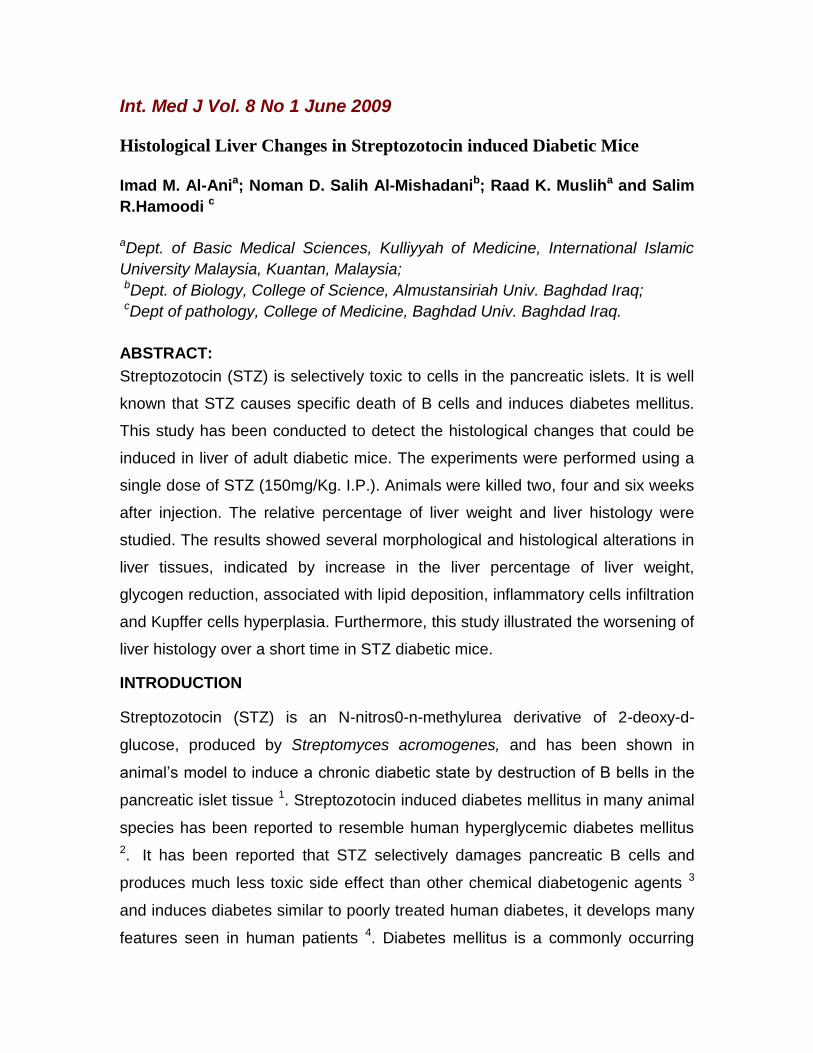
Furthermore, alterations in glycogen contents in the hepatocytes were obtained n
specime4ns stained with PAS stain (plates. 5, 6, 7).


DISCUSSION
Attention has long centered on the liver in diabetes mellitus because of the
importance of this organ in carbohydrate metabolism and regulation of blood
sugar. Many studies 9, 10 revealed the occurrence of hepatic changes in some
cases of diabetic patients. Liver biopsy showed an accumulation of fats into the
hepatocytes, lead to a significant increase in liver weight. Liver enlargement has
been indicated in experimental diabetic rats 11 and mice 12.
Diabetes mellitus is one of the most common causes of fatty liver and the
frequent increase in the liver size in patients with diabetes mellitus was
recognized 9, 10. In the present study, there was a significant increase in the
weight of liver tissue in STZ diabetic mice as compared with the control group.
These results are in agreement with those of Cefalu et al 11 and Kume et al 12
and were related to the accumulation of fat into the hepatocytes, fatty changes in
hepatocytes, hydropic changes and cloudy swelling were observed in the present
study. It is well established that if a fatty liver from any cause persists, cirrhosis

will develop 13, 14. In the chronic phase of diabetes, an increase in liver weight or
hepatomegaly might occur due to glycogen deposition or fatty metamorphosis 15.
After four weeks, STZ-diabetic mice showed more progressive changes, there
was severe congestion in the in the portal area with necrotic foci, hydropic
changes and aggregation and infiltration of lymphocytes between hepatocytes,
further more there were hyperplasia of kupffer cells and significant gradual
reduction in glycogen content of the hepatocytes. Hamilton 10 indicated that
diabetes mellitus is one of the most common causes of fatty liver where fats are
accumulated in the hepatocytes; he indicates that fat comprises as much as 40%
of liver weight in patients with diabetes mellitus (It is 5% in normal liver).
Herman et al 16 related the hepatomegaly observed in STZ-induced diabetes in
rats to the hyperplasia in early phase and to the decreased apoptosis in the later
stage. Itoh et al 17 have considered that fatty infiltration of liver as a precursor of
cirrhosis in diabetic patients. Falchuk et al 13 showed hepatic fatty steatosis and
pericentral fibrosis in diabetic patients. They recognized hepatocytes were
markedly swollen and suggested that these abnormalities may represent an
intermediate lesion between fatty steatosis and cirrhosis. Nanji et al 9 mentioned
that the damage was mainly to the plasma membrane of the hepatocytes and
could be attributed to the elevation in aspartate aminotransferases in patients
with fatty infiltrations. The activity of serum alkaline phosphatase, aspartate
aminotransferases, alanine aminotransferases and levels of biluribin and
cholesterol were increased significantly in STZ- diabetic mice 18. Papaccio et al 19
found that STZ interferes with cellular metabolic oxidative mechanisms. Satav
and Katyara 7 studied the effect of STZ-induced diabetes on the oxidative energy
metabolism in rat liver mitochondria and found reduction in respiratory activity.
Lukivskaye et al 20 related the liver pathological changes in alloxan diabetic rats
to the mitochondrial abnormalities. Many authors suggested that liver
mitochondrial dysfunction in diabetes is related to the oxidative stress enhanced
in diabetic animals 21 and patients 22.

The decreased cellularity within the islets of langerhans observed in rats 19 and
rabbits 8 reflect the cytotoxicity of STZ on the B-cells. Whether the present
histopathological finding in the liver of mice indicates that STZ has a direct
cytoxic effect on the liver hepatocytes or it is indirectly through the induced
diabetes mellitus need further investigations.
REFERENCES
1. Hardman, J.G. and Limbird, L.E. (2001): Goodman & Gilman’s, The
pharmacological Basis of Therapeutics. 10th Ed. New York: McGraw-Hill, P.
1399.
2. Weir, G.C., Clore, E.T., Zmachinski, C.J. and Bonner-Weir,S. (1981): Islet
secretion in a new experiment model for non-insulin dependent diabetes.
Diabetes, 30, 590-595.
3. Junod,A.; Lambert,A.E.; Stauffacher,W. & Renold,A.E. (1969). Diabetogenic
Action of STZ. Relationship of Dose to metabolic Response. J. Clin. Invest.
48: 2129-2139
4. Shafrir, E.(1996). Animal Models of Diabetes in Pregnancy. Diabetes
Reviews, 4 (1): 114-128.
5. Ido, Y; McHowat, J.; Chang K.C.; Arrigoni-Martelli,E.; Orfalain, Z.; Kilo, C.;
Corrp,B. & Williamson, J.R. (1994), Neural Dysfunction and Metabolic
Imbalances in Diabetic Rats. Diabetes, 43: 1479-1477.
6. Noor, A., Gunasekaran, S., Soosaihanickam, A. and Vijayalakshmi, M.A.
(2008): Antidaiabetic activity of Aloevera and histology of organs in
Streptozotocin induced diabetic rats. Current Sci. 94, 1070-1076.
7. Satav, J.G. and Katyare, S.S. (2004): Effect of Streptozotocin induced
diabetes on oxidative energy metabolism in rat liver. Indian J. Clin. Biochem.,
19, 23-31.

8. Sajad, H.M,, Abdui-Baqui, R.C., Bhgat, M.M. and Abdul-Wahid, S. (2008):
Biochemical and histomorphological study of Streptozotocin induced
diabetes mellitus in rabbits. Pakistan J. Nutrition, 7, 359-364.
9. Nanji, A.A.; French, S.W. & Freeman, J.B. (1986). Serum Alanine
Aminotransferases Ratio and Degree of Fatty Liver in Morbidly Obese
Patients. Enzyme, 36: 266-269.
10. Hamilton, H.K. (1987). Professional Guide to Diseases. An up to Date
Encyclopedia of Illness, Disorders and their Treatment, 2nd ed., Spring House
Corporation Book Division, USA, pp. 691-715.
11. Cefalu, W.T.; Wang, A.Q.; Bell-Farrow, A. & Ralapati, S. (1991). Liver and
Kidney Tissue Membrane as Tissue Markers for Non-Enzymatic
Glycosylation, 40: 902-907
12. Kume, E,, Ohmachi, Y., Hagaki, S. Tamura, K. and Doi, K. (1994): Hepatic
changes of mice in subacute phase of Streptozotocin induced diabetes. Exp.
Toxicol. Pathol. 46, 368-374.
13. Falchuk, K.R.; Fiske, S.C.; Haggitt, R.C.; Federman, M. & Treg, C. (1980).
Pericentral Hepatic Fibrosis & Intracellular Hyaline in Diabetes Mellitus,
Gastroenterology, 78:535-541
14. Mohammed, S.B.O. (1989). Liver Biochemical Abnormalities in Diabetic
Patients. M.Sc. Thesis. Univ. of Baghdad, Baghdad, Iraq.
15. DeMoria,N., Coantoni, A. and Van Thiel, D.H. (2001): The liver endocrine
function. In: Kenneth, L. Ed. Principle and Practic of Endocrinology and
Metabolism, 3rd. Ed. Philadeliphia: Lippincott Williams & Wilkins, P. 1870-
1885.

16. Herrman, C.E., Sanders, R.A., Klaunig, J.E. Schwarz, L.R. and Watkins, J.B.
(1999): Decreased apoptosis as a mechanism for hepatomegaly in
Streptozotocin induced diabetic rats. Toxicol Sci. 50, 146-145.
17. Itho, S.; Tsukada, Y.; Motomura, Y. & Ichinoe, A. (1979). Five Patients with
Nonalcoholic Diabetic Cirrhosis. Acta Hepatol Gastroenterol., 26:90-97.
18. Al-Mashhadany, N.D.S. (2000). Biochemical and Histopathological Studies
on liver of Streptozotocin Diabetic Mice. M.Sc. Thesis, Al-Mustansiriah Univ.
Baghdad, Iraq.
19. Papaccio, G., Pisanti, F.A., Latronico, M.V., Ammendola, A. and Galidieri, M.
(2000): Multiple low doses and single high dose treatment with Streptozotocin
do not generate nitric oxide. J. Cell Biochem., 77, 82-91.
20. Lukivskaya, O., Eleonora, P and Vyacheslva, U.B. (2007): Protective effect
of of ursodeoxycholic acid on liver mitochondrial function in rats with alloxan-
induced diabetes: Link with oxidative stress. Life Sci. 80, 2394-2402.
21. Kucharska, J., braunova, J., Ulicna, O., Zlatos, l. and Gvozdjakova, A.
(2000): Defect of co-enzyme Q in heart and liver mitochondria in rats with
Streptozotocin induced diabetes. Physiological Res., 49, 411-418.
22. Bukker, S.J., Uzerman, R.G., Teerlink, T. Resterhoff, H.V. and Henie R.J.
(2000): Cytsolic triglycerides and oxidative stress in central obesity: the
missing link between excessive atherosclerosis, endothelial dysfunction and
beta-cell failure? Atherosclerosis, 148, 17-21.
Correspondence
Department of BMS,
Faculty of amedicine
International Islamic University Malaysia