Effect of Ramadan Fasting on Diabetic.............
13
Z.U.M.J.Vol.20; N.2; March; 2014 Effect of Ramadan Fasting on Diabetic…………. -193- EFFECT OF RAMADAN FASTING ON DIABETIC MICRO-VASCULAR COMPLICATION Mohamed E.G.Kamar , Abbas A.Orabi , Ihab M.Salem and Arafa M.EL- Shabrawy Diabetes &endocrinology unit, internal medicine department, faculty of medicine, Zagazig University, EGYPT ABSTRACT Fasting during Ramadan, one of the five pillars of Islam is an obligatory duty for all healthy adult Muslims. During Ramadan, Muslims must abstain from eating, drinking, taking oral medications, and smoking from the exact time of dawn until time of sunset; there are no restrictions on food or fluid intake between sunset and dawn. Islamic rules allow patients who are diseased and fasting may be hard or harmful for them not to fast. This study aimed to study the relation between Ramadan fasting and course of micro vascular complication of DM, and to find advices for patient during fasting to help in regression of micro vascular complication of DM. We started the study on 74 diabetic patients but only 64 completed the clinical visits with us and the other 10 missed from the follow up visits. Before Ramadan and After Ramadan fasting all patients were subjected to:. Electroretinogram (ERG), Mean Conduction Velocity (MCV), and Urinary Albumin Creatinine Ratio (UACR) in addition to serum insulin, proinsulin. The results showed There was increase in the mean oscillatory potentials in the second (OS2 ) of both eyes from 8.96±3.96 to 9.49±4.77.and other changes in the ERG which indicate significant improvement of diabetic retinopathy after Ramadan fasting , MCV decreased from 43.92± 21.68 to 39.85±13.50 ms. and albumin creatinine ratio was increased from 98.41 ±160.49 to 141.49 ±228.62. Conclusion: Fasting of the whole month of Ramadan has a beneficial effect on β cell function. The relation between Ramadan fasting and micro-vascular complications is variable, with some improvement of diabetic retinopathy. neuropathy show adverse effects. Ramadan fasting increase the albumin creatinine ratio but there is improvement among patients with microalbuminuria. Key words: Ramadan, Fasting, Diabetes, micro-vascular. INTRODUCTION asting during Ramadan, one of the five pillars of Islam is an obligatory duty for all healthy adult Muslims. , During Ramadan, Muslims must abstain from eating, drinking, taking oral medications, and smoking from the exact time of dawn until time of sunset; there are no restrictions on food or fluid intake between sunset and dawn. 1 Although, Ramadan fasting is safe and could not induce any harmful effect in young healthy subjects, those with various diseases should consult their physicians and follow scientific recommendations. 2 Islamic rules allow patients who are diseased and fasting may be hard or harmful for them not to fast, Despite of this, patients usually insist to fast. 3 Previous studies showed that Ramadan fasting caused significant changes in body weight 4 and different biochemical parameters. 5-6 Few studies investigated the relation between Ramadan fasting and diabetic microvascular complications. This may be due to the general concept that one month is a short duration not sufficient to affect such chronic complications of chronic disease (diabetes). In this study we tried to explore the effect of Ramadan fasting on diabetic microvascular complications, does Ramadan fasting in diabetic patients affect microvascular complications or not? Diabetic retinopathy may be the most common microvascular complication of diabetes. It is responsible for ∼ 10,000 new cases of blindness every year in the United States alone. The risk of developing diabetic retinopathy or other microvascular complications of diabetes depends on both the duration and the severity of hyperglycemia. Retinopathy may begin to develop as early as 7 years before the diagnosis of diabetes in patients with type 2 diabetes. 7 Electroretinogram (ERG) and diabetic retinopathy. Evidence has begun to point to the fact that even before vascular complications begin to manifest, neuronal cell death and dysfunction have already begun. 7 Electrophysiological studies of humans with diabetes could be used to assess alterations 6 ; moreover alterations in oscillatory potentials have been shown to predict the onset of proliferative retinopathy better than vascular lesions seen on fundus photographs. 7 (OS1-OS4) were significantly reduced in amplitude and increased in implicit time in the no-DR and NPDR groups. OS4 amplitude correlated significantly with the retinal arteriolar caliber suggesting a correlation between retinal neuronal dysfunction and microvasculature changes. 8 It has been suggested that the b-wave of the electroretinogram is a particularly sensitive index of retinal ischemia and that, although the amount of reduction in b-wave amplitude during ischemia F
-
Upload
khangminh22 -
Category
Documents
-
view
0 -
download
0
Transcript of Effect of Ramadan Fasting on Diabetic.............

Z.U.M.J.Vol.20; N.2; March; 2014 Effect of Ramadan Fasting on Diabetic………….
-193-
EFFECT OF RAMADAN FASTING ON DIABETIC MICRO-VASCULAR
COMPLICATION Mohamed E.G.Kamar , Abbas A.Orabi , Ihab M.Salem and Arafa M.EL- Shabrawy
Diabetes &endocrinology unit, internal medicine department, faculty of medicine, Zagazig University, EGYPT
ABSTRACT
Fasting during Ramadan, one of the five pillars of Islam is an obligatory duty for all healthy adult Muslims.
During Ramadan, Muslims must abstain from eating, drinking, taking oral medications, and smoking from
the exact time of dawn until time of sunset; there are no restrictions on food or fluid intake between sunset
and dawn. Islamic rules allow patients who are diseased and fasting may be hard or harmful for them not to
fast. This study aimed to study the relation between Ramadan fasting and course of micro vascular
complication of DM, and to find advices for patient during fasting to help in regression of micro vascular
complication of DM. We started the study on 74 diabetic patients but only 64 completed the clinical visits
with us and the other 10 missed from the follow up visits. Before Ramadan and After Ramadan fasting all
patients were subjected to:. Electroretinogram (ERG), Mean Conduction Velocity (MCV), and Urinary
Albumin Creatinine Ratio (UACR) in addition to serum insulin, proinsulin. The results showed There was
increase in the mean oscillatory potentials in the second (OS2 ) of both eyes from 8.96±3.96 to
9.49±4.77.and other changes in the ERG which indicate significant improvement of diabetic retinopathy
after Ramadan fasting , MCV decreased from 43.92± 21.68 to 39.85±13.50 ms. and albumin creatinine ratio
was increased from 98.41 ±160.49 to 141.49 ±228.62.
Conclusion: Fasting of the whole month of Ramadan has a beneficial effect on β cell function. The relation
between Ramadan fasting and micro-vascular complications is variable, with some improvement of diabetic
retinopathy. neuropathy show adverse effects. Ramadan fasting increase the albumin creatinine ratio but
there is improvement among patients with microalbuminuria.
Key words: Ramadan, Fasting, Diabetes, micro-vascular.
INTRODUCTION
asting during Ramadan, one of the five pillars
of Islam is an obligatory duty for all healthy
adult Muslims. , During Ramadan, Muslims must
abstain from eating, drinking, taking oral
medications, and smoking from the exact time of
dawn until time of sunset; there are no restrictions
on food or fluid intake between sunset and dawn. 1
Although, Ramadan fasting is safe and could
not induce any harmful effect in young healthy
subjects, those with various diseases should
consult their physicians and follow scientific
recommendations.2
Islamic rules allow patients who are diseased
and fasting may be hard or harmful for them not
to fast, Despite of this, patients usually insist to
fast.3 Previous studies showed that Ramadan
fasting caused significant changes in body weight4
and different biochemical parameters.5-6
Few studies investigated the relation between
Ramadan fasting and diabetic microvascular
complications. This may be due to the general
concept that one month is a short duration not
sufficient to affect such chronic complications of
chronic disease (diabetes). In this study we tried
to explore the effect of Ramadan fasting on
diabetic microvascular complications, does
Ramadan fasting in diabetic patients affect
microvascular complications or not?
Diabetic retinopathy may be the most
common microvascular complication of
diabetes. It is responsible for ∼ 10,000 new
cases of blindness every year in the United
States alone. The risk of developing diabetic
retinopathy or other microvascular
complications of diabetes depends on both
the duration and the severity of
hyperglycemia. Retinopathy may begin to
develop as early as 7 years before the
diagnosis of diabetes in patients with type 2
diabetes.7
Electroretinogram (ERG) and diabetic
retinopathy.
Evidence has begun to point to the fact that even
before vascular complications begin to manifest,
neuronal cell death and dysfunction have already
begun.7Electrophysiological studies of humans
with diabetes could be used to assess alterations 6;
moreover alterations in oscillatory potentials have
been shown to predict the onset of proliferative
retinopathy better than vascular lesions seen on
fundus photographs.7 (OS1-OS4) were
significantly reduced in amplitude and increased
in implicit time in the no-DR and NPDR groups.
OS4 amplitude correlated significantly with the
retinal arteriolar caliber suggesting a correlation
between retinal neuronal dysfunction and
microvasculature changes.8
It has been suggested that the b-wave of the
electroretinogram is a particularly sensitive index
of retinal ischemia and that, although the amount
of reduction in b-wave amplitude during ischemia
F
Z.U.M.J.Vol.20; N.2; March; 2014 Effect of Ramadan Fasting on Diabetic………….
-194-
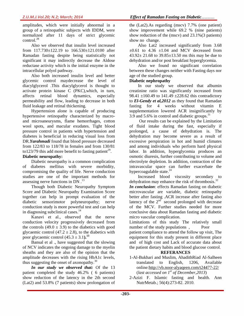
corresponds to the severity of the insult, the
degree of recovery of the b-wave during
reperfusion depends on the duration of ischemia.
In this sense the b-wave of the ERG represents a
functional measure for potential therapeutic
efficacy of drugs interacting with these
pathophysiological processes.9
Diabetic neuropathy is a common
complication of diabetes mellitus with severe
morbidity, compromising the quality of life.
Nerve conduction studies are one of the important
methods for assessing nerve functions in
DN.10
Though both Diabetic Neuropathy Symptom
Score and Diabetic Neuropathy Examination
Score together can help in prompt evaluation of
the diabetic sensorimotor polyneuropathy; nerve
conduction study is more powerful test and can
help in diagnosing subclinical cases.13
Diaetic nephropathy is the major cause of
end stage renal disease (ESRD) throughout the
world. It is defined clinically as the presence of
microalbuminuria or overt nephropathy in patients
with diabetes who lack indicators of other renal
diseases.12
Additionally, the reduced glomerular
filtration rate (GFR) and albuminuria caused by
DN are independent risk factors for CVD and
death.13
PATIENTS AND METHODS
This study had been carried out in internal
medicine outpatient clinic, diabetes and
endocrinology outpatient clinic, Rheumatology
outpatient clinic, Al-Hekma eye center, and
clinical pathology department at Zagazig
university hospital. This study was performed
during Ramadan of Hijri year 1432 and 1433
(August 2011and July- August 2012). In both
years Ramadan was 30 days. The average fasting
period was 14 h; the starting and finishing hours
of the fasting were approximately 3:30 a.m. and
5:30 p.m., respectively. We started the study on
74 diabetic patients but only 64 completed the
clinical visits with us and the other 10 missed
from the follow up visits. Their ages ranged from
20 to 64 years; with mean ± SD 44.07± 13.09
years and 25 of them were males and the other 19
were females. All participants provided informed
consent to share in this study.
Inclusion criteria: Diabetic patients willing to
fast during next Ramadan were included in this
study.
Exclusion criteria:
1--Patient with renal impairment (Cr >1.5)
2- Liver dysfunction (ALT > double normal or
total bilirubin >1)
3-Women who were pregnant or breast
feeding.
primary objective was The objectives:Study
to assess the effect of Ramadan fasting on diabetic
microvascular complication. Study methodology: This is a cross-sectional study
in which all patients were subjected to the
following:-
A) Before Ramadan:
Full history taking and detailed clinical
examination with particular consideration on:
1-Symptoms and sings of diabetes or its
complications (Neuropathy, retinopathy,
nephropathy).
2-Fundus examination to detect diabetic
retinopathy.
Routine laboratory investigations (Liver, kidney
function tests and Fasting blood glucose level,)
Specific investigations:
1-Albumin-creatinie ratio (ACR).
2-Sensory conduction velocity.
3-Electroretinogram(ERG)
4-insulin-proinsulin level
B) After Ramadan:
History of the number of fasting days, problems
that occurred during fasting (hypoglycemia) and
modification in the dose of oral anti-diabetic
drugs or insulin.
Laboratory investigations: fasting blood glucose,
insulin and proinsulin.
Specific investigations: 1-Albumin-creatinie ratio (ACR).
2-Sensory conduction velocity.
3-Electroretinogram(ERG)
Blood tests:
Blood samples were obtained from all
subjects at the beginning in the week before the
start of Ramadan (pre-Ramadan) and 4 days later
following the fasting period finished, immediately
after Bairam (post-Ramadan).
Samples were allowed to clot and the serum
centrifuged, divided into aliquots and stored at -
80°C until analyzed. All serum samples were
analyzed in a single batch to avoid day-to- day
laboratory variation.
They were measured in the central laboratory
of clinical pathology department, Zagazig
University.
Serum glucose was measured by the glucose
oxidase technique (Roche Diagnostics GmbH).
Serum insulin&Serum proinsulin was measured
by ELISA.
HOMA-IR is calculated using the following
formula :
International Formula:
Fasting Glucose (mmol/L) x fasting Insulin
(mU/L) / 22.5.
US Formula:

Z.U.M.J.Vol.20; N.2; March; 2014 Effect of Ramadan Fasting on Diabetic………….
-195-
Fasting Glucose (mg/dl) x fasting Insulin
(µU/mL) / 405.
Insulin sensitivity, resistance and β cell
function were calculated using the HOMA
Calculator that was released in 2004 by Hines et
al. This provides quick and easy access to use
model-derived estimates of β cell function
(HOMA B %) and insulin resistance (HOMA IR),
rather than linear approximations.17
creatinine ratio(ACR):-Albumin
Urine samples were obtained from all subjects
pre-Ramadan post-Ramadan. The second morning
Urine samples were collected in serialized urine
containers.
UACR is a ratio between two measured
substances. UACR is reported in mg/g and
approximates the albumin excretion in mg/day.
UACR in mg/g = Urine albumin (mg/dL) / Urine
creatinine (g/dL)
Nerve conduction velocity:
+[Nerve conduction study was done in
rheumatology outpatient clinic measuring sensory
conduction velocity in the right median nerve
before and after fasting. Median nerve was chosen
because it accessible and easier in examination.
Sensory NCS were performed by electrical
stimulation of a peripheral nerve and recording
from a purely sensory portion of the nerve.
Electroretinogram(ERG)
Full flash ERG was performed according to
the standards of the international society for
clinical electrophysiology of vision(ISCVE)
approved in 2008, for all subjects at the
beginning in the week before the start of Ramadan
(pre-Ramadan) and 4 days later following the
fasting period finished, immediately after Bairam
(post-Ramadan).
It were done in Alhekma Eye Center using
Roland-consult; The RETI-port33 system. The
RETI-port consists of the pattern stimulator unit
and computer analyzer system. The bio signal
amplifier includes a preamplifier near the patient.
All patient data and the results are stored in the
data base. The bio signal and averaged curves
from all channels can be displayed on the monitor.
6 responses were performed for all
patients. The ISCEV Standard specifies five
responses:18
(1)Dark-adapted 0.01 ERG (rod response);
(2) Dark-adapted 3.0 ERG (combined rod–cone
response);
(3)Dark-adapted 3.0 oscillatory potentials;
(4) Lightadapted3.0 ERG (cone response);
(5) Light-adapted 3.0 flicker (30 Hz flicker).
(6) Additional response: Dark-adapted 10.0 ERG
Preparation of the patient
The patient assumes a comfortable position
sitting up. The patient's eyes were dilated
beforehand with standard dilating eye drops.
Anesthetic drops are then placed in the eyes,
causing them to become numb.
Pre-adaptation to light or dark: 20 min of
dark adaptation before recording dark-
adaptedERGs and 10 min of light adaptation
beforerecording light-adapted ERGs.
ISCEV standard ERG:
1-Dark-adapted 0.01 ERG (Scotopic or rod
response): The Dark-adapted 0.01 ERG is the first
signal measured after dark adaptation, because it
is the most sensitive to light adaptation. The
stimulus is a dim white flash of 0.01 cd.s.m-2;
with a minimum interval of 2 s between flashes.
2-Dark-adapted 3.0 ERG (combined rod–
cone response): This is produced by a white 3.0
cd.s.m-2 flash in the dark-adapted eye. With an
interval of at least 10 s between stimuli.
3-Dark-adapted 3.0 oscillatory potentials:
Dark-adapted oscillatory potentials obtained from
the dark-adapted eye, using the 3.0 cd.s.m-2 flash
stimulus.
4-Light-adapted 3.0 ERG (photopic 3.0 ERG
GF or, single-flash cone response): Use a 3.0-
cd.s.m-2 stimulus, with at least 0.5 sbetween
flashes. The background luminance of 30 cd.m-2
measured at the surface of the full-field stimulus
bowl.
5-Light-adapted 3.0 flicker ERG (30 Hz
flicker) (photopic 3.0 flicker 30Hz ERG GF):
Flicker ERGs also reflect activity of the cone
system,and it was obtained with 3.0-cd.s.m-2
stimuli,under the same conditions of light-
adaptation as theLight-adapted 3.0 ERG.
Recording the flicker ERG inthe light-adapted
state reduces discomfort and allowsthe light
adaptation to be standardized. Flasheswas
presented at a rate of approximately 30stimuli per
second (30 Hz), and the rate that was
chosenconstant for all patient in our study. The
first ERGresponse to the flickering stimulus is
single flashwaveform; thus, the first few
waveforms discarded so that stable conditions are
reached.
Additional ERG
6-Dark-adapted 10.0 ERG: An additional
dark-adapted ERG to a stronger flash , it was
obtained with 10.0 cd.s.m-2.This stimulus gives a
larger a-wave with better definition (no double
trough), larger oscillatory potentials that are easier
to characterize, and more distinctive features of
negative ERG waveforms (for critical recognition
of diseases with related b-wave reduction). Also

Z.U.M.J.Vol.20; N.2; March; 2014 Effect of Ramadan Fasting on Diabetic………….
-196-
this stronger flash may give better signals in
patients with opaque media or immature retinae.
ERG analysis and reporting
Single flash ERGs: In general, b-wave
amplitude and time-to-peak (implicit time) is
measured for all ERGs (except oscillatory
potentials), and the a-wave should also be
measured when recognizable as a distinct
component. According to current convention, the
a-wave amplitude is measured from baseline to a-
wave trough; the b-wave amplitude is measured
from a-wave trough to b-wave peak; the a-wave
and b-wave implicit times are measured from the
time of the flash to the peak of the wave (see
Figure below).
Oscillatory potentials: Their appearance is highly dependent upon adaptation state and filter
characteristics of the amplifier, but there are usually three major peaks often followed by a fourth smaller one.
Simply observing the presence of the three peaks, and their normality relative to the standards of the laboratory,
may be adequate for many clinical purposes in keeping with our present state of knowledge.
Flicker ERG:The amplitude of flicker ERG is measured from the trough to the peak (averaging several
typical responses). The implicit time is measured from each stimulus onset to the corresponding peak.
Statistical Analysis:
Data entered and analyzed using Microsoft
Word. Data were then imported into Statistical
Package for the Social Sciences (SPSS version
16.0) software for analysis. Baseline characteristics
of the study population were presented as
frequencies and percentages (%) or mean values
and standard deviations (SD). According to the type
of data, the following tests were used to test
differences for significance; Paired t-test was used
to compare pre and post Ramadan fasting variables.
Differences were considered significant when p
values were less than 0.05. Chi square for
(qualitative variables). Correlation of numeric data
was done by person’s correlation (r).
RESULTS
Table (1) : the demographic data and clinical characteristics among studied diabetic patients:
Age (years) ± SD
Range (years)
44.07± 13.09.
(20 - 64)yrs.
Gender
Male
N Percent
36 56.8%
Female 28 43.2%
Duration (years )Range
± SD
(0.5-35) yrs.
8.90±7.977
Family history of DM
+ ve
- ve
26 (45.5 % )
38 (54.5 % )
Associated HTN
Yes
No
18 (25 % )
36 (75 % )
Table show that 40.6% of the study population have positive family history of DM
28.1% have associated HTN with DM
Z.U.M.J.Vol.20; N.2; March; 2014 Effect of Ramadan Fasting on Diabetic………….
-197-
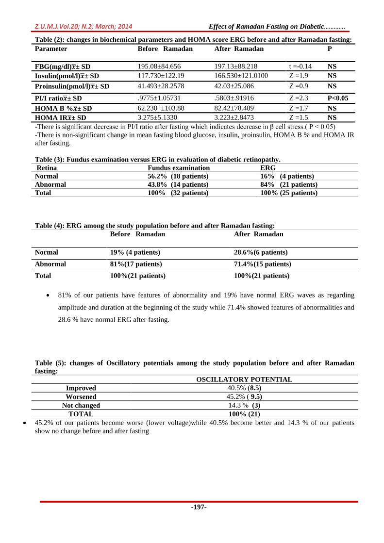
Table (2): changes in biochemical parameters and HOMA score ERG before and after Ramadan fasting:
Parameter Before Ramadan After Ramadan P
FBG(mg/dl) ± SD 195.08±84.656 197.13±88.218 t =-0.14 NS
Insulin(pmol/l) ± SD 117.730±122.19 166.530±121.0100 Z =1.9 NS
Proinsulin(pmol/l) ± SD 41.493±28.2578 42.03±25.086 Z =0.9 NS
PI/I ratio ± SD .9775±1.05731 .5803±.91916 Z =2.3 P<0.05
HOMA B % ± SD 62.230 ±103.88 82.42±78.489 Z =1.7 NS
HOMA IR ± SD 3.275±5.1330 3.223±2.8473 Z =1.5 NS
-There is significant decrease in PI/I ratio after fasting which indicates decrease in β cell stress.( P < 0.05)
-There is non-significant change in mean fasting blood glucose, insulin, proinsulin, HOMA B % and HOMA IR
after fasting.
Table (3): Fundus examination versus ERG in evaluation of diabetic retinopathy.
Retina Fundus examination ERG Normal 56.2% (18 patients) 16% (4 patients) Abnormal 43.8% (14 patients) 84% (21 patients) Total 100% (32 patients) 100% (25 patients)
Table (4): ERG among the study population before and after Ramadan fasting:
81% of our patients have features of abnormality and 19% have normal ERG waves as regarding
amplitude and duration at the beginning of the study while 71.4% showed features of abnormalities and
28.6 % have normal ERG after fasting.
Table (5): changes of Oscillatory potentials among the study population before and after Ramadan
fasting:
OSCILLATORY POTENTIAL
40.5% (8.5) Improved
45.2% ( 9.5) Worsened
14.3 % (3) Not changed
100% (21) TOTAL
45.2% of our patients become worse (lower voltage)while 40.5% become better and 14.3 % of our patients
show no change before and after fasting
Before Ramadan After Ramadan
Normal 19% (4 patients) 28.6%(6 patients)
Abnormal 81%(17 patients) 71.4%(15 patients)
Total 100%(21 patients) 100%(21 patients)

Z.U.M.J.Vol.20; N.2; March; 2014 Effect of Ramadan Fasting on Diabetic………….
-198-
Table (6): changes of Oscillatory potentials ERG before and after Ramadan fasting:
Parameter Before Ramadan After Ramadan T P
Right N2(ms) ± SD 20.95±1.75 21.29±1.79 -.69 NS
Left N2(ms) ± SD 20.90±1.34 21.33±2.03 -0.82 NS
Right P2 (ms) ± SD 25.52±1.57 24.43±3.65 1.31 NS
Left P2 (ms) ± SD 25.14±1.71 25.48±1.50 -0.92 NS
RightOS2(µv) ± SD 8.73±4.50 9.24±4.92 -0.47 NS
Left OS2(µv) ± SD 9.19±3.43 9.74±4.72 -0.66 NS
N2(ms) ± SD 20.93 ± 1.54 21.31 ±1.89 -1.08 NS
P2 (ms) ± SD 25.33 ±1.54 21.31 ±1.89 0.83 NS
OS2(µv) ± SD 8.96±3.96 9.49±4.77 -0.79 NS
The OS2 of both eyes increased from 8.96 ± 3.96 µv to 9.49 ± 4.77 µv. this indicates improvement of the retinal
function after fasting.
Table (7): protein dipstick versus ACR in diagnosis of diabetic nephropathy
nephropathy protein dipstick ACR yes 40% (12 patients) 63.3% (19 patients) no 60% (18 patients) 36.7% (11 patients) total 100% ( 30 patients) 100% ( 30 patients)
30 of our patients by both traditional protein dipstick and Albumin Creatinine Ratio (ACR).
By protein dipstick: 40 % (12 patients) had proteinuria and 60 % (18 patients) negative for protein in
urine.
By ACR: 63.3% (19 patients) are positive for microalbumin and 36.7 %( 11 patients) negative for
microalbuminuria .
Table (8): changes in ACR among the study population before and after Ramadan fasting:
Total Second year First year
11 7 4 improved
8 5 3 Worsened
1 1 0 No change
8 4 4 Drop out
2 1 1 No fasting
30 18 12 Total
Of the 20 patients included in the statistical analysis: 7 are found to be negative for micro-albumin(less than 30
mg/gm) before fasting of them 2 improved after fasting, 1 not changed and 4 become worse.
*11 are found to have microalbumiuria(30-300 mg/gm) before fasting of them 5 improved, 2 not changed and 4
become worse
*2 are found to have macroalbuminuria ( more than 300 mg/gm) one show slight improvement in ACR ratio
and the other show increase of the ACR by about 50% of the before fasting value.

Z.U.M.J.Vol.20; N.2; March; 2014 Effect of Ramadan Fasting on Diabetic………….
-199-
Diagram (1 ): ACR among the study population before and after Ramadan fasting:
Diagram show 7 are found to be negative for micro-albumin(less than 30 mg/gm),11 are found to have
microalbumiuria (30-300 mg/gm) and 2 are found to have macroalbuminuria ( more than 300 mg/gm) before
Ramadan fasting .
After Ramadan fasting 6 are found to be negative for micro-albumin, 11 are found to have
microalbumiuria and 3 are found to have macroalbuminuria.
Diagram ( 2 ): ACR changes among different categories of the study population before and after
Ramadan fasting:
Figure show that the highest number of improved patients is among patients with microalbumiuria.
Of the 7 patients negative for micro-albumin: 2 improved after fasting, 1 not changed and 4 become
worse.
Eleven patients had microalbumiuria 8 improved, and 3 become worse.
Two are found to have macroalbuminuria, one show slight improvement in ACR ratio and the other
show increase of the ACR.
0
2
4
6
8
10
12
before after
normal
microalbuminuria
macroalbuminuria
0
1
2
3
4
5
normal micro macro
improved
worse
no change

Z.U.M.J.Vol.20; N.2; March; 2014 Effect of Ramadan Fasting on Diabetic………….
-200-
Table (9): comparison between different categories as regarding change of ACR after Ramadan fasting:
x2 P
ACR Total
Normal Micro Macro
worse number 5 4 1 10
% 71.4% 36.4% 50.0% 50.0%
Improve number 2 7 1 10 2.1 NS
% 28.6% 63.6% 50.0% 50.0%
Not-changed number 5 4 1 10 worse
% 71.4% 36.4% 50.0% 50.0%
Total number 7 11 2 20
% 100.0% 100.0% 100.0% 100.0%
Table (10): comparison between different categories as regarding decrease of ACR after Ramadan
fasting
X2 P
ACR total
Normal Diabetic nephropathy
improve
count 2 8 10
% 28.6% 61.5% 50.0%
12.5 <0.001
total count 7 13 20
% 100.0% 100.0% 100.0%
Table (11): changes of ACR before and after Ramadan fasting:
Parameter Before Ramadan After Ramadan Z P
albumin 103.07±138.04 258.17±507.05 -0.936 NS
Creatinine 132.27±75.12 131.97±92.82 -0.024
NS
ACR 98.41 ±160.49 141.49 ±228.62 -1.328 NS
Table (12):Correlation between ACR changes with age and number of fasting days:
DACR
Fasting days r 0.447*
p-value 0.048
Age r 0.171
p-value 0.471

Z.U.M.J.Vol.20; N.2; March; 2014 Effect of Ramadan Fasting on Diabetic………….
-201-
Table (13) shows changes of nerve conduction velocity among the study populations:
Cv(m/s) Lat2
7.7% (1 patient) 46.2% (6 patients) BETTER
69.2 % (9 patients) 53.8% (7 patients) WORSE
23.1%(3 patients) - Not changed
100% (13 patients) 100% (13 patients) TOTAL
46.2% show reduction of the latency in the 2th second (Lat2) and 53.8% show prolongation of the (Lat2).
7.7% show improvement(increase)of mscv while 69.2 % show reduction of the (mscv) and 23.1% show no
change
Table (14) : ): changes of nerve conduction velocity (MCV) before and after Ramadan fasting:
Parameter Before Ramadan After Ramadan t P
Lat2 3.68 ±0.61 4.36 ±1.04 -2.13 0.05
Cv(m/s) 51.54± 9.58 43.85±6.69 2.06 NS
Paired t-test comparing Mean sensory conduction velocity (mscv) and the latency in the 2th second (Lat2)
before and after Ramadan fasting show increase in the lat2 mean and reduction in the MCV after Ramadan
fasting but statistically not significant (p-value > 0.05).
Table (15): Correlation between changes OF MCV and LAT2 with age and number of fasting days:
Dlat2 DMCV age Fasting days
D-lat2 r 1 -.502- -.445- -.303-
p-value .080 .127 .314
D-MCV r -.502- 1 .386 .290
p-value .080 .193 .337
Age r -.445- .386 1 .424
p-value .127 .193 .149
Fasting days r -.303- .290 .424 1
p-value .314 .337 .149

Z.U.M.J.Vol.20; N.2; March; 2014 Effect of Ramadan Fasting on Diabetic………….
-202-
DISCUSSION
Ramadan fasting is one of the five pillars of
Islam. One billion Muslim adults worldwide refrain
from food, water and oral drug intake from dawn to
sunset during Ramadan fasting. Ramadan fasting
could not induce any harmful effect in young
healthy subjects1 . However, it can induce several
complications in patients with diabetes. In this
study we tried to explore the effect of Ramadan
fasting on microvascular complications of DM. 2
It was observed that There was non-significant
change in mean fasting blood glucose which
indicates that Ramadan fasting not significantly
alter glycemic control and this correlates with
different studies that found that Ramadan fasting
has been reported not to alter glycemic control19
.
However this is contradictory to the EPIDIAR
study, 2001 who reported a decrease in fasting
glucose and an increase in the frequency of severe
hypoglycemia during Ramadan in a population
including both types 1 and 2 patients.5This variation
may be due to the amount or type of food
consumption, regularity of taking medications,
engorging after the fast is broken, or decreased
physical activities. In most cases, no episode of
acute complications (hypoglycemic or
hyperglycemic types) occurs in patients under
medical management.20
This is also contradictory to Norouzy et al,
2012 who showed that fasting during Ramadan
deteriorated the glycemic control in Type 2 diabetes
patients. This was more evident in patients using
oral hypoglycemic medication than diet controlled
patients21
.
Also This is contradictory to Fakhrzadeh et al,
2003 who found that fasting plasma glucose
decreased significantly in both men and women
after Ramadan fasting and to Khaled et al ,2006
who found significant decreases in fasting blood
glucose and glycosylated hemoglobin (HbA1c) in
obese women with type 2 diabetes mellitus after
Ramadan fasting22
.
Our study shows high levels in the mean
fasting insulin before Ramadan fasting (Normal
value: 57-79pmol/l) and this may be explained by
insulin resistance found in type 2 diabetic patients
and this correlates with Bergman et al, 1997 who
provided that the most practical way of assessing
insulin resistance is the measurement of plasma
insulin levels after overnight fasting condition23
.
Diabetic Retinopathy:
Evidence has begun to point to the fact that
even before vascular complications begin to
manifest, neuronal cell death and dysfunction have
alread begun8 (Miranda et al., 2011).
In our study we observed that oscillatory
potentials in the second (OS2) increased from
8.73±4.50 µv to 9.24±4.92 µv in the right eye and
from 9.19±3.43 µv to 9.74±4.72 µv in the left eye.
Also the mean OS2 of both eyes increased from
8.96±3.96 to 9.49±4.77.
Duration of P2 in the right eye reduced from
25.14±1.71 ms to25.48±1.50 ms, also duration of
P2 reduced in the left eye from 25.14±1.71to
25.48±1.50 and the mean duration of P2 decreased
from 25.33 ±1.54 to 21.31 ±1.89 but statically not
significant (p-value >0.05).
Duration of N2 in the right eye increased from
20.95±1.75 to 21.29±1.79 ms, also duration of N2
increased in the left eye from 20.90±1.34to
21.33±2.03 and the mean duration of N2 increased
from 20.93 ± 1.54 to21.31 ±1.89.
These changes indicate significant
improvement of diabetic retinopathy after Ramadan
fasting .this may be due Limitation of fluid intake
during the fast, especially if prolonged. And this can
be increased by perspiration in hot and humid
climates and among individuals who perform hard
physical labor.24
All these may decrease retinal
exudation despite increasing the risk of thrombosis.
Saada et al. (2010)25
; noted that glycosylated
haemoglobin (HbA1c) decreased slightly during the
last week of the month of Ramadan among the
diabetic patients. Also Bouguerra et al.29
Had found
that HBA1c significantly decreased from 8.8 % to
7.4% and from 10.6% to 7.1% correspondingly after
Ramadan fasting.
Diabetes Control and Complications Trial
(DCCT) found that achieving mean hemoglobin
A1C (A1C) of 7.9 % reduced the incidence of new
cases of retinopathy by as much as 76 %. The
reduction was directly related to the degree of
glycemic control as estimated from hemoglobin
A1C values. The United Kingdom Prospective
Diabetes Study found similar results in patients with
type 2 diabetes; each 1 percent point reduction in
A1C was associated with a 37 percent reduction in
development of retinopathy .27
So the improvement of our patients can be
explained by the better glycemic control after
Ramadan. This consistent with Frost-Larsen et al
that assessed the effect of short-term strict glycemic
control on OS amplitude and reported that OP
Z.U.M.J.Vol.20; N.2; March; 2014 Effect of Ramadan Fasting on Diabetic………….
-203-
amplitudes, which were initially abnormal in a
group of a retinopathic subjects with IDDM, were
normalized after 11 days of strict glycemic
control.28
Also we observed that insulin level increased
from 117.730±122.19 to 166.530±121.0100 after
Ramadan fasting despite being statistically not
significant it may indirectly decrease the Aldose
reductase activity which is the initial enzyme in the
intracellular polyol pathway.
Also both increased insulin level and better
glycemic control maydecrease the level of
diacylglycerol .This diacylglycerol is thought to
activate protein kinase C (PKC),which, in turn,
affects retinal blood dynamics, especially
permeability and flow, leading to decrease in both
fluid leakage and retinal thickening.
Hypertension alone is capable of producing
hypertensive retinopathy characterized by macro-
and microaneurysms, flame hemorrhages, cotton
wool spots, and macular exudates. Tight blood
pressure control in patients with hypertension and
diabetes is beneficial in reducing visual loss from
DR.Yarahmadi found that blood pressure decreased
from 122/83 to 118/78 in females and from 130/81
to123/79 this add more benefit to fasting patient29
.
Diabetic neuropathy:
Diabetic neuropathy is a common complication
of diabetes mellitus with severe morbidity,
compromising the quality of life. Nerve conduction
studies are one of the important methods for
assessing nerve functions in DN .13
Though both Diabetic Neuropathy Symptom
Score and Diabetic Neuropathy Examination Score
together can help in prompt evaluation of the
diabetic sensorimotor polyneuropathy; nerve
conduction study is more powerful test and can help
in diagnosing subclinical cases.14
Kanavi et al., observed that the nerve
conduction velocity progressively decreased from
the controls (49.0 ± 3.9) to the diabetics with good
glycaemic control (47.2 ± 2.8), to the diabetics with
poor glycaemic control (45.3 ± 3.1).13
Bansal et al ., have suggested that the slowing
of NCV indicates the ongoing damage to the myelin
sheaths and they are also of the opinion that the
amplitude decreases with the rising HbA1c levels,
thus suggesting the onset of axonopathy.30
In our study we observed that: Of the 13
patient completed the study 46.2% ( 6 patients)
show reduction of the latency in the 2th second
(Lat2) and 53.8% (7 patients) show prolongation of
the (Lat2).As regarding (mscv) 7.7% (one patient)
show improvement while 69.2 % (nine patients)
show reduction of the (mscv) and 23.1%(3 patients)
show no change.
Also Lat2 increased significantly from 3.68
±0.61 to 4.36 ±1.04 and MCV decreased from
43.92± 21.68 to 39.85±13.50 ms this may be due to
dehydration and/or post breakfast hyperglycemia.
Also we found no significant correlation
between these changes neither with Fasting days nor
age of the studied group.
Diabetic nephropathy
In our study we observed that albumin
creatinine ratio was significantly increased from
98.41 ±160.49 to 141.49 ±228.62 this contradictory
to El-Gendy et al,2012 as they found that Ramadan
fasting for 4 weeks without vitamin E
supplementation lowered ACR insignificantly by
3.9 and 5.6% in control and diabetic groups.31
Our results can be explained by the Limitation
of fluid intake during the fast, especially if
prolonged, a cause of dehydration is. The
dehydration may become severe as a result of
excessive perspiration in hot and humid climates
and among individuals who perform hard physical
labor. In addition, hyperglycemia produces an
osmotic diuresis, further contributing to volume and
electrolyte depletion. In addition, contraction of the
intravascular space can further exacerbate the
hypercoagulable state.24
Increased blood viscosity secondary to
dehydration may enhance the risk of thrombosis.32
In conclusion: effects Ramadan fasting on diabetic
microvascular are variable, diabetic retinopathy
better after fasting .ACR increase after fasting also
latency of the 2nd
second prolonged with decrease
of the MCV. Further studies needed for more
conclusive data about Ramadan fasting and diabetic
micro vascular complication.
Limitations of this study The relatively small
number of the study populations , Poor
patient compliance to attend the follow up visit, The
equipment for this study present in different place
and of high cost and Lack of accurate data about
the patient dietary habits and blood glucose control.
REFERANCES
1-Al-Bukhari and Muslim, AhadithRiad Al-Salheen
translated to English, 1206, Available
online:http://vb.noor-alyaqeen.com/t24477-22/
(last accessed on 1st of December,2013)
2-Azizi F. Islamic fasting and health. Ann
NutrMetab.; 56(4):273-82. 2010.

Z.U.M.J.Vol.20; N.2; March; 2014 Effect of Ramadan Fasting on Diabetic………….
-204-
3-Salti I, Be´nard E, Detournay B, et al., EPIDIAR
study group: A population based study of
diabetes and its characteristics during the
fasting month of Ramadan in 13 countries:
results of the epidemiology of diabetes and
Ramadan 1422/2001 (EPIDIAR) study.
Diabetes Care; 27:2306–2311: 2004.
4-Ziaee, V., Razaei, M., Ahmadinejad, et al.,: The
changes of metabolic profile and weight during
Ramadan fasting. Singapore Medical Journal
47(5), 409-414, 2006.
5-Dewanti L., Watanabe C., Sulistiawati et al.,.
Unexpected changes in blood pressure and
hematological parameters among fasting and
nonfasting workers during Ramadan in
Indonesia.Eur J ClinNutr. Jul;60(7):877-81,
2006.
6-Adlouni, A., Ghalim, N., Benslimane, A., et al.,
:Fasting during Ramadan induces a marked
increase inhigh-density lipoprotein cholesterol
and decrease in low-densitylipoprotein
cholesterol. Annals of Nutrition & Metabolism
41(4), 242-249, 1997.
7-Fong DS, Aiello LP, Ferris FL et al.,: Diabetic
retinopathy. Diabetes Care 27:2540-2553, 2004.
8- ar a iranda, ar a ictoria nche - illarejo,
Raquel lvare - lting, et al.,
Electroretinogram Alterations in
Diabetes?,Electroretinograms, Dr.
GregorBelusic (Ed.), ISBN: 978-953-307-383-
5, (2011).In Tech .Available from:(last
accessed on November 2013)
http://www.intechopen.com/books/electroretino
grams/electroretinogram-alterations-in-diabetes-
9- Ghirlanda, M. A. Di Leo, S. Caputo, B. et al., :
Detection of inner retina dysfunction by steady-
state focal electroretinogram pattern and flicker
in early IDDM. Diabetes, 40 9 September,
(1991), 1122 1127 . 0012-1797, 1991.
10-Bresnick G. H and Palta M.: Predicting
progression to severe proliferative diabetic
retinopathy. Archives of Ophthalmology, 105 6
June, (1987), 810- 814, 1987.
11-Luu,C. D..Szental,J. A. Lee,S. et al., Correlation
between retinal oscillatory potentials and retinal
vascular calibre in type 2 diabetes. Investigative
Ophthalmology & Visual Science, 51 1
January,), 482 486 . 1552-5783. 2010.
12-Kern T. S.,Miller C. M., Tang J et al.,.:
Comparison of three strains of diabetic rats with
respect to the rate at which retinopathy and
tactile allodynia develop. Molecular Vision, 16
August :1629 1639 . 1090-0535 , 2010.
13-Kanavi Roopa Shekharappa, Srinivasa.K., k.j.
Vedavathi et al.,: A Study on the Utility of
Nerve Conduction Studies in Type 2 Diabetes
Mellitus. Journal of Clinical and Diagnostic
Research. June, Vol-5(3): 529-531, 2011.
14-Asad A, Hameed MA, Khan et al.,
:Source:Army Medical College/NUST,
Rawalpindi. J Pak Med Assoc. Sep; 59(9):594-
8, 2009.
15-Ritz, E., Zeng X. and Rychlík I.: Clinical
manifestation and natural history of diabetic
nephropathy. ContribNephrol, 170: 19-27,
2011.
16-Shin D, Seung S, Yoon HE, et
al.:Microalbuminuria is Independently
Associated with Arterial Stiffness and Vascular
Inflammation but not with Carotid Intima-
Media Thickness in Patients with Newly
Diagnosed Type 2 Diabetes or Essential
Hypertension. J Korean Med Sci. 2013
February; 28(2): 252–260, 2013.
17-Holman R, Hines G, Kennedy I, et al., A
calculator for HOMA, Diabetologia; 47: Suppl
1: A222, 2004.
18-Marmor MF, Fulton A B, Holder GE, et al., (for
the International Society for Clinical
Electrophysiology of Vision) ISCEV Standard
for full-field clinical electroretinography (2008
update) Doc Ophthalmol 118:69–77, 2009.
19- Sari R, BalciMK, Akbas SH et al.: The effects
of diet, sulfonylurea, and Repaglinide therapy
on clinical and metabolic parameters in type 2
diabetic patients during Ramadan, Endocr. Res.
30 (2) (2004) 169–177.
20-Ewis A and Afifi NM. Ramadan fasting and
non-insulin-dependent diabetes mellitus : Effect
of regular exercise. Second International
Congress on Health and Ramadan. Dec. 1-3,
1997, Istanbul,Turkey, P 76, 1997.
21-Norouzy A, Mohajeri SM, Shakeri S, et al.,
Effect of Ramadan fasting on glycemic control
in patients with Type 2 diabetes.J Endocrinol
Invest. 2012 Sep;35(8):766-71,2012.
22-Fakhrzadeh H, Larijani B, Sanjari M, et al.,
Effect of Ramadan fasting on clinical and
biochemical parameters in health adults. Ann
Saudi Med; 23: 223-6, 2003.
23-Bergman RN, Finegood DT, Kahn SE. The
evolution of beta-cell dysfunction and insulin
resistance in type 2 diabetes. Eur J Clin Invest;
32(Suppl 3):S35–S45, 2002.
Z.U.M.J.Vol.20; N.2; March; 2014 Effect of Ramadan Fasting on Diabetic………….
-205-
24-Beckman JA, Creager MA and Libby P.
Diabetes and atherosclerosis: epidemiology,
pathophysiology, and management. JAMA
287:2570–2581, 2002.
25-SaadaAit, Selseletattou G.1, Belkacemi Let
al.:Effect of Ramadan fasting on glucose,
glycosylated haemoglobin, insulin, lipids and
proteinous concentrations in women with non-
insulin dependent diabetes mellitus. African
Journal of Biotechnology Vol. 9 (1), pp. 087-
094, 4 January, 2010, Available online at
http://www.academicjournals.org/AJB ISSN
1684–5315 © 2010 Academic Journals.
Accessedon 2nd of December 2013
26-Bouguerra R, Belkadhi A, Jabrane J, et al. Les
effetsmétaboliques du jeûne du mois de
Ramadan chez des diabétiques de type 2. East
Mediterr Health J 2003; 9: 1099-1108.
27-UK Prospective Diabetes Study (UKPDS)
Group. Intensive blood-glucose control with
sulphonylureas or insulin compared with
conventional treatment and risk of
complications in patients with type 2 diabetes
(UKPDS 33). Lancet 1998; 352:837.
28-Frost-Larsen k, Christiansen J, Sandahl H,et al.,
1983: The effect of strict short-term metabolic
control on retinal nervous system abnormalities
in newly diagnosed type 1 (insulin-dependent)
diabetic patients. Diabetologia, 24 3 March,),
207 209 . 0001-2186X. 2003.
29-Benaji B, Mounib N , Roky R., et al.,:Diabetes
and Ramadan: Review of the literature.Diabetes
Research and Clinical Practice 73, 117–125,
2006.
30-Bansal V, Kalita J and Misra UK: Diabetic
neuropathy. Postgrad Med J 82: 95-100, 2006.
31-El-Gendy Ola,Rokaya M, El-BataeHass et al.,:
Ramadan Fasting Improves Kidney Functions
and Ameliorates Oxidative Stress in Diabetic
Patients. World Journal of Medical Sciences 7
(1): 38-48, 2012.
32-Akhan G, Kutluhan S and Koyuncuoglu HR. Is
there any change in stroke incidence during
Ramadan? ActaNeurolScandin 101:259–261, 20