L-Serine Supplementation Blunts Fasting-Induced Weight ...
14
Citation: López-Gonzales, E.; Lehmann, L.; Ruiz-Ojeda, F.J.; Hernández-Bautista, R.; Altun, I.; Onogi, Y.; Khalil, A.E.; Liu, X.; Israel, A.; Ussar, S. L-Serine Supplementation Blunts Fasting-Induced Weight Regain by Increasing Brown Fat Thermogenesis. Nutrients 2022, 14, 1922. https://doi.org/10.3390/ nu14091922 Academic Editor: Roberto Iacone Received: 28 March 2022 Accepted: 29 April 2022 Published: 4 May 2022 Publisher’s Note: MDPI stays neutral with regard to jurisdictional claims in published maps and institutional affil- iations. Copyright: © 2022 by the authors. Licensee MDPI, Basel, Switzerland. This article is an open access article distributed under the terms and conditions of the Creative Commons Attribution (CC BY) license (https:// creativecommons.org/licenses/by/ 4.0/). nutrients Article L-Serine Supplementation Blunts Fasting-Induced Weight Regain by Increasing Brown Fat Thermogenesis Elena López-Gonzales 1,2 , Lisa Lehmann 1,2 , Francisco Javier Ruiz-Ojeda 1,2,3 , René Hernández-Bautista 1,2 , Irem Altun 1,2 , Yasuhiro Onogi 1,2 , Ahmed Elagamy Khalil 1,2 , Xue Liu 1,2 , Andreas Israel 1,2 and Siegfried Ussar 1,2,4, * 1 RG Adipocytes & Metabolism, Institute for Diabetes & Obesity, Helmholtz Center Munich, 85764 Munich, Germany; [email protected] (E.L.-G.); [email protected] (L.L.); [email protected] (F.J.R.-O.); [email protected] (R.H.-B.); [email protected] (I.A.); [email protected] (Y.O.); [email protected] (A.E.K.); [email protected] (X.L.); [email protected] (A.I.) 2 German Center for Diabetes Research (DZD), 85764 Munich, Germany 3 Department of Biochemistry and Molecular Biology II, Faculty of Pharmacy, University of Granada, 18071 Granada, Spain 4 Department of Medicine, Technical University of Munich, 80333 Munich, Germany * Correspondence: [email protected]; Tel.: +49-89-3187-2047 Abstract: Weight regain after fasting, often exceeding the pre-fasting weight, is a common phe- nomenon and big problem for the treatment of obesity. Thus, novel interventions maintaining reduced body weight are critically important to prevent metabolic disease. Here we investigate the metabolic effects of dietary L-serine supplementation, known to modulate various organ functions. C57BL/6N-Rj male mice were supplemented with or without 1% L-serine in their drinking water and fed with a chow or high-fat diet. Mice were fed either ad libitum or subjected to repeated overnight fasting. Body weight, body composition, glucose tolerance and energy metabolism were assessed. This was combined with a detailed analysis of the liver and adipose tissues, including the use of primary brown adipocytes to study mitochondrial respiration and protein expression. We find that L-serine supplementation has little impact on systemic metabolism in ad libitum-fed mice. Conversely, L-serine supplementation blunted fasting-induced body weight regain, especially in diet-induced obese mice. This reduction in body weight regain is likely due to the increased energy expenditure, based on elevated brown adipose tissue activity. Thus, L-serine supplementation during and after weight-loss could reduce weight regain and thereby help tackle one of the major problems of current obesity therapies. Keywords: L-serine; BAT; fasting; obesity; body weight; weight regain; energy expenditure 1. Introduction Obesity has reached endemic levels worldwide. In addition to the direct effects of excessive body fat, obesity increases the risk for the development of type 2 diabetes, car- diovascular disease and certain types of cancer [1–3]. Current pharmacological treatments show only moderate efficacy in weight loss and surgical interventions are not available to most patients [4]. Moreover, lifestyle interventions, such as changes in diet and exercise, do not provide long-lasting weight reduction and result in weight regain, often exceeding initial body weight [5]. Weight regain is especially problematic as it increases the risk for metabolic disease due to an inflammatory memory of previous obesity compared to subjects with the same body fat content without prior weight loss [6–8]. Thus, novel approaches promoting sustained weight loss or preventing weight regain are urgently needed. Impor- tantly, the ideal intervention would be initiated before the onset of metabolic complications, Nutrients 2022, 14, 1922. https://doi.org/10.3390/nu14091922 https://www.mdpi.com/journal/nutrients
-
Upload
khangminh22 -
Category
Documents
-
view
6 -
download
0
Transcript of L-Serine Supplementation Blunts Fasting-Induced Weight ...

Citation: López-Gonzales, E.;
Lehmann, L.; Ruiz-Ojeda, F.J.;
Hernández-Bautista, R.; Altun, I.;
Onogi, Y.; Khalil, A.E.; Liu, X.; Israel,
A.; Ussar, S. L-Serine
Supplementation Blunts
Fasting-Induced Weight Regain by
Increasing Brown Fat Thermogenesis.
Nutrients 2022, 14, 1922.
https://doi.org/10.3390/
nu14091922
Academic Editor: Roberto Iacone
Received: 28 March 2022
Accepted: 29 April 2022
Published: 4 May 2022
Publisher’s Note: MDPI stays neutral
with regard to jurisdictional claims in
published maps and institutional affil-
iations.
Copyright: © 2022 by the authors.
Licensee MDPI, Basel, Switzerland.
This article is an open access article
distributed under the terms and
conditions of the Creative Commons
Attribution (CC BY) license (https://
creativecommons.org/licenses/by/
4.0/).
nutrients
Article
L-Serine Supplementation Blunts Fasting-Induced WeightRegain by Increasing Brown Fat ThermogenesisElena López-Gonzales 1,2, Lisa Lehmann 1,2, Francisco Javier Ruiz-Ojeda 1,2,3 , René Hernández-Bautista 1,2,Irem Altun 1,2, Yasuhiro Onogi 1,2 , Ahmed Elagamy Khalil 1,2, Xue Liu 1,2 , Andreas Israel 1,2
and Siegfried Ussar 1,2,4,*
1 RG Adipocytes & Metabolism, Institute for Diabetes & Obesity, Helmholtz Center Munich,85764 Munich, Germany; [email protected] (E.L.-G.); [email protected] (L.L.);[email protected] (F.J.R.-O.); [email protected] (R.H.-B.);[email protected] (I.A.); [email protected] (Y.O.);[email protected] (A.E.K.); [email protected] (X.L.);[email protected] (A.I.)
2 German Center for Diabetes Research (DZD), 85764 Munich, Germany3 Department of Biochemistry and Molecular Biology II, Faculty of Pharmacy, University of Granada,
18071 Granada, Spain4 Department of Medicine, Technical University of Munich, 80333 Munich, Germany* Correspondence: [email protected]; Tel.: +49-89-3187-2047
Abstract: Weight regain after fasting, often exceeding the pre-fasting weight, is a common phe-nomenon and big problem for the treatment of obesity. Thus, novel interventions maintainingreduced body weight are critically important to prevent metabolic disease. Here we investigate themetabolic effects of dietary L-serine supplementation, known to modulate various organ functions.C57BL/6N-Rj male mice were supplemented with or without 1% L-serine in their drinking water andfed with a chow or high-fat diet. Mice were fed either ad libitum or subjected to repeated overnightfasting. Body weight, body composition, glucose tolerance and energy metabolism were assessed.This was combined with a detailed analysis of the liver and adipose tissues, including the use ofprimary brown adipocytes to study mitochondrial respiration and protein expression. We find thatL-serine supplementation has little impact on systemic metabolism in ad libitum-fed mice. Conversely,L-serine supplementation blunted fasting-induced body weight regain, especially in diet-inducedobese mice. This reduction in body weight regain is likely due to the increased energy expenditure,based on elevated brown adipose tissue activity. Thus, L-serine supplementation during and afterweight-loss could reduce weight regain and thereby help tackle one of the major problems of currentobesity therapies.
Keywords: L-serine; BAT; fasting; obesity; body weight; weight regain; energy expenditure
1. Introduction
Obesity has reached endemic levels worldwide. In addition to the direct effects ofexcessive body fat, obesity increases the risk for the development of type 2 diabetes, car-diovascular disease and certain types of cancer [1–3]. Current pharmacological treatmentsshow only moderate efficacy in weight loss and surgical interventions are not available tomost patients [4]. Moreover, lifestyle interventions, such as changes in diet and exercise,do not provide long-lasting weight reduction and result in weight regain, often exceedinginitial body weight [5]. Weight regain is especially problematic as it increases the risk formetabolic disease due to an inflammatory memory of previous obesity compared to subjectswith the same body fat content without prior weight loss [6–8]. Thus, novel approachespromoting sustained weight loss or preventing weight regain are urgently needed. Impor-tantly, the ideal intervention would be initiated before the onset of metabolic complications,
Nutrients 2022, 14, 1922. https://doi.org/10.3390/nu14091922 https://www.mdpi.com/journal/nutrients

Nutrients 2022, 14, 1922 2 of 14
thus requiring a very high safety profile. In this context, dietary supplements could supportother treatment strategies. Among the various supplements, L-serine has shown interestingbeneficial effects on insulin secretion and the protection of pancreatic beta cells in models oftype 1 diabetes as well as in reducing food intake [9,10]. Furthermore, human data show anassociation of high L-serine concentrations with improved insulin secretion and sensitivityas well as better glucose tolerance [11].
L-Serine is a central metabolite for various cellular functions. It is an importantproteinogenic amino acid. Moreover, serine is a substrate for purine, phosphatidylser-ine and sphingolipids synthesis and NADPH [12–14]. Thus, L-serine supplementationcould promote tumor growth [15,16]. However, L-serine supplementation is generallyconsidered safe by the Food and Drug Administration (FDA) [17]. Moreover, L-serinesupplementation shows neuroprotective effects and the enantiomer of L-serine, D-serine,is an important neurotransmitter functioning as a co-agonist of the N-methyl-D-aspartate(NMDA) receptor [3] and deficiency of D-serine is associated with the development ofmental disorders such as schizophrenia [18]. L-serine supplementation also amelioratesDSS (dextran sodium sulfate)-induced colitis and gut microbiota composition in mice [19].Thus, various health-promoting effects of L-serine supplementation have been documented.However, surprisingly little is known on the role of L-serine in the regulation of bodyweight, despite a role of D-serine in reducing high-fat diet consumption and body weightgain [18] and a weight-reducing effect of long term L-serine supplementation [9]. Here, wedescribe the metabolic effects of L-serine supplementation during ad libitum access to foodas well as upon fasting and refeeding.
2. Materials and Methods2.1. Animals
Male mice aged 3 or 7 weeks old C57BL/6N-Rj were purchased from Janvier (France),kept under a cycle of 12 h light and 12 h dark at a temperature of 22 ◦C and fed witheither regular chow diet (CD) (Altromin 131, Lage, Germany) or a 58% high-fat diet (HFD,Research Diets D12331, Research Diets, Inc. New Brunswick, NJ, USA) ad libitum for 8 or16 weeks. Mice were provided with sterile water or sterile water supplemented with 1% L-serine (84959-100G; Sigma-Aldrich, Schnelldorf, Germany). Body weight and blood glucosewere monitored weekly. We assigned 6–8 mice per condition. Body composition wasanalyzed by nuclear magnetic resonance (EchoMRI, Houston, TX, USA) at the beginningand the end of the experiment. A pyruvate tolerance test (PTT) was performed afterovernight fasting (16 h) by intraperitoneally injecting 2 g/kg sodium pyruvate (Sigma-Aldrich, Taufkirchen, Germany). A glucose tolerance test (GTT) was performed after 4 hfasting by injecting 2 g/kg glucose (Braun, Melsungen, Germany). Energy expenditure (EE),respiratory exchange ratio (RER) and food and water intake were measured by indirectcalorimetry (TSE PhenoMaster, Bad Homburg, Germany). Random fed or overnight-fastedmice were euthanized by overdosing anesthetics after 8 or 16 weeks. Animal experimentswere performed according to the German animal welfare law, guidelines and regulations ofthe district of Upper Bavaria (Munich, Germany), protocol number 55.2-1-54-2532-52-2016.
2.2. RNA Isolation, Transcription and RT-PCR
Total RNA was extracted from frozen perigonadal fat (PGF), subcutaneous fat (SCF),BAT (brown adipose tissue) and the hypothalamus using Qiazol lysis reagent (Qiagen,Hilden, Germany) and the RNeasy Mini kit (Qiagen, Hilden, Germany). RNA was reversetranscribed to cDNA (High-Capacity cDNA Reverse Transcription Kit; Applied Biosystems,Thermo Fisher Scientific, Darmstadt, Germany) following the manufacturer’s instructions.Quantitative PCR was performed using gene-specific primers (Table S1) and the iTaq Uni-versal SYBR Green Supermix (Biorad, Feldkirchen, Germany) in a CFX384 Touch (Biorad,Feldkirchen, Germany). Gene expression was analyzed with the BioRad CFX Manager 3.1Software version 3.1.1517.0823 (Biorad, Feldkirchen, Germany) and normalized by TATAbox-binding protein (Tbp). Expression levels were calculated with the ∆Ct method.

Nutrients 2022, 14, 1922 3 of 14
2.3. Western Blot for Protein Expression Measurement
Snap frozen tissues or cells were lysed with RIPA buffer (50 mM Tris; 150 mM sodiumchloride; 1 mM Ethylenediaminetetraacetic acid (EDTA); 1% Triton-X100; and 0.1% sodiumdodecyl sulfate (SDS), pH 7.4) supplemented with 1% protease inhibitor cocktail (Sigma-Aldrich, Taufkirchen, Germany), 1% phosphatase inhibitor II (Sigma-Aldrich, Taufkirchen,Germany), 1% phosphatase inhibitor III (Sigma-Aldrich, Taufkirchen, Germany). Proteinconcentration was measured with the BCA protein assay kit (Thermo Fisher Scientific,Darmstadt, Germany). Protein extracts were denatured by boiling them at 70 ◦C for 10 minwith 2.5% β-mercaptoethanol (Carl Roth, Karlsruhe, Germany) in LDS sample buffer (Nu-PAGE LDS, Invitrogen, Thermo Fisher Scientific, Darmstadt, Germany) and loaded on10% SDS poly-acrylamide gels. Proteins were blotted onto 0.45 µm polyvinylidene diflu-oride (PVDF) membranes (Thermo Fisher Scientific, Darmstadt, Germany) and blockedat room temperature for 1 h with 5% skim milk (Biomol, Hamburg, Germany) in 0.1%Tween 20 containing TBS (TBS-T). Then, membranes were incubated at 4 ◦C overnightwith the corresponding primary antibodies diluted in 5% bovine serum albumin (BSA)(Carl Roth, Karlsruhe, Germany) in TBS-T following incubation with the correspondingHRP-conjugated secondary antibodies in 5% skim milk in TBS-T. Primary antibodies: β-actin HRP (Santa Cruz Biotechnology, Inc., Dallas, TX, USA; 1:1000), Hormone-SensitiveLipase (HSL) (Cell Signalling, Beverly, MA, USA; 1:1000), phospho-HSL Ser563 (Cell Sig-naling, Beverly, MA, USA; 1:1000), phospho-HSL Ser565 (Cell Signaling, Beverly, MA, USA;1:1000), phosphor-HSL Ser660 (Thermo Fisher Scientific, Darmstadt, Germany; 1:1000) andUncoupled protein 1 (UCP1) (D9D6X. Cell Signaling, Beverly, MA, USA; 1:1000). Secondaryantibody: goat α-rabbit HRP (Invitrogen, Thermo Fisher Scientific, Darmstadt, Germany;1:5000). Membranes were developed with HRP substrate (immobilon western chemilumHRP substrate; EMD Millipore Corporation, Burlington, MA, USA) and imaged with aChemidoc MP (BioRad, Feldkirchen, Germany). Protein quantification was performedusing ImageJ version 1.52v software.
2.4. Histology
Tissues were fixed in 4% paraformaldehyde/phosphate-buffered saline (PFA/PBS)overnight and stored in 70% ethanol. Tissues were dehydrated with ethanol, cleared withxylol and incubated in paraffin overnight. The next day, tissues were paraffin-embedded incassettes, cut into 2 µm sections with the semi-automated microtome HM390E (ThermoFisher Scientific, Waldorf, Germany) and stained with hematoxylin and eosin (H&E).Images were taken with a Zeiss Scope A1 (Carl Zeiss Canada, Toronto, ON, Canada).
2.5. Serum and Liver Hormones and Metabolites
Serum insulin levels were determined with the Mouse Ultrasensitive Insulin ELISA kit(Alpco, Salem, NH, USA). Glycogen content in the liver was measured either with a starchassay (r-Biopharm, Pfungstadt, Germany) or a Glycogen colorimetric/fluorimetric assaykit (Biovision, Milpitas, CA, USA). Triglyceride content was measured with the TriglycerideQuantification colorimetric/fluorimetric kit (Biovision Milpitas, CA, USA). L-serine wasquantified in tissues with the DL-Serine Assay Kit (Fluorometric) (Biovision, Milpitas,CA, USA).
2.6. Ex Vivo Lipolysis Assay
PGF pads were obtained from wild-type mice after sacrifice by cervical dislocation.Tissues were cut into small pieces (3–5 mg) and pre-equilibrated in pre-warmed Dulbecco’sModified Eagle Medium (DMEM) (31966047; Life Technologies Thermo Fisher Scientific,Darmstadt, Germany) at 37 ◦C, 5% CO2 for 30 min. Then, tissues were incubated with200 µM L-serine, 400 µM L-serine, with or without 1 µM isoproterenol (Sigma Aldrich,Taufkirchen, Germany) or with 1 µM isoproterenol alone in DMEM for 3 h at 37 ◦C, 5%CO2. After the incubation, the media were used for glycerol quantification with the freeglycerol colorimetric assay kit (Biovision, Milpitas, CA, USA) and fat pads were delipidated

Nutrients 2022, 14, 1922 4 of 14
with 2:1 chloroform/methanol solution at 37 ◦C with shaking for 1 h. Delipidated tissueswere transferred to a 0.3 N NaOH/0.1% SDS solution and kept at 40 ◦C overnight withshaking for protein extraction. Protein measurement was done with the BCA protein assaykit (Thermo Fisher Scientific, Darmstadt, Germany). Lipolysis was calculated based on freeglycerol content in the assay media normalized by protein content.
2.7. Primary Brown Adipocytes Culture
Pre-adipocytes were obtained from the stromal vascular fraction (SVF) of the inter-scapular BAT of wild-type mice. BAT was minced into small pieces and digested usingmedia containing 1% BSA (Carl Roth, Karlsruhe, Germany) and 1 mg/mL Collagenase typeIV (Gibco, Thermo Fisher Scientific, Darmstadt, Germany) in DMEM (31966047; Life Tech-nologies, Thermo Fisher Scientific, Darmstadt, Germany), for 45 min at 37 ◦C with shaking.After washing and centrifugation twice for 5 min at 500× g, SVF was re-suspended inDMEM supplemented with 10% fetal bovine serum (FBS) (Gibco, Thermo Fisher Scientific,Darmstadt, Germany), 1% penicillin/streptomycin (Gibco, Thermo Fisher Scientific, Darm-stadt, Germany) and 100 µg/mL Normocin (InvivoGen, Toulouse France). Preadipocyteswere grown in a 6-well plate and re-plated in either 6-well or 24-well plates. The cell culturemedium was changed every other day. At confluence, preadipocytes were induced for2 days with induction media (DMEM supplemented with 10% FBS, 0.5 mM 3-Isobutyl-1-methylxanthine (IBMX) (Sigma Aldrich, Taufkirchen, Germany), 5 µM dexamethasone(Sigma Aldrich, Taufkirchen, Germany), 125 µM indomethacin (Sigma Aldrich, Taufkirchen,Germany), 100 nM human insulin (Sigma Aldrich, Taufkirchen, Germany) and 1 nM 3,3’,5’-Triiodo-L-thyronine (T3) (Calbiochem, Sigma Aldrich, Taufkirchen, Germany). Cells werethen incubated with differentiation media (DMEM, 10% FBS with 100 nM insulin and 1 nMT3) until the day of the experiment.
2.8. Cellular Respiration
Primary brown preadipocytes were cultured and differentiated on XF24 Seahorseplates (Agilent technologies Inc., Santa Clara, CA, USA). For each biological replicate,brown preadipocytes from two mice were pooled. At day 7 of differentiation, brownadipocytes were pre-treated with 400 µM L-serine for 3 h. Prior to measurement andfollowing the pre-treatment, DMEM was changed to XF Assay Medium-Modified DMEMAgilent technologies Inc., Santa Clara, CA, USA) supplemented with or without 400 µM L-serine. Assay ports were loaded with: (A) assay media, 4 mM L-serine, 10 µM isoproterenolor 4 mM L-serine and 10 µM isoproterenol, (B) 20 µg/mL oligomycin (Merck, Darmstadt,Germany), (C) 20 µM carbonyl cyanide-p-trifluoromethoxyphenylhydrazone (FCCP) (R&Dsystems, Wiesbaden, Germany), (D) 25 µM rotenone; 25 µM antimycin A (Sigma Aldrich,Taufkirchen, Germany) and 1 M 2-deoxy-D-glucose (Alfa Aesar, Kandel, Germany). Oxygenconsumption rates (OCR) were measured at day 7 of differentiation with the Seahorse XF24Analyzer (Seahorse Bioscience-Agilent, North Billerica, MA, USA). Each cycle consistedof 2 min mixing, 2 min waiting and 2 min measuring. Data analysed with Wave softwareversion 2.6.1.53 (Agilent technologies Inc., Santa Clara, CA, USA).
2.9. Adipocyte Size Measurements
Adipocyte size distribution as shown in Supplementary Figure S1c was measured asdescribed in [18]. Adipocyte sizes shown in Figure S2d were measured using the Adiposoftplug-in for ImageJ version 1.52v (https://imagej.net/plugins/adiposoft; Accessed on21 April 2022 using 3 pictures taken per adipose tissue from each mouse. Outliers wereremoved using ROUT (Q = 1%) in GraphPad Prism 8 version 8.4.3 (GraphPad software,La Jolla, CA, USA).
2.10. Statistical Analysis
All statistics were calculated using GraphPad Prism 8 version 8.4.3 (GraphPad soft-ware, La Jolla, CA, USA). Data are presented as mean ± standard error of the mean (SEM)

Nutrients 2022, 14, 1922 5 of 14
unless stated differently in the figure legend. Statistical significance was determined byunpaired Student’s t-test between two groups or, for multiple comparisons, using One-or Two-Way ANOVA, followed by Tukey’s multiple comparison test, or as stated in therespective figure legend. A univariate general linear model was performed to study theeffect of the diet on energy expenditure, adjusting for body weight in SPSS version 24 (SPPSInc., Chicago, IL, USA). The mean difference was calculated for each paired comparison.Differences reached statistical significance with p < 0.05.
3. Results3.1. L-Serine Supplementation Does Not Alter Body Weight or Glucose Homeostasis in AdLibitum-Fed Young Mice
To test the effects of L-serine supplementation on body weight development andmetabolic function, 4-week-old mice were supplemented with 1% L-serine in their drinkingwater for 8 weeks and fed either a CD or a HFD. L-serine supplementation did not alterbody weight (Figure 1a) or body composition (Figure 1b). Similarly, we did not observechanges in body weight when 8-week-old mice, fed either a CD or HFD were supplementedwith L-serine for 16 weeks (Figure S1a). L-serine supplementation also did not alterparameters of glucose homeostasis, such as randomly-fed blood glucose (Figure 1c) andinsulin levels (Figure 1d) or glucose tolerance (Figure 1e). H&E stainings of the liver,subcutaneous (SCF) and perigonadal (PGF) fat as well as qPCR analysis in adipose depotsdid not indicate changes in inflammation (Figures 1f and S1b) or adipocyte cell size (FigureS1c) between the control and L-serine-supplemented mice of each diet group. However,HFD feeding increased the proportion of large adipocytes compared to CD-fed mice,irrespective of L-serine supplementation. L-serine supplementation did not affect hepatictriglyceride (Figure S1d) or serine levels (Figure S1e). However, we observed a significantdecrease in the liver weight of serine-supplemented mice upon HFD feeding but not CDfeeding (Figure S1f), as well as a trend towards reduced glycogen levels in both CD-fedand HFD-fed mice (Figure S1g). The expression of Phosphoenolpyruvate carboxykinase(Pepck) was increased in the CD-fed mice supplemented with L-serine, which was notobserved upon HFD feeding (Figure S1h). We did not observe any differences in pyruvatetolerance between the serine-supplemented and control animals on either diet after 1 weekof supplementation (Figure S1i).
3.2. L-Serine Supplementation Blunts Body Weight Regain after Repeated Overnight Fasting
Pyruvate tolerance tests (PTTs), in contrast to insulin and glucose tolerance tests (GTTs),require an overnight fast to deplete hepatic glycogen stores. This results in a ~10–15%weight loss in mice, which is usually regained within a week. We observed a reducedweight regain in L-serine-supplemented HFD-fed mice following the PTT (Figure 2a). Totest the role of L-serine supplementation in weight regain, we subjected CD and HFD micewith or without L-serine supplementation to additional overnight fasting. Indeed, L-serinesupplementation in HFD-fed mice reduced body weight regain after each overnight fast(Figure 2a). The weight difference between the control and L-serine-supplemented miceincreased with every fasting/refeeding cycle (Figure 2a). Liver weight did not changein any condition (Figure S2a), but triglyceride content decreased in the CD with L-serinesupplementation (Figure S2b). In line with the triglyceride levels, liver histology showedfewer lipids in the CD-fed mice upon L-serine supplementation. Interestingly, L-serineseemed to decrease the size of the lipid droplets in the HFD, but not the number (Figure S2c).An assessment of body composition showed that the decrease in body weight was due toa reduction in fat but not lean mass (Figure 2b). This was also seen in a reduction in theweight of individual fat depots (Figure 2c) and the size of subcutaneous and perigonadaladipocytes (Figures 2d and S2d). A gene expression analysis of TNFα IL-6 and CD68expression in SCF and PGF revealed a significant downregulation of TNFα and a strongtrend toward reduced CD68 expression in the PGF of HFD-fed L-serine-supplementedmice compared to HFD-fed controls (Figure S2e). We did not observe any differences in

Nutrients 2022, 14, 1922 6 of 14
glucose tolerance between groups as the repeated fasting retained normal glucose tolerancein control HFD-fed mice when compared to the CD-fed mice (Figure 2e).
Nutrients 2022, 14, x FOR PEER REVIEW 6 of 15
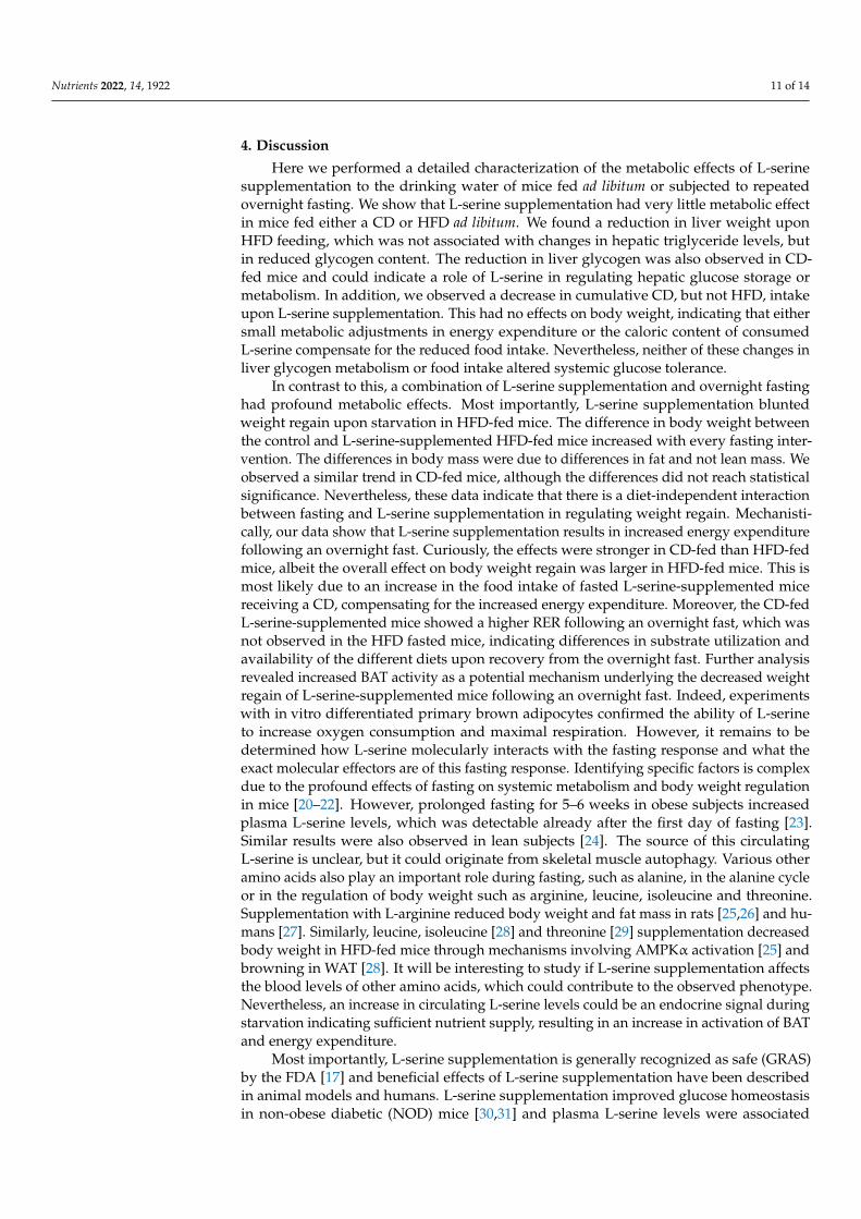
Figure 1. L-serine supplementation has no major effects on metabolism in ad libitum-fed mice. (a) Body weight of chow diet (CD) and high-fat diet (HFD) fed mice supplemented with or without L-serine for 8 weeks, starting at 4 weeks of age (n = 8). (b) Body compo-sition (fat and lean mass) of CD-fed and HFD-fed mice with and without L-serine sup-plementation assessed after 8 weeks (n = 8). (c) Randomly fed blood glucose levels of CD-fed and HFD-fed mice with and without L-serine supplementation after 3 weeks (n= 8). (d) Randomly fed insulin levels of CD-fed and HFD-fed mice with and without L-serine supplementation after 3 weeks. (e) Glucose Tolerance Test (GTT) and baseline subtracted area under the curve (AUC) of CD -fed and HFD-fed mice with and without L-serine supplementation at week 6 (n = 8). (f) Representative H&E stainings of perigonadal fat (PGF), SCF (subcutaneous fat) and liver of random CD-fed and HFD-fed mice with or without L-serine supplementation after 8 weeks. Scale bars represent 100 µM. Data are shown as mean ± SEM, one or two-way ANOVA with Tukey’s post-test. * p < 0.05.
3.2. L-Serine Supplementation Blunts Body Weight Regain after Repeated Overnight Fasting Pyruvate tolerance tests (PTTs), in contrast to insulin and glucose tolerance tests
(GTTs), require an overnight fast to deplete hepatic glycogen stores. This results in a ~10–
Figure 1. L-serine supplementation has no major effects on metabolism in ad libitum-fed mice.(a) Body weight of chow diet (CD) and high-fat diet (HFD) fed mice supplemented with or withoutL-serine for 8 weeks, starting at 4 weeks of age (n = 8). (b) Body composition (fat and lean mass)of CD-fed and HFD-fed mice with and without L-serine supplementation assessed after 8 weeks(n = 8). (c) Randomly fed blood glucose levels of CD-fed and HFD-fed mice with and without L-serinesupplementation after 3 weeks (n= 8). (d) Randomly fed insulin levels of CD-fed and HFD-fed micewith and without L-serine supplementation after 3 weeks. (e) Glucose Tolerance Test (GTT) andbaseline subtracted area under the curve (AUC) of CD -fed and HFD-fed mice with and withoutL-serine supplementation at week 6 (n = 8). (f) Representative H&E stainings of perigonadal fat (PGF),SCF (subcutaneous fat) and liver of random CD-fed and HFD-fed mice with or without L-serinesupplementation after 8 weeks. Scale bars represent 100 µM. Data are shown as mean ± SEM, one ortwo-way ANOVA with Tukey’s post-test. * p < 0.05.

Nutrients 2022, 14, 1922 7 of 14
Nutrients 2022, 14, x FOR PEER REVIEW 7 of 15
15% weight loss in mice, which is usually regained within a week. We observed a reduced weight regain in L-serine-supplemented HFD-fed mice following the PTT (Figure 2a). To test the role of L-serine supplementation in weight regain, we subjected CD and HFD mice with or without L-serine supplementation to additional overnight fasting. Indeed, L-ser-ine supplementation in HFD-fed mice reduced body weight regain after each overnight fast (Figure 2a). The weight difference between the control and L-serine-supplemented mice increased with every fasting/refeeding cycle (Figure 2a). Liver weight did not change in any condition (Figure S2a), but triglyceride content decreased in the CD with L-serine supplementation (Figure S2b). In line with the triglyceride levels, liver histology showed fewer lipids in the CD-fed mice upon L-serine supplementation. Interestingly, L-serine seemed to decrease the size of the lipid droplets in the HFD, but not the number (Figure S2c). An assessment of body composition showed that the decrease in body weight was due to a reduction in fat but not lean mass (Figure 2b). This was also seen in a reduction in the weight of individual fat depots (Figure 2c) and the size of subcutaneous and perigonadal adipocytes (Figures 2d and S2d). A gene expression analysis of TNFα IL-6 and CD68 expression in SCF and PGF revealed a significant downregulation of TNFα and a strong trend toward reduced CD68 expression in the PGF of HFD-fed L-serine-supple-mented mice compared to HFD-fed controls (Figure S2e). We did not observe any differ-ences in glucose tolerance between groups as the repeated fasting retained normal glucose tolerance in control HFD-fed mice when compared to the CD-fed mice (Figure 2e).
Figure 2. L-serine supplementation blunts fasting-induced body weight regain. (a) Body weight increase (%) of mice a fed chow diet (CD) or high-fat diet (HFD) with or without L-serine supple-mentation and repeated overnight fasting for 8 weeks starting at 8 weeks of age (n = 6). (b) Body
Figure 2. L-serine supplementation blunts fasting-induced body weight regain. (a) Body weightincrease (%) of mice a fed chow diet (CD) or high-fat diet (HFD) with or without L-serine supple-mentation and repeated overnight fasting for 8 weeks starting at 8 weeks of age (n = 6). (b) Bodycomposition (fat and lean mass) of mice shown in (a) after 8 weeks (n = 6). (c) Weight of perigonadalfat (PGF) and subcutaneous fat (SCF) of mice in (a) after week 8 (n = 6). (d) Representative H&Estainings of PGF and SCF from mice shown in (a) after 8 weeks. Scale bars show 100 µM. (e) GlucoseTolerance Test (GTT) of CD-fed and HFD-fed mice with and without L-serine supplementation atweek 6 (n = 6). Data are shown as mean ± SEM, one or two-way ANOVA with Tukey’s post-hoc test.* p < 0.05, ** p < 0.01, *** p < 0.001, **** p < 0.0001.
3.3. L-Serine Supplementation Increases Energy Expenditure following an Overnight Fast
To study the metabolic changes induced by the interaction of fasting and L-serinesupplementation, we studied CD-fed and HFD-fed mice either with or without serinesupplementation in metabolic cages (Figure S3a). After acclimatization, the CD (after7 weeks) and HFD (after 8 weeks) mice were fasted for 16 h in metabolic cages and foodintake, energy expenditure, activity and respiratory exchange ratios were assessed in the72 h thereafter (Figure 3a). As expected, daily food intake and cumulative food intakewere increased in the CD-fed mice following an overnight fast compared to ad libitum-fed mice (Figures 3b and S3b). A similar trend was observed in the HFD-fed mice. Theincrease in daily food intake was largely attributed to increased feeding during the lightcycle. L-serine supplementation increased food intake in CD-fed, but not HFD-fed mice(Figures 3b and S3b). A similar trend was observed for water intake (Figure S3c). In linewith the changes in food intake, we observed increased Neuropeptide Y (Npy) and de-

Nutrients 2022, 14, 1922 8 of 14
creased Proopiomelanocortin (Pomc) expression in the hypothalamus of overnight-fastedmice fed a CD, whereas no statistically significant differences were observed in mice feda HFD at the end of the 16-week L-serine supplementation (Figure S3d). No differencesin Npy or Pomc expression between the control and L-serine-supplemented mice wereobserved in either experimental condition (Figure S3d). Thus, L-serine supplementationincreased chow diet intake following an overnight fast compared to control animals. Con-versely, there was a trend for slightly reduced food intake during the light phase in theHFD-fed L-serine-supplemented mice following an overnight fast. These data suggestedthat differences in food intake per se could not explain the reduced weight regain ofHFD-fed mice upon serine supplementation. To this end, we investigated whether energyexpenditure and substrate utilization, assessed by the respiratory exchange ratio (RER),were altered by serine supplementation. Ad libitum CD-fed mice showed the typical diurnalrhythm of substrate utilization with the highest RER during the dark phase (active phase)and the lowest values, indicating fatty acid oxidation, during the light phase (resting phase),with no differences between the control and L-serine-supplemented mice (Figure 3c). Fast-ing reduced the RER in all groups and diets (Figure S3e). This also demonstrated thatconsumption of L-serine through the drinking water did not significantly contribute to en-ergy expenditure, as this would raise the RER. Refeeding rapidly increased the RER, whichremained elevated throughout the remaining time measured (Figure S3e). The absence ofdiurnal fluctuations can be explained by the increased food intake during the light phase.The RER of control mice increased its amplitude during the 72 h following fasting, whereasthe L-serine-supplemented mice remained at close to one throughout the whole time. Thiscould result from the increased food intake observed in the L-serine-supplemented miceor indicate preferential oxidation of carbohydrates. The RER pattern was similar in theHFD-fed mice, albeit the overall amplitude of oscillation was reduced due to the increaseddietary fats (Figure 3c). Moreover, fasted mice returned more rapidly after the next darkcycle to the diurnal oscillation observed in ad libitum-fed mice (Figure S3e). Interestingly,L-serine supplementation lowered the RER in the HFD-fed mice compared to controls,potentially indicating increased adipose tissue lipolysis (Figure S3e). To this end, wequantified HSL phosphorylation at the activating sites Ser563 and Ser660, as well as theinhibitory Ser565 site in SCF and PGF following an overnight fast (Figures 3d and S3f).We observed increased Ser660 and reduced Ser565 phosphorylation in HSL in the HFDcompared to the CD-fed animals in PGF. However, we did not observe any differencesbetween the control and L-serine-supplemented mice irrespective of the diet. In addition,serine supplementation led to a reduction in phosphorylation of Ser565 in the CD comparedto the HFD in SCF (Figure 3d). Thus, these data did not suggest an increased lipolysis inwhite adipose tissue in the interaction of fasting and L-serine supplementation. Moreover,ex vivo stimulation of PGF with either 200 µM or 400 µM L-serine did not increase lipolysisas measured by glycerol release into the medium (Figure 3e). We also did not observe aneffect of 400 µM L-serine on isoproterenol-induced lipolysis (Figure 3e).
3.4. L-Serine Supplementation Activates Brown Adipose Tissue Thermogenesis after Fasting
Neither food intake nor changes in lipolysis explained the decreased weight regainof L-serine-supplemented mice upon HFD feeding. In ad libitum CD-fed mice, L-serinesupplementation decreased energy expenditure (EE) (p = 0.002) (Figure 4a). Conversely,EE was increased with L-serine supplementation following an overnight fast (p = 0.0003),which is in line with the food intake data (Figure 3b). Similarly, L-serine supplementationshowed a trend towards reduced EE in ad libitum HFD-fed mice versus an increase followingan overnight fast (Figure 4a). The inverse relation between body weight and EE in fastedcontrol animals complicates the interpretation of the latter results. Nevertheless, thesedata suggest that increased EE could explain the observed reduction in weight regainin L-serine-supplemented HFD-fed mice. The total activity was not changed betweengroups (Figure S4a). Thus, we investigated a potential role of brown adipose tissue (BAT)activity in the observed phenotype. Histological analysis of BAT showed that upon fasting,

Nutrients 2022, 14, 1922 9 of 14
L-serine supplementation reduced lipid droplet size in both the CD-fed and HFD-fedmice (Figure 4b), which was also reflected by decreased BAT weight in CD-fed mice and atrend in HFD-fed mice (Figure S4b). Gene expression of important thermogenesis-relatedgenes were not changed in the BAT of any experimental group or any diet (Figure S4c).However, uncoupled protein 1 (UCP1) protein levels were significantly higher in L-serine-supplemented fasted mice fed a CD. This was not observed in HFD-fed mice, whichgenerally had much lower UCP1 protein levels (Figures 4c and S4d). Thus, we testedif L-serine has direct effects on mitochondrial respiration in primary brown adipocytes.Pretreatment with L-serine or an acute injection did not alter basal respiration. However,L-serine potentiated isoproterenol-induced and maximal respiration (Figure 4d).
Nutrients 2022, 14, x FOR PEER REVIEW 9 of 15
addition, serine supplementation led to a reduction in phosphorylation of Ser565 in the CD compared to the HFD in SCF (Figure 3d). Thus, these data did not suggest an increased lipolysis in white adipose tissue in the interaction of fasting and L-serine supplementa-tion. Moreover, ex vivo stimulation of PGF with either 200 µM or 400 µM L-serine did not increase lipolysis as measured by glycerol release into the medium (Figure 3e). We also did not observe an effect of 400 µM L-serine on isoproterenol-induced lipolysis (Figure 3e).
Figure 3. L-serine supplementation alters food intake and respiratory exchange ratio (RER), but notwhite adipose tissue (WAT) lipolysis. (a) Experimental setup for indirect calorimetry measurements.The figure was generated using Biorender.com. (b) Average food intake of random fed and fastedchow diet (CD) and high-fat diet (HFD)-fed mice with or without L-serine supplementation at week7 (CD) or 8 (HFD) (CD random fed: n = 5; CD fasted, CD+L-ser random fed, CD+L-ser fasted: n = 6;

Nutrients 2022, 14, 1922 10 of 14
HFD random fed: n = 4; HFD fasted, HFD+L-ser random fed, HFD+L-ser fasted: (n = 6). (c) Meanrespiratory exchange ratio (RER) of random fed and fasted CD and HFD fed mice with or withoutL-serine supplementation at weeks 7 (CD) or 8 (HFD) (CD random fed: n = 5; CD fasted, CD+L-serrandom fed, CD+L-ser fasted: n = 6; HFD random fed: n = 4; HFD fasted, HFD+L-ser random fed,HFD+L-ser fasted: n = 6). (d) Quantifications of Western blots for phosphorylated HSL (Ser563,Ser660, Ser565) and total HSL of PGF and SCF (n = 6) of fasted CD-fed and HFD-fed mice with orwithout L-serine supplementation. (e) Ex vivo lipolysis assay of PGF (n = 3). Data are shown asmean ± SEM, one or two-way ANOVA with Tukey’s post-hoc test. * p < 0.05, ** p < 0.01, *** p < 0.001,**** p < 0.001.
Nutrients 2022, 14, x FOR PEER REVIEW 11 of 15
Figure 4. L-serine supplementation alters energy expenditure and potentiates mitochondrial respi-ration. (a) Energy expenditure after overnight fasting in relation to body weight of random fed and fasted CD (chow diet) and HFD (high-fat diet)-fed mice with or without L-serine supplemen-tation at weeks 7 (CD) or 8 (HFD) (CD random fed: n = 5; CD fasted, CD+L-ser random fed, CD+L-ser fasted: n = 6; HFD random fed: n = 4; HFD fasted, HFD+L-ser random fed, HFD+L-ser fasted: (n = 6). (b) Representative H&E stainings of brown adipose tissue (BAT) from random fed and fasted CD-fed and HFD-fed mice with or without L-serine supplementation after 8 weeks starting at 4 (random fed) and 8 (fasted) weeks of age. Scale bars show 100 µM. (c) Quantification of uncoupled protein 1 (UCP1) protein content normalized to beta actin from BAT of random fed and fasted CD-fed and HFD-fed mice with or without L-serine supplementation (n = 6). (d) Oxygen consumption rate (OCR) of differentiated primary brown adipocytes pre-treated or not with 400 µM L-serine and acutely exposed (injected) to 400 µM L-serine and/or 1 µM isoproterenol (n = 3 replicates from 2 mice). Statistical analysis in panel a is described in Section 2.10. Data are shown as mean ± SEM, one or two-way ANOVA with Tukey’s post-hoc test. * p < 0.05, ** p < 0.01,*** p < 0.001.
Figure 4. L-serine supplementation alters energy expenditure and potentiates mitochondrial respira-tion. (a) Energy expenditure after overnight fasting in relation to body weight of random fed andfasted CD (chow diet) and HFD (high-fat diet)-fed mice with or without L-serine supplementationat weeks 7 (CD) or 8 (HFD) (CD random fed: n = 5; CD fasted, CD+L-ser random fed, CD+L-serfasted: n = 6; HFD random fed: n = 4; HFD fasted, HFD+L-ser random fed, HFD+L-ser fasted: (n = 6).(b) Representative H&E stainings of brown adipose tissue (BAT) from random fed and fasted CD-fedand HFD-fed mice with or without L-serine supplementation after 8 weeks starting at 4 (randomfed) and 8 (fasted) weeks of age. Scale bars show 100 µM. (c) Quantification of uncoupled protein1 (UCP1) protein content normalized to beta actin from BAT of random fed and fasted CD-fed andHFD-fed mice with or without L-serine supplementation (n = 6). (d) Oxygen consumption rate (OCR)of differentiated primary brown adipocytes pre-treated or not with 400 µM L-serine and acutelyexposed (injected) to 400 µM L-serine and/or 1 µM isoproterenol (n = 3 replicates from 2 mice).Statistical analysis in panel a is described in Section 2.10. Data are shown as mean ± SEM, one ortwo-way ANOVA with Tukey’s post-hoc test. * p < 0.05, ** p < 0.01,*** p < 0.001.
Nutrients 2022, 14, 1922 11 of 14
4. Discussion
Here we performed a detailed characterization of the metabolic effects of L-serinesupplementation to the drinking water of mice fed ad libitum or subjected to repeatedovernight fasting. We show that L-serine supplementation had very little metabolic effectin mice fed either a CD or HFD ad libitum. We found a reduction in liver weight uponHFD feeding, which was not associated with changes in hepatic triglyceride levels, butin reduced glycogen content. The reduction in liver glycogen was also observed in CD-fed mice and could indicate a role of L-serine in regulating hepatic glucose storage ormetabolism. In addition, we observed a decrease in cumulative CD, but not HFD, intakeupon L-serine supplementation. This had no effects on body weight, indicating that eithersmall metabolic adjustments in energy expenditure or the caloric content of consumedL-serine compensate for the reduced food intake. Nevertheless, neither of these changes inliver glycogen metabolism or food intake altered systemic glucose tolerance.
In contrast to this, a combination of L-serine supplementation and overnight fastinghad profound metabolic effects. Most importantly, L-serine supplementation bluntedweight regain upon starvation in HFD-fed mice. The difference in body weight betweenthe control and L-serine-supplemented HFD-fed mice increased with every fasting inter-vention. The differences in body mass were due to differences in fat and not lean mass. Weobserved a similar trend in CD-fed mice, although the differences did not reach statisticalsignificance. Nevertheless, these data indicate that there is a diet-independent interactionbetween fasting and L-serine supplementation in regulating weight regain. Mechanisti-cally, our data show that L-serine supplementation results in increased energy expenditurefollowing an overnight fast. Curiously, the effects were stronger in CD-fed than HFD-fedmice, albeit the overall effect on body weight regain was larger in HFD-fed mice. This ismost likely due to an increase in the food intake of fasted L-serine-supplemented micereceiving a CD, compensating for the increased energy expenditure. Moreover, the CD-fedL-serine-supplemented mice showed a higher RER following an overnight fast, which wasnot observed in the HFD fasted mice, indicating differences in substrate utilization andavailability of the different diets upon recovery from the overnight fast. Further analysisrevealed increased BAT activity as a potential mechanism underlying the decreased weightregain of L-serine-supplemented mice following an overnight fast. Indeed, experimentswith in vitro differentiated primary brown adipocytes confirmed the ability of L-serineto increase oxygen consumption and maximal respiration. However, it remains to bedetermined how L-serine molecularly interacts with the fasting response and what theexact molecular effectors are of this fasting response. Identifying specific factors is complexdue to the profound effects of fasting on systemic metabolism and body weight regulationin mice [20–22]. However, prolonged fasting for 5–6 weeks in obese subjects increasedplasma L-serine levels, which was detectable already after the first day of fasting [23].Similar results were also observed in lean subjects [24]. The source of this circulatingL-serine is unclear, but it could originate from skeletal muscle autophagy. Various otheramino acids also play an important role during fasting, such as alanine, in the alanine cycleor in the regulation of body weight such as arginine, leucine, isoleucine and threonine.Supplementation with L-arginine reduced body weight and fat mass in rats [25,26] and hu-mans [27]. Similarly, leucine, isoleucine [28] and threonine [29] supplementation decreasedbody weight in HFD-fed mice through mechanisms involving AMPKα activation [25] andbrowning in WAT [28]. It will be interesting to study if L-serine supplementation affectsthe blood levels of other amino acids, which could contribute to the observed phenotype.Nevertheless, an increase in circulating L-serine levels could be an endocrine signal duringstarvation indicating sufficient nutrient supply, resulting in an increase in activation of BATand energy expenditure.
Most importantly, L-serine supplementation is generally recognized as safe (GRAS)by the FDA [17] and beneficial effects of L-serine supplementation have been describedin animal models and humans. L-serine supplementation improved glucose homeostasisin non-obese diabetic (NOD) mice [30,31] and plasma L-serine levels were associated

Nutrients 2022, 14, 1922 12 of 14
with improved insulin sensitivity in humans [11]. Moreover, experimental and clinicaldata suggest that L-serine supplementation improves hereditary sensory and autonomicneuropathy type 1 (HSAN1) disease [32,33]. Furthermore, ongoing clinical trials investigateL-serine supplementation for treatment in Alzheimer’s disease [34] and GRIN-relatedencephalopathy in children [35]. Thus, the potential effects of L-serine supplementationon body weight regain in humans could be easily studied. This will be greatly importantas major species differences exist between mice and humans. Overnight fasting has muchmore pronounced metabolic consequences in mice than in humans due to much highermetabolic rate [36]. Thus, it will be important to define which hypocaloric weight lossprogram corresponds best to the overnight fasting described here. Moreover, we shouldbear in mind that BAT has a more important role in the regulation of systemic metabolismin mice than in humans. However, our data strongly suggest that L-serine supplementationduring and after weight-loss interventions could significantly reduce weight regain andthereby help tackle one of the major problems of current obesity therapies.
Supplementary Materials: The following supporting information can be downloaded at: https://www.mdpi.com/article/10.3390/nu14091922/s1, Table S1: qPCR primers, Figure S1: L-serinesupplementation does not affect white adipose (WAT) tissue inflammation or cell size in ad libitumfed mice, Figure S2: L-serine supplementation reduces white adipocyte size and white adipose tissue(WAT) inflammation upon fasting, Figure S3: L-serine supplementation increases cumulative highfat diet intake in fasted mice, Figure S4: L-serine supplementation does not alter thermogenic geneexpression in brown adipose tissue (BAT).
Author Contributions: Study design, E.L.-G., L.L. and S.U.; Experiments and data analysis, E.L.-G., L.L., F.J.R.-O., R.H.-B., I.A., Y.O., A.E.K., A.I. and X.L.; writing, E.L.-G. and S.U. All authorsparticipated in discussing the results. All authors have read and agreed to the published version ofthe manuscript.
Funding: Y.O. received support through the Post-doctoral Fellowship program from The UeharaMemorial Foundation, Japan (201830047) and the Alexander von Humboldt-Stiftung, Germany.
Institutional Review Board Statement: Animal experiments were performed according to the Ger-man animal welfare law and guidelines and regulations of the district of Upper Bavaria (Bayern,Germany), protocol number 55.2-1-54-2532-52-2016.
Data Availability Statement: All primary data can be obtained from the corresponding author uponreasonable request.
Acknowledgments: We would like to thank Annette Feuchtinger for help with the automatedadipocyte cell size measurements.
Conflicts of Interest: The authors declare no conflict of interests.
References1. World Health Organization. Overview Complications Prevention and Control; World Health Organization: Geneva, Switzerland,
2020; Available online: www.who.int/health-topics/obesity (accessed on 10 September 2021).2. Blüher, M. Obesity: Global epidemiology and pathogenesis. Nat. Rev. Endocrinol. 2019, 15, 288–298. [CrossRef] [PubMed]3. Holm, L.J.; Buschard, K. L-Serine: A neglected amino acid with a potential therapeutic role in diabetes. Apmis 2019, 127, 655–659.
[CrossRef] [PubMed]4. Salari, N.; Jafari, S.; Darvishi, N.; Valipour, E.; Mohammadi, M.; Mansouri, K.; Shohaimi, S. The best drug supplement for obesity
treatment: A systematic review and network meta-analysis. Diabetol. Metab. Syndr. 2021, 13, 1–12. [CrossRef]5. Gaesser, G.A.; Angadi, S.S. Obesity treatment: Weight loss versus increasing fitness and physical activity for reducing health risks.
iScience 2021, 24, 102995. [CrossRef] [PubMed]6. Heffron, S.P.; Parham, J.S.; Pendse, J.; Alemán, J.O. Treatment of Obesity in Mitigating Metabolic Risk. Circ. Res. 2020, 126,
1646–1665. [CrossRef]7. Fischer, I.P.; Irmler, M.; Meyer, C.W.; Sachs, S.J.; Neff, F.; de Angelis, M.H.; Beckers, J.; Tschöp, M.H.; Hofmann, S.; Ussar, S. A
history of obesity leaves an inflammatory fingerprint in liver and adipose tissue. Int. J. Obes. 2017, 42, 507–517. [CrossRef]8. Donnelly, J.E.; Blair, S.N.; Jakicic, J.M.; Manore, M.M.; Rankin, J.W.; Smith, B.K.; American College of Sports Medicine. American
College of Sports Medicine Position Stand. Appropriate Physical Activity Intervention Strategies for Weight Loss and Preventionof Weight Regain for Adults. Med. Sci. Sports Exerc. 2009, 41, 459–471. [CrossRef]

Nutrients 2022, 14, 1922 13 of 14
9. Zhou, X.; Zhang, H.; He, L.; Wu, X.; Yin, Y. Long-Term l-Serine Administration Reduces Food Intake and Improves OxidativeStress and Sirt1/NFκB Signaling in the Hypothalamus of Aging Mice. Front. Endocrinol. 2018, 9, 476. [CrossRef]
10. Holm, L.J.; Haupt-Jorgensen, M.; Larsen, J.; Giacobini, J.D.; Bilgin, M.; Buschard, K. L-serine supplementation lowers diabetesincidence and improves blood glucose homeostasis in NOD mice. PLoS ONE 2018, 13, e0194414. [CrossRef]
11. Vangipurapu, J.; Stancáková, A.; Smith, U.; Kuusisto, J.; Laakso, M. Nine Amino Acids Are Associated with Decreased InsulinSecretion and Elevated Glucose Levels in a 7.4-Year Follow-up Study of 5181 Finnish Men. Diabetes 2019, 68, 1353–1358. [CrossRef]
12. Parker, S.J.; Metallo, C.M. Chasing One-Carbon Units to Understand the Role of Serine in Epigenetics. Mol. Cell 2016, 61, 185–186.[CrossRef] [PubMed]
13. Kalhan, S.C.; Hanson, R.W. Resurgence of Serine: An Often Neglected but Indispensable Amino Acid. J. Biol. Chem. 2012, 287,19786–19791. [CrossRef] [PubMed]
14. Newsholme, P.; Stenson, L.; Sulvucci, M.; Sumayao, R.; Krause, M. Amino Acid Metabolism, 2nd ed.; Elsevier B.V.: Amsterdam,The Netherlands, 2011; Volume 1.
15. Muthusamy, T.; Cordes, T.; Handzlik, M.K.; You, L.; Lim, E.W.; Gengatharan, J.; Pinto, A.F.; Badur, M.G.; Kolar, M.J.;Wallace, M.; et al. Serine restriction alters sphingolipid diversity to constrain tumor growth. Nature 2021, 586, 790–795. [CrossRef][PubMed]
16. Tajan, M.; Hennequart, M.; Cheung, E.C.; Zani, F.; Hock, A.K.; Legrave, N.; Maddocks, O.D.K.; Ridgway, R.A.; Athineos, D.;Suárez-Bonnet, A.; et al. Serine synthesis pathway inhibition cooperates with dietary serine and glycine limitation for cancertherapy. Nat. Commun. 2021, 12, 1–16. [CrossRef]
17. U.S. Food & Drug Administration. L-Serine GRAS 172.320. Substances Added to Food (formerly EAFUS), Food Ingredientand Packaging Inventories. 2021. Available online: https://www.cfsanappsexternal.fda.gov/scripts/fdcc/index.cfm?set=FoodSubstances&id=SERINE (accessed on 20 January 2022).
18. Suwandhi, L.; Hausmann, S.; Braun, A.; Gruber, T.; Heinzmann, S.S.; Gálvez, E.J.; Buck, A.; Legutko, B.; Israel, A.;Feuchtinger, A.; et al. Chronic d-serine supplementation impairs insulin secretion. Mol. Metab. 2018, 16, 191–202. [CrossRef]
19. Zhang, H.; Hua, R.; Zhang, B.; Zhang, X.; Yang, H.; Zhou, X. Serine Alleviates Dextran Sulfate Sodium-Induced Colitis andRegulates the Gut Microbiota in Mice. Front. Microbiol. 2018, 9, 3062. [CrossRef]
20. Heijboer, A.C.; Donga, E.; Voshol, P.J.; Dang, Z.-C.; Havekes, L.M.; Romijn, J.A.; Corssmit, E.P.M. Sixteen hours of fastingdifferentially affects hepatic and muscle insulin sensitivity in mice. J. Lipid Res. 2005, 46, 582–588. [CrossRef]
21. Jensen, T.L.; Kiersgaard, M.K.; Sørensen, D.B.; Mikkelsen, L.F. Fasting of mice: A review. Lab. Anim. 2013, 47, 225–240. [CrossRef]22. Pedroso, J.A.; Wasinski, F.; Donato, J. Prolonged fasting induces long-lasting metabolic consequences in mice. J. Nutr. Biochem.
2020, 84, 108457. [CrossRef]23. Felig, P.; Owen, O.E.; Wahren, J.; Cahill, G.F. Amino acid metabolism during prolonged starvation Amino Acid Metabolism
during Prolonged Starvation. J. Clin. Investig. 1969, 48, 584–594. [CrossRef]24. Adibi, A. Influence of dietary deprivations on plasma concentration of free amino acids of man. J. Appl. Physiol. 1968, 25, 52–57.
[CrossRef] [PubMed]25. Fu, W.J.; Haynes, T.E.; Kohli, R.; Hu, J.; Shi, W.; Spencer, T.; Carroll, R.J.; Meininger, C.J.; Wu, G. Dietary L-Arginine Supplementa-
tion Reduces Fat Mass in Zucker Diabetic Fatty Rats. J. Nutr. 2005, 135, 714–721. [CrossRef] [PubMed]26. Tan, B.; Li, X.; Yin, Y.; Wu, Z.; Liu, C.; Tekwe, C.D.; Wu, G. Regulatory roles for L-arginine in reducing white adipose tissue. Front.
Biosci. 2012, 17, 2237–2246. [CrossRef] [PubMed]27. Lucotti, P.; Setola, E.; Monti, L.D.; Galluccio, E.; Costa, S.; Sandoli, E.P.; Fermo, I.; Rabaiotti, G.; Gatti, R.; Piatti, P. Beneficial effects
of a long-term oral l-arginine treatment added to a hypocaloric diet and exercise training program in obese, insulin-resistant type2 diabetic patients. Am. J. Physiol. Metab. 2006, 291, E906–E912. [CrossRef]
28. Ma, Q.; Zhou, X.; Hu, L.; Chen, J.; Zhu, J.; Shan, A. Leucine and isoleucine have similar effects on reducing lipid accumulation,improving insulin sensitivity and increasing the browning of WAT in high-fat diet-induced obese mice. Food Funct. 2020, 11,2279–2290. [CrossRef]
29. Ma, Q.; Zhou, X.; Sun, Y.; Hu, L.; Zhu, J.; Shao, C.; Meng, Q.; Shan, A. Threonine, but Not Lysine and Methionine, Reduces FatAccumulation by Regulating Lipid Metabolism in Obese Mice. J. Agric. Food Chem. 2020, 68, 4876–4883. [CrossRef]
30. Zhou, X.; He, L.; Zuo, S.; Zhang, Y.; Wan, D.; Long, C.; Huang, P.; Wu, X.; Wu, C.; Liu, G.; et al. Serine prevented high-fatdiet-induced oxidative stress by activating AMPK and epigenetically modulating the expression of glutathione synthesis-relatedgenes. Biochim. Biophys. Acta (BBA) Mol. Basis Dis. 2018, 1864, 488–498. [CrossRef]
31. Sim, W.-C.; Yin, H.-Q.; Choi, H.-S.; Choi, Y.-J.; Kwak, H.C.; Kim, S.-K.; Lee, B.-H. L-Serine Supplementation Attenuates AlcoholicFatty Liver by Enhancing Homocysteine Metabolism in Mice and Rats. J. Nutr. 2014, 145, 260–267. [CrossRef]
32. Fridman, V.; Suriyanarayanan, S.; Novak, P.; David, W.; Macklin, E.A.; McKenna-Yasek, D.; Walsh, K.; Aziz-Bose, R.;Oaklander, A.L.; Brown, R.; et al. Randomized trial of l-serine in patients with hereditary sensory and autonomic neuropathytype 1. Neurology 2019, 92, e359–e370. [CrossRef]
33. Garofalo, K.; Penno, A.; Schmidt, B.; Lee, H.-J.; Frosch, M.P.; Von Eckardstein, A.; Brown, R.H.; Hornemann, T.; Eichler, F.S. Orall-serine supplementation reduces production of neurotoxic deoxysphingolipids in mice and humans with hereditary sensoryautonomic neuropathy type 1. J. Clin. Investig. 2011, 121, 4735–4745. [CrossRef]

Nutrients 2022, 14, 1922 14 of 14
34. Stark, A.; Labs, B.C. Phase IIa L-serine Trial for ALzheimer’s Disease (LSPI-2). 2022. Available online: https://clinicaltrials.gov/ct2/show/NCT03062449 (accessed on 20 January 2022).
35. De Déu, S.J. Tolerability and Efficacy of L-Serine in Patients with GRIN-Related Encephalopathy. 2020. Available online:https://clinicaltrials.gov/ct2/show/record/NCT04646447 (accessed on 20 January 2022).
36. Demetrius, L. Of mice and men. EMBO Rep. 2005, 6, 39–45. [CrossRef] [PubMed]