Revolutionary NEW Pillow Resists Germ and Mold Growth To ...
Upload
khangminh22Category
view
0download
0
Original Article
Sex Dev 2020;14:80–97
Primordial Germ Cell Migration and Histological and Molecular Characterization of Gonadal Differentiation in Pachón Cavefish Astyanax mexicanus
Boudjema Imarazene
a, b Séverine Beille
a Elodie Jouanno
a Adéle Branthonne
a
Violette Thermes
a Manon Thomas
a Amaury Herpin
a Sylvie Rétaux
b
Yann Guiguen
a
aINRAE, Laboratoire de Physiologie et Génomique des poissons, Rennes, France; bUniversité Paris-Saclay, CNRS, Institut des Neurosciences Paris-Saclay, Gif-sur-Yvette, France
Received: July 31, 2020Accepted: September 16, 2020Published online: March 10, 2021
Yann GuiguenINRAELaboratoire de Physiologie et Génomique des poissonsCampus de Beaulieu, FR–35042 Rennes (France) yann.guiguen @ inrae.fr
© 2021 S. Karger AG, [email protected]/sxd
DOI: 10.1159/000513378
KeywordsGene expression profiling · Histology · nanos · Ovary · PGCs · Testis · vasa
AbstractThe genetic regulatory network governing vertebrate go-nadal differentiation appears less conserved than previously thought. Here, we investigated the gonadal development of Astyanax mexicanus Pachón cavefish by looking at primor-dial germ cells (PGCs) migration and proliferation, gonad his-tology, and gene expression patterns. We showed that PGCs are first detected at the 80% epiboly stage and then reach the gonadal primordium at 1 day post-fertilization (dpf). However, in contrast to the generally described absence of PGCs proliferation during their migration phase, PGCs num-ber in cavefish doubles between early neurula and 8–9 so-mites stages. Combining both gonadal histology and vasa (germ cell marker) expression patterns, we observed that ovarian and testicular differentiation occurs around 65 dpf in females and 90 dpf in males, respectively, with an impor-tant inter-individual variability. The expression patterns of dmrt1, gsdf, and amh revealed a conserved predominant male expression during cavefish gonadal development, but
none of the ovarian differentiation genes, i. e., foxl2a, cy-p19a1a, and wnt4b displayed an early sexually dimorphic ex-pression, and surprisingly all these genes exhibited predom-inant expression in adult testes. Altogether, our results lay the foundation for further research on sex determination and differentiation in A. mexicanus and contribute to the emerging picture that the vertebrate sex differentiation downstream regulatory network is less conserved than pre-viously thought, at least in teleost fishes.
© 2021 S. Karger AG, Basel
Introduction
Fishes, with over 35,000 recognized species, constitute the most diverse and abundant group of vertebrates [Fricke et al., 2020]. Besides their remarkable taxonomic diversity and the impressive range of habitats, behaviors, and morphological differences, they exhibit all kinds of reproductive strategies and sex determination (SD) mechanisms [Devlin and Nagahama, 2002; Bachtrog et al., 2014]. Along with this diversity of SD mechanisms, comparative studies revealed a high turnover of master sex-determining (MSD) genes [Pan et al., 2018]. These

Gonadal Differentiation in Cavefish 81Sex Dev 2020;14:80–97DOI: 10.1159/000513378
MSD genes control downstream sex differentiation cas-cades that include all the physiological, morphological, and cellular processes by which the undifferentiated go-nad will develop either into a testis or an ovary [Devlin and Nagahama, 2002]. In contrast to the observed high turnover of both SD systems and MSD genes, the down-stream sex differentiation processes are often considered to be relatively stable in fish [Nagahama, 2005; Ijiri et al., 2008]. However, studies on the evolution of the gene reg-ulatory network leading to sex differentiation in medaka, Oryzias latipes, suggest that this downstream regulation is not as conserved as previously thought [Herpin et al., 2013]. Hence, this variability makes teleost fishes inter-esting and important models for exploring the complex-ity of the gene network(s) underlying sexual differentia-tion in vertebrates.
In teleost fish, gonadal differentiation can follow di-verse trajectories. It has been explored in several species from either morphological, cellular, or physiological points of view, highlighting a large variety of reproductive strategies, including gonochorism with both differenti-ated and undifferentiated gonochoristic species and her-maphroditism [Yamamoto, 1969; Baroiller and Guiguen, 2001; Devlin and Nagahama, 2002; Nishimura and Tana-ka, 2014]. In differentiated gonochoristic species that rep-resent the majority of teleost fish, differentiation pro-ceeds from an undifferentiated gonad into a differentiat-ing testis in males or a differentiating ovary in females and remains stable throughout their complete life span [Ya-mamoto, 1969]. However, in undifferentiated gonocho-ristic species, all undifferentiated gonads proceed through ovarian development followed by a later and final differ-entiation step into either ovaries or testes [Yamamoto, 1969].
During the gonadal differentiation processes, primor-dial germ cell (PGC) specification, migration, prolifera-tion, and colonization of the gonadal anlage constitute the initial steps of early gonadogenesis. Following these early steps of PGC migration and colonization of the sex-ually undifferentiated embryonic gonad, a sexualization program, leading to a stepwise differentiation toward a testicular or ovarian fate, will be activated [Guerrero-Es-tévez and Moreno-Mendoza, 2010]. This sexually dicho-tomic differentiation program will induce morphological and physiological sex differences that are accompanied by sex-specific changes in the gonadal histology and gene expression patterns. For instance, early signs of female histological differentiation may include, among many criteria that could be variable between species, (i) the on-togenesis of an early ovarian cavity, (ii) an active prolif-
eration of germ cells, or (iii) an early meiotic activity and a size increase of female germ cells (auxocytosis) com-pared to male germ cells [Brusle and Brusle, 1983]. Con-comitantly, many genes involved in the gonadal differen-tiation process will be expressed in a sexually dimorphic fashion, ensuring the proper development and differen-tiation of the male and female gonads [Jørgensen et al., 2008; Guerrero-Estévez and Moreno-Mendoza, 2010; Wang et al., 2019]. For instance, dmrt1 (doublesex and mab-3 related transcription factor 1), amh (anti-Mülleri-an hormone), and gsdf (gonadal somatic cell derived fac-tor) have been described as conserved factors specifically involved in testicular differentiation [Ijiri et al., 2008; Guerrero-Estévez and Moreno-Mendoza, 2010]. On the other hand, cyp19a1a (cytochrome P450, family 19, sub-family A, polypeptide 1a), foxl2a (forkhead box L2a), and wnt4b (Wnt Family Member 4b) are often documented as classical ovarian differentiation factors [Guerrero-Es-tévez and Moreno-Mendoza, 2010; Guiguen et al., 2010; Herpin et al., 2013; Sreenivasan et al., 2014; Bertho et al., 2016]. But as mentioned above, SD and sex differentia-tion are extremely plastic processes in teleost fish, prompt-ing to new investigations in additional species to be able to better underline conserved and nonconserved sex dif-ferentiation pathways.
The Mexican tetra, Astyanax mexicanus, belongs to the Characiform group which contains about 2,300 spe-cies [Nelson et al., 2016], including a few important aqua-culture species like the Tambaqui, Colossoma macropo-mum. It is a native species from Central America rivers where this species is found in 2 morphotypes, inhabiting markedly different environments. Besides the “classical” pigmented and eyed river-dwelling fish, about 30 popula-tions of depigmented and blind cavefish live in the dark-ness of caves in North-East Mexico [Mitchell et al., 1977; Elliott, 2019]. Many resources, including some genome sequences [Di Palma et al., 2007; Hinaux et al., 2013; Mc-Gaugh et al., 2014; Herman et al., 2018], are now avail-able, making this species widely used in evolutionary, de-velopmental, and genetic studies for exploring many traits linked to cave adaptation [Keene et al., 2016]. But despite interesting reports pointing to some odd sex ra-tios in different A. mexicanus populations [Wilkens, and Strecker, 2017], sex differentiation has so far not been ex-plored in this emerging model.
To fill this knowledge gap and provide some essential ground information for further studies on sex determina-tion in A. mexicanus, we characterized gonadal differen-tiation in blind and depigmented laboratory-raised fish originating from the Pachón cave located in the north of
Imarazene et al.Sex Dev 2020;14:80–9782DOI: 10.1159/000513378
the Sierra de El Abra (Tamaulipas, Mexico) through a combination of PGCs tracking experiments, gonad his-tology, and gonadal gene expression. Our results show that the first signs of gonad histological differentiation occur around 65 days post-fertilization (dpf) in females and 90 dpf in males but with an important inter-individ-ual variability that is also found in gene expression pat-terns. Expression of well-known genes involved in testis differentiation in vertebrates revealed a conserved and significant predominant male expression in A. mexicanus with an early overexpression of gsdf in males. However, none of the classical ovarian differentiation genes that we analyzed, i. e., foxl2a, cyp19a1a, and wnt4b, displayed an early sexually dimorphic expression pattern during go-nadal development, and these genes were even detected as predominantly expressed during male gametogenesis and in adult testis. This revealed major divergences in the classical and rather conserved ovarian differentiation molecular network between A. mexicanus and other ver-tebrates. Altogether, our results lay a solid foundation for further research on sex determination and differentiation in A. mexicanus.
Materials and Methods
Animal SamplingLaboratory stocks of A. mexicanus Pachón cavefish were ob-
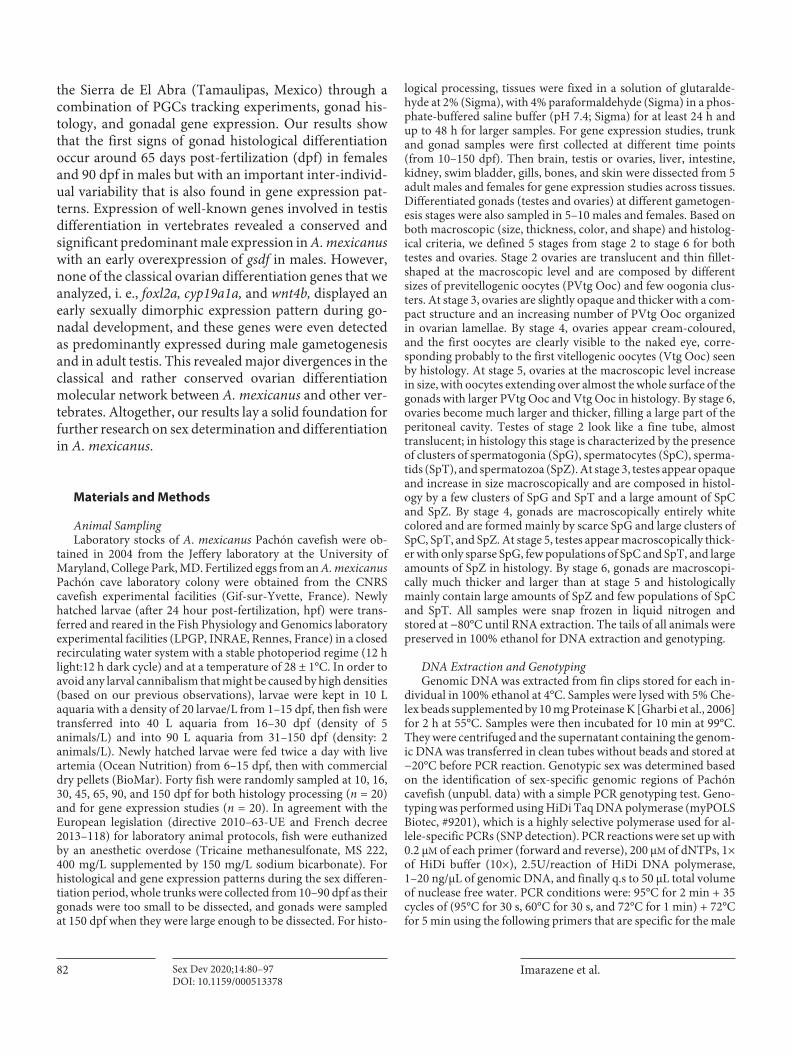
tained in 2004 from the Jeffery laboratory at the University of Maryland, College Park, MD. Fertilized eggs from an A. mexicanus Pachón cave laboratory colony were obtained from the CNRS cavefish experimental facilities (Gif-sur-Yvette, France). Newly hatched larvae (after 24 hour post-fertilization, hpf) were trans-ferred and reared in the Fish Physiology and Genomics laboratory experimental facilities (LPGP, INRAE, Rennes, France) in a closed recirculating water system with a stable photoperiod regime (12 h light:12 h dark cycle) and at a temperature of 28 ± 1°C. In order to avoid any larval cannibalism that might be caused by high densities (based on our previous observations), larvae were kept in 10 L aquaria with a density of 20 larvae/L from 1–15 dpf, then fish were transferred into 40 L aquaria from 16–30 dpf (density of 5 animals/L) and into 90 L aquaria from 31–150 dpf (density: 2 animals/L). Newly hatched larvae were fed twice a day with live artemia (Ocean Nutrition) from 6–15 dpf, then with commercial dry pellets (BioMar). Forty fish were randomly sampled at 10, 16, 30, 45, 65, 90, and 150 dpf for both histology processing (n = 20) and for gene expression studies (n = 20). In agreement with the European legislation (directive 2010–63-UE and French decree 2013–118) for laboratory animal protocols, fish were euthanized by an anesthetic overdose (Tricaine methanesulfonate, MS 222, 400 mg/L supplemented by 150 mg/L sodium bicarbonate). For histological and gene expression patterns during the sex differen-tiation period, whole trunks were collected from 10–90 dpf as their gonads were too small to be dissected, and gonads were sampled at 150 dpf when they were large enough to be dissected. For histo-
logical processing, tissues were fixed in a solution of glutaralde-hyde at 2% (Sigma), with 4% paraformaldehyde (Sigma) in a phos-phate-buffered saline buffer (pH 7.4; Sigma) for at least 24 h and up to 48 h for larger samples. For gene expression studies, trunk and gonad samples were first collected at different time points (from 10–150 dpf). Then brain, testis or ovaries, liver, intestine, kidney, swim bladder, gills, bones, and skin were dissected from 5 adult males and females for gene expression studies across tissues. Differentiated gonads (testes and ovaries) at different gametogen-esis stages were also sampled in 5–10 males and females. Based on both macroscopic (size, thickness, color, and shape) and histolog-ical criteria, we defined 5 stages from stage 2 to stage 6 for both testes and ovaries. Stage 2 ovaries are translucent and thin fillet-shaped at the macroscopic level and are composed by different sizes of previtellogenic oocytes (PVtg Ooc) and few oogonia clus-ters. At stage 3, ovaries are slightly opaque and thicker with a com-pact structure and an increasing number of PVtg Ooc organized in ovarian lamellae. By stage 4, ovaries appear cream-coloured, and the first oocytes are clearly visible to the naked eye, corre-sponding probably to the first vitellogenic oocytes (Vtg Ooc) seen by histology. At stage 5, ovaries at the macroscopic level increase in size, with oocytes extending over almost the whole surface of the gonads with larger PVtg Ooc and Vtg Ooc in histology. By stage 6, ovaries become much larger and thicker, filling a large part of the peritoneal cavity. Testes of stage 2 look like a fine tube, almost translucent; in histology this stage is characterized by the presence of clusters of spermatogonia (SpG), spermatocytes (SpC), sperma-tids (SpT), and spermatozoa (SpZ). At stage 3, testes appear opaque and increase in size macroscopically and are composed in histol-ogy by a few clusters of SpG and SpT and a large amount of SpC and SpZ. By stage 4, gonads are macroscopically entirely white colored and are formed mainly by scarce SpG and large clusters of SpC, SpT, and SpZ. At stage 5, testes appear macroscopically thick-er with only sparse SpG, few populations of SpC and SpT, and large amounts of SpZ in histology. By stage 6, gonads are macroscopi-cally much thicker and larger than at stage 5 and histologically mainly contain large amounts of SpZ and few populations of SpC and SpT. All samples were snap frozen in liquid nitrogen and stored at −80°C until RNA extraction. The tails of all animals were preserved in 100% ethanol for DNA extraction and genotyping.
DNA Extraction and GenotypingGenomic DNA was extracted from fin clips stored for each in-
dividual in 100% ethanol at 4°C. Samples were lysed with 5% Che-lex beads supplemented by 10 mg Proteinase K [Gharbi et al., 2006] for 2 h at 55°C. Samples were then incubated for 10 min at 99°C. They were centrifuged and the supernatant containing the genom-ic DNA was transferred in clean tubes without beads and stored at −20°C before PCR reaction. Genotypic sex was determined based on the identification of sex-specific genomic regions of Pachón cavefish (unpubl. data) with a simple PCR genotyping test. Geno-typing was performed using HiDi Taq DNA polymerase (myPOLS Biotec, #9201), which is a highly selective polymerase used for al-lele-specific PCRs (SNP detection). PCR reactions were set up with 0.2 μM of each primer (forward and reverse), 200 μM of dNTPs, 1× of HiDi buffer (10×), 2.5U/reaction of HiDi DNA polymerase, 1–20 ng/μL of genomic DNA, and finally q.s to 50 μL total volume of nuclease free water. PCR conditions were: 95°C for 2 min + 35 cycles of (95°C for 30 s, 60°C for 30 s, and 72°C for 1 min) + 72°C for 5 min using the following primers that are specific for the male
Gonadal Differentiation in Cavefish 83Sex Dev 2020;14:80–97DOI: 10.1159/000513378
allele: 5′-TGGACCTGCGGGACCTCG-3′ (forward primer) and 5′-CTTTAGACTTCCTACCGTGCCT-3′ (reverse primer). Con-sequently, a single band is amplified only in males, and this PCR protocol has been tested on 200 laboratory Pachón cavefish ani-mals that were reared under similar experimental conditions as the one used in our current experimentation. Among these 200 fish, we found 102 phenotypic males and 98 phenotypic females. The sex ratio was analyzed by χ2 test showing a 1:1 sex ratio (p < 0.05), and a complete sex linkage between phenotype and genotype was observed using our genetic marker. In addition, a complete sex linkage was also observed for all individuals used in the present study and sexed with confidence using histology. Moreover, PCR genotyping on animals used in this study indicated a final balanced sex ratio (p < 0.05) with 168 males and 163 females.
Histology and MicroscopyFixed larvae, juveniles, and adult gonadal tissues (testes and
ovaries) were washed in PBS and then in DEPC water (4 times each). They were dehydrated in increasing ethanol concentration (10, 30, 50, 70, and 100%) and finally stored in 100% ethanol at 4°C. A total of 8 animals (4 males and 4 females) per stage were embed-ded in historesin (Leica HistoResin) blocks that were sectioned at 4 μm using a Tungsten Carbide D-profile microtome knife (Mi-crom Microtech) and stained with hematoxylin-eosin-safran (HES) (Microm Microtech). Slides of all these samples were exam-ined with a Nikon 90i microscope and photographed with a Nikon DS Ri1 camera (Nikon corporation).
PGCs tracking with a Green Fluorescent Protein nanos1 3′UTR Construction (GFP-nos1 3′UTR)In vivo PGCs labeling was performed by injecting a GFP-nos1
3′UTR mRNA construct combined with the mmGFP5 open read-ing frame (ORF) [Siemering et al., 1996] cloned upstream of the 3′UTR of the zebrafish nanos1 gene [Köprunner et al., 2001; Her-pin et al., 2007]. This construction was injected into the cytoplasm of 1-cell stage fertilized eggs of Pachón cavefish before 1 hpf. The number of GFP-nos1 3′UTR positive cells was determined as soon as a clear cellular fluorescence was visible from the background signal at around 80% epiboly. Embryos were staged according to previously described developmental staging series [Hinaux et al., 2011]. Observation and imaging was performed from 8 hpf until 19 dpf using an Olympus SZX16 stereomicroscope (8–17 hpf) and a laser microscope (Eclipse C1 laser-scanning, Nikon) with a ×60 Nikon objective (PL APO, 1.4 NA) and the Nikon image software (26 hpf–19 dpf). Confocal images were solely adjusted for contrast and brightness.
Immunostaining, Ethyl Cinnamate (ECi) Clearing, and ImagingLarvae displaying GFP-nos1 3′UTR positive cells were collected
at 20 dpf, fixed overnight in 4% paraformaldehyde (PFA) in phos-phate buffer 0.12 M (PBS, pH7.4) at 4°C, and stored for several days in PBS with 0.5% sodium azide (Sigma-Aldrich) at 4°C. Before staining, specimens were thoroughly washed at room temperature in PBS (overnight), in PBS/0.1% Tween20 (PBSt) (5 min), in PBS/0.2% Triton (PBSTx) (2 × 30 min), and then at 37°C in PBS/0.2%Triton/20% dimethyl sulfoxide (DMSO) (PBSTxD) (30 min). Permeabilization was performed for 3 h at 37°C in PBSt/0.1% Triton/20% DMSO/0.1% deoxycholate/0.1% NP40/0.05% sodium azide. Specimens were rinsed twice in PBSTx for 15 min, preincu-
bated in PBSTx/0.3 M Glycine/0.05% sodium azide for 30 min at 37°C, and placed for 3 h at 37°C in a blocking solution containing PBSTxD/6% sheep serum and 0.05% sodium azide. Samples were then incubated for 3 days at 37°C with a chicken anti-GFP anti-body (1:500, ref. ab13970, Abcam) in PBSt/5% DMSO/3% sheep serum/10 μg/μL heparin and 0.05% sodium azide, and 2 days at 37°C with a goat anti-chicken Alexa Fluor 488-conjugate antibody (1:500, ref. A11039, Life Technologies) in the same solution. Meth-yl Green (MG, 80 μg/mL, ref. 323829, Sigma) and 1,1′-Dioctadecyl-3,3,3′,3′-tetramethylindocarbocyanine perchlorate (Dil, 10 µM, ref. 145311, Abcam) were added together with the secondary anti-body for nuclear staining and cell membrane labeling, respectively. Specimens were then washed overnight in PBS/0.1% Tween 20/0.1% heparin. Before imaging, larvae were optically cleared as described in Klingberg et al. [2017] with some modifications. Spec-imens were dehydrated at room temperature in successive 1 h baths of increasing methanol concentration (20, 40, 60, 80, 98%) containing 2% Tween20 under gentle agitation. They were then transferred for 1 h in 100% methanol and overnight in ethyl cin-namate (Eci, ref. 112372, Sigma-Aldrich). Imaging was performed in Eci under a Leica TCS SP8 laser scanning confocal microscope equipped with a Leica HC Fluotar L 16x/0.6 IMM CORR VISIR objective. Alexa Fluor 488, DiI, and MG were excited by 488, 552, and 638 nm lasers, respectively. Z-stack images (3 μm steps) were acquired with no laser compensation in depth. No image post-processing was performed and maximal projection was obtained from the z-stack acquisition.
RNA Isolation, cDNA Synthesis, and Real-Time PCRTotal RNA was extracted from trunks, gonads, and tissues us-
ing Tri-reagent (Molecular Research Center) according to the sup-plier’s protocol. RNA quantification concentration was measured with a NanoDrop ND 2000 spectrophotometer (Thermo Scien-tific). Complementary DNA (cDNA) templates were synthesized by denaturing 2 μg of RNA supplemented by 5 μL of 10 mM dNTP for 5 min at 70°C. The mixture was then placed on ice for 10 min before adding random hexamers and M-MLV reverse transcrip-tase (Promega). For each sample, negative controls without reverse transcriptase were included in the analysis. Subsequently, the mix-ture was incubated at 37°C for 60 min and then cooled at 4°C. cDNA samples were diluted 25-fold before real-time PCR. Primers for 7 sex-related genes (primer and gene names are listed in online suppl. suppl Table1; for all online suppl. material, see www.karger.com/doi/10.1159/513378), and 6 reference housekeeping genes: actb (actin b), rps18 (ribosomal protein s18), gapdh (glyceralde-hyde 3-phosphate dehydrogenase), eftud2 (elongation factor tu GTP binding domain containing 2), ubr2 (ubiquitin protein ligase E3 component N-recognin 2), and polr2 (RNA polymerase II sub-unit A) were designed on intron-exon junctions to avoid genomic DNA amplification, except for the foxl2 gene which is a single exon gene, using the Primer3web software version 4.1.0 [Koressaar and Remm, 2007; Kõressaar et al., 2018; Untergasser et al., 2012]. RT-qPCR was performed with SYBR Green reagents kits (Applied Bio-systems), and amplifications were detected with a LightCycler® 480 Instrument II (Roche). The PCR reaction consisted of 4 μL of diluted cDNA, 1 μL of diluted primers (10 μmol/mL), and 5 μL of SYBR Green master mix using the following cycling parameters: 95°C for 2 min + (95°C for 15 s, 60°C for 10 s, and 72°C for 10 s) for 40 cycles. All samples were analyzed in triplicate. Quantifica-tion cycle (Cq) values were normalized by the geometric mean of

Imarazene et al.Sex Dev 2020;14:80–9784DOI: 10.1159/000513378
50% epiboly 80% epiboly early neurula
16-17 somites 1 dpf (26 hpf)
20 dpf20 dpf20 dpf
4 dpf
Gu
Gu
LL
11 dpf 19 dpf
100 µm
100 µm
100 µm 100 µm
PGCs
8-9 somites
100 µm 100 µm 100 µm
PGCPGCs
PGCsPGCs
PGCs
PGCs PGCsPGCs
1 mm
PGCs
PGCsPGCs
100 µm
1 mm1 mm
1 mm
A B C
FED
G H I
J K L
Fig. 1. Migration of A. mexicanus Pachón cavefish PGCs during early embryogenesis and larval development from 50% epiboly (5–6 hpf) to 20 dpf. PGCs were labeled by GFP-zebrafish nos1 3′UTR mRNA injections at the 1-cell stage. A Lateral view of 50% epiboly stage embryo. No PGCs were clearly observed at this stage. B Scattered PGCs appeared at 80% epiboly stage (8–9 hpf). PGCs migration was visualized from that stage until hatching. C At early neurula stage, PGCs were scattered on either side of the body axis (about 10 hpf). D By the beginning of somitogenesis, PGCs moved anteriorly to the mid-trunk zone (13–14 hpf). E PGCs reached the mid-trunk zone and roughly aligned in lateral parts of the embryo
trunk (17 hpf). F PGCs reached the gonadal ridge at 1 dpf. G–I At 4, 11, and 19 dpf, respectively, PGCs took position at the upper surface of the gut. J–L A 2D view of the abdominal cavity shelter-ing the gonads at 20 dpf. Blue staining: Methyl Green nuclear staining (MG); red staining: membrane staining with DiI; green dots: GFP-nos1 3′UTR positive cells (PGCs). J Merge (MG + GFP). K Merge (MG + DiI). L Merge (MG + GFP + DiI). At this stage, PGCs had colonized the gonads which appeared as thin threads. Arrowheads in B–L point to PGCs. Rectangle in D shows the areas where PGCs were located. Squares in J, K refer to the zoomed area shown in L. Gu, Gut. Scale bars: A–E, J–L, 100 μm; F–I, 1 mm.
Gonadal Differentiation in Cavefish 85Sex Dev 2020;14:80–97DOI: 10.1159/000513378
the 6 reference housekeeping genes, and relative expression levels for each target gene were calculated using the following formula: 2−ΔCq(ΔCq = Cq target gene – Cq mean housekeeping gene) method.
Statistical AnalysesAll data are shown as mean ± standard error of the mean
(SEM). Statistical analyses were performed by non-parametric tests since our data did not confirm one or all of the assumptions for parametric tests (normal distribution, homogeneity of varianc-es, and homoscedasticity) using RStudio (Open Source version) considering the level of significance at p < 0.05. Statistical signifi-cance of the difference in expression at different time points (mul-tiple comparisons) were tested by Kruskal-wallis test followed by post hoc Pairwise Wilcoxon Rank Sum Test with Bonferroni cor-rections for adjustment of critical p values. For comparisons be-tween 2 groups, we used Wilcoxon Rank Sum Test.
Results
Visualization and Localization of PGCsStages of normal embryonic development in A. mexi-
canus have been described by Hinaux et al. [2011]. At 23°C, hatching of cavefish embryos occurs from 24 hpf onward (± 2 h). For monitoring PGCs formation and mi-
gration, a GFP-zebrafish nos1–3′UTR mRNA was inject-ed [Kurokawa et al., 2006; Saito et al., 2006; Herpin et al, 2007; Saito et al., 2010; Saito et al., 2014] during the first hour after fertilization, corresponding to 1- to 4-cell stag-es in A. mexicanus. PGC labeling is based on the fact that nanos1 (nos1) mRNAs are rapidly degraded in somatic cells and only stabilized in PGCs by interacting with sev-eral germ plasm components, including microRNA (miR-430), dnd (dead end), and dazl (deleted in azo-ospermia-like) [Köprunner et al., 2001; Giraldez et al., 2006; Mishima et al., 2006; Mishima, 2012].
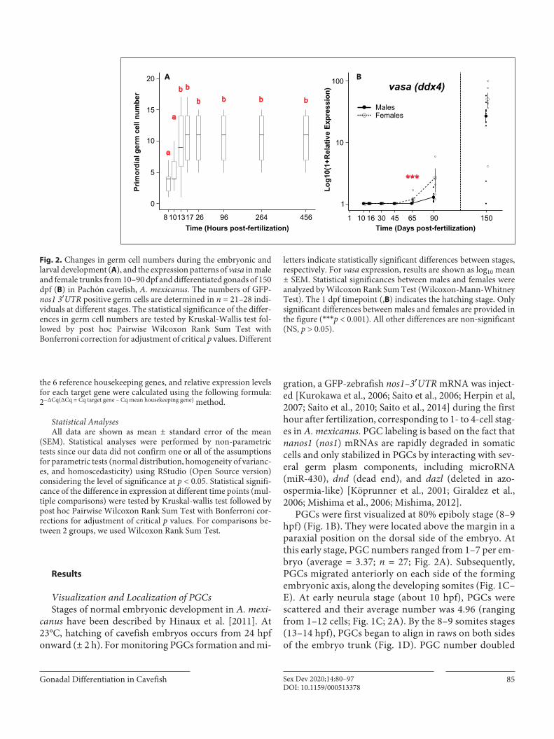
PGCs were first visualized at 80% epiboly stage (8–9 hpf) (Fig. 1B). They were located above the margin in a paraxial position on the dorsal side of the embryo. At this early stage, PGC numbers ranged from 1–7 per em-bryo (average = 3.37; n = 27; Fig. 2A). Subsequently, PGCs migrated anteriorly on each side of the forming embryonic axis, along the developing somites (Fig. 1C–E). At early neurula stage (about 10 hpf), PGCs were scattered and their average number was 4.96 (ranging from 1–12 cells; Fig. 1C; 2A). By the 8–9 somites stages (13–14 hpf), PGCs began to align in raws on both sides of the embryo trunk (Fig. 1D). PGC number doubled
Prim
ordi
al g
erm
cel
l num
ber
10
20
0
15
8
5
26101317 96 264 456
a
a
b b
b b b b
Time (Hours post-fertilization)
A B
Time (Days post-fertilization)
10
100
1
Log1
0(1+
Rel
ativ
e Ex
pres
sion
)
4510 16 30 65 90 150
***
MalesFemales
vasa (ddx4)
1
Fig. 2. Changes in germ cell numbers during the embryonic and larval development (A), and the expression patterns of vasa in male and female trunks from 10–90 dpf and differentiated gonads of 150 dpf (B) in Pachón cavefish, A. mexicanus. The numbers of GFP-nos1 3′UTR positive germ cells are determined in n = 21–28 indi-viduals at different stages. The statistical significance of the differ-ences in germ cell numbers are tested by Kruskal-Wallis test fol-lowed by post hoc Pairwise Wilcoxon Rank Sum Test with Bonferroni correction for adjustment of critical p values. Different
letters indicate statistically significant differences between stages, respectively. For vasa expression, results are shown as log10 mean ± SEM. Statistical significances between males and females were analyzed by Wilcoxon Rank Sum Test (Wilcoxon-Mann-Whitney Test). The 1 dpf timepoint (,B) indicates the hatching stage. Only significant differences between males and females are provided in the figure (***p < 0.001). All other differences are non-significant (NS, p > 0.05).
Imarazene et al.Sex Dev 2020;14:80–9786DOI: 10.1159/000513378
between early neurula and 8–9 somites stages (Fig. 2A), ranged from 1–17 at that stage (average = 9.52; n = 21), and remained constant up to 20 dpf. Subsequently, PGCs continued to migrate axially until reaching their prospective final position around the mid-trunk region at 16–17 somites stage (17 hpf). At that stage, PGCs
were aligned very close to the body axis (Fig. 1E). Fi-nally, in hatching larvae (26 hpf), PGCs moved medi-ally to the gonadal primordia where they formed 2 lines on both sides of the body axis (Fig. 1F). After hatching and until 20 dpf, PGCs were progressively found at the upper surface of the gut, posteriorly to the swim blad-
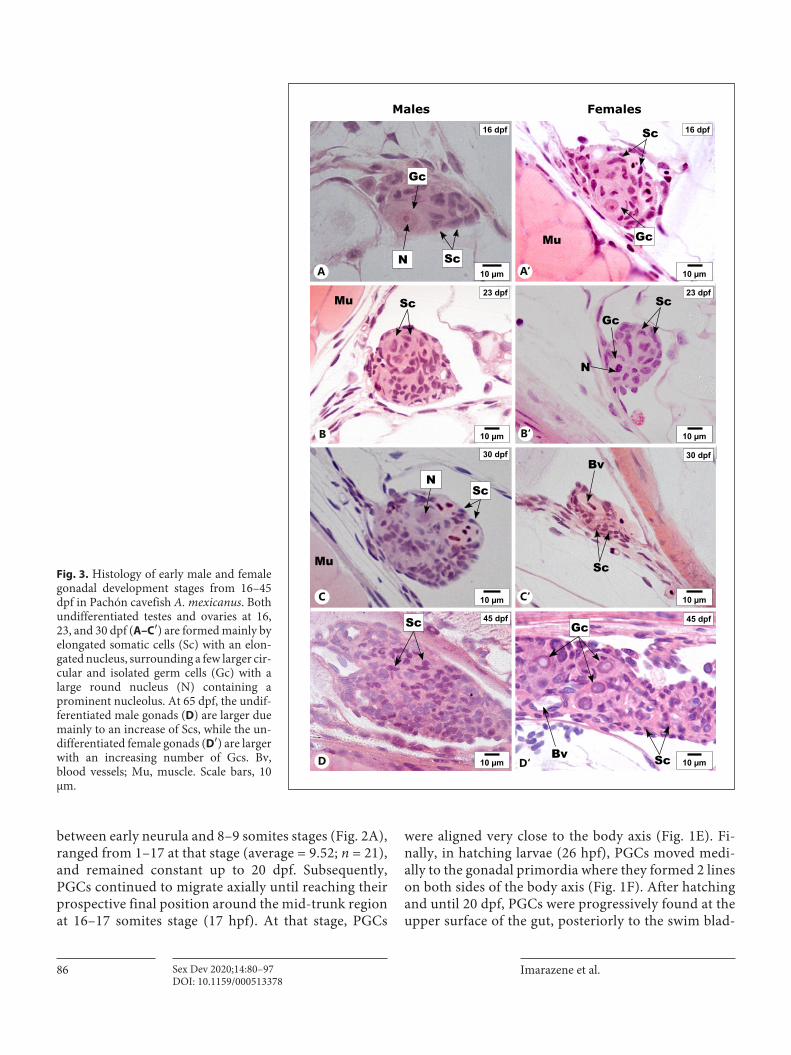
16 dpf
23 dpfScMu
30 dpf
45 dpf
30 dpf
23 dpf
45 dpf
ScGc
Sc
N
30 dpf
Bv
Sc
Bv
16 dpf
Mu
Sc
Sc
Males Females
Mu
Gc
Gc
Gc
10 µm10 µm
10 µm10 µm
10 µm10 µm
10 µm10 µm
ScN
ScNA A‘
B B‘
C‘C
D D‘
Fig. 3. Histology of early male and female gonadal development stages from 16–45 dpf in Pachón cavefish A. mexicanus. Both undifferentiated testes and ovaries at 16, 23, and 30 dpf (A–C′) are formed mainly by elongated somatic cells (Sc) with an elon-gated nucleus, surrounding a few larger cir-cular and isolated germ cells (Gc) with a large round nucleus (N) containing a prominent nucleolus. At 65 dpf, the undif-ferentiated male gonads (D) are larger due mainly to an increase of Scs, while the un-differentiated female gonads (D′) are larger with an increasing number of Gcs. Bv, blood vessels; Mu, muscle. Scale bars, 10 μm.

Gonadal Differentiation in Cavefish 87Sex Dev 2020;14:80–97DOI: 10.1159/000513378
der, where the future gonads will form (Fig. 1G–I). Af-ter clearing of the whole larvae at 20 dpf, a 2-dimen-sional view of the area where gonads are localized con-firmed that PGCs had already colonized the gonads (Fig. 1J−L). These results also confirmed that the GFP-nos1 3′UTR positive cells were bona fide PGCs.
Histological Gonadal DifferentiationThe time course of differentiation into male or female
gonads was then followed by histology. The genotypic
sex of larvae, juveniles, and young adult samples was de-termined based on sex-specific genomic regions of Pachón cavefish with a simple PCR genotyping test. In both males and females of A. mexicanus, the gonadal pri-mordia appeared as very thin and filiform paired organs, located dorsally in the peritoneal cavity on both sides of the swim bladder. Between 16 and 30 dpf, although al-ready composed of different types of somatic cells and a few scarce germ cells, the male and female differentiating gonads did not present any apparent dimorphism (Fig.
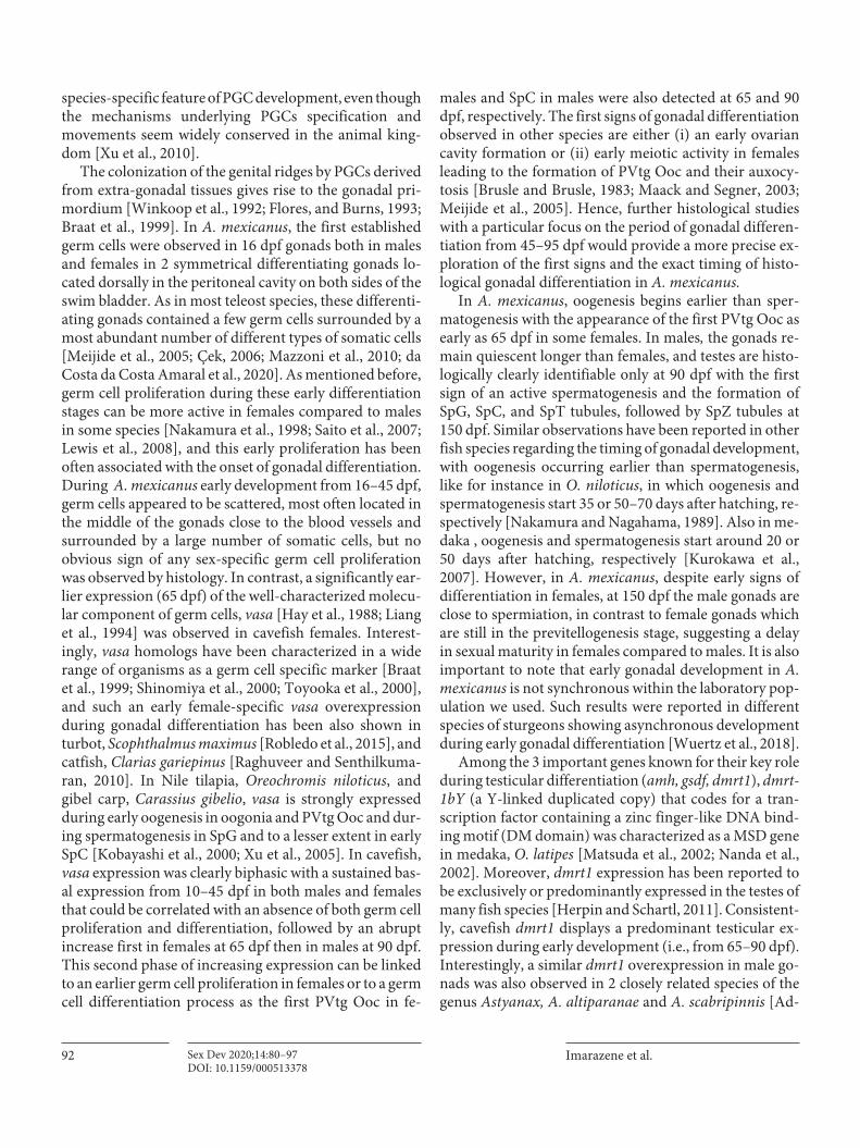
90 dpf
65 dpf
150 dpf
SpG
SpG
SpZ
SpC
SpT
Sc
Sc
SpC
PVtg OoC
65 dpf
90 dpf
N
Sc
Nu
Rbc
OoG
Males Females
150 dpf
SpT PVtg OoC
20 µm10 µm
10 µm 50 µm
10 µm 10 µm
Gc
Ol
PVtg OoC
SpC
A A‘
B B‘
C‘C
Fig. 4. Histology of differentiating male and female gonads from 65–150 dpf in Pachón cavefish A. mexicanus. At 65 dpf (A), the undifferentiated testes increase in size but they also display an in-creasing number of germ cells (Gc) compared to 45 dpf, while the ovaries (A′) are elongated, thin, and show previtellogenic oocytes (PVtg Ooc) indicating the onset of the ovarian differentiation. At
90 dpf (B) and 150 dpf (C), the testes are formed by spermatogonia (SpG), spermatocytes (SpC), spermatids (SpT), and spermatozoa (SpZ). Ovaries of 90 dpf (B′) and 150 dpf (C′) are characterized by a large number of PVtg Ooc that are clearly organized in ovarian lamellae (Ol) at 150 dpf. Mu, muscle; N, nucleus; Nu, nucleolus; Rbc, red blood cells.

Imarazene et al.Sex Dev 2020;14:80–9788DOI: 10.1159/000513378
3A–C′). At 45 dpf the gonad sizes increased both in males and females, with higher numbers of germ cells detected per section (Fig. 3D, D′), but no clear sign of histological differentiation could be detected neither in somatic nor in germ cells (Fig. 3D, D′). It is only around 65 dpf that previtellogenic oocytes became clearly visible in some fe-males (in 1 of 4 females examined at 65 dpf) (Fig. 4A′). In contrast to oogonias that were characterized by a rounded shape, a clear cytoplasm, and a large nucleus,
these previtellogenic oocytes were larger with a dense cy-toplasm and a nucleus with multiple peripherally local-ized nucleoli (Fig. 4A′, B′, C′). However, at 65 dpf all male gonads remained undifferentiated with regard to germ cell development (Fig. 4A). It is only at 90 dpf that the first signs of germinal differentiation could be seen with the first detection of spermatocytes and spermatid cysts (Fig. 4B). Hence, from 65 dpf onwards in females and 90 dpf onwards in males, gonads were engaged in active ga-
***60
0
30
90
Rel
ativ
e Ex
pres
sion gsdf
amh
dmrt1
cyp19a1a
foxl2a
wnt4b
vasa (ddx4)
MalesFemales
60
0
30
90
Rel
ativ
e Ex
pres
sion
60
0
30
90
Rel
ativ
e Ex
pres
sion
Bones
Tissues
Brain
Gills
Gonad
s
Intes
tine
Kidney
Liver
MuscleSkin
Swim-bl
adde
rBon
es
Tissues
Brain
Gills
Gonad
s
Intes
tine
Kidney
Liver
MuscleSkin
Swim-bl
adde
r
Bones
Tissues
Brain
Gills
Gonad
s
Intes
tine
Kidney
Liver
MuscleSkin
Swim-bl
adde
r
**
*
***
****
**
** **
Fig. 5. Tissue-specific expression patterns of gsdf, amh, dmrt1, cy-p19a1a, foxl2a, wnt4b, and vasa in adult male (light gray) and fe-male (dark gray) cavefish quantified by qRT-PCR. Results are pre-sented as boxplots with individual expression values displayed as dots, the expression median as a line, and the box displaying the
first and third quartiles of expression. Statistical significances for each tissue between males and females were tested with Wilcoxon Rank Test. **p < 0.01; *p < 0.05. All other differences are non-sig-nificant (p > 0.05).

Gonadal Differentiation in Cavefish 89Sex Dev 2020;14:80–97DOI: 10.1159/000513378
metogenesis. At 150 dpf, this resulted in clear ovarian lamellae with large PVtg Ooc in females (Fig. 4C′) and a testicular tissue with all spermatogenesis stages up to large cysts of spermatozoa in males (Fig. 4C). Altogether, these observations suggested that the differentiating go-nads of A. mexicanus Pachón cavefish remain histologi-cally undifferentiated at least until 45 dpf in females and 65 dpf in males. Thereafter, undifferentiated gonads dif-ferentiated directly into ovaries or testes. Comparison of the gonad histology of 4 males and 4 females sampled at
each stage showed that gonadal development was not synchronous between animals, like for instance in fe-males at 65 dpf for which we observed only 1 individual displaying some PVtg Ooc. Variability in gonadal devel-opment was even sometimes detected within the same animal, with for instance a marked left/right asymmetry of gonad histology in one 90 dpf female for which the right gonad was large and clearly differentiated while the left gonad was still at the onset of differentiation (online suppl. Fig. 1).
60
0
30
90
Rel
ativ
e Ex
pres
sion
MalesFemales
60
0
30
90
Rel
ativ
e Ex
pres
sion
60
0
30
90
Rel
ativ
e Ex
pres
sion
***
**
**
**
*
***
***** **
***
**
*
** **
***
**
*
**
**
*****
*
**
**
***
**
*****
gsdf
amh
dmrt1
cyp19a1a
foxl2a
wnt4b
*
*
****
ns
vasa (ddx4)
2 3 4 5 6Stages
2 3 4 5 6Stages
2 3 4 5 6Stages
Fig. 6. Expression patterns of gsdf, amh, dmrt1, cyp19a1a, foxl2a, wnt4b, and vasa in cavefish testes (light gray) and ovaries (dark gray) at different gametogenesis stages. Results are presented as boxplots with individual expression values displayed as dots, the
expression median as a line, and the box displaying the first and third quartiles of expression. Statistical significances for each stage between testis and ovaries were tested with Wilcoxon Rank Test. ***p < 0.01; **p < 0.01; *p < 0.05. ns, non-significant, p > 0.05

Imarazene et al.Sex Dev 2020;14:80–9790DOI: 10.1159/000513378
Expression Profiles of Sex-Related Genes in A. mexicanusIn order to better characterize gonadal differentiation
in A. mexicanus, we analyzed the expression profiles of 7
genes well-known as markers of germ cells (vasa), tes-ticular differentiation (amh, gsdf, dmrt1), and ovarian dif-ferentiation (foxl2a, cyp19a1a, wnt4b). Expression of all these marker genes was first checked in various adult tis-
Log1
0(1+
Rel
ativ
e Ex
pres
sion
)10
100
1
10
100
1
10
100
1
10
100
1
Log1
0(1+
Rel
ativ
e Ex
pres
sion
)Lo
g10(
1+R
elat
ive
Expr
essi
on)
gsdf
amh
cyp19a1a
foxl2a
*
***
*
**
10
100
1
45 15010 16 30 65 90Time (Days post-fertilization)
dmrt1
* **
MalesFemales
1
10
100
1
45 15010 16 30 65 90Time (Days post-fertilization)
wnt4b
1
Fig. 7. Expression patterns of gsdf, amh, dmrt1, cyp19a1a, foxl2a, and wnt4b in male and female trunks from 10–90 dpf and testes and ovaries at 150 dpf in cavefish A. mexicanus quantified by qRT-PCR (males: dark solid line; females: gray dashed line). Results are presented as log10 mean ± SEM. Statistical significances between males and females were tested with Wilcoxon Rank Sum Test (Wil-
coxon-Mann-Whitney Test). The 1 dpf time-point (A, B) indicates the hatching stage. Only significant differences between males and females are provided (**p < 0.01; *p < 0.05); all other differences are non-significant (p > 0.05). Black and gray dots represent the individual values of the relative expression in males and females, respectively.

Gonadal Differentiation in Cavefish 91Sex Dev 2020;14:80–97DOI: 10.1159/000513378
sues including gonads. dmrt1, gsdf, amh, and vasa all dis-played a clear predominant gonadal expression, confirm-ing their validity as bona fide gonadal differentiation markers in A. mexicanus. In addition, foxl2a, cyp19a1a, and wnt4b were all expressed at high levels in gonads but also, albeit at a lower extent, in some other tissues (Fig. 5). Surprisingly, apart from vasa that was expressed at high levels both in male and female gonads, all these genes were significantly overexpressed in the testis, including classical ovarian markers such as cyp19a1a, foxl2a, and wnt4b. Similarly, gonadal specific expression of markers involved in testicular (amh, gsdf, and dmrt1) and ovarian (foxl2a, cyp19a1a, and wnt4b) differentiation at different spermatogenesis and oogenesis stages shows that all these genes were overexpressed predominantly in the testes during gametogenesis (Fig. 6), excepted for vasa, which displayed a significantly higher expression level in the ovaries at stages 3, 4, and 6 compared to testes.
Because of the difficulty of sampling very small differen-tiating gonads during the early stages of cavefish develop-ment, we sampled trunks (whole fish without the head and the tail) between 10 and 90 dpf and we isolated gonads at 150 dpf (Fig. 7). During early development of cavefish, all gene expression patterns displayed an important variabili-ty, with coefficients of variation ranging from 107.3–347.12% (online suppl. Table 2) irrespective of the sex and stage sampled (Fig. 7). Such gene expression variability probably reflected the variability in the timing of gonadal development that we also detected by histology (online sup-pl. Fig. 1). But despite this variability, some global trends were clearly observed for germ cell (vasa, official symbol ddx4) and testicular differentiation gene markers (amh, dmrt1, gsdf). Expression of vasa in cavefish showed similar levels between sexes from 10–45 dpf (Fig. 2B), followed by a significant increase at 65 dpf in females (p = 0.0257) and at 90 dpf in males (p = 0.00086) with a significant (p < 0.001) difference between sexes at 65 dpf. Male overexpressions were also observed for testicular differentiation gene mark-ers with significant differences between sexes observed at 16, 45, 65, and 90 dpf for gsdf, at 90 dpf for amh, and at 65 and 90 dpf for dmrt1 (Fig. 7). In contrast, all female differ-entiation marker genes did not display any significant dif-ferences between the sexes (Fig. 7).
Discussion
Despite being an important emerging model species, the morphological and molecular mechanisms underly-ing A. mexicanus cavefish sex differentiation have not
been investigated yet. In the present study, we provide evidence that A. mexicanus belongs to the differentiated gonochoristic species like most teleost species. Of note, this is in contrast with a close relative characidae species, the black widow tetra, Gymnocorymbus ternetzi, de-scribed as an undifferentiated gonochoristic species [Mazzoni et al., 2015]. We also characterized the PGCs migration process and gonadal differentiation steps using both histological and molecular information in a Pachón cave laboratory population of A. mexicanus.
Injections of GFP-zebrafish nos1 3′UTR mRNAs at the 1-cell stage of A. mexicanus enabled a reliable PGC track-ing, as described in other teleost species [Kurokawa et al., 2006; Saito et al., 2006; Herpin et al., 2007; Saito et al., 2014]. The visualization of the first PGCs was possible at 80% epiboly in A. mexicanus. Comparatively, PGCs were detected in other fish species at different stages ranging from the 50% epiboly stage in Danio rerio[Saito et al., 2006] to the somitogenesis stage in Prochilodus lineatus [Coelho et al., 2019]. This variability in the first detection of a clear fluorescence of the GFP-zebrafish nos1 3′UTR mRNA reporter is likely due to a weak and variable signal of background noise resulting from the initial GFP ex-pression in both PGCs and somatic cells as described in other fish species [Saito et al., 2006; Saito et al., 2014]. In addition, the number of PGCs in A. mexicanus was very low (average 9.5 PGCs at the somitogenesis stage) com-pared to other species like zebrafish and medaka that have on average 21.2 and 22.5 PGCs at the same stage [Saito et al., 2006]. It has been reported that PGCs do not prolifer-ate during their migration to the genital crest [Linhartova et al., 2014], but in A. mexicanus we detected an almost significant doubling in PGC number between the early neurula and the 8–9 somites stages, followed by an ab-sence of further division until 20 dpf when the PGCs have already entered the differentiating gonads. In medaka, germ cells undergo 2 types of division after they reach the differentiating gonads: a first slow intermittent division (type I) where each germ cell divides into 2 daughter cells, and a synchronous continuous proliferation (type II) [Saito et al., 2007]. The early PGCs proliferation seen in A. mexicanus around the late neurula stage looks like the slow type I division of medaka germ cells. These results suggest that the absence of PGC divisions during their migration to the genital crest is not as conserved as previ-ously thought. Comparative studies on PGCs migration have evidenced that this process could be species-specific [Kurokawa et al., 2006; Saito et al., 2006; Coelho et al., 2019], suggesting that the early PGCs proliferation that we detected in the cavefish A. mexicanus could be also a
Imarazene et al.Sex Dev 2020;14:80–9792DOI: 10.1159/000513378
species-specific feature of PGC development, even though the mechanisms underlying PGCs specification and movements seem widely conserved in the animal king-dom [Xu et al., 2010].
The colonization of the genital ridges by PGCs derived from extra-gonadal tissues gives rise to the gonadal pri-mordium [Winkoop et al., 1992; Flores, and Burns, 1993; Braat et al., 1999]. In A. mexicanus, the first established germ cells were observed in 16 dpf gonads both in males and females in 2 symmetrical differentiating gonads lo-cated dorsally in the peritoneal cavity on both sides of the swim bladder. As in most teleost species, these differenti-ating gonads contained a few germ cells surrounded by a most abundant number of different types of somatic cells [Meijide et al., 2005; Çek, 2006; Mazzoni et al., 2010; da Costa da Costa Amaral et al., 2020]. As mentioned before, germ cell proliferation during these early differentiation stages can be more active in females compared to males in some species [Nakamura et al., 1998; Saito et al., 2007; Lewis et al., 2008], and this early proliferation has been often associated with the onset of gonadal differentiation. During A. mexicanus early development from 16–45 dpf, germ cells appeared to be scattered, most often located in the middle of the gonads close to the blood vessels and surrounded by a large number of somatic cells, but no obvious sign of any sex-specific germ cell proliferation was observed by histology. In contrast, a significantly ear-lier expression (65 dpf) of the well-characterized molecu-lar component of germ cells, vasa [Hay et al., 1988; Liang et al., 1994] was observed in cavefish females. Interest-ingly, vasa homologs have been characterized in a wide range of organisms as a germ cell specific marker [Braat et al., 1999; Shinomiya et al., 2000; Toyooka et al., 2000], and such an early female-specific vasa overexpression during gonadal differentiation has been also shown in turbot, Scophthalmus maximus [Robledo et al., 2015], and catfish, Clarias gariepinus [Raghuveer and Senthilkuma-ran, 2010]. In Nile tilapia, Oreochromis niloticus, and gibel carp, Carassius gibelio, vasa is strongly expressed during early oogenesis in oogonia and PVtg Ooc and dur-ing spermatogenesis in SpG and to a lesser extent in early SpC [Kobayashi et al., 2000; Xu et al., 2005]. In cavefish, vasa expression was clearly biphasic with a sustained bas-al expression from 10–45 dpf in both males and females that could be correlated with an absence of both germ cell proliferation and differentiation, followed by an abrupt increase first in females at 65 dpf then in males at 90 dpf. This second phase of increasing expression can be linked to an earlier germ cell proliferation in females or to a germ cell differentiation process as the first PVtg Ooc in fe-
males and SpC in males were also detected at 65 and 90 dpf, respectively. The first signs of gonadal differentiation observed in other species are either (i) an early ovarian cavity formation or (ii) early meiotic activity in females leading to the formation of PVtg Ooc and their auxocy-tosis [Brusle and Brusle, 1983; Maack and Segner, 2003; Meijide et al., 2005]. Hence, further histological studies with a particular focus on the period of gonadal differen-tiation from 45–95 dpf would provide a more precise ex-ploration of the first signs and the exact timing of histo-logical gonadal differentiation in A. mexicanus.
In A. mexicanus, oogenesis begins earlier than sper-matogenesis with the appearance of the first PVtg Ooc as early as 65 dpf in some females. In males, the gonads re-main quiescent longer than females, and testes are histo-logically clearly identifiable only at 90 dpf with the first sign of an active spermatogenesis and the formation of SpG, SpC, and SpT tubules, followed by SpZ tubules at 150 dpf. Similar observations have been reported in other fish species regarding the timing of gonadal development, with oogenesis occurring earlier than spermatogenesis, like for instance in O. niloticus, in which oogenesis and spermatogenesis start 35 or 50–70 days after hatching, re-spectively [Nakamura and Nagahama, 1989]. Also in me-daka , oogenesis and spermatogenesis start around 20 or 50 days after hatching, respectively [Kurokawa et al., 2007]. However, in A. mexicanus, despite early signs of differentiation in females, at 150 dpf the male gonads are close to spermiation, in contrast to female gonads which are still in the previtellogenesis stage, suggesting a delay in sexual maturity in females compared to males. It is also important to note that early gonadal development in A. mexicanus is not synchronous within the laboratory pop-ulation we used. Such results were reported in different species of sturgeons showing asynchronous development during early gonadal differentiation [Wuertz et al., 2018].
Among the 3 important genes known for their key role during testicular differentiation (amh, gsdf, dmrt1), dmrt-1bY (a Y-linked duplicated copy) that codes for a tran-scription factor containing a zinc finger-like DNA bind-ing motif (DM domain) was characterized as a MSD gene in medaka, O. latipes [Matsuda et al., 2002; Nanda et al., 2002]. Moreover, dmrt1 expression has been reported to be exclusively or predominantly expressed in the testes of many fish species [Herpin and Schartl, 2011]. Consistent-ly, cavefish dmrt1 displays a predominant testicular ex-pression during early development (i.e., from 65–90 dpf). Interestingly, a similar dmrt1 overexpression in male go-nads was also observed in 2 closely related species of the genus Astyanax, A. altiparanae and A. scabripinnis [Ad-
Gonadal Differentiation in Cavefish 93Sex Dev 2020;14:80–97DOI: 10.1159/000513378
olfi et al., 2015; Castro et al., 2019a; Martinez-Bengochea et al., 2020].
The anti-Müllerian hormone is a glycoprotein of the transforming growth factor β superfamily (TGF-β) [Lu-kas-Croisier et al., 2003]. In teleosts, Y-linked amh dupli-cated copies have been found to act as MSD genes in sev-eral species [Hattori et al., 2012; Li et al., 2015; Pan et al., 2019]. In A. mexicanus, amh was detected at roughly equal levels in both male and female undifferentiated go-nads and subsequently displayed a testicular overexpres-sion at 90 dpf. Such a dimorphic pattern has been found in many fish species at early differentiating stages [Viz-ziano et al., 2007; Ijiri et al., 2008; Robledo et al., 2015; Jiang et al., 2020] consistent with amh playing an impor-tant role in testicular differentiation. In addition, amh is highly expressed in adult testes compared to adult ovaries of A. scabripinnis and A. altiparanae [Castro et al., 2019b; Martinez-Bengochea et al., 2020].
The gonadal soma-derived factor (gsdf) is another TGF-β member that was first identified in rainbow trout Oncorhynchus mykiss [Sawatari et al., 2007], and its ex-pression is predominantly detected in Sertoli and granu-losa cells in several fish species [Sawatari et al., 2007; Shi-bata et al., 2010; Gautier et al., 2011a, b]. Interestingly, Y chromosome genes or alleles of gsdf were characterized or proposed as potential master sex determining genes in the Luzon ricefish Oryzias luzonensis [Myosho et al., 2012] and in sablefish Anoplopoma fimbria [Rondeau et al., 2013]. In cavefish, gsdf displays a clear dimorphic expres-sion with significantly higher levels in males than in fe-males as soon as 16 dpf. Such an early testicular overex-pression of gsdf was also reported in O. latipes [Shibata et al., 2010], O. niloticus [Kaneko et al., 2015], D. rerio [Yan et al., 2017], and Cynoglossus semilaevis [Zhu et al., 2018].
We have also studied ovarian differentiation genes such as foxl2b, cyp19a1a, and wnt4b, which have been suggested as key sex differentiation players across a wide range of animals [Chassot et al., 2008; Cutting et al., 2013; Herpin et al., 2013; Shen and Wang, 2014; Herpin and Schartl, 2015; Fajkowska et al., 2019]. The forkhead box protein L2 (foxl2) is a member of the large forkhead box gene family of transcription factors that is expressed pre-dominantly in female gonads of many fish species [Ber-tho et al., 2016]. For instance, in A. scabripinnis, foxl2a displays a dimorphic expression between adult testes and ovaries with higher expression in females compared to males [Castro et al., 2019b]. Besides foxl2, estrogens and cyp19a1a have been reported as crucial conserved actors of ovarian differentiation in fish [Guiguen et al., 2010], but surprisingly, we did not find any female overexpres-
sion of foxl2a and cyp19a1a during early development in A. mexicanus cavefish, contrasting with their known im-plication as classical ovarian differentiation genes [Guiguen et al., 2010; Bertho et al., 2016]. In addition, we also found that these 2 genes were highly expressed in cavefish testes with only low basal expression levels in ovaries during gametogenesis stages until adult gonads. Such a non-dimorphic expression of these classical ovar-ian differentiation genes was unexpected. However, it must be noted that A. mexicanus is not the only exception to the rule: similar results have been found for instance in the stellate sturgeon, Acipenser stellatus, in which no sig-nificant differences were found for foxl2 in male and fe-male gonads [Burcea et al., 2018], or in another Characi-form, the Tambaqui, C. macropomum, in which no ex-pression of cyp19a1a was detected in both males and females [Lobo et al., 2020]. Moreover, in A. altiparanae, both foxl2 and cyp19a1a are expressed equally in adult testes and ovaries [Martinez-Bengochea et al., 2020]. In mammalian species, foxl2 cooperates with wnt4 in regu-lating fst expression during ovarian development [Gar-cia-Ortiz et al., 2009], and growing evidence suggests that the Wnt signaling pathway might also may play a con-served role in fish sex differentiation [Wu and Chang, 2009; Chen et al., 2015]. In cavefish, we found that wnt4b expression, like foxl2a and cyp19a1a, did not show any sexually dimorphic expression during early gonadal dif-ferentiation. In fact, wnt4b is even expressed at higher levels in the testes than in the ovaries during gametogen-esis and up to adult gonads.
Although the sex differentiation downstream gene regulatory network was initially thought to be highly con-served in vertebrates because of the conservation of the genes involved in this process [Cutting et al., 2013], re-cent results are questioning this hypothesis [Herpin et al., 2013]. In fact, many “important genes” do not exhibit conserved spatiotemporal expression patterns. This is for instance the case in fish for both dmrt1 and gsdf that are dimorphically expressed with a different timing during the early gonadal development of medaka and zebrafish [Kurokawa et al., 2007; Jørgensen et al., 2008] compared to Nile tilapia [Kaneko et al., 2015]. In cavefish, gsdf is clearly expressed in a sexually dimorphic fashion much before dmrt1 and amh (from 16 dpf onwards vs. 65–90 dpf), supporting the idea that gsdf may act as an early switch promoting testis differentiation in cavefish as de-scribed in medaka O. latipes [Zhang et al., 2016]. In the same vein, although testicular differentiation genes dis-played obvious sexually dimorphic expression patterns, neither foxl2a and cyp19a1a nor wnt4b showed sexually
Imarazene et al.Sex Dev 2020;14:80–9794DOI: 10.1159/000513378
dimorphic patterns during cavefish gonadal differentia-tion. Given their well-known implication in fish ovarian differentiation and their tight interaction [Wu and Chang, 2009; Guiguen et al., 2010; Chen et al., 2015; Bertho et al., 2016; Bertho et al., 2018], these results appear surprising and would probably need additional confirmation. How-ever, estrogen-independent ovarian differentiation has already been suggested in medaka [Kawahara and Ya-mashita, 2000; Bertho et al., 2016, 2018].
In summary, combining both expression patterns of sex-related genes and histological changes in A. mexica-nus , we provided a first description of the gonadal dif-ferentiation process in this species. Based on gonadal his-tology and on the vasa germ cell specific expression, we delineated the beginning of sexually dimorphic fates in cavefish between 45 and 65 dpf. This sex differentiation period is corroborated both by histological features and by the sexually dimorphic expression patterns of testicu-lar differentiation genes such as dmrt1, amh, and gsdf. Altogether, these findings are of prime importance for a better understanding of the sex determination mecha-nisms in A. mexicanus. Our results also revealed major discrepancies with the canonical estrogen action on fish ovarian differentiation [Guiguen et al., 2010], pointing out the idea that the control of ovarian development in cavefish would be estrogen independent and that the downstream regulatory network of sex differentiation is not as conserved in teleost fish as initially thought.
Acknowledgements
We are grateful to the Deca team members from CNRS (Insti-tute of Neurosciences Paris-Saclay, Gif-sur-Yvette), particularly Victor Simon for assistance in obtaining eggs and Jorge Torres-Paz for help with injections. We thank A. Patinote and P-L. Sudan from the experimental facility (INRAE UR1037 LPGP, Rennes) for fish maintenance. We want to thank M. Policarpo (CNRS EGCE,
Gif-sur-Yvette) and C. Guyomar (INRAE UR1037 LPGP, Rennes) for their support with R software. We also want to thank D. Casane (CNRS EGCE, Gif-sur-Yvette) for statistical advice.
Statement of Ethics
Animal protocols were carried out in accordance with Euro-pean legislation (directive 2010–63-UE and French decree 2013–118). Animals were treated according to the French and European legislation for handling of animals in research. S.R.’s authorization for use of A. mexicanus in research is 91–116. The animal facility of the Institute received authorization 91272105 from the Veteri-nary Services of Essonne, France, in 2015.
Conflict of Interest Statement
The authors have no conflicts of interest to declare.
Funding Sources
This project was supported by funds from the “Agence Natio-nale de la Recherche” (ANR/DFG, PhyloSex project, 2014–2016) to Y.G. and an «Equipe FRM» grant [DEQ20150331745] from the Fondation pour la Recherche Médicale to S.R. The funders had no role in study design, data collection and analysis, decision to pub-lish, or preparation of the manuscript. B.I. PhD fellowship was supported by the Doctoral School of Ecology, Geosciences, Agron-omy, Nutrition of the university of Rennes 1 and INRAE.
Author Contributions
Funding acquisition: Y.G, S.R. Design, planning and discussing the project and experiments: B.I., A.H., S.R and Y.G. Executing experiments: B.I., E.J., A.B., and S.B. Carrying out the immunos-taining, clearing and confocal microscopy imaging: B.I., M.T., V.T. Analyzing the data: B.I. and Y.G. Writing original draft: B.I. Re-viewing and editing final manuscript: B.I., A.H., S.R., Y.G; Super-vising the project: A.H., S.R., and Y.G.
References
Adolfi MC, Carreira AC, Jesus LW, Bogerd J, Fu-nes RM, Schartl M, et al. Molecular cloning and expression analysis of dmrt1 and sox9 during gonad development and male repro-ductive cycle in the lambari fish, Astyanax al-tiparanae. Reprod Biol Endocrinol. 2015; 13:
2.Bachtrog D, Mank JE, Peichel CL, Kirkpatrick M,
Otto SP, Ashman TL, et al. Sex determination: why so many ways of doing it? PLoS Biol. 2014; 12(7): e1001899.
Baroiller JF, Guiguen Y. Endocrine and environ-mental aspects of sex differentiation in gono-choristic fish. In: Scherer G, Schmid M, eds. Genes and Mechanisms in Vertebrate Sex De-termination. Basel: Birkhäuser; 2001. p. 177–201 .EXS91
Bertho S, Pasquier J, Pan Q, Le Trionnaire G, Bobe J, Postlethwait JH, et al. Foxl2 and its relatives are evolutionary conserved players in gonadal sex differentiation. Sex Dev. 2016;
10(3): 111–29.
Bertho S, Herpin A, Branthonne A, Jouanno E, Yano A, Nicol B, et al. The unusual rainbow trout sex determination gene hijacked the ca-nonical vertebrate gonadal differentiation pathway. Proc Natl Acad Sci USA. 2018;
115(50): 12781–6.Braat AK, Zandbergen T, van de Water S, Goos
HJ, Zivkovic D. Characterization of zebrafish primordial germ cells: morphology and early distribution of vasa RNA. Dev Dyn. 1999;
216(2): 153–67.

Gonadal Differentiation in Cavefish 95Sex Dev 2020;14:80–97DOI: 10.1159/000513378
Brusle J, Brusle S. La gonadogenèse des Poissons. Reproduction Nutrition Développement. 1983; 23: 453–91.
Burcea A, Popa GO, Florescu Gune IE, Maereanu M, Dudu A, Georgescu SE, et al. Expression characterization of six genes possibly in-volved in gonad development for stellate stur-geon individuals (Acipenser stellatus, Pallas 1771). Int J Genomics. 2018; 2018: 7835637.
Castro JP, Hattori RS, Yoshinaga TT, Silva DMZA, Foresti F, Santos MH, et al. Differen-tial expression of dmrt1 in Astyanax scabri-pinnis (Teleostei, Characidade) is correlated with B chromosome occurrence. Zebrafish. 2019a; 16(2): 182–8.
Castro JP, Hattori RS, Yoshinaga TT, Silva DMZA, Ruiz-Ruano FJ, Foresti F, et al. Dif-ferential expression of genes related to sexual determination can modify the reproductive cycle of Astyanax scabripinnis (Characi-formes: Characidae) in B chromosome carrier individuals. Genes. 2019b; 10(11): 909.
Çek Ş. Early gonadal development and sex differ-entiation in rosy barb (Puntius conchonius). Animal Biol. 2006; 56(3): 335–50.
Chassot AA, Gregoire EP, Magliano M, Lavery R, Chaboissier MC. Genetics of ovarian differ-entiation: Rspo1, a major player. Sex Dev. 2008; 2(4-5): 219–27.
Chen H, Li S, Xiao L, Zhang Y, Li G, Liu X, et al. Wnt4 in protogynous hermaphroditic or-ange-spotted grouper (Epinephelus coioides): identification and expression. Comp Bio-chem Physiol B, Biochem Mol Biol. 2015; 183:
67–74.Coelho GCZ, Yo IS, Mira-López TM, Monzani
PS, Arashiro DR, Fujimoto T, et al. Prepara-tion of a fish embryo for micromanipulation: staging of development, removal of the cho-rion and traceability of PGCs in Prochilodus lineatus. Int J Dev Biol. 2019; 63(1-2): 57–65.
da Costa Amaral A, Lima AF, Ganeco-Kirschnik LN, Almeida FL. Morphological characteriza-tion of pirarucu Arapaima gigas (Schinz, 1822) gonadal differentiation. J Morphol. 2020; 281(4–5): 491–9.
Cutting A, Chue J, Smith CA. Just how conserved is vertebrate sex determination?. Dev Dyn. 2013; 242(4): 380–7.
Devlin RH, Nagahama Y. Sex determination and sex differentiation in fish: an overview of ge-netic, physiological, and environmental influ-ences. Aquaculture. 2002; 208(3-4): 191–364.
Di Palma F, Kidd C, Borowsky R, Kocher TD. Construction of bacterial artificial chromo-some libraries for the Lake Malawi cichlid (Metriaclima zebra), and the blind cavefish (Astyanax mexicanus). Zebrafish. 2007; 4: 41–7.
Elliott WR. The Astyanax Caves of Mexico: Cave-fishes of Tamaulipas, San Luis Potosí, and Guerrero. J Fish Biol. 2019; 94: 205.
Fajkowska M, Ostaszewska T, Rzepkowska M. Review: Molecular mechanisms of sex differ-entiation in sturgeons. Rev Aquacult. 2019;
12: 1003–27.
Flores JA, Burns JR. Ultrastructural study of em-bryonic and early adult germ cells, and their support cells, in both sexes of Xiphophorus (Teleostei: Poeciliidae). Cell Tissue Res. 1993;
271(2): 263–70.Fricke R, Eschmeyer WN, van der Laan R, eds.
2020. Eschmeyer’s Catalog of Fishes: Genera, Species, References. (accessed 15/03/2020 at http: //researcharchive.calacademy.org/re-search/ichthyology/catalog/fishcatmain.asp).
Garcia-Ortiz JE, Pelosi E, Omari S, Nedorezov T, Piao Y, Karmazin J, et al. Foxl2 functions in sex determination and histogenesis through-out mouse ovary development. BMC Dev Biol. 2009; 9: 36.
Gautier A, Le Gac F, Lareyre JJ. The gsdf gene lo-cus harbors evolutionary conserved and clus-tered genes preferentially expressed in fish previtellogenic oocytes. Gene. 2011a; 472(1-2): 7–17.
Gautier A, Sohm F, Joly JS, Le Gac F, Lareyre JJ. The proximal promoter region of the zebraf-ish gsdf gene is sufficient to mimic the spatio-temporal expression pattern of the endoge-nous gene in Sertoli and granulosa cells. Biol Reprod. 2011b; 85(6): 1240–51.
Gharbi K, Gautier A, Danzmann RG, Gharbi S, Sakamoto T, Høyheim B, et al. A linkage map for brown trout (Salmo trutta): Chromosome homeologies and comparative genome orga-nization with other salmonid fish. Genetics. 2006; 172(4): 2405–19.
Giraldez AJ, Mishima Y, Rihel J, Grocock RJ, Van Dongen S, Inoue K, et al. Zebrafish MiR-430 promotes deadenylation and clearance of ma-ternal mRNAs. Science. 2006; 312(5770): 75–9.
Guerrero-Estévez S, Moreno-Mendoza N. Sexual determination and differentiation in teleost fish. Rev Fish Biol Fisheries. 2010; 20: 101–21.
Guiguen Y, Fostier A, Piferrer F, Chang CF. Ovar-ian aromatase and estrogens: A pivotal role for gonadal sex differentiation and sex change in fish. Gen Comp Endocrinol. 2010; 165(3):
352–66.Hattori RS, Murai Y, Oura M, Masuda S, Majhi
SK, Sakamoto T, et al. A Y-linked anti-Mülle-rian hormone duplication takes over a critical role in sex determination. Proc Natl Acad Sci USA. 2012; 109(8): 2955–9.
Hay B, Jan LY, Jan YN. A protein component of Drosophila polar granules is encoded by vasa and has extensive sequence similarity to ATP-dependent helicases. Cell. 1988; 55(4): 577–87.
Herman A, Brandvain Y, Weagley J, Jeffery WR, Keene AC, Kono TJY, et al. The role of gene flow in rapid and repeated evolution of cave-related traits in Mexican tetra, Astyanax mex-icanus. Mol Ecol. 2018; 27(22): 4397–416.
Herpin A, Schartl M. Dmrt1 genes at the cross-roads: a widespread and central class of sexu-al development factors in fish. FEBS J. 2011;
278(7): 1010–9.Herpin A, Schartl M. Plasticity of gene-regulatory
networks controlling sex determination: of masters, slaves, usual suspects, newcomers, and usurpators. EMBO Rep. 2015; 16: 1260–74.
Herpin A, Rohr S, Riedel D, Kluever N, Raz E, Schartl M. Specification of primordial germ cells in medaka (Oryzias latipes). BMC Dev Biol. 2007; 7: 3.
Herpin A, Adolfi MC, Nicol B, Hinzmann M, Schmidt C, Klughammer J, et al. Divergent expression regulation of gonad development genes in medaka shows incomplete conserva-tion of the downstream regulatory network of vertebrate sex determination. Mol Biol Evol. 2013; 30(10): 2328–46.
Hinaux H, Pottin K, Chalhoub H, Père S, Elipot Y, Legendre L, et al. A developmental staging table for Astyanax mexicanus surface fish and Pachón cavefish. Zebrafish. 2011; 8(4): 155–65.
Hinaux H, Poulain J, Da Silva C, Noirot C, Jeffery WR, Casane D, et al. De novo sequencing of Astyanax mexicanus surface fish and Pachón cavefish transcriptomes reveals enrichment of mutations in cavefish putative eye genes. PLoS One. 2013; 8(1): e53553.
Ijiri S, Kaneko H, Kobayashi T, Wang DS, Sakai F, Paul-Prasanth B, et al. Sexual dimorphic expression of genes in gonads during early differentiation of a teleost fish, the Nile tilapia Oreochromis niloticus. Biol Reprod. 2008;
78(2): 333–41.Jiang M, Jia S, Chen J, Chen K, Ma W, Wu X, et
al. Timing of gonadal development and di-morphic expression of sex-related genes in gonads during early sex differentiation in the Yellow River carp. Aquaculture. 2020; 518:
734825.Jørgensen A, Morthorst JE, Andersen O, Rasmus-
sen LJ, Bjerregaard P. Expression profiles for six zebrafish genes during gonadal sex differ-entiation. Reprod Biol Endocrinol. 2008; 6: 25.
Kaneko H, Ijiri S, Kobayashi T, Izumi H, Kuramo-chi Y, Wang DS, et al. Gonadal soma-derived factor (gsdf), a TGF-beta superfamily gene, induces testis differentiation in the teleost fish Oreochromis niloticus. Mol Cell Endocrinol. 2015; 415: 87–99.
Kawahara T, Yamashita I. Estrogen-independent ovary formation in the medaka fish, Oryzias latipes. Zool Sci. 2000; 17(1): 65–8.
Keene AC, Yoshizawa M, McGaugh SE. Biology and Evolution of the Mexican Cavefish. Else-vier; 2016.
Klingberg A, Hasenberg A, Ludwig-Portugall I, Medyukhina A, Männ L, Brenzel A, et al. Ful-ly automated evaluation of total glomerular number and capillary tuft size in nephritic kidneys using lightsheet microscopy. J Am Soc Nephrol. 2017; 28(2): 452–9.
Kobayashi T, Kajiura-Kobayashi H, Nagahama Y. Differential expression of vasa homologue gene in the germ cells during oogenesis and spermatogenesis in a teleost fish, tilapia, Oreochromis niloticus. Mech Dev. 2000;
99(1-2): 139–42.Köprunner M, Thisse C, Thisse B, Raz E. A ze-
brafish nanos-related gene is essential for the development of primordial germ cells. Genes Dev. 2001; 15(21): 2877–85.

Imarazene et al.Sex Dev 2020;14:80–9796DOI: 10.1159/000513378
Koressaar T, Remm M. Enhancements and mod-ifications of primer design program Primer3. Bioinformatics. 2007; 23(10): 1289–91.
Kõressaar T, Lepamets M, Kaplinski L, Raime K, Andreson R, Remm M. Primer3_masker: in-tegrating masking of template sequence with primer design software. Bioinformatics. 2018;
34(11): 1937–8.Kurokawa H, Aoki Y, Nakamura S, Ebe Y, Ko-
bayashi D, Tanaka M. Time-lapse analysis re-veals different modes of primordial germ cell migration in the medaka Oryzias latipes. Dev Growth Differ. 2006; 48(3): 209–21.
Kurokawa H, Saito D, Nakamura S, Katoh-Fukui Y, Ohta K, Baba T, et al. Germ cells are essen-tial for sexual dimorphism in the medaka go-nad. Proc Natl Acad Sci USA. 2007; 104(43):
16958–63.Lewis ZR, McClellan MC, Postlethwait JH, Cres-
ko WA, Kaplan RH. Female-specific increase in primordial germ cells marks sex differen-tiation in threespine stickleback (Gasteroste-us aculeatus). J Morphol. 2008; 269(8): 909–21.
Li M, Sun Y, Zhao J, Shi H, Zeng S, Ye K, et al. A tandem duplicate of anti-Müllerian hormone with a missense SNP on the Y chromosome is essential for male sex determination in Nile tilapia, Oreochromis niloticus. PLoS Genet. 2015; 11(11): e1005678.
Liang L, Diehl-Jones W, Lasko P. Localization of vasa protein to the Drosophila pole plasm is independent of its RNA-binding and helicase activities. Development. 1994; 120(5): 1201–11.
Linhartova Z, Saito T, Psenicka M. Embryogene-sis, visualization and migration of primordial germ cells in tench (Tinca tinca). J Appl Ich-thyol. 2014; 30: 29–39.
Lobo IKC, Nascimento ÁRD, Yamagishi MEB, Guiguen Y, Silva GFD, Severac D, et al. Tran-scriptome of tambaqui Colossoma macropo-mum during gonad differentiation: Different molecular signals leading to sex identity. Ge-nomics. 2020; 112(3): 2478–88.
Lukas-Croisier C, Lasala C, Nicaud J, Bedecarrás P, Kumar TR, Dutertre M, et al. Follicle-stim-ulating hormone increases testicular Anti-Mullerian hormone (AMH) production through sertoli cell proliferation and a non-classical cyclic adenosine 5′-monophosphate-mediated activation of the AMH gene. Mol Endocrinol. 2003; 17(4): 550–61.
Maack G, Segner H. Morphological development of the gonads in zebrafish. J Fish Biol. 2003;
62(4): 895–906.Martinez-Bengochea A, Doretto L, Rosa IF,
Oliveira MA, Silva C, Silva DMZA, et al. Ef-fects of 17β-estradiol on early gonadal devel-opment and expression of genes implicated in sexual differentiation of a South American te-leost, Astyanax altiparanae. Comp Biochem Physiol Part B Biochem Mol Biol. 2020; 248-249: 110467.
Matsuda M, Nagahama Y, Shinomiya A, Sato T, Matsuda C, Kobayashi T, et al. DMY is a Y-specific DM-domain gene required for male development in the medaka fish. Nature. 2002; 417(6888): 559–63.
Mazzoni TS, Grier HJ, Quagio-Grassiotto I. Germline cysts and the formation of the ger-minal epithelium during the female gonadal morphogenesis in Cyprinus carpio (Teleostei: Ostariophysi: Cypriniformes). Anat Rec (Hoboken). 2010; 293(9): 1581–606.
Mazzoni TS, Grier HJ, Quagio-Grassiotto I. The basement membrane and the sex establish-ment in the juvenile hermaphroditism during gonadal differentiation of the Gymnocorym-bus ternetzi (Teleostei: Characiformes: Char-acidae). Anat Rec (Hoboken). 2015; 298(12):
1984–2010.McGaugh SE, Gross JB, Aken B, Blin M, Borowsky
R, Chalopin D, et al. The cavefish genome re-veals candidate genes for eye loss. Nat Com-mun. 2014; 5: 5307.
Meijide FJ, Lo Nostro FL, Guerrero GA. Gonadal development and sex differentiation in the cichlid fish Cichlasoma dimerus (Teleostei, perciformes): A light- and electron-micro-scopic study. J Morphol. 2005; 264(2): 191–210.
Mishima Y. Widespread roles of microRNAs dur-ing zebrafish development and beyond. Dev Growth Differ. 2012; 54(1): 55–65.
Mishima Y, Giraldez AJ, Takeda Y, Fujiwara T, Sakamoto H, Schier AF, et al. Differential reg-ulation of germline mRNAs in soma and germ cells by zebrafish miR-430. Curr Biol. 2006; 16(21): 2135–42.
Mitchell RW, Russell WH, Elliott WR. Mexican eyeless characin fishes, genus Astyanax: envi-ronment, distribution, and evolution. Lub-bock: Texas Tech Press; 1977.
Myosho T, Otake H, Masuyama H, Matsuda M, Kuroki Y, Fujiyama A, et al. Tracing the emer-gence of a novel sex-determining gene in me-daka, Oryzias luzonensis. Genetics. 2012;
191(1): 163–70.Nagahama Y. Molecular mechanisms of sex de-
termination and gonadal sex differentiation in fish. Fish Physiol Biochem. 2005; 31(2-3):
105–9.Nakamura M, Nagahama Y. Differentiation and
development of Leydig cells, and changes of testosterone levels during testicular differen-tiation in tilapia Oreochromis niloticus. Fish Physiol Biochem. 1989; 7(1-6): 211–9.
Nakamura M, Kobayashi T, Chang X-T, Nagaha-ma Y. Gonadal sex differentiation in teleost fish. J. Exp. Zool.. 1998; 281(5): 362–72.
Nanda I, Kondo M, Hornung U, Asakawa S, Win-kler C, Shimizu A, et al. A duplicated copy of DMRT1 in the sex-determining region of the Y chromosome of the medaka, Oryzias latipes. Proc Natl Acad Sci USA. 2002; 99(18):
11778–83.Nelson JS, Grande TC, Wilson MVH. Fishes of
the World. John Wiley & Sons, Ltd; 2016. p. 1–12.
Nishimura T, Tanaka M. Gonadal development in fish. Sex Dev. 2014; 8(5): 252–61.
Pan Q, Guiguen Y, Herpin A. Evolution of Sex Determining Genes in Fish, in Skinner MK, ed. Encyclopedia of Reproduction. Oxford: Academic Press; 2018. pp 168–75.
Pan Q, Feron R, Yano A, Guyomard R, Jouanno E, Vigouroux E, et al. Identification of the master sex determining gene in Northern pike (Esox lucius) reveals restricted sex chro-mosome differentiation. PLoS Genet. 2019;
15(8): e1008013.Raghuveer K, Senthilkumaran B. Cloning and dif-
ferential expression pattern of vasa in the de-veloping and recrudescing gonads of catfish, Clarias gariepinus. Comp Biochem Physiol, Part A Mol Integr Physiol. 2010; 157(1): 79–85.
Robledo D, Ribas L, Cal R, Sánchez L, Piferrer F, Martínez P, et al. Gene expression analysis at the onset of sex differentiation in turbot (Scophthalmus maximus). BMC Genomics. 2015; 16: 973.
Rondeau EB, Messmer AM, Sanderson DS, Jant-zen SG, von Schalburg KR, Minkley DR, et al. Genomics of sablefish (Anoplopoma fim-bria): expressed genes, mitochondrial phylog-eny, linkage map and identification of a puta-tive sex gene. BMC Genomics. 2013; 14: 452.
Saito D, Morinaga C, Aoki Y, Nakamura S, Mitani H, Furutani-Seiki M, et al. Proliferation of germ cells during gonadal sex differentiation in medaka: Insights from germ cell-depleted mutant zenzai. Dev Biol. 2007; 310(2): 280–90.
Saito T, Fujimoto T, Maegawa S, Inoue K, Tanaka M, Arai K, et al. Visualization of primordial germ cells in vivo using GFP-nos1 3’UTR mRNA. Int J Dev Biol. 2006; 50(8): 691–9.
Saito T, Goto-Kazeto R, Fujimoto T, Kawakami Y, Arai K, Yamaha E. Inter-species transplan-tation and migration of primordial germ cells in cyprinid fish. Int J Dev Biol. 2010; 54(10):
1481–6.Saito T, Pšenička M, Goto R, Adachi S, Inoue K,
Arai K, et al. The origin and migration of pri-mordial germ cells in sturgeons. PLoS One. 2014; 9(2): e86861.
Sawatari E, Shikina S, Takeuchi T, Yoshizaki G. A novel transforming growth factor-beta super-family member expressed in gonadal somatic cells enhances primordial germ cell and sper-matogonial proliferation in rainbow trout (Oncorhynchus mykiss). Dev Biol. 2007;
301(1): 266–75.Shen ZG, Wang HP. Molecular players involved
in temperature-dependent sex determination and sex differentiation in Teleost fish. Genet Sel Evol. 2014; 46: 26.
Shibata Y, Paul-Prasanth B, Suzuki A, Usami T, Nakamoto M, Matsuda M, et al. Expression of gonadal soma derived factor (GSDF) is spa-tially and temporally correlated with early tes-ticular differentiation in medaka. Gene Expr Patterns. 2010; 10(6): 283–9.
Gonadal Differentiation in Cavefish 97Sex Dev 2020;14:80–97DOI: 10.1159/000513378
Shinomiya A, Tanaka M, Kobayashi T, Nagahama Y, Hamaguchi S. The vasa-like gene, olvas, identifies the migration path of primordial germ cells during embryonic body formation stage in the medaka, Oryzias latipes. Dev Growth Differ. 2000; 42(4): 317–26.
Siemering KR, Golbik R, Sever R, Haseloff J. Mu-tations that suppress the thermosensitivity of green fluorescent protein. Curr Biol. 1996;
6(12): 1653–63.Sreenivasan R, Jiang J, Wang X, Bártfai R, Kwan
HY, Christoffels A, et al. Gonad differentia-tion in zebrafish is regulated by the canonical Wnt signaling pathway. Biol Reprod. 2014;
90(2): 45.Toyooka Y, Tsunekawa N, Takahashi Y, Matsui
Y, Satoh M, Noce T. Expression and intracel-lular localization of mouse Vasa-homologue protein during germ cell development. Mech Dev. 2000; 93(1-2): 139–49.
Untergasser A, Cutcutache I, Koressaar T, Ye J, Faircloth BC, Remm M, et al. Primer3--new capabilities and interfaces. Nucleic Acids Res. 2012; 40(15): e115.
Vizziano D, Randuineau G, Baron D, Cauty C, Guiguen Y. Characterization of early molecu-lar sex differentiation in rainbow trout, On-corhynchus mykiss. Dev Dyn. 2007; 236(8):
2198–206.Wang W, Zhu H, Dong Y, Dong T, Tian Z, Hu H.
Identification and dimorphic expression of sex-related genes during gonadal differentia-tion in sterlet Acipenser ruthenus, a primitive fish species. Aquaculture. 2019; 500: 178–87.
Wilkens H, Strecker U. Evolution in the Dark: Darwin’s Loss Without Selection. Berlin Hei-delberg: Springer-Verlag; 2017.
Winkoop A, Booms GHR, Dulos GJ, Timmer-mans LPM. Ultrastructural changes in pri-mordial germ cells during early gonadal de-velopment of the common carp (Cyprinus carpio L., teleostei). Cell Tissue Res. 1992; 267:
337–46.Wu GC, Chang CF. Wnt4 is associated with the
development of ovarian tissue in the protan-drous black porgy, Acanthopagrus schlegeli. Biol Reprod. 2009; 81(6): 1073–82.
Wuertz S, Güralp H, Pšenička M, Chebanov M. Sex Determination in Sturgeon. Sex Control in Aquaculture John Wiley & Sons, Ltd; 2018; pp 645–68.
Xu H, Gui J, Hong Y. Differential expression of vasa RNA and protein during spermatogen-esis and oogenesis in the gibel carp (Carassius auratus gibelio), a bisexually and gynogeneti-cally reproducing vertebrate. Dev Dyn. 2005;
233(3): 872–82.Xu H, Li M, Gui J, Hong Y. Fish germ cells. Sci
China Life Sci. 2010; 53(4): 435–46.Yamamoto T-O.Sex Differentiation,in Hoar WS,
Randall DJ (eds). Fish Physiology. Academic Press; 1969; pp 117–75.
Yan YL, Desvignes T, Bremiller R, Wilson C, Dil-lon D, High S, et al. Gonadal soma controls ovarian follicle proliferation through Gsdf in zebrafish. Dev Dyn. 2017; 246(11): 925–45.
Zhang X, Guan G, Li M, Zhu F, Liu Q, Naruse K, et al. Autosomal gsdf acts as a male sex initia-tor in the fish medaka. Sci Rep. 2016; 6: 19738–13.
Zhu Y, Meng L, Xu W, Cui Z, Zhang N, Guo H, et al. The autosomal Gsdf gene plays a role in male gonad development in Chinese tongue sole (Cynoglossus semilaevis). Sci Rep. 2018;
8(1): 17716.
Copyright © 2022 FDOKUMEN