Evaluation of hepatoprotective effect of Gentiana olivieri herbs on subacute administration and...
11
Evaluation of hepatoprotective effect of Gentiana olivieri herbs on subacute administration and isolation of active principle Didem Deliorman Orhan a , Mustafa Aslan a , Go ¨knur Aktay b , Ender Ergun c , Erdem Yesilada a, * , Fatma Ergun a a Department of Pharmacognosy, Faculty of Pharmacy, Gazi University, Etiler 06330, Ankara, Turkey b Institute of Forensic Medicine, Ankara University, Dikimevi 06100, Ankara, Turkey c Department of Pathology, Faculty of Dentistry, Ankara University, Besevler 06500, Ankara, Turkey Received 8 August 2002; accepted 5 December 2002 Abstract Hepatoprotective effect of Gentiana olivieri Griseb. (Gentianaceae) flowering herbs on subacute administration were studied using in vivo models in rats. For the activity assessment on carbon tetrachloride-induced hepatic damage following biochemical parameters were evaluated; plasma and hepatic tissue malondialdehyde formation, and liver tissue glutathione level, as well as plasma transaminase enzyme levels (aspartate transferase and alanine transferase). Results of biochemical tests were also confirmed by histopathological examination. Through bioassay-guided fractionation procedures isoorientin, a known C-glycosylflavone, was isolated from the ethyl acetate fraction as the active antihepatotoxic constituent by silica gel column chromatography. Isoorientin exhibited significant hepatoprotective effect at 15 mg/kg b.w. dose. D 2003 Published by Elsevier Science Inc. Keywords: Aspartate transferase; Alanine transferase; Gentiana olivieri; Glutathione; Hepatoprotective; Histopathological evaluation; Isoorientin; Lipid peroxidation; Malondialdehyde; Subacute administration Introduction The risk of liver intoxication has recently increased by the higher exposure to environmental toxins, pesticides and frequent use of chemotherapeutics. However, nature offers a wide range of sources in 0024-3205/03/$ - see front matter D 2003 Published by Elsevier Science Inc. doi:10.1016/S0024-3205(03)00117-6 * Corresponding author. Tel.: +90-312-222-4255; fax: +90-312-223-5018. E-mail address: [email protected] (E. Yesilada). www.elsevier.com/locate/lifescie Life Sciences 72 (2003) 2273 – 2283
-
Upload
kunfeyekun -
Category
Documents
-
view
0 -
download
0
Transcript of Evaluation of hepatoprotective effect of Gentiana olivieri herbs on subacute administration and...
Evaluation of hepatoprotective effect of Gentiana olivieri herbs on
subacute administration and isolation of active principle
Didem Deliorman Orhana, Mustafa Aslana, Goknur Aktayb, Ender Ergunc,Erdem Yesiladaa,*, Fatma Erguna
aDepartment of Pharmacognosy, Faculty of Pharmacy, Gazi University, Etiler 06330, Ankara, Turkeyb Institute of Forensic Medicine, Ankara University, Dikimevi 06100, Ankara, Turkey
cDepartment of Pathology, Faculty of Dentistry, Ankara University, Besevler 06500, Ankara, Turkey
Received 8 August 2002; accepted 5 December 2002
Abstract
Hepatoprotective effect of Gentiana olivieri Griseb. (Gentianaceae) flowering herbs on subacute administration
were studied using in vivo models in rats. For the activity assessment on carbon tetrachloride-induced hepatic
damage following biochemical parameters were evaluated; plasma and hepatic tissue malondialdehyde formation,
and liver tissue glutathione level, as well as plasma transaminase enzyme levels (aspartate transferase and alanine
transferase). Results of biochemical tests were also confirmed by histopathological examination. Through
bioassay-guided fractionation procedures isoorientin, a known C-glycosylflavone, was isolated from the ethyl
acetate fraction as the active antihepatotoxic constituent by silica gel column chromatography. Isoorientin
exhibited significant hepatoprotective effect at 15 mg/kg b.w. dose.
D 2003 Published by Elsevier Science Inc.
Keywords: Aspartate transferase; Alanine transferase; Gentiana olivieri; Glutathione; Hepatoprotective; Histopathological
evaluation; Isoorientin; Lipid peroxidation; Malondialdehyde; Subacute administration
Introduction
The risk of liver intoxication has recently increased by the higher exposure to environmental toxins,
pesticides and frequent use of chemotherapeutics. However, nature offers a wide range of sources in
0024-3205/03/$ - see front matter D 2003 Published by Elsevier Science Inc.
doi:10.1016/S0024-3205(03)00117-6
* Corresponding author. Tel.: +90-312-222-4255; fax: +90-312-223-5018.
E-mail address: [email protected] (E. Yesilada).
www.elsevier.com/locate/lifescie
Life Sciences 72 (2003) 2273–2283
order to protect or treat hepatic injuries, contrarily to conventional therapy. Among them silymarin,
artichoke, etc. have frequently been used in therapy, including mushroom poisoning.
In a previous study, the hepatoprotective effect of seven Turkish folk remedies was investigated on
carbon tetrachloride (CCl4)-induced liver injury. Consequently, the ethanol (EtOH) extract of Gentiana
olivieri Griseb. (Gentianaceae) was shown to possess a potent hepatoprotective effect on acute
administration in rat [1].
The present study was aimed to evaluate the antihepatotoxic effect of G. olivieri extracts and fractions
against carbon tetrachloride-induced hepatotoxicity on subacute administration. In order to assess the
activity some biochemical parameters of plasma and hepatic tissue [malondialdehyde formation (MDA)
in plasma and hepatic tissue, transaminase enzyme levels in plasma (aspartate transferase — AST- and
alanine transferase - ALT), and cellular glutathion (GSH) level in liver tissue] were monitored as well as
histopathological examination, and then through bioassay-guided fractionation procedure to isolate the
active antihepatotoxic constituent(s) and define the chemical structure by spectral techniques.
Material and methods
Material
Gentiana olivieri Griseb. was collected by one of the authors (M.A.) from Gaziantep (Turkey) in May
1999. A voucher of the plant is stored in the herbarium of Gazi University, Faculty of Pharmacy
(99G001). Cynara scolymus, artichoke, a well-known hepatoprotective plant was used as reference and
was bought from Bazaar in Ankara (in May 2000).
Extraction, fractionation and isolation procedures
Preparation of extract and fractions
The dried and coarsely powdered aerial parts of G. olivieri were macerated with 80j ethanol for 3 h
by continuous stirring at room temperature and then evaporated to dryness under reduced pressure
[EtOH extract]. The condensed extract was then dissolved in distilled water and fractionated through
successive extractions with chloroform, ethylacetate, n-butanol/saturated with water. Each fraction was
concentrated to dryness under reduced pressure and below (40-50 jC) on a rotary evaporator to give
[CHCl3-Fr.], [EtOAc Fr.], [n-BuOH Fr.] and the remaining aqueous fraction [R-H2O Fr.], respectively.
Column chromatographic fractionation of [EtOAc Fr.] and isolation of active constituent
In order to further fractionate the active fraction, [EtOAc Fr.], was applied to Kieselgel 60 (0.2–0.5
mm) (Merck, Darmstadt Art. No. 7733) column chromatography and eluted with CHCl3-MeOH-H2O
(90:31:4), (65:40:4) and (65:40:9), successively. Eluents were combined into two subfractions; [Fr. 1–
55], [Fr. 56–89], according to tlc behaviour using two solvent systems; CHCl3-MeOH-H2O (61:32:7)
and EtOAc-MeOH-H2O (100:16.5:13.5). The precipitate would occurred when was dissolved [Fr. 56–
89] in MeOH and kept in refrigerator was removed through filtration. This process was repeated several
times and each of the precipitate [Fr. 56–89#] and the upper part [Fr. 56–89z] subfractions were
combined separately. On tlc [using the same solvent systems as given above] and hplc [RP-18
LiChrosphere 5 Am, Supelco; H2O-MeOH-gl. AcH (65:35:5)] analysis of [Fr. 56–89#] was shown to
D. Deliorman Orhan et al. / Life Sciences 72 (2003) 2273–22832274
possess a single component. The structure was elucidated as isoorientin using TLC, UV, 1H and 13C-
NMR (in CD3OD and DMSO-d6) as well as 2D-NMR [DEPT-135, HETCOR, HMBC] and FAB-MS
techniques. 1H-NMR data (J values)[H attached to numbered C atom] (Hz in CD3OD): 6.53 s [H-3],
6.48 s [H-8], 7.36 br.s [H-2V], 6.90 d (8.50) [H-5V], 7.37 br d [H-6V], 4.90 d (10.0) [H-1VV], 4.15 t (9.2)
[H-2VV], 3.43–3.44 [H-3VVand H-4VV], 3.42 m [H-5VV], 3.87 dd (12.2) [H-6VVa], 3.75 dd (12.5) [H-6VVb]. 13C(y in DMSO-d6): 163.57 (C-2), 102.74 (C-3), 181.77 (C-4), 160.58 (C-5), 108.79 (C-6), 163.15 (C-7),
93.44 (C-8), 156.12 (C-9), 103.35 (C-10), 121.38 (C-1V), 113.27 (C-2V), 145.68 (C-3V), 149.69 (C-4V),115.98 (C-5V), 118.88 (C-6V), 72.99 (C-1VV), 70.54 (C-2VV), 78.87 (C-3VV), 70.18 (C-4VV), 81.45 (C-5VV),61.41 (C-6VV).
High Performance Liquid Chromatographic (hplc) analysis of isoorientin in extracts and fractions
In order to determine the concentration of active component (isoorientin) in extracts and fractions
following hplc conditions were used: Column: LiChrospher RP-18 (5 Am (250 mm � 4.6 mm) [Supelco
98040778], Wavelenght: 354 nm, Flow rate: 0.8 ml/min, Pressure: 183-184 barr, Mobile phase: Water:
Methanol: anhydrous acetic acid (65:35:5). Content of isoorientin in extracts and fractions were
determined as follows (w/w): in EtOH extr., 1.25%; in EtOAc fr., 10.40%; in n-BuOH fr., 3.01%; in
R-H2O fr., nil; in Fr. [56-89]z, 32%.
Pharmacological procedures
Test animals
Male Sprague-Dawley rats (150–180 g) were purchased from the Animal House of Gulhane Military
Academy of Medicine (Ankara) and were kept for 2 days before the experiments for acclimatization to
the experimental conditions with free access to food (standard diet) and water, but food was withdrawn
24 h before the experiment. Throughout the experiments, animals were processed according to the
suggested ethical guidelines for the care of laboratory animals. The animals were divided into 14 groups
consisting of six rats each.
Preparation and administration of test samples
The extracts, fractions and subfractions were suspended in 0.5% CMC in distilled water prior to oral
administration to experimental animals. Test groups of rats were orally treated with EtOH extract (in 250,
125, and 62.5 mg/kg bw. doses) (bw., body weight), n-BuOH Fr. and R-H2O Fr. (in 125 mg/kg bw.
dose), CHCl3 Fr. and EtOAc Fr. (in 125 and 250 mg/kg bw. doses) for 5 following days, daily once by
gastric gavage needle. The control group (untreated) and carbon tetrachloride group (positive control) of
rats were administered with 0.5% carboxymethyl cellulose (CMC) suspension for the same period.
As the natural originated drug, the suspension of lyophilized and powdered bracts of Cynara scolymus
(artichoke) in 0.5% CMC was directly administered to animals in 500 mg/kg bw. dose without any
extraction process [1].
Experimental procedure
60 min after the administration of the last dose on 5th day, except the control group rats, each of the
carbon tetrachloride (CCl4) group and test group of animals was challenged with 50% CCl4 in liquid
paraffin (2.5 ml/kg bw., per os) to induce hepatic injury. Twenty-four hours after the hepatotoxin
administration, blood samples were withdrawn by cardiac puncture and then the rats were sacrificed by
D. Deliorman Orhan et al. / Life Sciences 72 (2003) 2273–2283 2275
overdose of diethylether. Blood samples collected in heparinized tubes were centrifuged at 3000 � g (4
jC) for 10 min to obtain plasma. Plasma samples were used to determine the lipid peroxide levels as well
as to test AST and ALT activities. On the other hand, liver of each rat was promptly removed and a part
was used to determine the tissue levels of MDA and GSH.
Aspartate transferase (AST) and alanine transferase (ALT) in plasma
Biocon standard kits and DAX-48 autoanalyzer were used to measure AST and ALT activities in
plasma samples according to the method of Wilkinson et al. [14].
Lipid peroxidation in plasma
The methodology described by Kurtel et al. [10] was used. Briefly, one ml of plasma sample was
combined with 2.0 ml of trichloroacetic acid (TCA; 15%, w/v)-thiobarbituric acid (TBA; 0.375%)-0.25
N. HCl and mixed throughly and centrifuged at 10 000 � g for 5 min. The supernatant was mixed with
20 Al of butyl hydroxy toluene (BHT; 0.02% in 95% EtOH, w/v) to prevent further oxidation and heated
for 15 min in a boiling water bath. After cooling under running water, the flocculent precipitate was
removed by centrifugation at 10 000 � g for 5 min. The absorbance of the sample was measured at 532
nm against blank that contained all the reagents except plasma. 1,1,3,3-tetraethoxypropan was used as
standard for calibration of the curve.
Lipid peroxidation in liver tissue
The method of Ohkawa et al. [12] as modified by Jamall and Smith [7] was used to determine lipid
peroxidation in tissue samples. Rats were sacrificed by an overdose of diethylether. The liver of each rat
was immediately excised and chilled in ice-cold 0.9% NaCl and then perfused via the portal vein with
ice-cold 0.9% NaCl. After washing with 0.9% NaCl, 1.0 g of wet tissue was weighted exactly and
homogenized in 9 ml of 0.25 M sucrose using a Teflon homogenizer to obtain a 10% suspension. The
cytosolic fraction was obtained by a two-step centrifugation first at 1000 � g for 10 min and then at
2000 � g for 30 min at 4 jC. A volume of the homogenate (0.20 ml) was transferred to a vial and was
mixed with 0.2 ml of a 8.1% (w/v) Sodium dodecyl sulphate solution, 1.50 ml of a 20% acetic acid
solution (adjusted to pH 3.5 with NaOH) and 1.50 ml of a 0.8% (w/v) solution of TBA and the final
volume was adjusted to 4.0 ml with distilled water. Each vial was tightly capped and heated in boiling
water bath for 60 min. The vials were then cooled under running water.
Equal volumes of tissue blank or test sample and 10% TCAwere transferred into a centrifuge tube and
centrifuged at 1000 � g for 10 min. The absorbance of the supernatant fraction was measured at 532 nm
(Beckman DU 650 Spectrometer). Control experiment was processed using the same experimental
procedure except the TBA solution was replaced with distilled water due to the peroxidative effect of
CCl4 on tissue; livers of CCl4-treated rats were used as positive control. 1,1,3,3-tetraethoxypropan was
used as standard for calibration of the curve.
Nonprotein sulfhydryl groups (Cellular GSH) in liver tissue [13]
200 mg of liver was homogenized in 8.0 ml of 0.02 M EDTA in an ice bath. The homogenates were
kept in the ice bath until used. Aliquots of 5.0 ml of the homogenates were mixed in 15.0 ml test tubes
with 4.0 ml distilled water and 1.0 ml of 50% trichloroacetic acid (TCA). The tubes were centrifuged for
15 min. at approximately 3000 � g. 2.0 ml of supernatant was mixed with 4.0 ml of 0.4 M Tris buffer,
pH 8.9, 0.1 ml Ellman’s reagent [5,5V-dithiobis-(2-nitro-benzoic acid)] (DTNB) added, and the sample
D. Deliorman Orhan et al. / Life Sciences 72 (2003) 2273–22832276
shaken. The absorbance was read within 5 min of the addition of DTNB at 412 nm against a reagent
blank with no homogenate. Results were expressed as Amol GSH/ g tissue.
Statistical analysis
The data obtained were analyzed by one-way analysis of variance (ANOVA) and Student-Newman-
Keuls posthoc tests for the significant interrelation between the various groups using Instat computer
software. P < 0.05 was considered to be significant from the control.
Histopathological evaluation
Immediately after the sacrifice, the liver of each rat was removed and an approximately half part was
placed in 10% formalin to fix the tissues for microscopic examination. The paraffin sections were
prepared in an automatic tissue processor (Lipshaw) and sliced into 5-Am-thick sections in a rotary
microtome and then stained with haematoxylin-eosin dye (Merck) and mounted with Canada balsam.
The histopathological examination of slides were performed under a Carl Zeiss Jena amplual type
photomicroscope (3.2 � 10 and 10 � 10) and photographed. The parameters examined are as follows;
degeneration in hepatocytes and in hepatic cords, focal necrosis, congestion in central vein and in
sinusoids, infiltration of lymphocytes, Kupffer cell proliferation, deformation in hepatocytes, bleeding in
hepatic lobes. The following relative score system was employed for the assessment of histological
damage; 0: absent; +: few; ++: mild; +++: moderate; ++++: severe; +++++: extremely severe.
Results and discussion
As discussed in the previous paper [1], increased lipid peroxidation induced by free radical derivatives
is one of the main factors involved in CCl4-intoxication. As a result of this oxidative damage to the
structural integrity of the liver a marked increase in the serum transaminase levels are observed. Aerobic
cells have evolved a complex enzymatic and non-enzymatic mechanism to deal with this oxidative
phenomenon [2]. One of the principal cellular defence molecules is reduced glutathione (GSH). GSH, a
non-protein cysteine reservoir in the liver is involved in many cellular processes including the
detoxification of endogenous and exogenous compounds and is able to protect cellular constituents
from the toxic effects of free radicals [4]. Thus, in order to evaluate the antihepatotoxic activity in the
present study, effects of the test samples on elevated serum levels of hepatospecific enzymes, AST and
ALT, as well as on hepatic and plasma MDA formation and nonprotein sulfhydryl groups (cellular GSH)
in liver homogenates were monitored.
In a previous study, we examined the EtOH extracts of some plants which are used in Turkish folk
medicine to treat liver ailments and the extract of G. olivieri administered at a 500 mg/kg of single dose
was found as the most effective plant, in protecting the liver against CCl4-induced acute hepatotoxicity
model in rats [1]. In that study, it was reported that the extract decreased the plasma levels of liver
enzymes, i.e. 86.4% in ALT and 69.6% in AST as well as 70.5% and 32.3% in plasma and liver tissue
MDA levels, respectively. However, a moderate effect was observed on histopathological examination of
the liver sections, which was uncongenial with the results of biochemical studies.
Considering the single and high dose of the extract administered in that previous study might be the
cause of this consequence, we decided to administer the extract in lower doses and for longer period
(subacute administration). EtOH extract was administered in three dose levels, 62.5, 125 and 250 mg/kg
D. Deliorman Orhan et al. / Life Sciences 72 (2003) 2273–2283 2277
daily per os, for 5 following days before challenging with CCl4 as hepatotoxic agent, and effects of the
extract on biochemical and histopathological parameters were monitored.
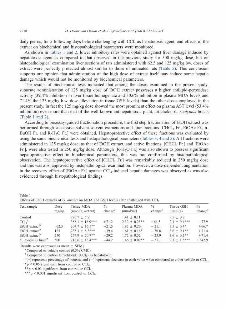
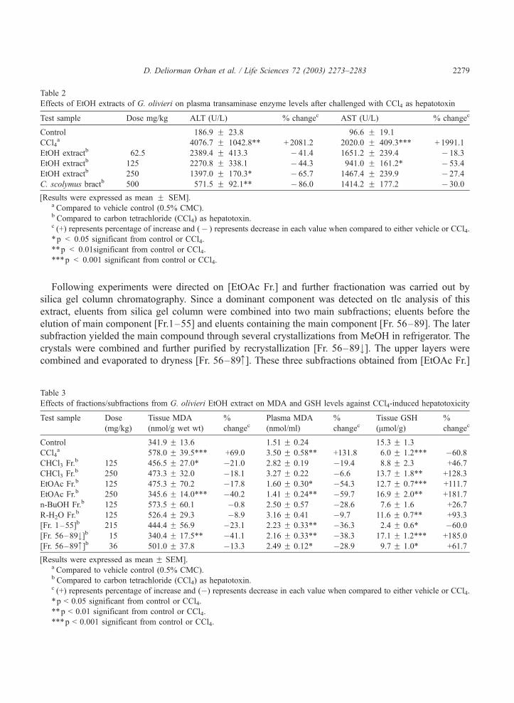
As shown in Tables 1 and 2, lower inhibitory rates were obtained against liver damage induced by
hepatotoxic agent as compared to that observed in the previous study for 500 mg/kg dose, but on
histopathological examination liver sections of rats administered with 62.5 and 125 mg/kg bw. doses of
extract were perfectly protected almost similar to those of untreated rats (Table 5). This conclusion
supports our opinion that administration of the high dose of extract itself may induce some hepatic
damage which would not be monitored by biochemical parameters.
The results of biochemical tests indicated that among the doses examined in the present study,
subacute administration of 125 mg/kg dose of EtOH extract possesses a higher antilipid-peroxidase
activity (39.4% inhibition in liver tissue homogenate and 30.6% inhibition in plasma MDA levels and
71.4% the 125 mg/kg b.w. dose alleviation in tissue GSH levels) than the other doses employed in the
present study. In fact the 125 mg/kg dose showed the most prominent effect on plasma AST level (53.4%
inhibition) even more than that of the well-known antihepatotoxic plant, artichoke, C. scolymus bracts
(Table 1 and 2).
According to bioassay-guided fractionation procedure, the first step fractionation of EtOH extract was
performed through successive solvent-solvent extractions and four fractions [CHCl3 Fr., EtOAc Fr., n-
BuOH Fr. and R-H2O Fr.] were obtained. Hepatoprotective effect of these fractions was evaluated by
using the same biochemical tests and histopathological parameters (Tables 3, 4 and 5). All fractions were
administered in 125 mg/kg dose, as that of EtOH extract, and active fractions, [CHCl3 Fr.] and [EtOAc
Fr.], were also tested in 250 mg/kg dose. Although [R-H2O Fr.] was also shown to possess significant
hepatoprotective effect in biochemical parameters, this was not confirmed by histopathological
observation. The hepatoprotective effect of [CHCl3 Fr.] was remarkably reduced in 250 mg/kg dose
and this was also approved by histopathological examination. However, a dose-dependent augmentation
in the recovery effect of [EtOAc Fr.] against CCl4-induced hepatic damages was observed as was also
evidenced through histopathological findings.
Table 1
Effects of EtOH extracts of G. olivieri on MDA and GSH levels after challenged with CCl4
Test sample Dose
mg/kg
Tissue MDA
(nmol/g wet wt)
%
changecPlasma MDA
(nmol/ml)
%
changecTissue GSH
(Amol/g)
%
changec
Control 226.7 F 5.8 1.41 F 0.11 9.5 F 0.8
CCl4a 388.1 F 18.9*** + 71.2 2.32 F 0.25** + 64.5 2.1 F 0.4*** � 77.9
EtOH extractb 62.5 304.7 F 16.5** �21.5 1.83 F 0.20 � 21.1 3.5 F 0.4* + 66.7
EtOH extractb 125 235.3 F 8.3*** �39.4 1.61 F 0.16* � 30.6 3.6 F 0.1** + 71.4
EtOH extractb 250 274.9 F 20.7** �29.2 1.72 F 0.52 � 25.9 3.6 F 0.2** + 71.4
C. scolymus bractb 500 216.6 F 13.4*** �44.2 1.46 F 0.09** � 37.1 9.3 F 1.5*** + 342.9
[Results were expressed as mean F SEM].a Compared to vehicle control (0.5% CMC).b Compared to carbon tetrachloride (CCl4) as hepatotoxin.c (+) represents percentage of increase and (�) represents decrease in each value when compared to either vehicle or CCl4.
*p < 0.05 significant from control or CCl4.
**p < 0.01 significant from control or CCl4.
***p < 0.001 significant from control or CCl4.
D. Deliorman Orhan et al. / Life Sciences 72 (2003) 2273–22832278
Following experiments were directed on [EtOAc Fr.] and further fractionation was carried out by
silica gel column chromatography. Since a dominant component was detected on tlc analysis of this
extract, eluents from silica gel column were combined into two main subfractions; eluents before the
elution of main component [Fr.1–55] and eluents containing the main component [Fr. 56–89]. The later
subfraction yielded the main compound through several crystallizations from MeOH in refrigerator. The
crystals were combined and further purified by recrystallization [Fr. 56–89#]. The upper layers were
combined and evaporated to dryness [Fr. 56–89z]. These three subfractions obtained from [EtOAc Fr.]
Table 2
Effects of EtOH extracts of G. olivieri on plasma transaminase enzyme levels after challenged with CCl4 as hepatotoxin
Test sample Dose mg/kg ALT (U/L) % changec AST (U/L) % changec
Control 186.9 F 23.8 96.6 F 19.1
CCl4a 4076.7 F 1042.8** + 2081.2 2020.0 F 409.3*** + 1991.1
EtOH extractb 62.5 2389.4 F 413.3 � 41.4 1651.2 F 239.4 � 18.3
EtOH extractb 125 2270.8 F 338.1 � 44.3 941.0 F 161.2* � 53.4
EtOH extractb 250 1397.0 F 170.3* � 65.7 1467.4 F 239.9 � 27.4
C. scolymus bractb 500 571.5 F 92.1** � 86.0 1414.2 F 177.2 � 30.0
[Results were expressed as mean F SEM].a Compared to vehicle control (0.5% CMC).b Compared to carbon tetrachloride (CCl4) as hepatotoxin.c (+) represents percentage of increase and (� ) represents decrease in each value when compared to either vehicle or CCl4.
*p < 0.05 significant from control or CCl4.
**p < 0.01significant from control or CCl4.
***p < 0.001 significant from control or CCl4.
Table 3
Effects of fractions/subfractions from G. olivieri EtOH extract on MDA and GSH levels against CCl4-induced hepatotoxicity
Test sample Dose
(mg/kg)
Tissue MDA
(nmol/g wet wt)
%
changecPlasma MDA
(nmol/ml)
%
changecTissue GSH
(Amol/g)
%
changec
Control 341.9 F 13.6 1.51 F 0.24 15.3 F 1.3
CCl4a 578.0 F 39.5*** +69.0 3.50 F 0.58** +131.8 6.0 F 1.2*** �60.8
CHCl3 Fr.b 125 456.5 F 27.0* �21.0 2.82 F 0.19 �19.4 8.8 F 2.3 +46.7
CHCl3 Fr.b 250 473.3 F 32.0 �18.1 3.27 F 0.22 �6.6 13.7 F 1.8** +128.3
EtOAc Fr.b 125 475.3 F 70.2 �17.8 1.60 F 0.30* �54.3 12.7 F 0.7*** +111.7
EtOAc Fr.b 250 345.6 F 14.0*** �40.2 1.41 F 0.24** �59.7 16.9 F 2.0** +181.7
n-BuOH Fr.b 125 573.5 F 60.1 �0.8 2.50 F 0.57 �28.6 7.6 F 1.6 +26.7
R-H2O Fr.b 125 526.4 F 29.3 �8.9 3.16 F 0.41 �9.7 11.6 F 0.7** +93.3
[Fr. 1–55]b 215 444.4 F 56.9 �23.1 2.23 F 0.33** �36.3 2.4 F 0.6* �60.0
[Fr. 56–89#]b 15 340.4 F 17.5** �41.1 2.16 F 0.33** �38.3 17.1 F 1.2*** +185.0
[Fr. 56–89z]b 36 501.0 F 37.8 �13.3 2.49 F 0.12* �28.9 9.7 F 1.0* +61.7
[Results were expressed as mean F SEM].a Compared to vehicle control (0.5% CMC).b Compared to carbon tetrachloride (CCl4) as hepatotoxin.c (+) represents percentage of increase and (�) represents decrease in each value when compared to either vehicle or CCl4.
*p < 0.05 significant from control or CCl4.
**p < 0.01 significant from control or CCl4.
***p < 0.001 significant from control or CCl4.
D. Deliorman Orhan et al. / Life Sciences 72 (2003) 2273–2283 2279
were also administered subacutely to rats in doses estimated from the concentrations in original extract.
As shown in Tables 3 and 4, a prominent hepatoprotective effect was shown by the precipitated part [Fr.
56–89#] and weaker effects by [Fr. 56–89z] and [Fr.1–55]. On histopathological evaluation a perfect
hepatoprotection was observed by both [Fr. 56–89#] and [Fr. 56–89z] almost similar to those of
untreated rats (Table 5). It should be notified that the subfraction [Fr. 56–89z], even after several
successive precipitation procedures, still contains the precipitated constituent as the major component
and thus might be the reason of the remarkable effect observed in histopathological examination.
Structure elucidation of the crystals of [Fr. 56–89#] was carried out by using TLC, UV, NMR an
FAB-MS techniques and determined as isoorientin, a known C-glycoside was previously reported from
the same plant [5] as well as several other species of the genera Gentiana [9]. However, this is the first
report on the in vivo antihepatotoxic effect of isoorientin.
The hplc analysis of the extracts and fractions obtained from the plant were also complying with the
biological test results. Due to the highest isoorientin content, EtOAc fraction (10.40%) showed the most
prominent activity among the solvent extracts against all parameters studied in the present study. But the
activity was not as potent as that of isoorientin [Fr. 56–89#] itself. Same correlation was also observed
for the subfraction [Fr. 56–89z] which contains 32% of isoorientin. However, R-H2O Fr. which is found
devoid of isoorientin, a considerable antihepatotoxic activity was determined. This is probably due to the
more polar isoorientin-O-glycosides existed in this fraction, which hydrolysed in the body to yield
isoorientin.
Previously, Hoffmannh-Bohm et al. [6] reported that C-glycosylflavones from Allophyllus edulis, a
Paraguayan traditional remedy for the treatment of liver ailments such as jaundice, possess remarkable in
vitro antihepatotoxic activity. They further discussed the in vitro structure-activity relationship of vitexin,
isovitexin and orientin (C-glycosylflavones) against CCl4- and galactosamine-induced hepatic damages.
Table 4
Effects of fractions/ubfractions obtained from G. olivieri EtOH extract on plasma transaminase enzyme levels against CCl4-
induced hepatotoxicity
Test sample Dose
(mg/kg)
ALT (U/L)
(mean F SEM)
% changec AST (U/L)
(mean F SEM)
% changec
Control 395.0 F 51.2 280.1 F 41.0
CCl4a 2630.0 F 223.4*** +565.8 2661.2 F 121.9*** +850.1
CHCl3 Fr.b 125 1320.0 F 34.6*** �49.8 1404.2 F 99.7*** �47.2
CHCl3 Fr.b 250 1485.3 F 313.9* �43.5 2322.0 F 713.5 �12.7
EtOAc Fr.b 125 1380.0 F 24.5*** �47.5 2090.0 F 872.7 �21.5
EtOAc Fr.b 250 1643.3 F 272.7* �37.5 1454.7 F 38.9*** �45.3
n-BuOH Fr.b 125 1815.0 F 637.0 �31.0 2430.0 F 595.7 �8.7
R-H2O Fr.b 125 1395.0 F 99.1*** �46.9 1404.0 F 180.6*** �47.2
[Fr. 1–55]b 215 1950.1 F 230.5 �25.9 1327.6 F 227.6*** �50.1
[Fr. 56–89#]b 15 656.6 F 203.9*** �75.0 510.7 F 201.9*** �80.8
[Fr. 56–89z]b 36 1900.5 F 38.0 �27.7 1811.9 F 82.2** �31.9a Compared to vehicle control (0.5% CMC).b Compared to carbon tetrachloride (CCl4) as hepatotoxin.c (+) represents percentage of increase and (�) represents decrease in each value when compared to either vehicle or CCl4.
*p < 0.05 significant from control or CCl4.
**p < 0.01 significant from control or CCl4.
***p < 0.001 significant from control or CCl4.
D. Deliorman Orhan et al. / Life Sciences 72 (2003) 2273–22832280
Table 5
Histopathological evaluation of the effects of extracts, fractions and subfractions against CCl4-induced hepatotoxicity on subacute administration
Test samples Dose Histopathological parameters
(mg/kg) Degeneration
in hepatocytes
Degeneration
in hepatic cords
Focal
Necrosis
Congestion
in central vein
Congestion
in sinusoids
Infiltration of
lymphocytes
Kupffer cell
proliferation
Deformation
in hepatocytes
Bleeding in
hepatic lobes
Control + u u + + + u u u
CCl4 ++ + + ++ + + + ++ + + + ++ + ++ + + ++ + + +
C. scolymus
bracts
500 + + + + + + u u ++ + + + + u
EtOH extract 62.5 + + + + + + + u u u + + + + +
125 + + u + + ++ + + + + +
250 ++ + + + + + + + + + ++ + + + + + u
n-BuOH Fr. 125 + + + + + u u u u + + + + u
R-H2O Fr. 125 + + + + + + + ++ + + ++ + + + + + + + u
CHCl3 Fr. 125 + + + + + + + ++ + + + + u + + + + + u
250 +++ + + ++ + + + ++ + + + +++ + ++ + ++ +++ + + + +
EtOAc Fr. 125 + + + + + + + u ++ u u + + u
250 + + + + + + + ++ u + + u
[Fr. 1–55] 215 + + + + + + + ++ ++ + + ++ + ++ + + + + + + + +
[Fr. 56–89#] 15 + + u u u ++ + + + u
[Fr. 56–89z] 36 + + + u u + + + +
u, absent; +: few, ++: mild, +++: moderate, ++++, severe; +++++, extremely severe.
D.Delio
rmanOrhanet
al./Life
Scien
ces72(2003)2273–2283
2281
Accordingly, 8-C-glycosylflavones (i.e. vitexin and orientin) exerted a cytotoxic effect on hepatocytes,
while isomeric 6-C-glycosylflavones (i.e. isovitexin) showed protective effects. However, both isomers
acquired antihepatotoxic activity through the attachment of a O-rhamnosyl moiety to the C-bound sugar.
The hepatoprotective activity was also markedly enhanced if the free hydroxy group at position 7 of 6-C-
glycosylflavones is replaced by an O-glucosyl moiety or by a methoxyl group. The results of the present
study has also supported the conclusions drawn by Hoffmann-Bohm et al. [6] that isoorientin, a 6-C-
glycosylflavone, would possess a prominent antihepatotoxic activity.
On the other hand, since the antihepatotoxic potency of the C-glycosylflavones have not been
evaluated through histopathological studies so far, the present study is also important to confirm the
results of in vitro (i.e. [6]) and in vivo (present study) pharmacological experiments by microscopic
techniques.
Several studies have reported that specific polyphenols are able to scavenge superoxide and hydroxyl
radicals to reduce lipid peroxyl radicals and to inhibit lipid peroxidation. Ko et al. [8] studied the
antioxidant activity of isoorientin-6-O-glucoside from Gentiana arisanensis and suggested it as an
effective water-soluble antioxidant that can prevent LDL against oxidation. Since isoorientin bears 3V,4Vcatechol group known as a main structural requirement for the antioxidant activity of phenolic
compounds, the antihepatotoxic effect reported in the present study might possibly based on the potent
antioxidant activity of the molecule as also evident from the MDA and GSH experiments in the present
study. However, C-glycosylflavones such as vitexin and isovitexin reported in Hoffmann-Bohm’s study
[6] do not bear this function. Budzianowski et al. [3] studied the antioxidant activity of ten C-
glycosylflavones including orientin, isoorientin, vitexin, isovitexin and only first two were found to
possess remarkable activity. Lin et al. [11] also reported only a weak antioxidant activity for isovitexin
on xanthine oxidase system.
Since the highly reactive trichloromethyl radical which attacks membrane phospholipid stimulating
lipid peroxidation and cell lysis [2] is reported as the main reason for CCl4-induced hepatotoxicity, the
remarkable hepatoprotective activity of G. olivieri might possibly due to the potent antioxidant activity
of isoorientin. Further studies should be carried out on the fractions as well as on active constituent,
isoorientin, using other in vivo and in vitro hepatotoxicity models in order to elucidate effect mechanism.
Acknowledgements
This study was financially supported by the Research Fund of Gazi University (EF 02/99-13).
References
[1] Aktay G, Deliorman D, Ergun E, Ergun F, Yesilada E, Cevik C. Hepatoprotective effects of Turkish folk remedies on
experimental liver injury. Journal of Ethnopharmacology 2000;73:121–9.
[2] Brent JA, Rumack BH. Role of free radicals in toxic hepatic injury II. Clinical Toxicology 1993;31:173–96.
[3] Budzianowski J, Pakulski G, Robak J. Studies on antioxidative activity of some C-glycosylflavones. Polish Journal of
Pharmacology and Pharmacy 1991;43:395–401.
[4] Comporti M, Maellaro E, Del Bello B, Casini AF. Glutathion depletion, its effect on other antioxidant systems and
hepatocellular damage. Xenobiotica 1991;21:1067–76.
[5] Ersoz T, Calıs I. C-Glucosylflavones from Gentiana olivieri. Hacettepe University Journal of Pharmacy 1991;11:29–38.
D. Deliorman Orhan et al. / Life Sciences 72 (2003) 2273–22832282
[6] Hoffmann-Bohm K, Lotter H, Seligmann O, Wagner H. Antihepatotoxic C-glycosylflavones from the leaves of Allo-
phyllus edulis var. edulis and gracilis. Planta Medica 1992;58:544–8.
[7] Jamall IS, Smith JC. Effects of cadmium on glutathione peroxidase, superoxide dismutase, and lipid peroxidation in the rat
heart: a possible mechanism of cadmium cardiotoxicity. Toxicology and Applied Pharmacology 1985;80:33–42.
[8] Ko FN, Chu CC, Lin CN, Chang CC, Teng C-M. Isoorientin-6VV-O-glucoside, a water-soluble antioxidant isolated from
Gentiana arisanensis. Biochimica et Biophysica Acta 1998;1389:81–90.
[9] Kuo SH, Yen MH, Chung MI, Lin CN. A flavone C-glycoside and an aromatic glucoside from Gentiana species.
Phytochemistry 1996;41:309–12.
[10] Kurtel H, Granger DN, Tso P, Grisham MB. Vulnerability of intestinal interstitial fluid oxidant stress. American Journal of
Physiology 1992;268:573–8.
[11] Lin CM, Chen CS, Chen CT, Liang YC, Lin JK. Molecular modeling of flavonoids that inhibits xanthine oxidase.
Biochemical and Biophysical Research Communications 2002;31:167–72.
[12] Ohkawa H, Ohishi N, Yagi K. Assay for lipid peroxides in animal tissues by thiobarbituric acid reaction. Analytical
Biochemistry 1979;95:351–8.
[13] Sedlak J, Lindsay RH. Estimation of total protein-band and nonprotein sulfhydryl group in tissue with Ellman’s reagent.
Analytical Biochemistry 1968;25:192–205.
[14] Wilkinson JH, Baron DN, Moss DW, Wolter PG. Standardization of clinical enzyme assays: Reference method for
aspartate and alanine transaminases. Journal of Clinical Pathology 1972;25:940.
D. Deliorman Orhan et al. / Life Sciences 72 (2003) 2273–2283 2283