Original Article Hepatoprotective Effect of Methanolic Leaf Extract of Anacardium occidentale...
8
Sub-Saharan African Journal of Medicine /Vol 1/Issue 3/Jul-Sep 2014 124 liver. It is involved with biochemical pathways related to growth, fight against disease, nutrient supply, and energy provision. [1] Besides performing physiological functions, the liver also protect against the hazards of harmful drugs and chemicals (xenobiotics and toxins) by metabolism. During metabolism, excessive free radicals are generated and may cause liver damage. [2] Liver injury can be induced by various factors, and hepatotoxins, such as carbon-tetrachloride (CCl 4 ), ethanol, and acetaminophen, which are metabolized by cytochrome Hepatoprotective Effect of Methanolic Leaf Extract of Anacardium occidentale (Cashew) on Carbon-Tetrachloride-Induced Liver Toxicity in Wistar Rats Daniel Ikyembe, Coston Pwavodi, Abel Nosereme Agbon Department of Human Anatomy, Faculty of Medicine, Ahmadu Bello University, Zaria, Nigeria ABSTRACT Context: Anacardium occidentale (cashew) leaf is used medicinally to treat various kinds of diseases such as diabetes, fever, bronchitis, and cetera, in different parts of the world, including Nigeria. Liver diseases are a worldwide problem. Medicinal plants are being increasingly utilized to treat a wide variety of diseases, liver disorder inclusive. Aim: This study investigated the hepatoprotective activity of pre-treatment with methanolic leaf extract of A. occidentale (MLAO) against carbon-tetrachloride (CCl 4 )-induced hepatotoxicity in Wistar rats. Materials and Methods: A total of 20 Wistar rats were divided into five groups I–V of four rats each. Group I was the control, while groups II–V were treatment groups. Groups V and IV were administered MLAO (500 mg/kg and 1000 mg/kg, oral, respectively); III administered silymarin (100 mg/kg, oral) as the reference drug and group II administered CCl 4 only. Treatment lasted a week. Twelve hours after the last treatment, hepatotoxicity was induced in the treatment group animals by administering a single dose of CCl 4 (1 ml; 1:1 solution:oil, oral). Hepatoprotective effect was studied by histological and serum marker enzymes (aspartate aminotransferase [AST], alanine aminotransferase [ALT], and alkaline phosphatase [ALP]) analysis. MLAO showed significant (P < 0.05) hepatoprotective effect by lowering the levels serum liver enzymes: AST, ALT, and ALP, which were in turn confirmed by histopathological examinations of liver sections and are comparable with the reference drug. Statistical Analysis Used: One-way analysis of variance. Results: Histological and biochemical examinations of the liver showed CCl 4 -induced hepatotoxicity. Pretreatment with MLAO, especially at dose 500 mg/kg, revealed hepatoprotective effect against chemically induced acute liver toxicity by preserving the histoarchitecture of the liver and significantly (P < 0.05) reducing of the serum marker enzymes. Conclusion: MLAO possesses significant hepatoprotective activity. Keywords: Anacardium occidentale, carbon-tetrachloride, hepatoprotective activity, hepatotoxicity, liver, Wistar rats Address for correspondence: Mr. Abel Nosereme Agbon, Department of Human Anatomy, Faculty of Medicine, Ahmadu Bello University, Zaria, Nigeria. E-mail: [email protected] Received: 05-02-14, Accepted: 08-06-14 Access this article online Quick Response Code: Website: www.ssajm.org DOI: 10.4103/2384-5147.138938 Original Article INTRODUCTION In the body, the key organ regulating homeostasis is the [Downloaded free from http://www.ssajm.org on Friday, February 27, 2015, IP: 93.182.126.173] || Click here to download free Android application for this journal
Transcript of Original Article Hepatoprotective Effect of Methanolic Leaf Extract of Anacardium occidentale...
Sub-Saharan African Journal of Medicine /Vol 1/Issue 3/Jul-Sep 2014 124
liver. It is involved with biochemical pathways related to growth, fight against disease, nutrient supply, and energy provision.[1] Besides performing physiological functions, the liver also protect against the hazards of harmful drugs and chemicals (xenobiotics and toxins) by metabolism. During metabolism, excessive free radicals are generated and may cause liver damage.[2]
Liver injury can be induced by various factors, and hepatotoxins, such as carbon-tetrachloride (CCl4), ethanol, and acetaminophen, which are metabolized by cytochrome
Hepatoprotective Effect of Methanolic Leaf Extract of Anacardium occidentale (Cashew) on Carbon-Tetrachloride-Induced Liver Toxicity in Wistar RatsDaniel Ikyembe, Coston Pwavodi, Abel Nosereme AgbonDepartment of Human Anatomy, Faculty of Medicine, Ahmadu Bello University, Zaria, Nigeria
ABSTRACTContext: Anacardium occidentale (cashew) leaf is used medicinally to treat various kinds of diseases such as diabetes, fever, bronchitis, and cetera, in different parts of the world, including Nigeria. Liver diseases are a worldwide problem. Medicinal plants are being increasingly utilized to treat a wide variety of diseases, liver disorder inclusive. Aim: This study investigated the hepatoprotective activity of pre-treatment with methanolic leaf extract of A. occidentale (MLAO) against carbon-tetrachloride (CCl4)-induced hepatotoxicity in Wistar rats. Materials and Methods: A total of 20 Wistar rats were divided into five groups I–V of four rats each. Group I was the control, while groups II–V were treatment groups. Groups V and IV were administered MLAO (500 mg/kg and 1000 mg/kg, oral, respectively); III administered silymarin (100 mg/kg, oral) as the reference drug and group II administered CCl4 only. Treatment lasted a week. Twelve hours after the last treatment, hepatotoxicity was induced in the treatment group animals by administering a single dose of CCl4 (1 ml; 1:1 solution:oil, oral). Hepatoprotective effect was studied by histological and serum marker enzymes (aspartate aminotransferase [AST], alanine aminotransferase [ALT], and alkaline phosphatase [ALP]) analysis. MLAO showed significant (P < 0.05) hepatoprotective effect by lowering the levels serum liver enzymes: AST, ALT, and ALP, which were in turn confirmed by histopathological examinations of liver sections and are comparable with the reference drug. Statistical Analysis Used: One-way analysis of variance. Results: Histological and biochemical examinations of the liver showed CCl4-induced hepatotoxicity. Pretreatment with MLAO, especially at dose 500 mg/kg, revealed hepatoprotective effect against chemically induced acute liver toxicity by preserving the histoarchitecture of the liver and significantly (P < 0.05) reducing of the serum marker enzymes. Conclusion: MLAO possesses significant hepatoprotective activity.
Keywords: Anacardium occidentale, carbon-tetrachloride, hepatoprotective activity, hepatotoxicity, liver, Wistar rats
Address for correspondence: Mr. Abel Nosereme Agbon, Department of Human Anatomy, Faculty of Medicine, Ahmadu Bello University, Zaria, Nigeria. E-mail: [email protected]
Received: 05-02-14, Accepted: 08-06-14
Access this article onlineQuick Response Code:
Website:
www.ssajm.org
DOI:
10.4103/2384-5147.138938
Original Article
INTRODUCTIONIn the body, the key organ regulating homeostasis is the
[Downloaded free from http://www.ssajm.org on Friday, February 27, 2015, IP: 93.182.126.173] || Click here to download free Android application for this journal
Ikyembe, et al.: Hepatoprotective effect of A. occidentale
125 Sub-Saharan African Journal of Medicine /Vol 1/Issue 3/Jul-Sep 2014
P450 2E1 (CYP2E1).[3] CCl4, the classic hepatotoxin, is widely used to induce liver damage in animal models and to investigate the role of lipid peroxidation as a mediator of hepatic injury.[4,5] The widely accepted mechanism of CCl4-induced acute liver injury is that, CCl4 is metabolized to a highly reactive trichloromethyl radical (CCl3
−) by cytochrome P450 in liver. CCl3
− in the liver can induce lipid peroxidation and consequently, results to hepatocellular membrane damage.[5-7]
Liver diseases are a worldwide problem.[8,9] In spite of tremendous scientific advancement in the field of hepatology in recent years, liver problems are on the rise;[10] high mortality and morbidity, its medical management is still inadequate as conventional or synthetic drugs used in the treatment of liver diseases are inadequate and sometimes can have serious side-effects.[11] Medicinal plants are being increasingly utilized to treat a wide variety of diseases,[2] and are promising natural source of hepatoprotective and antioxidant compounds valuable in the treatment of liver disorder and in the protection against poisoning from chemical and environmental toxins.[12,13] Phytochemicals found in many medicinal plants, phenolic compounds such as flavonoids and isoflavonoids have been proved to play an important role in the treatment of many diseases.[9] Hence, people, including those in developed countries, are looking at the traditional systems of medicine for remedies to hepatic disorders.[11]
Anacardium occidentale (cashew) is a tree in the family of the flowering plant Anacardiaceae. The family contains 73 genera and about 600 species.[14] Anacardium contains eight species, native to tropical America, of which the cashew is by far the most important economically.[15] It is a multipurpose tree of the Amazon that grows up to 15 m high.[15,16] A. occidentale has been in use as a folk remedy for some diseases, for example, diabetes mellitus.[17,18] Extracts from roots, stems and fruits of A. occidentale have been used by the Cameroonian and other countries’ folk medicine.[19] In the traditional Nigerian and Brazilian pharmacopoeia, stem bark of A. occidentale is known for its anti-inflammatory effects.[20-22] The leaves and/or the bark is also used in Brazil for eczema, psoriasis, scrofula, dyspepsia, genital problems, and venereal diseases, as well as for impotence, bronchitis, cough, intestinal colic, leishmaniasis, and syphilis-related skin disorders.[23] A occidentale fruit is a rich source of vitamins, minerals, and other essential nutrients. It has up to five times vitamin C than oranges and contains a high amount of mineral salts.[24] Leaves extract of A. occidentale possess phyto-constituents such as saponins, tannins, and flavonoids, which have been reported to exert antioxidant activities;[25,26] a strong antioxidant capacity was also observed against hepatocarcinogenesis induced by aflatoxin B1 in Wistar mice.[27]
The aim of this study was to histologically and biochemically evaluate the hepatoprotective effect of methanolic leave extract of A. occidentale (MLAO) (cashew) on CCl4-induced liver toxicity in adult Wistar rats.
MATERIALS AND METHODSPlant Material Collection and IdentificationAnacardium occidentale (cashew) leaves were collected from the Sculpture Garden in Ahmadu Bello University, Zaria. Plant material was authenticated and deposited in the Herbarium Unit of the Department of Biological Science, Faculty of Sciences, Ahmadu Bello University, Zaria, Kaduna State, Nigeria with the Voucher Specimen Number: 184.
Extraction of Plant MaterialsPreparation of A. occidentale leaves extract was conducted in the Department of Pharmacognosy, Faculty of Pharmaceutical Sciences, Ahmadu Bello University, Zaria, Kaduna, Nigeria. The under-shade air-dried leaves were pulverized into powder. About 360 g of the powder was soaked in 70% methanol. After 24 h, the solution was filtered and evaporated to dryness using H-H Digital Thermometer Water Bath (McDonald Scientific International –22050Hz1.0A International Number) at 65°C.
Experimental AnimalsExperimental animals, Wistar rats, of either sex (150–200 g), obtained from Department of Human Anatomy Animal House, Faculty of Medicine, Ahmadu Bello University, Zaria, Kaduna, Nigeria, were housed in wired cages and were acclimatized for 2 weeks prior to the commencement of the experiments. The animals were housed under standard laboratory condition, light and dark cycles of 12 h, and were provided with standard rodent pellet diet and water ad libitum. The animals were categorized into control and treatment groups. Animals of treatment group were administered, in addition to feed and water, MLAO and/or CCl4.
Experimental ProcedureA total of 20 Wistar rats of either sex were divided into five groups I–V of four rats each. Group I served as control, while groups II, III, IV, and V were treatment groups. Liver toxicity was induced in experimental animals by the administration of CCl4. Groups V and IV were administered MLAO (500 mg/kg and 1000 mg/kg, oral gavages, respectively) and group III was administered silymarin (100 mg/kg, oral gavage) as the reference drug for a period of 7 days. Group II was administered a single dose CCl4 (1 ml, oral gavage). After 12 h of last extract or silmarin administration, that is, on the 8th day, all animals in the treatment group were administered a single dose of CCl4 (1 ml; 1:1 solution:oil, oral). After 6 h of CCl4 administration, all experimental animals were humanely
[Downloaded free from http://www.ssajm.org on Friday, February 27, 2015, IP: 93.182.126.173] || Click here to download free Android application for this journal
Ikyembe, et al.: Hepatoprotective effect of A. occidentale
Sub-Saharan African Journal of Medicine /Vol 1/Issue 3/Jul-Sep 2014 126
sacrificed under chloroform anesthesia and liver organs harvested for routine histological tissue processing and blood samples collected for biochemical analysis.
Hepatoprotective activity was studied by histological examination of liver tissue sections and serum liver (marker) enzymes (aspartate aminotransferase [AST]; alanine aminotransferase [ALT], and alkaline phosphatase [ALP]) analysis.
Histological StudiesThe liver organs of all the experimental animals were fixed in 10% buffered formalin in labeled bottles. Tissues were processed routinely and embedded in paraffin wax. Sections of 5 μ thickness were cut, stained with hematoxylin and eosin (H and E) and examined under the light microscope.
Data AnalysisResults obtained were analyzed using the statistical software, Statistical Package for Social Scientist (SPSS version 18.0, SPSS Inc., 233 South Wacker Drive, 11th
Floor, Chicago, IL 60606-641, USA) and Microsoft Office Excel 2007 for chart. Significant difference among means of the groups was determined using one-way analysis of variance with least significant difference posthoc test for significance. Values were considered as significant when P < 0.05.
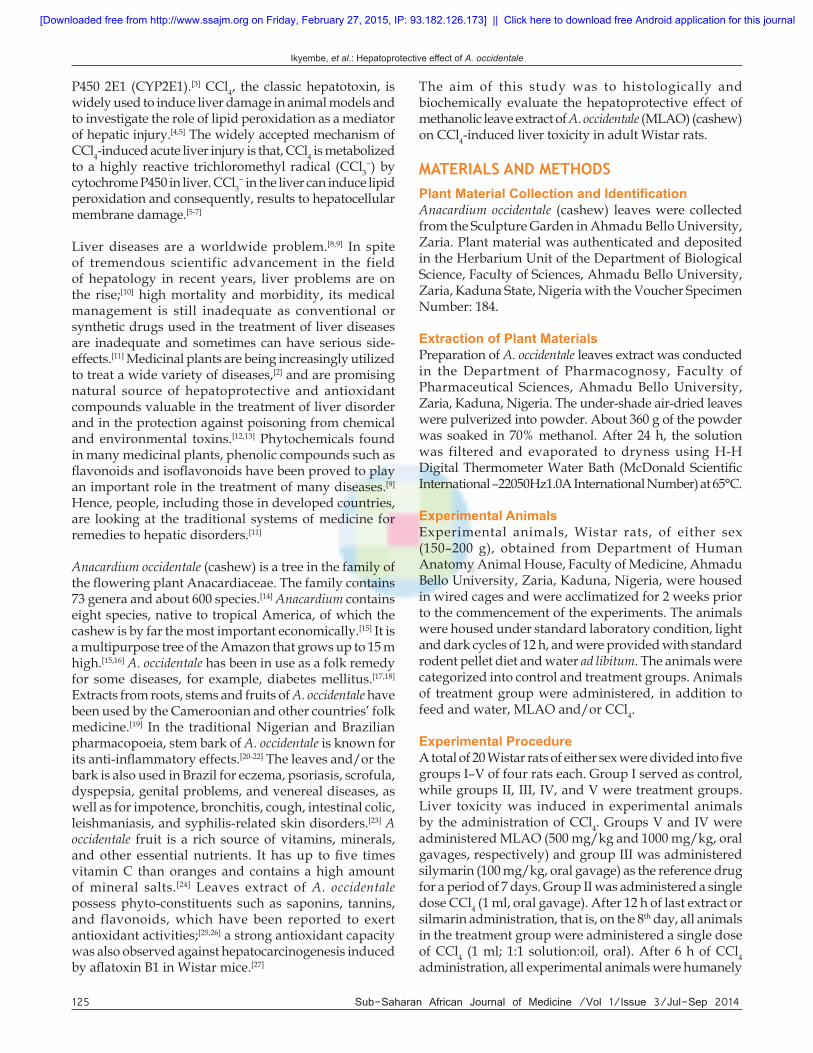
RESULTSHistological ExaminationHistological examination of tissue sections of the liver revealed the following: In the control group showed normal histoarchitecture of the liver parenchyma; the characteristic appearance of hepatic lobule unit: Centrilobular venules (central veins), array of anatomizing plates of liver cells (hepatocytes) radiating from the central vein separated by vascular spaces (sinusoids) and portal tract/triad (hepatic portal vein, hepatic vein, and bile duct) [Figure 1a].
Wistar rats treated with 1 ml of CCl4 revealed severe histoarchitectural distortion of the liver parenchyma, which manifested as diffused necrosis of hepatocytes with pyknotic nuclei and hepatocellular vacuolation. Infiltration of inflammatory cells, congestion and sinusoidal dilations were also observed [Figure 1b].
The silymarin-treated Wistar rats followed by the administration of CCl4 showed normal cytoarchitecture of the liver with mild histoarchitectural distortion, such as, infiltration of inflammatory cells and central vein congestion [Figure 1c].
In the group administered with high dose (1000 mg/kg)
MLAO followed by the administration of CCl4, the liver sections revealed histoarchitectural distortion of liver parenchyma manifesting as hepatocellular necrosis, central vein congestion and of inflammatory cells infiltration. However, observed distortion was not as severe when compared to CCl4-treated group [Figure 1d].
In the group administered low dose (500 mg/kg) MLAO followed by CCl4 administration, the liver sections showed normal cytoarchitecture of the liver with mild histoarchitectural distortion, such as, central vein congestion [Figure 1e].
Biochemical Analysis - Serum Liver EnzymesAnalysis of blood biochemical parameters of experimental animals revealed elevation of serum liver enzymes
Figure 1: Micrograph of liver of Wistar rat H and E stain (×100). (a) Liver section of control group showing normal histology of the liver. Central vein (C); hepatocyte (H); sinusoids (S). (b) Liver section of group administered CCl4 only showing severe distortion of the histoarchitecture of the liver. Congested central vein (C); necrosis (N); sinusoidal dilatation (S); vacuolation (V). (c) Liver section of group pretreated with silymarin (100 mg/kg) followed by CCl4 showing mild distortion of the histoarchitecture of the liver. Congested central vein (C); inflammatory cells infiltration (I). (d) Liver section of group pretreated with methanolic leave extract of Anacardium occidentale (MLAO) (1000 mg/kg) followed by CCl4 showing distortion of the histoarchitecture of the liver. Congested central vein (C); inflammatory cells infiltration (I); necrosis (N). (e) Liver section of group pretreated with MLAO (500 mg/kg) followed by CCl4 showing mild distortion of the histoarchitecture of the liver. Congested central vein (C); hepatocyte (H)
a b
d
e
c
[Downloaded free from http://www.ssajm.org on Friday, February 27, 2015, IP: 93.182.126.173] || Click here to download free Android application for this journal
Ikyembe, et al.: Hepatoprotective effect of A. occidentale
127 Sub-Saharan African Journal of Medicine /Vol 1/Issue 3/Jul-Sep 2014
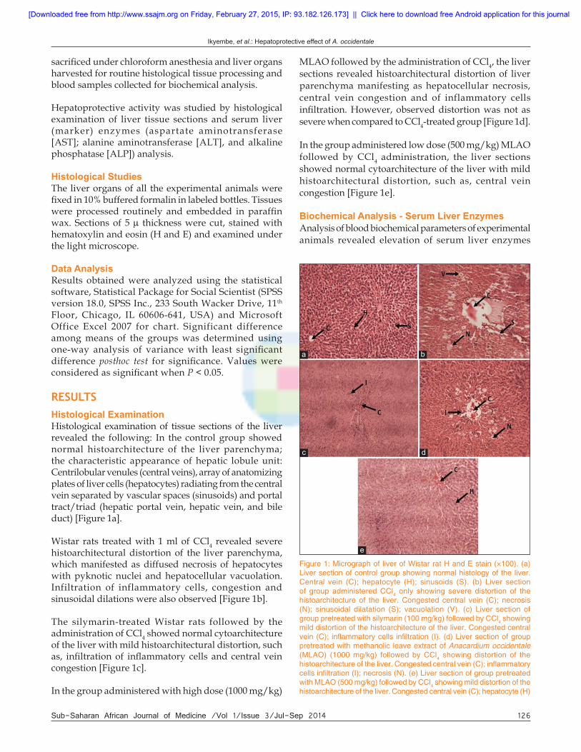
(AST, ALT, and ALP) levels in the treatment groups. Elevation of AST level was observed in CCl4-treated group, silymarin and CCl4-treated group and extract (1000 mg/kg MLAO) and CCl4-treated group, while extract (500 mg/kg MLAO) and CCl4-treated group showed decreased enzyme (AST) level when compared to the control [Figure 2].
Decrease AST levels were observed with silymarin and extract pretreated groups when compared with group administered CCl4 only. Significant (P < 0.01) decrease was observed only at dose 500 mg/kg of MLAO.
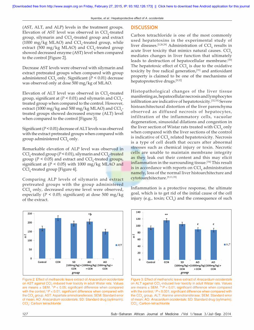
Elevation of ALT level was observed in CCl4-treated group, significant at (P < 0.01) and silymarin and CCl4-treated group when compared to the control. However, extract (1000 mg/kg and 500 mg/kg MLAO) and CCl4-treated groups showed decreased enzyme (ALT) level when compared to the control [Figure 3].
Significant (P < 0.01) decrease of ALT levels was observed with the extract pretreated groups when compared with group administered CCl4 only.
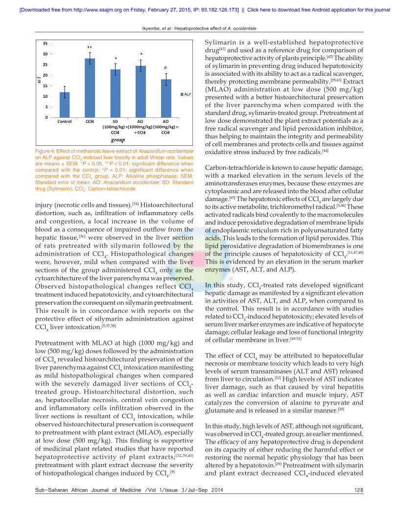
Remarkable elevation of ALP level was observed in CCl4-treated group (P < 0.01), silymarin and CCl4-treated group (P < 0.05) and extract and CCl4-treated groups, significant at (P < 0.05) with 1000 mg/kg MLAO and CCl4-treated group [Figure 4].
Comparing ALP levels of silymarin and extract pretreated groups with the group administered CCl4 only, decreased enzyme level were observed, especially (P < 0.05; significant) at dose 500 mg/kg of the extract.
Figure 2: Effect of methanolic leave extract of Anacardium occidentale on AST against CCl4-induced liver toxicity in adult Wistar rats. Values are means ± SEM. *P < 0.05; significant difference when compared with the control; bP < 0.01; significant difference when compared with the CCl4 group. AST: Aspartate aminotransferase; SEM: Standard error of mean; AO: Anacardium occidentale; SD: Standard drug (sylimarin); CCl4: Carbon tetrachloride
Figure 3: Effect of methanolic leave extract of Anacardium occidentale on ALT against CCl4-induced liver toxicity in adult Wistar rats. Values are means ± SEM. **P < 0.01; significant difference when compared with the control; cP < 0.001; significant difference when compared with the CCl4 group. ALT: Alanine aminotransferase; SEM: Standard error of mean; AO: Anacardium occidentale; SD: Standard drug (sylimarin); CCl4: Carbon-tetrachloride
DISCUSSIONCarbon tetrachloride is one of the most commonly used hepatotoxins in the experimental study of liver diseases.[5,28,29] Administration of CCl4 results in acute liver toxicity that mimics natural causes. CCl4 mediates changes in liver function that ultimately leads to destruction of hepatocellular membrane.[30] The hepatotoxic effect of CCl4 is due to the oxidative toxicity by free radical generation,[31] and antioxidant property is claimed to be one of the mechanisms of hepatoprotective drugs.[9,32]
Histopathological changes of the l iver tissue manifesting as, hepatocellular necrosis and lymphocytes infiltration are indicative of hepatotoxicity.[32,33] Severe histoarchitectural distortion of the liver parenchyma observed as diffused necrosis of hepatocytes, infiltration of the inflammatory cells, vacuolar degeneration, sinusoidal dilations and congestion in the liver section of Wistar rats treated with CCl4 only when compared with the liver sections of the control is indicative of CCl4 related hepatotoxicity. Necrosis is a type of cell death that occurs after abnormal stresses such as chemical injury or toxin. Necrotic cells are unable to maintain membrane integrity as they leak out their content and this may elicit inflammation in the surrounding tissue.[34] This result is in accordance with reports on CCl4 administration namely, loss of the normal liver histoarchitecture and cytotoarchitecture.[9,11,35]
Inflammation is a protective response, the ultimate goal, which is to get rid of the initial cause of the cell injury (e.g., toxin; CCl4) and the consequence of such
[Downloaded free from http://www.ssajm.org on Friday, February 27, 2015, IP: 93.182.126.173] || Click here to download free Android application for this journal
Ikyembe, et al.: Hepatoprotective effect of A. occidentale
Sub-Saharan African Journal of Medicine /Vol 1/Issue 3/Jul-Sep 2014 128
injury (necrotic cells and tissues).[34] Histoarchitectural distortion, such as, infiltration of inflammatory cells and congestion, a local increase in the volume of blood as a consequence of impaired outflow from the hepatic tissue,[36] were observed in the liver section of rats pretreated with silymarin followed by the administration of CCl4. Histopathological changes were, however, mild when compared with the liver sections of the group administered CCl4 only as the cytoarchitecture of the liver parenchyma was preserved. Observed histopathological changes reflect CCl4 treatment induced hepatotoxicity, and cytoarchitectural preservation the consequent on silymarin pretreatment. This result is in concordance with reports on the protective effect of silymarin administration against CCl4 liver intoxication.[9,37,38]
Pretreatment with MLAO at high (1000 mg/kg) and low (500 mg/kg) doses followed by the administration of CCl4 revealed histoarchitectural preservation of the liver parenchyma against CCl4 intoxication manifesting as mild histopathological changes when compared with the severely damaged liver sections of CCl4-treated group. Histoarchitectural distortion, such as, hepatocellular necrosis, central vein congestion and inflammatory cells infiltration observed in the liver sections is resultant of CCl4 intoxication, while observed histoarchitectural preservation is consequent to pretreatment with plant extract (MLAO), especially at low dose (500 mg/kg). This finding is supportive of medicinal plant related studies that have reported hepatoprotective activity of plant extracts;[32,39,40] pretreatment with plant extract decrease the severity of histopathological changes induced by CCl4.[9]
Sylimarin is a well-established hepatoprotective drug[41] and used as a reference drug for comparison of hepatoprotective activity of plants principle.[42] The ability of sylimarin in preventing drug induced hepatotoxicity is associated with its ability to act as a radical scavenger, thereby protecting membrane permeability.[29,43] Extract (MLAO) administration at low dose (500 mg/kg) presented with a better histoarchitectural preservation of the liver parenchyma when compared with the standard drug, sylimarin-treated group. Pretreatment at low dose demonstrated the plant extract potentials as a free radical scavenger and lipid peroxidation inhibitor, thus helping to maintain the integrity and permeability of cell membranes and protects cells and tissues against oxidative stress induced by free radicals.[44]
Carbon-tetrachloride is known to cause hepatic damage, with a marked elevation in the serum levels of the aminotransferases enzymes, because these enzymes are cytoplasmic and are released into the blood after cellular damage.[45] The hepatotoxic effects of CCl4 are largely due to its active metabolite, trichloromethyl radical.[5,46] These activated radicals bind covalently to the macromolecules and induce peroxidative degradation of membrane lipids of endoplasmic reticulum rich in polyunsaturated fatty acids. This leads to the formation of lipid peroxides. This lipid peroxidative degradation of biomembranes is one of the principle causes of hepatotoxicity of CCl4.[11,47,48] This is evidenced by an elevation in the serum marker enzymes (AST, ALT, and ALP).
In this study, CCl4-treated rats developed significant hepatic damage as manifested by a significant elevation in activities of AST, ALT, and ALP, when compared to the control. This result is in accordance with studies related to CCl4-induced hepatotoxicity; elevated levels of serum liver marker enzymes are indicative of hepatocyte damage; cellular leakage and loss of functional integrity of cellular membrane in liver.[49-51]
The effect of CCl4 may be attributed to hepatocellular necrosis or membrane toxicity which leads to very high levels of serum transaminases (ALT and AST) released from liver to circulation.[52] High levels of AST indicates liver damage, such as that caused by viral hepatitis as well as cardiac infarction and muscle injury, AST catalyzes the conversion of alanine to pyruvate and glutamate and is released in a similar manner.[29]
In this study, high levels of AST, although not significant, was observed in CCl4-treated group, as earlier mentioned. The efficacy of any hepatoprotective drug is dependent on its capacity of either reducing the harmful effect or restoring the normal hepatic physiology that has been altered by a hepatotoxin.[29] Pretreatment with silymarin and plant extract decreased CCl4-induced elevated
Figure 4: Effect of methanolic leave extract of Anacardium occidentale on ALP against CCl4-induced liver toxicity in adult Wistar rats. Values are means ± SEM. *P < 0.05, **P < 0.01; significant difference when compared with the control; aP < 0.01; significant difference when compared with the CCl4 group. ALP: Alkaline phosphatase; SEM: Standard error of mean; AO: Anacardium occidentale; SD: Standard drug (Sylimarin); CCl4: Carbon-tetrachloride
[Downloaded free from http://www.ssajm.org on Friday, February 27, 2015, IP: 93.182.126.173] || Click here to download free Android application for this journal
Ikyembe, et al.: Hepatoprotective effect of A. occidentale
129 Sub-Saharan African Journal of Medicine /Vol 1/Issue 3/Jul-Sep 2014
enzyme level when compared to the CCl4-intoxicated group; decrease was significant only in 500 mg/kg extract-treated group. This result is in accordance with reports related to hepatoprotective medicinal plants; decreased levels of serum liver enzymes consequent on plant extract pretreatment against CCl4-induced elevations indicates protection of structural integrity of hepatocytic cell membrane or regeneration of damaged hepatocytes.[29,53]
ALT is more specific to the liver, and is thus a better parameter for detecting liver injury. Elevated level of serum ALT is indicative of cellular leakage and loss of functional integrity of cell membrane in liver.[29,50] In this study, significant high level of ALT was observed in CCl4-treated group, as earlier mentioned. Pretreatment with the extract significantly decreased CCl4-induced elevated enzyme level when compared to the CCl4-intoxicated group. It was also observed that, pretreatment with the extract, especially at dose 500 mg/kg, decreased enzyme level when compared to the control, this was however not significant. Previous studies have reported that oxidative stress plays an essential role in the hepatic injury mediated by CCl4.[46,55,56] Decrease in the level of these enzymes with MLAO is an indication of the stabilization of plasma membrane as well as repair of liver damage caused by CCl4, and is similar to earlier reports.[32]
Phosphatases are key enzymes responsible for various biological functions such as metabolism, detoxification and biosynthesis of energy macromolecules for different essential functions. Any alteration in the activity of these enzymes causes tissue lesion and cellular impairment and dysfunction.[29] Serum ALP level is related to the function of hepatic cell. Elevation in serum ALP level is due to increased synthesis, in the presence of increased biliary pressure.[12,29] In this study, all treatment rats showed elevated levels of ALP when compared to the control consequent to CCl4 intoxication. Pretreatment with the extract, significantly, at dose 500 mg/kg, decreased enzyme level when compared to the CCl4-intoxicated group. This result is in accordance to reports on hepatoprotective medicinal plant; the lowering of enzymes level are definite indication of hepatoprotective action of the drug.[11,43] Hepatoprotective action may be due to the prevention of the leakage of intracellular enzymes by its membrane stabilizing activity.[32]
Hepatoprotective effect suggests that the plant may possess antioxidant properties that helped to combat the CCl4-induced oxidative stress in the liver. The ability of natural compounds to attenuate toxin-induced hepatotoxicity is believed to be related to their intrinsic antioxidant properties.[11,56] Flavanoids[57] and saponins,[58] plant phytochemical constituent, are well known for their
antioxidant and hepatoprotective activities. Flavonoids have been reported to protect against toxicity induced by environmental toxicants[26,56,59] such as CCl4. The chemoprotective activities of flavonoids are related to their ability to inhibit peroxidative damage caused by environmental toxicants.[60-63] The flavonoids and saponins present in the MLAO may have, perhaps, played a major role in the hepatoprotective action.
CONCLUSIONThe result of this study suggests that, the MLAO was effective, histologically and biochemically, in preventing CCl4-induced acute liver injury in Wistar rats, especially at dose of 500 mg/kg. The hepatoprotective activity of the plant extract, which could be of therapeutic potentials, may be consequent to the antioxidant activities of constituent phytochemicals.
ACKNOWLEDGMENTSWe wish to acknowledge Mr. Peter S. Akpulu for technical support and the Department of Human Anatomy, Faculty of Medicine, Ahmadu Bello University, Zaria for providing the facilities to conduct this study.
REFERENCES1. Ward FM, Daly MJ. Hepatic disease. In: Walker R, Edward
C, editors. Clinical Pharmacy and Therapeutics. New York: Churchill Livingston; 1999. p. 195-212.
2. Biswas K, Kumar A, Babaria BA, Prabhu K, Ramachandra SS. Hepatoprotective effect of leaves of Peltophorum pterocarpum against paracetamol induced acute liver damage in rats. J Basic Clin Pharm 2010;1:10-5.
3. Sun F, Hamagawa E, Tsutsui C, Ono Y, Ogiri Y, Kojo S. Evaluation of oxidative stress during apoptosis and necrosis caused by carbon tetrachloride in rat liver. Biochim Biophys Acta 2001;1535:186-91.
4. Brattin WJ, Glende EA Jr, Recknagel RO. Pathological mechanisms in carbon tetrachloride hepatotoxicity. J Free Radic Biol Med 1985;1:27-38.
5. Gong F, Yin Z, Xu Q, Kang W. Hepatoprotective effect of Mitragyna rotundifolia Kuntze on CCl4-induced acute liver injury in mice. Afr J Pharm Pharmacol 2012;6:330-5.
6. Drill VA. Hepatotoxic agents; mechanism of action and dietary interrelationship. Pharmacol Rev 1952;4:1-42.
7. Ohta Y, Sasaki E, Nishida K, Kongo M, Hayashi T, Nagata M, et al. Contribution of the antilipidperoxidative action of Dai-saiko-to extract to its preventive effect on carbon tetrachloride-induced acute liver injury in rats. Phytother Res 1998;12:5-8.
8. World Health Organization (WHO). Viral hepatitis: Report by the WHO Secretariat; 2010.
9. Soufy NI. Hepatoprotective and antioxidant effects of Silybum marianum plant against hepatotoxicity induced by carbon tetrachloride in rats. J Am Sci 2012;8:479-786.
10. Perz JF, Armstrong GL, Farrington LA, Hutin YJ, Bell BP. The contributions of hepatitis B virus and hepatitis C virus infections to cirrhosis and primary liver cancer worldwide. J Hepatol 2006;45:529-38.
11. Prakash T, Fadadu SD, Sharma UR, Surendra V, Goli D,
[Downloaded free from http://www.ssajm.org on Friday, February 27, 2015, IP: 93.182.126.173] || Click here to download free Android application for this journal
Ikyembe, et al.: Hepatoprotective effect of A. occidentale
Sub-Saharan African Journal of Medicine /Vol 1/Issue 3/Jul-Sep 2014 130
Stamina P, et al. Hepatoprotective activity of leaves of Rhododendron arboreum in CCl4 induced hepatotoxicity in rats. J Med Plants Res 2008;2:315-20.
12. Muriel P, Garciapiña T, Perez-Alvarez V, Mourelle M. Silymarin protects against paracetamol-induced lipid peroxidation and liver damage. J Appl Toxicol 1992;12:439-42.
13. Barar RS. Essentials of Pharmacotherapeutics. 3rd ed. New Delhi: Chand and Company Ltd.; 2000. p. 340-1.
14. Correa MP. Dicionário de plantas úteis do Brasil. Ministério da Agricultura. Rio de Janeiro: IBDF; 1978. p. 55 [in Portuguese].
15. Morton JF. Fruits of Warm Climates. Florida: Flair Books; 1987. ISBN: 0-9610184-1-0.
16. de Souza CP, Mendes NM, Jannotti-Passos LK, Pereira JP. The use of the shell of the cashew nut, Anacardium occidentale, as an alternative molluscacide. Rev Inst Med Trop Sao Paulo 1992;34:459-66.
17. Longuefosse JL, Nossin E. Medical ethnobotany survey in Martinique. J Ethnopharmacol 1996;53:117-42.
18. Kamtchouing P, Sokeng SD, Moundipa PF, Watcho P, Jatsa HB, Lontsi D. Protective role of Anacardium occidentale extract against streptozotocin-induced diabetes in rats. J Ethnopharmacol 1998;62:95-9.
19. Sokeng DS, Kamtchouing P, Watcho P, Jatsa HB, Moundipa PF, Ngounou FN, et al. Hypoglycemic activity of Anacardium occidentale L. aqueous extract in normal and streptozotocin-induced diabetic rats. Diabetes Res 2001;36:001-9.
20. Mota ML, Thomas G, Barbosa Filho JM. Anti-inflammatory actions of tannins isolated from the bark of Anacardium occidentale L. J Ethnopharmacol 1985;13:289-300.
21. Chen SC, Chung KT. Mutagenicity and antimutagenicity studies of tannic acid and its related compounds. Food Chem Toxicol 2000;38:1-5.
22. Ojewole JA. Laboratory evaluation of the hypoglycemic effect of Anacardium occidentale Linn. (Anacardiaceae) stem-bark extracts in rats. Methods Find Exp Clin Pharmacol 2003;25:199-204.
23. França F, Cuba CA, Moreira EA, Miguel O, Almeida M, das Virgens Mde L, et al. An evaluation of the effect of a bark extract from the cashew (Anacardium occidentale L.) on infection by Leishmania (Viannia) braziliensis. Rev Soc Bras Med Trop 1993-Sep;26:151-5.
24. Dare SS, Hamman WO, Musa S, Goji AD, Oyewale AA, Abba S, et al. Effects of aqueous extract of Anacardium occidentale (cashew) leaf on pregnancy outcome of wistar rats. Int J Anim Vet Adv 2011;3:77-82.
25. Gonçalves JL, Lopes RC, Oliveira DB, Costa SS, Miranda MM, Romanos MT, et al. In vitro anti-rotavirus activity of some medicinal plants used in Brazil against diarrhea. J Ethnopharmacol 2005 14;99:403-7.
26. Jaiswal YS, Tatke PA, Gabhe SY, Vaidya A. Antioxidant activity of various extracts of leaves of Anacardium Occidentale (cashew). Res J Pharm Biol Chem Sci 2010;1:112-9.
27. Premalatha B, Sachdanandam P. Semecarpus anacardium L. nut extract administration induces the in vivo antioxidant defence system in aflatoxin B1 mediated hepatocellular carcinoma. J Ethnopharmacol 1999;66:131-9.
28. Rincón AR, Covarrubias A, Pedraza-Chaverrí J, Poo JL, Armendáriz-Borunda J, Panduro A. Differential effect of CCl4 on renal function in cirrhotic and non-cirrhotic rats. Exp Toxicol Pathol 1999;51:199-205.
29. Palanivel MG, Rajkapoor B, Senthil KR, Einstein JW, Kumar EP, Rupesh KM, et al. Hepatoprotective and antioxidant effect of Pisonia aculeata l. Against CCl4-induced hepatic damage in rats. Sci Pharm 2008;76:203-15.
30. de Andrade Belo MA, Soares VE, de Souza LM, da Rosa Sobreira
MF, Cassol DM, Toma SB. Hepatoprotective treatment attenuates oxidative damages induced by carbon tetrachloride in rats. Exp Toxicol Pathol 2012;64:155-65.
31. Pandit S, Sur TK, Jana U, Debnath PK, Sen S, Bhattacharyya D. Prevention of carbon tetrachloride-induced hepatotoxicity in rats by Adhatoda vasica leaves. Indian J Pharmcol 2004;36:312-3.
32. Sahreen S, Khan MR, Khan RA. Evaluation of antioxidant activities of various solvent extracts of Carissa opaca fruits. Food Chem 2010;122:1205-11.
33. Nabeshima Y, Tazuma S, Kanno K, Hyogo H, Iwai M, Horiuchi M, et al. Anti-fibrogenic function of angiotensin II type 2 receptor in CCl4-induced liver fibrosis. Biochem Biophys Res Commun 2006;346:658-64.
34. Kumar V, Alla SR, Krishnan KS, Ramaswami M. Syndapin is dispensable for synaptic vesicle endocytosis at the Drosophila larval neuromuscular junction. Mol Cell Neurosci 2009;40:234-41.
35. Padhy BM, Srivastava A, Kumar VL. Calotropis procera latex affords protection against carbon tetrachloride induced hepatotoxicity in rats. J Ethnopharmacol 2007;113:498-502.
36. Ramzi SC, Vinay K, Tucker C. Robbinn’s Pathologic Basis of Diseases. 6th ed. Philadelphia: W.B. Sauders Company; 1999. p. 913-27.
37. Rao PV, Jayaraj R, Bhaskar AS. Protective efficacy and the recovery profile of certain chemoprotectants against lethal poisoning by microcystin-LR in mice. Toxicon 2004;44:723-30.
38. Wahsha M, Al-Jassabi S. The role of silymarin in the protection of mice liver damage against microcystin-LR toxicity. Jordan J Biol Sci 2009;2:63-8.
39. Al-Qarawi A, Mousa HM, Ali BH, Abdel-Rahman H, El-Mougy SA. Protective effect of extracts from dates (Phoenix dactylifera L.) on carbon tetrachloride-induced hepatotoxicity in rats. Int J Appl Res Vet M 2004;2:176-80.
40. Ahmed MB, Hasona NA, Selemain HA. Protective effects of extract from dates (Phoenix dactylifera L.) and ascorbic acid on thioacetamide-induced hepatotoxicity in rats. Iran J Pharm Res 2008;7:193-201.
41. Pradhan SC, Girish C. Hepatoprotective herbal drug, silymarin from experimental pharmacology to clinical medicine. Indian J Med Res 2006;124:491-504.
42. Usmani S, Kushwaha P. A study on hepatoprotective activity of Calotropis gigantea leaves extract. Int J Pharm Pharm Sci 2010;2:101-3.
43. Song Z, Deaciuc I, Song M, Lee DY, Liu Y, Ji X, et al. Silymarin protects against acute ethanol-induced hepatotoxicity in mice. Alcohol Clin Exp Res 2006;30:407-13.
44. Naik SR, Panda VS. Antioxidant and hepatoprotective effects of Ginkgo biloba phytosomes in carbon tetrachloride-induced liver injury in rodents. Liver Int 2007;27:393-9.
45. Recknagel RO, Glende EA Jr, Dolak JA, Waller RL. Mechanisms of carbon tetrachloride toxicity. Pharmacol Ther 1989;43:139-54.
46. Johnston DE, Kroening C. Mechanism of early carbon tetrachloride toxicity in cultured rat hepatocytes. Pharmacol Toxicol 1998;83:231-9.
47. Recknagael R. Carbontetrachloride hepatotoxicity. Pharmacol Rev 1967;19:145-96.
48. Kaplowitz N, Aw TY, Simon FR, Stolz A. Drug-induced hepatotoxicity. Ann Intern Med 1986;104:826-39.
49. Drotman RB, Lawhorn GT. Serum enzymes as indicators of chemically induced liver damage. Drug Chem Toxicol 1978;1:163-71.
50. Tirkey N, Pilkhwal S, Kuhad A, Chopra K. Hesperidin, a citrus bioflavonoid, decreases the oxidative stress produced by carbon tetrachloride in rat liver and kidney. BMC Pharmacol 2005;5:2.
[Downloaded free from http://www.ssajm.org on Friday, February 27, 2015, IP: 93.182.126.173] || Click here to download free Android application for this journal

Ikyembe, et al.: Hepatoprotective effect of A. occidentale
131 Sub-Saharan African Journal of Medicine /Vol 1/Issue 3/Jul-Sep 2014
51. Anand KV, Anandhi R, Pakkiyaraj M, Geraldine P. Protective effect of chrysin on carbon tetrachloride (CCl4)-induced tissue injury in male Wistar rats. Toxicol Ind Health 2011;27:923-33.
52. Achliya GS, Kotgale SG, Wadodkar AK, Dorle AK. Hepatoprotective activity of Panchgavya Gritha in carbon tetrachloride induced hepatotoxicity in rats. Indian J Pharmacol 2003;35:311.
53. Osman M, Ahmed M, Mahfouz S, Elaby S. Biochemical studies on the hepatoprotective effects of pomegranate and guava ethanol extracts. N Y Sci J 2011;4:27-41.
54. Weber LW, Boll M, Stampfl A. Hepatotoxicity and mechanism of action of haloalkanes: Carbon tetrachloride as a toxicological model. Crit Rev Toxicol 2003;33:105-36.
55. Lin HM, Tseng HC, Wang CJ, Lin JJ, Lo CW, Chou FP. Hepatoprotective effects of Solanum nigrum Linn. extract against CCl(4)-induced oxidative damage in rats. Chem Biol Interact 2008;171:283-93.
56. Farombi EO. Locally derived natural antioxidant substances in Nigeria: Potential role as new chemotherapeutic agents. In: Bahorun T, Gurib-Fakim A, editors. Molecular and Therapeutic Aspects of Redox Biochemistry. UK: OICA International Limited; 2003. p. 208-25.
57. Yoshikawa M, Morikawa T, Kashima Y, Ninomiya K, Matsuda H. Structures of new dammarane-type Triterpene Saponins from the flower buds of Panax notoginseng and hepatoprotective effects of principal Ginseng Saponins. J Nat Prod 2003;66:922-7.
58. Baek NL, Kim YS, Kyung JS, Park KH. Isolation of antihepatotoxic agent from the roots of Astragalus membranaceous. Korean J Phamacogn 1996;27:111-7.
59. Anosike CA, Ugwu UB, Nwakanma O. Effect of ethanol extract of Pyrenacantha staudtii leaves on carbontetrachloride induced hepatotoxicity in rats. Biokemistri 2008;20:17-22.
60. Sahreen S, Khan MR, Khan RA. Evaluation of antioxidant activities of various solvent extracts of Carissa opaca fruits. Food Chem 2010;122:1205-11.
61. Etuk EU, Agaie BM, Ladan MJ, Garba I. The modulatory effect of Cochlospermum tinctorium a rich aqueous root extract on liver damage induced by carbon tetrachloride in rats. Afr J Pharm Pharmacol 2009;3:151-7.
62. Muriel P, Mourelle M. Prevention by silymarin of membrane alterations in acute CCl4 liver damage. J Appl Toxicol 1990;10:275-9.
63. Faure M, Lissi E, Torres R, Vidella L. Antioxidant activities of lignan and flavonoids. Phytochemistry 1990;29:3773-5.
How to cite this article: Ikyembe D, Pwavodi C, Agbon AN. Hepatoprotective effect of methanolic leaf extract of anacardium occidentale (cashew) on carbon-tetrachloride-induced liver toxicity in wistar rats. Sub-Saharan Afr J Med 2014;1:124-31.
Source of Support: Nil, Conflict of Interest: None declared.
“QUICK RESPONSE CODE” LINK FOR FULL TEXT ARTICLESThe journal issue has a unique new feature for reaching to the journal’s website without typing a single letter. Each article on its first page has a “Quick Response Code”. Using any mobile or other hand-held device with camera and GPRS/other internet source, one can reach to the full text of that particular article on the journal’s website. Start a QR-code reading software (see list of free applications from http://tinyurl.com/yzlh2tc) and point the camera to the QR-code printed in the journal. It will automatically take you to the HTML full text of that article. One can also use a desktop or laptop with web camera for similar functionality. See http://tinyurl.com/2bw7fn3 or http://tinyurl.com/3ysr3me for the free applications.
Announcement
[Downloaded free from http://www.ssajm.org on Friday, February 27, 2015, IP: 93.182.126.173] || Click here to download free Android application for this journal