Characterization of Troponin Responses in Isoproterenol-Induced Cardiac Injury in the Hanover Wistar...
13
http://tpx.sagepub.com/ Toxicologic Pathology http://tpx.sagepub.com/content/35/4/606 The online version of this article can be found at: DOI: 10.1080/01926230701389316 2007 35: 606 Toxicol Pathol Griffiths, Matthew Whayman, Thomas Williams and John Turton Malcolm York, Cheryl Scudamore, Sally Brady, Christabelle Chen, Sharon Wilson, Mark Curtis, Gareth Evans, William Characterization of Troponin Responses in Isoproterenol-Induced Cardiac Injury in the Hanover Wistar Rat Published by: http://www.sagepublications.com On behalf of: Society of Toxicologic Pathology can be found at: Toxicologic Pathology Additional services and information for http://tpx.sagepub.com/cgi/alerts Email Alerts: http://tpx.sagepub.com/subscriptions Subscriptions: http://www.sagepub.com/journalsReprints.nav Reprints: http://www.sagepub.com/journalsPermissions.nav Permissions: What is This? - Jun 1, 2007 Version of Record >> at China University of Petroleum - Beijing on October 11, 2013 tpx.sagepub.com Downloaded from at China University of Petroleum - Beijing on October 11, 2013 tpx.sagepub.com Downloaded from at China University of Petroleum - Beijing on October 11, 2013 tpx.sagepub.com Downloaded from at China University of Petroleum - Beijing on October 11, 2013 tpx.sagepub.com Downloaded from at China University of Petroleum - Beijing on October 11, 2013 tpx.sagepub.com Downloaded from at China University of Petroleum - Beijing on October 11, 2013 tpx.sagepub.com Downloaded from at China University of Petroleum - Beijing on October 11, 2013 tpx.sagepub.com Downloaded from at China University of Petroleum - Beijing on October 11, 2013 tpx.sagepub.com Downloaded from at China University of Petroleum - Beijing on October 11, 2013 tpx.sagepub.com Downloaded from at China University of Petroleum - Beijing on October 11, 2013 tpx.sagepub.com Downloaded from at China University of Petroleum - Beijing on October 11, 2013 tpx.sagepub.com Downloaded from at China University of Petroleum - Beijing on October 11, 2013 tpx.sagepub.com Downloaded from at China University of Petroleum - Beijing on October 11, 2013 tpx.sagepub.com Downloaded from
-
Upload
independent -
Category
Documents
-
view
0 -
download
0
Transcript of Characterization of Troponin Responses in Isoproterenol-Induced Cardiac Injury in the Hanover Wistar...

http://tpx.sagepub.com/Toxicologic Pathology
http://tpx.sagepub.com/content/35/4/606The online version of this article can be found at:
DOI: 10.1080/01926230701389316
2007 35: 606Toxicol PatholGriffiths, Matthew Whayman, Thomas Williams and John Turton
Malcolm York, Cheryl Scudamore, Sally Brady, Christabelle Chen, Sharon Wilson, Mark Curtis, Gareth Evans, WilliamCharacterization of Troponin Responses in Isoproterenol-Induced Cardiac Injury in the Hanover Wistar Rat
Published by:
http://www.sagepublications.com
On behalf of:
Society of Toxicologic Pathology
can be found at:Toxicologic PathologyAdditional services and information for
http://tpx.sagepub.com/cgi/alertsEmail Alerts:
http://tpx.sagepub.com/subscriptionsSubscriptions:
http://www.sagepub.com/journalsReprints.navReprints:
http://www.sagepub.com/journalsPermissions.navPermissions:
What is This?
- Jun 1, 2007Version of Record >>
at China University of Petroleum - Beijing on October 11, 2013tpx.sagepub.comDownloaded from at China University of Petroleum - Beijing on October 11, 2013tpx.sagepub.comDownloaded from at China University of Petroleum - Beijing on October 11, 2013tpx.sagepub.comDownloaded from at China University of Petroleum - Beijing on October 11, 2013tpx.sagepub.comDownloaded from at China University of Petroleum - Beijing on October 11, 2013tpx.sagepub.comDownloaded from at China University of Petroleum - Beijing on October 11, 2013tpx.sagepub.comDownloaded from at China University of Petroleum - Beijing on October 11, 2013tpx.sagepub.comDownloaded from at China University of Petroleum - Beijing on October 11, 2013tpx.sagepub.comDownloaded from at China University of Petroleum - Beijing on October 11, 2013tpx.sagepub.comDownloaded from at China University of Petroleum - Beijing on October 11, 2013tpx.sagepub.comDownloaded from at China University of Petroleum - Beijing on October 11, 2013tpx.sagepub.comDownloaded from at China University of Petroleum - Beijing on October 11, 2013tpx.sagepub.comDownloaded from at China University of Petroleum - Beijing on October 11, 2013tpx.sagepub.comDownloaded from

Toxicologic Pathology, 35:606–617, 2007Copyright C© by the Society of Toxicologic PathologyISSN: 0192-6233 print / 1533-1601 onlineDOI: 10.1080/01926230701389316
Characterization of Troponin Responses in Isoproterenol-InducedCardiac Injury in the Hanover Wistar Rat
MALCOLM YORK,1 CHERYL SCUDAMORE,1 SALLY BRADY,2 CHRISTABELLE CHEN,2 SHARON WILSON,1 MARK CURTIS,1GARETH EVANS,3 WILLIAM GRIFFITHS,4 MATTHEW WHAYMAN,1 THOMAS WILLIAMS,1 AND JOHN TURTON2
1GlaxoSmithKline Research and Development, Park Road, Ware, Herts SG12 0DP, UK2Centre for Toxicology, Department of Pharmacology, School of Pharmacy, University of London, London WC1N 1AX, UK
3Safety Assessment Alderley, AstraZeneca Pharmaceuticals, Alderley Park, Macclesfield, Cheshire SK10 4TG, UK4Department of Pharmaceutical and Biological Chemistry, School of Pharmacy, University of London, London WC1N 1AX, UK
ABSTRACT
The investigations aimed to evaluate the usefulness of cardiac troponins as biomarkers of acute myocardial injury in the rat. Serum from femaleHanover Wistar rats treated with a single intraperitoneal (IP) injection of isoproterenol (ISO) was assayed for cardiac troponin I (cTnI) (ACS: 180SE,Bayer), cTnI (Immulite 2000, Diagnostic Products Corporation) and cardiac troponin T (cTnT) (Elecsys 2010, Roche). In a time-course study (50.0mg/kg ISO), serum cTnI (ACS:180SE) and cTnT increased above control levels at 1 hour postdosing, peaking at 2 hours (cTnI , 4.30 µg/L; cTnT,1.79 µg/L), and declined to baseline by 48 hours, with histologic cardiac lesions first seen at 4 hours postdosing. The Immulite 2000 assay gaveminimal cTnI signals, indicating poor immunoreactivity towards rat cTnI. In a dose-response study (0.25 to 20.0 mg/kg ISO), there was a trend forincreasing cTnI (ACS:180SE) values with increasing ISO dose levels at 2 hours postdosing. By 24 hours, cTnI levels returned to baseline althoughchronic cardiac myodegeneration was present. We conclude that serum cTnI and cTnT levels are sensitive and specific biomarkers for detecting ISOinduced myocardial injury in the rat. Serum troponin values reflect the development of histopathologic lesions; however peak troponin levels precedemaximal lesion severity.
Keywords. Cardiac troponin I; cardiotoxicity; isoproterenol; rat; toxicity; troponin.
INTRODUCTION
In man, serum levels of the cardiac troponins (cTn), par-ticularly troponin I (cTnI) and troponin T (cTnT), are widelyused in the detection of acute myocardial infarction and arange of other cardiac conditions (Mair, 1997; Christensonand Azzazy, 1998; Panteghini, 2004; Adamcova et al., 2005;Apple et al., 2005; Dybdahl et al., 2005). However, the useof serum cTnI and cTnT in the detection of cardiac injuryin laboratory animal species is not as widespread (Hermanet al., 1998; Bertsch et al., 1999; Bertinchant et al., 2000;Adamcova et al., 2005; Walker, 2006). Wallace et al. (2004)reviewed the usefulness of serum troponins as biomarkersof drug-induced cardiac toxicity in preclinical safety assess-ment studies as part of the role of the Expert Working Group(EWG) of the Nonclinical Studies Subcommittee (NCSS),which had been established by the US Food and Drug Ad-
Address correspondence to: Cheryl Scudamore, GSK, R&D, Park Road,Ware, Herts SG12 0DP, UK; e-mail: [email protected]
Abbreviations: ACPS, Advisory Committee for Pharmaceutical Sci-ences; ALT, Alanine aminotransferase; AST, Aspartate aminotransferase;CK, Total creatine kinase; CKBB, Creatine kinase isoenzyme BB; CKMB,Creatine kinase isoenzyme MB; CKMM Creatine kinase isoenzyme MM;cTn, Cardiac troponin; cTnI, Cardiac troponin I; cTnT, Cardiac troponinT; CV, Coefficient of variation; EWG, Expert Working Group; GLD, Glu-tamate dehydrogenase; H&E, Haematoxylin and eosin; IP, intraperitoneal;ISO, Isoproterenol; LD, Total lactate dehydrogenase; LD1 to LD5, Lactatedehydrogenase isoenzymes 1 to 5; NAD, No abnormalities detected; NCSS,Nonclinical Studies Subcommittee; NS, Not statistically significant; OM,Original magnification; PBS, Phosphate-buffered saline; RT, Room tem-perature; SD, Standard deviation.
ministration, and which reported to the Advisory Committeefor Pharmaceutical Sciences (ACPS).
In considering the case for advocating the increased useof cTn, the NCSS wished to improve the accuracy by whichpreclinical studies predict a clinical outcome with respectto potential adverse drug reactions, and also propose newbiomarkers that would strengthen the interface betweenpreclinical laboratory studies and clinical trials (Wallaceet al., 2004). In addition, the EWG also set out the desiredcharacteristics of the ideal serum/plasma biomarker for thedetection of myocardial injury, and suggested that these were:specificity, sensitivity, predictability and robustness (Wallaceet al., 2004). Nevertheless, the EWG also concluded that ad-ditional work was required for the further validation of theutility of cTn, and put forward a series of future goals, whichincluded:
1. Evaluation of the kinetics of release and return to baseline(i.e. the diagnostic window) of cTn, and the correlationwith histopathology, following the administration of com-pounds that cause distinct forms of cardiomyocyte injury.
2. Determination of whether there is a threshold for the in-crease in serum cTnI, and below which there is no sub-stantial or sustained cardiomyocyte injury.
3. Establish whether there is a diagnostic advantage of mea-suring serum cTnI, or cTnT, or cTnI and cTnT, in thevarious forms of cardiomyocyte injury.
4. Establish whether the variation in the cTnI assay platformswill influence diagnostic sensitivity within and across lab-oratory animal species.
606

Vol. 35, No. 4, 2007 TROPONIN RESPONSES IN THE RAT 607
With the above proposals in mind, we have carried out aseries of studies involving isoproterenol (ISO) induced car-diac injury in the female Hanover Wistar rat. ISO was thefirst pure β-adrenoceptor agonist to be synthesised (Searsand Lotvall, 2005). The drug was developed in the 1940s andquickly became widely used for the relief of the symptomsof asthma (Waldeck, 2002). However, although ISO is a verypotent β agonist, with almost no action on α-adrenoceptors,the drug does not distinguish between β1 and β2 recep-tors. As a result, ISO has significant extrapulmonary sideeffects, such as tachycardia, arrhythmias and palpitations,because the drug stimulates β1 receptors in the heart. ISO isa synthetic catecholamine and undergoes rapid metabolism,resulting in a very short duration of action (Dollery,1998).
The drug is no longer used in the UK and USA for therelief of asthma symptoms (BNF, 2006). However, ISO hasbecome widely used in toxicological studies as a model drugto induce cardiac muscle injury with myocardial ischemiaand the formation of infarct-like lesions (Rona et al., 1959;Handforth, 1962; Judd and Wexler, 1974; Bleuel et al., 1995);the mechanism of toxicity is therefore closely related to thepharmacological action of the drug. In the rat, at high doses,ISO quickly stimulates β1 and β2 receptors in the heart, in-ducing an abnormally rapid heart rate (β1 activity) and a fallin blood pressure (β2 activity), resulting in cardiac tissueanoxia/hypoxia due to elevated oxygen demand and bring-ing about severe myocardial necrosis (Rona et al., 1959).These changes are associated with significant increases inboth serum cTnI and cTnT (Bleuel et al., 1995; Bertinchantet al., 2000; Wallace et al., 2004). To induce cardiac lesionsin the rat, ISO is generally administered by the subcuta-neous, intraperitoneal or intravenous routes (Wexler and Kit-tinger, 1963; Leszkovszky and Gal, 1967; Kahn et al., 1969;Gryglewski et al., 1971; Benjamin et al., 1989; Bleuel et al.,1995; Mohan and Bloom, 1999; Inamoto et al., 2000; O’Brienet al., 2006) and elevations in the levels of serum cTn aregenerally evident within 4 hours of drug administration; theincreases in serum cTn levels are closely correlated with theseverity of myocardial injury (Wallace et al., 2004; Walker,2006).
The purposes of the present investigations were: (1) toassess the linearity and precision of the cTnI assay using theACS: 180SE (Bayer), together with the storage stability ofthe rat-specific immunoreactive cTnI signal; (2) to identifythe onset of the release of cTn and correlate this timepointwith the nature and severity of the histopathologic lesionin the heart from 1 to 48 hours after the administration ofISO; (3) to identify the magnitude of the cTn signal withincreasing dose levels of ISO over the dose range 0.25 to20.0 mg/kg; (4) to correlate the magnitude and duration ofthe cTnI signal (ACS: 180SE; Bayer Healthcare Diagnostics)with the cTnT signal (Elecsys 2010; Roche Diagnostics) andalso relate these 2 signals to the cTnI signal obtained withthe DPC Immulite 2000 (Diagnostic Products Corporation);and, (5) to correlate the magnitude and duration of the cTnIsignal with the more traditional biomarkers of cardiomyocyteinjury, and in particular with both creatine kinase (CK) andlactate dehydrogenase (LD) isoenzymes. Preliminary reportsof these findings have been published in abstract form (Chenet al., 2004; Brady et al., 2005).
MATERIALS AND METHODS
AnimalsFemale Hanover Wistar rats (B and K Universal Ltd, Grim-
stone, Aldbrough, Hull, UK or Harlan UK Ltd, Bicester,Oxon, UK) were caged in groups of 3 to 6. Animals werebedded on wood shavings with diet (Rat and Mouse No. 1,SDS Ltd, Witham, Essex, UK) and mains drinking water adlibitum. A temperature of 19 to 21◦C was maintained, witha relative humidity of 45% to 65% and a light: dark cycleof 12:12 h (lights on at 07.00 hours). Animals were allowedto acclimatise for at least 7 days before each experiment andwere observed daily or more frequently for signs of ill health;body weights were determined at appropriate times. All ani-mal procedures were carried out under local Ethical Commit-tee guidelines and approval, and followed the Home Office(1989) “Code of Practice for the Housing and Care of Ani-mals Used in Scientific Procedures.”
Administration of IsoproterenolIsoproterenol (ISO; Sigma Chemical Co, Poole, Dorset,
UK) (also known as isoprenaline in the UK) was dissolvedin phosphate buffered saline (PBS) and administered by in-traperitoneal (IP) injection; control animals were treated withPBS (vehicle) by the same route.
Autopsy Procedures and Sample CollectionRats were sacrificed by IP injection of pentobarbitone
sodium (Sagatal; Rhone Merieux Ltd., Harlow, Essex, UK)and blood was removed from the abdominal aorta and col-lected into serum separator tubes (Microtainer; Becton Dick-inson and Co, Franklin Lakes, NJ, USA) and clotted at roomtemperature for 2 hours before centrifugation (6,000 rpm,5 min). The serum was harvested and stored at −80◦C priorto assessment.
Serum Clinical ChemistrySerum cTnI levels were measured by automated immuno-
chemiluminescence on the ACS: 180SE kit (Bayer Health-care Diagnostics, Newbury, UK). The assay consists of anti-bodies directed against distinct sites of the cTnI protein. Thecapture antibody (solid phase) consists of a combination of2 monoclonal antibodies; 80% of the solid phase is made upof a monoclonal antibody that is focused on the stable regionof cTnI between amino acids 70–110, and 20% of the solidphase is made up of a second monoclonal antibody that isfocussed on the P2 region, between amino acids 11–26. Thislast region is the cardiac-specific region (1–31) and was in-corporated to assure capture of as many fragments of cTnIas possible. The “Lite reagent” tracer detection antibody is apolyclonal antibody which has been affinity purified againstthe P3 epitope and is made up of amino acids 27–40; this isthe cardiac-specific area.
Serum cTnT levels were measured by automated immuno-chemiluminescence on the Elecsys 2010 (Roche Diagnostics,Lewes, UK). This assay used a biotinylated mouse mono-clonal antibody for antigen capture (antibody M11.7 recog-nizing amino acid residues 136–147) linked to streptavidin-coated paramagnetic beads. The mouse monoclonal detec-tion antibody (M7 recognizing amino acid residues 125–131)was labelled with ruthenium trisbypyridyl complex (Muller-Bardorff et al., 1997).

608 YORK ET AL. TOXICOLOGIC PATHOLOGY
Additional assays of serum cTnI concentrations in selectedexperiments were also performed by automated immuno-chemiluminescence on the Immulite 2000 (Diagnostic Prod-ucts Corporation, Llanberis, UK). This assay used a mousemonoclonal capture antibody and an alkaline phosphatase-conjugated polyclonal detection antibody, both directed to-wards epitopes betweeen amino acid residues 33–110 of cTnI(Scharnhost et al., 2002).
Total creatine kinase (CK), total lactate dehydrogenase(LD), alanine aminotransferase (ALT), aspartate aminotrans-ferase (AST), glutamate dehydrogenase (GLD) and aldolaseactivities in serum were measured by automated spectropho-tometric methods on the Advia 1650 (Bayer Healthcare Di-agnostics, Newbury, UK) using commercial diagnostic kits.Electrophoresis of CK and LD isoenzymes was performedon the REP automated electrophoresis system (Helena Lab-oratories Corporation, Gateshead, UK).
Histopathological ExaminationAt autopsy, the heart was removed, weighed and imme-
diately immersed in 10.5% phosphate-buffered formalin fix-ative. Following fixation, the hearts were trimmed using astandard pattern (Isaacs, 1998) to provide a transverse sectionthrough the middle of the ventricles and longitudinal sectionsof the apex and base of the heart. Tissues were processed rou-tinely into paraffin and 3 µm sections cut and stained withhematoxylin and eosin (H&E). A single standard section fromeach animal was examined (blind review) microscopically byone operator and lesions graded as minimal (grade 1) (occa-sional individual myofibre injury), mild (grade 2) (multipleindividual myofibre injury), moderate (grade 3) (larger focalto locally extensive areas of myofibre injury), or moderatelysevere (grade 4) (the majority of fibres in the myocardiumaffected).
Statistical Analysis and Data PresentationISO treated and control groups were compared using an un-
paired 2-tailed Student’s t-test (Microsoft Excel; MicrosoftCorporation). Data is presented as results from individual an-imals, or as means (SD) for groups of animals; the numberof animals in a group is shown as n. In certain instances, thedifference in a parameter between a vehicle treated (control)group and an ISO treated group is expressed as a “fold in-crease”; here, the increase in the mean value of a parameterin the ISO treated group, or, the increase in the value of aparameter in an ISO treated individual animal, is comparedwith the mean value of the parameter in the concurrent con-trol group of animals. Where histopathologic changes in theheart are quantified, the method of quantification is describedin the relevant table of results.
EXPERIMENTAL DESIGN
Experiment 1: Characterization of the cTnI assay using theACS: 180SE (Bayer)
Experiment 1a: Immunoreactivity and Linearity of the cTnIResponse: Immunoreactivity and linearity of the cTnI re-sponse within the working range (0–50 µg/L) was assessedusing (i) varying dilutions of rat cardiac homogenate pre-pared freshly in PBS; (ii) an experimentally-induced in vivogenerated rat-specific cTnI in serum; (iii) purified commer-cial calibration materials (Hytest, Turku, Finland) prepared
from rat cardiac homogenates. Dilutions of the material wereprepared in species-specific cTnI negative serum.
Experiment 1b: Study on the Stability of cTnI in Serum:Female Hanover Wistar rats (n = 27; mean body weight172.7 g; SD, 8.7 g; B and K Universal) were randomized into2 groups; 22 animals were treated with ISO at 50.0 mg/kgby IP injection, and 5 (controls) were dosed with PBS (ve-hicle) by the same route. At 2 hours postdosing, rats weresacrificed by the IP injection of pentobarbitone sodium andblood removed from the abdominal aorta and collected intoserum separator tubes that were maintained at room temper-ature (RT). At 2 hours after the autopsy, serum was preparedand assayed for cTnI. The mean level of cTnI, measured onthe ACS: 180SE, for the ISO treated animals (n = 22) was25.30 (SD: 13.28) µg/L, and for the control animals (n = 5),<0.03 (SD: 0.00) µg/L.
Additional measurement of cTnT was also performed toprovide correlation data (reported later). The serum sampleswere then pooled to provide cTnI positive material; the levelof cTnI in the pooled sample was 27.50 µg/L. The pooledsample was then divided into aliquots to investigate the im-munoreactive cTnI stability in serum at RT, 4◦C, −20◦C, and−80◦C.
Experiment 2: Time-Course Study (1 to 48 hours postdos-ing): Female Hanover Wistar rats (n = 32; mean body weight111.2 g; SD, 6.8 g; B and K Universal) were randomized into8 groups of 4 animals each. ISO was given as a single IP in-jection at 50.0 mg/kg to 7 groups of animals, and one group(controls) received PBS (vehicle). Animals were autopsied at0 hours (vehicle treated control), and at 1, 2, 4, 6, 12, 24, and48 hours postdosing. Blood was removed for serum prepara-tion and hearts were taken for histopathologic investigation.Serum cTnI levels were measured using the ACS: 180SE(Bayer) and with the Immulite 2000 (Diagnostic Products);cTnT was assayed using the Elecsys 2010 (Roche).
Experiment 3: Dose-Response Study (8.0 to 48.0 mg/kgIsoproterenol): Female Hanover Wistar rats (n = 35; meanbody weight 116.4 g; SD, 10.0 g; B and K Universal) wererandomized into 7 groups of 5 animals each. ISO was ad-ministered by IP injection at 8.0, 16.0, 24.0, 32.0, 40.0 and48.0 mg/kg; control animals were dosed with PBS (vehicle).Animals were autopsied at 5 hours postdosing. Blood was re-moved for the preparation of serum and hearts were taken forhistologic study. As in Experiment 2, serum cTnI levels wereassayed with the ACS: 180SE and with the DPC Immulite2000; values for cTnT were determined using the Elecsys2010.
Experiment 4: Dose-Response Study (0.25 to 20.0 mg/kgIsoproterenol): Female Hanover Wistar rats (n = 108; meanbody weight 124.1 g; SD, 15.4 g; Harlan UK) were ran-domised into 12 dose level groups of 9 animals each. ISOwas administered by IP injection at 0.25, 0.5 and 1.0 mg/kg(part 1 dose level grouping); 2.0, 4.0 and 6.0 mg/kg (part 2dose level grouping); and 8.0, 10.0 and 20.0 mg/kg (part 3dose level grouping); each grouping of 3 ISO dose levels hada separate control group which was treated with PBS (vehi-cle) by the IP route. Of the 9 rats in each of the 12 part 1, 2,and 3 groupings, 5 were autopsied at 2 hours postdosing, andthe remaining 4 animals were autopsied at 24 hours postdos-ing. At autopsy, blood was taken for the preparation of serumand hearts were placed in fixative for histologic study.

Vol. 35, No. 4, 2007 TROPONIN RESPONSES IN THE RAT 609
Serum was assayed for cTnI (ACS: 180SE) and for thelevels of several serum enzymes conventionally used to as-sess cardiac toxicity, including LD (total activity) and LDisoenzymes (LD1, 2, 3, 4, and 5), CK (total activity) andCK isoenzymes (CKMB, CKBB and CKMM); also, ALT,AST, GLD and aldolase activities were measured. Histologicassessment of cardiac histopathology was carried out and in-volved the quantification of lesions on a scale of 0 to 4, andthe mean lesion score at each ISO dose level was expressedas the myodegeneration score.
RESULTS
Experiment 1a: Immunoreactivity and Linearity of the cTnIResponse: Reproducible linear responses were found with allthe immunoreactive cTnI materials tested over the dynamicrange of the assay (0.03 to 50 µg/L). Serum cTnI levels incontrol/untreated rats were found to be below the lower limitof quantitation (<0.03 ug/L). Interassay coefficients of vari-ation (CV: n = 50) were 11.1% (0.27 µg/L) and 6.3% (13.3,33.2 µg/L).
Experiment 1b: Stability: Stability studies demonstratedthat immunoreactive rat serum cTnI was stable for 24 hours(RT), 14 days (4◦C) and up to 3 months (−20◦C and −80◦C).Additionally, no significant loss of immunoreactivity was ob-served following multiple freeze-thaw cycles (up to n = 5cycles).
Experiment 2: Time-Course Study (1 to 48 Hours Dosing):Levels of cTnI and cTnT, and the results from the histopatho-logic assessment of cardiac tissues, are presented in Table 1.The level of serum cTnI measured with the ACS: 180SE forvehicle treated (control) rats was <0.03 µg/L. ISO treatedrats (n = 4) first showed a positive response at 1 hour post-ISO dosing. All 4 animals gave a positive response at 1 hour,with values ranging from 0.30 to 8.01 (mean 4.27, SD 3.86)µg/L. However, there was at this time some variability in theresponse. At 2 hours postdosing, values of cTnI measuredwith the ACS: 180SE ranged from 2.25 to 5.20 (mean 4.30,SD 1.38) µg/L. Levels of serum cTnI then showed a gradual
TABLE 1.—Experiment 2. Mean (SD) of cardiac troponin I (cTnI) and cardiac troponin T (cTnT) levels in the serum of control and isoproterenol (ISO) treatedfemale Hanover Wistar rats at time points from 0 hours (control) to 48 hours postdosing; the incidence and morphologic description of cardiac lesions is also given.a
Histopathology (number of animals)Time
postdosing(h)
cTnI; ACS:180SE(µg/L)
cTnI; DPCImmulite(µg/L)
cTnT;Elecsys 2010
(µg/L) NADMyofibre
eosinophiliaAcute
myodegenerationChronic
myodegeneration
0 (Control) <0.030 (0.000) 0.368 (0.038) <0.010 (0.000) 4 0 0 01 4.265 (3.857)
[141.2]0.480 (0.101)
[0.3]1.535 (1.366)
[152.5]4 0 0 0
2 4.295 (1.376)∗∗∗[142.2]
0.373 (0.096) 1.790 (0.415)∗∗∗[178.0]
4 0 0 0
4 2.003 (1.586)∗[65.8]
0.358 (0.057) 0.756 (0.537)∗[74.6]
3 1 0 0
6 2.713 (1.443)∗∗[89.4]
0.373 (0.051) 1.033 (0.517)∗∗[102.3]
1 2 1 0
12 0.508 (0.569)[15.9]
0.338 (0.079) 0.197 (0.194)[18.7]
1 0 3 0
24 0.110 (0.147)[2.7]
0.285 (0.045)∗ 0.069 (0.089)[5.9]
0 0 0 4
48 <0.030 (0.000) 0.295 (0.142) 0.058 (0.060)[4.8]
0 0 0 4
aISO was administered at 50.0 mg/kg by IP injection; there were n = 4 animals at each time point; cTnI was assayed with the ACS: 180SE (Bayer) and with the DPC Immulite (DiagnosticProducts); cTnT was assayed with the Elecsys 2010 (Roche); [] indicates the “fold increase” of the mean value over the control mean value; NAD = no abnormalities detected; ∗significantlydifferent to the control animals, p < 0.05; ∗∗ p < 0.01; ∗∗∗ p < 0.001.
decline from 4 to 24 hours postdosing; at 24 hours, individualvalues ranged from <0.03 to 0.33 (mean 0.11, SD 0.15) µg/Lwith only 2 of the 4 rats showing increases above baseline(control) values.
At 48 hours after dosing, cTnI values measured with theACS: 180SE had fallen to control levels (<0.03 µg/L) in all 4animals investigated. Therefore, from Table 1, it is seen thatthe peak concentration of cTnI was at 2 hours (p < 0.001) af-ter the administration of ISO, with a mean result of 4.30 µg/L,a 142-fold increase above the mean control value of <0.03µg/L; also, clear evidence of cTnI positivity was demon-strated from 1 to 6 hours postdosing.
Levels of serum cTnT measured with the Elecsys 2010showed a similarity in the pattern of responses to the resultsobtained for cTnI with the ACS: 180SE (Table 1); however,the fold responses for cTnT were generally greater than thefold increases for cTnI. Levels for cTnT were first positive at 1hour postdosing with ISO; here, all 4 animals examined werepositive, with values ranging from 0.13 to 2.86 (mean 1.54,SD 1.37) µg/L (mean control baseline levels were <0.01,SD 0.00 µg/L). As with cTnI levels, at 2 hours after ISOadministration values of cTnT assayed with the Elecsys 2010were higher than at 1 hour, the mean result being 1.79 µg/L(p < 0.001).
As with cTnI, levels of cTnT then showed a general gradualdecline from 4 to 24 hours postdosing, and at the 24- and 48-hour time points, only 4 of the 8 animals investigated showeda positive response above the baseline value of <0.01 µg/L.The release of cTnT paralleled the findings for cTnI, withcTnT values peaking at 2 hours postdosing; the mean level ofcTnT at this time (1.79µg/L) representing a 178-fold increaseover the baseline control value. Similarly, as with cTnI, clearevidence of cTnT positivity was evident from 1 to 6 hourspost-ISO dosing.
When serum cTnI concentrations were measured using theImmulite 2000 platform (Table 1), in contrast with the abovefindings for cTnI and cTnT, peak mean values were evidentat 1 hour postdosing (0.48 µg/L), but this level represented

610 YORK ET AL. TOXICOLOGIC PATHOLOGY
FIGURE 1.—H & E stained sections of progressive changes in cardiac myofibres in the left ventricular inner myocardium following the administration ofisoproterenol (ISO), at 50.0 mg/kg (intraperitoneal injection) to female Hanover Wistar rats. (A) Control rat showing the normal appearance of cardiac myofibres;original magnification (OM) ×400. (B) Early changes in the myocardium at 4 hours post-ISO dosing showing increased myofibre swelling and eosinophilia (OM×400). (C) At 6 hours post ISO administration there is acute myodegeneration with swollen eosinophilic myofibres and a neutrophil infiltrate (OM ×400). (D)Chronic myodegeneration at 24 hours after the administration of ISO with the loss of myofibres and a mononuclear cell infiltration (graded mild to moderate) (OM×400).
less than a 1-fold increase over the control baseline value(0.37 µg/L). At other time points the results also indicatedpoor immunoreactivity and sensitivity (Table 1) with thisassay.
Histopathologic examination of hearts from control andISO treated rats showed that the earliest changes in the my-ocardium were evident at 4 hours after the administration ofISO (Figure 1) when a single animal of the 4 studied showedminimal (grade 1) myofibre eosinophilia (characterized byswelling and increased eosinophilia of cardiac myofibres).At 6 hours, 2 rats of the 4 examined demonstrated similarminimal to mild changes, with a third animal showing acutemyodegeneration (characterized by swollen eosinophilic my-ofibres with a neutrophil infiltrate). At 12 hours dosing, 3 ofthe 4 rats investigated had minimal to mild acute degenera-tion, and at 24 hours and 48 hours, all animals showed mildto moderate chronic myodegeneration (characterized by aloss of cardiac myofibers and a mononuclear cell infiltration,graded as mild to moderate).
These results are presented in Table 1 and the relationshipbetween the onset of histopathologic lesions and serum lev-els of cTnI and cTnT are illustrated in Figure 2. The firstappearance of cardiac lesions, at 4 to 6 hours postdosing, isseen to be clearly demarcated from the times of onset of cTnIand cTnT positivity in the serum at 1 and 2 hours after theadministration of ISO.
Experiment 3: Dose-Response Study (8.0 to 48.0 mg/kgIsoproterenol): Results are presented in Table 2. It is seenthat for cTnI levels assayed with the ACS: 180SE, from 8.0to 48.0 mg/kg ISO, the mean fold increases of the 6 doselevel groups ranged from 45.1 (at 16.0 mg/kg ISO) to 111.9
FIGURE 2.—Group mean levels of serum cardiac troponin I (cTnI) measuredwith the ACS: 180SE (Bayer) and the DPC Immulite (Diagnostic Products),and cardiac troponin T (cTnT) measured with the Elecsys 2010 (Roche), in thefemale Hanover Wistar rat at a series of time points after the administration of asingle dose of isoproterenol at 50.0 mg/kg (intraperitoneal injection; 4 animalsat each time point). The time of onset of histopathological findings in the heartis also shown.

Vol. 35, No. 4, 2007 TROPONIN RESPONSES IN THE RAT 611
TABLE 2.—Experiment 3. Mean (SD) levels of cardiac troponin I (cTnI) and cardiac troponin T (cTnT) in the serum of female Hanover Wistar rats treated withisoproterenol (ISO) at 0 (control), 8.0, 16.0, 24.0, 32.0, 40.0 and 48.0 mg/kg, and autopsied at 5 hours postdosinga. Mean histopathological changes in the heart arealso presented by severity grade.
ISO dose(mg/kg) cTnI; ACS: 180 (µg/L) cTnI; DPC Immulite (µg/L) cTnT; Elecsys 2010 (µg/L)
Heart histopathological
changesb
0 0.046 (0.036) 0.246 (0.025) <0.010 (0.000) 0.0 (0.0)8.0 3.552 (4.074) 0.328 (0.079) 1.634 (1.848) 0.6 (0.5)∗
[76.2] [0.3] [162.4] —16.0 2.120 (1.185)∗∗ 0.324 (0.042)∗∗ 1.136 (0.634)∗∗ 0.4 (0.5)
[45.1] [0.3] [112.6] —24.0 2.730 (1.510)∗∗ 0.345 (0.102) 1.394 (0.669)∗∗ 1.0 (0.7)∗
[58.3] [0.4] [138.4] —32.0 3.532 (2.129)∗∗ 0.392 (0.121)∗ 1.618 (0.716)∗∗∗ 1.2 (0.4)∗∗∗
[75.8] [0.6] [160.8] —40.0 2.762 (1.409)∗∗ 0.322 (0.031)∗∗ 1.638 (0.943)∗∗ 1.2 (0.8)∗c
[59.0] [0.3] [162.8] —48.0 5.192 (5.504) 0..398 (0.143)∗ 1.730 (1.368)∗ 1.0 (0.7)∗
[111.9] [0.6] [172.0] —
aFor cTnI and cTnT values, [] indicates the “fold increase” of group mean value over the mean control value. *Significantly different to the control animals, p < 0.05; ∗∗ p < 0.01;∗∗∗ P < 0.001. cTnI was assayed with the ACS: 180 (Bayer) and with the DPC Immulite (Diagnostic Products), and cTnT was assayed with the Elecsys 2010 (Roche). n = 5 animals ateach ISO dose level.
bHistopathological changes in the heart were categorized as: 0 (no abnormalities detected); minimal (grade 1) myofibre eosinophilia; mild (grade 2) myofibre eosinophilia.cOne animal treated with 40 mg/kg ISO had mild acute myodegeneration.
(at 48.0 mg/kg ISO); the mean of these 6 mean fold increasevalues was 71.05. However, there was no evidence of a dose-response relationship. For cTnI levels assayed with the Im-mulite, the mean fold increases of the 6 ISO dose level groupsranged from 0.3 (at 8.0, 16.0, and 40.0 mg/kg ISO) to 0.6 (at32.0 and 48.0 mg/kg ISO); the mean of these 6 mean fold in-creases was 0.42. There was no evidence of a dose-responserelationship. For cTnT levels assayed with the Elecsys 2010,the mean fold increases of the 6 ISO dose level groups rangedfrom 112.6 (at 16.0 mg/kg ISO) to 172.0 (at 48.0 mg/kg ISO);the mean of these 6 mean fold increases was 151.50. Again,a dose-response relationship was not evident.
These fold increases, in general terms, compared with theresults obtained in Experiment 2 (above) and emphasize thatresults determined for cTnI with the ACS: 180SE, and forcTnT with the Elecsys 2010, show meaningful increases overthe baseline control values. However, increases for cTnI ob-tained with the DPC Immulite, showed mean fold increasesof 0.6 or less over the mean control value, suggesting lowimmunoreactivity.
An examination of the cTnI values for individual animals(data not shown) confirms the above findings. The 2 high-est results for cTnI assayed with the ACS: 180SE were ratnumbers 9 (8.0 mg/kg ISO) and 32 (48.0 mg/kg ISO), givingvalues of 9.67 µg/L (a 209-fold increase) and 14.47 µg/L(a 313-fold increase), respectively. With the Elecsys 2010,the values for cTnT for rat number 9 and 32 were 4.75 µg/L(a 474-fold increase) and 3.95 µg/L (a 394-fold increase),respectively; these 2 individual animals also had the high-est cTnT levels of the 30 ISO treated rats. With the DPCImmulite, the highest values for cTnI were rat number 25(32.0 mg/kg ISO) and 32 (48.0 mg/kg ISO), with values of0.56 (a 1.3-fold increase) and 0.61 µg/L (a 1.5-fold increase),respectively.
Serum enzyme findings (data not shown) demonstratedthat mean levels of ALT activity in ISO treated rats showedno clear or consistent increases above the vehicle treated con-trol animals; similar negative results were found for meanAST levels. Results for mean levels of GLD, LD, CK andaldolase, in ISO treated animals, also showed no consis-
tent dose-related increases above the mean baseline controlvalues.
When the findings for serum enzyme levels in individualanimals were considered, the only significant result appearedto be for rat number 9 given 8.0 mg/kg ISO, and rat num-ber 32 given 48.0 mg/kg ISO. Rat 9 had a high cTnI value of9.67 µg/L, and a cTnT value of 4.75 µg/L, which represented209 and 474-fold increases, respectively. This individual an-imal had an AST level of 246 U/L (a fold increase of 1.67above the baseline control mean value). Rat 32 demonstrateda cTnI level of 14.47 µg/L and a cTnT level of 3.95 µg/L,these being fold increases of 313 and 394, respectively. TheAST activity of rat 32 was 249 U/L (a fold increase of 1.69above the control mean value). The findings for rat 9 and rat32 demonstrate clearly that the fold increases for cTnI andcTnT are very considerably greater than the fold increase ofthe more generally used serum enzyme AST, a conventionalmarker of cardiotoxicity.
The histologic assessment of hearts taken from ISO-treatedrats at the autopsy at 5 hours postdosing (Table 2) demon-strated evidence of myofibre eosinophilia in a proportion ofthe animals from each ISO dose level group. This was gradedas minimal (grade 1) in animals treated at 8.0 and 16.0 mg/kgISO. The incidence of the lesions was increased at the higherISO dose levels reaching a maximum in animals treated with≥24.0 mg/kg. The increased incidence at ≥24.0 mg/kg wasaccompanied by an increase in severity grade in some ani-mals (to mild myofibre eosinophilia) with a single animal (ratnumber 29) treated with 40.0 mg/kg ISO showing progres-sion of the lesion type to mild acute myodegeneration. Thelesions were predominantly localized to the left ventricularinner myocardium particularly affecting the apex of the heartand the papillary muscles. There appeared to be a trend forthe magnitude of the histologic lesions to be associated withrats having higher cTnI values.
Experiment 4: Dose-Response Study (0.25 to 20.0 mg/kgIsoproterenol): In this study, rats were dosed with ISO atlower dose levels than in Experiments 2 and 3. Selected serumclinical biochemistry results and histologic findings are pre-sented in Table 3 (2 hours postdosing) and Table 4 (24 hours
612 YORK ET AL. TOXICOLOGIC PATHOLOGY
TABLE 3.—Experiment 4. Mean (SD) of levels of cardiac troponin I (cTnI), total lactate dehydrogenase (LD), lactate dehydrogenase isoenzymes (LD1 and LD2),total creatine kinase (CK) and creatine kinase isoenzyme MB (CKMB) in the serum of control and isoproterenol (ISO) treated female Hanover Wistar rats at doselevels from 0.25 to 20.0 mg/kg, and autopsied at 2 hours postdosing.a,b The mean (SD) severity grade of the cardiac lesions is presented as the myodegenerationscore.c
ISO dose (mg/kg) cTnI (µg/L) LD (U/L) LD1 (%) LD2 (%) CK (U/L) CKMB (%)Myodegeneration
score
0 (Control) <0.030 (0.000) 2324.2 (656.1) 1.90 (0.22) 2.08 (0.43) 849.2 (255.2) 14.20 (2.59) 0 (0)0.25 0.796 (0.581)* 1803.6 (385.6) 3.86 (1.05)∗∗ 4.02 (1.55)∗ 671.2 (145.9) 9.98 (0.90)∗ 0 (0)
[25.5] — [1.0] [0.9] — — —0.5 1.192 (1.602) 1702.0 (311.3) 3.16 (1.14) 3.18 (1.60) 619.8 (94.5) 11.42 (4.71) 0 (0)
[38.7] — [0.7] [0.5] — — —1.0 0.790 (1.124) 1993.2 (371.0) 2.84 (0.59)∗ 2.76 (0.95) 669.0 (120.0) 9.06 (2.87)* 0 (0)
[25.3] — [0.5] [0.3] — — —0 (Control) 0.060 (0.042) 2303.2 (387.4) 1.82 (0.43) 1.82 (0.33) 871.8 (161.2) 9.86 (1.15) 0 (0)2.0 8.498 (4.629)∗∗ 1670.0 (439.4)∗ 5.08 (1.86)∗∗ 6.76 (3.33)∗ 568.8 (107.6)∗∗ 15.40 (2.82)∗∗ 0 (0)
[140.6] — [1.8] [2.7] — [0.6] —4.0 15.722 (10.080)∗∗ 1551.2 (380.6)∗ 7.82 (4.04)∗ 10.26 (6.41)∗ 523.6 (151.4)∗∗ 15.80 (1.37)∗∗∗ 0 (0)
[261.0] — [3.3] [4.6] — [0.6] —6.0 15.626 (17.366) 1650.8 (660.0) 5.66 (2.86)∗ 7.78 (4.67)∗ 479.0 (159.4)∗∗ 13.16 (4.00) 0 (0)
[259.4] — [2.1] [3.3] — [0.3] —0 (Control) <0.030 (0.000) 2528.0 (754.1) 1.92 (0.53) 1.44 (0.05) 969.6 (255.6) 11.38 (2.43) 0.2 (0.4)8.0 14.804 (8.980)∗∗ 3486.2 (1067.8) 3.86 (0.74)∗∗∗ 5.74 (1.68)∗∗∗ 1165.6 (255.8) 10.70 (1.83) 0.2 (0.4)
[492.5] [0.4] [1.0] [3.0] [0.2] — —10.0 7.726 (7.421)* 2612.8 (568.7) 4.72 (1.42)∗∗ 6.54 (2.20)∗∗∗ 978.2 (141.5) 13.04 (3.06) 0.2 (0.4)
[256.5] — [1.5] [3.5] — [0.1]20.0 3.200 (3.264) 2642.2 (402.1) 3.84 (1.53)∗ 3.90 (2.36)∗ 819.4 (118.4) 12.30 (3.70) 0 (0)
[105.7] — [1.0] [1.7] — [0.1] —
aThe experiment was conducted in 3 parts, according to the dose level of ISO administered; part 1, at 0.25, 0.5, 1.0 mg/kg; part 2, at 2.0, 4.0, 6.0 mg/kg; part 3, at 8.0, 10.0, 20.0mg/kg; each part of the experiment had a separate control group. There were 5 animals in each dose level group. ∗Significantly different from the control animals, p < 0.05; ∗∗ p < 0.01;∗∗∗ p < 0.001.
bcTnI was measured on the ACS: 180SE. LD1 and LD2 are expressed as % of total LD1 to LD5. CKMB is expressed as % of total CKMB, CKMM and CKBB. [] indicates the “foldincrease” of the mean value over the control mean value.
cCardiac lesions were graded on a scale: 0 = absent; 1 = minimal; 2 = slight; 3 = moderate; 4 = moderately severe; the mean of the severity grade score is presented as the myodegenerationscore.
postdosing). At 2 hours post ISO dosing, serum cTnI posi-tivity was evident at 0.25 mg/kg ISO, the lowest dose levelof the drug administered; the mean value was 0.80 µg/L (P< 0.05), an increase of 25-fold above the mean control levelof <0.03 µg/L (Table 3). Individual cTnI values for the 5 an-imals treated with ISO at 0.25 mg/kg were 0.23, 0.42, 0.67,0.94, and 1.72 µg/L. This finding therefore clearly demon-
TABLE 4.—Experiment 4. Mean (SD) of levels of cardiac troponin I (cTnI), total lactate dehydrogenase (LD), lactate dehydrogenase isoenzymes (LD1 and LD2),total creatine kinase (CK) and creatine kinase isoenzyme MB (CKMB) in the serum of control and isoproterenol (ISO) treated female Hanover Wistar rats at doselevels from 0.25 to 20.0 mg/kg, and autopsied at 24 hours postdosing. The mean (SD) severity grade of the cardiac lesions is presented as the myodegenerationscore.a
ISO dose(mg/kg) cTnI (µg/L) LD (U/L) LD1 (%) LD2 (%) CK (U/L) CKMB (%)
Myodegenerationscore
0 (Control) <0.030 (0.000) 2174.3 (1030.8) 2.08 (1.15) 1.20 (0.45) 897.5 (417.4) 13.73 (4.32) 0 (0)0.25 <0.030 (0.000) 2243.5 (421.4) 3.40 (0.71) 2.08 (0.51)∗ 945.0 (278.2) 12.60 (4.52) 1.5 (0.6)∗∗
- — [0.6] [0.7] [0.1] — —0.5 <0.030 (0.000) 2103.5 (706.1) 2.35 (0.96) 1.48 (0.46) 647.8 (262.6) 9.48 (4.67) 1.5 (0.6)∗∗
- — [0.1] [0.2] — — —1.0 0.320 (0.600) 2253.5 (362.2) 4.13 (2.45) 2.03 (0.85) 733.0 (127.3) 6.13 (0.22)∗ 2.0 (0.8)∗∗
[9.7] — [1.0] [0.7] — — —0 (Control) <0.030 (0.000) 2007.8 (108.5) 2.65 (0.90) 1.48 (0.36) 690.5 (80.9) 9.10 (1.53) 0 (0)2.0 <0.030 (0.000) 3012.8 (1830.7) 2.18 (0.68) 1.45 (0.19) 874.8 (348.4) 16.03 (10.53) 1.0 (0.8)∗
— [0.5] — — [0.3] [0.8] —4.0 <0.030 (0.000) 1649.5 (748.1) 3.40 (1.79) 1.73 (0.75) 570.8 (215.4) 11.28 (7.01) 2.3 (1.0)∗∗
— — [0.3] [0.2] — [0.2] —6.0 0.653 (1.205) 2497.8 (1016.4) 4.45 (2.56) 2.40 (1.00) 687.8 (170.7) 11.30 (3.21) 3.0 (1.2)∗∗
[20.8] [0.2] [0.7] [0.6] — [0.2] —0 (Control) <0.030 (0.000) 2017.0 (379.3) 2.80 (0.37) 1.50 (0.08) 759.0 (109.6) 15.35 (2.23) 0 (0)8.0 0.188 (0.198) 1967.5 (253.5) 4.15 (1.39) 1.85 (0.19)∗ 804.3 (41.4) 10.65 (1.82)∗ 2.8 (0.5)∗∗∗
[5.3] — [0.5] [0.2] [0.1] — —10.0 0.573 (1.085) 1718.0 (420.9) 4.38 (1.69) 2.08 (0.67) 598.0 (158.1) 12.03 (3.04) 3.3 (0.5)∗∗∗
[18.1] — [0.6] [0.4] — — —20.0 6.200 (6.657) 1825.8 (403.8) 9.33 (4.35)∗ 4.98 (4.75) 592.5 (80.7)∗ 10.23 (1.72)∗ 3.3 (1.0)∗∗∗
[205.7] — [2.3] [2.3] — — —
aAll other information as Table 3, except there were 4 animals in each ISO dose level group.
strated that in this experiment the dose level of 0.25 mg/kgISO was above “threshold” (i.e., a threshold dose was notidentified). Above the dose level of 0.25 mg/kg ISO, therewas a general trend for an increase in serum cTnI values withincreasing dose levels of ISO.
The maximum positivity of cTnI, at 2 hours postdosing,was evident at 4.0, 6.0, and 8.0 mg/kg ISO with mean levels

Vol. 35, No. 4, 2007 TROPONIN RESPONSES IN THE RAT 613
of 15.72 15.63, and 14.80 µg/L respectively, giving 261-fold,259-fold, and 493-fold increases above the concurrent controlvalues of 0.06, 0.06, and 0.03 µg/L, respectively. However,mean levels of cTnI at 10.0 and 20.0 mg/kg ISO were, at 2hours postdosing, lower than at 4.0 and 6.0 mg/kg ISO, withmean values of 7.73 and 3.20 µg/L, respectively; the reasonsfor this result are unclear. Another feature of the cTnI valuesat these high dose levels of ISO was the variability of theresponse. For example, at 10.0 and 20.0 mg/kg ISO, 1 of the5 animals at each dose level gave a result of <0.03 µg/L,the baseline (control) value. Indeed, baseline values wereevident in 6 of the 45 animals treated with ISO at the 2 hourpostdosing autopsy.
At 24 hours after the administration of ISO (Table 4), cTnIlevels, in general, had fallen to baseline values at dose levelsof 0.25 to 4.0 mg/kg ISO. However, at dose levels of 6.0, 8.0,and 10.0 mg/kg ISO there was still some evidence of mildpositivity at 24 hours postdosing in some individual animals,with 5 out of a total of 12 animals treated at these ISO doselevels showing values of cTnI above baseline control values(<0.03 µg/L); this involved 2 out of 4 animals at 6.0 mg/kgISO, 2 of 4 animals at 8.0 mg/kg, and 1 of 4 animals at 10.0mg/kg. Nevertheless, at 20.0 mg/kg ISO at 24 hours postdos-ing, troponin positivity was still clearly evident in 3 animalstreated at this dose level where cTnI values of 2.92, 6.33, and15.43 µg/L, were observed. Nonetheless, this mean value of6.20 µg/L, although not statistically significant, representeda 206-fold increase above the control baseline value of <0.03µg/L. This finding of cTnI positivity at 24 hours postdosing atthe high ISO dose level of 20.0 mg/kg was of interest, demon-strating that cTnI positivity and the magnitude of responsewas maintained at this time point.
Measurement of enzymes that have been conventionallyused to assess injury in the heart, were examined at 2 hourspost ISO dosing (Table 3). LD1 and LD2 isoenzymes werestudied and results for both isoenzymes are given separately.These data are expressed as percentages of the total LD val-ues, which are also presented (LD U/L) in Table 3. It is seenthat statistically significant increased activities for LD1 andLD2 were evident at 0.25 mg/kg ISO. Indeed, the activity ofLD1 was significantly increased at all dose levels of ISO at2 hours postdosing (except at 0.5 mg/kg ISO), and activitiesof LD2 were significantly raised at dose levels of 2.0 mg/kgISO and above. The maximum increase of LD1 was at 4.0mg/kg ISO; here the activity was 7.8%, a 3.3-fold increaseabove the baseline control value of 1.8%.
For LD2, the maximum increase was also at 4.0 mg/kgISO, and here there was a 4.6-fold increase above the baselinevalue. It is of interest to note that the results for LD1 and LD2,which show a pattern of response with a maximum peak at4.0 mg/kg ISO, are, in general terms, similar to the patternof response for cTnI, which also demonstrated a maximalpeak at 4.0 mg/kg ISO. However, the fold increases for LD1(3.3) and LD2 (4.6) at 4.0 mg/kg are considerably less thanthe fold increase (261) for cTnI at 2 hours postdosing withISO.
It is also of interest to note that the mean results for LD2(as % of the total LD values), at 2 hours after dosing withISO, are equal to or above the mean results for LD1 (%) atall ISO dose levels except at 1.0 mg/kg ISO (Table 3). Thispattern of change is also evident in the fold increases of LD1
and LD2: the fold increases for LD2 above baseline valuesat dose levels of 2.0 mg/kg ISO and above, are all higher thatthe fold increases for LD1; this appears to be a consistentobservation.
At 24 hours post ISO dosing (Table 4), the mean activitiesof LD1 and LD2 had, in overall terms, returned to approxi-mately normal levels; however, at 20.0 mg/kg ISO the levelsof LD1 and LD2 remained above the baseline control valuesshowing 2.3-fold increases in each case (p < 0.05 for LD1,NS for LD2). An examination of the activities of LD1 andLD2 in individual animals at 24 hours post ISO dosing indi-cated that in rats where serum levels were increased for theseparameters above baseline control values, these individualanimals also had cTnI levels that remained high at this timepoint.
Another point of interest to emerge from the examinationof the LD isoenzyme data at 24 hours post-ISO dosing (Table4), is that where mean LD1 activities did show levels (as %or as fold increases) above baseline control values (NS orp < 0.05), these levels were, in general, often higher thatthe levels for LD2. This finding would suggest that serumlevels of LD2 return to baseline more rapidly than levels ofLD1. This effect was seen for example at 20.0 mg/kg ISO at24 hours, where LD1 remained significantly increased aboveLD2. This effect, in the presence of cTnI positivity, whereLD2 was raised above LD1 at 2 hours, to be followed at 24hours where LD2 activities are reduced below those of LD1,is sometimes referred to as the “LD1-LD2 flip,” and relatesto the different half-lives of the 2 isoenzymes in serum (LD1having a longer half-life than LD2).
For the isoenzyme CKMB at 2 hours post ISO dosing,the peak activity (as %) was at 4.0 mg/kg (Table 3), whichagain paralleled the findings for cTnI, LD1, and LD2; in-deed, the increase for CKMB above control levels at 2.0mg/kg ISO was 0.6-fold (p < 0.01), and at 4.0 mg/kg ISOthere was also a 0.6-fold (p < 0.001) increase. However, inmany instances, at a particular ISO dose level, the levels ofCKMB showed considerable variability, and the individualresults in ISO-treated animals were often directly compara-ble to baseline control values, even when levels of cTnI weregreatly increased. At 24 hours postdosing, the mean activitiesof CKMB had fallen below baseline values at all ISO doselevels, except at 2.0, 4.0, and 6.0 mg/kg (Table 4).
At 2 hours postdosing with ISO, levels of the serum en-zymes ALT, AST, GLD, LD, CK, and aldolase showed, ingeneral terms, no consistent, or dose-related increases (datanot shown). However, in the case of AST, there was someevidence of small increases in activity in individual animalsat the higher ISO dose levels. These small increases for ASTwere, for example, to 174, 195, 207, and 244 U/L and in theseinstances the levels of cTnI in these individual animals werealso very high, being 15.07, 41.32, 17.40, and 28.42 µg/L,respectively. The mean concurrent control activity of ASTwas 119.0 U/L. For CK, at some higher ISO dose levels (2.0,4.0 and 6.0 mg/kg) there was evidence of small but statisti-cally significant decreases in the mean levels of activity at2 hours postdosing (p < 0.01 at each ISO dose level). TotalLD activity similarly showed little demonstration of usefuldiagnostic change in ISO-treated rats; indeed, there were sta-tistically significant decreases in mean levels of activity at 2.0and 4.0 mg/kg ISO (p < 0.05 at each dose level). Levels of
614 YORK ET AL. TOXICOLOGIC PATHOLOGY
ALT, GLD, and aldolase showed little evidence of meaningfulchange in ISO treated animals. At 24 hours postdosing withISO, mean levels of ALT, AST, GLD, LD, CK, and aldolaseshowed no evidence of significant, or dose-related changes,and therefore the levels of these enzymes gave no indicationof having any diagnostic value.
The histopathologic assessment of cardiac lesions at 2hours postdosing is set out in Table 3, with the changes quan-tified by severity grade on a scale of 0 to 4, and the mean (SD)of the grades expressed as the myodegeneration score. At 2hours postdosing, 3 animals, 1 control and 1 each treated with8.0 and 10.0 mg/kg ISO, showed minimal (grade 1) lesionscharacterized as chronic myodegeneration. Given the resultsfrom the time course study (Experiment 2), these observablelesions were considered to predate the start of the study andtherefore represent background pathology. However, it is ofinterest to note that the 2 ISO treated individual animals hadextremely high cTnI levels, 28.42 and 18.92 µg/L, respec-tively.
At 24 hours, post ISO dosing, all lesions seen in the heartsexamined were characterized as chronic myodegenerationand the mean myodegeneration score showed a trend to-wards increasing severity with increasing dose levels of ISO(Table 4). At dose levels of 0.25 to 4.0 mg/kg ISO, the meanmyodegeneration scores ranged from 1.0 to 2.3 (with p valuesfrom <0.05 to <0.01). In the case of the animals treated withISO at 6.0, 8.0, 10.0, and 20.0 mg/kg, the mean myodegen-eration scores were from 2.8 (p < 0.001; 8.0 mg/kg), to 3.3at 10.0 and 20.0 mg/kg ISO (p < 0.001 at both dose levels).In addition, there was a trend for animals with the more se-vere forms of chronic myodegeneration to have higher serumlevels of cTnI; for example at 20.0 mg/kg ISO, the myode-generation scores of the 4 rats in the group were 2, 3, 4 and 4,and the cTnI values of these animals were 0.12, 15.43, 2.92,and 6.33 µg/L, respectively.
DISCUSSION
In Experiment 1, the assay for cTnI using the Bayer ACS:180SE platform was found to be immunoreactive and pro-vide a linear response to rat-specific cardiac homogenate,purified standards and in vivo generated cTnI in rat serumover a dynamic range of 0.03 to 50 µg/L. Between batchprecision of the assay (11.3%) was demonstrated to concen-trations of 0.3 ug/L. Additionally the robustness of the assaywas highlighted by exceptionally good stability of the im-munoreactive cTnI signal when stored at 4◦C or when frozen(−20◦C and −80◦C), and when samples were subjected torepetitive freeze-thaw cycles. Our findings in identifying thesuitability of this assay to assess cTnI responses in the rat aresupported by other workers (O’Brien et al., 2006) where theBayer Centaur cTnI assay (which uses the same methodologyand reagents as the ACS: 180SE) was evaluated.
In Experiment 2, it became evident that in this rat modelof ISO-induced cardiac injury, there was a close associationbetween the histopathologic assessment of acute/chronic my-odegeneration and the serum levels of both cTnI and cTnT(Table 1), although there is clearly a temporal disconnectwith maximal cTn responses preceding maximal severity ofthe histopathologic lesion observed. In Experiment 3 and 4(Tables 2, 3, 4), serum cTnI and cTnT levels clearlydemonstrated superiority over the previously used conven-
tional/historical biomarkers of cardiac injury, such as AST,total LD, the LD isoenzymes LD1 and LD2, total CK, and theCK isoenzyme CKMB. Indeed, in the ISO treated rats (Ta-bles 3, 4), AST, total LD, and total CK levels showed no clearor consistent dose-related increase above the vehicle treatedcontrol animals; there was however some evidence of smallincreases in AST activity in individual animals at higher ISOdose levels. Nevertheless, in general terms, the serum levelsof the enzymes AST, total LD and total CK showed no evi-dence of having any useful diagnostic value in the evaluationof cardiac injury.
For the LD isoenzymes LD1 and LD2 in Experiment 4, themaximum fold increases at 2 hours postdosing (4.0 mg/kgISO) were 3.3 and 4.6, respectively (Table 3); however, atthis time point the minimum and maximum fold increasesof cTnI in individual ISO treated rats were 25.3 and 492.5,respectively, demonstrating the clear superiority of cTnI as adiagnostically sensitive serum marker of cardiac injury. Forthe isoenzyme CKMB in Experiment 4, the maximum foldincrease in ISO treated rats at 2 hours postdosing was 0.6(4.0 mg/kg ISO) (Table 3); this poor result again confirmsthe clear advantage of cTnI as a serum marker of choice inISO induced cardiac injury.
The increased relative activity of the isoenzymes LD1 andLD2 following cardiac injury, and the variation in the half-life of the 2 isoenzymes, has been discussed in several re-ports (Wolf et al., 1986; Preus et al., 1989; Ladi et al., 1990;Bertinchant et al., 2000; Walker, 2006). LD1 has been foundto have the longest circulating half-life of the LD isoenzymesand relative shifts in the serum LD1/LD2 ratio have beenshown to correlate with the severity and duration of cardiacinjury (Wolf et al., 1986; Preus et al., 1988). At 2 hours post-dosing in Experiment 4 (Table 3), activities of LD2 in ISOtreated rats were generally equal to or higher than the activ-ities of LD1. However, at 24 hours postdosing, LD1 activitywas greater than LD2 (Table 4). These alterations in LD1and LD2 isoenzyme levels over a 24-hour period have beenreferred to as the “LD1-LD2 flip,” and it has been suggestedthat these changes can be accounted for by LD1 having alonger half-life than LD2 (Walker, 2006).
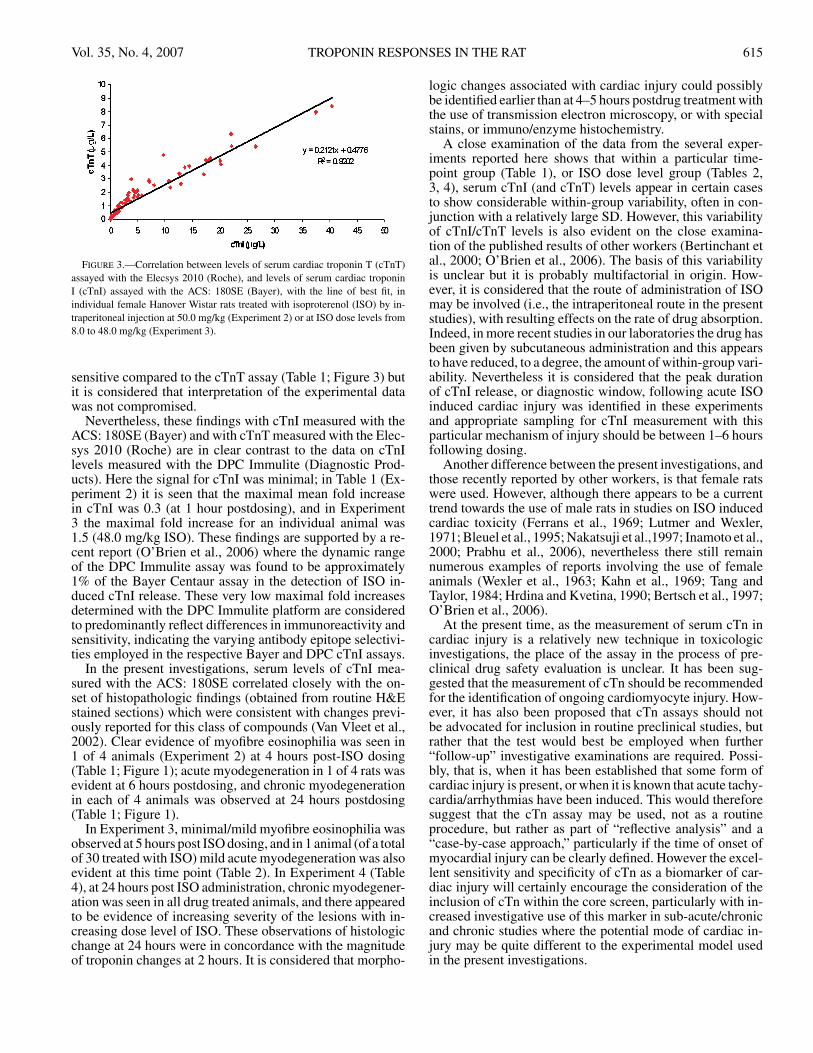
It is seen (Table 1 and 2) that the magnitude of the increasein cTnT in the serum of ISO treated rats, above the base-line control values, is slightly greater than the magnitude ofchange in serum cTnI above baseline control values. For ex-ample, in Table 1, the maximum mean fold increase at 2 hourspostdosing was 142.2 for cTnI and 178.0 for cTnT; in Table2, the maximum mean fold increase at 5 hours postdosingwas 111.9 for cTnI and 172.0 for cTnT with maximum foldincreases for individual animals (data not shown) of 314 forcTnI and 474 for cTnT. However, in overall terms it is con-sidered that the sensitivity of both biomarkers appears to beapproximately similar in this particular experimental model.Furthermore, when serum samples from individual animalsin Experiments 1, 2, and 3 were assayed for both cTnI andcTnT (n = 83), and the data re-analyzed and plotted with theline of best fit (Figure 3), cTnI and cTnT levels were shown tobe closely correlated (R2 = 0.9202). This result would there-fore indicate that either serum cTnI, or serum cTnT can beused as a suitable biomarker of ISO induced cardiac injuryin the rat. However at cTnI levels below 0.3 µg/L with theACS: 180SE, the precision of the assay was considered less
Vol. 35, No. 4, 2007 TROPONIN RESPONSES IN THE RAT 615
FIGURE 3.—Correlation between levels of serum cardiac troponin T (cTnT)assayed with the Elecsys 2010 (Roche), and levels of serum cardiac troponinI (cTnI) assayed with the ACS: 180SE (Bayer), with the line of best fit, inindividual female Hanover Wistar rats treated with isoproterenol (ISO) by in-traperitoneal injection at 50.0 mg/kg (Experiment 2) or at ISO dose levels from8.0 to 48.0 mg/kg (Experiment 3).
sensitive compared to the cTnT assay (Table 1; Figure 3) butit is considered that interpretation of the experimental datawas not compromised.
Nevertheless, these findings with cTnI measured with theACS: 180SE (Bayer) and with cTnT measured with the Elec-sys 2010 (Roche) are in clear contrast to the data on cTnIlevels measured with the DPC Immulite (Diagnostic Prod-ucts). Here the signal for cTnI was minimal; in Table 1 (Ex-periment 2) it is seen that the maximal mean fold increasein cTnI was 0.3 (at 1 hour postdosing), and in Experiment3 the maximal fold increase for an individual animal was1.5 (48.0 mg/kg ISO). These findings are supported by a re-cent report (O’Brien et al., 2006) where the dynamic rangeof the DPC Immulite assay was found to be approximately1% of the Bayer Centaur assay in the detection of ISO in-duced cTnI release. These very low maximal fold increasesdetermined with the DPC Immulite platform are consideredto predominantly reflect differences in immunoreactivity andsensitivity, indicating the varying antibody epitope selectivi-ties employed in the respective Bayer and DPC cTnI assays.
In the present investigations, serum levels of cTnI mea-sured with the ACS: 180SE correlated closely with the on-set of histopathologic findings (obtained from routine H&Estained sections) which were consistent with changes previ-ously reported for this class of compounds (Van Vleet et al.,2002). Clear evidence of myofibre eosinophilia was seen in1 of 4 animals (Experiment 2) at 4 hours post-ISO dosing(Table 1; Figure 1); acute myodegeneration in 1 of 4 rats wasevident at 6 hours postdosing, and chronic myodegenerationin each of 4 animals was observed at 24 hours postdosing(Table 1; Figure 1).
In Experiment 3, minimal/mild myofibre eosinophilia wasobserved at 5 hours post ISO dosing, and in 1 animal (of a totalof 30 treated with ISO) mild acute myodegeneration was alsoevident at this time point (Table 2). In Experiment 4 (Table4), at 24 hours post ISO administration, chronic myodegener-ation was seen in all drug treated animals, and there appearedto be evidence of increasing severity of the lesions with in-creasing dose level of ISO. These observations of histologicchange at 24 hours were in concordance with the magnitudeof troponin changes at 2 hours. It is considered that morpho-
logic changes associated with cardiac injury could possiblybe identified earlier than at 4–5 hours postdrug treatment withthe use of transmission electron microscopy, or with specialstains, or immuno/enzyme histochemistry.
A close examination of the data from the several exper-iments reported here shows that within a particular time-point group (Table 1), or ISO dose level group (Tables 2,3, 4), serum cTnI (and cTnT) levels appear in certain casesto show considerable within-group variability, often in con-junction with a relatively large SD. However, this variabilityof cTnI/cTnT levels is also evident on the close examina-tion of the published results of other workers (Bertinchant etal., 2000; O’Brien et al., 2006). The basis of this variabilityis unclear but it is probably multifactorial in origin. How-ever, it is considered that the route of administration of ISOmay be involved (i.e., the intraperitoneal route in the presentstudies), with resulting effects on the rate of drug absorption.Indeed, in more recent studies in our laboratories the drug hasbeen given by subcutaneous administration and this appearsto have reduced, to a degree, the amount of within-group vari-ability. Nevertheless it is considered that the peak durationof cTnI release, or diagnostic window, following acute ISOinduced cardiac injury was identified in these experimentsand appropriate sampling for cTnI measurement with thisparticular mechanism of injury should be between 1–6 hoursfollowing dosing.
Another difference between the present investigations, andthose recently reported by other workers, is that female ratswere used. However, although there appears to be a currenttrend towards the use of male rats in studies on ISO inducedcardiac toxicity (Ferrans et al., 1969; Lutmer and Wexler,1971; Bleuel et al., 1995; Nakatsuji et al.,1997; Inamoto et al.,2000; Prabhu et al., 2006), nevertheless there still remainnumerous examples of reports involving the use of femaleanimals (Wexler et al., 1963; Kahn et al., 1969; Tang andTaylor, 1984; Hrdina and Kvetina, 1990; Bertsch et al., 1997;O’Brien et al., 2006).
At the present time, as the measurement of serum cTn incardiac injury is a relatively new technique in toxicologicinvestigations, the place of the assay in the process of pre-clinical drug safety evaluation is unclear. It has been sug-gested that the measurement of cTn should be recommendedfor the identification of ongoing cardiomyocyte injury. How-ever, it has also been proposed that cTn assays should notbe advocated for inclusion in routine preclinical studies, butrather that the test would best be employed when further“follow-up” investigative examinations are required. Possi-bly, that is, when it has been established that some form ofcardiac injury is present, or when it is known that acute tachy-cardia/arrhythmias have been induced. This would thereforesuggest that the cTn assay may be used, not as a routineprocedure, but rather as part of “reflective analysis” and a“case-by-case approach,” particularly if the time of onset ofmyocardial injury can be clearly defined. However the excel-lent sensitivity and specificity of cTn as a biomarker of car-diac injury will certainly encourage the consideration of theinclusion of cTn within the core screen, particularly with in-creased investigative use of this marker in sub-acute/chronicand chronic studies where the potential mode of cardiac in-jury may be quite different to the experimental model usedin the present investigations.

616 YORK ET AL. TOXICOLOGIC PATHOLOGY
A recent search of the literature has demonstrated that instudies involving cTn, the compound of frequent choice forthe induction of cardiac lesions is ISO; also it would appearthat ISO-induced changes in the heart are beginning to becharacterized and reported in the literature, and related toserum levels of cTn. Nevertheless, this highlights the situa-tion that in other types of cardiac injury, induced with othermodel compounds, cTn responses have not been well de-scribed and may have prolonged diagnostic windows for op-timal measurement in comparison with ISO induced cardiacinjury. This has become clear in studies on cardiac biomarkerevaluation in our own laboratories where attention has nowturned to examine different models of experimental injurywith a series of other cardiotoxic agents.
In conclusion, and in agreement with the reports of Wallaceet al. (2004), O’Brien et al. (2006) and Walker (2006), it isconsidered that in the present studies, cTn was confirmed asbeing a specific and sensitive biomarker for the detection ofmyocardial damage in the rat. On injury to the heart, cTnI israpidly released, and in serum, cTnI is a robust marker andshows good stability in storage at 4◦C, −20◦C and −80◦C.The duration of release of cTnI in the blood is also adequatelylong in acute injury. Increased levels of serum cTnI showan association with the development of cardiac lesions andprecede maximal lesion severity in acute models of cardiacinjury. Assays of cTnI in serum are rapid, specific, simple, ac-curate, relatively inexpensive, and easy to perform, althoughthere is some evidence that technical improvements in theassay are required at low serum concentrations. The useful-ness of cTnI as a biomarker is enhanced as it is not expressedin nontarget tissues and also because cTn bridges betweenpreclinical and clinical studies.
ACKNOWLEDGMENTS
We gratefully acknowledge the assistance of the technicalstaff at the School of Pharmacy for care of the animals andthe technical support of the Clinical Pathology and Histologygroups of GlaxoSmithKline, UK and the Clinical PathologyLaboratories of AstraZeneca, UK. SB acknowledges the sup-port of GlaxoSmithKline, UK, and the School of Pharmacy.
REFERENCES
Adamcova, M., Sterba, M., Simunek, T., Potacova, A., Popelova, O., Mazurova,Y., and Gersl, V. (2005). Troponin as a marker of myocardiac damage indrug-induced cardiotoxicity. Expert Opin Drug Saf 4, 457–72.
Apple, F. S., Wu, A. H. B., Mair, J., Ravkilde, J., Panteghini, M., Tate, J.,Pagani, F., Christenson, R. H., Mockel, M., Danne, O., and Jaffe, A. S.(2005). Future biomarkers for detection of ischemia and risk stratificationin acute coronary syndromes. Clin Chem 51, 810–24.
Benjamin, I. J., Jalil, J. E., Tan, L. B., Cho, K., Weber, K. T., and Clark, W. A.(1989). Isoproterenol-induced myocardial fibrosis in relation to myocytenecrosis. Circ Res 65, 657–70.
Bertinchant, J. P., Robert, E., Polge, A., Marty-Double, C., Fabbro-Peray, P.,Poirey, S., Aya, G., Juan, J. M., Ledermann, B., de la Coussaye, J. E.,and Dauzat, M. (2000). Comparison of the diagnostic value of cardiactroponin I and T determinations for detecting early myocardial damageand the relationship with histological findings after isoprenaline-inducedcardiac injury in rats. Clin Chim Acta 298, 13–28.
Bertsch, T., Bleuel, H., Aufenanger, J., and Rebel, W. (1997). Comparison ofcardiac troponin T and cardiac troponin I concentrations in peripheralblood during orciprenaline induced tachycardia in rats. Exp Toxic Pathol49, 467–8.
Bertsch, T., Bleuel, H., Deschl, U., and Rebel, W. (1999). A new sensitivecardiac troponin T rapid test (TROPT) for the detection of experimentalacute myocardial damage in rats. Exp Toxic Pathol 51, 565–9.
Bleuel, H., Deschl, U., Bertsch, T., Bolz, G., and Rebel, W. (1995). Diagnosticefficiency of troponin T measurements in rats with experimental myocar-dial cell damage. Exp Toxic Pathol 47, 121–7.
Brady, S., Jamalfar, S., York, M., Scudamore, C., Roman, I., Stamp, C., Swain,A., Williams, T., Griffiths, W., Patterson, L., and Turton, J. (2005). Car-diotoxicity of isoproterenol and levels of serum cardiac troponin I in theHan Wistar rat: a threshold dose response study. Toxicology 213, 244.
British National Formulary (BNF). (2006). British Medical Association and theRoyal Pharmaceutical Society of Great Britain, London.
Chen, C., Turton, J., Evans, G., Scudamore, C., Whayman, M., Williams, T.,and York, M. (2004). Characterisation of cardiac troponin I release andanalytical methods following isoprenaline-induced acute cardiomyocyteinjury in the rat. Toxicology 202, 87–8.
Christenson, R. H., and Azzazy, H. M. E. (1998). Biochemical markers of theacute coronary syndromes. Clin Chem 44, 1855–64.
Dollery, C. (1998). Therapeutic Drugs. Churchill Livingstone, Edinburgh.Dybdahl, B., Slørdahl, S. A., Waage, A., Kierulf, P., Espevik, T., and Sundan,
A. (2005). Myocardial ischaemia and the inflammatory response: releaseof heat shock protein 70 after myocardial infarction. Heart 91, 299–304.
Ferrans, V. J., Hibbs, R. G., Walsh, J. J., and Burch, G. E. (1969). Histochemicaland electron microscopical studies on the cardiac necroses produced bysympathomimetic agents. Ann N Y Acad Sci 156, 309–32.
Gryglewski, R. J., Kulig, A., and Kostka-Trabka, E. (1971). Myocardial necrosesproduced by intravenous infusion into rats. J Pharm Pharmacol 23, 926–30.
Handforth, C. P. (1962). Isoproterenol-induced myocardial infarction in animals.Arch Pathol 73, 161–5.
Herman, E. H., Lipshultz, S. E., Rifai, N., Zhang, J., Papoian, T., Yu, Z.-X.,Takeda, K., and Ferrans, V. J. (1998). Use of cardiac troponin T levels asan indicator of doxorubicin-induced cardiotoxicity. Cancer Res 58, 195–7.
Home Office (1989). Code of practice for the housing and care of animals usedin scientific procedures. Her Majesty’s Stationery Office, London.
Hrdina, R., and Kvetina, J. (1990). Changes in cardiac output, hepatic and renalfunctions in rats with isoprenaline-induced heart damage. Cor Vasa 32,149–56.
Inamoto, S., Murao, S., Yokoyama, M., Kitazawa, S., and Maeda, S. (2000).Isoproterenol-induced myocardial injury resulting in altered S100A4 andS100A11 protein expression in the rat. Pathol Int 50, 480–5.
Isaacs, K. R. (1998). The cardiovascular system. In: Target Organ Pathology: ABasic Text (J. Turton and J. Hooson, eds.), pp. 141–76. Taylor & Francis,London.
Judd, J. T., and Wexler, B. C. (1974). Myocardial glycoprotein changes withisoproterenol-induced necrosis and repair in the rat. Am J Pathol 226,597–602.
Kahn, D. S., Rona, G., and Chappel, C. I. (1969). Cardiac pathology induced byisoproterenol and related amines. Ann N Y Acad Sci 156, 285–93.
Ladi, R. N., Hollaar, L., Souverijn, J. H., and van der Laarse, A. (1990). Quan-titation of cumulative release of lactate dehydrogenase isoenzyme-1 inplasma of patients with acute myocardial infarction using a commerciallyavailable test. Clin Physiol Biochem 8, 250–5.
Leszkovszky, G. P., and Gal, G. (1967). Observations on isoprenaline-inducedmyocardial necroses. J Pharm Pharmacol 19, 226–30.
Lutmer, R. F., and Wexler, B. C. (1971). Myocardial and serum lactatechanges during isoproterenol-induced infarction. Am Heart J 81, 516–20.
Mair, J. (1997). Progress in myocardial damage detection: new biochemicalmarkers for clinicians. Crit Rev Clin Lab Sci 34, 1–66.
Mohan, P., and Bloom, S. (1999). Lipolysis is an important determinant ofisoproterenol-induced myocardial necrosis. Cardiovasc Pathol 8, 255–61.
Muller-Bardoff, M., Hallermayer, K., Schorder, A., Ebert, C., Botgya, A.,Gerhadt, G., Remppid, A., Zehelin, J., and Katus, H. (1997). Improvedtroponin T ELISA specific for cardiac troponin T isoform: assay de-velopment and analytical and clinical validation. Clin Chem 43, 458–66.
Vol. 35, No. 4, 2007 TROPONIN RESPONSES IN THE RAT 617
Nakatsuji, S., Yamate, J., Kuwamura, M., Kotani, T., and Sakuma, S. (1997).In vivo responses of macrophages and fibroblasts in the healing followingisoproterenol-induced myocardial injury in rats. Virchows Arch 430, 63–69.
O’Brien, P. J., Smith, D. E. C., Knechtel, T. J., Marchak, M. A.,Pruimboom-Brees, I., Brees, D. J., Spratt, D. P., Archer, F. J., But-ler, P., Potter, A. N., Provost, J. P., Richard, J., Snyder, P. A., andReagan, W. J. (2006). Cardiac troponin I is a sensitive, specificbiomarker of cardiac injury in laboratory animals. Lab Anim 40, 153–71.
Panteghini, M. (2004). Role and importance of biochemical markers in clinicalcardiology. Eur Heart J 25, 1187–96.
Prabhu, S., Jainu, M., Sabitha, K. E., and Shyamala Devi, C. S. (2006). Ef-fect of mangiferin on mitochondrial energy production in experimen-tally induced myocardial infarcted rats. Vascul Pharmacol 44, 519–25.
Preus, M., Bhargava, A. S., Khater, A. E., and Gunzel, P. (1988). Diagnosticvalue of serum creatine kinase and lactate dehydrogenase isoenzyme de-terminations for monitoring early cardiac damage in rats. Toxicol Lett 42,225–33.
Preus, M., Karsten, B., and Bhargava, A. S. (1989). Serum isoenzyme patternof creatine kinase and lactate dehydrogenase in various animal species. JClin Chem Clin Biochem 27, 787–90.
Rona, G., Chappel, C.I., Balazs, T., and Gaudry R. (1959). An infarct-like my-ocardial lesion and other toxic manifestations produced by isoproterenolin the rat. Arch Pathol 67, 443–55.
Scharnhost, V., Vader H. L., and van der Graaf, F. (2002). Characteristics of thecardiac troponin I assay on the Immulite 2000 analyser. Clin Chem 48,1626–7.
Sears, M. R., and Lotvall, J. (2005). Past, present and future: β2-adrenoceptoragonists in asthma management. Respir Med 99, 152–70.
Tang, Q., and Taylor, P. B. (1984). Development of isoproterenol-induced cardiachypertrophy. Can J Physiol Pharmacol 62, 384–9.
Van Vleet, D. F., Ferrans, V. J., and Herman, E. (2002). Cardiovascular andskeletal muscle systems. In: Handbook of Toxicologic Pathology, Vol 2,2nd edition (W. M. Haschek, C. G. Rousseaux, and M. A. Wallig, eds.),pp. 373–95. Academic Press, San Diego, California.
Waldeck, B. (2002). β-Adrenoceptor agonists and asthma: 100 years of devel-opment. Eur J Pharmacol 445, 1–12.
Walker, D. B. (2006). Serum chemical biomarkers of cardiac injury for nonclin-ical safety testing. Toxicol Pathol 34, 94–104.
Wallace, K. B., Hausner, E., Herman, E., Holt, G. D., MacGregor, J. T., Metz,A. L., Murphy, E., Rosenblum, I. Y., Sistare, F. D., and York, M. J. (2004).Serum troponins as biomarkers of drug-induced cardiac toxicity. ToxicolPathol 32, 106–21.
Wexler, B. C., and Kittinger, G. W. (1963). Myocardial necrosis in rats: serumenzymes, adrenal steroid and histopathological alterations. Circ Res 13,159–71.
Wolf, R. E., Graeber, G. M., Burge, J. R., DeShong, J. L., MacDonald, J. L.,and Zajtchuk, R. (1986). Evaluation of serum creatine kinase and lactatedehydrogenase in experimental myocardial infarction, atriotomies, andthoractomies. Ann Thorac Surg 41, 378–86.