The maximum spacing estimation for multivariate observations

Jou
rnal
of P
hysi
olog
y
A well-studied mechanism by which myocardial function
is tuned to the haemodynamic load is through b-adrenoceptor
stimulation and subsequent activation of cAMP-dependent
protein kinase (PKA). Signalling through this pathway is
associated with increased rates of relaxation and
contraction in the myocardium (Ross & Sobel, 1972; Luo
et al. 1994) and is believed to be predominantly due to
phosphorylation of phospholamban by PKA leading to
enhanced reuptake of Ca2+ by the sarcoplasmic reticulum
(Kranias, 1985; Luo et al. 1994; Solaro & Van Eyk, 1996;
Wolska et al. 1996). PKA additionally targets contractile
proteins, specifically troponin I (TnI) and myosin binding
protein C (MyBP-C), and results in a decrease in myo-
filament Ca2+ sensitivity (Solaro et al. 1976; Garvey et al.1988). It has been suggested that phosphorylation of
myofilament proteins may affect cardiac contractility
independently of the alterations in the dynamics of Ca2+
handling (Kentish et al. 2001). Although a regulatory role
for MyBP-C cannot be altogether eliminated in the light of
an emerging literature (Kunst et al. 2000; Winegrad, 2000),
there is good evidence to suggest that the PKA effect in the
myocardium is due to phosphorylation of TnI and not
MyBP-C (Fentzke et al. 1999; Kentish et al. 2001).
Other studies have shown that replacement of cardiac TnI
(cTnI) with the slow skeletal isoform (ssTnI) in the heart can
alter contractile properties of heart muscle by increasing Ca2+
sensitivity (Fentzke et al. 1999) and reducing the response to
changes in muscle length (Arteaga et al. 2000). The TnI
isoforms differ by the presence of a unique 32 amino acid
extension at the N-terminus of cTnI. Since this extension
contains the two targets for PKA, ssTnI cannot be
phosphorylated by PKA (Fentzke et al. 1999; Kentish et al.2001). Though the specific role of this N-terminus has not
Troponin I in the murine myocardium: influence on length-dependent activation and interfilament spacingJohn P. Konhilas, Thomas C. Irving*, Beata M. Wolska†, Eias E. Jweied, Anne F. Martin, R. John Solaroand Pieter P. de Tombe†
Program in Cardiovascular Sciences, Department of Physiology and Biophysics, †Section of Cardiology, University of Illinois at Chicago, College ofMedicine, Chicago, IL 60612 and *CSRRI and Department of BCPS, Illinois Institute of Technology, Chicago, IL 60616, USA
Cyclic AMP-dependent protein kinase (PKA) targets contractile proteins, troponin-I (TnI) and
myosin binding protein C (MyBP-C) in the heart and induces a decrease in myofilament Ca2+
sensitivity. Yet, the effect of sarcomere length (SL) change on Ca2+ sensitivity (length-dependent
activation: LDA) following PKA-dependent phosphorylation is not clear. To clarify the role of PKA-
dependent phosphorylation of TnI and MyBP-C on LDA in the heart, we examined LDA in skinned
myocytes from a non-transgenic (NTG) and a transgenic murine model in which the native cardiac
isoform (cTnI) was completely replaced by the slow skeletal isoform of TnI (ssTnI-TG) lacking the
phosphorylation sites for PKA, while retaining PKA sites on MyBP-C. In NTG myocytes, PKA
treatment decreased Ca2+ sensitivity at each SL, but enhanced the impact of SL change on Ca2+
sensitivity. Despite a greater sensitivity to Ca2+ and a reduction in LDA, neither Ca2+ responsiveness
nor LDA was affected by PKA treatment in ssTnI-TG myocytes. To determine whether the above
observations could be explained by the lateral separation between thick and thin filaments, as
suggested by others, we measured interfilament spacing by X-ray diffraction as a function of SL in
skinned cardiac trabeculae in the passive state from both NTG and ssTnI-TG models before and
following treatment with PKA. Phosphorylation by PKA increased lattice spacing at every SL in
NTG trabeculae. However, the relationship between SL and myofilament lattice spacing in ssTnI-
TG was markedly shifted downward to an overall decreased myofilament lattice spacing following
PKA treatment. We conclude: (1) PKA-dependent phosphorylation enhances length-dependent
activation in NTG hearts; (2) replacement of native TnI with ssTnI increases Ca2+ sensitivity of
tension but reduces length-dependent activation; (3) MyBP-C phosphorylation by PKA does not
alter calcium responsiveness and induces a decrease in myofilament lattice spacing at all sarcomere
lengths and (4) length-dependent activation in the heart cannot be entirely explained by alterations
in myofilament lattice spacing.
(Resubmitted 18 December 2002; accepted after revision 20 December 2002; first published online 24 January 2003)
Corresponding author P. P. de Tombe: Department of Physiology and Biophysics (M/C 902), College of Medicine, University ofIllinois at Chicago, 900 South Ashland Avenue, Chicago, IL 60607-7171, USA. Email: [email protected]
J Physiol (2003), 547.3, pp. 951–961 DOI: 10.1113/jphysiol.2002.038117
© The Physiological Society 2003 www.jphysiol.org

Jou
rnal
of P
hysi
olog
y
fully been elucidated, these latter studies implicate TnI as an
important modulator of both Ca2+ sensitivity and length-
dependent activation, a phenomenon believed to be the
cellular mechanism for the Frank-Starling law of the heart
(ter Keurs et al. 1980).
The increase in Ca2+ sensitivity upon an increase in
sarcomere length may be due to a decrease in interfilament
spacing with an increase in length (Rome, 1968) thereby
influencing cross-bridge reactivity (Godt & Maughan,
1981; McDonald & Moss, 1995; Fuchs & Wang, 1996).
That is, cross-bridges may be more likely to enter into a
strong binding state at a given level of activator Ca2+ when
interfilament spacing is reduced. The correlation between
interfilament spacing, alterations in Ca2+ sensitivity, and
length-dependent activation following TnI phosphorylation
or TnI isoform composition are not known. Given the
significance of sarcomere length, TnI isoform composition
and TnI phosphorylation status on myocardial contraction,
several questions arise. (1) Does PKA-mediated phos-
phorylation of cTnI affect the sarcomere length dependency
of myofilament activation, and if so, is this effect due to the
phosphorylation of cTnI or MyBP-C? (2) What is the role
of interfilament spacing in determining myofilament Ca2+
sensitivity and length-dependent myofilament activation
under these conditions?
In experiments reported here, we determined the effect of
PKA-mediated phosphorylation on sarcomere length-
(SL)-dependent activation in skinned single cardiac myocytes
obtained from either non-transgenic mice or transgenic
mice in which cTnI was completely replaced by ssTnI in
the heart. In addition, interfilament spacing was measured
over a wide range of sarcomere lengths in skinned cardiac
trabeculae using synchrotron X-ray diffraction. We found
that: (1) PKA-mediated phosphorylation induces an increase
in SL-dependent activation; (2) this effect is due to
phosphorylation of cTnI and not MyBP-C; and
(3) alterations in Ca2+ sensitivity and SL-dependent activation
upon PKA-mediated myofilament phosphorylation or
TnI isoform composition cannot be predicted, solely, by
alterations in interfilament spacing.
METHODSMyocyte preparationAll experiments were performed according to institutionalguidelines concerning the care and use of experimental animals.Mice (NTG and ssTnI-TG; 20–30 g) were anaesthetized (sodiumpentobarbital 50 mg (kg body weight)_1
I.P. injection) and thehearts were rapidly excised and retrogradely perfused with amodified Krebs-Henseleit (K-H) solution (de Windt et al. 1999)(mmol l_1): NaCl 118.5, KCl 5, MgSO4 1.2, NaH2PO4 2, ethylene-diaminetetraacetic acid, EDTA 0.5, D(+)-glucose 10, NaHCO3 25,pyruvic acid 1.5, CaCl2 3.0. In addition, insulin (5 u l_1) andpropranolol (5 mmol l_1), to block non-specific b-adrenergicactivation, and carbamyl choline (10 mmol l_1), to enhancephosphatase activity (Gupta et al. 1994) were added to the
perfusate. 3-Butanedione monoxime (20 mmol l_1) was alsoadded to the K-H solution to inhibit contraction, presumablythrough the energetic stabilization of the unattached state of themyosin molecule (Backx et al. 1994). Skinned myocytes ormyocyte fragments were obtained by mechanical isolation asdescribed previously (Fan et al. 1997; Fentzke et al. 1999). Briefly,during perfusion with the K-H solution, a section of the leftventricle was minced into 1–2 mm pieces. Next, the pieces weremechanically disrupted at high speed for 15–20 s (Power Gen700D; Fisher Scientific) in ice-cold standard relaxing solution(mmol l_1): Na+-ATP 4, MgCl2 1, ethylene glycol-bis(b-aminoethyl ether)-N,N,N‚,N‚-tetraacetic acid (EGTA) 2,KCl 100, imidazole 10. The homogenized tissue was thencentrifuged at 800 r.p.m. (Marathon 22 Kbr; Fisher Scientific) for1 min at 4 °C. The supernatant was discarded and the tissue wasresuspended in standard relaxing solution containing 0.3 %Triton X-100 (Pierce Chemical) for 6 min to remove thesarcolemma and all remaining membranous structures. Afterwashing twice with the relaxing solution, the cells and cellfragments were resuspended in standard relaxing solution andstored on ice for < 10 h before data collection.
Experimental apparatus and protocolThe experiments were performed on the stage of an invertedmicroscope (Olympus) that allowed visualization of the cell or cellfragments. The cell was attached at either end to a sensitive forcetransducer (Cambridge model 403A; ~300 Hz resonantfrequency) and a high-speed servomotor (Cambridge model 308;~1 ms 90 % step response) with stainless-steel minutien tips andsilicone rubber. The sarcomere length was measured by video-microscopy; each horizontal pixel line was transformed by fastFourier transformation (FFT) into a spatial frequency domainand converted into median sarcomere length across the region(Fan et al. 1997). Cell length was adjusted by using the servomotor,which was controlled via computer (Apple PowerPC) usingcustom-designed software (LabVIEW, National Instruments;Austin, TX, USA). Both minutien tips were connected to X-Y-Zmanipulators (Newport) permitting accurate positioning of theattached cell. Cell length and width were measured by video-microscopy. Myocyte thickness was measured by placing a smallmirror under a 45 deg angle close to the cell by using a three-dimensional manipulator. The myocyte was suspended above amovable stage that contained several solution wells (temperaturecontrolled at 15°C). Rapid solution changes were made bytranslating the stage laterally via stage manipulators such thatanother solution-containing well was brought underneath the cell(Fan et al. 1997). The myofilament Ca2+ sensitivity of tension wascharacterized at two sarcomere lengths (2.25 and 1.95 mm) bymeasuring steady-state tension development followed by a rapidslackening to assess total tension. Active tension at each [Ca2+] wasthe difference between total tension and relaxed, passive tension(passive tension was not measurable at the short sarcomere lengthand was minimal at the long sarcomere length). Cells that did notmaintain 80 % of initial maximal tension or a visible striationpattern were discarded. In addition, if the extent of internalshortening due to end-compliance in the preparation wasextensive (>100 nm) the data were also discarded.
The ionic strength of the solutions was kept at 180 mmol l_1 byadding the appropriate amount of potassium propionate. Inaddition, all solutions contained the following (mmol l_1):phosphocreatine 10, N,N-bis[2-hydroxyethyl]-2-aminoethane-sulphonic acid (BES) 100, leupeptin 0.1, phenylmethylsulphonylfluoride (PMSF) 0.1, dithiothreitol (DTT) 1 and 4 U ml l_1 creatine
J. P. Konhilas and others952 J Physiol 547.3
Jou
rnal
of P
hysi
olog
y
phosphokinase. The free Mg2+ and Mg-ATP concentration wascalculated at 1 and 5 mmol l_1, respectively. To achieve a range offree [Ca2+], activating and relaxing solutions were appropriatelymixed with an apparent stability constant of the Ca2+-ethyleneglycol-bis(b-aminoethyl ether)-N,N,N‚,N‚-tetraacetic acid (EGTA)complex of 106.39 assumed. The pH was adjusted to 7.0 at 15.0 °Cwith KOH. Force was stored on computer via an A/D converterusing the custom software for off-line analysis (10 kHz perchannel sampling frequency).
X-ray diffraction experimentsThe overall experimental arrangement, sample preparation andprotocol have been described in detail previously (Irving et al.2000). Briefly, experiments were performed on the BioCATundulator-based beamline at the Advanced Photon Source,Argonne National Laboratory. Following rapid excision, the heartwas perfused with the identical K-H solution as described above.Trabeculae were dissected from the right ventricular free wall inboth NTG and ssTnI-TG mice and demembranated overnightwith 1 % Triton X-100 in standard relaxing solution. Preliminaryexperiments indicated robust PKA-dependent phosphorylationin cardiac trabeculae prepared in this manner (data not shown).For the X-ray studies, trabeculae were mounted between a forcetransducer and a servo-motor in a small trough, which allowedsimultaneous collection of the X-ray patterns and viewing of thestriation pattern using a long working distance objective (w 40) ofan inverted microscope equipped with a CCD video camera. Low-angle X-ray diffraction patterns were collected on a CCD-basedX-ray detector. Spacings between the 1,0 and 1,1 equatorialreflections in the diffraction pattern were converted to d10 latticespacings using Bragg’s law.
Individual trabeculae were stretched to arbitrary sarcomere lengthsbetween 1.9 and 2.4 mm at the beginning of the experiment.Sarcomere lengths were determined from the video-image asdescribed previously (Irving et al. 2000) and checked both beforeand immediately after the X-ray exposure. Fibre length was thensystematically lengthened or shortened to collect at least 10–12 (atleast every 50 nm) consecutive data points describing therelationship between sarcomere length and interfilament latticespacing. Next, an additional 2–4 data points were collected at varioussarcomere lengths back along the curve to check for reproducibilityor hysteresis. The level of passive tension in the lattice was recordedwhen the X-ray exposure was taken.
Data analysisEach individual Ca2+–force relationship was fitted to a modifiedHill equation:
Frel = [Ca2+]nH/(EC50nH + [Ca2+]nH),
where Frel is the force relative to maximum Ca2+ saturated force,EC50 is the [Ca2+] at which force is half-maximal and nH is the slopeof the Ca2+–force relationship (Hill coefficient). The DEC50 wascalculated as the difference in EC50 at SL = 1.95 and 2.25 mm foreach experiment. Likewise, the DLS was calculated as the differencein LS calculated at SL = 1.95 and 2.25 mm for each experiment.Separation between the symmetrical equatorial 1,0 and 1,1 X-raydiffraction reflections was measured directly from detectorimages using the program FIT2D (Hammersley, 1998) on a UNIXworkstation and converted to d10 lattice spacing using Bragg’s law,which can then, in turn, be converted to the inter-thick filamentspacing by multiplying d10 by 2/«3. The differences among EC50,DEC50, LS and DLS for each group at each SL were analysed with aone-way ANOVA Student’s t test with a post hoc Bonferronicorrection to assess differences among mean values. All data areshown as means ± S.E.M.
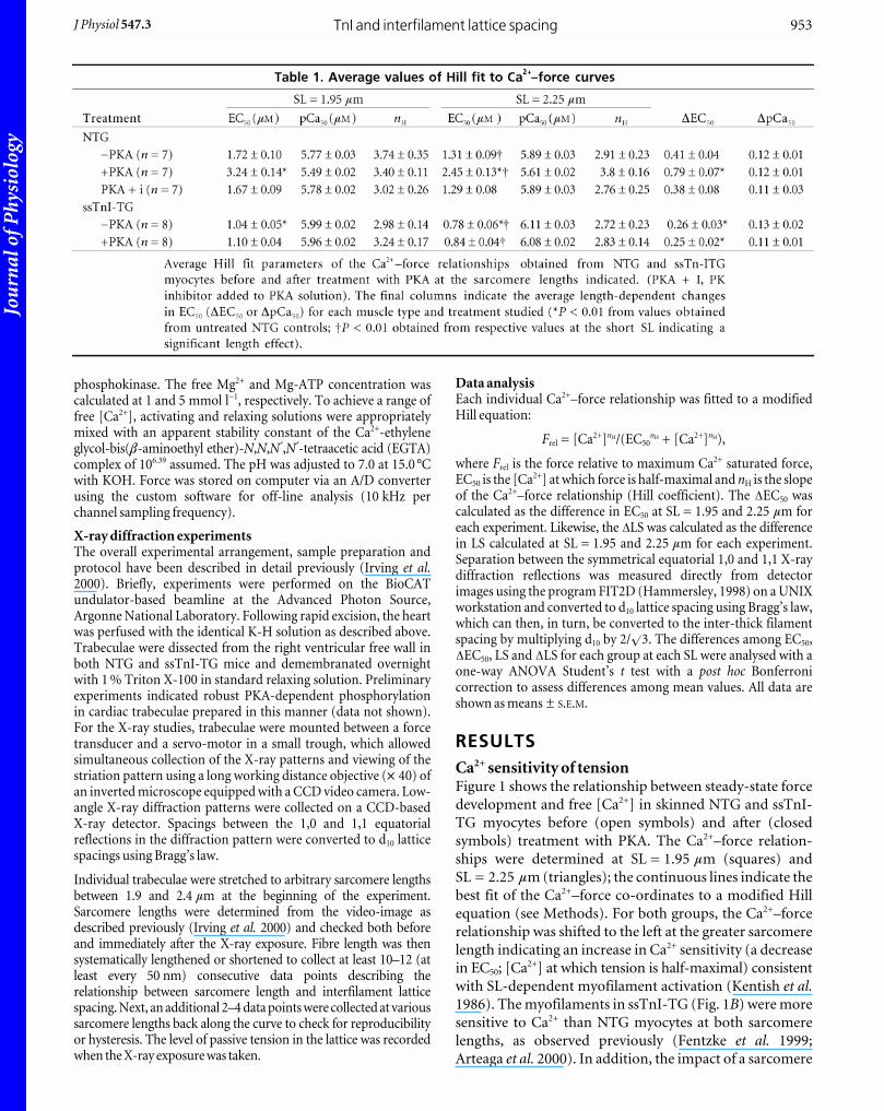
RESULTSCa2+ sensitivity of tensionFigure 1 shows the relationship between steady-state force
development and free [Ca2+] in skinned NTG and ssTnI-
TG myocytes before (open symbols) and after (closed
symbols) treatment with PKA. The Ca2+–force relation-
ships were determined at SL = 1.95 mm (squares) and
SL = 2.25 mm (triangles); the continuous lines indicate the
best fit of the Ca2+–force co-ordinates to a modified Hill
equation (see Methods). For both groups, the Ca2+–force
relationship was shifted to the left at the greater sarcomere
length indicating an increase in Ca2+ sensitivity (a decrease
in EC50; [Ca2+] at which tension is half-maximal) consistent
with SL-dependent myofilament activation (Kentish et al.1986). The myofilaments in ssTnI-TG (Fig. 1B) were more
sensitive to Ca2+ than NTG myocytes at both sarcomere
lengths, as observed previously (Fentzke et al. 1999;
Arteaga et al. 2000). In addition, the impact of a sarcomere
TnI and interfilament lattice spacingJ Physiol 547.3 953
Jou
rnal
of P
hysi
olog
y
length change on Ca2+ sensitivity, as calculated as the
difference in the EC50 at each sarcomere length (DEC50),
was significantly reduced in ssTnI-TG myocytes. Similar
to results reported previously, there was no difference in
maximum tension between NTG and TG preparations at
either SL (Wolska et al. 2001). Earlier studies have employed
pCa50 (_log[Ca2+]) to index Ca2+ sensitivity (Godt &
Maughan, 1981; McDonald & Moss, 1995; Fuchs & Wang,
1996). Employing this method revealed statistically significant
differences in Ca2+ sensitivity between NTG and ssTnI-TG
myocytes. However, length-dependent activation determined
as the difference in pCa50 at the two sarcomere lengths
(DpCa50) indicated no significant differences in length-
dependent activation between NTG and ssTnI-TG
myocytes. The average parameters of the Hill fit from NTG
and ssTnI-TG myocytes are summarized in Table 1.
Figure 1 illustrates the effect of treatment of NTG and
ssTnI-TG myocytes with the catalytic subunit of PKA
(3 mg protein (ml standard relaxing solution)_1; Sigma
Chemicals). As described in Methods, muscle preparation
was designed to prevent additional, non-specific adrenergic
phosphorylation or to enhance phosphatase activity
resulting in minimal phosphorylation of TnI and MyBP-C.
This was supported by robust 32P incorporation (data not
shown) by TnI and MyBP-C in NTG myocardium and
MyBP-C in ssTnI-TG preparations, as we have previously
shown using this transgenic murine model (Fentzke et al.1999; Kentish et al. 2001). Consistent with our previous
studies (Solaro, 1995; Janssen & de Tombe, 1997), treatment
of the NTG skinned myocardium with PKA induced a
rightward shift of the Ca2+–force relationship at both
sarcomere lengths, as indicated by an increase in the EC50
(Fig. 1A). In addition, the impact of a sarcomere length
change (DEC50) on the Ca2+–force relationship was
significantly enhanced following PKA treatment, indicating
an increase in length-dependent activation properties
following PKA treatment. Again, using the pCa50 parameter,
there was a significant decrease in Ca2+ sensitivity following
PKA treatment, yet the effect of length change on pCa50
(DpCa50) was the same before and following PKA treatment
(Table 1). If a protein kinase inhibitor was added to the
solution (63 mg protein (ml standard relaxing solution)_1;
Sigma Chemicals), the effect of PKA was completely
abolished independent of parameter choice (Fig. 1C).
Furthermore, PKA did not alter maximum tension at either
SL, as reported previously (Janssen & de Tombe, 1997).
PKA targets two serine residues (22, 23) at the N-terminus of
cTnI, amino acid sites that are not found in ssTnI, and
therefore, in ssTnI-TG hearts TnI is not phosphorylated by
PKA (Fentzke et al. 1999; Kentish et al. 2001). However, PKA
targets an additional thick filament protein, myosin-binding
protein C (MyBP-C; Winegrad, 2000). Treatment of ssTnI-
TG myocytes with PKA did not affect Ca2+ sensitivity of
tension at either sarcomere length (Fig. 1B). Consequently,
J. P. Konhilas and others954 J Physiol 547.3
Figure 1. Ca2+-dependent tension development in cardiacmyocytes from NTG and ssTnI-TG hearts A and B, Ca2+–force relation of NTG (A) and ssTnI-TG (B) cardiacmyocytes before (open symbols) and following (closed symbols)treatment with the catalytic subunit of PKA (3 mg protein (mlstandard relaxing solution)_1) at SL = 2.25 mm (9) andSL = 1.95 mm (ª). C, Ca2+–force relation of NTG cardiac myocytesat two sarcomere lengths before (open symbols) and following(closed symbols) treatment with a PK inhibitor (PKI) added toPKA solution. Lines indicate the Hill fit to the data obtained at theindicated sarcomere length before and following PKA treatment.

Jou
rnal
of P
hysi
olog
y
there was no impact on DEC50, or DpCa50, and thus no effect
on length-dependent activation. These data show that the
PKA effect on Ca2+ sensitivity and length-dependent
activation in NTG hearts can be attributed, solely, to
phosphorylation of TnI and not of MyBP-C. Again, the
average parameters of the Hill fit from NTG and ssTnI-TG
myocytes, both before and after PKA treatment, are
summarized in Table 1.
Relationship between interfilament spacing andsarcomere lengthTo determine the influence of PKA-dependent phos-
phorylation of cTnI and MyBP-C in the heart on inter-
filament spacing, we simultaneously measured myofilament
lattice spacing and sarcomere length in relaxed skinned
cardiac trabeculae of NTG and ssTnI-TG mice before and
after PKA treatment using synchrotron X-ray diffraction
(Irving et al. 2000). Figure 2 illustrates typical X-ray
diffraction patterns obtained in trabeculae at SL = 2.1 mm
from NTG and ssTnI-TG hearts before and after treatment
with PKA. These data show clearly resolved symmetrical
pairs of 1,0 and 1,1 equatorial X-ray reflections. The adjacent
panels illustrate line scans obtained from the 2-D diffraction
patterns. Measurement of the distance between the
symmetrical sets of reflections, indicated by the continuous
vertical lines, allowed lattice spacing to be determined within
TnI and interfilament lattice spacingJ Physiol 547.3 955
Figure 2. Typical X-ray diffraction patterns obtainedin skinned cardiac trabeculae from NTG andssTnI-TG hearts before and following PKAtreatment at SL = 2.10 µmA, CCD images of the diffraction patterns. X-ray diffractionby the myofilament lattice gives rise to two sets ofsymmetrical, equatorial reflections, delineated by thecontinuous vertical lines. B, intensity profiles of the2-dimensional CCD images shown in A. Arrows indicate thepeak intensities, corresponding to the 1,0 and 1,1 reflections.These clearly resolved reflections or intensity peaks allowedlattice spacing to be determined within 0.25 % accuracy,equivalent to a myofilament lattice spacing resolution of0.1 nm.
Figure 3. Average myofilament lattice spacing as a function of sarcomere length in skinnedcardiac trabeculae from NTG and ssTnI-TG hearts (n = 8–10 for each group)The relationships were obtained by averaging the data into 0.05 mm wide sarcomere length bins.A, relationship between lattice spacing and sarcomere length in NTG trabeculae prior to (1) and following(0) treatment with PKA. B, relationship between lattice spacing and sarcomere length in ssTnITG trabeculaeprior to (1) and following (0) treatment with PKA. Each arrow indicates the sarcomere length at which theCa2+–force relationships were determined.
Jou
rnal
of P
hysi
olog
y
0.25 % accuracy, equivalent to a myofilament lattice spacing
resolution of 0.1 nm (Irving et al. 2000). There was an
evident inward displacement of the reflections following
PKA treatment in NTG trabeculae, indicative of an increased
myofilament lattice spacing. In contrast, in ssTnI-TG
trabeculae, PKA treatment increased the distance between
reflections, indicative of a reduction in myofilament lattice
spacing.
As is shown in Fig. 3, myofilament lattice spacing (LS) was
highly dependent on SL, consistent with other and our
previous results in mouse myocardium (Fig. 3) (Konhilas
et al. 2000; Cazorla et al. 2001) and in striated muscle
(Rome, 1968; Bagni et al. 1994; Irving et al. 2000).
Furthermore, there was a parallel increase in LS at every SL
in NTG trabeculae following treatment with PKA (Fig. 3A).
Treatment of ssTnI-TG trabeculae, on the other hand,
induced contraction of the myofilament lattice at all
sarcomere lengths (Fig. 3B). It is also important to note
that in both NTG and ssTnI-TG hearts, PKA-dependent
phosphorylation greatly reduced the experimental variability
among trabeculae, as indicated by a reduction in the
standard error of each binned data point. The implication
may be that, given the effect of phosphorylation status on
LS, there existed a greater variation in basal phos-
phorylation levels under control conditions (untreated
trabeculae). This variation was reduced by PKA treatment,
presumably by maximally phosphorylating myofilament
proteins. Indeed, preliminary back-phosphorylation
experiments aimed at quantifying this variability in basal
phosphorylation status revealed 27 % variation of 32P
incorporation.
To quantify the effects of SL on lattice spacing, lattice spacing
was determined at precisely the same sarcomere lengths at
which the Ca2+ sensitivity of tension was measured
(SL = 1.95 and 2.25 mm). The magnitude of the effect of SL
change on lattice spacing was defined as the difference in the
lattice spacing at these two SLs: DLS. DLS was similar in
untreated NTG (DLS = 1.57 ± 0.15 nm) and ssTnI-TG
(DLS = 1.56 ± 0.24 nm) trabeculae. Furthermore, in NTG
hearts, PKA treatment did not alter the effect of SL change on
lattice spacing, i.e. DLS was similar before and following PKA
treatment (DLS = 1.54 ± 0.12 nm). However, PKA treatment
of ssTnI-TG trabeculae significantly reduced DLS
(DLS = 0.87 ± 0.09 nm) relative to that of untreated ssTnI-
TG hearts.
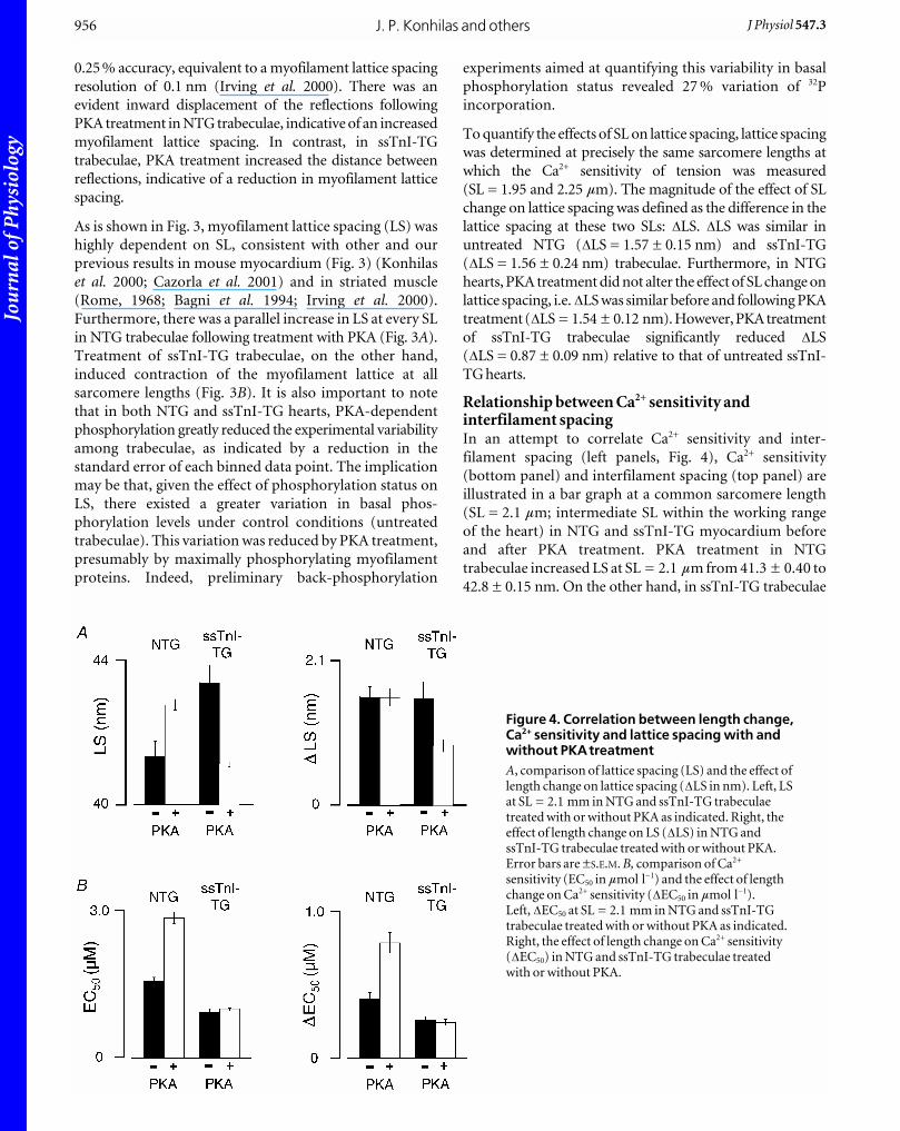
Relationship between Ca2+ sensitivity andinterfilament spacingIn an attempt to correlate Ca2+ sensitivity and inter-
filament spacing (left panels, Fig. 4), Ca2+ sensitivity
(bottom panel) and interfilament spacing (top panel) are
illustrated in a bar graph at a common sarcomere length
(SL = 2.1 mm; intermediate SL within the working range
of the heart) in NTG and ssTnI-TG myocardium before
and after PKA treatment. PKA treatment in NTG
trabeculae increased LS at SL = 2.1 mm from 41.3 ± 0.40 to
42.8 ± 0.15 nm. On the other hand, in ssTnI-TG trabeculae
J. P. Konhilas and others956 J Physiol 547.3
Figure 4. Correlation between length change,Ca2+ sensitivity and lattice spacing with andwithout PKAtreatmentA, comparison of lattice spacing (LS) and the effect oflength change on lattice spacing (DLS in nm). Left, LSat SL = 2.1 mm in NTG and ssTnI-TG trabeculaetreated with or without PKA as indicated. Right, theeffect of length change on LS (DLS) in NTG andssTnI-TG trabeculae treated with or without PKA.Error bars are ±S.E.M. B, comparison of Ca2+
sensitivity (EC50 in mmol l_1) and the effect of lengthchange on Ca2+ sensitivity (DEC50 in mmol l_1).Left, DEC50 at SL = 2.1 mm in NTG and ssTnI-TGtrabeculae treated with or without PKA as indicated.Right, the effect of length change on Ca2+ sensitivity(DEC50) in NTG and ssTnI-TG trabeculae treatedwith or without PKA.
Jou
rnal
of P
hysi
olog
y
PKA treatment induced a reduction in LS from 43.4 ± 0.49
to 41.1 ± 0.07 nm. Whereas PKA treatment significantly
reduced Ca2+ sensitivity in NTG myocytes, PKA treatment
did not affect Ca2+ sensitivity in ssTnI-TG cells. In
addition, the right panels of Fig. 4 show the relative impact
of a change in SL on Ca2+ sensitivity (bottom panel) and
lattice spacing (top panel), as indexed by the DLS andDEC50 parameters before and after treatment with PKA.
Treatment with PKA increased length-dependent activation
in NTG trabeculae (increased DEC50), but did not affect
the relative impact of SL on lattice spacing (DLS).
Treatment of ssTnI-TG skinned trabeculae induced a
reduction in DLS, but did not affect length-dependent
activation (DEC50). If Ca2+ sensitivity and the length-
dependent changes in Ca2+ sensitivity were directly
influenced by changes in interfilament spacing, the relative
changes in each Ca2+ sensitivity parameter should be in
parallel with changes in interfilament spacing. From the
bar graphs in each group, clearly, changes in interfilament
spacing with PKA treatment or with length did not always
correlate with changes in myofilament Ca2+ sensitivity, in
either NTG or ssTnI-TG myocardium.
DISCUSSIONMyofilament activation requires co-operative and
allosteric interactions among bound cross-bridges and
thin filament proteins (Bremel & Weber, 1972; Swartz &
Moss, 1992; Lehrer, 1994; Lehman et al. 2000). TnI has
been shown be an important regulatory component
during the macromolecular rearrangement of myofilament
proteins that results in myofilament force development.
Contractile activation is initiated by binding of Ca2+ to
troponin C (Solaro & Rarick, 1998) inducing translocation
of tropomyosin (Tm) such that potential cross-bridge
binding sites on actin are revealed (Huxley, 1969; Huxley
& Simmons, 1971). Strong cross-bridge binding further
induces a conformational change in Tm into a third
position on actin (Lehman et al. 2000; Tobacman &
Butters, 2000). In this study, we provide further evidence
that TnI plays an integral role in the signalling cascade
during myofilament activation. Furthermore, the central
mechanisms underlying TnI action on cardiac myofilament
activation did not appear to involve interfilament spacing.
Impact of TnI composition or PKA treatment onCa2+ sensitivity.It is well established that phosphorylation of the cardiac
sarcomere mediated by protein kinase A (PKA) induces a
reduction in myofilament Ca2+ sensitivity (Solaro et al.1976; de Tombe & Stienen, 1995; Janssen & de Tombe,
1997), an observation that was confirmed in the present
study. As for the impact of length change on Ca2+-dependent
tension development, previous results have been quite
variable. A study employing intact electrically stimulated
isolated ferret papillary muscle found that treatment with
isoprenaline (an agonist of the b-adrenergic pathway)
induced an increase in the muscle length dependency of
the intracellular [Ca2+]–tension relationship (Komukai &
Kurihara, 1997). Others have found either no alteration
(van der Velden et al. 2000) or a decrease (Kajiwara et al.2000) in length-dependent activation following PKA
treatment. In the present study, the impact of sarcomere
length on myofilament Ca2+ sensitivity following treatment
with PKA depended on the choice of indexing parameter.
Using the EC50 as the measure of Ca2+ sensitivity, length-
dependent activation, calculated as the difference in EC50
at the two sarcomere lengths, was enhanced following
PKA treatment. However, length-dependent activation
determined when Ca2+ sensitivity was indexed by pCa50
remained unaffected.
As indicated by our study, the choice of parameter to index
myofilament Ca2+ sensitivity could potentially affect data
interpretation. As it stands, the extent of length dependency
has a direct impact on the shape of the length–isometric
tension relationship in skinned cardiac fibres and the
shape of the end-systolic sarcomere length–tension
relationship in intact cardiac fibres (Kentish et al. 1986).
Thus, at a given inotropic state, i.e. a single Ca2+
concentration, the magnitude of tension development
depends on sarcomere length. Similarly, at a given
activation level (Ca2+ concentration), PKA treatment
augments isometric tension development following a
length change to a greater extent than in muscle fibres in
the unphosphorylated state. Since tension is measured at a
single concentration, this effect is independent of
parameter choice. By reasoning, the use of EC50 more
closely illustrates the physiological impact of PKA-
dependent phosphorylation on length-dependent activation.
It should be noted that the relation between pCa
(_log[Ca2+]) and [Ca2+] is non-linear. For these reasons,
we believe that the use of the EC50 parameter to index
myofilament Ca2+ sensitivity is more appropriate than
pCa50. Nevertheless, whether other factors, besides choice
of Ca2+ sensitivity parameter, play a role in determining the
impact of PKA on length dependency of myofilament
activation awaits further study.
The length effect notwithstanding, our present study
shows that replacement of cardiac TnI (cTnI) with the
slow skeletal isoform (ssTnI) in the heart can impart
contractile properties of slow skeletal muscle (Konhilas etal. 2002a), consistent with previous reports from our
laboratory (Fentzke et al. 1999; Arteaga et al. 2000). The
predominant difference between these two isoforms is the
presence of a unique 32 amino acid N-terminus extension
in cTnI that contains target sites for PKA-dependent
phosphorylation (Solaro & Rarick, 1998). Given that PKA
targets both cTnI and cardiac MyBP-C and that PKA
treatment did not affect either Ca2+ sensitivity or
sarcomere length dependency of Ca2+ in the ssTnI
TnI and interfilament lattice spacingJ Physiol 547.3 957
Jou
rnal
of P
hysi
olog
y
transgenic myocardium lacking the PKA phosphorylation
sites on TnI, it is likely that the effects of PKA treatment on
myofilament activation are due, solely, to phosphorylation
of cTnI and not MyBP-C. Phosphorylation of TnI alters
the Ca2+ binding characteristics of TnC (Robertson et al.1982) and the structural interaction between TnI and TnC
(Dong et al. 1997; Finley et al. 1999).
Thus, the changes in Ca2+ sensitivity and length-
dependent activation upon structural modification on the
N-terminus of TnI may be due to a change in the kinetics
of thin filament protein–protein interactions in the
molecular steps leading from Ca2+ binding to cross-bridge
attachment. Although the impact of PKA-mediated phos-
phorylation in wild-type (non-transgenic) myocardium is
well established, the importance of this N-terminus
extension in regulation of the cardiac myofilament
activation is not entirely clear. It is clear, however, that
interfilament spacing plays, at best, only a minor role (see
below). Similarly, the physiological function of MyBP-C in
muscle contraction in not entirely clear based on these
studies. Still, a role for MyBP-C as a regulator of cardiac
contractility, in particular Ca2+ sensitivity, is emerging in
the literature (Flavigny et al. 1999; Yang et al. 1999; Kunst
et al. 2000; Winegrad, 2000). Finally, the generation of the
ssTnI-TG mouse model (Fentzke et al. 1999) has provided
an important tool to examine the mechanism of length-
dependent activation, and to clarify the role of TnI protein
isoform and PKA-dependent phosphorylation of cTnI in
imparting the phenotypic differences between slow
skeletal muscle and cardiac muscle.
Impact of TnI composition or PKA treatment oninterfilament spacingWe also studied the impact of troponin isoform composition
and PKA treatment on interfilament spacing by X-ray
diffraction techniques. Several observations were apparent
from these experiments. First, PKA treatment induced a
parallel expansion of the myofilament lattice at all
sarcomere lengths in wild-type relaxed skinned trabeculae.
That is, phosphorylation by PKA of both cTnI and MyBP-
C increased the separation between the thick filaments in
equal proportion at each sarcomere length. Second,
substitution of cTnI with ssTnI induced a parallel expansion
of the myofilament lattice. Third, phosphorylation of
MyBP-C, in ssTnI-TG trabeculae, induced a non-parallel
contraction of the myofilament lattice such that inter-
filament spacing became less dependent on sarcomere
length following PKA treatment.
Although the factors that determine the separation
between thick filaments in the sarcomere are not completely
understood, it is clear that electrostatic repulsion of the
contractile filaments plays a prominent role in this process
(Millman, 1998). It is conceivable that the addition of a
negative charge at the N-terminus of cTnI upon PKA
treatment induced the expansion of the cardiac myofilament.
Alternatively, or in addition, lattice expansion may be due
the molecular rearrangement of cTnI that has been
observed previously upon phosphorylation by PKA
(Chandra et al. 1997).
The molecular mechanism underlying the expansion in
skinned ssTnI-TG trabeculae, however, is less clear. The
structural differences between thin filaments containing
either ssTnI or cTnI have not been defined. There are
charge differences between ssTnI (pI = 9.61) and cTnI
(pI = 9.87) making the slow skeletal variant a less
positively charged protein than the cardiac variant.
Cardiac cTnI has 14 positively charged residues in excess of
the negatively charged residues, whereas ssTnI has 11.
Moreover, there are charge differences in functionally
important micro-domains, which we have proposed are
important in the relative insensitivity of TG myofilaments
containing ssTnI to deactivation by acidic pH (Li et al.2001; Wolska et al. 2001). For example, one potentially
significant amino acid difference in cTnI is at Gln157,
which exists as Lys124 in ssTnI. These residues are in a
region critical to interaction with TnC. Other significant
charge differences exist at Ala164, Glu166 and His173 of
cTnI, which are replaced by His131, Val133 and Asn140 in
ssTnI. Since it is the outermost net negative charge on the
thin filament that is most important in regard to lattice
expansion (Millman & Nickel, 1980; Millman & Irving,
1988), removing a relatively small amount of positive
charge could have a disproportionately large impact by
unmasking negative charge at a relatively high radius on
the thin filament. Interfilament spacing is much more
sensitive to the charge radius than to the amount of charge
(Millman & Nickel, 1980; Millman & Irving, 1988). Hence,
without exact knowledge as to the location of the charge, it
is difficult to make quantitative predictions regarding the
impact on interfilament spacing. Future studies, using
specific charge mutants of TnI, could provide insight into
the role of specific charge distribution alterations on
interfilament spacing.
Perhaps even more difficult to interpret is the non-parallel
contraction of the myofilament lattice following PKA
treatment of ssTnI-TG trabeculae. Since ssTnI lacks PKA
phosphorylation sites, the lattice spacing changes must be
attributed to phosphorylation of MyBP-C protein (Garvey
et al. 1988; Fentzke et al. 1999). Despite an overall collapse
of the myofilament lattice as observed in this study, it has
been shown that phosphorylation of MyBP-C extends the
myosin heads away from the backbone of isolated thick
filaments and increases their apparent structural order
(Weisberg & Winegrad, 1996, 1998). In contrast, preliminary
analysis of the d10/d11 reflection intensity ratio from our
data suggests a shift of mass away from the thin filament
following phosphorylation of MyBP-C. Hence, complex
thick filament structural changes may have occurred
following phosphorylation of MyBP-C under our conditions.
J. P. Konhilas and others958 J Physiol 547.3

Jou
rnal
of P
hysi
olog
y
In any case, phosphorylation of MyBP-C in the presence of
ssTnI on the thin filament appears to inhibit compression
of the myofilament lattice upon an increase in sarcomere
length. Whether this is purely the result of a structural
change, a change in charge distribution, or a combination
of both phenomena cannot be determined from the
current study. Given the effect of phosphorylation on TnI
(Chandra et al. 1997) coupled with the effect of net charge
on lattice spacing, it is reasonable to propose that the
structural and/or charge change of MyBP-C following
PKA-mediated phosphorylation in the presence of a
distinct TnI (ssTnI) could have a non-parallel consequence
to the myofilament interfilament spacing–sarcomere
length relationship.
Interfilament spacing did not correlate with Ca2+ sensitivity
(Fig. 4). This result suggests that the contribution of inter-
filament spacing to length-dependent activation of the
cardiac sarcomere is negligible, consistent with our recent
study in skinned rat cardiac trabeculae (Konhilas et al.2002b). The increase in length-dependent activation
following PKA treatment indicates that phosphorylation
regulates the signalling between thin filament proteins
such that transmission of the length signal is promoted,
perhaps by a release of a prevailing limitation that may be
imposed upon the co-operative influence of cross-bridge
attachment. Likewise, the reverse of this same molecular
mechanism may cause the reduction of length signal
transmission in face of a lack of the N-terminus extension
on troponin I.
Implications for the ventricular Frank-Starling lawrelationOur results have implications with regard to the slope of
the relation between end-systolic pressure (ESP) and end-
systolic volume (ESV), which is rooted in length-dependent
activation and is a manifestation of the Frank-Starling
mechanism. We found that length dependence of activation
is a variable function of the state of phosphorylation of
cTnI. At the short SL, desensitization following PKA
treatment was greater than at longer SL. As a result theDEC50 was greater after PKA phosphorylation than before.
We (Arteaga et al. 2000) have predicted, on the basis of a
simple model relating length-dependent activation to the
slope of the ESP–ESV relation, that this ‘disproportional
desensitization’ would result in a change in slope. In
preliminary experiments (Nowak et al. 2001), we have
tested this idea using a conductance catheter and
comparing the ESP–ESV relation in ssTnI-TG hearts with
NTG hearts beating in situ in the presence of isoprenaline
stimulation. In this case, there was a correlation between
the changes in length-dependent activation and the
ESP–ESV relation.
LimitationsIn this study we determined active Ca2+-dependent force
development in single skinned myocytes, while interfilament
spacing was measured in relaxed skinned cardiac trabeculae.
It has been illustrated in skeletal muscle that skinned fibres in
a rigor state possess an interfilament spacing that is smaller
than that of the same muscle fibre under relaxed conditions
(Brenner & Yu, 1985; Kawai et al. 1993). The suggestion is
that the measured lattice spacing in relaxed muscle at a
particular SL may not functionally be equivalent to that of
activated fibres at the same SL. Thus, although a role for
interfilament spacing in the regulation of cardiac muscle
contraction appears unlikely, it cannot altogether be
excluded based on the present study. Differences in Ca2+
activation properties between skinned myocytes and
skinned cardiac trabeculae are not likely to have affected our
data; we found similar values for Ca2+ sensitivity and length-
dependent activation in either skinned papillary or RV
trabeculae preparations from either NTG or ssTnI-TG mice
(Konhilas et al. 1998; Arteaga et al. 2000). Also, our study
employed skinned muscle preparations in all protocols. It
has been suggested that length-dependent activation is less
pronounced in intact myocardium as compared with
skinned myocardium (Komukai & Kurihara, 1997), despite
a similar impact of sarcomere length on interfilament
spacing (Irving et al. 2000). Hence, there appear to be
differences between intact and skinned muscle, such that our
results on skinned muscle preparations may not be
applicable to intact muscle.
Finally, our data suggest that phosphorylation of either
cardiac TnI or MyBP-C may have a large, and possibly
opposite, impact on interfilament. Although preliminary
studies indicate similar phosphorylation levels of C-
protein in NTG and ssTnI-TG animals, we cannot exclude
other, compensatory phosphorylation-induced myofilament
lattice expansion, as opposed to the lack of the N-terminus
extension in ssTnI-TG myocardium. In addition, the
small, but variable, level of basal phosphorylation in NTG
myocardium may have caused us to overestimate length-
dependent activation and Ca2+ sensitivity in NTG
myocardium that would have existed in the absence of
basal phosphorylation.
However, it is unlikely that this phenomenon would
hamper the comparison between NTG and ssTnI-TG Ca2+
sensitivity and length-dependent activation. We have
recently observed in preliminary experiments (Smith et al.2002) that extraction–reconstitution of either cTnI or
ssTnI in skinned rat myocardium resulted in differences in
myofilament Ca2+ sensitivity and length-dependent
activation similar to those reported here and in our
previous study (Fentzke et al. 1999). Finally, recent studies
indicate additional myofilament targets (titin and myomesin)
for PKA-dependent phosphorylation (Obermann et al.1998; Yamasaki et al. 2002). The studies described in this
paper have not addressed the role that these myofilament
proteins may play and thus cannot be excluded as potential
regulators of the PKA effect.
TnI and interfilament lattice spacingJ Physiol 547.3 959
Jou
rnal
of P
hysi
olog
y
ConclusionsIn the present study, we found that treatment of skinned
myocardium with PKA induced a reduction in Ca2+
sensitivity and an increase in sarcomere length dependency
of contractile activation. This effect was due, solely, to
phosphorylation of cardiac troponin I (cTnI), not cardiac
MyBP-C. In addition, complete substitution of cTnI by
ssTnI in the cardiac sarcomere induced an increase in Ca2+
sensitivity concomitant with a decrease in sarcomere-
length dependency of myofilament activation. Both PKA
treatment and TnI isoform composition had a profound
and opposite impact on interfilament spacing. Inter-
filament spacing, however, did not appear to mediate
alterations in either myofilament Ca2+ sensitivity or the
sarcomere length dependency of myofilament activation.
REFERENCESArteaga GM, Palmiter KA, Leiden JM & Solaro RJ. (2000).
Attenuation of length dependence of calcium activation in
myofilaments of transgenic mouse hearts expressing slow skeletal
troponin I. J Physiol 526, 541–549.
Backx PH, Gao WD, Azan-Backx MD & Marban E. (1994).
Mechanism of force inhibition by 2,3-butanedione monoxime in
rat cardiac muscle: roles of [Ca2+]i and cross-bridge kinetics.
J Physiol 476, 487–500.
Bagni MA, Cecchi G, Griffiths PJ, Maeda Y, Rapp G & Ashley CC
(1994). Lattice spacing changes accompanying isometric tension
development in intact single muscle fibers. Biophys J 67,
1965–1975.
Bremel RD & Weber A (1972). Cooperation with actin filament in
vertebrate skeletal muscle. Nat New Biol 238, 97–101.
Brenner B & Yu LC (1985). Equatorial x-ray diffraction from single
skinned rabbit psoas fibers at various degrees of activation.
Changes in intensities and lattice spacing. Biophys J 48, 829–834.
Cazorla O, Wu Y, Irving TC & Granzier H (2001). Titin-based
modulation of calcium sensitivity of active tension in mouse
skinned cardiac myocytes. Circ Res 88, 1028–1035.
Chandra M, Dong W, Pan B, Cheung HC & Solaro RJ (1997). Effects
of protein kinase A phosphorylation on signaling between cardiac
troponin I and the N-terminal domain of cardiac troponin C.
Biochemistry 36, 13 305–13 311.
de Tombe PP & Stienen GJM (1995). Protein kinase A does not alter
economy of force maintenance in skinned cardiac trabeculae.
Circ Res 76, 734–741.
de Windt LJ, Willems J, Reneman RS, Van der Vusse GJ, Arts T &
Van Bilsen M (1999). An improved isolated, left ventricular
ejecting, murine heart model. Functional and metabolic
evaluation. Pflugers Arch 437, 182–190.
Dong W, Chandra M, Xing J, She M, Solaro RJ & Cheung HC (1997).
Phosphorylation-induced distance change in a cardiac muscle
troponin I mutant. Biochemistry 36, 6754–6761.
Fan D, Wannenburg T & De Tombe PP (1997). Decreased myocyte
tension development and calcium responsiveness in rat right
ventricular pressure overload. Circulation 95, 2312–2317.
Fentzke RC, Buck SH, Patel JR, Lin H, Wolska BM, Stojanovic MO,
Martin AF, Solaro RJ, Moss RL & Leiden JM (1999). Impaired
cardiomyocyte relaxation and diastolic function in transgenic
mice expressing slow skeletal troponin I in the heart. J Physiol 517,
143–157.
Finley N, Abbott MB, Abusamhadneh E, Gaponenko V, Dong W,
Gasmi-Seabrook G, Howarth JW, Rance M, Solaro RJ, Cheung HC
& Rosevear PR (1999). NMR analysis of cardiac troponin C–
troponin I complexes: effects of phosphorylation. FEBS Lett 453,
107–112.
Flavigny J, Souchet M, Sebillon P, Berrebi-Bertrand I, Hainque B,
Mallet A, Bril A, Schwartz K & Carrier L (1999). COOH-terminal
truncated cardiac myosin-binding protein C mutants resulting
from familial hypertrophic cardiomyopathy mutations exhibit
altered expression and/or incorporation in fetal rat
cardiomyocytes. J Mol Biol 294, 443–456.
Fuchs F & Wang YP (1996). Sarcomere length versus interfilament
spacing as determinants of cardiac myofilament Ca2+ sensitivity
and Ca2+ binding. J Mol Cell Cardiol 28, 1375–1383.
Garvey JL, Kranias EG & Solaro RJ (1988). Phosphorylation of C-
protein, troponin I and phospholamban in isolated rabbit hearts.
Biochem J 249, 709–714.
Godt RE & Maughan DW (1981). Influence of osmotic compression
on calcium activation and tension in skinned muscle fibers of the
rabbit. Pflugers Arch 391, 334–337.
Gupta RC, Neumann J, Boknik P & Watanabe AM (1994). M2-
specific muscarinic cholinergic receptor-mediated inhibition of
cardiac regulatory protein phosphorylation. Am J Physiol 266,
H1138–1144.
Hammersley AP (1998). FIT2DV9.129 Reference Manual Version 3.1.European Synchrotron Radiation Facility, Grenoble, France.
Huxley AF & Simmons RM (1971). Proposed mechanism of force
generation in striated muscle. Nature 233, 533–538.
Huxley HE (1969). The mechanism of muscular contraction. Science164, 1356–1365.
Irving TC, Konhilas JP, Perry D, Fischetti R & de Tombe PP (2000).
Myofilament lattice spacing as a function of sarcomere length in
isolated rat myocardium. Am J Physiol Heart Circ Physiol 279,
H2568–2573.
Janssen PML & de Tombe PP (1997). Protein kinase A does not alter
unloaded velocity of sarcomere shortening in skinned rat cardiac
trabeculae. Am J Physiol 273, H2415–2422.
Kajiwara H, Morimoto S, Fukuda N, Ohtsuki I & Kurihara S (2000).
Effect of troponin I phosphorylation by protein kinase A on
length-dependence of tension activation in skinned cardiac muscle
fibers. Biochem Biophys Res Commun 272, 104–110.
Kawai M, Wray JS & Zhao Y (1993). The effect of lattice spacing
change on cross-bridge kinetics in chemically skinned rabbit psoas
muscle fibers: I. Proportionality between the lattice spacing and
the fiber width. Biophys J 64, 187–196.
Kentish JC, McCloskey DT, Layland J, Palmer S, Leiden JM, Martin
AF & Solaro RJ (2001). Phosphorylation of troponin i by protein
kinase a accelerates relaxation and crossbridge cycle kinetics in
mouse ventricular muscle. Circ Res 88, 1059–1065.
Kentish JC, ter Keurs HE, Ricciardi L, Bucx JJ & Noble MI (1986).
Comparison between the sarcomere length–force relations of
intact and skinned trabeculae from rat right ventricle. Influence of
calcium concentrations on these relations. Circ Res 58, 755–768.
Komukai K & Kurihara S (1997). Length dependence of
Ca(2+)–tension relationship in aequorin-injected ferret papillary
muscles. Am J Physiol 273, H1068–1074.
Konhilas JP, Irving TC & de Tombe PP (2002a). Length-dependent
activation in three striated muscle types of the rat. J Physiol 544,
225–236.
Konhilas JP, Irving TC & de Tombe PP (2002b). Myofilament
calcium sensitivity in skinned rat cardiac trabeculae: role of
interfilament spacing. Circ Res 90, 59–65.
J. P. Konhilas and others960 J Physiol 547.3
Jou
rnal
of P
hysi
olog
y
Konhilas JP, Wolska B, Martin AF, Solaro RJ & de Tombe PP (2000).
PKA modulates length-dependent activation in murine
myocardium. Biophys J 78, 108A.
Konhilas JP, Wolska BM, Martin AF, Palmiter KA, Leiden JM,
Fentzke R, Solaro RJ & de Tombe PP (1998). Troponin I
composition modulates length-dependent activation in murine
myocardium. Circulation 98, I–836.
Kranias EG (1985). Regulation of calcium transport by protein
phosphatase activity associated with cardiac sarcoplasmic
reticulum. J Biol Chem 260, 11 006–11 010.
Kunst G, Kress KR, Gruen M, Uttenweiler D, Gautel M & Fink RH
(2000). Myosin binding protein C, a phosphorylation-dependent
force regulator in muscle that controls the attachment of myosin
heads by its interaction with myosin S2. Circ Res 86, 51–58.
Lehman W, Hatch V, Korman V, Rosol M, Thomas L, Maytum R,
Geeves MA, Van Eyk JE, Tobacman LS & Craig R (2000).
Tropomyosin and actin isoforms modulate the localization of
tropomyosin strands on actin filaments. J Mol Biol 302, 593–606.
Lehrer SS (1994). The regulatory switch of the muscle thin filament:
Ca2+ or myosin heads? J Muscle Res Cell Motil 15, 232–236.
Li G, Martin AF & Solaro JR (2001). Localization of regions of
troponin I important in deactivation of cardiac myofilaments by
acidic pH. J Mol Cell Cardiol 33, 1309–1320.
Luo W, Grupp IL, Harrer J, Ponniah S, Grupp G, Duffy JJ,
Doetschman T & Kranias EG (1994). Targeted ablation of the
phospholamban gene is associated with markedly enhanced
myocardial contractility and loss of beta-agonist stimulation. CircRes 75, 401–409.
McDonald KS & Moss RL (1995). Osmotic compression of single
cardiac myocytes eliminates the reduction in Ca2+ sensitivity of
tension at short sarcomere length. Circ Res 77, 199–205.
Millman BM (1998). The filament lattice of striated muscle. PhysiolRev 78, 359–391.
Millman BM & Irving TC (1988). Filament lattice of frog striated
muscle. Radial forces, lattice stability, and filament compression in
the A-band of relaxed and rigor muscle. Biophys J 54, 437–447.
Millman BM & Nickel BG (1980). Electrostatic forces in muscle and
cylindrical gel systems. Biophys J 32, 49–63.
Nowak G, Pena JR, Arteaga GM, Geenen DL, Pieples K, Wieczorek
DF, Solaro RJ & Wolska BM (2001). Correlations between
alterations in length-dependent Ca2+ activation of cardiac
myofilaments and the end systolic pressure–volume relation.
Circulation 104, 1507.
Obermann WM, van der Ven PF, Steiner F, Weber K & Furst DO
(1998). Mapping of a myosin-binding domain and a regulatory
phosphorylation site in M-protein, a structural protein of the
sarcomeric M band. Mol Biol Cell 9, 829–840.
Robertson SP, Johnson JD, Holroyde MJ, Kranias EG, Potter JD &
Solaro RJ (1982). The effect of troponin I phosphorylation the
Ca2+-binding properties of the Ca2+-regulatory site of bovine
cardiac troponin. J Biol Chem 257, 260–263.
Rome E (1968). X-ray diffraction studies of the filament lattice of
striated muscle in various bathing media. J Mol Biol 37, 331–344.
Ross J Jr & Sobel BE (1972). Regulation of cardiac contraction. AnnRev Physiol 34, 47–90.
Smith SH, Versluis JP, Martin AF, Solaro RJ & de Tombe PP (2002).
Role of troponin I in the sarcomere length dependence of calcium
sensitivity in skinned rat trabeculae. Circulation 106, 101–102.
Solaro RJ (1995). Control mechanisms regulating contractile activity
of cardiac myofilaments. In Physiology and Pathophysiology of theHeart, ed. Sperelakis N, pp. 355–365. Kluwer Publishers, Boston.
Solaro RJ, Moir AJG & Perry SV (1976). Phosphorylation of
troponin I and the inotropic effect of adrenaline in the perfused
rabbit heart. Nature 262, 615–617.
Solaro RJ & Rarick HM (1998). Troponin and tropomyosin: proteins
that switch on and tune in the activity of cardiac myofilaments.
Circ Res 83, 471–480.
Solaro RJ & Van Eyk J (1996). Altered interactions among thin
filament proteins modulate cardiac function. J Mol Cell Cardiol 28,
217–230.
Swartz DR & Moss RL (1992). Influence of a strong-binding myosin
analogue on calcium-sensitive mechanical properties of skinned
skeletal muscle fibers. J Biol Chem 267, 20 497–20 506.
ter Keurs HE, Rijnsburger WH, van Heuningen R & Nagelsmit MJ
(1980). Tension development and sarcomere length in rat cardiac
trabeculae. Evidence of length-dependent activation. Circ Res 46,
703–714.
Tobacman LS & Butters CA (2000). A new model of cooperative
myosin-thin filament binding. J Biol Chem 275, 27 587–27 593.
van der Velden J, de Jong JW, Owen VJ, Burton PB & Stienen GJ
(2000). Effect of protein kinase A on calcium sensitivity of force
and its sarcomere length dependence in human cardiomyocytes.
Cardiovasc Res 46, 487–495.
Weisberg A & Winegrad S (1996). Alteration of myosin cross bridges
by phosphorylation of myosin-binding protein C in cardiac
muscle. Proc Natl Acad Sci U S A 93, 8999–9003.
Weisberg A & Winegrad S (1998). Relation between crossbridge
structure and actomyosin ATPase activity in rat heart. Circ Res 83,
60–72.
Winegrad S (2000). Myosin binding protein C, a potential regulator
of cardiac contractility. Circ Res 86, 6–7.
Wolska BM, Stojanovic MO, Luo W, Kranias EG & Solaro RJ (1996).
Effect of ablation of phospholamban on dynamics of cardiac
myocyte contraction and intracellular Ca2+. Am J Physiol 271,
C391–397.
Wolska BM, Vijayan K, Arteaga GM, Konhilas JP, Phillips RM, Kim
R, Naya T, Leiden JM, Martin AF, de Tombe PP & Solaro RJ
(2001). Expression of slow skeletal troponin I in adult transgenic
mouse heart muscle reduces the force decline observed during
acidic conditions. J Physiol 536, 863–870.
Yamasaki R, Wu Y, McNabb M, Greaser M, Labeit S & Granzier H
(2002). Protein kinase A phosphorylates titin’s cardiac-specific
N2B domain and reduces passive tension in rat cardiac myocytes.
Circ Res 90, 1181–1188.
Yang Q, Sanbe A, Osinska H, Hewett TE, Klevitsky R & Robbins J
(1999). In vivo modeling of myosin binding protein C familial
hypertrophic cardiomyopathy. Circ Res 85, 841–847.
Acknowledgements This work was supported in part by the Cardiovascular SciencesProgram Training Grant T32 07692 (J. P. Konhilas), a nationalGrant In Aid from the American Heart Association 9950459N(T. C. Irving), NIH R37 HL 22231 and NIH PO1 HL 62426(Project 1, R. J. Solaro and Project 4, P. P. de Tombe) andNIH HL 52322 and HL 63704 (P. P. de Tombe). We would like tothank Dr Karyn Bischoff for help with data analysis, and RonMcKinney for assistance with autoradiography. Use of theAdvanced Photon Source was supported by the US Department ofEnergy, Basic Energy Sciences, Office of Energy Research, underContract No. W-31–109-ENG-38. BioCAT is a US NationalInstitutes of Health-supported Research Center RR08630. Dr deTombe was an established investigator of the American HeartAssociation during the time these studies were performed.
TnI and interfilament lattice spacingJ Physiol 547.3 961
Copyright © 2022 FDOKUMEN