Postischemic cardiac recovery in heme oxygenase-1 transgenic ischemic/reperfused mouse myocardium
10
Postischemic cardiac recovery in heme oxygenase-1 transgenic ischemic/reperfused mouse myocardium Bela Juhasz a , Balazs Varga a , Attila Czompa a , Istvan Bak a , Istvan Lekli a , Rudolf Gesztelyi a , Judit Zsuga b , Adam Kemeny-Beke c , Miklos Antal d , Levente Szendrei a , Arpad Tosaki a, * a Department of Pharmacology, Health Science Center, Faculty of Pharmacy, University of Debrecen, Debrecen, Hungary b Clinic of Neurology, Health Science Center, University of Debrecen, Debrecen, Hungary c Clinic of Ophthalmology, Health Science Center, University of Debrecen, Debrecen, Hungary d Department of Anatomy, Histology and Embryology, Faculty of Medicine, Medical and Health Science Center, University of Debrecen, Debrecen, Hungary Received: February 17, 2010; Accepted: July 6, 2010 Abstract Heme oxygenase-1 (HO-1) transgenic mice (Tg) were created using a rat HO-1 genomic transgene. Transgene expression was detected by RT-PCR and Western blots in the left ventricle (LV), right ventricle (RV) and septum (S) in mouse hearts, and its function was demon- strated by the elevated HO enzyme activity. Tg and non-transgenic (NTg) mouse hearts were isolated and subjected to ischemia/ reperfusion. Significant post-ischemic recovery in coronary flow (CF), aortic flow (AF), aortic pressure (AOP) and first derivative of AOP (AOPdp/dt) were detected in the HO-1 Tg group compared to the NTg values. In HO-1 Tg hearts treated with 50 mol/kg of tin protoporphyrin IX (SnPPIX), an HO enzyme inhibitor, abolished the post-ischemic cardiac recovery. HO-1 related carbon monoxide (CO) production was detected in NTg, HO-1 Tg and HO-1 Tg SnPPIX treated groups, and a substantial increase in CO production was observed in the HO-1 Tg hearts subjected to ischemia/reperfusion. Moreover, in ischemia/reperfusion-induced tissue Na and Ca 2 gains were reduced in HO-1 Tg group in comparison with the NTg and HO-1 Tg SnPPIX treated groups; furthermore K loss was reduced in the HO-1 Tg group. The infarct size was markedly reduced from its NTg control value of 37 4% to 20 6% (P 0.05) in the HO-1 Tg group, and was increased to 47 5% (P 0.05) in the HO-1 knockout (KO) hearts. Parallel to the infarct size reduc- tion, the incidence of total and sustained ventricular fibrillation were also reduced from their NTg control values of 92% and 83% to 25% (P 0.05) and 8% (P 0.05) in the HO-1 Tg group, and were increased to 100% and 100% in HO-1 KO / hearts. Immunohistochemical staining of HO-1 was intensified in HO-1 Tg compared to the NTg myocardium. Thus, the HO-1 Tg mouse model suggests a valuable therapeutic approach in the treatment of ischemic myocardium. Keywords: heme oxygenase-1 • ischemia • reperfusion J. Cell. Mol. Med. Vol 15, No 9, 2011 pp. 1973-1982 © 2011 The Authors Journal of Cellular and Molecular Medicine © 2011 Foundation for Cellular and Molecular Medicine/Blackwell Publishing Ltd doi: 10.1111/j.1582-4934.2010.01153.x Introduction With respect to gene control and regulation, a great number of inducer-responsive elements have been identified and characterized within the 5-flanking regions of the mouse, rat and human heme oxygenase-1 (HO-1). In the past decades, substantial progress has been made in our understanding of the regulation and function of HO-1 and its isozymes. Thus, heat shock proteins including HO-1, known as 32 kD stress-inducible protein, provides signifi- cant protection against a variety of tissue injuries [1]. Different isozymes of HO have been identified, cloned and studies demon- strated that HO-1 is induced in response to various interventions causing oxidative stress including ultraviolet irradiation, hypoxia and ischemia [2–6]. By degrading oxidant heme and generating antioxidant bilirubin, and a gaseous molecule, endogen carbon monoxide (CO), and HO-1 induction protect tissues from death caused by various pathological processes [7–10]. Thus, in various diseases that are induced by different factors, the lack of HO-1 or its expression substantially modifies tissue and organ functions. Given the cytoprotective role of HO-1, there is a growing interest *Correspondence to: Arpad TOSAKI, Department of Pharmacology, Faculty of Pharmacy, Health and Science Center, University of Debrecen, Nagyerdei krt. 98, 4032-Debrecen, Hungary. Tel.: 36-52-255586 Fax: 36-52-255586 E-mail: [email protected]
Transcript of Postischemic cardiac recovery in heme oxygenase-1 transgenic ischemic/reperfused mouse myocardium

Postischemic cardiac recovery in heme oxygenase-1
transgenic ischemic/reperfused mouse myocardium
Bela Juhasz a, Balazs Varga a, Attila Czompa a, Istvan Bak a, Istvan Lekli a, Rudolf Gesztelyi a, Judit Zsuga b, Adam Kemeny-Beke c, Miklos Antal d, Levente Szendrei a, Arpad Tosaki a, *
a Department of Pharmacology, Health Science Center, Faculty of Pharmacy, University of Debrecen, Debrecen, Hungaryb Clinic of Neurology, Health Science Center, University of Debrecen, Debrecen, Hungary
c Clinic of Ophthalmology, Health Science Center, University of Debrecen, Debrecen, Hungaryd Department of Anatomy, Histology and Embryology, Faculty of Medicine, Medical and Health Science Center,
University of Debrecen, Debrecen, Hungary
Received: February 17, 2010; Accepted: July 6, 2010
Abstract
Heme oxygenase-1 (HO-1) transgenic mice (Tg) were created using a rat HO-1 genomic transgene. Transgene expression was detectedby RT-PCR and Western blots in the left ventricle (LV), right ventricle (RV) and septum (S) in mouse hearts, and its function was demon-strated by the elevated HO enzyme activity. Tg and non-transgenic (NTg) mouse hearts were isolated and subjected to ischemia/reperfusion. Significant post-ischemic recovery in coronary flow (CF), aortic flow (AF), aortic pressure (AOP) and first derivative of AOP(AOPdp/dt) were detected in the HO-1 Tg group compared to the NTg values. In HO-1 Tg hearts treated with 50 �mol/kg of tin protoporphyrin IX (SnPPIX), an HO enzyme inhibitor, abolished the post-ischemic cardiac recovery. HO-1 related carbon monoxide (CO)production was detected in NTg, HO-1 Tg and HO-1 Tg � SnPPIX treated groups, and a substantial increase in CO production wasobserved in the HO-1 Tg hearts subjected to ischemia/reperfusion. Moreover, in ischemia/reperfusion-induced tissue Na� and Ca2�
gains were reduced in HO-1 Tg group in comparison with the NTg and HO-1 Tg � SnPPIX treated groups; furthermore K� loss wasreduced in the HO-1 Tg group. The infarct size was markedly reduced from its NTg control value of 37 � 4% to 20 � 6% (P � 0.05)in the HO-1 Tg group, and was increased to 47 � 5% (P � 0.05) in the HO-1 knockout (KO) hearts. Parallel to the infarct size reduc-tion, the incidence of total and sustained ventricular fibrillation were also reduced from their NTg control values of 92% and 83% to 25%(P � 0.05) and 8% (P � 0.05) in the HO-1 Tg group, and were increased to 100% and 100% in HO-1 KO�/� hearts.Immunohistochemical staining of HO-1 was intensified in HO-1 Tg compared to the NTg myocardium. Thus, the HO-1 Tg mouse modelsuggests a valuable therapeutic approach in the treatment of ischemic myocardium.
Keywords: heme oxygenase-1 • ischemia • reperfusion
J. Cell. Mol. Med. Vol 15, No 9, 2011 pp. 1973-1982
© 2011 The AuthorsJournal of Cellular and Molecular Medicine © 2011 Foundation for Cellular and Molecular Medicine/Blackwell Publishing Ltd
doi:10.1111/j.1582-4934.2010.01153.x
Introduction
With respect to gene control and regulation, a great number ofinducer-responsive elements have been identified and characterizedwithin the 5�-flanking regions of the mouse, rat and human hemeoxygenase-1 (HO-1). In the past decades, substantial progresshas been made in our understanding of the regulation and function
of HO-1 and its isozymes. Thus, heat shock proteins includingHO-1, known as 32 kD stress-inducible protein, provides signifi-cant protection against a variety of tissue injuries [1]. Differentisozymes of HO have been identified, cloned and studies demon-strated that HO-1 is induced in response to various interventionscausing oxidative stress including ultraviolet irradiation, hypoxiaand ischemia [2–6]. By degrading oxidant heme and generatingantioxidant bilirubin, and a gaseous molecule, endogen carbonmonoxide (CO), and HO-1 induction protect tissues from deathcaused by various pathological processes [7–10]. Thus, in variousdiseases that are induced by different factors, the lack of HO-1 orits expression substantially modifies tissue and organ functions.Given the cytoprotective role of HO-1, there is a growing interest
*Correspondence to: Arpad TOSAKI, Department of Pharmacology, Faculty of Pharmacy, Health and Science Center, University of Debrecen, Nagyerdei krt. 98, 4032-Debrecen, Hungary.Tel.: �36-52-255586Fax: �36-52-255586E-mail: [email protected]

1974
in the importance of HO-1 and its by-products, e.g. iron andendogenous CO, in the recovery of cardiac injury caused byischemia [11, 12].
In our previous studies, we observed a reduction in HO-1mRNA and protein expression causing decreased enzyme activityleading ultimately to poor recovery of the ischemic/reperfused fibrillated myocardium [13, 14]. To conclusively demonstratewhether HO-1 plays a critical role in the recovery of post-ischemicmyocardium, we generated HO-1 transgenic mice (Tg). We sug-gested that the vulnerability of HO-1 Tg to reduce the incidence ofventricular fibrillation (VF), attenuation of infarct size and improvedrecovery of post-ischemic function during ischemia/reperfusioninjury would be related to HO-1 protein expression and its by-products including the endogenous CO or iron. We also speculatedthat HO-1 signalling and protein expression would play an essen-tial role in the improvement post-ischemic cardiac aortic flow (AF),coronary flow (CF), aortic pressure (AOP), the first derivative ofAOP (AOPdp/dt), myocardial ion contents including Na�, K� andCa2�. Various mechanisms have been proposed to explain thepost-ischemic recovery and the causes of arrhythmias, but rela-tively little attention has been paid, to our knowledge, in order toclarify the mechanism(s) of VF at a gene expression level inischemic/reperfused hearts. For instance, the long QT syndromeand idiopathic VF, as known until now, are cardiac disorders basedon genetic mutation of potassium channels and cause sudden car-diac death from ventricular arrhythmias [15–17]. The aim of ourpresent study was to generate and use HO-1 Tg to directly analysethe impact and role of HO-1 on post-ischemic recovery in themyocardium. In some additional studies, we made attempt to esti-mate the incidence of reperfusion-induced VF and infarct size inHO-1 Tg mouse heart in comparison with HO-1 knockout (KO)�/�
and wild-type mouse myocardium. We believe that the presentstudy provides novel insights into the function of HO-1 and its con-tribution to the recovery of post-ischemic cardiac function andreduction of arrhythmias in the heart.
Methods
Transgenic mice
Tg were generated as described by Araujo et al. [9]. In brief, CFY mouseeggs were injected with 52 kb rat HO-1 construct containing 27.7 kb of the5� upstream region, 8.3 kb of HO-1 gene and 16 kb of the 3� downstreamregion. Cloning was carried out by PCR of rat genomic P1 library with twosets of PCR primers corresponding to the first exon (5�-GCT-TCG-GTG-GGT-TAT-CTG-CCG-TTA-T-3� and 5�-CAG-TCT-TAC-AGG-CGG-GGA-ATG-TGA-G-3�), and the fifth exon of rat HO-1 gene (5�-GAG-ACG-CCC-CGA-GGA-AAA-TCC-CAG-AT-3� and 5�-CCC-AAG-AAA-AGA-GAG-CCA-GGC-AAG-AT-3�). Two clones were identified as positives, and restriction map-ping showed that both of them included the same insert. The 52 kb bandwas equivalent to the HO-1 gene and flanking regions. This 52 kb fragment,including the native HO-1 gene and its promoter, was excised and digestedwith �-agarase, and then microinjected. Mice were genotyped by PCRusing the set of primers specific for rat HO-1 exon 5.
Animals and heart preparation
Male wild-type (non-transgenic, NTg), HO-1 Tg and HO-1 KO�/� mice(25–35 g) (Charles Rivers Laboratories, Sulzfeld, Germany), were used forall studies. Animals received humane care in compliance with the‘Principles of Laboratory Animal Care’, formulated by the National Societyfor Medical Research and the Guide for the Care and Use of LaboratoryAnimals prepared by the National Academy of Sciences and published bythe National Institute of Health (NIH Publication No. 86–23, revised 1996).Mice were anaesthetized with 60 mg/kg of pentobarbital sodium. After i.p.administration of heparin (1000 IU/kg) the chest was opened, the heartwas rapidly excised and mounted to a ‘working’ perfusion apparatusdescribed by Hewett et al. [18]. The perfusion was established with a mod-ified oxygenated Krebs–Henseleit buffer with the following concentrations(in mM): 118.4 NaCl, 4.1 KCl, 2.5 CaCl2, 25 NaHCO3, 1.17 KH2PO4, 1.46 MgCl2 and 11.1 glucose. The perfusion buffer was previously satu-rated with a mixture of 95% O2 and 5% CO2, pH 7.4 at 37C. To prevent themyocardium from drying out, the heart chamber, in which hearts were sus-pended, was covered and the humidity was kept at a constant level(90–95%). In all experiments global ischemia was induced for 20 min. fol-lowed by 2 hrs of reperfusion. In additional experiments, mice were i.p.injected with 50 �mol/kg of tin protoporphyrin IX (SnPPIX), an HO enzymeinhibitor, 1 day prior to the isolation of the heart and induction of ischemiaand reperfusion. HO enzyme activities and recovery of post-ischemic car-diac function were measured in hearts obtained from NTg, HO-1 Tg andHO-1 Tg-SnPPIX treated mice. The schematic presentation of the experi-mental time course and parameters measured are shown in Figure 1.
Registration of VF and measurement of cardiac function
Epicardial electrocardiograms (ECGs) were recorded throughout the exper-imental period by two silver electrodes attached directly to the heart andconnected to a data acquisition system (ADInstruments, Powerlab, CastleHill, Australia). ECGs were analysed to determine the presence or absenceof VF. Hearts were considered to be in VF if an irregular undulating base-line was apparent on ECGs. If the duration of VF was longer than 2 min.,the VF was defined as sustained VF, otherwise, the VF was non-sustained.If VF developed and the sinus rhythm did not spontaneously return withinthe first 2 min. of reperfusion, hearts were electrically defibrillated by adefibrillator using two silver electrodes and 15 V square-wave pulse of 1 msec. duration and reperfused. AF and CF rates were measured by atimed collection of the aortic and coronary effluents that dripped from theheart. Before ischemia and during reperfusion, heart rate (HR), CF and AFwere registered. AOP and AOPdp/dt were measured by a computer acqui-sition system (ADInstruments).
Measurement of infarct size
Infarct size was measured, at the end of each experiment, with 10 ml of 1%triphenyl tetrazolium solution in phosphate buffer (Na2HPO4 88 mM,NaH2PO4 1.8 mM) injected via the side arm of the aortic cannula thenstored at �70C for later analysis. Frozen hearts were sliced transversely[19] in a plane perpendicular to the apico-basal axis into 1–2-mm-thicksections, weighted, blotted dry, placed in between microscope slides andscanned on a Hewlett-Packard Scanjet 5p single pass flat bed scanner(Hewlett-Packard, Palo Alto, CA, USA). Using the NIH Image 1.61 image
© 2011 The AuthorsJournal of Cellular and Molecular Medicine © 2011 Foundation for Cellular and Molecular Medicine/Blackwell Publishing Ltd

J. Cell. Mol. Med. Vol 15, No 9, 2011
1975
processing software, each digitized image was subjected to equivalentdegrees of background subtraction, brightness and contrast enhancementfor improved clarity and distinctness. Infarct zones of each slice weretraced and the respective areas were calculated in terms of pixels [20]. Theareas were measured by computerized planimetry software and theseareas were multiplied by the weight of each slice, then the results summedup to obtain the weight of the risk zone (total weight of the left ventricle[LV], mg) and the infarct zone (mg). Infarct size was expressed as the ratio,in percent, of the infarct zone to the risk zone.
Northern blots and RT-PCR
RNA was prepared from the LVs and right ventricles (RVs), and septum (S; about 50 mg) by guanidine isothiocyanate acid/phenol method [21],and 5 �g total RNA was used to synthesize first strand cDNA by theSuperScript First-Strand Synthesis System for RT-PCR (Invitrogen, SanDiego, CA, USA). The product of cDNA was amplified by PCR with specificprimers for rat HO-1 exon 5 (CCC-TTC-CTG-TGT-CTT-CCT-TTG and ACA-GCC-GCC-TCT-ACC-GAC-CAC-A). The product of RT-PCR was separatedusing 1.5% agarose gel, and visualized by ethidium bromide.
Western blots
Myocardial samples from LV, RV and S were homogenized in Tris-HCl(13.2 mM/l), glycerol (5.5%), SDS (0.44%) and �-mercaptoethanol. Thesame amount of soluble protein (50 �g) was fractionated by Tris-glycine-SDS-PAGE (12%) electrophoresis, and Western blot was carried out asdescribed by Pellacani et al. [22] with the use of an antibody for recombi-nant rat HO-1 protein.
Measurement of HO activity
Fifty milligrams of tissue were homogenized in 10 ml of 200 mM phos-phate buffer, and then centrifuged at 19,000 g 4C for 10 min. Thesupernatant was removed and recentrifuged at 100,000 g 4C for 60 min., and precipitated fractions were suspended in 2 ml of 100 mM K-phosphate buffer. Biliverdin reductase was crudely purified as describedby Tenhunen et al. [23], and HO activity was assayed as described by
Yoshida et al. [24]. Reaction mixtures consisted of (final volume 2 ml) 100 �M K-phosphate (pH 7.4), 15 nM hemin, 300 �M bovine serum albu-min, 1 mg biliverdin reductase and 1 mg microsomal fraction of cardiactissue. The reaction was allowed to proceed for 1 hr at 37C in dark in ashaking water bath and was stopped by placing the test on ice. Incubationmixtures were then scanned using a scanning spectrophotometer, and theamount of bilirubin was calculated as the difference between absorbanceat 464 and 530 nm [25]. Proteins were determined by the method of Lowryet al. [26], in microsomal fractions.
Measurement of CO
Tissue CO content using gas chromatography was detected as described byCook et al. [27]. Briefly, hearts were homogenized in 4 volumes of 0.1 Mphosphate-buffer (pH: 7.4) using an 520 homogenizer (IngenieurburoCAT, M. Zipperer GmbH, Staufen, Germany). The homogenates were cen-trifuged at 4C for 15 min. at 12,800 g and the supernatant fractions wereused for the determination of tissue CO content. The reaction mixtures con-tain: 150 �l of supernatant, 60 �l of nicotinamide adenine dinucleotidephosphate (NADPH) (4.5 mM) and 50 �l of 3.5/0.35 mM methemalbumin,and for blank samples 60 �l phosphate buffer was used instead of NADPH.Samples were pre-incubated at 37C for 5 min., then the headspace waspurged and the incubation was continued for 1 hr in dark at 37C. The reac-tion stopped by placing the samples on ice and the headspace gas wasanalysed. One thousand microlitres of the headspace gas from each vial wasinjected into the gas chromatograph using a gastight syringe (Hamilton Co.,Reno, NV, USA) in hydrogen gas flow with a speed of 30 ml/min.. Analysistook place during the next 150 sec. on a 200 cm stainless-steel column witha 0.3 cm inner diameter. The detector was a thermal conductivity detectorwith an AC current of 80 mA. The individual value was expressed in milli-volts. The column was packed with Molselect 5 Å and maintained at 30C.The temperature of the injector and detector was controlled and kept at 50C.
Immunohistochemistry
Paraffin sections (7 �m) of tissue were incubated in the presence of poly-clonal antibody and purified liver HO-1 obtained from rats (Stress GenBiotech., Victoria, BC, Canada). Reactions were visualized by immunoper-oxidase colour reaction in NTg, HO-1 Tg and HO-1 KO ischemic/reperfusedmouse myocardium. Formalin fixed tissues were paraffin embedded and
© 2011 The AuthorsJournal of Cellular and Molecular Medicine © 2011 Foundation for Cellular and Molecular Medicine/Blackwell Publishing Ltd
Fig. 1 Schematic presentation of the experimentaltime course.
1976
7 �m sections were placed on poly-l-lysine coated glass slides (Sigma-Aldrich, St. Louis, MO, USA). Following deparaffinization and rehydration,samples were used for quenching endogenous peroxidases and blockingnon-specific binding sites by 3% H2O2 in normal goat serum. Sectionswere incubated for another 2 hrs with HO-1 antibody at a dilution of 1:750as described in the manufacturer’s manual. After 10 min. of phosphate-buffered solution washing, slides were incubated for additional 30 min. inthe presence of purified biotinylated anti/rabbit IgG (Vector, Burlingame,CA, USA) at a dilution of 1:200. Slides were rewashed in phosphate-buffered solution and incubated with peroxidase conjugated streptavidin(Zymed, San Francisco, CA, USA) for 30 min. followed by red colourdevelopment using 3-amino-9-ethylcarbazole in 0.1 M of acetate buffer(pH 5.2). Sections were counter-stained with Gill’s haematoxylin for 15–20 sec., rinsed with deionized water and dipped in 1% of lithiumcarbonate. After draining off water, slides were placed in oven for 30 min.at 80C, and then covered with permount cover slips. Sections werephotographed using a Zeiss (Göttingen, Germany) light microscope.
Measurement of cellular Na�, K� and Ca2�
Cellular cations were measured as previously described [28]. In brief,hearts were rapidly cooled to 0–5C by submersion in, and then perfusedfor 5 min. with an ice-cold ion-free buffer solution containing 100 mmol/lof trishydroxy-methyl-amino-methane and 220 mmol/l of sucrose towash out ions from the extracellular space and to stop enzyme activitiesresponsible for membrane ion transport processes. Five minutes of coldwashing of the heart washed out �90% of the ions from the extracellu-lar space [29]. Following the wash out period, left ventricular tissueswere dried for 48 hrs at 100C, and made ash at 550C for 20 hrs. Theash was dissolved in 5 ml of 3 M nitric acid and diluted 10-fold with ion-free deionized water. Cellular Na� was measured at a wavelength of330.3 nm, K� was measured at 404.4 nm, and Ca2� at 422.7 nm in air-acetylene flame using an atomic absorption spectrophotometer(Perkin-Elmer 1100-B, Perkin-Elmer, Waltham, MA, USA). This methodfor the measurement of cellular ion contents has been previouslydescribed in the myocardium [29, 30] and central nervous system [31].
Statistics
The data for HR, CF, AF, AOP, AOPdp/dt, cellular Na�, K�, Ca 2� and infarctsize were expressed as the mean � S.E.M. ANOVA was first carried out to testfor any differences between the mean values of groups. If differences wereestablished the values of NTg group were compared to those of HO-1 Tg,and HO-1 KO�/� groups by multiple t-test followed by Bonferroni test.Because of the non-parametric distribution of the incidence of VF (sustainedand non-sustained), the chi-square test was used to compare individualgroups. A change of P � 0.05 was considered to be statistically significant.
Results
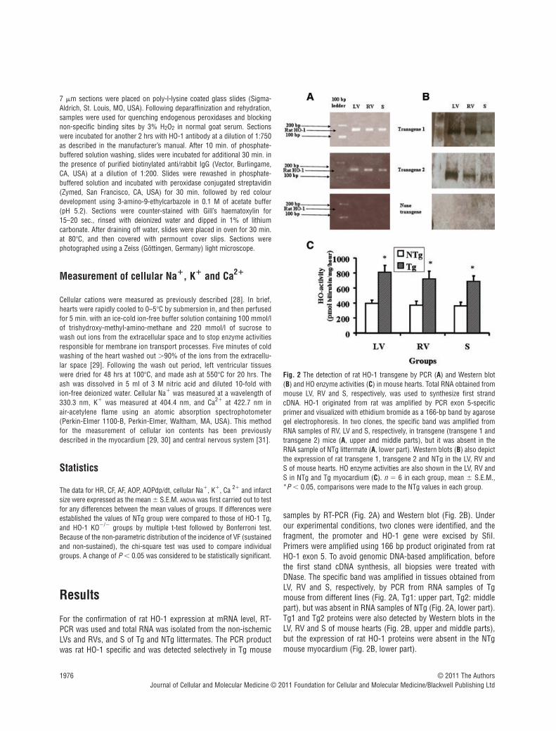
For the confirmation of rat HO-1 expression at mRNA level, RT-PCR was used and total RNA was isolated from the non-ischemicLVs and RVs, and S of Tg and NTg littermates. The PCR productwas rat HO-1 specific and was detected selectively in Tg mouse
samples by RT-PCR (Fig. 2A) and Western blot (Fig. 2B). Underour experimental conditions, two clones were identified, and thefragment, the promoter and HO-1 gene were excised by SfiI.Primers were amplified using 166 bp product originated from ratHO-1 exon 5. To avoid genomic DNA-based amplification, beforethe first stand cDNA synthesis, all biopsies were treated withDNase. The specific band was amplified in tissues obtained fromLV, RV and S, respectively, by PCR from RNA samples of Tgmouse from different lines (Fig. 2A, Tg1: upper part, Tg2: middlepart), but was absent in RNA samples of NTg (Fig. 2A, lower part).Tg1 and Tg2 proteins were also detected by Western blots in theLV, RV and S of mouse hearts (Fig. 2B, upper and middle parts),but the expression of rat HO-1 proteins were absent in the NTgmouse myocardium (Fig. 2B, lower part).
© 2011 The AuthorsJournal of Cellular and Molecular Medicine © 2011 Foundation for Cellular and Molecular Medicine/Blackwell Publishing Ltd
Fig. 2 The detection of rat HO-1 transgene by PCR (A) and Western blot(B) and HO enzyme activities (C) in mouse hearts. Total RNA obtained frommouse LV, RV and S, respectively, was used to synthesize first strandcDNA. HO-1 originated from rat was amplified by PCR exon 5-specificprimer and visualized with ethidium bromide as a 166-bp band by agarosegel electrophoresis. In two clones, the specific band was amplified fromRNA samples of RV, LV and S, respectively, in transgene (transgene 1 andtransgene 2) mice (A, upper and middle parts), but it was absent in theRNA sample of NTg littermate (A, lower part). Western blots (B) also depictthe expression of rat transgene 1, transgene 2 and NTg in the LV, RV andS of mouse hearts. HO enzyme activities are also shown in the LV, RV andS in NTg and Tg myocardium (C). n � 6 in each group, mean � S.E.M.,*P � 0.05, comparisons were made to the NTg values in each group.

J. Cell. Mol. Med. Vol 15, No 9, 2011
1977
HO enzyme activities were also measured in the LV, RV and Sobtained from NTg and Tg mice (Fig. 2C). The results show that HOactivities were significantly increased in samples obtained from theLV, RV and S of Tg mouse hearts in comparison with NTg values(Fig. 2C) indicating the function of Tg1 and Tg2 genes in the mousemyocardium. It is important to note that HO-1 mRNA and proteinwere not detected in NTg mouse myocardium either by PCR (Fig. 2A, lower part) or Western blot (Fig. 2B, lower part), but HOenzyme activities were present in the samples of NTg mousemyocardium including LV, RV and S, respectively, indicating thatmeasured HO enzyme activity involves all isoforms such as HO-1,HO-2, HO-3 and HO-4 (Fig. 2C). Thus, the differences in HO enzymeactivities (Fig. 2C) between NTg and Tg in the LV, RV and S wereclearly related to the rat HO-1 transgene in the mouse myocardium.
Table 1 shows the recovery of post-ischemic cardiac functionin NTg and HO-1 Tg hearts subjected to 20 min. of normothermicglobal ischemia followed by 2 hrs of reperfusion. Before the induc-tion of ischemia significant changes were not detected betweenNTg and HO-1 Tg groups in HR, CF, AF, AOP and AOPdp/dt. Uponreperfusion, the post-ischemic recovery of CF, AF, AOP andAOPdp/dt were observed in the HO-1 Tg group in comparison withthe NTg values without any significant differences in HR. Thus, forinstance, after 30 and 120 min. of reperfusion, AF was signifi-cantly increased from its NTg control values of 0.9 � 0.1 ml/min.and 0.8 � 0.1 ml/min. to 2.3 � 0.2 ml/min. (P � 0.05) and 2.2 �0.2 ml/min. (P � 0.05) in the Tg group, respectively (Table 1). Thesame pattern was observed in the post-ischemic recovery in CF,AOP and AOPdp/dt (Table 1). The results also show (Table 1) that50 �mol/kg of SnPPIX, an HO enzyme inhibitor, abolished thepost-ischemic recovery of cardiac function in comparison with theobserved protection in the HO-1 Tg group. Thus, the valuesobtained in the HO-1 Tg � SnPPIX group were not statisticallysignificant in comparison with the NTg values (Table 1).
Figure 3 depicts representative curves of endogenous CO pro-duction detected by GC in NTg, Tg and Tg SnPPIX treatedmyocardium subjected to 20 min. ischemia followed by 120 min.of reperfusion. It is shown that in HO-1 Tg hearts subjected to 20 min. of ischemia followed by 120 min. of reperfusion (Fig. 3B,chromatogram), a substantial increase in CO production wasobserved in comparison with the NTg myocardium (Fig. 3A, chro-matogram). However, the endogenous production of CO in theHO-1 Tg myocardium treated with SnPPIX was detected at rela-tively low level. (Fig. 3C, chromatogram).
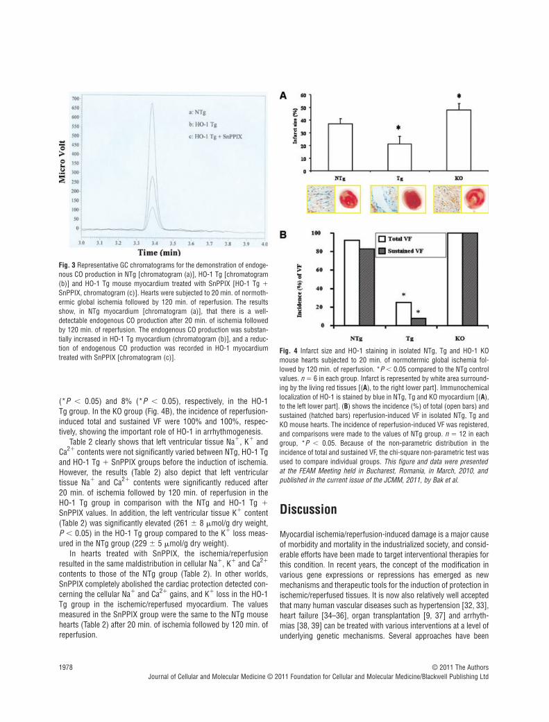
Figure 4 shows representative pictures of infarct size (Fig. 4A,lower part, to the right) and immunohistochemical staining of HO-1 (Fig. 4A, lower part to the left) in isolated mouse hearts sub-jected to ischemia and reperfusion. The infarct size (Fig. 4A) wasmarkedly reduced from its NTg control value of 37 � 4% to 20 �6% (*P � 0.05) in the HO-1 Tg group (Fig. 4A). However, in theHO-1 KO hearts, the infarct size was significantly increased to 47 � 5% (*P � 0.05) in comparison with NTg group (Fig. 4A).
In addition, Figure 4A shows the representative immunohisto-chemical localization of HO-1 (Fig. 4A, lower part – to the left) inNTg, Tg and HO-1 KO ischemic/reperfused mouse hearts. Leftventricular cardiac samples were obtained from NTg, HO-1 Tg andHO-1 KO mouse myocardium, respectively. Cytoplasmic proteinstaining (blue) of HO-1 in NTg myocardium was detected (Fig. 4A,lower part – to the left) which was more intense in Tgmyocardium, and markedly reduced in KO myocardium after 20 min. of ischemia followed by 120 min. of reperfusion (Fig. 4A,lower part – to the left).
Figure 4 also shows the incidence (%) of reperfusion-inducedVF (Fig. 4B) as total (sustained and non-sustained) and sustainedVF in NTg, Tg and KO mouse groups. The incidence of reperfu-sion-induced total and sustained VF (Fig. 4B) was significantlyreduced from their NTg control values of 92% and 83% to 25%
© 2011 The AuthorsJournal of Cellular and Molecular Medicine © 2011 Foundation for Cellular and Molecular Medicine/Blackwell Publishing Ltd
Table 1 Effects of 20 min. of ischemia followed by 30 and 120 min. of reperfusion on the recovery of cardiac function in NTg and HO-1 Tg, and the effect of SnPPIX
n � 6 in each group, mean � S.E.M.; HR: heart rate, beats/min.; CF: coronary flow, ml/min.; AF: aortic flow, ml/min.; AOP: aortic pressure, mmHg;AOPdp/dt: first derivative of aortic pressure, mmHg/sec.; SnPPIX: tin protoporphyrin IX. *P � 0.05, comparisons were made to the time-matchedNTg values.
Before ischemia After reperfusion (RE)
30 min. 120 min.
HR CF AF AOPAOPdp/dt
HR CF AF AOPAOPdp/dt
HR CF AF AOPAOPdp/dt
NTg 288 �
93.0 �
0.23.5 �
0.2157 �
73794 �
73255 �
92.0 �
0.10.9 �
0.172 � 6
945 �
44262 �
92.1 �
0.10.8 �
0.167 � 4
930 �
51
HO-1Tg
292 �
83.2 �
0.33.4 �
0.2149 �
63834 �
85261 �
72.8 �
0.1*2.3 �
0.2*95 � 7*
2000 �
63*269 �
82.9 �
0.2*2.2 �
0.2*91 �
6*1880 �
80*
HO-1Tg �
SnPPIX
294 �
93.1 �
0.23.2 �
0.3152 �
93815 �
94265 �
82.2 �
0.21.1 �
0.278 � 7
1018 �
80270 �
92.0 �
0.31.1 �
0.272 � 8
1000 �
77
1978
(*P � 0.05) and 8% (*P � 0.05), respectively, in the HO-1 Tg group. In the KO group (Fig. 4B), the incidence of reperfusion-induced total and sustained VF were 100% and 100%, respec-tively, showing the important role of HO-1 in arrhythmogenesis.
Table 2 clearly shows that left ventricular tissue Na�, K� andCa2� contents were not significantly varied between NTg, HO-1 Tgand HO-1 Tg � SnPPIX groups before the induction of ischemia.However, the results (Table 2) also depict that left ventricular tissue Na� and Ca2� contents were significantly reduced after 20 min. of ischemia followed by 120 min. of reperfusion in theHO-1 Tg group in comparison with the NTg and HO-1 Tg �
SnPPIX values. In addition, the left ventricular tissue K� content(Table 2) was significantly elevated (261 � 8 �mol/g dry weight,P � 0.05) in the HO-1 Tg group compared to the K� loss meas-ured in the NTg group (229 � 5 �mol/g dry weight).
In hearts treated with SnPPIX, the ischemia/reperfusionresulted in the same maldistribution in cellular Na�, K� and Ca2�
contents to those of the NTg group (Table 2). In other worlds,SnPPIX completely abolished the cardiac protection detected con-cerning the cellular Na� and Ca2� gains, and K� loss in the HO-1Tg group in the ischemic/reperfused myocardium. The valuesmeasured in the SnPPIX group were the same to the NTg mousehearts (Table 2) after 20 min. of ischemia followed by 120 min. ofreperfusion.
Discussion
Myocardial ischemia/reperfusion-induced damage is a major causeof morbidity and mortality in the industrialized society, and consid-erable efforts have been made to target interventional therapies forthis condition. In recent years, the concept of the modification invarious gene expressions or repressions has emerged as newmechanisms and therapeutic tools for the induction of protection inischemic/reperfused tissues. It is now also relatively well acceptedthat many human vascular diseases such as hypertension [32, 33],heart failure [34–36], organ transplantation [9, 37] and arrhyth-mias [38, 39] can be treated with various interventions at a level ofunderlying genetic mechanisms. Several approaches have been
© 2011 The AuthorsJournal of Cellular and Molecular Medicine © 2011 Foundation for Cellular and Molecular Medicine/Blackwell Publishing Ltd
Fig. 3 Representative GC chromatograms for the demonstration of endoge-nous CO production in NTg [chromatogram (a)], HO-1 Tg [chromatogram(b)] and HO-1 Tg mouse myocardium treated with SnPPIX [HO-1 Tg �SnPPIX, chromatogram (c)]. Hearts were subjected to 20 min. of normoth-ermic global ischemia followed by 120 min. of reperfusion. The resultsshow, in NTg myocardium [chromatogram (a)], that there is a well-detectable endogenous CO production after 20 min. of ischemia followedby 120 min. of reperfusion. The endogenous CO production was substan-tially increased in HO-1 Tg myocardium (chromatogram (b)], and a reduc-tion of endogenous CO production was recorded in HO-1 myocardiumtreated with SnPPIX [chromatogram (c)].
Fig. 4 Infarct size and HO-1 staining in isolated NTg, Tg and HO-1 KOmouse hearts subjected to 20 min. of normotermic global ischemia fol-lowed by 120 min. of reperfusion. *P � 0.05 compared to the NTg controlvalues. n � 6 in each group. Infarct is represented by white area surround-ing by the living red tissues [(A), to the right lower part]. Immunochemicallocalization of HO-1 is stained by blue in NTg, Tg and KO myocardium [(A),to the left lower part]. (B) shows the incidence (%) of total (open bars) andsustained (hatched bars) reperfusion-induced VF in isolated NTg, Tg andKO mouse hearts. The incidence of reperfusion-induced VF was registered,and comparisons were made to the values of NTg group. n � 12 in eachgroup, *P � 0.05. Because of the non-parametric distribution in theincidence of total and sustained VF, the chi-square non-parametric test wasused to compare individual groups. This figure and data were presented at the FEAM Meeting held in Bucharest, Romania, in March, 2010, andpublished in the current issue of the JCMM, 2011, by Bak et al.

J. Cell. Mol. Med. Vol 15, No 9, 2011
1979
made to manipulate cell phenotypes including the inhibition ofgenes associated with cell proliferation by preventing the develop-ment of atherosclerosis and restenosis [40–42] and the productionof growth factors to promote angiogenesis [43–47] for the treat-ment of peripheral vascular disease and myocardial ischemia.
In our previous studies, we found a substantial reduction in theexpression of HO-1 mRNA and its protein with a causative reduc-tion in the HO-1 enzyme activity in fibrillated ischemic/reperfusedrat myocardium [13]. Moreover, we have shown that, in thisrespect, no difference exists between non-diabetic and diabeticheart [48]. Furthermore, HO-1 KO mouse hearts displayed areduced left ventricular function after ischemia/reperfusion [14]. IfHO-1 system has a crucial role in the protection againstischemic/reperfusion-induced injury in the myocardium, with theenhanced expression of the HO-1, more ischemic tissue can besalvaged. Therefore, in the present study, because the HO-1 sys-tem can play a crucial role in the pathology of the ischemic/reper-fused myocardium, we decided to approach the question from adifferent angle using HO-1 Tg mouse hearts.
We generated HO-1 overexpressing Tg mice with an elevationin baseline HO enzyme activity by about 50% in the LV, RV and S,respectively. Thus, the application of a genome-based transgeneresulted in expression levels in relation with increased HO enzymeactivities in comparison with those of HO activities levels detectedin the LV, RV and S obtained from NTg mice. Although, in ourstudies, the very similar homology between the mouse and ratHO-1 made it impossible to differentiate the HO-1 and Tg-inducedenzyme activities, but we were able to distinguish them at mRNAlevel. In addition, our results obtained in NTg, HO-1 Tg and Tg-SnPPIX treated mouse hearts, we favour the idea that increasedendogenous CO production levels in the myocardium representsthe major signal transduction responsible for HO-1 related protec-tion reflected in the improvement of the recovery in post-ischemiccardiac function, reduction of the incidence of reperfusion-induced VF and infarct size connected to the recovery of cellularion contents. Our results further supported by the complete lossof cardiac protection in the Tg ischemic/reperfused mice treatedwith SnPPIX, the inhibitor of HO enzyme.
The exact mechanism by which HO-1 induces cardiac protec-tion remains to be elucidated; however, one of the most significantprotective mechanisms of the HO system can be explained by the
generation of endogenous CO and its signalling mechanism. COcould suppress cell apoptosis [37] suggesting that the anti-apop-totic effect of HO-1 is mediated via the generation of CO. In addi-tion, anti-inflammatory property of CO suppresses the expressionof pro-inflammatory genes associated with the activation ofmonocyte/M [49].
Another possible cellular signalling mechanism of CO is asso-ciated with the guanylyl cyclase activation and tissue levels ofguanosine monophosphate (cGMP) and adenosine monophos-phate (cAMP) in isolated ischemic/reperfused myocardium [50].Very low concentrations of CO, in the perfusion buffer, afford asignificant protection against the ischemia/reperfusion-induceddamage in isolated buffer perfused hearts. The isolated buffer per-fused heart could be an ideal model to study the direct effect ofexogenous CO on cardiac function and molecular signallingbecause the blood and its elements are excluded from the model,thus the oxygen transport to cells and tissues is not directly dam-aged via the CO/blood/haemoglobin system. Previous results haveshown that low concentrations of exogenous CO protect the iso-lated ischemic/reperfused myocardium and act by increasing tis-sue cAMP and cGMP levels [50]. Indeed, a moderate increase incAMP content could lead to arrhythmogenesis and developmentof various arrhythmias by elevating cytosolic calcium levels [51]in the ischemic and post-ischemic myocardium. It is of interest tonote that multiple increases in cGMP levels could mask and inter-fere with the arrhythmogenic effects of cAMP leading to the sup-pression of reperfusion-induced VF in the myocardium [50]. Thesignificant increase in cGMP levels can be related to guanylatecyclase activities in CO-treated myocardium, and suggests thatinduction of guanylate cyclase-cGMP system via CO signalling isessential for cardioprotection in ischemic/reperfused hearts.However, not only the absolute levels of cGMP and cAMP but alsothe ratio of these two nucleotides are critical factors that deter-mine the recovery of post-ischemic cardiac function afterischemia and reperfusion [52]. Thus, the lower the cAMP/cGMPratio is during ischemia, the better is the post-ischemic cardiacrecovery achieved in isolated ischemic/reperfused hearts.
It has to be noted that Lakkisto et al. have recently found thatcadioprotection induced by hemin, a well-known inducer ofhaemoxigenase 1, is independent of cGMP levels and, at least inpart, being mediated via the preservation of C43 [53]. They
© 2011 The AuthorsJournal of Cellular and Molecular Medicine © 2011 Foundation for Cellular and Molecular Medicine/Blackwell Publishing Ltd
Table 2 Cellular Na�, K� and Ca2� contents (�mol/g dry weight) in the left ventricular tissue before ischemia and after 20 min. of ischemiafollowed by 120 min. of reperfusion in NTg and HO-1 Tg, and the effect of SnPPIX
n � 6 in each group, mean � S.E.M., *P� 0.05, comparisons were made to the time-matched NTg values; SnPPIX: tin protoporphyrin IX .
Group Before ischemia After 20 min. ischemia followed by 120 min. reperfusion
Na�� K�� Ca2�� Na�� K�� Ca2��
NTg 31 � 3 282 � 6 1.6 � 0.1 84 � 5 229 � 5 4.1 � 0.3
HO-1 Tg 27 � 4 277 � 7 1.7 � 0.2 58 � 7* 261 � 8* 2.5 � 0.5*
HO-1 Tg � SnPPIX 35 � 5 286 � 9 1.9 � 0.3 90 � 9 219 � 9 4.5 � 0.7

1980
found that elevated HO-1 levels, induced by hemin, are not accom-panied by elevated cGMP levels. In our previous study [50], wehave found that cGMP levels are elevated after exogenous COapplication; moreover, after exogenous CO administration the ratioof cAMP/cGMP is reduced. In their study [53], the authors meas-ured only the level of cGMP and not the ratio of cAMP/cGMP. Thedifference between their and our results is probably due to thehigher CO concentration after the exogenous administration.
Although not specifically studied in the present investigation atmolecular level, it is of interest to note the findings of Piantadosiet al. [54] with myocardial cells. In their studies, Piantadosi et al.implicated the role of HO-1/CO pathway in mitochondrial biogen-esis through Akt1 activation [55] involving nuclear factor erythroid2-related factor (Nrf)2 expression, downstream GSK-3� blockadeand Nrf2 nuclear translocation leading to Nrf2-dependent activa-tion of nuclear respiratory factor (NRF)-1 transcription. Theirfinding suggest HO-1/CO, by sequentially activating the aforemen-tioned two transcription factors, as a remarkable component of aprosurvival program of mitochondrial biogenesis connected tocellular antioxidant defence mechanisms [56, 57].
Today, when so many advances and efforts are being made inmolecular biology and genetics, we tend to lose sight of the impor-tance of basic ions such as Na�, K� and Ca2� in both clinical andexperimental studies. As a gap, in changes between various geneexpression and/or repression and the function of myocardial tissue,could closely be connected with changes in myocardial tissue ioncontents. An important finding such a final endpoint of our presentstudy was the myocardial K� – conserving effect’ related to the pre-vention of cellular Na� and Ca2� gains in Tg myocardium, and thiscardiac protection can be reversed by SnPPIX, an HO-1 inhibitor.HO-1–bilirubin system and one of its by-products, CO, are essen-tial for regulation of cardiac function and ion transport across cellmembranes via the stabilization of various Na, K and Ca ion chan-nels possibly by an effect exerted on Na-K-ATPase, and thesetransient outward and inward currents are thought to underline thedelayed after depolarization that can be observed after the actionpotential in Ca-loaded cardiac tissue propagating rhythm distur-bances [58]. There are significant differences in the cellularsignalling mechanisms induced by CO when they are compared
with the actions of other interventions that elevate cyclicnucleotides in tissues. CO has been proposed to increase KCa chan-nel Ca2� sensitivity in arterial smooth muscle cells [59], and thedata of Perez et al. [60] suggest that CO increases Ca2� spark cou-pling by shifting the Ca2� sensitivity of KCa channels produced byCa2� sparks. Thus, CO may act upon KCa channel � subunits toenhance the coupling relationship leading to cellular K� loss, andCa2� and Na� gains as we observed in our present studies.
In our study we have shown that CO level was significantlyincreased in the HO-1 Tg mouse myocardium. However, we didnot measure CO levels during reperfusion; therefore, we believethat the elevated CO production can be responsible for the protec-tion during reperfusion in Tg myocardium. Besides this, we can-not rule out the contribution of other HO-1 related molecules suchas biliverdin and bilirubin in the observed cardiac protection.
In conclusion, using HO-1 Tg, we have demonstrated the directcausative role of HO-1 in protection against ischemia/reperfusion-induced injury. Specifically, our results show a significant recoveryof post-ischemic cardiac function, prevention of the developmentof reperfusion-induced VF and reduction in infarct size. In addi-tion, we have provided new insights into the ionic mechanisms bywhich HO-1 may contribute to the attenuation of reperfusion-induced arrhythmias in HO-1 Tg. We believe that these studieswould help in developing novel therapies based on targeting HO-1for the treatment of ischemia/reperfusion injury in patients withcoronary artery disease.
Acknowledgements
This study was supported by grants from OTKA (K-72315, OTKA 78223),TAMOP 4.2.2–08/1–2008-0007 and TAMOP 4.2.1.B-2010.
Conflict of interest
The authors confirm that there are no conflicts of interest.
References
1. Ryter SW, Choi AM. Heme oxygenase-1:redox regulation of a stress protein in lungand cell culture models. Antioxid RedoxSignal. 2005; 7: 80–91.
2. Maines MD. The heme oxygenase system:a regulator of second messenger gases.Annu Rev Pharmacol Toxicol. 1997; 37:517–54.
3. Lee PJ, Jiang BH, Chin BY, et al.Hypoxia-inducible factor-1 mediates tran-scriptional activation of the heme oxyge-nase-1 gene in response to hypoxia. J BiolChem. 1997; 272: 5375–81.
4. Otterbein LE, Lee PJ, Chin BY, et al.Protective effects of heme oxygenase-1 inacute lung injury. Chest. 1999; 116: 61S–3S.
5. Reeve VE, Tyrrell RM. Heme oxygenaseinduction mediates the photoimmunopro-tective activity of UVA radiation in themouse. Proc Natl Acad Sci USA. 1999; 96:9317–21.
6. Yet SF, Tian R, Layne MD, et al. Cardiac-specific expression of heme oxygenase-1protects against ischemia and reperfusioninjury in transgenic mice. Circ Res. 2001;89: 168–73.
7. Choi AM, Alam J. Heme oxygenase-1:function, regulation, and implication of anovel stress-inducible protein in oxidant-induced lung injury. Am J Respir Cell MolBiol. 1996; 15: 9–19.
8. Minamino T, Christou H, Hsieh CM, et al.Targeted expression of heme oxygenase-1prevents the pulmonary inflammatory andvascular responses to hypoxia. Proc NatlAcad Sci USA. 2001; 98: 8798–803.
9. Araujo JA, Meng L, Tward AD, et al.Systemic rather than local heme oxyge-nase-1 overexpression improves cardiac
© 2011 The AuthorsJournal of Cellular and Molecular Medicine © 2011 Foundation for Cellular and Molecular Medicine/Blackwell Publishing Ltd
J. Cell. Mol. Med. Vol 15, No 9, 2011
1981
allograft outcomes in a new transgenicmouse. J Immunol. 2003; 171: 1572–80.
10. Sawle P, Foresti R, Mann BE, et al.Carbon monoxide-releasing molecules(CO-RMs) attenuate the inflammatoryresponse elicited by lipopolysaccharide inRAW264.7 murine macrophages. Br JPharmacol. 2005; 145: 800–10.
11. Liu X, Wei J, Peng DH, et al. Absence ofheme oxygenase-1 exacerbates myocar-dial ischemia/reperfusion injury in diabeticmice. Diabetes. 2005; 54: 778–84.
12. Tang YL, Tang Y, Zhang YC, et al.Improved graft mesenchymal stem cellsurvival in ischemic heart with a hypoxia-regulated heme oxygenase-1 vector. J AmColl Cardiol. 2005; 46: 1339–50.
13. Bak I, Papp G, Turoczi T, et al. The role ofheme oxygenase-related carbon monoxideand ventricular fibrillation in ischemic/reperfused hearts. Free Radic Biol Med.2002; 33: 639–48.
14. Bak I, Szendrei L, Turoczi T, et al. Hemeoxygenase-1-related carbon monoxideproduction and ventricular fibrillation inisolated ischemic/reperfused mousemyocardium. FASEB J. 2003; 17: 2133–5.
15. Hong K, Piper DR, Diaz-Valdecantos A,et al. De novo KCNQ1 mutation responsi-ble for atrial fibrillation and short QT syn-drome in utero. Cardiovasc Res. 2005; 68:433–40.
16. Splawski I, Timothy KW, Sharpe LM, et al. Ca(V)1.2 calcium channel dysfunctioncauses a multisystem disorder includingarrhythmia and autism. Cell. 2004; 119:19–31.
17. Splawski I, Timothy KW, Decher N, et al.Severe arrhythmia disorder caused by car-diac L-type calcium channel mutations.Proc Natl Acad Sci USA. 2005; 102:8089–98.
18. Hewett TE, Grupp IL, Grupp G, et al.Alpha-skeletal actin is associated withincreased contractility in the mouse heart.Circ Res. 1994; 74: 740–6.
19. Schultz JE, Yao Z, Cavero I, et al.Glibenclamide-induced blockade of ischemicpreconditioning is time dependent in intactrat heart. Am J Physiol. 1997; 272:H2607–15.
20. Dickson EW, Blehar DJ, Carraway RE, et al. Naloxone blocks transferred precon-ditioning in isolated rabbit hearts. J MolCell Cardiol. 2001; 33: 1751–6.
21. Chomczynski P, Sacchi N. Single-stepmethod of RNA isolation by acid guani-dinium thiocyanate-phenol-chloroformextraction. Anal Biochem. 1987; 162:156–9.
22. Pellacani A, Wiesel P, Sharma A, et al.Induction of heme oxygenase-1 duringendotoxemia is downregulated by trans-forming growth factor-beta1. Circ Res.1998; 83: 396–403.
23. Tenhunen R, Ross ME, Marver HS, et al.Reduced nicotinamide-adenine dinu-cleotide phosphate dependent biliverdinreductase: partial purification and charac-terization. Biochemistry. 1970; 9:298–303.
24. Yoshida T, Takahashi S, Kikuchi G. Partialpurification and reconstitution of the hemeoxygenase system from pig spleen micro-somes. J Biochem. 1974; 75: 1187–91.
25. Morita T, Perrella MA, Lee ME, et al.Smooth muscle cell-derived carbonmonoxide is a regulator of vascular cGMP.Proc Natl Acad Sci USA. 1995; 92:1475–9.
26. Lowry OH, Rosebrough NJ, Farr AL, et al.Protein measurement with the Folin phenolreagent. J Biol Chem. 1951; 193: 265–75.
27. Cook MN, Nakatsu K, Marks GS, et al.Heme oxygenase activity in the adult rataorta and liver as measured by carbonmonoxide formation. Can J PhysiolPharmacol. 1995; 73: 515–8.
28. Tosaki A, Balint S, Szekeres L. Protectiveeffect of lidocaine against ischemia andreperfusion-induced arrhythmias andshifts of myocardial sodium, potassium,and calcium content. J CardiovascPharmacol. 1988; 12: 621–8.
29. Pridjian AK, Levitsky S, Krukenkamp I, et al. Developmental changes in reperfu-sion injury. A comparison of intracellularcation accumulation in the newborn,neonatal, and adult heart. J ThoracCardiovasc Surg. 1987; 93: 428–33.
30. Alto LE, Dhalla NS. Myocardial cationcontents during induction of calcium para-dox. Am J Physiol. 1979; 237: H713–9.
31. Szabo ME, Gallyas E, Bak I, et al. Hemeoxygenase-1-related carbon monoxide andflavonoids in ischemic/reperfused ratretina. Invest Ophthalmol Vis Sci. 2004;45: 3727–32.
32. Mattson DL, Dwinell MR, Greene AS, et al. Chromosome substitution revealsthe genetic basis of Dahl salt-sensitivehypertension and renal disease. Am JPhysiol Renal Physiol. 2008; 295:F837–42.
33. Tsai CT, Hwang JJ, Lai LP, et al.Interaction of gender, hypertension, andthe angiotensinogen gene haplotypes onthe risk of coronary artery disease in alarge angiographic cohort. Atherosclerosis.2009; 203: 249–56.
34. Davis J, Wen H, Edwards T, et al. Alleleand species dependent contractile defectsby restrictive and hypertrophic cardiomy-opathy-linked troponin I mutants. J MolCell Cardiol. 2008; 44: 891–904.
35. Monti J, Fischer J, Paskas S, et al.Soluble epoxide hydrolase is a susceptibil-ity factor for heart failure in a rat model ofhuman disease. Nat Genet. 2008; 40:529–37.
36. Zhang L, Hu A, Yuan H, et al. A missensemutation in the CHRM2 gene is associatedwith familial dilated cardiomyopathy. CircRes. 2008; 102: 1426–32.
37. Sato K, Balla J, Otterbein L, et al. Carbonmonoxide generated by heme oxygenase-1suppresses the rejection of mouse-to-ratcardiac transplants. J Immunol. 2001;166: 4185–94.
38. Groh WJ, Groh MR, Saha C, et al.Electrocardiographic abnormalities andsudden death in myotonic dystrophy type1. N Engl J Med. 2008; 358: 2688–97.
39. Knollmann BC, Roden DM. A geneticframework for improving arrhythmia ther-apy. Nature. 2008; 451: 929–36.
40. Zahradka P, Wright B, Fuerst M, et al.Peroxisome proliferator-activated receptoralpha and gamma ligands differentiallyaffect smooth muscle cell proliferation andmigration. J Pharmacol Exp Ther. 2006;317: 651–9.
41. Honda T, Kaikita K, Tsujita K, et al.Pioglitazone, a peroxisome proliferator-activated receptor-gamma agonist, attenu-ates myocardial ischemia-reperfusioninjury in mice with metabolic disorders. J Mol Cell Cardiol. 2008; 44: 915–26.
42. Wayman NS, Hattori Y, McDonald MC, et al. Ligands of the peroxisome prolifera-tor-activated receptors (PPAR-gamma andPPAR-alpha) reduce myocardial infarctsize. FASEB J. 2002; 16: 1027–40.
43. Sharma S, Dewald O, Adrogue J, et al.Induction of antioxidant gene expressionin a mouse model of ischemic cardiomy-opathy is dependent on reactive oxygenspecies. Free Radic Biol Med. 2006; 40:2223–31.
44. Arab S, Konstantinov IE, Boscarino C, et al. Early gene expression profiles dur-ing intraoperative myocardial ischemia-reperfusion in cardiac surgery. J ThoracCardiovasc Surg. 2007; 134: 74–81.
45. Liu X, Simpson JA, Brunt KR, et al.Preemptive heme oxygenase-1 gene deliv-ery reveals reduced mortality and preser-vation of left ventricular function 1 yr afteracute myocardial infarction. Am J PhysiolHeart Circ Physiol. 2007; 293: H48–59.
© 2011 The AuthorsJournal of Cellular and Molecular Medicine © 2011 Foundation for Cellular and Molecular Medicine/Blackwell Publishing Ltd

1982
46. Thirunavukkarasu M, Penumathsa SV,Koneru S, et al. Resveratrol alleviates car-diac dysfunction in streptozotocin-induceddiabetes: role of nitric oxide, thioredoxin,and heme oxygenase. Free Radic Biol Med.2007; 43: 720–9.
47. Penumathsa SV, Koneru S, Zhan L, et al.Secoisolariciresinol diglucoside inducesneovascularization-mediated cardioprotec-tion against ischemia-reperfusion injury inhypercholesterolemic myocardium. J MolCell Cardiol. 2008; 44: 170–9.
48. Csonka C, Varga E, Kovacs P, et al. Hemeoxygenase and cardiac function inischemic/reperfused rat hearts. Free RadicBiol Med. 1999; 27: 119–26.
49. Otterbein LE, Bach FH, Alam J, et al.Carbon monoxide has anti-inflammatoryeffects involving the mitogen-activatedprotein kinase pathway. Nat Med. 2000; 6:422–8.
50. Bak I, Varadi J, Nagy N, et al. The role ofexogenous carbon monoxide in the recov-ery of post-ischemic cardiac function in
buffer perfused isolated rat hearts. CellMol Biol. 2005; 51: 453–9.
51. du Toit EF, Opie LH. Modulation of severityof reperfusion stunning in the isolated ratheart by agents altering calcium flux at onsetof reperfusion. Circ Res. 1992; 70: 960–7.
52. Du Toit EF, Meiring J, Opie LH. Relationof cyclic nucleotide ratios to ischemic andreperfusion injury in nitric oxide-donortreated rat hearts. J Cardiovasc Pharmacol.2001; 38: 529–38.
53. Lakkisto P, Csonka C, Fodor G, et al. Theheme oxygenase inducer hemin protectsagainst cardiac dysfunction and ventricularfibrillation in ischaemic/reperfused rathearts: role of connexin 43. Scand J ClinLab Invest. 2009; 69: 209–18.
54. Piantadosi CA, Carraway MS, Babiker A,et al. Heme oxygenase-1 regulates cardiacmitochondrial biogenesis via Nrf2-mediatedtranscriptional control of nuclear respiratoryfactor-1. Circ Res. 2008; 103: 1232–40.
55. Suliman HB, Carraway MS, Tatro LG, et al. A new activating role for CO in car-
diac mitochondrial biogenesis. J Cell Sci.2007; 120: 299–308.
56. Wagener FA, Volk HD, Willis D, et al.Different faces of the heme-heme oxyge-nase system in inflammation. PharmacolRev. 2003; 55: 551–71.
57. Maines MD. The heme oxygenase system:update 2005. Antioxid Redox Signal. 2005;7: 1761–6.
58. Kass RS, Lederer WJ, Tsien RW, et al.Role of calcium ions in transient inwardcurrents and aftercontractions induced bystrophanthidin in cardiac Purkinje fibres. JPhysiol. 1978; 281: 187–208.
59. Wang R, Wu L, Wang Z. The direct effectof carbon monoxide on KCa channels invascular smooth muscle cells. PflugersArch. 1997; 434: 285–91.
60. Perez GJ, Bonev AD, Nelson MT.Micromolar Ca(2�) from sparks activatesCa(2�)-sensitive K(�) channels in rat cerebral artery smooth muscle. Am J Physiol Cell Physiol. 2001; 281:C1769–75.
© 2011 The AuthorsJournal of Cellular and Molecular Medicine © 2011 Foundation for Cellular and Molecular Medicine/Blackwell Publishing Ltd