Erythropoietin: Powerful protection of ischemic and post-ischemic brain
Upload
independentCategory
view
0download
0
Inhibition of stress-activated protein kinase in the ischemic/reperfusedheart: role of magnesium tanshinoate B in preventing apoptosis
Kathy K.W. Au-Yeunga, Da-yuan Zhub, Karmin Oa, Yaw L. Siowa,c,*aDepartment of Pharmacology, Faculty of Medicine, The University of Hong Kong, 1/F, Li Shu Fan Building, 5 Sassoon Road, Pokfulam, Hong Kong
SAR, ChinabState Key Laboratory for Drug Research, Shanghai Institute of Materia Medica, Shanghai Institute of Biological Sciences, Chinese Academy of
Sciences, Shanghai, ChinacSchool of Traditional Chinese Medicine, Faculty of Medicine, The University of Hong Kong, Hong Kong SAR, China
Received 29 June 2000; accepted 16 November 2000
Abstract
The activation of stress-activated protein (SAP) kinase may lead to an induction of apoptosis that is responsible for part of thecardiomyocyte death in reperfusion injury. The objective of the present study was to investigate the mechanism by which magnesiumtanshinoate B (MTB), a bioactive compound isolated from Danshen, prevents apoptosis in cardiomyocytes in the ischemic/reperfused heart.Isolated adult rat hearts were perfused by the Langendorff mode with medium containing MTB prior to the induction of normothermic globalischemia. At the end of the 30-min ischemic period, the heart was reperfused with the same medium with or without MTB for an additional20 min. In the MTB-treated ischemic/reperfused heart, the number of apoptotic nuclei was reduced by 2.5-fold in comparison to that inuntreated ischemic/reperfused controls [236 4 vs 576 7 (mean6 SD) TUNEL-positive cells, respectively, N5 3–4,P , 0.001]. SAPkinase activity was elevated 1.7-fold in ischemic/reperfused rat hearts [35.66 3.8 vs 21.26 3.3 (control) (mean6 SEM) relativedensitometric units, N5 4–6, P , 0.05]. Treatment with MTB abolished this elevation in SAP kinase activity (25.06 5.2 relativedensitometric units), which was also decreased by 40% in the nucleus. When the heart was subjected to ischemia alone, there was nosignificant change in SAP kinase activity in the presence or absence of MTB. MTB did not appear to affect the p38 mitogen-activated proteinkinase activity in this model system. In conclusion, MTB was shown to have cardioprotective activity against apoptosis, probably throughthe inhibition of SAP kinase activity. © 2001 Elsevier Science Inc. All rights reserved.
Keywords:Apoptosis; Ischemia; Protein kinases; Protein phosphorylation; Reperfusion
1. Introduction
Over the last decade, a new superfamily of protein ki-nases activated by mitogens and growth factors was recog-nized. Broadly referred to as the MAP kinases, they includethe extracellular signal-regulated protein kinases, the SAPkinases and p38 MAPK [1]. Cellular stresses, e.g. ischemia/reperfusion, heat shock, hyperosmotic conditions, UV irra-
diation, protein synthesis inhibitors such as anisomycin, aswell as the proinflammatory cytokines like tumor necrosisfactor-a, have been reported to activate the latter two MAPkinase homologues, namely SAP kinase and p38 MAPK[1].
The activation of SAP kinase and p38 MAPK may playa critical role in the genetic response of components of thecardiovascular system to disease states such as ischemia,atherosclerosis, heart failure, and restenosis [2]. The diseaseprocess can produce oxygen free radicals and factors (e.g.cytokines), which can activate the SAP kinases [3–5] inaddition to causing oxidation of cellular components, oxi-dation of the membrane (causing leakage), and modificationof membrane receptors [6]. The activated SAP kinases canthen translocate to the nucleus [4], where they may inducea host of immediate early genes [7], leading to apoptosis.Activation of the SAP kinase cascade is one of the mecha-
* Corresponding author. Tel.:1852-2819-2864; fax:1852-2817-0859.
E-mail address:[email protected] (Y.L. Siow).Abbreviations: JIP, JNK-interacting protein; MAP, mitogen-activated
protein; p38 MAPK, p38 MAP kinase; MTB, magnesium tanshinoate B;PARP, poly(ADP-ribose) polymerase; PMSF, phenylmethylsulfonyl fluo-ride; SAP, stress-activated protein; and TUNEL, terminal deoxyribonucle-otide transferase (TdT)-mediated dUTP nick-end labeling.
Biochemical Pharmacology 62 (2001) 483–493
0006-2952/01/$ – see front matter © 2001 Elsevier Science Inc. All rights reserved.PII: S0006-2952(01)00686-4
nisms for the induction of apoptosis [8–11]. Recent evi-dence suggests that a proportion of the cardiomyocytespreferentially undergoes apoptosis following myocardialischemia/reperfusion [12,13]. In ischemic/reperfusion in-jury, a sustained activation of SAP kinases has been re-ported [4,5,14]. This may account for the apoptotic death ofcardiomyocytes. Any attenuation of the activation of SAPkinase may, in turn, reduce the loss of cardiomyocytes, thusproducing a partial offset to reperfusion injury.
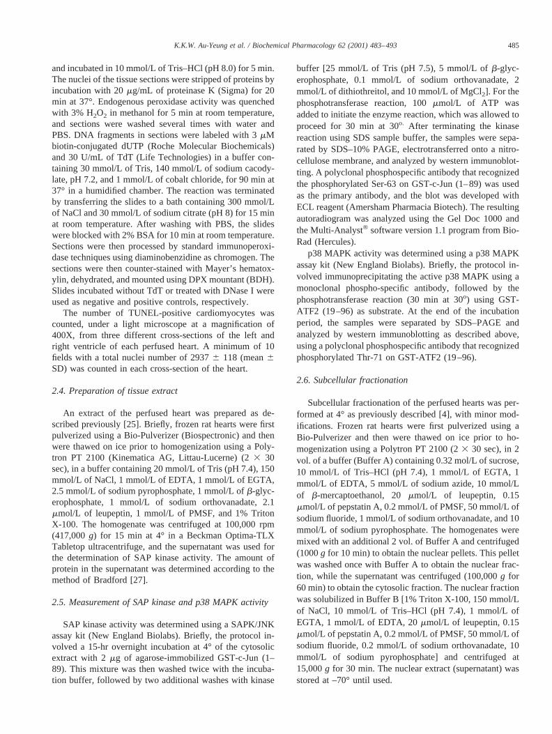
Danshen (Radix Salvia miltiorrhizae) has been used inthe treatment of cardiovascular disease in China [15–17]and has been shown to lower blood pressure [18–21]. In theisolated rat heart, Zhou and Ruigrok [21] showed that Dan-shen had a negative inotropic effect and increased coronaryblood flow. Furthermore, Danshen-treated hearts had betterpost-ischemic reperfusion recovery of ventricular developedpressure and less contracture than untreated hearts [21].Clinically, in a double-blind study using phenolphthalein asa placebo, Danshen was reported to be effective in thetreatment of coronary heart disease [22]. Other studies haverevealed that Danshen was able to dilate the rat aorta in anendothelium-dependent manner [20,23]. This vasodilatoryaction of Danshen might be due, in part, to the action of oneof its active ingredients, tanshinone II-A. Tanshinone II-A isa naturally occurring calcium antagonist and can causecoronary and peripheral vasodilation by reducing the influxof calcium into myocardial and smooth muscle cells [21].Another active component of Danshen is MTB (Fig. 1), alsoknown as magnesium lithospermate B. Infusion of MTBinto the post-ischemic rabbit heart has been shown to reducedamage to the myocardium when compared with the saline-treated control [24]. Futhermore, intravenous injection ofMTB (30 mg/kg) into rats resulted in a decrease in bloodpressure with no changes in the heart rate [20]. However,the mechanisms of these MTB actions are largely unknown.
In the present study, the effect of MTB on ischemia/reperfusion-induced apoptosis was investigated. Our resultsdemonstrated that MTB can protect the ischemic/reperfusedheart against apoptotic cell death. Further investigationsrevealed that MTB can directly inhibit SAP kinase activityand its translocation into the nucleus. This may representone of the mechanisms by which MTB exerts its cardiopro-tective effect.
2. Materials and methods
2.1. Materials
MTB was isolated fromRadix Salvia miltiorrhizaebystandard chromatographic techniques [24], and its puritywas determined by HPLC to be. 98%. SAP kinase and p38MAPK assay kits, and rat-specific polyclonal antibodyagainst cleaved PARP (D214) were purchased from NewEngland Biolabs and Cell Signaling Technology, respec-tively. The expression plasmid pGEX-GST-ATF2 (19–96)was a gift from Dr. J. Silvio Gutkind of NIDR, NIH;recombinant GST-ATF2 (19–96) was prepared according tothe protocols of Amersham Pharmacia Biotech. All otherchemicals were of reagent grade and were obtained from theSigma Chemical Co. or BDH Chemicals. For heart perfu-sion, male Sprague–Dawley rats (200–250 g) were used.All experimental protocols involving the use of animalswere approved by the Committee for the Use of Live Ani-mals for Teaching and Research of The University of HongKong.
2.2. Heart perfusion
Perfusion of the rat heart was performed as previouslydescribed [25]. Male Sprague–Dawley rats (200–250 g)were anaesthetized by i.p. injection of sodium pentobarbi-tone (7 mg/100 g body weight) and heparin (20 IU/100 gbody weight). Hearts were then excised and perfused in aLangendorff apparatus with Krebs-Henseleit buffer contain-ing 120 mmol/L of NaCl, 25 mmol/L of NaHCO3, 5.5mmol/L of dextrose, 4.76 mmol/L of KCl, 1.2 mmol/L ofmagnesium sulfate, 1.2 mmol/L of KH2PO4, and 1.27mmol/L of CaCl2, saturated with 95% O2/5% CO2 for 10min at 37°. This was followed by perfusion with freshKrebs-Henseleit buffer containing MTB for 20 min. Subse-quently, normothermic global ischemia was induced bystopping the flow of buffer for 30 min (unless stated other-wise) while maintaining the temperature at 37°. At the endof the ischemic period, the hearts either were processedimmediately or were reperfused with the same mediumcontaining MTB for an additional 120 min, unless statedotherwise. Perfused hearts were processed as described be-low or were frozen in liquid nitrogen and stored at –70°until used.
2.3. In situ labeling of DNA fragments (TUNEL)
DNA fragmentation was detectedin situ by usingTUNEL in the perfused heart [26]. After perfusion, heartswere fixed in phosphate-buffered 4% paraformaldehyde at4° overnight prior to embedding in paraffin. Embeddedtissue (5mm sections) was adhered to slides that had beenpretreated with VectabondTM (Vector Laboratories Inc.).Paraffin was removed from sections by immersing in xy-lene; rehydrated through 100, 96, 80, 70, and 0% ethanol;
Fig. 1. Structure of magnesium tanshinoate B (MTB; magnesium litho-spermate B).
484 K.K.W. Au-Yeung et al. / Biochemical Pharmacology 62 (2001) 483–493
and incubated in 10 mmol/L of Tris–HCl (pH 8.0) for 5 min.The nuclei of the tissue sections were stripped of proteins byincubation with 20mg/mL of proteinase K (Sigma) for 20min at 37°. Endogenous peroxidase activity was quenchedwith 3% H2O2 in methanol for 5 min at room temperature,and sections were washed several times with water andPBS. DNA fragments in sections were labeled with 3mMbiotin-conjugated dUTP (Roche Molecular Biochemicals)and 30 U/mL of TdT (Life Technologies) in a buffer con-taining 30 mmol/L of Tris, 140 mmol/L of sodium cacody-late, pH 7.2, and 1 mmol/L of cobalt chloride, for 90 min at37° in a humidified chamber. The reaction was terminatedby transferring the slides to a bath containing 300 mmol/Lof NaCl and 30 mmol/L of sodium citrate (pH 8) for 15 minat room temperature. After washing with PBS, the slideswere blocked with 2% BSA for 10 min at room temperature.Sections were then processed by standard immunoperoxi-dase techniques using diaminobenzidine as chromogen. Thesections were then counter-stained with Mayer’s hematox-ylin, dehydrated, and mounted using DPX mountant (BDH).Slides incubated without TdT or treated with DNase I wereused as negative and positive controls, respectively.
The number of TUNEL-positive cardiomyocytes wascounted, under a light microscope at a magnification of400X, from three different cross-sections of the left andright ventricle of each perfused heart. A minimum of 10fields with a total nuclei number of 29376 118 (mean6SD) was counted in each cross-section of the heart.
2.4. Preparation of tissue extract
An extract of the perfused heart was prepared as de-scribed previously [25]. Briefly, frozen rat hearts were firstpulverized using a Bio-Pulverizer (Biospectronic) and thenwere thawed on ice prior to homogenization using a Poly-tron PT 2100 (Kinematica AG, Littau-Lucerne) (23 30sec), in a buffer containing 20 mmol/L of Tris (pH 7.4), 150mmol/L of NaCl, 1 mmol/L of EDTA, 1 mmol/L of EGTA,2.5 mmol/L of sodium pyrophosphate, 1 mmol/L ofb-glyc-erophosphate, 1 mmol/L of sodium orthovanadate, 2.1mmol/L of leupeptin, 1 mmol/L of PMSF, and 1% TritonX-100. The homogenate was centrifuged at 100,000 rpm(417,000g) for 15 min at 4° in a Beckman Optima-TLXTabletop ultracentrifuge, and the supernatant was used forthe determination of SAP kinase activity. The amount ofprotein in the supernatant was determined according to themethod of Bradford [27].
2.5. Measurement of SAP kinase and p38 MAPK activity
SAP kinase activity was determined using a SAPK/JNKassay kit (New England Biolabs). Briefly, the protocol in-volved a 15-hr overnight incubation at 4° of the cytosolicextract with 2mg of agarose-immobilized GST-c-Jun (1–89). This mixture was then washed twice with the incuba-tion buffer, followed by two additional washes with kinase
buffer [25 mmol/L of Tris (pH 7.5), 5 mmol/L ofb-glyc-erophosphate, 0.1 mmol/L of sodium orthovanadate, 2mmol/L of dithiothreitol, and 10 mmol/L of MgCl2]. For thephosphotransferase reaction, 100mmol/L of ATP wasadded to initiate the enzyme reaction, which was allowed toproceed for 30 min at 30o. After terminating the kinasereaction using SDS sample buffer, the samples were sepa-rated by SDS–10% PAGE, electrotransferred onto a nitro-cellulose membrane, and analyzed by western immunoblot-ting. A polyclonal phosphospecific antibody that recognizedthe phosphorylated Ser-63 on GST-c-Jun (1–89) was usedas the primary antibody, and the blot was developed withECL reagent (Amersham Pharmacia Biotech). The resultingautoradiogram was analyzed using the Gel Doc 1000 andthe Multi-Analyst® software version 1.1 program from Bio-Rad (Hercules).
p38 MAPK activity was determined using a p38 MAPKassay kit (New England Biolabs). Briefly, the protocol in-volved immunoprecipitating the active p38 MAPK using amonoclonal phospho-specific antibody, followed by thephosphotransferase reaction (30 min at 30o) using GST-ATF2 (19–96) as substrate. At the end of the incubationperiod, the samples were separated by SDS–PAGE andanalyzed by western immunoblotting as described above,using a polyclonal phosphospecific antibody that recognizedphosphorylated Thr-71 on GST-ATF2 (19–96).
2.6. Subcellular fractionation
Subcellular fractionation of the perfused hearts was per-formed at 4° as previously described [4], with minor mod-ifications. Frozen rat hearts were first pulverized using aBio-Pulverizer and then were thawed on ice prior to ho-mogenization using a Polytron PT 2100 (23 30 sec), in 2vol. of a buffer (Buffer A) containing 0.32 mol/L of sucrose,10 mmol/L of Tris–HCl (pH 7.4), 1 mmol/L of EGTA, 1mmol/L of EDTA, 5 mmol/L of sodium azide, 10 mmol/Lof b-mercaptoethanol, 20mmol/L of leupeptin, 0.15mmol/L of pepstatin A, 0.2 mmol/L of PMSF, 50 mmol/L ofsodium fluoride, 1 mmol/L of sodium orthovanadate, and 10mmol/L of sodium pyrophosphate. The homogenates weremixed with an additional 2 vol. of Buffer A and centrifuged(1000g for 10 min) to obtain the nuclear pellets. This pelletwas washed once with Buffer A to obtain the nuclear frac-tion, while the supernatant was centrifuged (100,000g for60 min) to obtain the cytosolic fraction. The nuclear fractionwas solubilized in Buffer B [1% Triton X-100, 150 mmol/Lof NaCl, 10 mmol/L of Tris–HCl (pH 7.4), 1 mmol/L ofEGTA, 1 mmol/L of EDTA, 20mmol/L of leupeptin, 0.15mmol/L of pepstatin A, 0.2 mmol/L of PMSF, 50 mmol/L ofsodium fluoride, 0.2 mmol/L of sodium orthovanadate, 10mmol/L of sodium pyrophosphate] and centrifuged at15,000g for 30 min. The nuclear extract (supernatant) wasstored at –70° until used.
485K.K.W. Au-Yeung et al. / Biochemical Pharmacology 62 (2001) 483–493
2.7. Statistical analysis
All values are expressed as means6 SEM. Comparisonsbetween groups were assessed by one-way ANOVA withTukey’s post hoc-analysis. Spearman rank order correlationcoefficient was calculated with SPSS for Windows (Version10) (SPSS Inc.). Statistical significance was defined as avalue ofP , 0.05.
3. Results
3.1. Effect of MTB on apoptosis in the isolated ischemic/reperfused heart
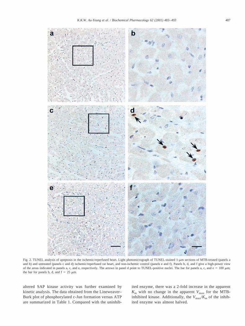
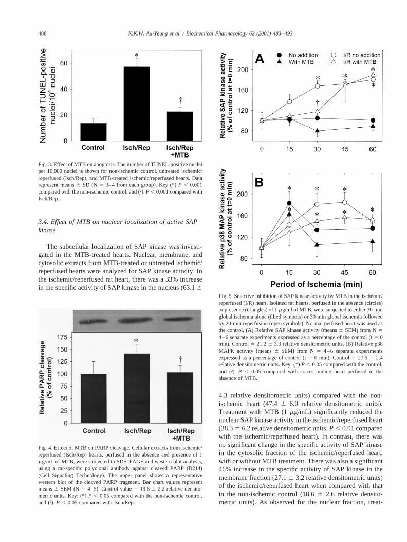
Since MTB has been shown to reduce myocardial dam-age in the post-ischemic rabbit heart [24], its effect oncardiomyocyte apoptotic cell death was first investigated.Apoptosis in cardiomyocytes was assessed by counting thenumber of TUNEL-positive cells. TUNEL positivity wascharacterized by focal nuclear staining [26]. As shown inFig. 2, in apoptotic cells, the nuclear and cell membraneintegrity remained intact. In our model system of ischemia/reperfusion, there was no difference in the number ofTUNEL-positive nuclei between the left and right ventri-cles. The reaction product was dark brown, and there wasminimal background. The number of TUNEL-positive nu-clei in the heart was increased significantly (by 2.5-fold)after ischemia/reperfusion (Figs. 2, c and d, and 3). Incontrast, when the isolated rat heart was perfused in thepresence of 1mg/mL of MTB (Fig. 2, a and b), the numberof TUNEL-positive nuclei after ischemia/reperfusion wascomparable to that of the non-ischemic/perfused heart (Fig.2, e and f). A significant 2.5-fold reduction in the number ofTUNEL-positive nuclei was observed in the MTB-treatedischemic/reperfused heart (236 4 nuclei, mean6 SD)when compared with the untreated ischemic/reperfused con-trol (57 6 7 nuclei) (Fig. 3).
Apoptosis was also assessed by the detection of PARPcleavage in the cell. The proteolytic cleavage of the 116 kDaPARP into the 24 kDa N-terminal DNA binding domain andthe 89 kDa C-terminal catalytic domain by caspase, whichis known to mediate the apoptotic process, has been usedextensively as a marker of apoptosis [28]. Figure 4 showsthat ischemia/reperfusion increased the level of the C-ter-minal 89 kDa PARP fragment, indicating increased apopto-sis in these cardiomyocytes. When the heart was perfused inthe presence of MTB, there was a significant reduction inthe level of the PARP fragment (Fig. 4).
3.2. Effect of MTB on SAP kinase and p38 MAPK activityin the isolated heart
Activation of SAP kinase is one of the mechanismsleading to apoptosis. The effect of MTB on SAP kinaseactivity was first studied using the isolated rat heart as a
model system. Rat hearts were perfused in the presence orabsence of 1 and 10mg/mL of MTB and subjected toischemia or ischemia/reperfusion; then the cellular SAPkinase activity was determined. As previously demonstratedby other investigators [3,5], there was no activation of SAPkinase when the rat heart was subjected to ischemia alone(Fig. 5A). The addition of MTB produced a slight but notsignificant inhibition of SAP kinase activity. In contrast,SAP kinase activity was elevated significantly (by 1.7-fold)during ischemia/reperfusion. Figure 5A shows that this el-evation in SAP kinase activity was transiently abolished(i.e. in the first 30 min) in the presence of 1mg/mL of MTB.Similar results were obtained when hearts were perfusedwith 10 mg/mL of MTB (data not shown).
Since p38 MAPK has also been implicated in apoptosisassociated with ischemia/reperfusion [3], the effect of MTBon this kinase activity was also investigated in our modelsystem. Consistent with published reports, there was a tran-sient activation of p38 MAPK activity during ischemia (Fig.5B). In the presence of MTB, there was no significantdecrease in the p38 MAPK activity. When rat hearts weresubjected to ischemia/reperfusion, there was a significantincrease in p38 MAPK activity after 30 min of ischemia.Similarly, there was no significant decrease in p38 MAPKactivity in the ischemic/reperfused heart in the presence ofMTB.
3.3. Direct effect of MTB on SAP kinase activity
The direct effect of MTB on SAP kinase activity wasthen investigated. Ischemic/reperfused heart extracts wereused as the source of SAP kinase for this part of the study.Figure 6 shows that in the presence of 0.01mg/mL of MTB,there was a 27% reduction in SAP kinase activity. Furtheraddition of the compound up to 10mg/mL resulted in agreater reduction of the SAP kinase activity (to 56%) com-pared with control activity. Addition of a higher concentra-tion of MTB (100mg/mL) did not result in greater inhibitionof SAP kinase activity (data not shown).
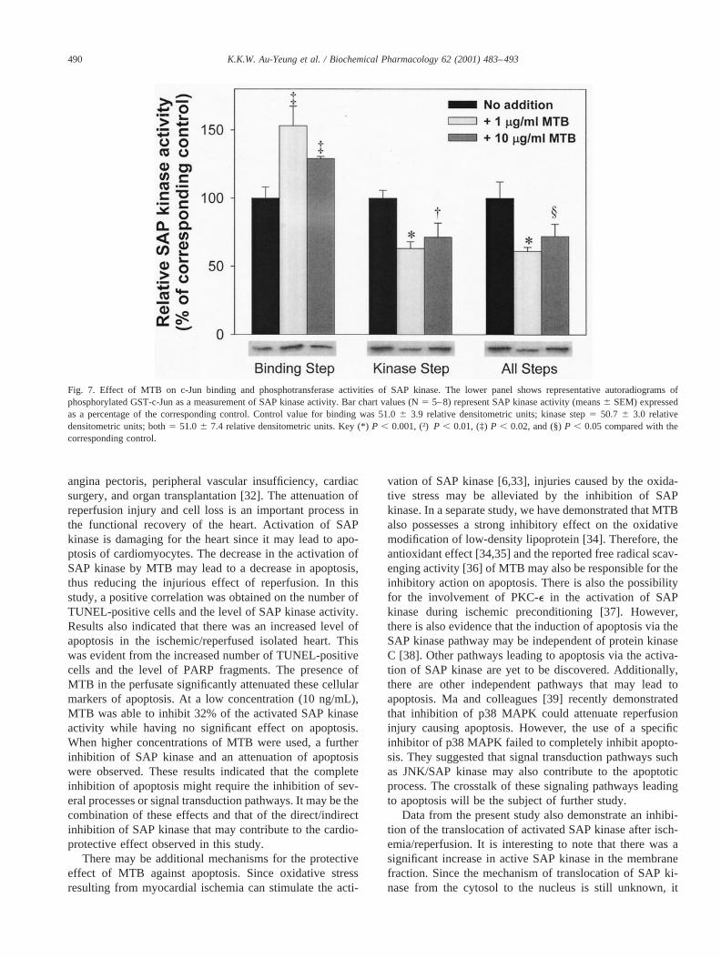
Next, the mechanism for the MTB inhibition of SAPkinase was investigated. The determination of SAP ki-nase activity involved two separate steps. In the first step(binding step), immobilized c-Jun was used to “pulldown” SAP kinase in the tissue extract, while the secondstep (kinase step) involved the actual phosphotransferasereaction. Rat hearts were homogenized in cell lysisbuffer, and the soluble extracts were used for determina-tion of SAP kinase activity. As depicted in Fig. 7, in thepresence of MTB (1mg/mL), the c-Jun binding activityof SAP kinase was enhanced significantly by 50%. Incontrast, phosphotransferase activity of the SAP kinasewas reduced to 63% of control. However, the addition ofMTB resulted in an overall 44% inhibition of the SAPkinase reaction. Similar results were observed when 10mg/mL of MTB was included in the assays for SAPkinase activity (Fig. 7). The mechanism by which MTB
486 K.K.W. Au-Yeung et al. / Biochemical Pharmacology 62 (2001) 483–493
altered SAP kinase activity was further examined bykinetic analysis. The data obtained from the Lineweaver–Burk plot of phosphorylated c-Jun formation versus ATPare summarized in Table 1. Compared with the uninhib-
ited enzyme, there was a 2-fold increase in the apparentKm with no change in the apparentVmax for the MTB-inhibited kinase. Additionally, theVmax/Km of the inhib-ited enzyme was almost halved.
Fig. 2. TUNEL analysis of apoptosis in the ischemic/reperfused heart. Light photomicrograph of TUNEL-stained 1-mm sections of MTB-treated (panels aand b) and untreated (panels c and d) ischemic/reperfused rat heart, and non-ischemic control (panels e and f). Panels b, d, and f give a high-power viewof the areas indicated in panels a, c, and e, respectively. The arrows in panel d point to TUNEL-positive nuclei. The bar for panels a, c, and e5 100 mm;the bar for panels b, d, and f5 25 mm.
487K.K.W. Au-Yeung et al. / Biochemical Pharmacology 62 (2001) 483–493
3.4. Effect of MTB on nuclear localization of active SAPkinase
The subcellular localization of SAP kinase was investi-gated in the MTB-treated hearts. Nuclear, membrane, andcytosolic extracts from MTB-treated or untreated ischemic/reperfused hearts were analyzed for SAP kinase activity. Inthe ischemic/reperfused rat heart, there was a 33% increasein the specific activity of SAP kinase in the nucleus (63.16
4.3 relative densitometric units) compared with the non-ischemic heart (47.46 6.0 relative densitometric units).Treatment with MTB (1mg/mL) significantly reduced thenuclear SAP kinase activity in the ischemic/reperfused heart(38.36 6.2 relative densitometric units,P , 0.01 comparedwith the ischemic/reperfused heart). In contrast, there wasno significant change in the specific activity of SAP kinasein the cytosolic fraction of the ischemic/reperfused heart,with or without MTB treatment. There was also a significant46% increase in the specific activity of SAP kinase in themembrane fraction (27.16 3.2 relative densitometric units)of the ischemic/reperfused heart when compared with thatin the non-ischemic control (18.66 2.6 relative densito-metric units). As observed for the nuclear fraction, treat-
Fig. 3. Effect of MTB on apoptosis. The number of TUNEL-positive nucleiper 10,000 nuclei is shown for non-ischemic control, untreated ischemic/reperfused (Isch/Rep), and MTB-treated ischemic/reperfused hearts. Datarepresent means6 SD (N 5 3–4 from each group). Key (*)P , 0.001compared with the non-ischemic control, and (†)P , 0.001 compared withIsch/Rep.
Fig. 4. Effect of MTB on PARP cleavage. Cellular extracts from ischemic/reperfused (Isch/Rep) hearts, perfused in the absence and presence of 1mg/mL of MTB, were subjected to SDS–PAGE and western blot analysis,using a rat-specific polyclonal antibody against cleaved PARP (D214)(Cell Signaling Technology). The upper panel shows a representativewestern blot of the cleaved PARP fragment. Bar chart values representmeans6 SEM (N 5 4–5). Control value5 19.66 2.2 relative densito-metric units. Key: (*)P , 0.05 compared with the non-ischemic control,and (†)P , 0.05 compared with Isch/Rep.
Fig. 5. Selective inhibition of SAP kinase activity by MTB in the ischemic/reperfused (I/R) heart. Isolated rat hearts, perfused in the absence (circles)or presence (triangles) of 1mg/ml of MTB, were subjected to either 30-minglobal ischemia alone (filled symbols) or 30-min global ischemia followedby 20-min reperfusion (open symbols). Normal perfused heart was used asthe control. (A) Relative SAP kinase activity (means6 SEM) from N 54–6 separate experiments expressed as a percentage of the control (t5 0min). Control5 21.26 3.3 relative densitometric units. (B) Relative p38MAPK activity (means6 SEM) from N 5 4–6 separate experimentsexpressed as a percentage of control (t5 0 min). Control5 27.5 6 2.4relative densitometric units. Key: (*)P , 0.05 compared with the control;and (†) P , 0.05 compared with corresponding heart perfused in theabsence of MTB.
488 K.K.W. Au-Yeung et al. / Biochemical Pharmacology 62 (2001) 483–493

ment with MTB significantly reduced the membrane-boundSAP kinase activity in the ischemic/reperfused heart(19.66 0.9 relative densitometric units).
3.5. Correlation between MTB inhibition of SAP kinaseactivity and apoptosis
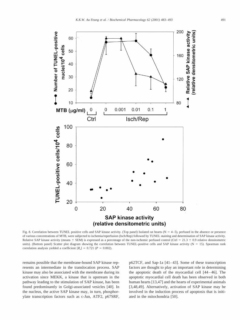
To evaluate the relationship between the number ofTUNEL-positive cells and SAP kinase, a concentration–response experiment using the isolated ischemic/reperfusedrat heart was performed. In the presence of 0.01mg/mL ofMTB, the activated SAP kinase activity was inhibited by32% (Fig. 8, top panel). There was no change in the numberof TUNEL-positive cells at this MTB concentration. Whenthe concentration of MTB was increased to 0.1mg/mL, thepercent inhibition of activated SAP kinase activity wasincreased to 36%. At this concentration, the number ofTUNEL-positive cells was also decreased from 586 6 to47 6 5 cells (Fig. 8, top panel). At 1mg/mL of MTB, theactivation of SAP kinase was abolished, and there was nodifference in the number of TUNEL-positive cells in theischemic/reperfused heart when compared with that of the
non-ischemic control. The bottom panel of Fig. 8 shows acorrelation between the number of TUNEL-positive cellsand SAP kinase activity (Spearman rank correlation coeffi-cient [Rs] 5 0.721,P 5 0.002).
4. Discussion
The present study demonstrated that MTB, a bioactivecompound isolated from Danshen, protected the ischemic/reperfused heart against apoptosis. Our results suggest thatthis cardioprotective effect of MTB may have resulted froman attenuation of SAP kinase activity in the heart, repre-senting one of the mechanisms contributing to the anti-apoptotic effect.
A novel finding of this study is that the administration ofMTB markedly attenuated the activation of SAP kinasefollowing myocardial ischemia/reperfusion. The inhibitoryactivity of MTB is through (i) a direct inhibition of thephosphotransferase activity of SAP kinase, and (ii) a re-duced nuclear translocation of the activated kinase. At lowmicromolar concentrations, MTB was effective in inhibitingSAP kinase activity. Kinetic analysis revealed that the ap-parentKm value for ATP was greater in the presence ofMTB, indicating a decrease in the affinity of the kinase forATP. In combination with an unchangedVmax, a competi-tive type of inhibition by MTB is indicated. Furthermore, adecreased apparentVmax/Km ratio also suggested a de-creased efficiency in the utilization of ATP by SAP kinasein the presence of MTB. This competitive inhibition of SAPkinase by MTB is probably reversible. If MTB interactsirreversibly with SAP kinase, then the omission of MTBonly during the second phosphotransferase step of the SAPkinase assay should still result in an inhibition of kinaseactivity. However, this was not the case. When MTB waspresent only during the incubation period for binding ofSAP kinase to c-Jun, an increase in the activity of SAPkinase was detected. It is interesting that MTB at the sametime also enhanced the binding of SAP kinase to c-Jun,which may result in a more rapid enzyme reaction (phos-photransferase reaction). Karin and colleagues [29] havedetermined the protein domain in c-Jun that was involved inthe binding of SAP kinase. MTB may act by opening up thisregion of c-Jun to allow its binding to SAP kinase. Recently,the presence of a JNK(SAP kinase)-interacting protein (JIP)has been reported. It has been proposed that JIP may act asa scaffold protein that can link upstream kinases to SAPkinase, thus providing specificity and enhanced activationof the stress-activated kinase cascade [30]. It remains to beinvestigated whether MTB enhances binding of SAP kinaseto c-Jun via the interaction with JIP.
Apoptosis has been suggested to account for a significantproportion of cardiomyocyte death observed in reperfusioninjury [13,31]. Reperfusion is a phenomenon that normallyfollows ischemic episodes, which underlies myocardial in-farction, thrombotic stroke, embolic vascular occlusions,
Fig. 6. In vitro effect of MTB on SAP kinase activity. Phosphotransferaseactivity of SAP kinase was determined in the absence (control, 0mg/mL 554.46 6.6 relative densitometric units) or presence of various concentra-tions of MTB. Western blot and the subsequent quantitation of the phos-phorylated GST-c-Jun were performed. Each value represents the mean6SEM (N 5 4–6). Key: (*) P , 0.05 compared with the control.
Table 1Apparent kinetic parameters of SAP kinase in the presence and absenceof MTB
Experiment Km
(mM)Vmax
(fmol/min)Vmax/Km
(fmol/min z mM)
No MTB 10.7 2.8 0.26MTB (1 mg/mL) 18.8 2.7 0.14
Values of the apparentKm and Vmax were determined from linearregression analysis of two experiments. Experimental differences were lessthan 10%.
489K.K.W. Au-Yeung et al. / Biochemical Pharmacology 62 (2001) 483–493
angina pectoris, peripheral vascular insufficiency, cardiacsurgery, and organ transplantation [32]. The attenuation ofreperfusion injury and cell loss is an important process inthe functional recovery of the heart. Activation of SAPkinase is damaging for the heart since it may lead to apo-ptosis of cardiomyocytes. The decrease in the activation ofSAP kinase by MTB may lead to a decrease in apoptosis,thus reducing the injurious effect of reperfusion. In thisstudy, a positive correlation was obtained on the number ofTUNEL-positive cells and the level of SAP kinase activity.Results also indicated that there was an increased level ofapoptosis in the ischemic/reperfused isolated heart. Thiswas evident from the increased number of TUNEL-positivecells and the level of PARP fragments. The presence ofMTB in the perfusate significantly attenuated these cellularmarkers of apoptosis. At a low concentration (10 ng/mL),MTB was able to inhibit 32% of the activated SAP kinaseactivity while having no significant effect on apoptosis.When higher concentrations of MTB were used, a furtherinhibition of SAP kinase and an attenuation of apoptosiswere observed. These results indicated that the completeinhibition of apoptosis might require the inhibition of sev-eral processes or signal transduction pathways. It may be thecombination of these effects and that of the direct/indirectinhibition of SAP kinase that may contribute to the cardio-protective effect observed in this study.
There may be additional mechanisms for the protectiveeffect of MTB against apoptosis. Since oxidative stressresulting from myocardial ischemia can stimulate the acti-
vation of SAP kinase [6,33], injuries caused by the oxida-tive stress may be alleviated by the inhibition of SAPkinase. In a separate study, we have demonstrated that MTBalso possesses a strong inhibitory effect on the oxidativemodification of low-density lipoprotein [34]. Therefore, theantioxidant effect [34,35] and the reported free radical scav-enging activity [36] of MTB may also be responsible for theinhibitory action on apoptosis. There is also the possibilityfor the involvement of PKC-e in the activation of SAPkinase during ischemic preconditioning [37]. However,there is also evidence that the induction of apoptosis via theSAP kinase pathway may be independent of protein kinaseC [38]. Other pathways leading to apoptosis via the activa-tion of SAP kinase are yet to be discovered. Additionally,there are other independent pathways that may lead toapoptosis. Ma and colleagues [39] recently demonstratedthat inhibition of p38 MAPK could attenuate reperfusioninjury causing apoptosis. However, the use of a specificinhibitor of p38 MAPK failed to completely inhibit apopto-sis. They suggested that signal transduction pathways suchas JNK/SAP kinase may also contribute to the apoptoticprocess. The crosstalk of these signaling pathways leadingto apoptosis will be the subject of further study.
Data from the present study also demonstrate an inhibi-tion of the translocation of activated SAP kinase after isch-emia/reperfusion. It is interesting to note that there was asignificant increase in active SAP kinase in the membranefraction. Since the mechanism of translocation of SAP ki-nase from the cytosol to the nucleus is still unknown, it
Fig. 7. Effect of MTB on c-Jun binding and phosphotransferase activities of SAP kinase. The lower panel shows representative autoradiograms ofphosphorylated GST-c-Jun as a measurement of SAP kinase activity. Bar chart values (N5 5–8) represent SAP kinase activity (means6 SEM) expressedas a percentage of the corresponding control. Control value for binding was 51.06 3.9 relative densitometric units; kinase step5 50.7 6 3.0 relativedensitometric units; both5 51.06 7.4 relative densitometric units. Key (*)P , 0.001, (†)P , 0.01, (‡)P , 0.02, and (§)P , 0.05 compared with thecorresponding control.
490 K.K.W. Au-Yeung et al. / Biochemical Pharmacology 62 (2001) 483–493
remains possible that the membrane-bound SAP kinase rep-resents an intermediate in the translocation process. SAPkinase may also be associated with the membrane during itsactivation since MEKK, a kinase that is upstream in thepathway leading to the stimulation of SAP kinase, has beenfound predominantly in Golgi-associated vesicles [40]. Inthe nucleus, the active SAP kinase may, in turn, phosphor-ylate transcription factors such as c-Jun, ATF2, p67SRF,
p62TCF, and Sap-1a [41–43]. Some of these transcriptionfactors are thought to play an important role in determiningthe apoptotic death of the myocardial cell [44–46]. Theapoptotic myocardial cell death has been observed in bothhuman hearts [13,47] and the hearts of experimental animals[3,48,49]. Alternatively, activation of SAP kinase may beinvolved in the induction process of apoptosis that is initi-ated in the mitochondria [50].
Fig. 8. Correlation between TUNEL positive cells and SAP kinase activity. (Top panel) Isolated rat hearts (N5 4–5), perfused in the absence or presenceof various concentrations of MTB, were subjected to ischemia/reperfusion (Isch/Rep) followed by TUNEL staining and determination of SAP kinase activity.Relative SAP kinase activity (means6 SEM) is expressed as a percentage of the non-ischemic perfused control (Ctrl5 21.36 0.9 relative densitometricunits). (Bottom panel) Scatter plot diagram showing the correlation between TUNEL-positive cells and SAP kinase activity (N5 15). Spearman rankcorrelation analysis yielded a coefficient [Rs] 5 0.721 (P 5 0.002).
491K.K.W. Au-Yeung et al. / Biochemical Pharmacology 62 (2001) 483–493
In summary, the present study clearly demonstrates theanti-apoptotic effect of MTB, a purified compound fromDanshen. This cardioprotective effect of MTB may be ex-erted via its direct/indirect inhibition of SAP kinase activityand the reduction in nuclear translocation of the activekinase.
Acknowledgments
The authors wish to acknowledge Mr. W.M.K. Leung forhis excellent technical assistance. The work described herewas supported by grants from the Research Grant Councilof Hong Kong Special Administrative Region, China (HKU7297/98M and 7356/00M), the National Science Founda-tion of China/Research Grant Council of HKSAR JointResearch Scheme (NSFC/HKU 39), and an OutstandingResearcher Award to Y.L.S. and a Sir Edward Youde Me-morial Fellowship to K.K.W.A-Y.
References
[1] Ip YT, Davis RJ. Signal transduction by the c-Jun N-terminal kinase(JNK)—from inflammation to development. Curr Opin Cell Biol1998;10:205–19.
[2] Force T, Pombo CM, Avruch JA, Bonventre JV, Kyriakis JM. Stress-activated protein kinases in cardiovascular disease. Circ Res 1996;78:947–53.
[3] Yin T, Sandhu G, Wolfgang CD, Burrier A, Webb RL, Rigel DF, HaiT, Whelan J. Tissue-specific pattern of stress kinase activation inischemic/reperfused heart and kidney. J Biol Chem 1997;272:19943–50.
[4] Mizukami Y, Yoshioka K, Morimoto S, Yoshida K. A novel mech-anism of JNK1 activation. Nuclear translocation and activation ofJNK1 during ischemia and reperfusion. J Biol Chem 1997;272:16657–62.
[5] Bogoyevitch MA, Gillespie-Brown J, Ketterman AJ, Fuller SJ, Ben-Levy R, Ashworth A, Marshall CJ, Sugden PH. Stimulation of thestress-activated mitogen-activated protein kinase subfamilies in per-fused heart. p38/RK mitogen-activated protein kinases and c-JunN-terminal kinases are activated by ischemia/reperfusion. Circ Res1996;79:162–73.
[6] Ferrari R, Ceconi C, Curello S, Alfieri O, Visioli O. Myocardialdamage during ischaemia and reperfusion. Eur Heart J 1993;14(SupplG):25–30.
[7] Webster KA, Discher DJ, Bishopric NH. Regulation offos and junimmediate-early genes by redox or metabolic stress in cardiac myo-cytes. Circ Res 1994;74:679–86.
[8] Zanke BW, Lee C, Arab S, Tannock IF. Death of tumor cells afterintracellular acidification is dependent on stress-activated proteinkinases (SAPK/JNK) pathway activation and cannot be inhibited byBcl-2 expression or interleukin 1b-converting enzyme inhibition.Cancer Res 1998;58:2801–8.
[9] He H, Li HL, Lin A, Gottlieb RA. Activation of the JNK pathway isimportant for cardiomyocyte death in response to simulated ischemia.Cell Death Differ 1999;6:987–91.
[10] Haunstetter A, Izumo S. Apoptosis: basic mechanisms and implica-tions for cardiovascular disease. Circ Res 1998;82:1111–29.
[11] Gottlieb RA, Engler RL. Apoptosis in myocardial ischemia-reperfu-sion. Ann NY Acad Sci 1999;874:412–26.
[12] Bromme HJ, Holtz J. Apoptosis in the heart: when and why? Mol CellBiochem 1996;163–164:261–75.
[13] Olivetti G, Abbi R, Quaini F, Kajstura J, Cheng W, Nitahara JA,Quaini E, Di Loreto C, Beltrami CA, Krajewski S, Reed JC, AnversaP. Apoptosis in the failing human heart. N Engl J Med 1997;336:1131–41.
[14] Yue TL, Ma XL, Wang X, Romanic AM, Liu GL, Louden C, Gu JL,Kumar S, Poste G, Ruffolo RR Jr, Feuerstein GZ. Possible involve-ment of stress-activated protein kinase signaling pathway and Fasreceptor expression in prevention of ischemia/reperfusion-inducedcardiomyocyte apoptosis by carvedilol. Circ Res 1998;82:166–74.
[15] Pharmacopoeia of People’s Republic of China, vol. I. Beijing: Chem-ical Industry Press, 2000. p. 57–8.
[16] Chinese herbal medicine: Materia Medica. Revised edition. Chapter10. Herbs that regulate the blood. (Compiled and translated by Ben-sky D and Gamble A, with Kaptchuk T). Seattle: Eastland Press,1993. p. 267–8.
[17] Zhu Y-P, Herbs promoting blood circulation and dissolving bloodstasis. In: Chinese Materia Medica: Chemistry, pharmacology andapplications. Amsterdam: Harwood Academic Publishers, 1998. p.459–63.
[18] Lei X-L, Chiou GCY. Cardiovascular pharmacology ofPanax noto-ginseng(Burk) F.H. Chen andSalvia miltiorrhiza.Am J Chin Med1986;14:145–52.
[19] Lei X-L, Chiou GCY. Studies on cardiovascular actions ofSalviamiltiorrhiza. Am J Chin Med 1986;14:26–32.
[20] Kamata K, Noguchi M, Nagai M. Hypotensive effects of lithospermicacid B isolated from the extract ofSalviae miltiorrhizae Radixin therat. Gen Pharmacol 1994;25:69–73.
[21] Zhou W, Ruigrok TJC. Protective effect of Danshen during myocar-dial ischemia and reperfusion: an isolated rat heart study. Am J ChinMed 1990;18:19–24.
[22] Shanghai Cooperative Group for the Study of Tanshinone IIA. Ther-apeutic effect of sodium tanshinone IIA sulfonate in patients withcoronary heart disease. A double blind study. J Trad Chin Med1984;4:20–4.
[23] Kamata K, Iizuka T, Nagai M, Kasuya Y. Endothelium-dependentvasodilator effects of the extract fromSalviae miltiorrhizae radix: Astudy on the identification of lithospermic acid B in the extracts. GenPharmacol 1993;24:977–81.
[24] Fung KP, Zeng LH, Wu J, Wong HNC, Lee CM, Hon PM, ChangHM, Wu TW. Demonstration of the myocardial salvage effect oflithospermic acid B isolated from the aqueous extract ofSalviamiltiorrhiza. Life Sci 1993;52:PL239–44.
[25] Siow YL, Choy PC, Leung WMK, O K. Effect ofFlos carthamionstress-activated protein kinase activity in the isolated reperfused ratheart. Mol Cell Biochem 2000;207:41–7.
[26] Ben-Sasson SA, Sherman Y, Gavrieli Y. Identification of dyingcells—In situ staining. In: Schwartz LM, Osborne BA, editors. Celldeath, vol. 46. San Diego: Academic Press, 1995. p. 29–39.
[27] Bradford MM. A rapid and sensitive method for the quantitation ofmicrogram quantities of protein utilizing the principle of protein-dyebinding. Anal Biochem 1976;72:248–54.
[28] Duriez PJ, Shah GM. Cleavage of poly(ADP-ribose) polymerase: asensitive parameter to study cell death. Biochem Cell Biol 1997;75:337–49.
[29] Kallunki T, Deng T, Hibi M, Karin M. c-Jun can recruit JNK tophosphorylate dimerization partners via specific docking interactions.Cell 1996;87:929–39.
[30] Whitmarsh AJ, Cavanagh J, Tournier C, Yasuda J, Davis RJ. Amammalian scaffold complex that selectively mediates MAP kinaseactivation. Science 1998;281:1671–4.
[31] Veinot JP, Gattinger DA, Fliss H. Early apoptosis in human myocar-dial infarcts. Hum Pathol 1997;28:485–92.
[32] Maxwell SR, Lip GY. Reperfusion injury: a review of the pathophys-iology, clinical manifestations and therapeutic options. Int J Cardiol1997;58:95–117.
[33] Clerk A, Michael A, Sugden PH. Stimulation of multiple mitogen-activated protein kinase sub-families by oxidative stress and phos-
492 K.K.W. Au-Yeung et al. / Biochemical Pharmacology 62 (2001) 483–493

phorylation of the small heat shock protein, HSP25/27, in neonatalventricular myocytes. Biochem J 1998;333:581–9.
[34] O K, Lynn EG, Vazhappilly R, Au-Yeung KW, Zhu D-Y, Siow YL.Magnesium tanshinoate B (MTB) inhibits low density lipoproteinoxidation. Life Sci 2001;68:903–12.
[35] Fung KP, Wu J, Zeng LH, Wong HN, Lee CM, Hon PM, Chang HM,Wu TW. Lithospermic acid B as an antioxidant-based protector ofcultured ventricular myocytes and aortic endothelial cells of rabbits.Life Sci 1993;53:L189–93.
[36] Yokozawa T, Chung HY, Dong E, Oura H. Confirmation that mag-nesium lithospermate B has a hydroxyl radical-scavenging action.Exp Toxicol Pathol 1995;47:341–4.
[37] Ping P, Zhang J, Huang S, Cao X, Tang XL, Li RC, Zheng YT, QiuY, Clerk A, Sugden P, Han J, Bolli R. PKC-dependent activation ofp46/p54 JNKs during ischemic preconditioning in conscious rabbits.Am J Physiol 1999;277:H1771–85.
[38] Yu R, Mandlekar S, Tan T-H, Kong A-NT. Activation of p38 andc-jun N-terminal kinase pathways and induction of apoptosis bychelerythrine do not require inhibition of protein kinase C. J BiolChem 2000;275:9612–9.
[39] Ma XL, Kumar S, Gao F, Louden CS, Lopez BL, Christopher TA,Wang C, Lee JC, Feuerstein GZ, Yue TL. Inhibition of p38 mitogen-activated protein kinase decreases cardiomyocyte apoptosis and im-proves cardiac function after myocardial ischemia and reperfusion.Circulation 1999;99:1685–91.
[40] Fanger GR, Johnson NL, Johnson GL. MEK kinases are regulated byEGF and selectively interact with Rac/Cdc42. EMBO J 1997;16:4961–72.
[41] Brand T, Sharma HS, Fleischmann KE, Duncker DJ, McFalls EO,Verdouw PD, Schaper W. Proto-oncogene expression in porcine
myocardium subjected to ischemia and reperfusion. Circ Res 1992;71:1351–60.
[42] Janknecht R, Hunter T. Activation of the Sap-1a transcription factorby the c-Jun N-terminal kinase (JNK) mitogen-activated protein ki-nase. J Biol Chem 1997;272:4219–24.
[43] Webster KA, Discher DJ, Bishopric NH. Induction and nuclear ac-cumulation offos and jun proto-oncogenes in hypoxic cardiac myo-cytes. J Biol Chem 1993;268:16852–8.
[44] Estus S, Zaks WJ, Freeman RS, Gruda M, Bravo R, Johnson EM Jr.Altered gene expression in neurons during programmed cell death:identification of c-jun as necessary for neuronal apoptosis. J Cell Biol1994;127:1717–27.
[45] Roffler-Tarlov S, Gibson Brown JJ, Tarlov E, Stolarov J, ChapmanDL, Alexiou M, Papaioannou VE. Programmed cell death in theabsence of c-Fos and c-Jun. Development 1996;122:1–9.
[46] Xia Z, Dickens M, Raingeaud J, Davis RJ, Greenberg ME. Opposingeffects of ERK and JNK-p38 MAP kinases on apoptosis. Science1995;270:1326–31.
[47] Schwartz K, Mercadier JJ. Molecular and cellular biology of heartfailure. Curr Opin Cardiol 1996;11:227–36.
[48] Black SC, Huang JQ, Rezaiefar P, Radinovic S, Eberhart A, Nichol-son DW, Rodger IW. Co-localization of the cysteine proteasecaspase-3 with apoptotic myocytes afterin vivo myocardial ischemiaand reperfusion in the rat. J Mol Cell Cardiol 1998;30:733–42.
[49] Piot CA, Padmanaban D, Ursell PC, Sievers RE, Wolfe CL. Ischemicpreconditioning decreases apoptosis in rat heartsin vivo. Circulation1997;96:1598–604.
[50] Jacotot E, Costantini P, Laboureau E, Zamzami N, Susin SA, Kroe-mer G. Mitochondrial membrane permeabilization during the apopto-tic process. Ann NY Acad Sci 1999;887:18–30.
493K.K.W. Au-Yeung et al. / Biochemical Pharmacology 62 (2001) 483–493
Copyright © 2022 FDOKUMEN