In-vitro dissolution of magnesium-calcium binary alloys: Clarifying the unique role of calcium...
10
In-vitro dissolution of magnesium–calcium binary alloys: Clarifying the unique role of calcium additions in bioresorbable magnesium implant alloys Nicholas T. Kirkland, 1 Nick Birbilis, 2 Jemimah Walker, 3 Tim Woodfield, 1,4 George J. Dias, 3 Mark P. Staiger 1 1 BioMate Group, Department of Mechanical Engineering, University of Canterbury, Private Bag 4800, Christchurch, New Zealand 2 Department of Materials Engineering, Monash University, Clayton, Victoria 3800, Australia 3 Department of Anatomy & Structural Biology, University of Otago, P.O. Box 913, Dunedin, New Zealand 4 Department of Orthopedic Surgery, University of Otago Christchurch, PO Box 4345, Christchurch, New Zealand Received 16 February 2010; revised 23 April 2010; accepted 10 May 2010 Published online 19 August 2010 in Wiley Online Library (wileyonlinelibrary.com). DOI: 10.1002/jbm.b.31687 Abstract: A systematic investigation of a series of magne- sium–calcium binary alloys is presented to reveal the influence of increasing calcium (Ca) additions on the in vitro degradation of magnesium (Mg). Because of its prevalence in structural tissues, Ca is among the most biologically viable additions to orthopedic-intended Mg-based biomaterials. Hence, a funda- mental electrochemical study of Ca additions to Mg biomateri- als is essential to its continued role as an alloying addition. In this work, in vitro degradation conditions closer to the physio- logical environment were implemented through the addition of proteins to simulated body fluid and maintenance of a con- stant pH, with tests conducted using Hanks solution, minimum essential medium (MEM), and MEM containing fetal bovine serum. Alloying with Ca leads to the formation of Mg 2 Ca intermetallic particles that result in systematically enhanced dissolution kinetics. This observation is rationalized via micro- electrochemical tests upon the Mg 2 Ca intermetallic in isolation, which reveals rapid anodic kinetics. Hence, the extent of Mg- Ca alloy dissolution can be modified depending on the amount of Mg 2 Ca present, suggesting that Ca can be deployed as a functional addition capable of not only enhancing bio- dissolution of the alloy, but being able to do this in a system- atic, controllable manner depending on its volume fraction. In addition, up to a 3-fold reduction in the corrosion rate is observed with corrosion testing in an albumin-containing medium when compared to Hanks solution, the results highlighting that the use of a physiologically ‘‘correct’’ medium is essential for the in vitro screening of Mg-based alloys suitable for orthopaedic applications. V C 2010 Wiley Periodicals, Inc. J Biomed Mater Res Part B: Appl Biomater 95B: 91–100, 2010. Key Words: magnesium, calcium, electrochemical corrosion, microelectrochemical cell, simulated body fluid, biomaterials, proteins INTRODUCTION Magnesium (Mg) alloys are a promising new class of degradable, structural biomaterials for orthopedic applica- tions. The field of Mg-based biomaterials was first reviewed by Staiger et al., 1 and again recently by Zeng et al., 2 and Witte et al. 3 At no other time in history has the potential of Mg-based metallic alloys as biomedical implant materials been so intensely researched with over 75 journal papers published in the last 3 years. Mg alloys are purported to provide a number of benefits over current orthopedic biomaterials, including a high strength to weight ratio; elastic modulus closer to that of bone compared with titanium 1 ; biocompatibility, osseointe- gration, and enhanced growth of bone-like apatite in vivo 4–6 ; increased osteoconduction and osteogenesis 7,8 ; and the ability to biodegrade in vivo. 9,10 The latter characteristic is the primary advantage of Mg over other metallic biomateri- als. The biodegradable properties of Mg present a number of benefits for its use as an orthopedic biomaterial. For example, degradable implants require only a single opera- tion, eliminating the time and cost associated with a second- ary surgery, or replacing, the primary implant. Furthermore, the period the implant is exposed to in vivo factors such as stress shielding and inflammation is reduced. 11 Finally, the metabolism of Mg and its excretion via the kidneys is a natural physiological process that is well understood. 12–14 Some notable pioneering efforts in the first half of the last century examined the potential of Mg alloys as orthope- dic biomaterials. 15–20 Until recently, sustained research into Mg-based biomaterials was virtually nonexistent, likely due to problems with rapid corrosion and the advent of stain- less steel and titanium alloy development. The corrosion of Mg produces hydrogen gas that accumulates in vivo adjacent to the implant. This accretion of hydrogen gas and Correspondence to: M. P. Staiger; e-mail: [email protected] Contract grant sponsor: New Zealand Foundation for Research, Science and Technology (FRST) V C 2010 WILEY PERIODICALS, INC. 91
-
Upload
canterbury-nz -
Category
Documents
-
view
0 -
download
0
Transcript of In-vitro dissolution of magnesium-calcium binary alloys: Clarifying the unique role of calcium...

In-vitro dissolution of magnesium–calcium binary alloys:Clarifying the unique role of calcium additions inbioresorbable magnesium implant alloys
Nicholas T. Kirkland,1 Nick Birbilis,2 Jemimah Walker,3 Tim Woodfield,1,4
George J. Dias,3 Mark P. Staiger1
1BioMate Group, Department of Mechanical Engineering, University of Canterbury, Private Bag 4800,
Christchurch, New Zealand2Department of Materials Engineering, Monash University, Clayton, Victoria 3800, Australia3Department of Anatomy & Structural Biology, University of Otago, P.O. Box 913, Dunedin, New Zealand4Department of Orthopedic Surgery, University of Otago Christchurch, PO Box 4345, Christchurch, New Zealand
Received 16 February 2010; revised 23 April 2010; accepted 10 May 2010
Published online 19 August 2010 in Wiley Online Library (wileyonlinelibrary.com). DOI: 10.1002/jbm.b.31687
Abstract: A systematic investigation of a series of magne-
sium–calcium binary alloys is presented to reveal the influence
of increasing calcium (Ca) additions on the in vitro degradation
of magnesium (Mg). Because of its prevalence in structural
tissues, Ca is among the most biologically viable additions to
orthopedic-intended Mg-based biomaterials. Hence, a funda-
mental electrochemical study of Ca additions to Mg biomateri-
als is essential to its continued role as an alloying addition. In
this work, in vitro degradation conditions closer to the physio-
logical environment were implemented through the addition
of proteins to simulated body fluid and maintenance of a con-
stant pH, with tests conducted using Hanks solution, minimum
essential medium (MEM), and MEM containing fetal bovine
serum. Alloying with Ca leads to the formation of Mg2Ca
intermetallic particles that result in systematically enhanced
dissolution kinetics. This observation is rationalized via micro-
electrochemical tests upon the Mg2Ca intermetallic in isolation,
which reveals rapid anodic kinetics. Hence, the extent of Mg-
Ca alloy dissolution can be modified depending on the
amount of Mg2Ca present, suggesting that Ca can be deployed
as a functional addition capable of not only enhancing bio-
dissolution of the alloy, but being able to do this in a system-
atic, controllable manner depending on its volume fraction. In
addition, up to a 3-fold reduction in the corrosion rate is
observed with corrosion testing in an albumin-containing
medium when compared to Hanks solution, the results
highlighting that the use of a physiologically ‘‘correct’’ medium
is essential for the in vitro screening of Mg-based alloys
suitable for orthopaedic applications. VC 2010 Wiley Periodicals,
Inc. J Biomed Mater Res Part B: Appl Biomater 95B: 91–100, 2010.
Key Words: magnesium, calcium, electrochemical corrosion,
microelectrochemical cell, simulated body fluid, biomaterials,
proteins
INTRODUCTION
Magnesium (Mg) alloys are a promising new class ofdegradable, structural biomaterials for orthopedic applica-tions. The field of Mg-based biomaterials was first reviewedby Staiger et al.,1 and again recently by Zeng et al.,2 andWitte et al.3 At no other time in history has the potential ofMg-based metallic alloys as biomedical implant materialsbeen so intensely researched with over 75 journal paperspublished in the last 3 years.
Mg alloys are purported to provide a number of benefitsover current orthopedic biomaterials, including a highstrength to weight ratio; elastic modulus closer to that ofbone compared with titanium1; biocompatibility, osseointe-gration, and enhanced growth of bone-like apatite in vivo4–6;increased osteoconduction and osteogenesis7,8; and theability to biodegrade in vivo.9,10 The latter characteristic isthe primary advantage of Mg over other metallic biomateri-
als. The biodegradable properties of Mg present a numberof benefits for its use as an orthopedic biomaterial. Forexample, degradable implants require only a single opera-tion, eliminating the time and cost associated with a second-ary surgery, or replacing, the primary implant. Furthermore,the period the implant is exposed to in vivo factors such asstress shielding and inflammation is reduced.11 Finally, themetabolism of Mg and its excretion via the kidneys is anatural physiological process that is well understood.12–14
Some notable pioneering efforts in the first half of thelast century examined the potential of Mg alloys as orthope-dic biomaterials.15–20 Until recently, sustained research intoMg-based biomaterials was virtually nonexistent, likely dueto problems with rapid corrosion and the advent of stain-less steel and titanium alloy development. The corrosion ofMg produces hydrogen gas that accumulates in vivo adjacentto the implant. This accretion of hydrogen gas and
Correspondence to: M. P. Staiger; e-mail: [email protected]
Contract grant sponsor: New Zealand Foundation for Research, Science and Technology (FRST)
VC 2010 WILEY PERIODICALS, INC. 91
subsequent formation of subcutaneous hydrogen bubbleswas a strong disincentive to further clinical use.21 Thus, thecontrolled release of Mg in vivo is a major challenge yet tobe resolved for the safe and effective use of Mg in ortho-pedic implants.
The development of suitable biodegradable implantalloys is a multidisciplinary challenge, since freedom in alloydesign must be confined to a range of alloying additionsthat are nontoxic in a medical sense. This rules out a largenumber of alloying elements common to commercial Mgalloys, including appreciable amounts of rare earthelements, some heavy metals, and to a large extent, alumin-ium.22,23 A promising and biocompatible element is calcium(Ca), and while some limited studies to date have reportedthe use of Ca,24–26 a systematic and fundamental study ofthe role of Ca additions upon dissolution of Mg alloysin vitro is warranted and timely. Fundamental studies, suchas the work described herein, are the necessary precursorfrom an alloy design perspective, for the engineering ofalloys with predictable and customized dissolution rates,by helping to understand the composition-microstructure-electrochemistry paradigm of these new biomaterials.
To date, the majority of in vitro degradation studies ofMg have been performed using a simulated body fluid (SBF)as the electrolyte,6,25,27-35 most commonly Hanks’ BalancedSalt Solution (Hanks solution).34,36–38 The composition of anSBF is based on a mixture of inorganic ionic salts that arein similar proportions to those found in human bloodplasma (Table II). However, corrosion tests performed byClarke and Hickman39 demonstrated, early on, the impor-tance of including organic components to create a morerealistic SBF when measuring mass loss for a range of met-als. In addition, the use of SBFs similar in composition toin vivo conditions combined with a controlled pH and tem-perature have been found to be critical in the evaluation ofbioactivity of biomaterials.40 The inclusion of proteins, suchas albumin, in an SBF has been shown to dramatically affectthe corrosion properties of metallic biomaterials.41,42 Recently,this has been further confirmed in a range of tests on pure Mg,as well as Mg alloyed with rare earth elements.43,44 To under-stand the role of Ca more generally, an electrochemical study ina variety of electrolyte media is required.
In part, it is not surprising that attempts to predictin vivo degradation behavior by testing Mg in inorganic-based SBF at ambient temperatures and without pH controlhave failed to find a correlation between the in vivo andin vitro degradation rates.29 The pH of the SBF has not beenmeasured in some studies,35,45 whereas others have onlycontrolled pH at the outset of the testing.6,29,37 However, the
pH of human serum is kept constant by the complexbiochemistry of the physiological system. In contrast, a cor-rosion cell is a confined static environment of variable pHas corrosion proceeds. For instance, OH� ions are releasedduring the corrosion of Mg, thereby raising the pH, which inturn reduces the corrosion rate.46 Hiromoto et al.47 stressedthe importance of pH control when attempting to accuratelypredict the corrosion rate of Mg and its alloys in vitro. Thus,corrosion rates measured in vitro are of little relevance tothe physiological condition if in vivo pH levels are notmaintained.6,37
The primary objective of the this study is twofold: (i)elucidate the electrochemical influence of Ca additions toMg, via a microstructural and electrochemical deconstruc-tion approach; and (ii) compare the effects of different cor-rosive media (Hanks solution, minimum essential medium(MEM) and MEM containing fetal bovine serum (FBS) onthe in vitro degradation properties of Mg-Ca alloys.
EXPERIMENTAL METHODS
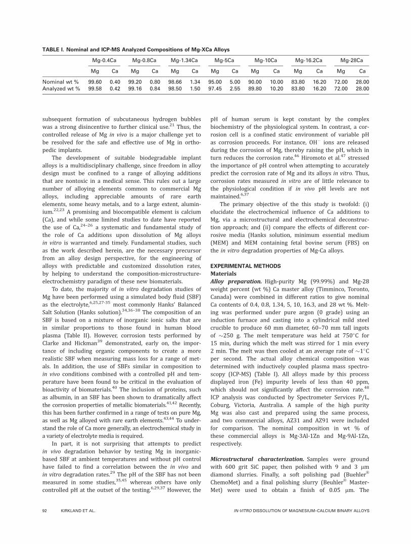
MaterialsAlloy preparation. High-purity Mg (99.99%) and Mg-28weight percent (wt %) Ca master alloy (Timminco, Toronto,Canada) were combined in different ratios to give nominalCa contents of 0.4, 0.8, 1.34, 5, 10, 16.3, and 28 wt %. Melt-ing was performed under pure argon (0 grade) using aninduction furnace and casting into a cylindrical mild steelcrucible to produce 60 mm diameter, 60–70 mm tall ingotsof �250 g. The melt temperature was held at 750�C for15 min, during which the melt was stirred for 1 min every2 min. The melt was then cooled at an average rate of �1�Cper second. The actual alloy chemical composition wasdetermined with inductively coupled plasma mass spectro-scopy (ICP-MS) (Table I). All alloys made by this processdisplayed iron (Fe) impurity levels of less than 40 ppm,which should not significantly affect the corrosion rate.48
ICP analysis was conducted by Spectrometer Services P/L,Coburg, Victoria, Australia. A sample of the high purityMg was also cast and prepared using the same process,and two commercial alloys, AZ31 and AZ91 were includedfor comparison. The nominal composition in wt % ofthese commercial alloys is Mg-3Al-1Zn and Mg-9Al-1Zn,respectively.
Microstructural characterization. Samples were groundwith 600 grit SiC paper, then polished with 9 and 3 lmdiamond slurries. Finally, a soft polishing pad (BuehlerV
R
ChemoMet) and a final polishing slurry (BeuhlerVR
Master-Met) were used to obtain a finish of 0.05 lm. The
TABLE I. Nominal and ICP-MS Analyzed Compositions of Mg-XCa Alloys
Mg-0.4Ca Mg-0.8Ca Mg-1.34Ca Mg-5Ca Mg-10Ca Mg-16.2Ca Mg-28Ca
Mg Ca Mg Ca Mg Ca Mg Ca Mg Ca Mg Ca Mg Ca
Nominal wt % 99.60 0.40 99.20 0.80 98.66 1.34 95.00 5.00 90.00 10.00 83.80 16.20 72.00 28.00Analyzed wt % 99.58 0.42 99.16 0.84 98.50 1.50 97.45 2.55 89.80 10.20 83.80 16.20 72.00 28.00
92 KIRKLAND ET AL. IN-VITRO DISSOLUTION OF MAGNESIUM–CALCIUM BINARY ALLOYS

as-polished surfaces were observed to display differentphases and grain boundaries without further etching. Thesurfaces were analyzed using an inverted optical microscope(LeicaV
R
DM IRM). Images were obtained with an AxioCamdigital camera and were processed using AxioVision 4.7(Sony Corporation, USA).
Scanning electron microscopy and electron backscatterdiffraction (EBSD) were carried out on an FEI Quanta 3D-FEG, with a Hikari EBSD camera and TSL OIM software. Toachieve high quality Kikuchi patterns, a gentle localized ionmill was performed immediately prior to the collection ofthe pattern, as the heterogeneous nature of the alloysprevented ultra high-quality mechanical polishing (for thepurposes of EBSD). The execution of spot EBSD wasperformed to identify the exact nature of the intermetallicphase via crystal structure determination.
Degradation testing of Mg-Ca alloysPreparation of corrosion media. Three different mediawere used for the experiments: (i) Hanks Balanced SaltSolution (or Hanks solution) (Sigma-Aldrich), (ii) MEM(Sigma-Aldrich), and (iii) a combination of MEM and FBS(MEM þ FBS) (Sigma-Aldrich). Hanks solution, the mostsimple of the three, is also the most commonly used in priorstudies of the corrosion and behaviour of Mg in SBFs. MEM,often used as a tissue culture media, was chosen as it iscloser to physiological conditions due to the addition of avariety of amino acids and vitamins that are present inhuman plasma (Sigma Aldrich, M0643). The third medium,with MEM plus 10% FBS, differed primarily through thepresence of proteins. The compositions of these can befound in Table II. Each media was filter sterilized (0.22 lm)before use (Millipore StericupV
R
). The final pH of the allthree media was measured at 7.15 6 0.05 after sterilizationusing a Metler Toledo SevenEasy S20 pH meter. The pH waskept constant across all experiments in corrosion media byconstant monitoring every 15 min. The temperature wasalso controlled to 37�C 6 1�C using a digital temperatureprobe attached to the pH meter. All three media werebuffered with 5.6 g/L of 2-(4-(2-hydroxyethyl)-1-pipera-zinyl) ethanesulfonic acid (HEPES).
Electrochemical testingBulk alloy testing. Sample preparation and cleaning for
the corrosion tests was carried out according to ASTM-G1-03.50 The ingots were cut into semicircular shapes leavingapproximately 10 cm2 on one side for corrosion experi-ments. Sample surfaces were prepared with sequential 240,400, 800, 1200, and 2000 grit paper and washed with pureethanol between each grinding step. Samples were finallywashed with 70% ethanol-water mix.
A three-electrode ‘‘flat-cell’’ (Princeton Applied Research)was used for the electrochemical measurements asdescribed previously.51 A saturated calomel electrode (SCE)reference and platinum counter electrode were employed.The cell contained approximately 300 mL of SBF to which 1cm2 of sample (working electrode) was exposed. Measure-ments were carried via a BioLogicV
R
VMP-3Z potentiostat
with a potential scan rate of 1 mV/s. Data was recordedand analyzed using EC-Lab v9.32.
Open circuit and potentiodynamic polarization (PDP)tests were used to obtain values of the corrosion potential(Ecorr) and current density (icorr). Specimens were allowedto equilibrate for 10 min to reach a suitably stable opencircuit potential (OCP) before commencing the PDP test.The icorr values were determined using a Tafel-type extra-polation of the polarization curve. The icorr value was alsoconverted into corrosion rate as shown in Eq. (1).52
CR ¼ K1icorrW
gq(1)
where CR is the corrosion rate (mm/year), K1 is a constantthat defines the units for the corrosion rate (3.27 � 10�3
mm g/lA cm year), icorr is the corrosion current density(lA/cm2), W is the atomic weight (Mg: 24.312, Ca: 40.078),g is the number of equivalents (Mg: 2, Ca: 2), q is the den-sity of the alloy (Mg: 1.74, Ca: 1.55 g/cm3).
Microelectrochemical testingMicroelectrochemical testing was carried out to evaluate theelectrochemical response of the intermetallic phase in isola-tion. The novel technique used was the micro-electrochemi-cal cell method, as outlined previously in Refs. 53–55 andreviewed by one of the authors in Ref. 56. In this method,the working electrode area is defined by the area of metalthat comes into contact with the opening of a microcapil-lary. The microcapillary is filled with electrolyte (in thiscase, preheated media), while containing a small-wire coun-ter electrode and electrolytic contact with a saturatedcalomel reference electrode. A silicone seal was applied tothe open end of the capillary to avoid any solution leakageand to allow an interference contact with the working elec-trode. The capillary opening is generally in the vicinity of10–100 lm in diameter and will vary with each capillary,such a size being sufficient to individually isolate inter-metallic particles in the alloys produced with the largest Ca
TABLE II. Compositions of All Media Used
Component Blood Hanks MEM MEM þ FBS
Naþ 140 145 117.4 119.66Kþ 5 5.8 5.4 5.36Mg2þ 1 0.4 0.4 0.46Ca2þ 2.5 1.3 1.8 1.87Cl� 100 144.6 123.5 121.15HPO4
2� 0.8 0.8 1 0.98SO4
2� 0.5 0.4 0.4 0.41D-Glucose 5 5.5 5.5 5.45Phenol red 0 0 0.03 0.03HEPES 0 25 25 25Amino acids
(g/L)No dataavailable
0 0.95 0.95
Fetal bovineserum (mL/L)
0 0 0 100
All concentrations in mmol/L unless otherwise stated. Concentra-
tions of inorganic blood contents given as in Ref. 49.
ORIGINAL RESEARCH REPORT
JOURNAL OF BIOMEDICAL MATERIALS RESEARCH B: APPLIED BIOMATERIALS | OCT 2010 VOL 95B, ISSUE 1 93

FIGURE 1. Photographs of the microelectrochemical apparatus showing the (A) entire setup, (B) microelectrode during testing, and (C) a close-
up of the microcapillary. [Color figure can be viewed in the online issue, which is available at wileyonlinelibrary.com.]
FIGURE 2. Optical light micrographs of the microstructure of (A) Mg-0.8Ca, (B) Mg-1.34Ca, (C) Mg-5Ca, and (D) Mg-10Ca binary alloys. The a-Mg
(1) and interdendritic eutectic (2) phases are also denoted with arrows.
94 KIRKLAND ET AL. IN-VITRO DISSOLUTION OF MAGNESIUM–CALCIUM BINARY ALLOYS

additions used for these studies (where particle sizesapproach many tens of microns). The microcell used inthese studies was incorporated into a lens-piece of an opti-cal microscope in the arrangement illustrated in Figure 1.
RESULTS
Microstructure of Mg-Ca alloysIt has been reported previously that the microstructure ofMg alloys has an appreciable effect on corrosion perform-ance.46,57 The corrosion of Mg alloys may be considered asummary response of the electrochemistry of the matrixand intermetallic secondary phases present. In regards tothe latter, the type, number density, and size range distribu-tions have a large impact on corrosion, as depicted classi-cally in Refs. 51 and 58. In this study, the microstructure ofMg-xCa alloys consisted of primary a-Mg dendrites and aninterdendritic eutectic phase consisting of a-Mg and Mg2Ca(Figure 2). The phase composition of Mg-Ca binary alloyshas been examined in detail previously, where the appear-ance of a Mg2Ca intermetallic phase is typical for Mg-Caalloys to a composition window up to �45 wt % Ca6,24,59,60
as predicted by the phase diagram.61 The process of rela-tively slow cooling (�0.1�C/s) from the melt leads to theformation of a dendritic microstructure. There was anincreased refinement of the a-Mg dendritic structure andMg2Ca phase from about 3 to up to 24 vol % with increas-ing Ca content from Mg-0.8Ca to Mg-28Ca, respectively.
Figure 3(A) reveals an SEM image of Mg-28Ca, wherethe intermetallic phase is clearly evident, along with a well-
defined divorced eutectic component of the microstructure.Spot EBSD upon the intermetallic phase [Figure 3(B)]reveals that the phase has the crystal structure and latticecharacteristics of Mg2Ca, in accordance with known latticestructures.61 The Mg2Ca intermetallic is unique amongst thephases typical of Mg alloys, in that the intermetallic hasthe same crystal structure (hexagonal) as the Mg itself, andthe only difference is that the Mg2Ca has lattice parametersalmost twice as large as the a-Mg.
FIGURE 3. An example of phase analysis performed by EBSD for Mg-28Ca. (A) Scanning electron micrograph of the microstructure. (B) Kikuchi
pattern of Mg2Ca phase as captured by the EBSD camera. (C) Virtual Kikuchi pattern of Mg2Ca phase as calculated by TSL OIM software. [Color
figure can be viewed in the online issue, which is available at wileyonlinelibrary.com.]
FIGURE 4. Potentiodynamic polarization curves of selected Mg-xCa
alloys in MEM þ FBS media. [Color figure can be viewed in the online
issue, which is available at wileyonlinelibrary.com.]
ORIGINAL RESEARCH REPORT
JOURNAL OF BIOMEDICAL MATERIALS RESEARCH B: APPLIED BIOMATERIALS | OCT 2010 VOL 95B, ISSUE 1 95

Electrochemical characterizationMg-Ca alloys. An example of the polarization curves forselected Mg-xCa alloys in the MEM þ FBS media is illus-trated in Figure 4. It can be readily seen that, with increas-ing calcium content, the corrosion potential becomes morenegative, with a concomitant increase in corrosion rate. Thissame overall trend was also observed (data not shown) inthe Hanks and MEM media, as summarised below.
Mg2Ca. The polarization response of the Mg2Ca intermetal-lic alone is seen in Figure 5, along with pure Mg as a com-parison. It is apparent that Mg2Ca phase overwhelminglysupports higher rates of reaction, and thus corrosion, acrossa wide range of potentials.
Effect of media composition on corrosion rateIt was observed that all alloys displayed a trend towardsreduced corrosion rate as the medium more closely mim-icked human serum (Figure 6). The addition of FBS to theMEM had a significant effect on the corrosion rate of all thealloys when compared to the Hanks and standard MEMmediums, with a decrease in the corrosion rate of between10 and 290%. Furthermore, it was observed that below thesolid solubility limit of �1.34 wt % Ca, the corrosion ratestayed the same or even slightly decreased with increasingCa additions, while above this limit, corrosion ratesincreased with increasing Ca content [Figure 6(A)]. Thecorresponding corrosion potential of each of the alloys canbe seen in Figure 6(B). Overall, the MEM þ FBS corrosionpotential values exhibited an ennoblement of the alloyscompared to the other media (Figure 7). However, therewas no clear trend when comparing the Hanks and theMEM results.
DISCUSSION
Dissolution of Mg-Ca binary alloysMicrostructural investigations described herein reveal andconfirm that alloying Mg with Ca leads to the formation ofthe intermetallic phase Mg2Ca. The presence of this second
FIGURE 5. Potentiodynamic polarization curves of Mg2Ca phase and
pure Mg from microelectrochemical testing. [Color figure can be
viewed in the online issue, which is available at wileyonlinelibrary.
com.]
FIGURE 6. The corrosion rates (A) and potentials (B) of Mg-xCa and
AZ alloys as a function of the alloy composition and corrosion media.
[Color figure can be viewed in the online issue, which is available at
wileyonlinelibrary.com.]
FIGURE 7. Potentiodynamic polarization curves of Mg-10Ca alloy as a
function of the corrosion media. [Color figure can be viewed in the
online issue, which is available at wileyonlinelibrary.com.]
96 KIRKLAND ET AL. IN-VITRO DISSOLUTION OF MAGNESIUM–CALCIUM BINARY ALLOYS

phase has a significant impact upon the measured anodickinetics of pure Mg as shown by a shift in the anodicbranches of the polarization curves toward significantlyhigher currents with increasing Ca additions (Figure 4). Incontrast, the corresponding cathodic branches are relativelyunaffected by Ca additions. If one were to extrapolate thecathodic branch of the polarization in Figure 4 for Mg-0.8Catowards increasingly negative potentials, the extrapolatedcurve would overlap the curves of the high Ca containingalloys. This result therefore suggests that Mg2Ca is a moreefficient anode than a-Mg.
The notion that Mg2Ca is more electrochemically activethan a-Mg is further confirmed by the microelectrochemicaltesting. We have demonstrated for the first time the polar-ization response of Mg2Ca, which clearly reveals very highrates of reaction (when compared to other metals or inter-metallics in engineering alloys). Nominally when intermetal-lics are present in Mg, the intermetallic assumes the role ofthe cathode, since the intermetallic—which is richer in alloy-ing element—cannot nominally support anodic reactions atthe rate of Mg, which is itself an active metal. This is not thecase with Ca additions.
In part, the genesis of the rapid dissolution of Mg itselfis owing to the fact that Mg cannot produce a stable surfaceoxide of complete coverage, as judged by a Pilling-Bedworthratio (PBR) < 1.61 Thus, the volume of the oxide producedupon Mg is lower than the volume of the metal consumed,indicating that a continuous oxide is not capable of formingin most conditions. There are only very few instances in na-ture when a PBR < 1 is encountered. If we now recall thatthe phase we identified as Mg2Ca has the identical crystalstructure as Mg, however, twice the lattice parameter mag-nitudes (i.e. Mg: a ¼ 0.32093, c ¼ 0.52107, Mg2Ca: a ¼0.623, c ¼ 1.012), then the notional expectation is thatvolume of metal consumed in Mg2Ca will be greater (for agiven number of atoms), and hence the PBR will be evenfurther below unity (i.e. �1).61 This situation—to theauthors’ knowledge—can only occur with Ca additions. It isalso noteworthy that Ca itself is amongst the only otherelements that can sustain a PBR < 1.62
This work therefore reveals quite starkly that Ca is aunique alloying addition to Mg in the context of biodegrad-able implants, in that it is an element that is both nontoxicand biocompatible, but can be deployed in an engineeringsense as a functional addition capable of not only enhancingbiodissolution of the alloy, but being able to do this in asystematic, controllable manner depending on its volumefraction. This has not been unequivocally stated before, andis not only important in the field, but gives significant creedto the notion of using the deconstruction approach hereinfor unlocking the microstructure-corrosion relationshipsthat will enable functional (i.e. customizable) implants to bedeveloped.
Influence of electrolyteThe type of media was found to strongly influence theoverall dissolution rate of Mg-Ca alloys. Given that experi-ments were conducted in various media to isolate the
impact of materials structure and reveal a general corrosionresponse, some discussion regarding electrolyte influencesis warranted. It was observed that, for a given alloy compo-sition, the anodic branch was altered, while the cathodicbranch remained essentially unaffected for media that moreclosely mimicked human serum i.e. MEM þ FBS (Figure 7).This apparent ennoblement of Ecorr is due to the reductionin anodic reaction kinetics in the presence of serum pro-teins. In fact, based upon the polarization data, the presenceof proteins appear to act as mild anodic inhibitors, leadingto a considerable decrease in icorr by stifling the overallanodic reaction rate (and hence moving the anodic branchtowards the left as seen in Figure 7.
Furthermore, the degree of ennoblement of Mg due tochanging corrosive media was not consistent across all alloycompositions. It was observed that disparities in Ecorr, asmeasured in the various media, were reduced for Ca addi-tions of 10 wt % or greater, indicating an apparentdecreased ennoblement by the MEM þ FBS medium. Theunderlying mechanism for this is unknown, although thereare three possible explanations that exist: (i) the increase insecondary phases with �10 wt % Ca may present a less‘‘attractive’’ site for protein adsorption, (ii) additional cal-cium phosphate may form on the surface, conceivably in anamorphous state, altering the corrosion kinetics and proteinadsorption,63,64 and/or (iii) a localized increase in pH at thesurface due to rapid degradation (and OH— evolution)reduced the rate of protein adsorption. For example, Sharpeet al. 65 similarly found that a higher pH decreases the rateof protein adsorption on hydroxyapatite surfaces. Furtherwork is required, however, to better understand the effectof localized pH changes on the surface–protein interactionin Mg alloys.
Many reported in vitro corrosion studies have beencarried out in Hanks solution or similar media. Chiu et al.38
found that the corrosion rate of pure Mg was 3.7 mm/yearin Hanks solution. This is three times greater than corrosionrates described in this study. However, in the study by Chiuet al., a small quantity (50 mL) of medium was used andonly changed every 2 days, which means an altered pHcould have had a significant effect on the corrosion overthis period. Moreover, the samples were left for 1 h to reachOCP, during which significant corrosion could occur beforestarting the potentiodynamic testing. With regard to corro-sion rates measured in this study, Gu et al.66 found a similarrate of corrosion for pure Mg in Hanks solution (0.36 mm/year). Zeng et al.28 also reported similar results for icorr ofAZ31 in Hanks solution, whereas Kannan and Raman25
reported similar values for icorr of AZ91 in a medium similarto Hanks solution. Wu et al.67 reported a slightly highercorrosion rate for AZ91, although they used a higher con-centration of NaCl in the medium. A comparison betweendata reported in the literature and values from the presentwork can be seen in Figure 8.
Compared with Hanks solution, MEM resulted in a lowercorrosion rate at Mg-Ca alloy levels above 5%, as well as forAZ alloys and pure Mg. In terms of media composition, themain difference between MEM and Hanks solutions was the
ORIGINAL RESEARCH REPORT
JOURNAL OF BIOMEDICAL MATERIALS RESEARCH B: APPLIED BIOMATERIALS | OCT 2010 VOL 95B, ISSUE 1 97

inclusion of amino acids at concentrations similar to thosein human serum (Table II). The effect of amino acids onthe corrosion resistance of metals is still presently poorlyunderstood, with no consensus as to whether they increase,decrease or have any effect on the corrosion rate.43,68
The addition of FBS to MEM is of primary importancedue to the presence of serum proteins in the solution.Proteins adsorb onto the surface of a biomaterial through acomplicated process that involves van der Waals forces,hydrophobic and electrostatic interactions, and hydrogenbonding.69 Protein–surface interactions are known to bean important factor in the corrosion behavior of bio-materials.2,6,33,44,70,71 Khan et al.72 found that the presenceof serum proteins reduced the corrosion of titanium alloys.Proteins have been found to dramatically affect the corro-sion rate of Mg with their addition to SBF, although thespecific reasons for this are not clear.33,44 Several theorieshave been proposed to describe the effect of the presenceof proteins on corrosion rate and are summarized asfollows: (i) proteins affect metallic corrosion by changingeither anodic or cathodic processes, or both.6 Proteinsappear to more strongly inhibit the anodic reaction basedon this study; (ii) interactions between the proteins andimplant surface alter dissolution due to a protein layer thatfurther inhibits the corrosion of the Mg.33 For example,albumin has an oval, compact, globular structure that packswell and resists denaturation during the adsorption process,thereby blocking reaction sites64; (iii) proteins form aninsoluble salt layer dense enough to provide additionalprotection through the incorporation of Ca and P at thesurface43; and (iv) a protein layer inhibits the effects ofchanges in pH and may hinder the diffusion of ions.69
General discussionWe have revealed that increasing the Ca content up to thesolid solubility limit, slightly alters the overall corrosionrate of Mg, while additions above this significantly increasethe corrosion rate.73 This is seen clearly in Figure 6(A),whereby following additions greater than a Cs (the solid
solubility limit of Ca in Mg) of �1.34 wt % Ca, the corrosionrate increases monotonically.
While Ca has been shown to improve the bone remodel-ling rate when alloyed with Mg,74 the authors do note,however, that at the highest levels of Ca additions testedherein the rate of dissolution is very rapid, and likely toorapid for use in general bioimplants. Using the calculatedcorrosion rate (mm/year), the approximate Mg mass loss ofvarious Mg-xCa alloys can be plotted as a linear function ofsurface area for different sized rectangular plates with afixed thickness:width:length ratio of 0.1:1:4 (Figure 9).Superimposed on this plot is the level of Mg (2.4–3.5 g)that the human kidneys are able to filter per day.13 Thissimplified approach is intended to illustrate that with moredetailed knowledge of Mg degradation, it will be possible todesign different medical devices by tailoring the alloy com-position and the corresponding allowable surface area ofthe device. It is important to note that the calculated degra-dation rates assume uniform corrosion, and do not take intoaccount localized forms of corrosion that are prevalent inMg alloys.49 Thus, this simple approach is likely to be alower bound estimate for the actual corrosion rate.52 Theprecise quantification of corrosion rates in the human bodyis a major challenge that lies ahead for the design and
FIGURE 8. A comparison of the corrosion rates observed in this study
to that of others. [Color figure can be viewed in the online issue,
which is available at wileyonlinelibrary.com.]
FIGURE 9. Mg mass loss rates as a function of surface area: (A) Hanks
solution, (B) MEM þ FBS. [Color figure can be viewed in the online
issue, which is available at wileyonlinelibrary.com.]
98 KIRKLAND ET AL. IN-VITRO DISSOLUTION OF MAGNESIUM–CALCIUM BINARY ALLOYS

clinical adoption of Mg biomaterials. However, the workdescribed herein reveals that it is possible to developcriteria for the design of suitable materials by deconvolvingthe microstructure and its electrochemistry.
CONCLUSIONS
The influence of Ca on the dissolution and electrochemicalresponse of Mg-Ca alloys was determined in the presence ofvarious media that mimic human serum to differing degrees.The following conclusions can be drawn from this work:
Increasing the content of Ca in a binary Mg alloy atlevels greater than the solubility limit greatly increases thecorrosion rate, to rates that are effectively the most rapid ofany Mg alloy reported.
We have presented for the first time the electrochemicalresponse of Mg2Ca uniquely. It is seen that this phase ishighly reactive. Over a wide range of potentials, Mg2Casustains dissolution rates about an order of magnitudegreater than Mg, which is already highly reactive. This isposited to be owing to the chemistry and crystal structureof Mg2Ca, and concomitant PBR � 1 which is expected,allowing the phase to be very reactive.
The presence of proteins principally affects the anodicreaction kinetics upon Mg alloys, reducing the rate of metal-lic dissolution. This translates to an inhibitive effect fromthe presence of proteins, and emphasizing that proteins area vital component to SBF to more fully emulate the body’senvironment, and thus obtain more realistic data fromin vitro degradation tests. Use of more physiologicallyrelevant test environments are a necessary next step in theevolution of in vitro tests towards effectively screeningcorrosion and biodegradation behavior in magnesium alloys.
It is unknown whether the positive effects that calciummight have on bone growth and osteoconduction wouldcounterbalance the rapid corrosion. Hence, in vivo studiesare needed to compare the results of the current in vitrotests.
This work reveals quite starkly that Ca is a unique alloy-ing addition to Mg in the context of biodegradable implants,since it is an element that can be deployed as a functionaladdition capable of enhancing biodissolotion and hence is atool in the development of implants with customisabledissolution.
ACKNOWLEDGMENTS
NB would like to thank the Australian Research Council (ARC)and the Monash Centre for Electron Microscopy (MCEM).
REFERENCES1. Staiger MP, Pietak AM, Huadmai J, Dias G. Magnesium and its
alloys as orthopedic biomaterials: A review. Biomaterials 2006;27:
1728–1734.
2. Zeng R, Dietzel W, Witte F, Hort N, Blawert C. Progress and chal-
lenge for magnesium alloys as biomaterials. Adv Eng Mater 2008;
10:B3–B14.
3. Witte F, Hort N, Vogt C, Cohen S, Kainer KU, Willumeit R, Feyera-
bend F. Degradable biomaterials based on magnesium corrosion.
Curr Opin Solid State Mater Sci 2008;12:63–72.
4. Duygulu O, Kaya RA, Oktay G, Kaya AA. Investigation on the
potential of magnesium alloy AZ31 as a bone implant. Mater Sci
Forum 2007;546–549:421–424.
5. Witte F, Reifenrath J, Muller PP, Crostack HA, Nellesen J, Bach
FW, Bormann D, Rudert M. Cartilage repair on magnesium scaf-
folds used as a subchondral bone replacement. Materialwissen-
schaft und Werkstofftechnik 2006;37:504–508.
6. Li Z, Gu X, Lou S, Zheng Y. The development of binary Mg-Ca
alloys for use as biodegradable materials within bone. Biomateri-
als 2008;29:1329–1344.
7. Howlett CR, Zreiqat H, Dell RO, Noorman J, Evans P, Dalton BA.
The effect of magnesium ion implantation into alumina upon the
adhesion of human bone derived cells. J Mater Sci: Mater Med
1994;9:715.
8. Okuma T. Magnesium and bone strength. Nutrition 2001;17:679–680.
9. Witte F, Kaese V, Haferkamp H, Switzer E, Meyer-Lindenberg A,
Wirth CJ, Windhagen H. In vivo corrosion of four magnesium alloys
and the associated bone response. Biomaterials 2005;26:3557–3563.
10. Ren Y, Wang H, Huang J, Zhang B, Yang K. Study of biodegrada-
tion of pure magnesium. Key Eng Mater 2007;342–343:601–604.
11. Galante JO, Lemons JE, Spector M, Wilson PD Jr, Wright RM.
The biological effects of implant materials. J Orthoped Res 1991;
9:760–775.
12. Saris N-EL, Mervaala E, Karppanen H, Khawaja JA, Lewenstam A.
Magnesium: An update on physiological, clinical and analytical
aspects. Clin Chim Acta 2000;294:1–26.
13. Vormann J. Magnesium: nutrition and metabolism. Mol Aspects
Med 2003;24:27–37.
14. Wolf FI, Cittadini A. Chemistry and biochemistry of magnesium.
Mol Aspects Med 2003;24:3–9.
15. Troitskii VV, Tsitrin DN. The resorbing metallic alloy ‘osteosinthe-
zit’ as material for fastening broken bone. Khirurgiia 1944;8:41–44.
16. Maier O. Uber die Verwendbarkeit von Leichtmetallen in der
Chirurgie (metallisches Magnesium als? Reizmittel zur Knochen-
neubildung). Dlsche. Z. Chir. 1940.
17. McBride BD. Absorbable metal in bone surgery. J Am Med Assoc
1938;111:2464–2467.
18. Verbrugge J. Le Materiel Metallique Resorbable En Chirurgie
Osseuse. La Press Medicale 1934;23:460–465.
19. Lambotte A. L’utilisation du magnesium comme materiel perdu
dans l’osteosynthese. Bull Mem Soc Nat Chir 1932;28:1325–1334.
20. Znamenskii MS. Metallic osteosynthesis by means of and appara-
tus made of resorbing metal. Khirurgiia 1945;12:60–63.
21. McCord CP. Chemical gas gangrene from metallic magnesium.
Ind Med 1942;11:71–79.
22. Flatten TP. Aluminium as a risk factor in Alzheimer’s disease,
with emphasis on drinking water. Brain Res Bull 2001;55:187–196.
23. Ganrot PO. Metabolism and possible health effects of aluminium.
Environ Health Perspect 1986;65:363–441.
24. Hassel T, Bach FW, Golovko AN, Krause A. Investigation of the
mechanical properties and the corosion behaviour of low alloyed
magnesium-calcium-alloys for use as absorbable biomaterial in
the implant technique. In: Pekguleryuz M, editor; Conference of
Metallurgy: Magnesium Technology in the Global Age, Montreal,
Quebec, Canada; 2006. pp 359–369.
25. Kannan MB, Raman RKS. In vitro degradation and mechanical in-
tegrity of calcium-containing magnesium alloys in modified-simu-
lated body fluid. Biomaterials 2008;29:2306–2314.
26. Kim W-C, Kim J-G, Lee J-Y, Seok H-K. Influence of Ca on the
corrosion properties of magnesium for biomaterials. Mater Lett
2008;62:4146–4148.
27. Denkena B, Witte F, Podolsky C, Lucas A. Degradable Implants
Made of Magnesium Alloys. France: Montpellier; 2005.
28. Zeng RC, Chen J, Dietzel W, Hort N, Kainer KU. Electrochemical
behavior of magnesium alloys in simulated body fluids. Trans
Nonferrous Metals Soc China 2007;17:S166–S170.
29. Witte F, Nellesen J, Crostack H-A, Kaese V, Pisch A, Beckmann F,
Windhagen H. In vitro and in vivo corrosion measurements
of magnesium alloys. Biomaterials 2006;27:1013–1018.
30. Wang H, Shi ZM, Yang K. Magnesium and magnesium alloys as
degradable metallic biomaterials. Adv Mater Res 2008;32:207–210.
31. Wang H, Estrin Y, Zuberova Z. Bio-corrosion of a magnesium alloy
with different processing histories. Mater Lett 2008;62:2476–2479.
ORIGINAL RESEARCH REPORT
JOURNAL OF BIOMEDICAL MATERIALS RESEARCH B: APPLIED BIOMATERIALS | OCT 2010 VOL 95B, ISSUE 1 99

32. Wan Y, Xiong G, Luo H, He F, Huang Y, Zhou X. Preparation and
characterization of a new biomedical magnesium-calcium alloy.
Mater Des 2008;29:2034–2037.
33. Liu C, Xin Y, Tian X, Chu PK. Degradation susceptibility of surgi-
cal magnesium alloy in artificial biological fluid containing albu-
min. J Mater Res 2007;22:1806–1814.
34. Liu C, Xin Y, Tang G, Chu PK. Influence of heat treatment on
degradation behavior of bio-degradable die-cast AZ63 magnesium
alloy in simulated body fluid. Mater Sci Eng: A 2007;456:350–357
35. Kuwahara H, Al-Abdullat Y, Ohta M, Tsutsumi S, Ikeuchi K,
Mazaki N, Aizawa T. Surface reaction of magnesium in hank’s
solutions. In: Materials Science Forum (Switzerland). Nagaoka
City, Japan: Trans Tech Publications; 2000. pp 349–358.
36. Kokubo T. Formation of biologically active bone-like apatite on
metals and polymers by a biomimetic process. Thermochim Acta
1996;280–281:479–490.
37. Lopez HY, Cortes DA, Escobedo S, Mantovani D. In vitro bioactiv-
ity assessment of metallic magnesium. Key Eng Mater 2006;
309–311:453–456.
38. Chiu KY, Wong MH, Cheng FT, Man HC. Characterization and cor-
rosion studies of fluoride conversion coating on degradable Mg
implants. Surf Coat Technol 2007;202:590–598.
39. Clarke EGC, Hickman J. An Investigation into the correlation
between the electrical potentials of metals and their behaviour in
biological fluids. J Bone Joint Surg 1953;35B:467–473.
40. Bohner M, Lemaitre J. Can bioactivity be tested in vitro with SBF
solution?. Biomaterials 2009;30:2175–2179.
41. Clarke EGC. In vitro aspects of metallic corrosion in tissue fluids.
Proc R Soc Med 1953;46:17.
42. Merrit K, Brown SA. Fretting corrosion in saline and serum.
J Biomed Mater Res 1981;15:479–488.
43. Yamamoto A, Hiromoto S. Effect of inorganic salts, amino acids
and proteins on the degradation of pure magnesium in vitro.
Mater Sci Eng: C 2009;29:1559–1568.
44. Rettig R, Virtanen S. Time-dependent electrochemical charac-
terization of the corrosion of a magnesium rare-earth alloy in
simulated body fluids. J Biomed Mater Res Part A 2008;85A:
167–175.
45. Lu SK, Yeh HI, Tian TY, Lee WH. Degradation of magnesium
alloys in biological solutions and reduced phenotypic expression
of endothelial cell grown on these alloys. In: Ibrahim F, editor.
3rd Kuala Lumpur International Conference on Biomedical Engi-
neering, 11–14 December, 2006, Kuala Lumpar, Malaysia:
Springer-Verlag. pp 98–101.
46. Bender S, Goellner J, Heyn A, Boese E. Corrosion and corrosion
testing of magnesium alloys. Mater Corrosion 2007;58:977–982.
47. Hiromoto S, Yamamoto A, Maruyama N, Somekawa H, Mukai T.
Influence of pH and flow on the polarisation behaviour of pure
magnesium in borate buffer solutions. Corrosion Sci 2008;50:
3561–3568.
48. Shaw BA, Wolfe RC. Corrosion of magnesium and magnesium-
base alloys. In: Cramer SD, Convino BS, editors. ASM Handbook.
2005. Metals Park: ASM International, pp 205–227.
49. Ghali E. Corrosion and protection of magnesium alloys. In: Mate-
rials Science Forum (Switzerland). Nagaoka City, Japan: Trans Tech
Publications; 2000. pp 261–272.
50. ASTM International. ASTM Standard G1-03, ‘‘Standard Practice
for Preparing, Cleaning, and Evaluating Corrosion Test Specimens’’.
West Conshohocken, PA: ASTM International; 2003.
51. Sudholz AD, Birbilis N, Bettles CJ, Gibson MA Corrosion behav-
iour of Mg-alloy AZ91E with atypical alloying additions. J Alloys
Compounds 2009;471:109–115.
52. ASTM International. ASTM Standard G102-89, ‘‘Standard Practice
for Calculation of Corrosion Rates and Related Information from
Electrochemical Measurements’’. West Conshohocken, PA: ASTM
International; 2004.
53. Suter T, Bohni H. Microelectrodes for studies of localized corro-
sion processes. Electrochimica Acta 1998;43:2843–2849.
54. Birbilis N, Buchheit RG. Electrochemical characteristics of inter-
metallic phases in aluminum alloys. J Electrochem Soc 2005;152:
B140–B151.
55. Lohrengel MM. Electrochemical capillary cells. Corrosion Eng Sci
Technol 2004;39:53–58.
56. Birbilis N, Padgett BN, Buchheit RG. Limitations in microelectro-
chemical capillary cell testing and transformation of electrochemi-
cal transients for acquisition of microcell impedance data. Electrochim
Acta 2005;50:3536–3544.
57. Amira S, Dube D, Tremblay R, Ghali E. Influence of the micro-
structure on the corrosion behavior of AXJ530 magnesium alloy
in 3.5% NaCl solution. Mater Character 2008;59:1508–1517.
58. Birbilis N, Easton MA, Sudholz AD, Zhu SM, Gibson MA. On the
corrosion of binary magnesium-rare earth alloys. Corrosion Sci
2009;51:683–689.
59. Bamberger M, Levi G, Vander Sande JB. Precipitation hardening
in Mg-Ca-Zn alloys. Metal Mater Trans A: Phys Metal Mater Sci
2006;37:481–487.
60. Nie JF, Muddle BC. Precipitation hardening of Mg-Ca(-Zn) alloys.
Acta Metal Sin 1997;37:1475–1481.
61. Nayeb-Hashemi AA. The Ca-Mg (Calcium-Magnesium) System.
In: International A, editor. Bulletin of Alloy Phase Diagrams; 1987.
pp 58–65.
62. ASM Handbook: Corrosion: Fundamentals, Testing and Protec-
tion. 9th ed. Materials Park: ASM International, 1987.
63. Cui FZ, Yang JX, Jiao YP, Yin QS, Zhang Y. Calcium phosphate
coating on magnesium alloy for modification of degradation
behavior. Frontiers Mater Sci China 2008;2:143–148.
64. Cheng X, Roscoe SG. Corrosion behavior of titanium in the pres-
ence of calcium phosphate and serum proteins. Biomaterials
2005;26:7350–7356.
65. Sharpe JR, Sammons RL, Marquis PM. Effect of pH on protein
adsorption to hydroxyapatite and tricalcium phosphate ceramics.
Biomaterials 1997;18:471–476.
66. Gu X, Zheng Y, Cheng Y, Zhong S, Xi T. In vitro corrosion and
biocompatibility of binary magnesium alloys. Biomaterials 2009;
30:484–498.
67. Wu G, Fan Y, Gao H, Zhai C, Zhu YP. The effect of Ca and rare
earth elements on the microstructure, mechanical properties and
corrosion behavior of AZ91D. Mater Sci Eng: A 2005;408:255–
263.
68. Solar RJ, Pollack S, Korostoff E. In vitro corrosion testing of
titanium surgical implan alloys: An approach to understanding
titanium release from implants. J Biomed Mater Res 1979;13:
217–250.
69. Mueller W-D, Mele MFLd, Nascimento ML, Zeddies M. Degrada-
tion of magnesium and its alloys: Dependence on the composi-
tion of the synthetic biological media. J Biomed Mater Res Part A
2008;9999:NA.
70. Latour RA. Biomaterials: Protein-surface interactions. In: Bowlin
GL, editor. Encyclopedia of Biomaterails and Biomedical Engi-
neering. London: Informa Healthcare; 2005, pp 270–285.
71. Mueller WD, Nascimento ML, Zeddies M, Corsico M, Gassa LM,
Mele MAFLD. Magnesium and its alloys as degradable biomateri-
als: Corrosion studies using potentiodynamic and EIS electro-
chemical techniques. Mater Res 2007;10:5–10.
72. Khan MA, Williams RL, Williams DF. The corrosion behaviour of
Ti-6Al-4V. Ti-6Al-7Nb and Ti-13Nb-13Zr in protein solutions. Bio-
materials 1999;20:631–637.
73. Wan Y, Xiong G, Luo H, He F, Huang Y, Zhou X. Preparation and
characterization of a new biomedical magnesium-calcium alloy.
Mater Design 2008;29:2034–2037.
74. Thomann M, Krause C, Bormann D, von der Hoh N, Windhagen
H, Meyer-Lindenberg A. Comparison of the resorbable magne-
sium alloys LAE442 and MgCa0.8 concerning their mechanical
properties, their progress of degradation and the bone-implant-
contact after 12 months implantation duration in a rabbit model.
Materialwissenschaft und Werkstofftechnik 2009;40:82–87.
100 KIRKLAND ET AL. IN-VITRO DISSOLUTION OF MAGNESIUM–CALCIUM BINARY ALLOYS