
Heme oxygenase-1 modulates fetal growth in the rat
6
Heme Oxygenase-1 Modulates Fetal Growth in the Rat Doron Kreiser, Xuandai Nguyen, Ron Wong, Daniel Seidman, David Stevenson, Shou Quan, Nader Abraham, and Phyllis A. Dennery Department of Pediatrics (DK, XN, RW, DSt, PAD), Stanford University School of Medicine, Stanford, California; Department of Obstetrics and Gynecology (DK, DSe), Sheba Medical Center, Tel-Hashomer, Tel-Aviv University, Israel; and Department of Pharmacology (SQ, NA), New York University, Valhalla, New York SUMMARY: Intrauterine growth restriction is associated with increased perinatal morbidity and mortality as well as with lifelong cardiovascular and metabolic complications. Deficiency of heme oxygenase 1 (HO-1) is associated with growth restriction in mice and in humans, suggesting a role for HO-1 in fetal growth and maintenance of pregnancy. We hypothesized that modulation of HO-1 in the pregnant rat would alter fetal growth. In pregnant dams, placental HO activity was significantly inhibited with zinc deuteroporphyrin IX 2,4 bis glycol, and HO-1 protein was increased by transducing adenoviral human HO-1. Inhibition of HO-1 by zinc deuteroporphyrin IX 2,4 bis glycol resulted in a significant decrease in pup size, whereas transfection with hHO-1 resulted in increased pup size. Furthermore, the expression of IGF binding protein-1 and its receptor paralleled the expression of HO-1 in the placenta and were significantly modulated by modification of HO-1 along with the expression of vascular endothelial growth factor. These observations demonstrate that HO-1 modulates fetal growth by its effects on placental growth factors. (Lab Invest 2002, 82:687–692). I ntrauterine growth restriction (IUGR) is defined as a birth weight below the 10th percentile for gesta- tional age (Goldenberg et al, 1989). In the United States, 8.6% of live births are growth-restricted (Hediger et al, 1998). Fetal growth in utero is influenced by extrinsic factors such as maternal vascular disease or intrinsic factors resulting from cell or organ dysfunction. Adverse effects of IUGR are seen immediately at birth, but the long-term morbidity and mortality of IUGR manifest into adulthood. A direct correlation between low birth weight and several adult onset diseases such as Syndrome X (hypertension, non- insulin-dependent diabetes mellitus, high serum trig- lycerides, and low serum high-density lipoproteins), cardiovascular diseases, and arterial hypertension has been demonstrated (Barker et al, 1993). The genetic contribution to IUGR is suggested by an increased prevalence of growth restriction in some families. A mutation in the IGF-1 gene can also cause IUGR (Woods et al, 1996). Furthermore, fetal cord serum IGF levels are increased in large-for-gestation- age infants and decreased in IUGR infants (Giudice et al, 1995). However, this correlation is not universal (Wang et al, 1991). A family of binding proteins (BP) mediates the biologic actions of IGFs (Han et al, 1996). The type 1 IGF receptor (IGF-1R) is a transmembrane tyrosine kinase that is widely expressed in fetal tis- sues. Activation of the receptor after binding of IGF-1 or IGF-2 results in cell proliferation and protection from apoptosis (Granerus and Engstrom, 2001), sug- gesting an important pathway for intrauterine growth. In fact, targeted mutations of the IGF-1R gene reduce mouse embryonic growth (Accili et al, 1999). In con- trast, IGF-BP binds to IGF-1 and inhibits its growth- promoting actions (Price et al, 1992; Woodall et al, 1996). Vascular endothelial growth factor (VEGF) can alter placental function and thereby affect fetal growth (Ahmed and Perkins, 2000; Cheung, 1997). Targeted disruption of this gene results in embryonic lethality even in the heterozygote state (Ferrara et al, 1996). Another gene that has been implicated in placental vascular proliferation and cell growth is heme oxygenase-1 (HO-1) (Ahmed et al, 2000; Lyall et al, 2000). A case of HO-1 deficiency demonstrated se- vere growth restriction (Yachie et al, 1999), as with the HO-1 null mutant mice (Poss and Tonegawa, 1997). HO, which catalyzes the conversion of heme to biliru- bin and carbon monoxide (CO) is found in most tissues, including the uterus and the placenta (Odrcich et al, 1998). In human placenta HO-1 is expressed at low levels throughout pregnancy (Lyall et al, 2000). In the rat, HO-1 protein levels peak on Day 16 in the uterus and on Day 19 in the placenta, and decline thereafter (Kreiser et al, 2001). This pattern approxi- mates that of VEGF and of fetal liver erythropoietic activity (Joshima, 1996). HO-1 is also found at higher levels in the fetal liver (Abraham et al, 1988). Other researchers have suggested that HO-1 expression DOI: 10.1097/01.LAB.0000017167.26718.F2 Received October 15, 2001. This work was funded by the U.S.-Israel Binational Science Foundation (DK and PD), the National Institutes of Health (NIH) (HD-39248 and HL-58752) (PD) and NIH grant PO1 HL34300 (NA and SQ), the Hess and Court Ballinger Funds from Stanford University (DK), and by the American Heart Association grant 50948T (SQ). Address reprint requests to: Dr. Phyllis A Dennery, Departments of Pediat- rics, Stanford University School of Medicine, 750 Welch Rd #315, Palo Alto, CA 94304. E-mail: [email protected] 0023-6837/02/8206-687$03.00/0 LABORATORY INVESTIGATION Vol. 82, No. 6, p. 687, 2002 Copyright © 2002 by The United States and Canadian Academy of Pathology, Inc. Printed in U.S.A. Laboratory Investigation • June 2002 • Volume 82 • Number 6 687
-
Upload
independent -
Category
Documents
-
view
0 -
download
0
Transcript of Heme oxygenase-1 modulates fetal growth in the rat

Heme Oxygenase-1 Modulates Fetal Growth in the RatDoron Kreiser, Xuandai Nguyen, Ron Wong, Daniel Seidman, David Stevenson,Shou Quan, Nader Abraham, and Phyllis A. Dennery
Department of Pediatrics (DK, XN, RW, DSt, PAD), Stanford University School of Medicine, Stanford, California;
Department of Obstetrics and Gynecology (DK, DSe), Sheba Medical Center, Tel-Hashomer, Tel-Aviv University,
Israel; and Department of Pharmacology (SQ, NA), New York University, Valhalla, New York
SUMMARY: Intrauterine growth restriction is associated with increased perinatal morbidity and mortality as well as with lifelongcardiovascular and metabolic complications. Deficiency of heme oxygenase 1 (HO-1) is associated with growth restriction in miceand in humans, suggesting a role for HO-1 in fetal growth and maintenance of pregnancy. We hypothesized that modulation ofHO-1 in the pregnant rat would alter fetal growth. In pregnant dams, placental HO activity was significantly inhibited with zincdeuteroporphyrin IX 2,4 bis glycol, and HO-1 protein was increased by transducing adenoviral human HO-1. Inhibition of HO-1by zinc deuteroporphyrin IX 2,4 bis glycol resulted in a significant decrease in pup size, whereas transfection with hHO-1 resultedin increased pup size. Furthermore, the expression of IGF binding protein-1 and its receptor paralleled the expression of HO-1in the placenta and were significantly modulated by modification of HO-1 along with the expression of vascular endothelial growthfactor. These observations demonstrate that HO-1 modulates fetal growth by its effects on placental growth factors. (Lab Invest2002, 82:687–692).
I ntrauterine growth restriction (IUGR) is defined asa birth weight below the 10th percentile for gesta-
tional age (Goldenberg et al, 1989). In the United States,8.6% of live births are growth-restricted (Hediger et al,1998). Fetal growth in utero is influenced by extrinsicfactors such as maternal vascular disease or intrinsicfactors resulting from cell or organ dysfunction.
Adverse effects of IUGR are seen immediately atbirth, but the long-term morbidity and mortality ofIUGR manifest into adulthood. A direct correlationbetween low birth weight and several adult onsetdiseases such as Syndrome X (hypertension, non-insulin-dependent diabetes mellitus, high serum trig-lycerides, and low serum high-density lipoproteins),cardiovascular diseases, and arterial hypertension hasbeen demonstrated (Barker et al, 1993).
The genetic contribution to IUGR is suggested by anincreased prevalence of growth restriction in somefamilies. A mutation in the IGF-1 gene can also causeIUGR (Woods et al, 1996). Furthermore, fetal cordserum IGF levels are increased in large-for-gestation-age infants and decreased in IUGR infants (Giudice etal, 1995). However, this correlation is not universal(Wang et al, 1991). A family of binding proteins (BP)
mediates the biologic actions of IGFs (Han et al, 1996).The type 1 IGF receptor (IGF-1R) is a transmembranetyrosine kinase that is widely expressed in fetal tis-sues. Activation of the receptor after binding of IGF-1or IGF-2 results in cell proliferation and protectionfrom apoptosis (Granerus and Engstrom, 2001), sug-gesting an important pathway for intrauterine growth.In fact, targeted mutations of the IGF-1R gene reducemouse embryonic growth (Accili et al, 1999). In con-trast, IGF-BP binds to IGF-1 and inhibits its growth-promoting actions (Price et al, 1992; Woodall et al,1996).
Vascular endothelial growth factor (VEGF) can alterplacental function and thereby affect fetal growth(Ahmed and Perkins, 2000; Cheung, 1997). Targeteddisruption of this gene results in embryonic lethalityeven in the heterozygote state (Ferrara et al, 1996).
Another gene that has been implicated in placentalvascular proliferation and cell growth is hemeoxygenase-1 (HO-1) (Ahmed et al, 2000; Lyall et al,2000). A case of HO-1 deficiency demonstrated se-vere growth restriction (Yachie et al, 1999), as with theHO-1 null mutant mice (Poss and Tonegawa, 1997).HO, which catalyzes the conversion of heme to biliru-bin and carbon monoxide (CO) is found in mosttissues, including the uterus and the placenta (Odrcichet al, 1998). In human placenta HO-1 is expressed atlow levels throughout pregnancy (Lyall et al, 2000). Inthe rat, HO-1 protein levels peak on Day 16 in theuterus and on Day 19 in the placenta, and declinethereafter (Kreiser et al, 2001). This pattern approxi-mates that of VEGF and of fetal liver erythropoieticactivity (Joshima, 1996). HO-1 is also found at higherlevels in the fetal liver (Abraham et al, 1988). Otherresearchers have suggested that HO-1 expression
DOI: 10.1097/01.LAB.0000017167.26718.F2
Received October 15, 2001.This work was funded by the U.S.-Israel Binational Science Foundation(DK and PD), the National Institutes of Health (NIH) (HD-39248 andHL-58752) (PD) and NIH grant PO1 HL34300 (NA and SQ), the Hessand Court Ballinger Funds from Stanford University (DK), and by theAmerican Heart Association grant 50948T (SQ).Address reprint requests to: Dr. Phyllis A Dennery, Departments of Pediat-rics, Stanford University School of Medicine, 750 Welch Rd #315, PaloAlto, CA 94304. E-mail: [email protected]
0023-6837/02/8206-687$03.00/0LABORATORY INVESTIGATION Vol. 82, No. 6, p. 687, 2002Copyright © 2002 by The United States and Canadian Academy of Pathology, Inc. Printed in U.S.A.
Laboratory Investigation • June 2002 • Volume 82 • Number 6 687
decreases placental vasoconstriction via CO(McLaughlin et al, 2000; Yoshiki et al, 2000) and avasodilator molecule (Zhang et al, 2001), and a directeffect of HO inhibition on placental function has beendemonstrated in vitro (Lyall et al, 2000). Furthermore,HO-1 protein is decreased in placentas of motherswith pre-eclampsia and with IUGR fetuses (Ahmed etal 2000), suggesting a role for HO-1 in endothelial cellprotection. The increased expression of HO-1 in de-velopment, the association of impaired growth withHO-1 disruption, and the effect of HO on placentalvasodilatation strongly suggest a role for HO-1 in fetalgrowth. We hypothesized that modulation of HO-1 inthe pregnant rat would alter fetal growth and that thiswould be associated with changes in placental growthfactor expression in vivo.
Results
Ventilatory Excretion of Carbon Monoxide (VeCO)
Transduction with adenoviral human HO-1 construct(hHO-1) or injection of zinc deuteroporphyrin IX 2,4 bisglycol (ZnBG) did not significantly alter VeCO (Fig. 1),suggesting that modulation of placental HO-1 did notalter systemic HO activity.
Tissue HO Activity
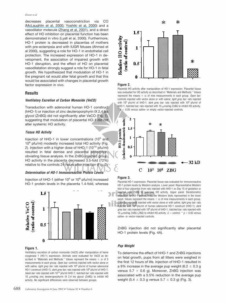
Injection of hHO-1 in lower concentrations (105 and108 pfu/ml) modestly increased total HO activity (Fig.2). Injection with a higher dose of hHO-1 (1010 pfu/ml)resulted in fetal demise and placental absorption,obviating tissue analysis. In the ZnBG-injected group,HO activity in the placenta decreased 3.5-fold (72%)relative to the controls 24 hours after injection (Fig. 2).
Determination of HO-1 Immunoreactive Protein Levels
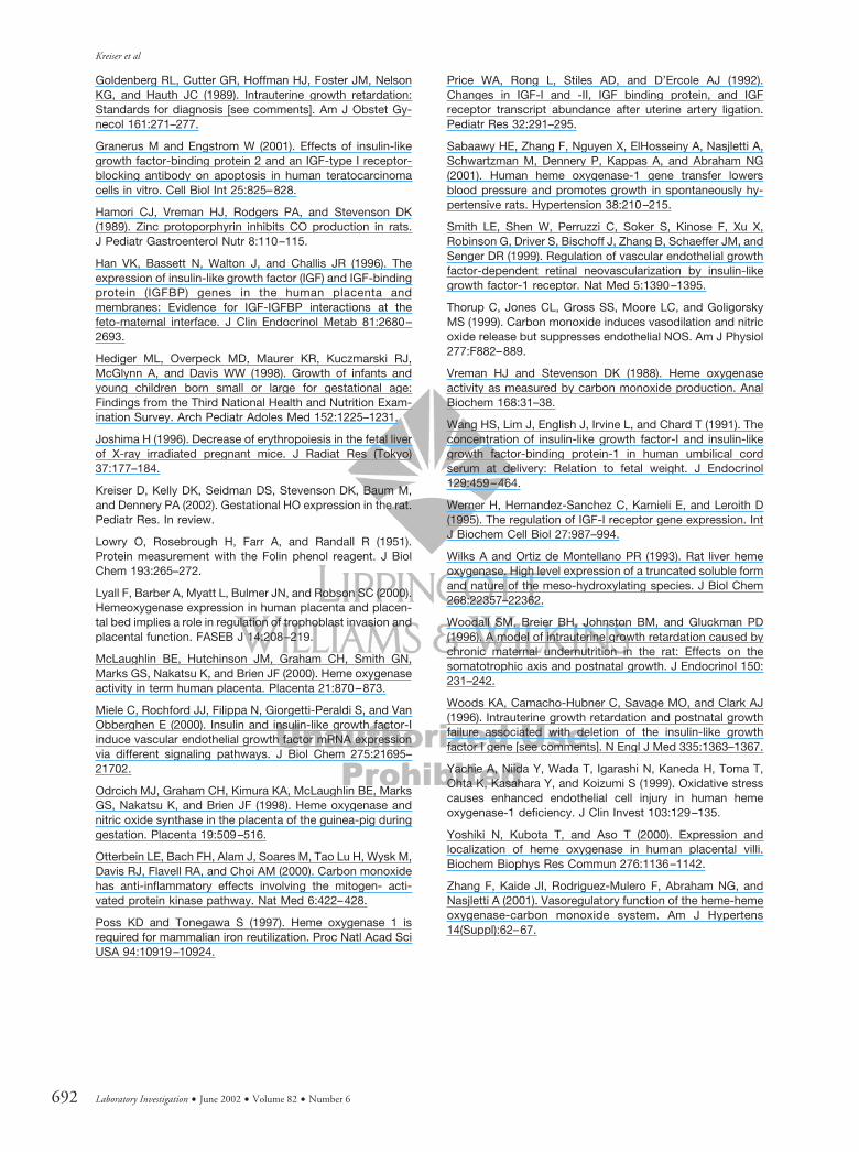
Injection of hHO-1 (either 105 or 108 pfu/ml) increasedHO-1 protein levels in the placenta 1.4-fold, whereas
ZnBG injection did not significantly alter placentalHO-1 protein levels (Fig. 4A).
Pup Weight
To determine the effect of hHO-1 and ZnBG injectionson fetal growth, pups from all litters were weighed inthe first 12 hours of life. Injection of hHO-1 resulted ina 9% increase in the average pup weight (6.2 � 0.3 gversus 5.7 � 0.6 g). Moreover, ZnBG injection wasassociated with a 5.5% reduction in the average pupweight (5.4 � 0.3 g versus 5.7 � 0.3 g) (Fig. 3).
Figure 1.Ventilatory excretion of carbon monoxide (VeCO) after manipulation of hemeoxygenase 1 (HO-1) expression. Animals were evaluated for VeCO as de-scribed in “Materials and Methods.” Values represent the means � SE of 5measurements in each group. Open bar: controls injected with vector alone orwith saline; light grey bar: rats injected with 105 pfu/ml of human adenoviralHO-1 construct (hHO-1); dark grey bar: rats injected with 108 pfu/ml of hHO-1;black bar: rats injected with 1010 pfu/ml hHO-1; hatched bar: rats injected with10 �mol/kg zinc deuteroporphyrin IX 2,4 bis glycol (ZnBG) to inhibit HOactivity. No significant differences were observed between groups.
Figure 2.Placental HO activity after manipulation of HO-1 expression. Placental tissuewas evaluated for HO activity as described in “Materials and Methods.” Valuesrepresent the means � SE of nine measurements in each group. Open bar:controls injected with vector alone or with saline; light grey bar: rats injectedwith 105 pfu/ml of hHO-1; dark grey bar: rats injected with 108 pfu/ml ofhHO-1; hatched bar: rats injected with 10 �mol/kg ZnBG to inhibit HO activity.* p � 0.05 versus saline- or empty vector-injected controls.
Figure 3.Placental HO-1 expression. Placental tissue was evaluated for immunoreactiveHO-1 protein levels by Western analysis. Lower panel: Representative Westernblot of four placentas from rats injected with hHO-1 on Day 15 of gestation orinjected with ZnBG to suppress HO activity. Upper panel: Densitometricevaluation of HO-1 protein from the Western blots represented in the lowerpanel. Values represent the means � SE of nine measurements in each group.Open bar: controls injected with vector alone or with saline; light grey bar: ratsinjected with 105 pfu/ml of human adenoviral HO-1 construct (hHO-1); darkgrey bar: rats injected with 108 pfu/ml of hHO-1; hatched bar: rats injected with10 �mol/kg ZnBG (ZBG) to inhibit HO activity. C � control. * p � 0.05 versussaline- or vector-injected controls.
Kreiser et al
688 Laboratory Investigation • June 2002 • Volume 82 • Number 6

Determination of IGF-1R, IGF-BP-1, and VEGFImmunoreactive Protein Levels
Because some of the byproducts of the HO reaction(in particular CO) serve as signaling molecules (Otter-bein et al, 2000), protein levels for various growthfactors were evaluated to determine whether HO-1mediated its effects by modulating IGF-1 or VEGFexpression. Because the highest concentration ofIGF-1 is in serum (Wang et al, 1991) and we were mostinterested in placental tissue, we determined the con-centration of IGF-1R and IGF-BP-1 in tissues ratherthan IFG-1 itself. Although injection of 108 pfu/mlhHO-1 had no effect on IGF-BP-1 in the placenta,ZnBG injection caused a significant decrease (60%)(Fig. 4B). Additionally, IGF-1R protein levels increased2.1-fold with hHO-1 adenovirus injection, whereasthere was a 57% decline after ZnBG injection (Fig. 4C).As to VEGF, adenoviral transduction of hHO-1 wasassociated with a dramatic increase in placental ex-pression. Injection of ZnBG did not alter placentalVEGF expression compared with controls (Fig. 5).
Discussion
IUGR is associated with many long-term conse-quences and has many etiologies. Deficiency of HO ina human has been described (Yachie et al, 1999) and,as with the HO-1 null mutant mice (Poss andTonegawa, 1997), there was growth restriction. Aneffect of HO-1 on cell proliferation (Clark et al, 1997;Deramaudt et al, 1998), vasodilatation (Thorup et al,1999; Zhang et al, 2001), and postnatal growth(Sabaawy et al, 2001) has been documented. Genetransfer of hHO-1 into coronary endothelial cells pro-moted angiogenesis (Deramaudt et al, 1998) and in-hibitors of HO increased placental resistance (Lyall etal, 2000). Overall, these examples suggest a role forHO-1 in placental vascularization and fetal growth;however the mechanism by which this is mediated isnot well defined.
Placental vascularization and an adequate fetopla-cental circulation are essential for promoting fetalgrowth. In the present work, the fact that inhibition ofHO suppressed growth and that increased HO-1 ex-pression promoted growth suggests that increasedHO activity improves placental function, perhapsthrough the vasodilator effects of CO and the vaso-proliferative effects of HO-1. Although, HO-1 wasmodified by ZnBG and hHO-1 locally, there were nochanges in the systemic excretion of CO, suggestingtight control of systemic CO excretion.
The increased expression of IGF-1R and VEGF afterhHO-1 transduction suggests that HO-1 or its byprod-ucts serve in signaling processes to enhance growthfactor expression and activation. Inhibition of IGF-related proteins by ZnBG further attests to the role ofHO-1 to modulate growth factor action. This has notbeen previously demonstrated.
Other researchers have hypothesized that increasedCO inhibits VEGF expression via a cGMP-mediatedpathway (Ghiso et al, 1999). We clearly demonstrateincreased VEGF expression in the placenta afterhHO-1 transduction. This implies differential cell-specific effects of CO or HO-1 on VEGF, or that COregulates other factors that in turn modulate VEGF inthe placenta. In a retinal model, IGF-1R activationresulted in increased VEGF activation of a mitogen-activated protein (MAP) kinase but did not modulateVEGF protein expression (Smith et al, 1999). However,in NIH3T3 fibroblasts, incubation with IGF-1 increasedVEGF mRNA via MAP kinase activation (Miele et al,2000). This demonstrates that IGF-1 can both increaseVEGF expression and increase its activation. It istherefore plausible to suggest that CO or HO-1 couldmodulate VEGF through IGF-1, but this does notexplain how HO-1 mediates increased IGF-1.
The IGF-1R gene has response elements in thepromoter that allow for increased transcription(Werner et al, 1995). Recent evidence suggests thatHO-1 can precede activation of other genes, such as
Figure 4.Effect of hHO-1 transduction on placental growth factors. RepresentativeWestern blot of 4 placentas from rats injected with 108 pfu/ml of hHO-1 on Day15 of gestation to enhance HO activity. Lanes are: C, control injected withvector alone; and hHO-1, rats injected with hHO-1 (2 examples are shown).Equal loading was verified with Coomassie blue staining. IGF-1R � insulin-likegrowth factor-1 receptor; IGF-BP � insulin-like growth factor-1 bindingprotein; VEGF � vascular endothelial growth factor.
Figure 5.Effect of HO inhibition on placental growth factors. Representative Westernblot of four placentas from rats injected with10 �mol/kg ZnBG on Day 15 ofgestation to suppress HO activity. Lanes are: C, controls injected with saline;and ZBG, rats injected with ZnBG (two examples are shown). Equal loadingwas verified with Coomassie blue staining. IGF-1R � insulin-like growthfactor-1 receptor; IGF-BP1 � insulin-like growth factor-1 binding protein;VEGF � vascular endothelial growth factor.
Heme Oxygenase and Growth
Laboratory Investigation • June 2002 • Volume 82 • Number 6 689

superoxide dismutase (Frankel et al, 2000), and thatCO can result in signaling via the p38 MAP kinase(Otterbein et al, 2000). Perhaps IGF-1R gene tran-scription is enhanced via the effects of CO or anotherbyproduct of the HO reaction. One such byproduct,iron, is important in regulating many genes (Alcantaraet al, 2001; Fogg et al, 1999), although no evidenceexists as to its regulation of IGF-1 gene expression.
Overall, our results indicate that changes in HO-1expression influence growth in utero. This is associ-ated with changes in growth factor expression thatcould affect fetal growth and placental function. Wespeculate that one of the byproducts of the reaction,such as CO, could result in signaling and increasedregulation of these growth factors.
Materials and Methods
Sixty-day-old timed pregnant Wistar rats (SimonsonLabs, Gilroy, California) were housed singly in atemperature-controlled room (25° C � 4° C) on a12-hour light cycle. The animals were allowed freeaccess to food and water. All animal care was inaccordance with National Institutes of Health guide-lines and under Institutional Animal Care and UseCommittee approval.
Pregnant dams were intraperitoneally injected witheither 5 �mol/kg of body weight ZnBG, on Day 16 ofpregnancy, or with 300 �l of hHO-1 adenovirus inthree distinct dilutions (105, 108, and 1010 pfu/ml) onDay 15 of pregnancy. Control groups were injectedwith normal saline or an empty vector. VeCO measure-ments were obtained before and 24 hours after injec-tion. After 24 hours, placental tissue was obtained forHO activity and for HO-1, IGF-1R, and IGF-BP-1protein levels by Western analysis. On Day 1 of life,pups from all groups were weighed.
ZnBG Preparation
ZnBG (2.87 mg) was dissolved in 240 �l of Na3PO4,0.4 M. An additional 1 ml of H2O was added and thesolution was slowly titrated to pH 7.8 with 1 M HCl(approximately 100 �l). The final volume was adjustedto 8 ml with normal saline.
Construction of Adenoviral hHO-1 cDNA Vector
A 987-bp hHO-1 cDNA fragment was released fromthe plasmid pRc-CMV-hHO1, and cloned at the HindIIIsite of the plasmid pGEM-7zf(�) (Promega, Madison,Wisconsin). The resulting vector pGEM-hHO1 wasthen linearized with EcoRI, and end-blunted withdNTP and Klenow. After digestion with BamHI, ahHO-1 cDNA fragment was released from pGEM7z-hHO1, and cloned at the PmeI/BglII sites of theadenoviral vector pAdCMV5GFP. The resulting ad-enoviral vector was designated as pAdCMV5GFP-HO.The linearized pAdCMV5GFP-HO by the digestionwith EcoRI was cotransfected with E1�, E3� adenovirallong-arm DNA (QBI-viral DNA, Quantum, Montreal, Que-bec, Canada) into a human embryonic cell line, 293Acells. The hHO-1 adenovirus construct was replicated
and encapsulated into an infectious virus. After a 5-dayincubation period, the virus plaque locations weremarked on the flasks, and the resultant cytopathic effecton the monolayers was observed microscopically untilthe plaque reached an adequate size. The plaques werepurified and checked for the presence of hHO-1 by PCRwith HO-1–specific primer, and amplified by propagationin the 293 cell line. The HO-1 adenovirus was releasedand collected by rupturing the infected cells throughthree freeze/thaw cycles 48 hours after infection. Afterthree rounds of plaque purification, recombinant adeno-virus was large-scale amplified and purified. Titers ofeach cesium chloride–purified viral stock were deter-mined from the absorbance at 260 nm (1 absorbanceunit � 1010 pfu/ml) and were confirmed by plaque assay.The virus was stored at 80° C until use.
Determination of HO Activity
Measurement of HO activity in placental tissue wasobtained by pooling placentas from each animal.Tissues were homogenized in four volumes of 0.01 M
sodium potassium phosphate buffer at pH 7.4 andthen centrifuged for 60 seconds at 12,500 �g. Thesupernatant was then analyzed for HO activity aspreviously described (Vreman and Stevenson, 1988).Twenty microliters of tissue supernatant, representing4 mg fresh weight of tissue, were incubated in 2-mlamber glass vials with 20 �l of 1.5 �M methemalbuminin 150 �l BSA and 20 �l of 4.5 mM NADPH for the totalreaction vials. For blank reaction vials, the NADPHwas replaced with an equal volume of buffer. The vialswere sealed with septum caps and placed in 37° Cwater bath for 5 minutes of temperature equilibrationand then purged with CO-free air. After 15 minutes offurther incubation, placing vials on powdered dry ice(�78° C) terminated the reactions. The CO generatedin the reaction medium and effused into the vialsheadspace was quantitated by gas chromatographywith a reduction gas analyzer (Trace Analytical, MenloPark, California). Analyzer response to CO was re-corded with an integrating recorder (CR-3A; ShimadzuScientific Instruments, Columbia, Maryland) throughmeasurement of peak area. The reduction gas ana-lyzer was standardized daily with volumes of 10.8 �l ofCO/l air (482 nM). Homogenates were analyzed forprotein content by the method of Lowry et al (1951)and read at 595 nm. HO activity was defined asNADPH-dependent CO production and calculated asthe difference between the CO in the total and blankreaction vials. HO activity was expressed as mean �SD pmol CO/�g protein/hour and then normalized tocontrols.
VeCO Measurements
The animals were weighed and placed in sealedPlexiglas tubes supplied with CO-free air at flow rateof 100 ml/minute. After a 30-minute equilibration pe-riod, gas leaving the chamber was analyzed for COconcentration by gas chromatography. For each timepoint, a minimum of five readings per animal was
Kreiser et al
690 Laboratory Investigation • June 2002 • Volume 82 • Number 6

taken. VeCO was expressed as mean � SD microlitersper hour per kilogram of body weight (Hamori et al,1989) and then normalized to control values.
Antibodies
Polyclonal rabbit anti-rat HO-1 antibody was raisedagainst a 30 kd soluble HO-1 protein expressed in Ecoli from rat liver cDNA (Wilks and Ortiz de Montel-lano, 1993) (gift of Angela Wilks, University of Califor-nia San Francisco, California) as previously described(Dennery et al, 1997). Rabbit anti-goat IGF-BP-1 andgoat anti-rabbit IGF-1 receptor antibodies were ob-tained from Santa Cruz Biotechnologies (Santa Cruz,California). HRP-conjugated anti-rabbit and anti-goatpolyclonal IgG was obtained from Santa CruzBiotechnologies.
Determination of HO-1, IGF-1R, and IGF-BP-1Immunoreactive Protein Levels (Western Analysis)
One hundred and eighty microgram aliquots of uterineor placental homogenates for HO-1, IGF-1R, andIGF-BP-1 were subjected to electrophoresis on a 12%polyacrylamide gel and then transferred to polyvinyli-dene difluoride membrane (PVDF) (Millipore Corpora-tion, Bedford, Massachusetts). Membranes were in-cubated for 1 hour with a 1:500 dilution of the primaryantibody washed three times for 10 minutes with0.05% Tween PBS (Dennery et al, 1997). The mem-branes were then incubated for 1 hour with a 1:800dilution of anti-goat or anti-rabbit polyclonal IgG atroom temperature. Antigen-antibody complexes werevisualized with the horseradish peroxidase chemilumi-nescence system (Super Signal; Pierce Chemical,Rockford, Illinois) according to the manufacturer’sinstructions. Equal loading of samples was verified byCoomassie blue stain. Densitometric evaluation wasconducted (SGI, Sunnyvale, California) and valuesfrom each blot normalized to controls to allow forcomparison between blots.
Statistical Analysis
For comparisons between the study groups, the nullhypothesis that there was no difference betweengroup means was tested by a single factor ANOVA forcontinuous variables using Bonferroni multiple com-parisons t test for multiple groups or unpaired t test fortwo groups or the Fisher Exact tests for dichotomousdata (Stat-View 4.02; Abacus Concepts, Berkeley,California). Statistical significance was assumed at p� 0.05.
Acknowledgements
We thank Dr. Henk Vreman, Dr. Yi-Hao Weng, andDr. Guang Yang for their expert technical assistance.
ReferencesAbraham NG, Lin JH, Mitrione SM, Schwartzman ML, LevereRD, and Shibahara S (1988). Expression of heme oxygenase
gene in rat and human liver. Biochem Biophys Res Commun150:717–722.
Accili D, Nakae J, Kim JJ, Park BC, and Rother KI (1999).Targeted gene mutations define the roles of insulin and IGF-1receptors in mouse embryonic development. J Pediatr En-docrinol Metab 12:475–485.
Ahmed A and Perkins J (2000). Angiogenesis and intrauterinegrowth restriction. Baillieres Best Pract Res Clin ObstetGynaecol 14:981–998.
Ahmed A, Rahman M, Zhang X, Acevedo CH, Nijjar S,Rushton I, Bussolati B, and St John J (2000). Induction ofplacental heme oxygenase-1 is protective against TNFalpha-induced cytotoxicity and promotes vessel relaxation. MolMed 6:391–409.
Alcantara O, Kalidas M, Baltathakis I, and Boldt DH (2001).Expression of multiple genes regulating cell cycle and apo-ptosis in differentiating hematopoietic cells is dependent oniron. Exp Hematol 29:1060–1069.
Barker DJ, Hales CN, Fall CH, Osmond C, Phipps K, andClark PM (1993). Type 2 (non-insulin-dependent) diabetesmellitus, hypertension and hyperlipidaemia (syndrome X):Relation to reduced fetal growth. Diabetologia 36:62–67.
Cheung CY (1997). Vascular endothelial growth factor: Pos-sible role in fetal development and placental function. J SocGynecol Investig 4:169–177.
Clark JE, Green CJ, and Motterlini R (1997). Involvement ofthe heme oxygenase-carbon monoxide pathway in keratino-cyte proliferation. Biochem Biophys Res Commun 241:215–220.
Dennery P, Sridhar K, Lee C, Wong H, Shokoohi V, RodgersP, and Spitz D (1997). Heme oxygenase-mediated resistanceto oxygen toxicity in hamster fibroblasts. J Biol Chem 272:14937–14942.
Deramaudt BM, Braunstein S, Remy P, and Abraham NG(1998). Gene transfer of human heme oxygenase into coro-nary endothelial cells potentially promotes angiogenesis.J Cell Biochem 68:121–127.
Ferrara N, Carver-Moore K, Chen H, Dowd M, Lu L, O’SheaKS, Powell-Braxton L, Hillan KJ, and Moore MW (1996).Heterozygous embryonic lethality induced by targeted inac-tivation of the VEGF gene. Nature 380:439–442.
Fogg S, Agarwal A, Nick HS, and Visner GA (1999). Ironregulates hyperoxia-dependent human heme oxygenase 1gene expression in pulmonary endothelial cells. Am J RespirCell Mol Biol 20:797–804.
Frankel D, Mehindate K, and Schipper HM (2000). Role ofheme oxygenase-1 in the regulation of manganese superox-ide dismutase gene expression in oxidatively-challengedastroglia. J Cell Physiol 185:80–86.
Ghiso N, Rohan RM, Amano S, Garland R, and Adamis AP(1999). Suppression of hypoxia-associated vascular endo-thelial growth factor gene expression by nitric oxide viacGMP. Invest Ophthalmol Vis Sci 40:1033–1039.
Giudice LC, de Zegher F, Gargosky SE, Dsupin BA, de lasFuentes L, Crystal RA, Hintz RL, and Rosenfeld RG (1995).Insulin-like growth factors and their binding proteins in theterm and preterm human fetus and neonate with normal andextremes of intrauterine growth. J Clin Endocrinol Metab80:1548–1555.
Heme Oxygenase and Growth
Laboratory Investigation • June 2002 • Volume 82 • Number 6 691
Goldenberg RL, Cutter GR, Hoffman HJ, Foster JM, NelsonKG, and Hauth JC (1989). Intrauterine growth retardation:Standards for diagnosis [see comments]. Am J Obstet Gy-necol 161:271–277.
Granerus M and Engstrom W (2001). Effects of insulin-likegrowth factor-binding protein 2 and an IGF-type I receptor-blocking antibody on apoptosis in human teratocarcinomacells in vitro. Cell Biol Int 25:825–828.
Hamori CJ, Vreman HJ, Rodgers PA, and Stevenson DK(1989). Zinc protoporphyrin inhibits CO production in rats.J Pediatr Gastroenterol Nutr 8:110–115.
Han VK, Bassett N, Walton J, and Challis JR (1996). Theexpression of insulin-like growth factor (IGF) and IGF-bindingprotein (IGFBP) genes in the human placenta andmembranes: Evidence for IGF-IGFBP interactions at thefeto-maternal interface. J Clin Endocrinol Metab 81:2680–2693.
Hediger ML, Overpeck MD, Maurer KR, Kuczmarski RJ,McGlynn A, and Davis WW (1998). Growth of infants andyoung children born small or large for gestational age:Findings from the Third National Health and Nutrition Exam-ination Survey. Arch Pediatr Adoles Med 152:1225–1231.
Joshima H (1996). Decrease of erythropoiesis in the fetal liverof X-ray irradiated pregnant mice. J Radiat Res (Tokyo)37:177–184.
Kreiser D, Kelly DK, Seidman DS, Stevenson DK, Baum M,and Dennery PA (2002). Gestational HO expression in the rat.Pediatr Res. In review.
Lowry O, Rosebrough H, Farr A, and Randall R (1951).Protein measurement with the Folin phenol reagent. J BiolChem 193:265–272.
Lyall F, Barber A, Myatt L, Bulmer JN, and Robson SC (2000).Hemeoxygenase expression in human placenta and placen-tal bed implies a role in regulation of trophoblast invasion andplacental function. FASEB J 14:208–219.
McLaughlin BE, Hutchinson JM, Graham CH, Smith GN,Marks GS, Nakatsu K, and Brien JF (2000). Heme oxygenaseactivity in term human placenta. Placenta 21:870–873.
Miele C, Rochford JJ, Filippa N, Giorgetti-Peraldi S, and VanObberghen E (2000). Insulin and insulin-like growth factor-Iinduce vascular endothelial growth factor mRNA expressionvia different signaling pathways. J Biol Chem 275:21695–21702.
Odrcich MJ, Graham CH, Kimura KA, McLaughlin BE, MarksGS, Nakatsu K, and Brien JF (1998). Heme oxygenase andnitric oxide synthase in the placenta of the guinea-pig duringgestation. Placenta 19:509–516.
Otterbein LE, Bach FH, Alam J, Soares M, Tao Lu H, Wysk M,Davis RJ, Flavell RA, and Choi AM (2000). Carbon monoxidehas anti-inflammatory effects involving the mitogen- acti-vated protein kinase pathway. Nat Med 6:422–428.
Poss KD and Tonegawa S (1997). Heme oxygenase 1 isrequired for mammalian iron reutilization. Proc Natl Acad SciUSA 94:10919–10924.
Price WA, Rong L, Stiles AD, and D’Ercole AJ (1992).Changes in IGF-I and -II, IGF binding protein, and IGFreceptor transcript abundance after uterine artery ligation.Pediatr Res 32:291–295.
Sabaawy HE, Zhang F, Nguyen X, ElHosseiny A, Nasjletti A,Schwartzman M, Dennery P, Kappas A, and Abraham NG(2001). Human heme oxygenase-1 gene transfer lowersblood pressure and promotes growth in spontaneously hy-pertensive rats. Hypertension 38:210–215.
Smith LE, Shen W, Perruzzi C, Soker S, Kinose F, Xu X,Robinson G, Driver S, Bischoff J, Zhang B, Schaeffer JM, andSenger DR (1999). Regulation of vascular endothelial growthfactor-dependent retinal neovascularization by insulin-likegrowth factor-1 receptor. Nat Med 5:1390–1395.
Thorup C, Jones CL, Gross SS, Moore LC, and GoligorskyMS (1999). Carbon monoxide induces vasodilation and nitricoxide release but suppresses endothelial NOS. Am J Physiol277:F882–889.
Vreman HJ and Stevenson DK (1988). Heme oxygenaseactivity as measured by carbon monoxide production. AnalBiochem 168:31–38.
Wang HS, Lim J, English J, Irvine L, and Chard T (1991). Theconcentration of insulin-like growth factor-I and insulin-likegrowth factor-binding protein-1 in human umbilical cordserum at delivery: Relation to fetal weight. J Endocrinol129:459–464.
Werner H, Hernandez-Sanchez C, Karnieli E, and Leroith D(1995). The regulation of IGF-I receptor gene expression. IntJ Biochem Cell Biol 27:987–994.
Wilks A and Ortiz de Montellano PR (1993). Rat liver hemeoxygenase. High level expression of a truncated soluble formand nature of the meso-hydroxylating species. J Biol Chem268:22357–22362.
Woodall SM, Breier BH, Johnston BM, and Gluckman PD(1996). A model of intrauterine growth retardation caused bychronic maternal undernutrition in the rat: Effects on thesomatotrophic axis and postnatal growth. J Endocrinol 150:231–242.
Woods KA, Camacho-Hubner C, Savage MO, and Clark AJ(1996). Intrauterine growth retardation and postnatal growthfailure associated with deletion of the insulin-like growthfactor I gene [see comments]. N Engl J Med 335:1363–1367.
Yachie A, Niida Y, Wada T, Igarashi N, Kaneda H, Toma T,Ohta K, Kasahara Y, and Koizumi S (1999). Oxidative stresscauses enhanced endothelial cell injury in human hemeoxygenase-1 deficiency. J Clin Invest 103:129–135.
Yoshiki N, Kubota T, and Aso T (2000). Expression andlocalization of heme oxygenase in human placental villi.Biochem Biophys Res Commun 276:1136–1142.
Zhang F, Kaide JI, Rodriguez-Mulero F, Abraham NG, andNasjletti A (2001). Vasoregulatory function of the heme-hemeoxygenase-carbon monoxide system. Am J Hypertens14(Suppl):62–67.
Kreiser et al
692 Laboratory Investigation • June 2002 • Volume 82 • Number 6