Regulation of Heme Oxygenase-1 by Redox Signals Involving Nitric Oxide

Up-Regulation of Heme Oxygenase-1 in Rat Spleen FollowingAniline Exposure
Jianling Wanga, Huaxian Maa, Paul J. Boora, V. M. Sadagopa Ramanujamb, G.A.S. Ansaria,and M. Firoze Khana,*aDepartment of Pathology, University of Texas Medical Branch, Galveston, TX, USAbDepartment of Preventive Medicine and Community Health, University of Texas Medical Branch,Galveston, TX, USA
AbstractSplenic toxicity of aniline is characterized by vascular congestion, hyperplasia, fibrosis anddevelopment of a variety of sarcomas in rats. However, underlying mechanisms by which anilineelicits splenotoxic response are not well understood. Previously we have shown that aniline exposurecauses oxidative damage to the spleen. To further explore the oxidative mechanism of aniline toxicity,we evaluated the potential contribution of heme oxygenase-1 (HO-1), which catalyzes hemedegradation and releases free iron. Male SD rats were given 1 mmol/kg/day aniline in water by gavagefor 1, 4 or 7 days, while respective controls received water only. Aniline exposure led to significantincreases in HO-1 mRNA expression in the spleen (2- and 2.4-fold at days 4 and 7, respectively)with corresponding increases in protein expression, as confirmed by ELISA and Western blotanalyses. Furthermore, immunohistochemical assessment of spleen showed strongerimmunostaining for HO-1 in the spleens of rats treated for 7 days, confined mainly to the red pulpareas. No changes were observed in mRNA and protein levels of HO-1 following 1 day exposure.The increase in HO-1 expression was associated with increases in total iron (2.4- and 2.7- fold), freeiron (1.9- and 3.5-fold), and ferritin levels (1.9- and 2.1-fold) at 4 and 7 days of aniline exposure.Our data suggest that HO-1 up-regulation in aniline-induced splenic toxicity could be a contributingpro-oxidant mechanism, mediated through iron release, and leading to oxidative damage.
KeywordsHeme oxygenase-1; oxidative stress; iron; ferritin; aniline; spleen
IntroductionAniline, a toxic aromatic amine, is an extensively used industrial chemical. Exposure to anilineis known to cause toxicity to the hematopoietic system [1-5]. Aniline toxicity is generallycharacterized by methemoglobinemia, hemolysis and hemolytic anemia [6-9], and by thedevelopment of splenic hyperplasia, fibrosis, and a variety of primary sarcomas after chronicexposure in rats [2,10-13]. While mechanisms of erythrocyte damage have been the focus of
* Address for Correspondence: M. Firoze Khan, Ph.D., Professor, Department of Pathology, University of Texas Medical Branch,Galveston, TX 77555-0438, Tel: 409-772-6881, Fax: 409-747-1763, [email protected]'s Disclaimer: This is a PDF file of an unedited manuscript that has been accepted for publication. As a service to our customerswe are providing this early version of the manuscript. The manuscript will undergo copyediting, typesetting, and review of the resultingproof before it is published in its final citable form. Please note that during the production process errors may be discovered which couldaffect the content, and all legal disclaimers that apply to the journal pertain.
NIH Public AccessAuthor ManuscriptFree Radic Biol Med. Author manuscript; available in PMC 2011 February 15.
Published in final edited form as:Free Radic Biol Med. 2010 February 15; 48(4): 513. doi:10.1016/j.freeradbiomed.2009.11.027.
NIH
-PA Author Manuscript
NIH
-PA Author Manuscript
NIH
-PA Author Manuscript

many studies, little attention has been given to the delineation of molecular mechanisms inaniline-induced toxicity to the spleen.
Heme oxygenase-1 (HO-1) is a rate-limiting microsomal enzyme that catalyzes the oxidativedegradation of heme moiety of hemoglobin to biliverdin, carbon monoxide and free iron [14,15]. HO-1 transcription can be induced by a whole array of stressors, including transition-metals [16,17], heme, hemoglobin and other heme proteins [18,19], and oxidative/nitrosativestress [20-22]. HO-1 can exert cytoprotective and cytotoxic effects through severalmechanisms, including serving as a molecular chaperone, degrading pro-oxidant heme toproduce antioxidants (bilirubin and carbon monoxide) and releasing iron [23-25]. Even thoughan antioxidant role of HO-1 has been extensively studied [15,24], several studies also supporta pro-oxidant role for HO-1 [14,26-28]. However, a specific role for HO-1 in aniline-inducedsplenic oxidative damage is not known.
Earlier studies have demonstrated that severity of toxic responses in the spleen is closelyassociated with erythrocyte damage [2,3,12]. The deposition and subsequent breakdown ofdamaged erythocytes during aniline insult will result in release and accumulation of iron/iron-storage proteins in the spleen. Indeed, studies have shown that aniline exposure in rats leadsto iron release/overload [3,4,8,12,29] and oxidative stress in the spleen [4,8,30-32]. Wehypothesize that up-regulation of HO-1 contributes to oxidative damaging reactions in thespleen by catalyzing the oxidative degradation of the heme moiety of hemoglobin and releasingfree iron. This study was, therefore, focused on evaluating the regulation of HO-1, release offree iron and status of iron storage protein, ferritin, in the spleens of rats exposed to aniline.
Materials and methodsAnimals and treatments
Male Sprague-Dawley rats (∼200 g), obtained from Harlan (Indianapolis, IN), were housed inwire-bottom cages over absorbent paper with free access to tap water and Purina rat chow. Theanimals were acclimatized in a controlled-environment animal room (temperature, 22 °C;relative humidity, 50%; photoperiod, 12-h light/dark cycle) for 7 days prior to treatment. Theexperiments were performed in accordance with the guidelines of the National Institutes ofHealth and were approved by the Institutional Animal Care and Use Committee of Universityof Texas Medical Branch. The animals, in groups of 6 each, were given 1mmol/kg/day anilinein drinking water by gavage for 1, 4 or 7 days; respective control animals received water only.The choice of aniline dose was based on our earlier studies that showed significant increasesin lipid peroxidation, protein oxidation and DNA damage (oxidative stress) in the spleen [4,29,30]. The rats were euthanized 24 h after the last dose under nembutal (sodium pentoparbital)anesthesia and the spleens were removed immediately, blotted, weighted and stored at -80 °Cuntil further analysis. A portion of spleen was snap-frozen in liquid nitrogen and stored at -80°C for RNA isolation. Also, portions of the spleen from control and aniline-treated rats werefixed in 10% neutral buffered formalin for immunohistological processing.
Real-time RT-PCR for HO-1 mRNARNA isolation—Total RNA was isolated from spleen tissues using RiboPure Kit (Ambion,Austin, TX) as per manufacturer's instructions. To eliminate genomic DNA contamination,isolated RNA was treated with RNase free DNase I (DNA-free kit, Ambion, Austin, TX). Thetotal RNA concentration was determined by measuring the absorbance at 260nm. RNAintegrity was verified electrophoretically by ethidium bromide staining and by measuringA260/A280 ratio.
Wang et al. Page 2
Free Radic Biol Med. Author manuscript; available in PMC 2011 February 15.
NIH
-PA Author Manuscript
NIH
-PA Author Manuscript
NIH
-PA Author Manuscript

Real-time RT-PCR—Real-time RT-PCR was performed essentially as described earlier[32-34]. First-strand cDNA was prepared from total RNA by using SuperScript First-StrandSynthesis Kit (Invitrogen, Carlsbad, CA) as per manufacturer's instructions. Real-time PCRemploying a two-step cycling protocol (denaturation and annealing/extension) was carried outusing Mastercycler realplex (Eppendorf, Westbury, N.Y.) and using primer sense 5′-TGGAAGAGGAGATAGAGCGA and antisense, 5′-TGTTGAGCAGGAAGGCGGTC. Foreach cDNA sample, parallel reactions were performed in triplicate for the detection of 18S andrat HO-1. The reaction samples in a final volume of 25 μl contained 2 μl of cDNA templates,2 μl primer pair, 12.5 μl iQ SYBR Green Supermix and 8.5 μl water. Amplification conditionswere identical for all reactions: 95 °C for 2 min for template denaturation and hot start priorto PCR cycling. A typical cycling protocol consisted of three stages: 15s at 95 °C fordenaturation, 30s at 65 °C for annealing, 30s at 72 °C for extension, and an additional 20s holdfor fluorescent signal acquisition [33]. To avoid non-specific signal from primer-dimers, thefluorescence signal was detected 2 °C below the melting temperature (Tm) of individualamplicon and above the Tm of the primer-dimers [35,36]. A total of 40 cycles were performed.
Quantitation of PCR was done using the comparative CT method as described in User BulletinNo. 2 of Applied Biosystems (Foster City, CA), and reported as fold difference relative to thecalibrator cDNA (QuantumRNA Universal 18S Standards, Ambion). The fold change in HO-1cDNA (target gene) relative to the 18S endogenous control was determined by:
Fold change = 2-ΔΔCT, where ΔΔCT = (CT Aniline - CT 18S) – (CT Control – CT 18S).
HO-1 ELISA—HO-1 protein levels in the spleen tissue lysates (prepared by using the lysisbuffer provided with ELISA kit) were quantitated by using rat HO-1 ELISA Kit (AssayDesigns, Ann Arbor, MI). OD was read at 450 nm on a Bio-Rad Benchmark Plus microplatespectrophotometer.
Western blot for HO-1—Protein samples from control and aniline-treated rats weresubjected to Western blot analysis as described earlier [31]. Briefly, spleen tissue lysates wereprepared using the lysis buffer essentially as described by the manufacturer (Cell Signaling,Beverly, MA). The lysate proteins (50 μg) were subjected to 10% SDS-PAGE and transferredto a PVDF membrane (Amersham, Arlington Heights, IL). After blocking with non-fat drymilk (5%, w/v), HO-1 protein band was detected using anti-HO-1 antibody (1:3000; SantaCruz Biotech, Santa Cruz, CA) and goat anti-rabbit IgG-HRP (1:5000; Santa Cruz Biotech) assecondary antibody. ECL Plus (Amersham, Arlington Heights, IL) was used to detect thesignals. HO-1 bands were quantitated by densitometry and normalized using the actin signalto correct for differences in loading of proteins from control and experimental groups. Fordensitometric analysis, the protein bands on the blot were measured by Eagle Eye II software.Protein concentration in the lysates was determined by Bio-Rad Protein Assay kit (Bio-RadLaboratories).
Immunohistochemistry of HO-1 and CD68—Paraffin sections were cut anddeparaffinized in an oven at 55 °C for 1 h and treated with xylene and various concentrationsof ethanol, and finally rehydrated with water [32,37]. The slides were incubated with sodiumcitrate buffer at 95 °C for 20 min for antigen retrieval and subsequently incubated with variousreagents, which included quenching of endogenous peroxidase activity with 0.3% H2O2 inPBS, blocking the non-specific binding sites with 5% non-fat dry milk, 1% BSA, 5% goatserum (horse serum in case of CD68) in PBS, respectively, and avidin and biotin block solutions(Vector Laboratories, Burlingame, CA). The sections were then incubated with antibodies toHO-1 (monoclonal, 1:50 dilution, Stressgen Bioreagents, Ann Arbor, MI,) or CD68 (amacrophage marker; 1:100 dilution, Abcam Inc., Cambridge, MA) overnight at 4 °C. Sectionswere washed with PBS and incubated with secondary antibodies. Immunoreactivity was
Wang et al. Page 3
Free Radic Biol Med. Author manuscript; available in PMC 2011 February 15.
NIH
-PA Author Manuscript
NIH
-PA Author Manuscript
NIH
-PA Author Manuscript

detected by the ABC Method (Vectastain Elite ABC kit, Vector Laboratories, Burlingame,CA) with color development using 3,3′-diaminobenzidine (DAB). Mayer's Hematoxylin wasthen added as a counterstain for 1 min. The negative controls were immunostained as above,but with goat or horse serum instead of the anti-HO-1 or anti-CD68 antibody. Histologicalevaluation for staining was done with an OLYMPUS BX51 Microscope (Leads Instruments,Inc., Irving, TX).
Total iron and free iron analysis—Total iron in the spleens was analyzed by atomicabsorption spectrophotometer as described earlier [4,38]. Analysis of low molecular weightchelatable iron (LMWC-Fe, free iron) was done as described earlier [13,29].
Ferritin Determination—Spleen tissues from control and aniline-treated rats were cut intosmall pieces and homogenized in a buffer containing 30 mM Tris-Cl, pH 8.0, 15 mM EDTA,1.0 mM DTT, 0.5 mM spermine and spermidine, 15% glycerol and a complete proteaseinhibitor cocktail (Roche, Germany). After homogenization, 0.2% Triton X-100 and 5.0 MNaCl (to achieve a final concentration of 150 mM) were added. After incubating 30 minuteson ice, the extracts were centrifuged at 14,000 g for 30 min. The supernatant was saved andused for ferritin assay.
Ferritin was quantitated using an ELISA kit, essentially as described by the manufacturer(Innovative Research Inc., Novi, MI). Briefly, 100 μl of the standard or samples from controland aniline-treated rats (in duplicate) were added to ELISA plate wells precoated with anti-ferritin antibodies, and incubated at room temperature for 1 h with gentle mixing. Afterincubation, the plate was washed thoroughly with the wash buffer and 100 μl of the diluted(1:100) enzyme-antibody conjugate was added and incubated for 10 min at room temperature.After washing with wash buffer, 100 μl TMB substrate (3′,3′,5′,5′-tetramethylbenzidine)solution was added to each well. The plate was then incubated at room temperature for 10 minwith continuous shaking followed by addition of 100 μl stop solution (0.3 M H2SO4) toterminate the reaction. The OD was read at 450 nm on a Bio-Rad Benchmark plus microplatespectrophotometer (Bio-Rad Laboratories, Hercules, CA).
Statistical analysis—Data are expressed as means ± SD. One-way ANOVA followed byTukey-Kramer multiple comparison test (GraphPad InStat 3 software, La Jolla, CA) wasperformed for the statistical analysis. A p value of <0.05 was considered to be statisticallysignificant.
ResultsHO-1 mRNA expression in response to aniline exposure
To evaluate the impact of aniline exposure on the expression of HO-1 in the spleen, HO-1mRNA levels were analyzed by real-time PCR and their levels are shown in Fig. 1. Anilineexposure for 4 and 7 days resulted in 2.0- and 2.4-fold increases, respectively, in HO-1 mRNAexpression as compared to their respective controls. No change in HO-1 mRNA was observedat day 1 following aniline exposure.
Effect of aniline exposure on HO-1 protein expression in the spleenTo investigate whether increases in HO-1 gene expression were also associated with increasesin protein levels, HO-1 protein levels were quantitated by a rat specific HO-1 ELISA kit in thespleen tissue lysates and results are shown in Fig. 2. Aniline exposure led to significantincreases of 1.9- and 2.6-fold in HO-1 levels at 4 and 7 days, respectively, whereas no changewas observed at day 1.
Wang et al. Page 4
Free Radic Biol Med. Author manuscript; available in PMC 2011 February 15.
NIH
-PA Author Manuscript
NIH
-PA Author Manuscript
NIH
-PA Author Manuscript

To further validate and confirm our quantitative ELISA results, HO-1 protein expression inthe spleen lysates was also determined by Western blotting. As evident from Fig. 3, the HO-1protein levels in the tissue lysates of aniline-treated rats increased by 2 and 2.5 fold at 4 and 7days, respectively. However, similar to mRNA and ELISA results, no changes in HO-1 proteinlevels were observed 24 h following a single dose of aniline.
Immunohistochemical assessment of HO-1 and CD68 in the spleen of rats exposed to anilineTo demonstrate HO-1 expression and localization in the spleens of experimental animals,immunohistochemical studies were conducted. Since mRNA and protein expression of HO-1showed no change at 1 day and maximum increases at 7 days, HO-1 immunohistochemicalassessment was done at 1 and 7 days only. While spleens from control rats showed sparseimmunostaining for HO-1 (Fig. 4A), increased immunostaining was evident in rats treated withaniline for 7 days (Fig. 4B). The immunoreactivity for HO-1 appeared predominantly in thered pulp areas of the spleen (Fig. 4B). Consistent with HO-1 mRNA and protein results, nochanges in HO-1 immunoreactivity were observed 24 h following a single dose of aniline (datanot included). To evaluate the contribution of macrophages in the increased expression of HO-1in the spleen, immunoreactivity for CD68+ cells was also investigated. As evident from Fig.4, aniline treatment for 7 days resulted in an increased immunoreactivity for CD68+ cells inthe red pulp areas of the spleen (D) in comparison to controls (C), suggesting that macrophagesare one of the major contributors to the increased HO-1 expression in the spleen in responseto aniline exposure.
Effect of aniline exposure on total and free ironAniline exposure is associated with erythrocyte damage [2,3]. Excessive deposition anddegradation of erythrocytes in the spleen could potentially lead to iron release/overload.Therefore, changes in both total and free iron were quantitated by atomic absorptionspectrophotometry. As evident from Fig. 5, exposure to a single dose of aniline did not resultin any change in the total iron levels. However, total iron content increased significantly at 4days (2.4 fold) and 7 days (2.7 fold) in comparison to their respective controls. LMWC-Fe(free iron) followed a similar pattern, showing increases of 1.9- and 3.5-fold at 4 and 7 days,respectively (Fig. 6), while no change after day 1.
Effect of aniline exposure on ferritin levels in spleenFerritin is an iron-storage protein which could exert antioxidant properties by mediating thesequestration and storage of reactive iron, thus preventing ferryl or hydroxyl radical formation[39]. On the other hand, ferritin could also be a potential source of catalytic iron duringoxidative stress [40,41]. Therefore, in view of the anti- and pro-oxidant potential of ferritin,its status was evaluated in the spleens of rats exposed to aniline. ELISA determination of totalferritin levels in the spleens of aniline-treated rats showed significant increases of 1.9- and 2.1-fold at days 4 and 7, respectively, in comparison to respective controls (Fig. 7). Exposure to asingle dose of aniline, however, did not result in any change in ferritin levels in the spleen.
DiscussionAniline and substituted anilines are known to cause splenic toxicity [2,12,13,31]. Among themost significant splenotoxic changes, preceding the formation of splenic sarcomas that resultfrom exposure to aniline and/or structurally-related compounds, are splenomegaly, sinusoidalcongestion, capsular hyperplasia and diffuse fibrosis [10-13]. Splenotoxicity could also leadto reduced ability of the spleen to participate in the immune response and/or its phagocyticfunction of clearing damaged erythrocytes and infectious organisms from the blood. Thisnecessitates a clear understanding of early molecular events in the spleen that could contributeto such toxic responses. It has been established previously that aniline exposure leads to
Wang et al. Page 5
Free Radic Biol Med. Author manuscript; available in PMC 2011 February 15.
NIH
-PA Author Manuscript
NIH
-PA Author Manuscript
NIH
-PA Author Manuscript

erythrocyte damage [2,3,7] and oxidative stress in the spleen [4,13,30]. Recognizing the roleof HO-1 in catalyzing the oxidative degradation of heme moiety of hemoglobin to release freeiron, it was of particular interest to evaluate its contribution to the splenic toxicity of aniline.It is evident from our data that HO-1 expression indeed increases in the spleen following anilineexposure, and its overexpression could be a pro-oxidant action in the splenic toxicity.
A major finding of this study is the significant increase in the expression of HO-1 (both atmRNA and protein levels) in the spleen following aniline exposure. The increases were highlysignificant in both repeated dose groups (4 and 7 days). Interestingly, the HO-1 was localizedmostly in the red pulp of the spleen – areas which also showed intense iron staining (Prussianblue) [4]. To our knowledge, this is the first study to show an up-regulation of HO-1 in spleenfollowing aniline exposure. Interestingly, the increases in HO-1 expression in this study alsocorresponded with previous findings of increased oxidative stress observed under similarexperimental conditions [4]. While oxidative/nitrosative stress is known to induce HO-1[20-22], up-regulation of HO-1, as observed in this study, could contribute to increasedoxidative stress [14,26-28]. Other hemolytic agents such as phenylhydrazine and phenacetinalso cause upregulation of HO-1 gene expression in rat spleens [42], and, thus, complimentour findings.
Aniline exposure in this study also led to significant increases in the total iron content – afinding consistent with earlier observations [4,13]. We also observed a substantial increase inLMWC-Fe (free iron) after repeated dose administration of aniline. Iron overload is associatedwith carcinogenesis in animal models and also in human diseases such as genetichemochromatosis and asbestosis [43,44]. Free iron is redox active and can promote, throughthe Fenton reaction, the formation of harmful oxygen species such as hydroxyl radicals [45,46]. Thus, the iron-catalyzed oxyradicals can damage cellular lipids, proteins and nucleic acids,resulting in wide-ranging impairment of cellular function and integrity. Our earlier studies didshow that aniline exposure was indeed associated with significant increases in lipidperoxidation [4,13], protein oxidation [13], DNA damage [29,32], activation of redox-sensitivetranscription factors NF-κB and AP-1 [31,33] and up-regulation of fibrogenic cytokines [30,31,33,34] in the spleen.
There has been considerable debate concerning the pro-oxidant or antioxidant nature of HO-1[14,24]. Heme has pro-oxidant properties, so its removal by HO-1 can be considered anantioxidant effect [15,47]. However, heme breakdown by HO-1 leads to the formation ofLMWC-Fe (free iron), which is a more versatile catalyst of oxidative damage than heme[48]. Interestingly, exogenously added bilirubin at an equimolar concentration to low-molecular-mass iron present in both microsomal and liposomal systems was unable to preventthe pro-oxidant effect of the iron [14], further supporting a pro-oxidant role for HO-1.
Normally, iron is transported and stored in specific proteins (ferritin, transferrin andlactoferritin), which prevent or minimize its reaction with reduced oxygen derivatives [49,50]. Iron released from HO-mediated reactions stimulates ferritin synthesis, which ultimatelyprovides an iron detoxification mechanism that may offer long-term cytoprotection [24].However, if iron transport and storage proteins do not rapidly and effectively remove thisredox-active iron, HO-1 induction could be a pro-oxidant effect. The aniline exposure protocolused in this study represents an experimental condition known to cause oxidative stress [4,30,32]. This protocol led to increases in ferritin levels, along with increases in free iron,suggesting that despite greater presence of ferritin, increased cytoprotection was not evidentand tissue damaging oxidative reactions still prevailed [4,30]. Our findings are in generalagreement with others, showing increased ferritin levels along with increased lipidperoxidation following ozone exposure [51], L-methionine treatment [52] and during non-alcoholic steatohepatitis [53]. Further support to this is evident from the finding that patients
Wang et al. Page 6
Free Radic Biol Med. Author manuscript; available in PMC 2011 February 15.
NIH
-PA Author Manuscript
NIH
-PA Author Manuscript
NIH
-PA Author Manuscript

with iron overload exhibit increased iron and oxidative stress, despite up-regulated ferritinlevels [54].
It is possible that ferritin can also be a potential source of catalytic iron during oxidative stress.Superoxide generating systems such as xanthine/xanthine oxidase or compounds/drugs whichundergo redox cycling to produce superoxide radicals, may promote the release of ferritin iron[40,55]. Thus, ferritin may promote oxidative damage under conditions where excessivesuperoxide formation occurs [40]. Indeed, studies suggest that aniline and its metabolites havethe potential to generate superoxide radicals [56]. Despite increased presence of ferritin, it isalso possible that intracellular iron may overwhelm the ability of ferritin to sequester redox-active iron and thus result in oxidative stress.
In conclusion, our studies clearly show that aniline exposure results in HO-1 up-regulation,increased ferritin levels, and excessive release and availability of iron (free iron) in the spleen.Greater release and presence of redox-active iron as a result of exaggerated expression of HO-1would result in increased oxidative stress, as observed previously [4,30,32,33]. Our data thussuggest that HO-1 up-regulation could be a significant pro-oxidant mechanism in the splenictoxicity of aniline, leading to oxidative damage through iron release in the spleen.
AcknowledgmentsThis work was supported by Grant ES06476 from the National Institute of Environmental Health Sciences (NIEHS),NIH, and its contents are solely the responsibility of the authors and do not necessarily represent the official views ofthe NIEHS, NIH.
References1. Kim YC, Carlson GP. The effect of an unusual work shift on chemical toxicity II. Studies on
theexposure of rats to aniline. Fundam Appl Toxicol 1986;7:144–152. [PubMed: 3732666]2. Bus JS, Popp JA. Perspectives on the mechanism of action of the splenic toxicity of aniline and
structurally related compounds. Food Chem Toxicol 1987;25:619–626. [PubMed: 3305247]3. Khan MF, Boor PJ, Kaphalia BS, Alcock NW, Ansari GAS. Hematopoietic toxicity of linoleic acid
anilide: importance of aniline. Fundam Appl Toxicol 1995;25:224–232. [PubMed: 7665006]4. Khan MF, Gu Y, Alcock NW, Boor PJ, Ansari GAS. Oxidative stress in splenotoxicity of aniline.
Fundam Appl Toxicol 1997;35:22–30. [PubMed: 9024670]5. Pauluhn J. Subacute inhalation toxicity of aniline in rats: Analysis of time-dependence and
concentration dependence of hematotoxic and splenic effects. Toxicol Sci 2004;81:198–215.[PubMed: 15187235]
6. Harrison JH, Jollow DJ. Role of aniline metabolites in aniline-induced hemolytic anemia. J PharmacolExp Ther 1986;238:1045–1054. [PubMed: 3746658]
7. Khan MF, Wu X, Kaphalia BS, Boor PJ, Ansari GAS. Acute hematopoietic toxicity of aniline in rats.Toxicol Lett 1997;92:31–37. [PubMed: 9242355]
8. Ciccoli L, Ferrali M, Rossi V, Signorini C, Alessandrini C, Comporti M. Hemolytic drugs aniline anddapsone induce iron release in erythrocytes and increase the free iron pool in spleen and liver. ToxicolLett 1999;110:57–66. [PubMed: 10593595]
9. Mellert W, Deckardt K, Gembardt C, Zwirner-Baier I, Jackh R, Ravenzwaay BV. Aniline: earlyindiators of toxicity in male rats and their relevance to spleen carcinogenicity. Human Exptl Toxicol2004;23:379–389.
10. Goodman DG, Ward JM, Reichardt WD. Splenic fibrosis and sarcomas in F344 rats fed dietscontaining aniline hydrochloride, p-chloroaniline, azobenzene, o-toluidine hydrochloride,4,4'sulfonyldianiline, or DRC Red No. 9. J Natl Cancer Inst 1984;73:265–273. [PubMed: 6588231]
11. Weinberger MA, Albert RH, Montgomery SB. Splenotoxicity associated with splenic sarcomas inrats fed high doses of DRC Red No. 9 or aniline hydrochloride. J Natl Cancer Inst 1985;75:681–690.[PubMed: 3862900]
Wang et al. Page 7
Free Radic Biol Med. Author manuscript; available in PMC 2011 February 15.
NIH
-PA Author Manuscript
NIH
-PA Author Manuscript
NIH
-PA Author Manuscript

12. Khan MF, Kaphalia BS, Boor PJ, Ansari GAS. Subchronic toxicity of aniline hydrochloride in rats.Arch Environ Contant Toxicol 1993;24:368–374.
13. Khan MF, Wu X, Boor PJ, Ansari GAS. Oxidative modification of proteins and lipids in aniline-induced splenic toxicity. Toxicol Sci 1999;48:134–140. [PubMed: 10330693]
14. Lamb NJ, Quinlan GJ, Mumby S, Evans TW, Gutteridge JMC. Haem oxygenase shows pro-oxidantactivity in microsomal and cellular systems: implications for the release of low-molecular-mass iron.Biochem J 1999;344:153–158. [PubMed: 10548545]
15. Prawan A, Kundu JK, Surh YJ. Molecular basis of heme oxygenase-1 induction: Implications forchemoprevention and chemoprotection. Antioxid Redox Signal 2005;7:1688–1703. [PubMed:16356130]
16. Maines MD, Kappas A. Metals as regulators of heme metabolism. Science 1977;198:1215–1221.[PubMed: 337492]
17. Brown KE, Dennery PA, Ridnour LA, Fimmel CJ, Kladney ED, Brunt RM, Spitz DR. Effect of ironoverload and dietary fat on indices of oxidative stress and hepatic fibrogeneis in rats. Liver Intl2003;23:232–242.
18. Alam J, Shibahara S, Smith A. Transcriptional activation of the heme oxygenase gene by heme andcadmium in mouse hepatoma cells. J Biol Chem 1989;264:6371–6375. [PubMed: 2703493]
19. Nath KA, Balla G, Vercellotti GM, Balla J, Jacob HS, Levitt MD, Rosenberg ME. Induction of hemeoxygenase is a rapid, protective response in rhabdomyolysis in the rat. J Clin Invest 1992;90:267–270. [PubMed: 1634613]
20. Bauer M, Bauer I. Heme oxygenase-1: Redox regulation and role in the hepatic response to oxidativestress. Antioxid Redox Signal 2002;4:749–758. [PubMed: 12470502]
21. Naughton P, Foresti R, Brains SK, Hoque M, Green CJ, Motterlini R. Induction of heme oxygenase-1by nitrosative stress. A role for nitroxyl anion. J Biol Chem 2002;277:40666–40674. [PubMed:12194971]
22. Ryter SW, Choi AMK. Heme-oxygenase-1: Molecular mechanisms of gene expression in oxygenrelated stress. Antioxid Redox Signal 2002;4:625–632. [PubMed: 12230874]
23. Suttner DM, Dennery PA. Reversal of HO-1 related cytoprotection with increased expression is dueto reactive iron. FASEB J 1999;13:1800–1809. [PubMed: 10506583]
24. Ryter SW, Tyrrell RM. The heme synthesis and degradation pathways: Role in oxidant sensitivity.Free Radic Biol Med 2000;28:289–309. [PubMed: 11281297]
25. Hemdan S, Almazan G. Iron contributes to dopamine-induced toxicity in oligodendrocyte progenitors.Neuropath Appl Neurobiol 2006;32:428–440.
26. Chen S, Khan ZA, Barbin Y, Chakrabarti S. Pro-oxidant role of heme oxygenase in mediating glucose-induced endothelial cell damage. Free Radic Res 2004;38:1301–1310. [PubMed: 15763954]
27. Khan ZA, Barbin YP, Cukiernik M, Adams PC, Chakrabarti S. Heme-oxygenase-mediated ironaccumulation in the liver. Can J Physiol Pharmacol 2004;82:448–456. [PubMed: 15389291]
28. Wang J, Dore S. Heme oxygenase-1 exacerbates early brain injury after intracerebral haemorrhage.Brain 2007;130:1643–1652. [PubMed: 17525142]
29. Wu X, Kannan S, Sadagopa Ramanujam VM, Khan MF. Iron release and oxidative DNA damage insplenic toxicity of aniline. J Toxicol Environ Health, Part A 2005;68:657–666. [PubMed: 15901093]
30. Khan MF, Wu X, Wang J. Up-regulation of transforming growth factor-β1 in the spleen of anilinetreated rats. Toxicol Appl Pharmacol 2003;187:22–28. [PubMed: 12628581]
31. Khan MF, Kannan S, Wang J. Activation of transcription factor AP-1 and mitogen-activated proteinkinases in aniline-induced splenic toxicity. Toxicol Appl Pharmacol 2006;210:86–93. [PubMed:16169568]
32. Ma H, Wang J, Abdel-Rehman SZ, Boor PJ, Khan MF. Oxidative DNA damage and its repair in ratspleen following subchronic exposure to aniline. Toxicol Appl Pharmacol 2008;233:247–253.[PubMed: 18793663]
33. Wang J, Kannan S, Li H, Khan MF. Cytokine gene expression and activation of NF-kappa B in aniline-induced splenic toxicity. Toxicol Appl Pharmacol 2005;15:36–44. [PubMed: 15694462]
Wang et al. Page 8
Free Radic Biol Med. Author manuscript; available in PMC 2011 February 15.
NIH
-PA Author Manuscript
NIH
-PA Author Manuscript
NIH
-PA Author Manuscript
34. Wang J, Wang G, Ansari GAS, Khan MF. Activation of oxidative stress-responsive signalingpathways in early splenotoxic response of aniline. Toxicol Appl Pharmacol 2008;230:227–234.[PubMed: 18420242]
35. Simpson DA, Feeney S, Boyle C, Stitt AW. Retinal VEGF mRNA measured by SYBR green Ifluorescence: a versatile approach to quantitative. PCR Mol Vision 2000;6:178–183.
36. Rajeevan MS, Ranamukhaarachchi DG, Vernon SD, Unger ER. Use of realtime quantitative PCR tovalidate the results of cDNA array and differential display PCR technologies. Methods 2001;25:443–4451. [PubMed: 11846613]
37. Khan MF, Wu X, Tipnis UR, Ansari GAS, Boor PJ. Protein adducts of malondialdehyde and 4-hydroxynonenal in livers of iron loaded rats:quantitation and localization. Toxicology 2002;173:193–201. [PubMed: 11960672]
38. Alcock NW. A hydrogen peroxide digestion system for tissue trace metal analysis. Biol Trace ElemRes 1987;13:363–370.
39. Klausner RD, Rouault TA, Harford JB. Regulating the fate of mRNA: the control of cellular ironmetabolism. Cell 1993;72:19–28. [PubMed: 8380757]
40. Reif DW. Ferritin as a source of iron for oxidative damage. Free Radic Biol Med 1992;12:417–427.[PubMed: 1317328]
41. Aust SD. Ferritin as a source of iron and protection from iron-induced toxicities. Toxicol Lett1995;82-83:941–944. [PubMed: 8597165]
42. Rokushima M, Omi K, Imura K, Arari A, Furukawa N, Itoh F, Miyazaki M, Yamamoto J, RokushimaM, Okada M, Torii M, Kato I, Ishizaki J. Toxicogenomics of drug-induced hemolytic anemia byanalyzing gene expression profiles in the spleen. Toxicol Sci 2007;100:290–302. [PubMed:17698508]
43. Bradbear RA, Bain C, Siskind V, Schofield FD, Webb S, Azelsen EM, Halliday JW, Bassett ML,Powell LW. Cohort study of internal malignancy in genetic hemochromatosis and other chronic non-alcoholic liver diseases. J Natl Cancer Ins 1985;75:81–84.
44. Toyokuni S. Iron-induced carcinogenesis: The role of redox regulation. Free Radic Biol Med1996;20:553–566. [PubMed: 8904296]
45. Halliwell B, Gutteridge JMC. Oxygen toxicity, oxygen radicals, transition metals and disease.Biochem J 1985;219:1–14. [PubMed: 6326753]
46. Ferrali M, Signorini C, Sugherini L, Pompella A, Lodovici M, Caciotti B, Ciccolit L, Comporti M.Release of free, redox-active iron in the liver and DNA oxidative damage following phenylhydrazineintoxication. Biochem Pharmacol 1997;53:1743–1751. [PubMed: 9264328]
47. Stocker R. Induction of haem oxygenase as a defence against oxidative stress. Free Radic ResCommun 1990;9:101–112. [PubMed: 2189794]
48. Gutteridge JMC. Iron promoters of the Fenton reaction and lipid peroxidation can be released fromhaemoglobin by peroxides. FEBS Lett 1986;201:291–295. [PubMed: 2423372]
49. Crichton RR, Charloteaux-Wauters M. Iron transport and storage. Eur J Biochem 1987;164:485–506.[PubMed: 3032619]
50. Ceccarelli D, Gallesi D, Giovannini F, Ferrali M, Masini A. Relationship between free iron level andrat liver mitochondrial dysfunction in experimental dietary iron overload. Biochem Biophys ResCommun 1995;209:53–59. [PubMed: 7726863]
51. Ghio AJ, Turi JL, Madden MC, Dailey LA, Richards JD, Stonehuerner JG, Morgan DL, SingletonS, Garrick LM, Garrick MD. Lung injury after ozone exposure is iron dependent. Am J Physiol LungCell Mol Physiol 2007;292:L134–L143. [PubMed: 16905637]
52. Mori N, Hirayama K. Long-term consumption of a methionine-supplemented diet increases iron andlipid peroxide levels in rat liver. J Nutr 2000;130:2349–2355. [PubMed: 10958834]
53. Malaguarnera L, Madeddu R, Palio E, Arena N, Malaguarnera M. Heme oxygenase-1 levels andoxidative stress-related parameters in non-alcoholic fatty liver disease patients. J Hepatol2005;42:585–591. [PubMed: 15763346]
54. Kassab-Chekir A, Laradi S, Ferchichi S, Haj Khelil A, Feki M, Amri F, Selmi H, Bejaoui M, MiledA. Oxidant, antioxidant status and metabolic data in patients with beta-thalassemia. Clin Chim Acta2003;338:79–86. [PubMed: 14637270]
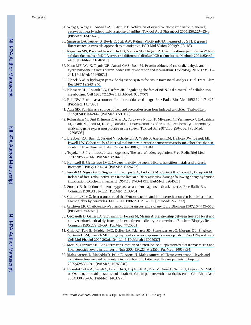
Wang et al. Page 9
Free Radic Biol Med. Author manuscript; available in PMC 2011 February 15.
NIH
-PA Author Manuscript
NIH
-PA Author Manuscript
NIH
-PA Author Manuscript

55. Agrawal R, Sharma P, Rao GS. Release of iron from ferritin by metabolites of benzene and superoxideradical generating agents. Toxicology 2001;168:223–230. [PubMed: 11684319]
56. Brennan RJ, Schiestl RH. Aniline and its metabolites generate free radicals in yeast. Mutagenesis1997;12:215–220. [PubMed: 9237764]
Wang et al. Page 10
Free Radic Biol Med. Author manuscript; available in PMC 2011 February 15.
NIH
-PA Author Manuscript
NIH
-PA Author Manuscript
NIH
-PA Author Manuscript

Fig. 1.Real-time PCR analysis of HO-1 gene expression in the spleens of control and aniline-treatedrats. Total RNA was extracted from spleens, real-time PCR was performed, and the fold changein mRNA expression (2-ΔΔCT) was determined. Values are means ± SD (n=3). *p<0.05 vs.respective controls.
Wang et al. Page 11
Free Radic Biol Med. Author manuscript; available in PMC 2011 February 15.
NIH
-PA Author Manuscript
NIH
-PA Author Manuscript
NIH
-PA Author Manuscript

Fig. 2.HO-1 protein levels in the spleens of rats treated with aniline. HO-1 was quantitated using arat-specific ELISA kit. Values are means ± SD (n=6). *p < 0.05 vs. respective controls; # p <0.05 vs. 4 days aniline group.
Wang et al. Page 12
Free Radic Biol Med. Author manuscript; available in PMC 2011 February 15.
NIH
-PA Author Manuscript
NIH
-PA Author Manuscript
NIH
-PA Author Manuscript

Fig. 3.HO-1 protein expression in rat spleens following aniline exposure. (A) Western blot detectionof HO-1 in the spleens of control and aniline-treated [1, 4 or 7 days (D)] rats. (B) Densitometricanalysis of HO-1 bands. Values are means ± SD (n=3). *p < 0.05 vs. respective controls; # p< 0.05 vs. 4 days aniline group.
Wang et al. Page 13
Free Radic Biol Med. Author manuscript; available in PMC 2011 February 15.
NIH
-PA Author Manuscript
NIH
-PA Author Manuscript
NIH
-PA Author Manuscript

Fig. 4.Immunohistochemistry of HO-1 and CD68 in rat spleen. Spleen from control rats (A) showedcomparatively mild staining for HO-1 with occasional heavily stained cells (arrows), whereasspleen from rats treated with aniline for 7 days showed consistently strong immunoreactivityfor HO-1 (B), with many heavily stained cells confined to the red pulp areas of the spleen.Similarly, there was an increased presence of CD68+ (macrophage marker) cells in the red pulpareas of spleen from rats treated with aniline for 7 days (D) compared to the controls (C).
Wang et al. Page 14
Free Radic Biol Med. Author manuscript; available in PMC 2011 February 15.
NIH
-PA Author Manuscript
NIH
-PA Author Manuscript
NIH
-PA Author Manuscript

Fig. 5.Splenic total iron content in the control and aniline-treated rats. Total iron was analyzed byatomic absorption spectrophotometry. Values are means ± SD (n=6). *p < 0.05 vs. respectivecontrols.
Wang et al. Page 15
Free Radic Biol Med. Author manuscript; available in PMC 2011 February 15.
NIH
-PA Author Manuscript
NIH
-PA Author Manuscript
NIH
-PA Author Manuscript

Fig. 6.LMWC-Fe (free iron) in the spleens of control and aniline-treated rats. Values are means ± SD(n=4-5). *p < 0.05 vs. controls.
Wang et al. Page 16
Free Radic Biol Med. Author manuscript; available in PMC 2011 February 15.
NIH
-PA Author Manuscript
NIH
-PA Author Manuscript
NIH
-PA Author Manuscript

Fig. 7.Ferritin levels in the spleens of control and aniline-treated rats. Total ferritin was quantitatedusing an ELISA. Values are means ± SD (n=5-6). *p < 0.05 vs. respective controls.
Wang et al. Page 17
Free Radic Biol Med. Author manuscript; available in PMC 2011 February 15.
NIH
-PA Author Manuscript
NIH
-PA Author Manuscript
NIH
-PA Author Manuscript
Copyright © 2022 FDOKUMEN