Ultrasound Cases - Spleen - Wendy Blount, DVM
86
Ultrasound Cases - Spleen Ultrasound Cases - Spleen Wendy Blount, DVM
-
Upload
khangminh22 -
Category
Documents
-
view
1 -
download
0
Transcript of Ultrasound Cases - Spleen - Wendy Blount, DVM

Ultrasound Cases - SpleenUltrasound Cases - SpleenWendy Blount, DVM
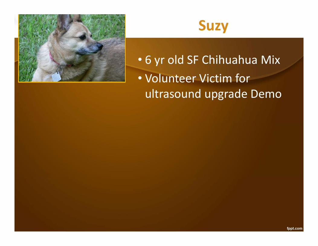
Suzy
• 6 yr old SF Chihuahua Mix• Volunteer Victim for
ultrasound upgrade Demo

Suzy
• 6 yr old SF Chihuahua Mix• Volunteer Victim for
ultrasound upgrade Demo
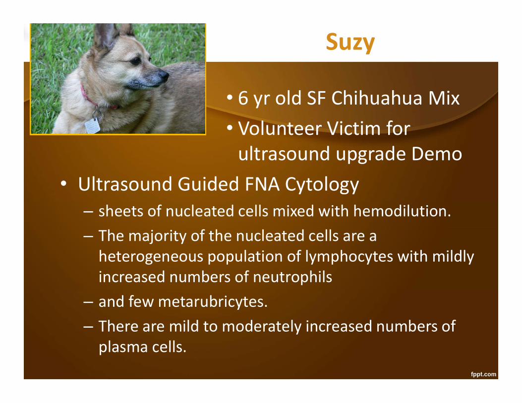
Suzy
• 6 yr old SF Chihuahua Mix• Volunteer Victim for
ultrasound upgrade Demo• Ultrasound Guided FNA Cytology
– sheets of nucleated cells mixed with hemodilution. – The majority of the nucleated cells are a
heterogeneous population of lymphocytes with mildly increased numbers of neutrophils
– and few metarubricytes. – There are mild to moderately increased numbers of
plasma cells.

Suzy
• 6 yr old SF Chihuahua Mix• Volunteer Victim for
ultrasound upgrade Demo• Ultrasound Guided FNA Cytology
– The splenic nodule aspirated may be due to a reactive follicle… there are no abnormal cells seen to indicate neoplasia. However, small cell lymphosarcoma is a possible differential diagnosis as cytologically small cell lymphoid neoplasia cannot be differentiated from hyperplasia. A biopsy is warranted if neoplasia is still a consideration.

Suzy
• 6 yr old SF Chihuahua Mix• Volunteer Victim for
ultrasound upgrade Demo• Plan: Recheck ultrasound in 30 days• 30 day Recheck – no change in sonogram or cytology• 90 day recheck – no change again• 6 month recheck – no change• Checked annually for the rest of her life, with no
change

Lessons from SuzySplenic Follicles and Cysts• Benign hypoechoic lesions,
unless polycystic disease• Rarely change over time• If fluid filled, sometimes can get larger• Cytology:
– Extramedullary hematopoiesis– Lymphoid hyperplasia, nodular hyperplasia– Benign fluid– Normal splenic aspirate
• Similar lesions can be found in the liver, biliary tract or pancreas
• Surgical treatment if abscessation

Lessons from SuzySplenic Follicles and Cysts• I have found splenic nodules
during ultrasound machinedemos in 2 dogs and 1 cat• 1 nodular hyperplasia with EMH and 2
lymphoid hyperplasia• EMH was large with Doppler flow, and ruptured
at the time of surgery• The other 2 small and without Doppler flow,
and not removed• Benign splenic nodules in well animals are not
uncommon

Georgia
Geriatric SF DLH
Head of the Spleen
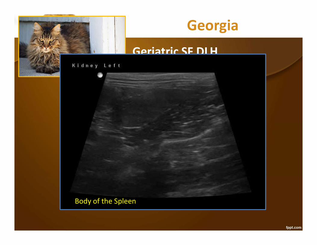
Georgia
Geriatric SF DLH
Body of the Spleen
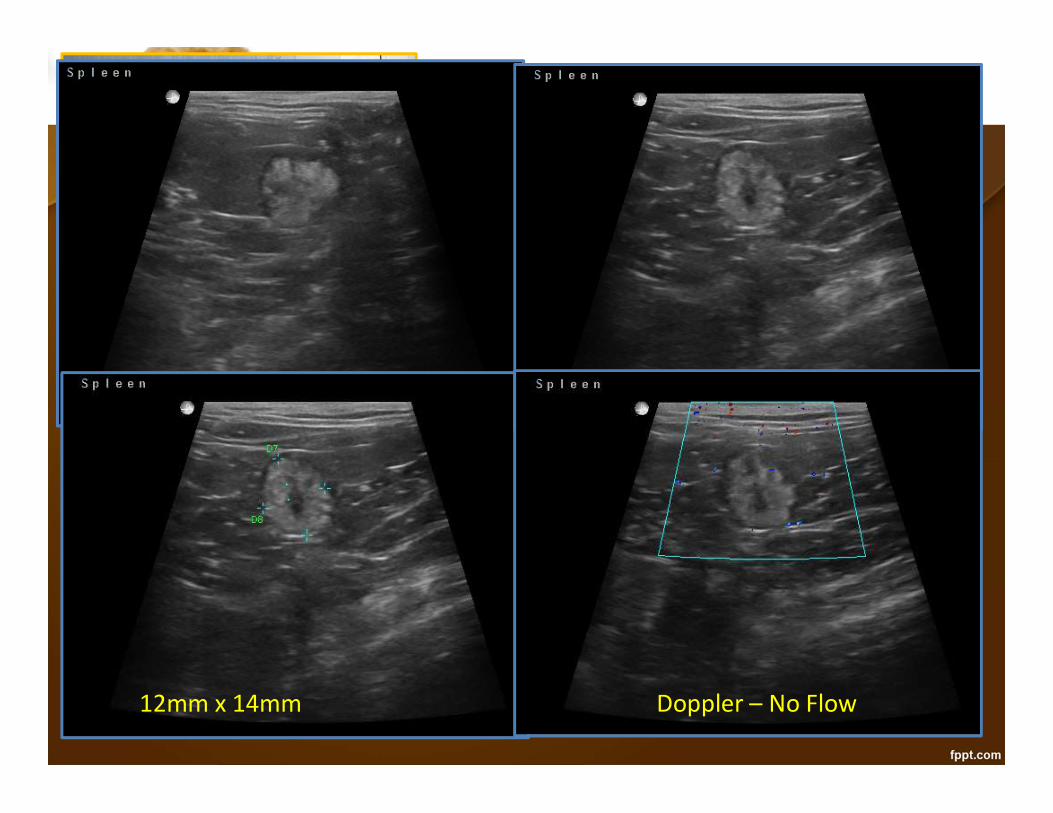
Georgia
Geriatric SF DLH
Doppler – No Flow12mm x 14mm

Tess
Middle aged SF Schnauzer
Bladder - Sagittal

Tess
Middle aged SF Schnauzer
Bladder - Transverse

Tess
Middle aged SF Schnauzer
Spleen Head

Tess
Middle aged SF Schnauzer
Spleen Hilus

Tess
Middle aged SF Schnauzer
Spleen Tail

Tess
Middle aged SF Schnauzer
Doppler – Flow through Tumor
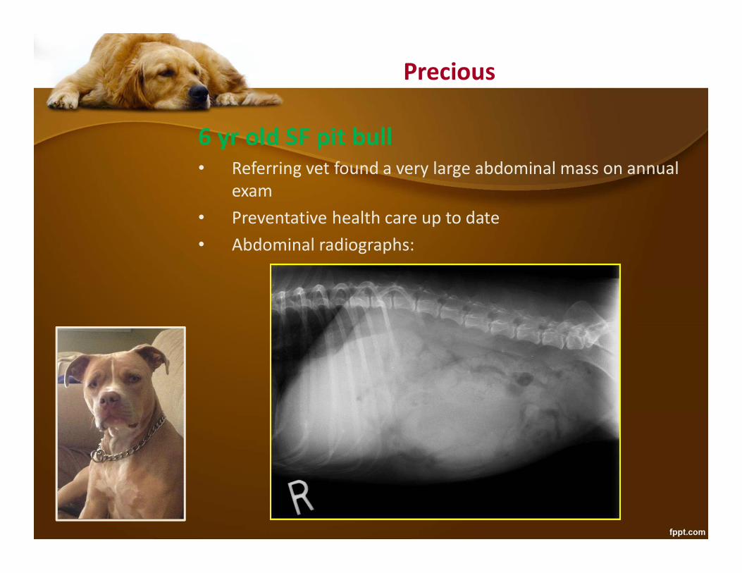
Precious
6 yr old SF pit bull• Referring vet found a very large abdominal mass on annual
exam• Preventative health care up to date• Abdominal radiographs:

Precious
6 yr old SF pit bull• Referring vet found a very large abdominal mass on
annual exam• Preventative health care up to date• Abdominal radiographs: enlarged spleen?
Exam• T- 101.2, P - 112, R - 24• Football size mid abdominal mass• BAR, well hydrated, in good body condition
CBC – normal
Chemistries - normal
UA – normal
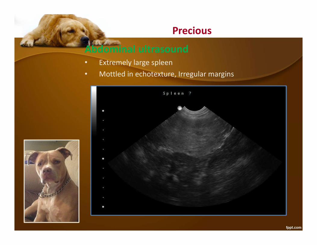
Precious
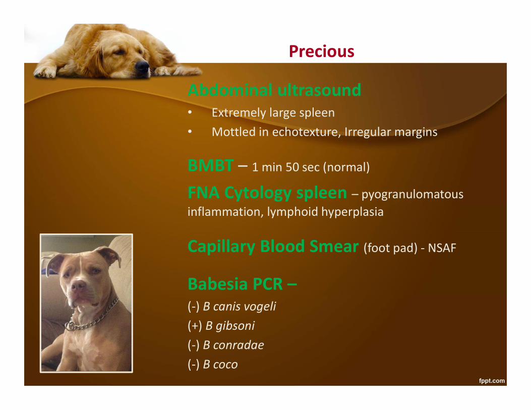
Abdominal ultrasound• Extremely large spleen• Mottled in echotexture, Irregular margins
Precious
Abdominal ultrasound• Extremely large spleen• Mottled in echotexture, Irregular margins
BMBT – 1 min 50 sec (normal)
FNA Cytology spleen – pyogranulomatous inflammation, lymphoid hyperplasia
Capillary Blood Smear (foot pad) - NSAF
Babesia PCR –(-) B canis vogeli(+) B gibsoni(-) B conradae(-) B coco

Precious
Treatment:• Doxycycline 10 mg/kg/day x 14d will palliate• Clindamycin 25 mg.kg PO BID x 14 days palliates
• Doxycycline, clindamycin + metronidazole may clear B gibsoni (unknown duration)
• Metronidazole 15 mg/kg PO BID
• Doxycycline, enrofloxacin, metronidazole may clear B gibsoni (unknown duration)
• Enrofloxacin 5 mg/kg/day PO
• Imidocarb 6.6 mg/kg IM – 2 doses 2 weeks apart• Diminazine 3-7 mg/kg – 2 doses 2 weeks apart• Likely to cure B canis vogeli, palliates B gibsoni
• Atovaquone and azithromycin – 50-85% effective

Hepatosplenic Abscess• Rare condition
• Can appear cystic, or like a solid mass• Can be infected or sterile
– Bartonella spp. – “bacillary peliosis”• More often mixed echogenicity with thick wall • Presence of gas is a tip off to abscessation• Remember that tumors can abscess – cannot rule
neoplasia out based on cytology• FNA cytology + culture/sensitivity is diagnostic, but can
risk rupture and peritonitis if infected– Indistinguishable sonographically from HSA/hematoma

Binkley11 yr old CM Mini SchnauzerCC: pants often, esp. at night• Preventative health current
• PE normal, neurologic exam normal• 3/6 systolic murmur – no CHF on thoracic rads
Binkley - Dry lung CHF-Pulmonary Edema
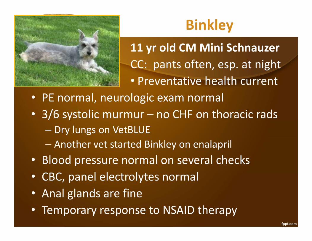
Binkley11 yr old CM Mini SchnauzerCC: pants often, esp. at night• Preventative health current
• PE normal, neurologic exam normal• 3/6 systolic murmur – no CHF on thoracic rads
– Dry lungs on VetBLUE– Another vet started Binkley on enalapril
• Blood pressure normal on several checks• CBC, panel electrolytes normal• Anal glands are fine• Temporary response to NSAID therapy
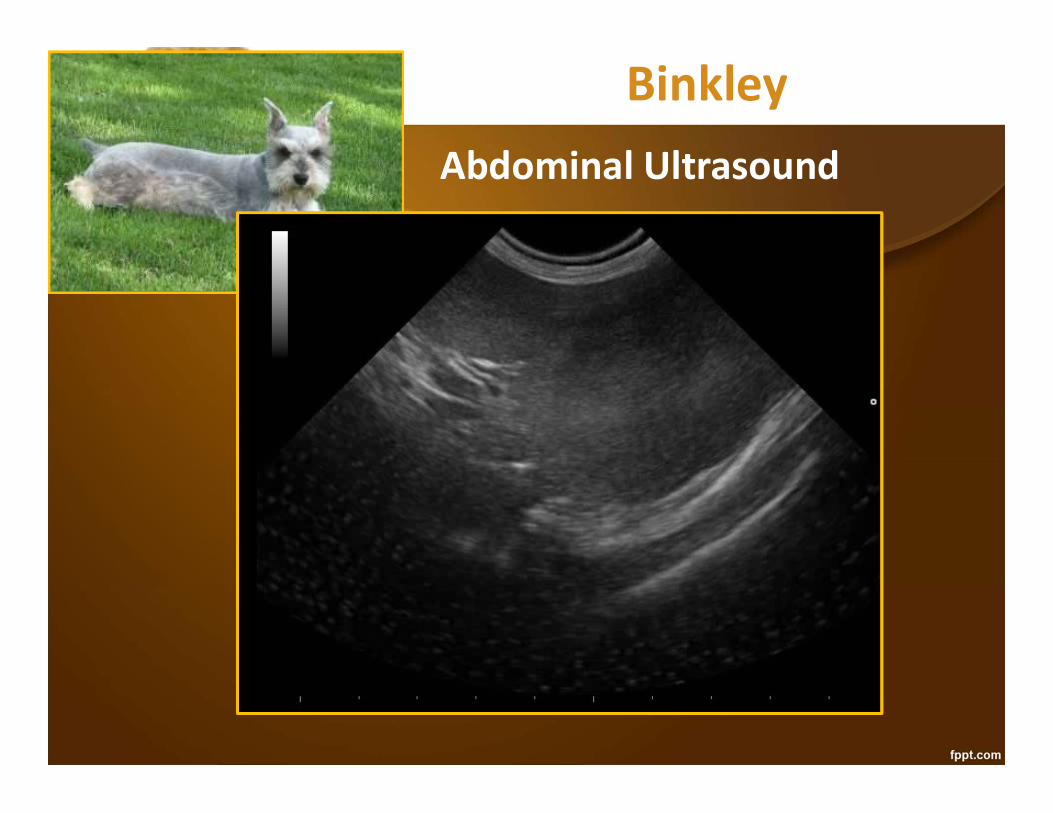
BinkleyAbdominal Ultrasound

BinkleyAbdominal Ultrasound• mostly solid mass at spleen
tail, not well encapsulated• No lesions in the liver• No masses on 3 views of thorax (RLat, LLat, DV)• No nodules on VetBLUE

BinkleyAbdominal Ultrasound• mostly solid mass at spleen
tail, not well encapsulated• No lesions in the liver• No masses on 3 views of thorax (RLat, LLat, DV)• No nodules on VetBLUE• PT, aPTT, BMBT normal• Splenectomy – splenic sarcoma• Panting episodes stopped• Recheck abdominal sonogram at 30 days, 90
days and 6 months clean

Binkley1 year later• difficulty breathing, not
coughing• Worse than panting episodes
• Exam – mildly pale mm, CRT 3 seconds, HR 180-190, RR 80, muffled heart sounds, weak peripheral pulses, distended jugular veins
• Thoracic radiographs

Binkley1 year later• difficulty breathing, not
coughing• Worse than panting episodes
• Exam – mildly pale mm, CRT 3 seconds, HR 180-190, RR 80, muffled heart sounds, weak peripheral pulses, distended jugular veins
• Thoracic radiographs

Binkley1 year later• difficulty breathing, not
coughing• Worse than panting episodes
• Exam – mildly pale mm, CRT 3 seconds, HR 180-190, RR 80, muffled heart sounds, weak peripheral pulses, distended jugular veins
• Thoracic radiographs• TFAST® – R-CTS, L-CTS – Dry Lungs + glide
- R-PCS

Binkley1 year later• difficulty breathing, not
coughing• Worse than panting episodes
• Exam – mildly pale mm, CRT 3 seconds, HR 180-190, RR 80, muffled heart sounds, weak peripheral pulses, distended jugular veins
• Thoracic radiographs• TFAST® – R-CTS, L-CTS – Dry Lungs + glide
- R-PCS

Binkley1 year later• difficulty breathing, not
coughing• Worse than panting episodes
• Exam – mildly pale mm, CRT 3 seconds, HR 180-190, RR 80, muffled heart sounds, weak peripheral pulses, distended jugular veins
• Thoracic radiographs• TFAST® – R-CTS, L-CTS – Dry Lungs + glide
- R-PCS

Binkley1 year later• difficulty breathing, not
coughing• Worse than panting episodes
• Exam – mildly pale mm, CRT 3 seconds, HR 180-190, RR 80, muffled heart sounds, weak peripheral pulses, distended jugular veins
• Thoracic radiographs• TFASTSM – R-CTS, L-CTS – Dry Lungs + glide
- R-PCS Cr – Pericardial Effusion- Tapped – PCV 32%- Echo – MR due to MV dysplasia

Lessons from Binkley• Histopathology is not always
entirely correct• Splenic masses can present for
abdominal pain• Pain can present as panting• Dogs with heart murmur and dyspnea aren’t
always in CHF• Bleeding RA tumors are not always visible on echo• Common things happen commonly (splenic
hemangiosarcoma)• Uncommon things happen uncommonly (splenic
fibrosarcoma cured by surgery)

Hemangiosarcoma• Splenic sonograms

Hemangiosarcoma• Splenic sonograms
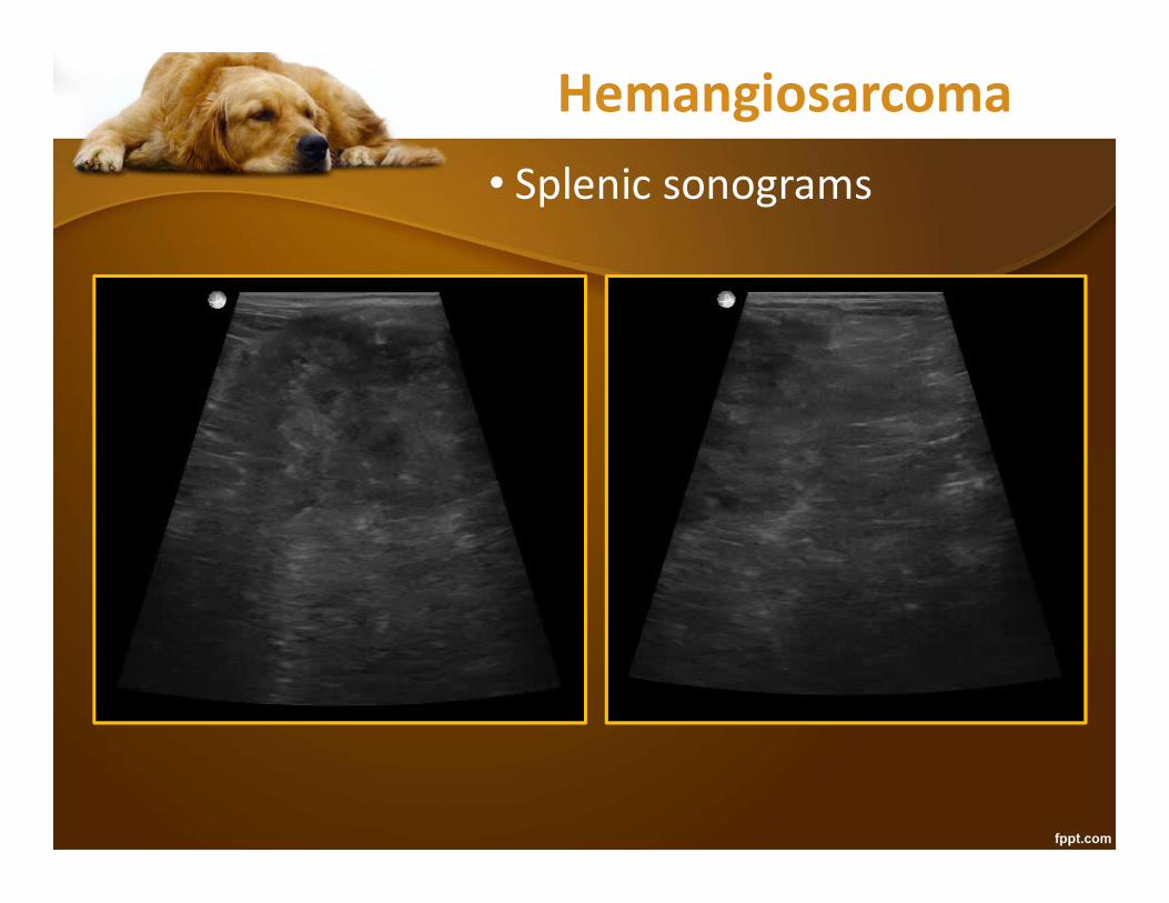
Hemangiosarcoma• Splenic sonograms

Hemangiosarcoma• Splenic sonograms

Hemangiosarcoma• Abdominal Radiographs

Hemangiosarcoma• Abdominal Radiographs

Hemangiosarcoma• Abdominal Radiographs
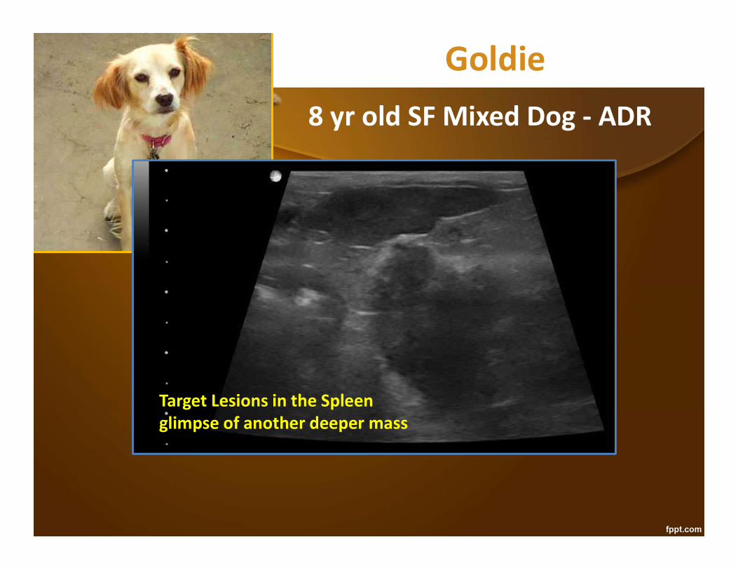
Goldie8 yr old SF Mixed Dog - ADR
Target Lesions in the Spleenglimpse of another deeper mass

Goldie8 yr old SF Mixed Dog - ADR
Pancreatic mass, suspect metastasis to the spleen
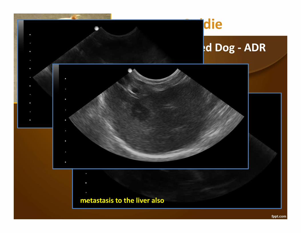
Goldie8 yr old SF Mixed Dog - ADR
metastasis to the liver also
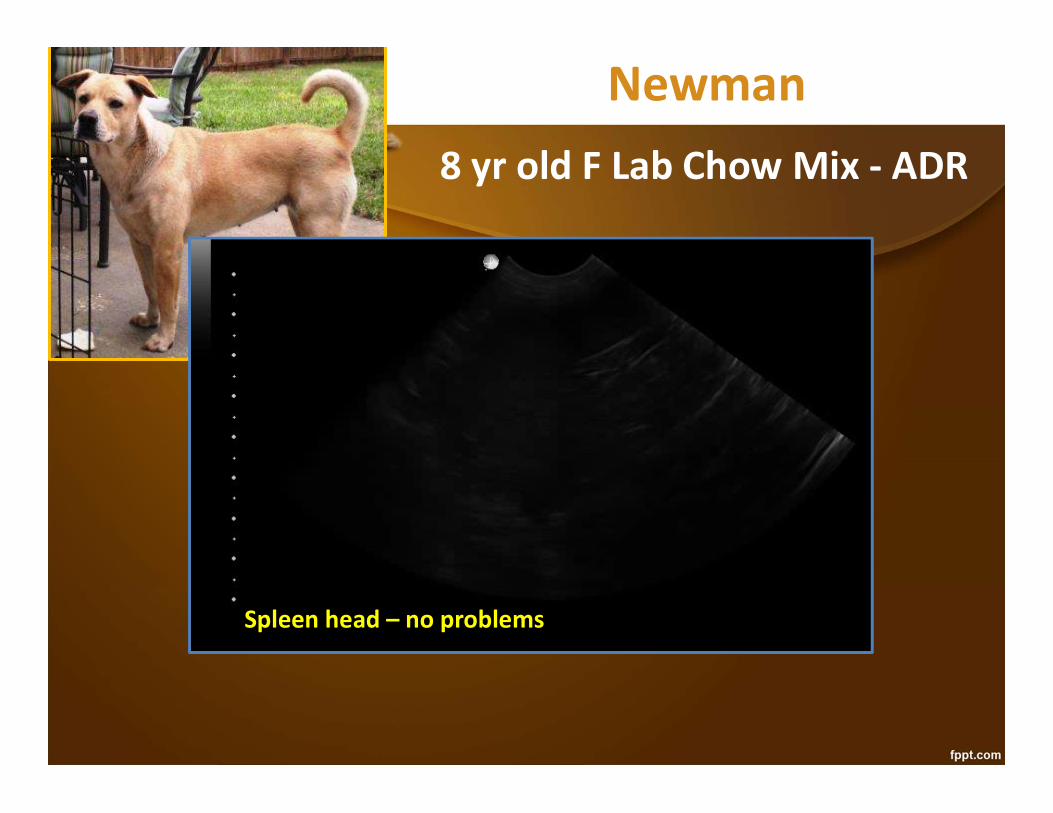
Newman8 yr old F Lab Chow Mix - ADR
Spleen head – no problems
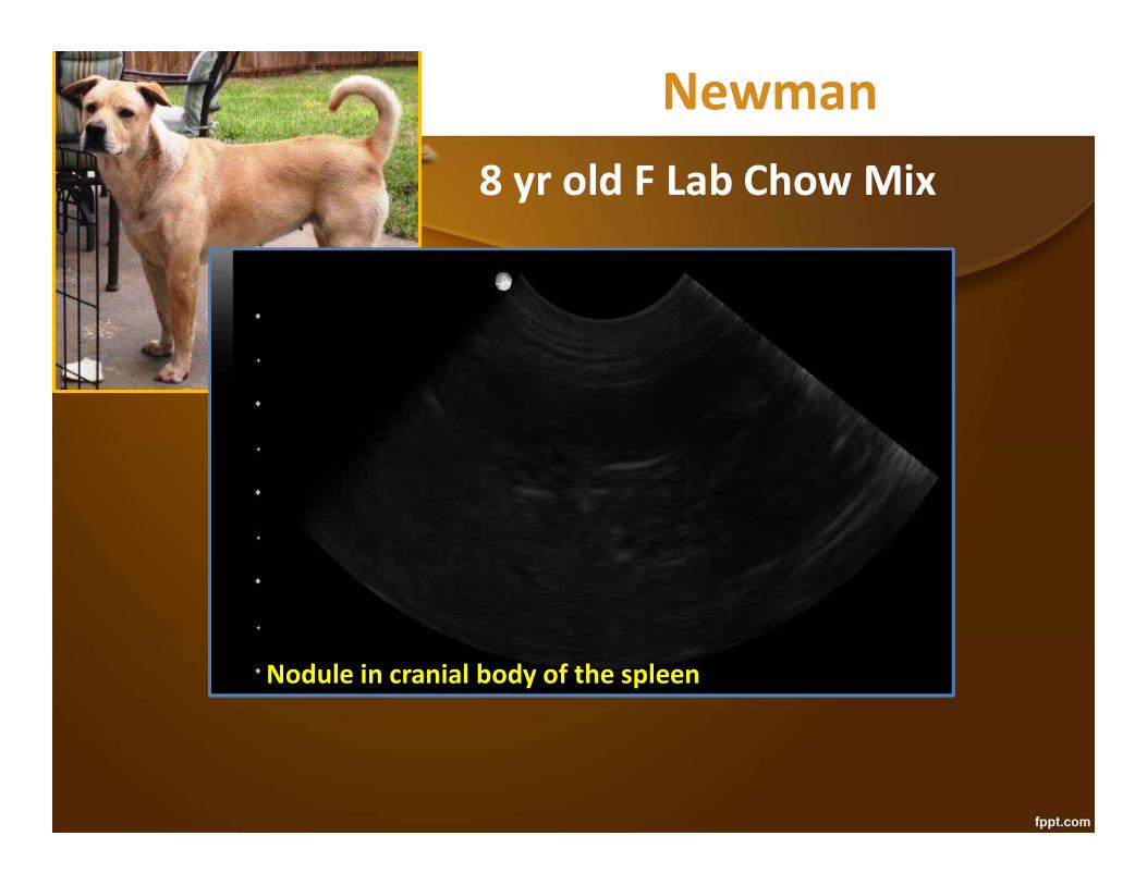
Newman8 yr old F Lab Chow Mix
Nodule in cranial body of the spleen
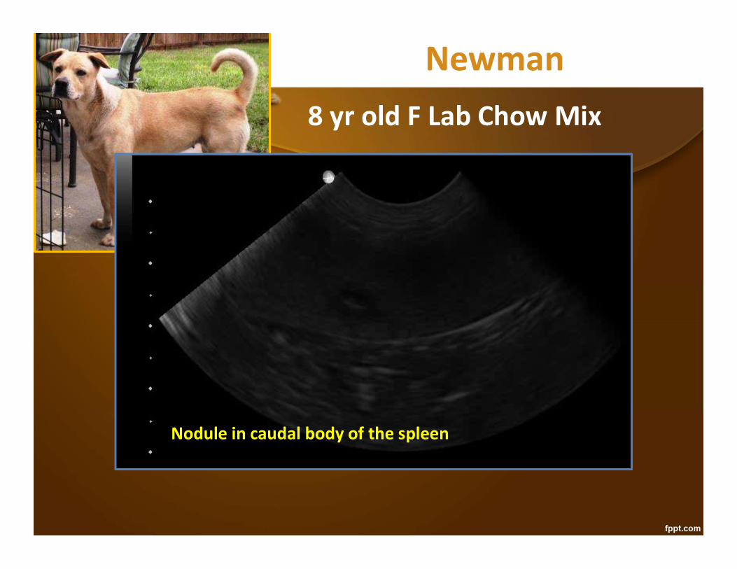
Newman8 yr old F Lab Chow Mix
Nodule in caudal body of the spleen
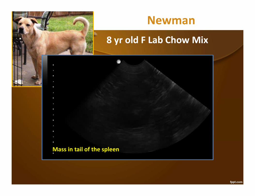
Newman8 yr old F Lab Chow Mix
Mass in tail of the spleen

Newman8 yr old F Lab Chow Mix
Liver – no problems

Newman
8 yr old F Lab Chow Mix
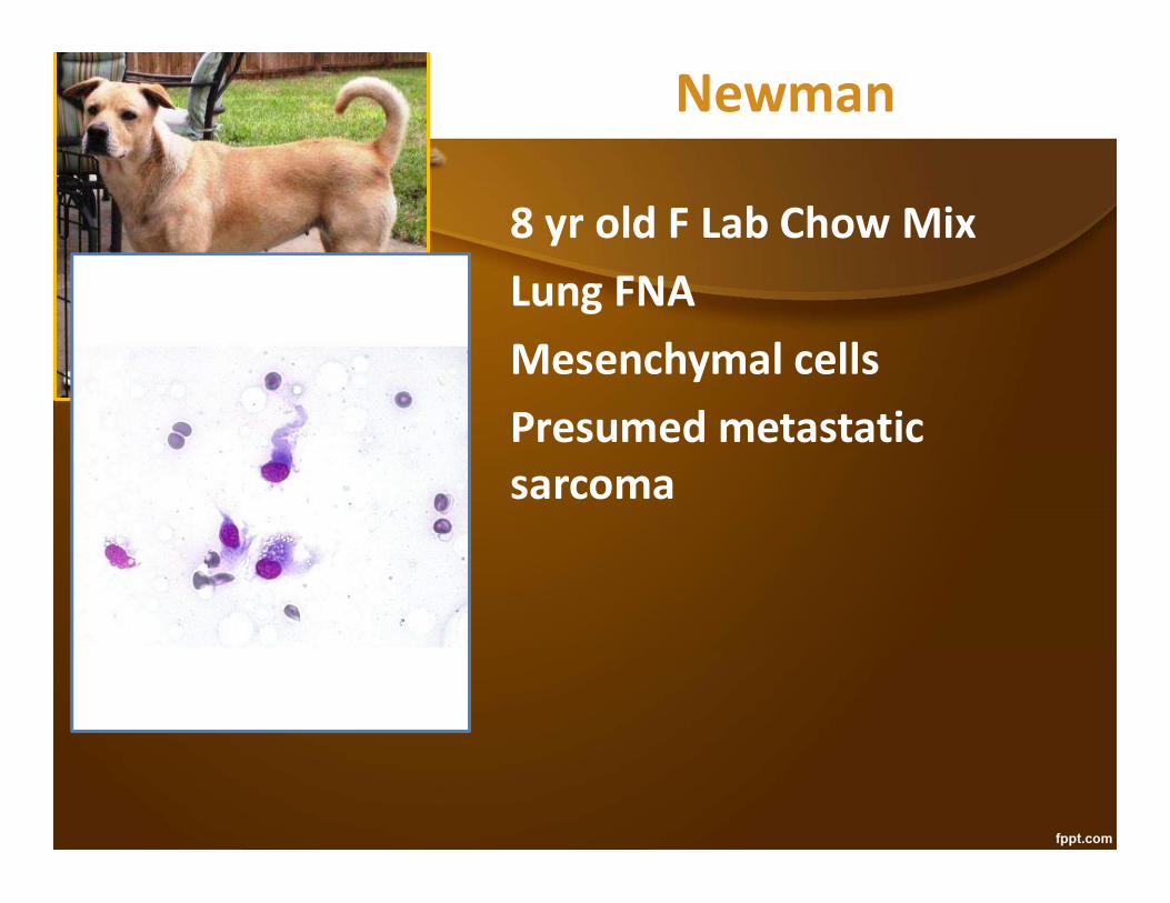
Newman
8 yr old F Lab Chow MixLung FNAMesenchymal cellsPresumed metastatic sarcoma
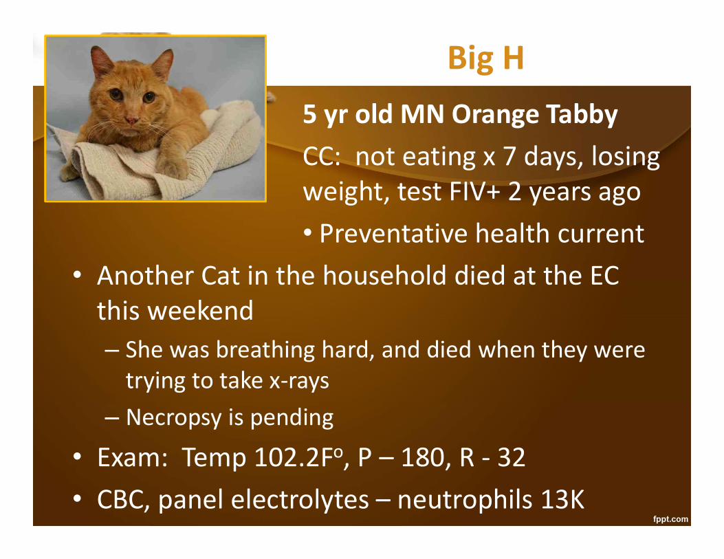
Big H5 yr old MN Orange TabbyCC: not eating x 7 days, losing weight, test FIV+ 2 years ago• Preventative health current
• Another Cat in the household died at the EC this weekend– She was breathing hard, and died when they were
trying to take x-rays– Necropsy is pending
• Exam: Temp 102.2Fo, P – 180, R - 32• CBC, panel electrolytes – neutrophils 13K
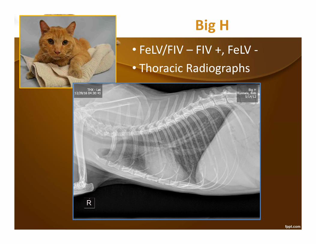
Big H• FeLV/FIV – FIV +, FeLV -• Thoracic Radiographs

Big H• FeLV/FIV – FIV +, FeLV -• Thoracic Radiographs

Big H• FeLV/FIV – FIV +, FeLV -• Thoracic Radiographs
• Peribronchiolar pattern
• Abdominal Ultrasound:

Big H• FeLV/FIV – FIV +, FeLV -• Thoracic Radiographs
• Peribronchiolar pattern
• Abdominal Ultrasound: hypoechoic spleen
• VetBLUE®:
Big H• FeLV/FIV – FIV +• Thoracic Radiographs
• Peribronchiolar pattern
• Abdominal Ultrasound: hypoechoic spleen
• VetBLUE®: very small nodules (1mm)
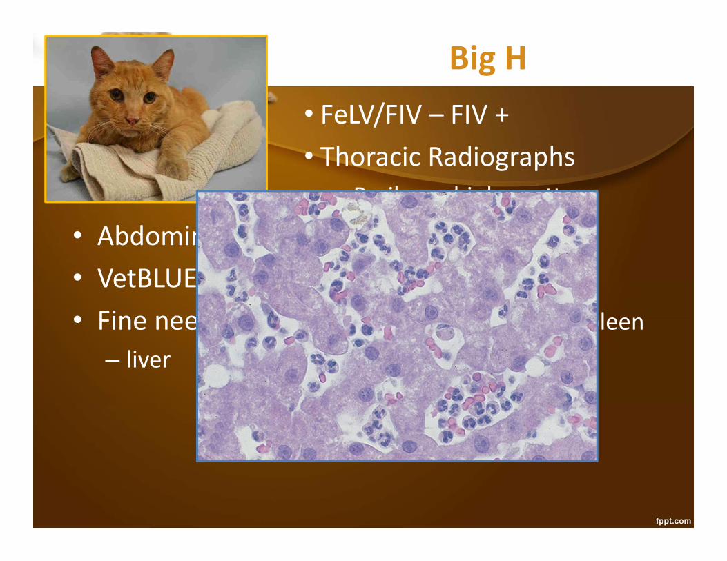
• Fine needle aspiration cytology of liver & spleen– liver
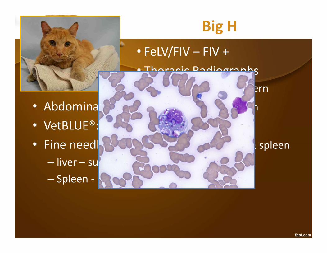
Big H• FeLV/FIV – FIV +• Thoracic Radiographs
• Peribronchiolar pattern
• Abdominal Ultrasound: hypoechoic spleen
• VetBLUE®: very small nodules (1mm)
• Fine needle aspiration cytology of liver & spleen– liver – suppurative hepatitis– Spleen -

Big H• FeLV/FIV – FIV +• Thoracic Radiographs
• Peribronchiolar pattern
• Abdominal Ultrasound: hypoechoic spleen
• VetBLUE: very small nodules (1mm)
• Fine needle aspiration cytology of liver & spleen– liver – suppurative hepatitis– Spleen – Histoplasma capsulatum
• Despite poor Px of Histoplasma + FIV, owner says, “We are here to save Big H.”

Big HTreatment: feeding tube
• Fluconazole 50mg PO BID x 7-14 days, then 50 mg PO SID
• Liposomal Amphotericin B (Abelcet®):– $200-300 for a lyophilized vial (refrigerate)– Reconstitute by adding aseptically 10 ml sterile H20, to
produce a 5 mg/ml solution (vial good for a year)– Dose is 1-2 mg/kg – dilute in 60 ml D5W and give IV over 1-
2 hours (dilution is good for 24 hours)– Treat 3x a week until cat improves, or total dose of 15-20
mg/kg is reached– Cat usually gets better within 2-3 weeks, if they respond– Check BUN before 3rd dose and every dose thereafter– Big H died at home after 3 treatments

Lessons from Big H• Histoplasma cats do not always
have miliary lung disease on rads• Liver, spleen, lymph nodes, bone
marrow, intestinal– FIV isn’t always a death sentence, but it can be– Western blot can rule out FIV false +– Histoplasma prognosis – 1/3 respond and do not
relapse, 1/3 relapse and need chronic meds, 1/3 do not respond
– Always aspirate a large spleen in a cat– MiraVista Histoplasma antigen urine assay is very
sensitive – use for diagnosis and monitoring ($$)– VetBLUE® can reveal tiny nodules not seen on rads

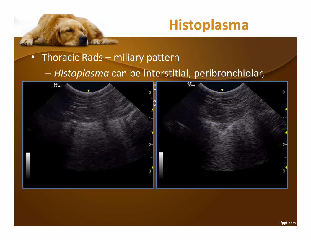
Histoplasma
• Thoracic Rads
Mike Connolly, DVM

Histoplasma
• Thoracic Rads

Histoplasma
• Thoracic Rads – miliary pattern– Histoplasma can be interstitial, peribronchiolar,
alveolar, or some combination thereof– Enlarged perihilar lymph nodes
• VetBLUE®
Histoplasma
• Thoracic Rads – miliary pattern– Histoplasma can be interstitial, peribronchiolar,
alveolar, or some combination thereof– Enlarged perihilar lymph nodes
• VetBLUE®

Histoplasma
• Thoracic Rads – miliary pattern– Histoplasma can be interstitial, peribronchiolar,
alveolar, or some combination thereof– Enlarged perihilar lymph nodes
• VetBLUE® – nodules, shreds, tissue sign• Cytology:
Mike Connolly, DVM
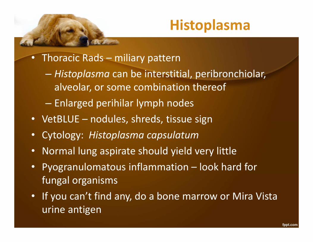
Histoplasma
• Thoracic Rads – miliary pattern– Histoplasma can be interstitial, peribronchiolar,
alveolar, or some combination thereof– Enlarged perihilar lymph nodes
• VetBLUE – nodules, shreds, tissue sign• Cytology: Histoplasma capsulatum• Normal lung aspirate should yield very little• Pyogranulomatous inflammation – look hard for
fungal organisms• If you can’t find any, do a bone marrow or Mira Vista
urine antigen

Raven10 yr old SF GSDCC: 20% weight loss over 6 months, now not eating
• Cannot spend more than $300• Just wants to know if it is time to euthanize• Exam: Subtle thickness in the mid abdomen• Office Visit - $50• GlobalFAST® = AFAST®, TFAST®, VetBLUE®• $67 each part = $201
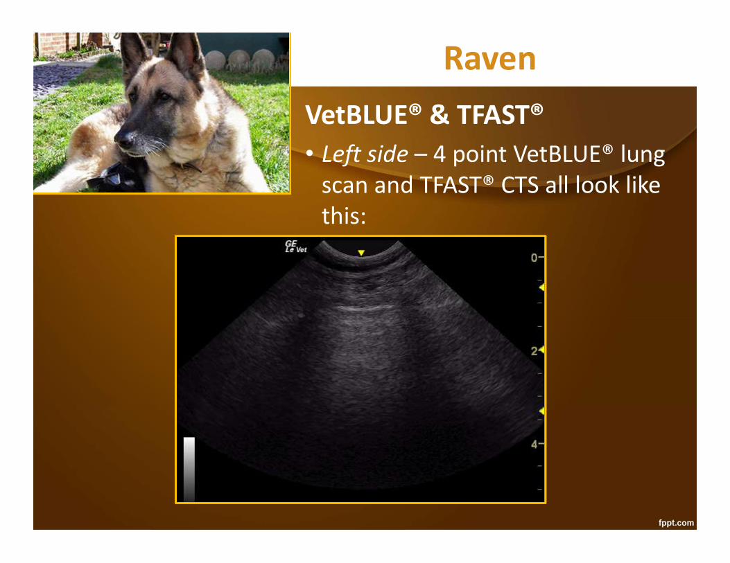
RavenVetBLUE® & TFAST®• Left side – 4 point VetBLUE® lung
scan and TFAST® CTS all look like this:
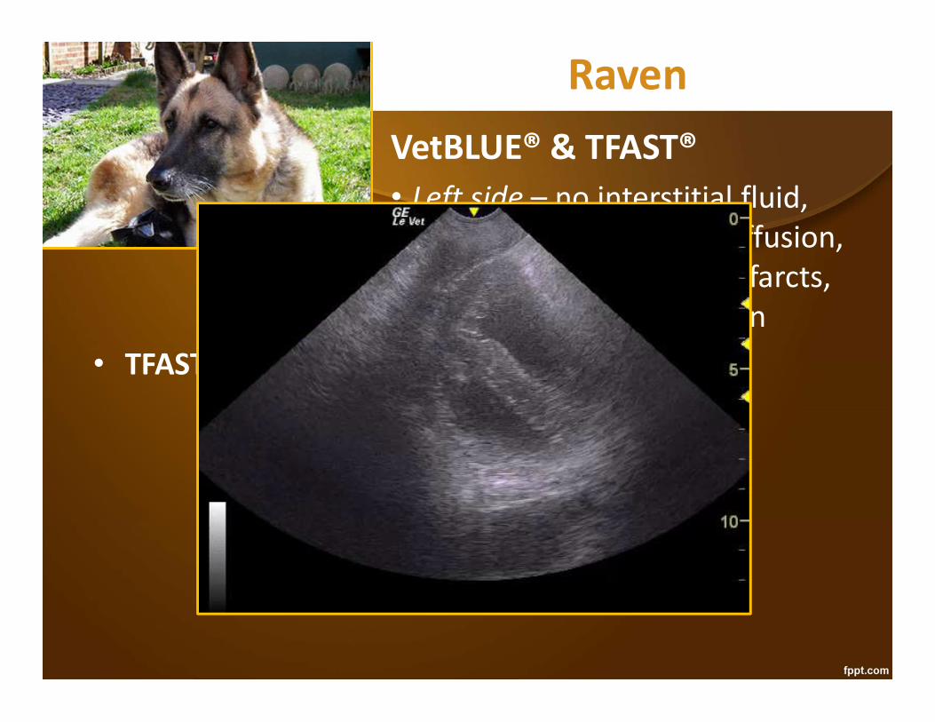
RavenVetBLUE® & TFAST®• Left side – no interstitial fluid,
pneumothorax, pleural effusion, nodules, alveolar fluid, infarcts, nor any lung consolidation
• TFAST® Left Pericardial scan

RavenVetBLUE® & TFAST®• Left side – no interstitial fluid,
pneumothorax, pleural effusion, nodules, alveolar fluid, infarcts, nor any lung consolidation
• TFAST® Left Pericardial scan – no pleural effusion, no pericardial effusion, no right heart problems, no left heart problems
• VetBLUE® & TFAST® Right side – just like the left side• AFAST® - 4 point abdominal scan

RavenVetBLUE® & TFAST®• Left side – no interstitial fluid, no
pneumothorax, pleural effusion, nodules, alveolar fluid, infarcts, nor any lung consolidation
• TFAST® Left Pericardial scan – no pleural effusion, no pericardial effusion, no right heart problems, no left heart problems
• VetBLUE® & TFAST® Right side – just like the left side• AFAST® - 4 point abdominal scan
DH View – Diaphragmatic-Hepatic View

RavenVetBLUE® & TFAST®• Left side – no interstitial fluid, no
pneumothorax, pleural effusion, nodules, alveolar fluid, infarcts, nor any lung consolidation
• TFAST® Left Pericardial scan – no pleural effusion, no pericardial effusion, no right heart problems, no left heart problems
• VetBLUE® & TFAST® Right side – just like the left side• AFAST® - 4 point abdominal scan – DH no
abnormalities, SR View – Spleno-Renal View

RavenVetBLUE® & TFAST®• Left side – no interstitial fluid, no
pneumothorax, pleural effusion, nodules, alveolar fluid, infarcts, nor any lung consolidation
• TFAST® Left Pericardial scan – no pleural effusion, no pericardial effusion, no right heart problems, no left heart problems
• VetBLUE® & TFAST® Right side – just like the left side• AFAST® - 4 point abdominal scan – DH no
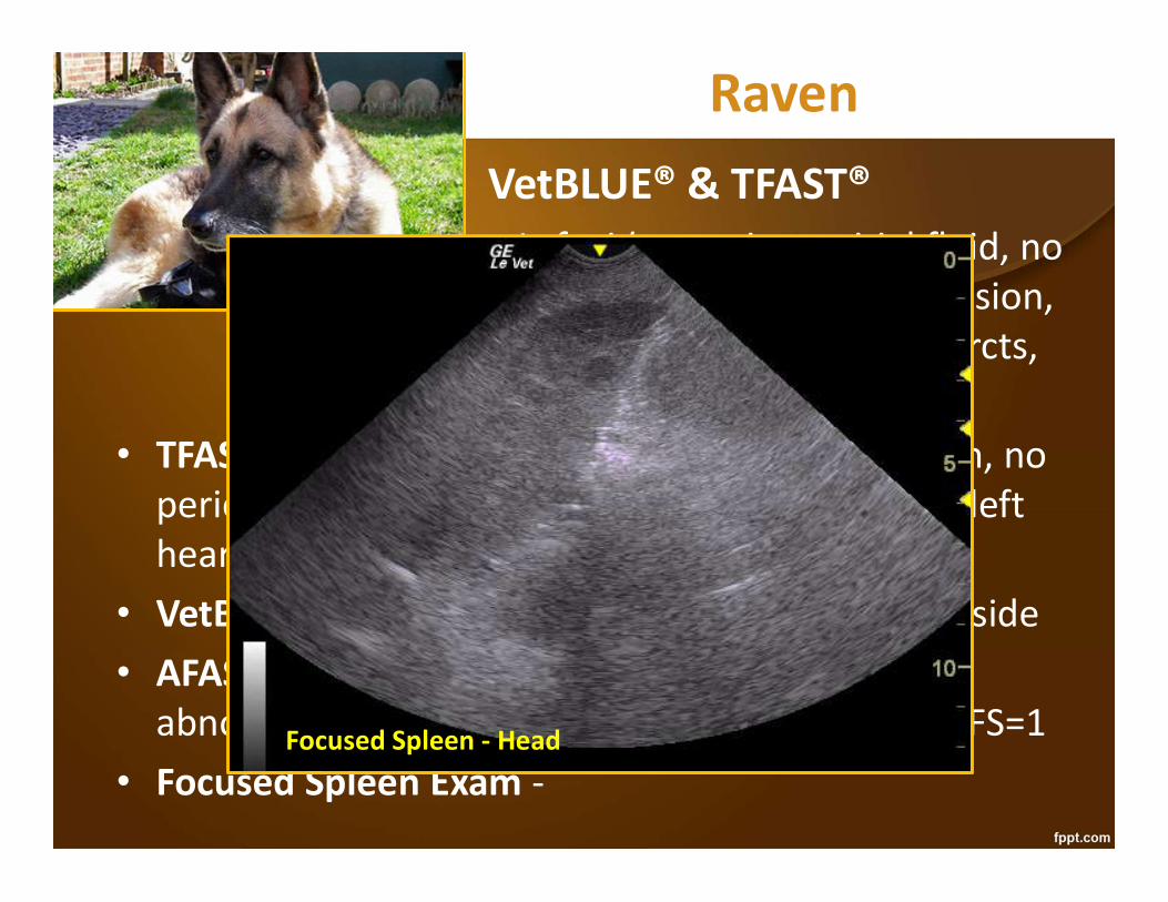
abnormalities, SR ascites & honeycomb spleen AFS=1• Focused Spleen Exam -
RavenVetBLUE® & TFAST®• Left side – no interstitial fluid, no
pneumothorax, pleural effusion, nodules, alveolar fluid, infarcts, nor any lung consolidation
• TFAST® Left Pericardial scan – no pleural effusion, no pericardial effusion, no right heart problems, no left heart problems
• VetBLUE® & TFAST® Right side – just like the left side• AFAST® - 4 point abdominal scan – DH no
abnormalities, SR ascites & honeycomb spleen AFS=1• Focused Spleen Exam -
Focused Spleen - Head

RavenVetBLUE® & TFAST®• Left side – no interstitial fluid, no
pneumothorax, pleural effusion, nodules, alveolar fluid, infarcts, nor any lung consolidation
• TFAST® Left Pericardial scan – no pleural effusion, no pericardial effusion, no right heart problems, no left heart problems
• VetBLUE® & TFAST® Right side – just like the left side• AFAST® - 4 point abdominal scan – DH no
abnormalities, SR ascites & honeycomb spleen AFS=1• Focused Spleen Exam -
Focused Spleen – Cranial Body

RavenVetBLUE® & TFAST®• Left side – no interstitial fluid, no
pneumothorax, pleural effusion, nodules, alveolar fluid, infarcts, nor any lung consolidation
• TFAST® Left Pericardial scan – no pleural effusion, no pericardial effusion, no right heart problems, no left heart problems
• VetBLUE® & TFAST® Right side – just like the left side• AFAST® - 4 point abdominal scan – DH no
abnormalities, SR ascites & honeycomb spleen AFS=1• Focused Spleen Exam -
Focused Spleen

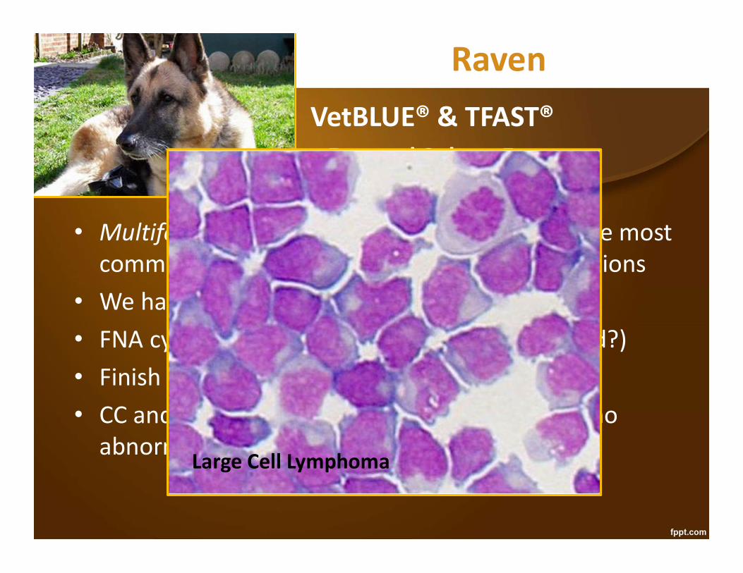
RavenVetBLUE® & TFAST®• Focused Spleen Exam –DDx Honeycomb Spleen
• Multifocal infiltrative disease – MCT and LSA are most common > plasma cell myeloma, atypical infections
• We have $41 left• FNA cytology after a conversation (spleen? fluid?)• Finish the AFAST® while the slide are drying
CC – Cysto-Colic View

RavenVetBLUE® & TFAST®• Focused Spleen Exam –DDx Honeycomb Spleen
• Multifocal infiltrative disease – MCT and LSA are most common > plasma cell myeloma, atypical infections
• We have $41 left• FNA cytology after a conversation (spleen? fluid?)• Finish the AFAST® while the slide are drying• CC and HR (Hepato-Renal/Home Run) views – no
abnormalities seen
RavenVetBLUE® & TFAST®• Focused Spleen Exam –DDx Honeycomb Spleen
• Multifocal infiltrative disease – MCT and LSA are most common > plasma cell myeloma, atypical infections
• We have $41 left• FNA cytology after a conversation (spleen? fluid?)• Finish the AFAST® while the slide are drying• CC and HR (Hepato-Renal/Home Run) views – no
abnormalities seenLarge Cell Lymphoma
RavenNow What??• We have $9 left• No enlarged lymph nodes
Large cell lymphoma of the spleen• Chemo not an option for these owners ($4-5K)• Prednisone only – 30-90 days• Marginal Zone Lymphoma (spleen only)
– Need immunohistochemistry to diagnose ($150)– Then need to stage ($500-600)– Then splenectomy only ($800-900)– Survival 1-2 years with splenectomy only
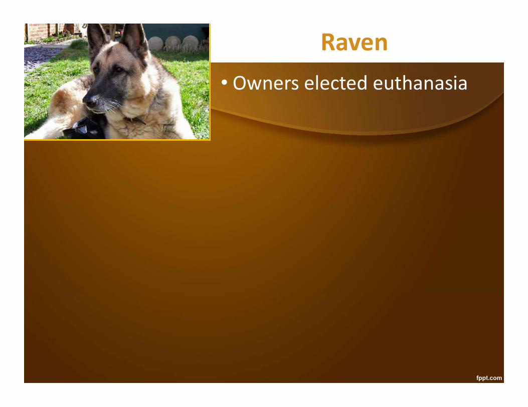
Raven• Owners elected euthanasia

•PowerPoint – Spleen Ultrasound Cases•.pdf of PowerPoints – Spleen Ultrasound Cases
•1 slide per page•6 slides per page
SummarySummary
Acknowledgements
Stephanie Lisciandro, ACVIM (Internal Medicine)Chapter 3: Focused or COAST3 – Liver and Gall Bladder
Focused Ultrasound Techniques for the Small Animal Practitioner
Editor Greg Lisciandro – 2014
Dr. Mike Connolly - Connolly Animal Clinic“Junebug” and “Suzy”
Eastex Vet Clinic - “Binkley”
Drs. Shawn Penn & Doug AshburnSouthwood Drive Animal Clinic - “Big H”