Endobronchial ultrasound-guided procedures - Medical ...
136
Endobronchial ultrasound-guided procedures March 2008 MSAC application 1108 Assessment report
-
Upload
khangminh22 -
Category
Documents
-
view
4 -
download
0
Transcript of Endobronchial ultrasound-guided procedures - Medical ...

Endobronchial
ultrasound-guided procedures
March 2008
MSAC application 1108
Assessment report

© Commonwealth of Australia 2008
ISBN (Print) 1-74186-638-3
ISBN (Online) 1-74186-639-1
ISSN (Print) 1443-7120
ISSN (Online) 1443-7139
First printed June 2008
Paper-based publications © Commonwealth of Australia 2008 This work is copyright. Apart from any use as permitted under the Copyright Act 1968, no part may be reproduced by any process without prior written permission from the Commonwealth. Requests and inquiries concerning reproduction and rights should be addressed to the Commonwealth Copyright Administration, Attorney General’s Department, Robert Garran Offices, National Circuit, Barton ACT 2600 or posted at http://www.ag.gov.au/cca Internet sites © Commonwealth of Australia 2008 This work is copyright. You may download, display, print and reproduce this material in unaltered form only (retaining this notice) for your personal, non-commercial use or use within your organisation. Apart from any use as permitted under the Copyright Act 1968, all other rights are reserved. Requests and inquiries concerning reproduction and rights should be addressed to Commonwealth Copyright Administration, Attorney General’s Department, Robert Garran Offices, National Circuit, Barton ACT 2600 or posted at http://www.ag.gov.au/cca
Electronic copies of the report can be obtained from the Medical Service Advisory Committee’s Internet site at http://www.msac.gov.au/
Printed copies of the report can be obtained from:
The Secretary Medical Services Advisory Committee Department of Health and Ageing Mail Drop 106 GPO Box 9848 Canberra ACT 2601
Enquiries about the content of the report should be directed to the above address.
The Medical Services Advisory Committee (MSAC) is an independent committee which has been established to provide advice to the Minister for Health and Ageing on the strength of evidence available on new and existing medical technologies and procedures in terms of their safety, effectiveness and cost-effectiveness. This advice will help to inform government decisions about which medical services should attract funding under Medicare.
MSAC recommendations do not necessarily reflect the views of all individuals who participated in the MSAC evaluation.
This report was prepared by the Medical Services Advisory Committee with the assistance of Marc Bevan, Marianne Chau, Jun Feng and Koji Makino from IMS Health. The report was edited by Ann Jones. The report was endorsed by the Minister for Health and Ageing on 20 May 2008.
Publication approval number: P3-3920

Endobronchial ultrasound-guided procedures i
Contents
Contents .................................................................................................................... i
Executive summary................................................................................................. ix The procedure............................................................................................................................... ix Medical Services Advisory Committee—role and approach ................................................. ix MSAC’s assessment of EBUS-guided procedures for investigation of non-small cell lung cancer, mediastinal/hilar masses, endobronchial cancer and peripheral lung lesions.................................................................................................................................... ix
Clinical need .................................................................................................................... ix Safety ............................................................................................................................... x Effectiveness .................................................................................................................. x Economics ...................................................................................................................... x
Recommendation ....................................................................................................................... xii
Introduction ..............................................................................................................1
Background.............................................................................................................. 2 The procedure................................................................................................................................ 2
Endobronchial ultrasound-transbronchial biopsy ...................................................... 2 Endobronchial ultrasound-transbronchial needle aspiration .................................... 3
Intended purpose .......................................................................................................................... 3 Clinical need ................................................................................................................................... 4
Incidence and mortality .................................................................................................. 5 Eligible population ........................................................................................................................ 6
Central, mediastinal and hilar tumours......................................................................... 6 Peripheral lung lesions .................................................................................................... 7
Current treatment.......................................................................................................................... 8 Existing procedures ...................................................................................................................... 9
Imaging techniques.......................................................................................................... 9 Sampling techniques...................................................................................................... 10
Lymph node stations .................................................................................................................. 12 Comparator .................................................................................................................................. 14 Marketing status of the technology .......................................................................................... 15 Current reimbursement arrangement ....................................................................................... 15
Approach to assessment .........................................................................................16 Research questions and clinical pathways................................................................................ 16
Non-small cell lung cancer staging.............................................................................. 16 Mediastinal/hilar masses of unknown origin ............................................................ 18 Depth diagnosis of endobronchial cancers................................................................ 20 Peripheral lung lesion.................................................................................................... 22
Assessment framework............................................................................................................... 24

ii Endobronchial ultrasound-guided procedures
Types of evidence ..........................................................................................................24 Review of the literature...............................................................................................................24
Search strategy ................................................................................................................24 Selection criteria .............................................................................................................25 Search results ..................................................................................................................28
Study appraisal..............................................................................................................................29 Assessment of eligible studies ......................................................................................29
Data analysis .................................................................................................................................31 Data extraction ...............................................................................................................31 Measurement of test accuracy ......................................................................................31 Statistics ...........................................................................................................................33
Expert advice................................................................................................................................33 Assessment of the body of evidence ........................................................................................33
Results of assessment............................................................................................. 35 Summary .......................................................................................................................................35 Is it safe?........................................................................................................................................36 Is it effective? ...............................................................................................................................37
Linked evidence..............................................................................................................37 Diagnostic accuracy .......................................................................................................37 Patient management ......................................................................................................46 Treatment effectiveness ................................................................................................47
Economic considerations....................................................................................... 48 Summary .......................................................................................................................................48
Background and approach............................................................................................49 Estimated extent of financial implications .................................................................50 Published evidence regarding endobronchial ultrasound cost-effectiveness ........57 Cost analyses of EBUS-guided techniques.................................................................58 Results..............................................................................................................................63
Discussion.....................................................................................................................................67
Other considerations .............................................................................................. 70 Generalisability of evidence .......................................................................................................70 Endobronchial ultrasound and endoscopic ultrasound .........................................................70 Prospective studies ......................................................................................................................70
Research recommendations ................................................................................... 72
Conclusions ............................................................................................................ 75 Safety .............................................................................................................................................75 Effectiveness ................................................................................................................................75 Economic analyses ......................................................................................................................76
Recommendation ................................................................................................... 77

Endobronchial ultrasound-guided procedures iii
Appendix A MSAC terms of reference and membership ................................... 78
Appendix B Advisory panel................................................................................. 80
Appendix C Supportive data ................................................................................81
Appendix D Included studies ............................................................................. 89
Appendix E Excluded studies ........................................................................... 97
Appendix F Literature search ............................................................................ 103
Appendix G Quality criteria ............................................................................... 106
Appendix H Staging classification .................................................................... 108
Appendix I Additional information for economic evaluation .......................... 110
Abbreviations ........................................................................................................ 114
References ............................................................................................................. 116

iv Endobronchial ultrasound-guided procedures
Tables
Table 1 Estimated number of patients per year eligible to undergo pathological assessment for NSCLC staging and the diagnosis of mediastinal masses of unknown origin.................................................................. 6
Table 2 Rates of comparator procedures for peripheral lung lesion diagnosis 2003–2004.................................................................................................................. 7
Table 3 Number of patients with peripheral lung lesions < 3 cm diameter .................. 8 Table 4 Comparative nodal accessibility............................................................................ 13 Table 5 Australian Register of Therapeutic Goods listing for EBUS devices
and components...................................................................................................... 15 Table 6 PPICO criteria for the use of EBUS-TBNA in the invasive (nodal)
staging of patients with presumed or known NSCLC ...................................... 16 Table 7 PPICO criteria for the use of EBUS-TBNA in the invasive diagnosis
of patients with mediastinal/hilar masses of unknown origin ......................... 18 Table 8 PPICO criteria for the use of EBUS with or without EBBX in
diagnosing the depth of endobronchial cancers................................................. 20 Table 9 PPICO criteria for the use of EBUS-TBBX in the invasive diagnosis
of patients with peripheral lung lesions ............................................................... 22 Table 10 Electronic databases searched during the review of EBUS-guided
transbronchial sampling procedures .................................................................... 24 Table 11 Selection criteria for studies of EBUS-TBNA in the invasive (nodal)
staging of patients with presumed or known NSCLC ...................................... 25 Table 12 Selection criteria for studies of EBUS-TBNA in invasive diagnosis of
patients with mediastinal/hilar masses of unknown origin .............................. 26 Table 13 Selection criteria for studies of EBUS with or without EBBX in the
depth diagnosis of endobronchial cancers .......................................................... 26 Table 14 Selection criteria for studies of EBUS-TBBX in the invasive diagnosis
of patients with peripheral lung lesions ............................................................... 27 Table 15 NHMRC dimensions of evidence ........................................................................ 30 Table 16 Designations of levels of evidence according to research question ................ 30 Table 17 Grading system for appraisal of studies evaluating diagnostic tests................ 31 Table 18 Body of evidence assessment matrix ................................................................... 34 Table 19 Pneumothorax and bleeding adverse events associated with EBUS-
guided procedures and comparator technologies .............................................. 37 Table 20 Characteristics of the included comparative studies assessing
diagnostic accuracy and yield of EBUS-TBNA among patients referred for lung cancer staging or diagnosis of mediastinal/hilar masses of unknown origin..................................................................................... 39
Endobronchial ultrasound-guided procedures v
Table 21 Diagnostic yields of included comparative studies assessing EBUS-TBNA among patients referred for NSCLC staging and diagnoses of mediastinal/hilar masses of unknown origin...................................................... 40
Table 22 Accuracy of EBUS-TBNA compared with EUS-FNA in patients referred for NSCLC staging and diagnosis of mediastinal/hilar masses of unknown origin .................................................................................................. 41
Table 23 Assessment of the comparative body of evidence for EBUS-TBNA in NSCLC staging and diagnosis of mediastinal/hilar masses of unknown origin ....................................................................................................... 42
Table 24 Accuracy of EBUS-TBBX compared with fluoroscopy-TBBX in patients with peripheral lesions............................................................................. 43
Table 25 Characteristics of the included comparative studies assessing the diagnostic accuracy/yield of EBUS-TBBX in patients with peripheral lung lesions .............................................................................................................. 44
Table 26 Diagnostic yield of the included comparative studies assessing EBUS-TBBX in patients with peripheral lung lesions ...................................... 45
Table 27 Assessing the comparative body of evidence for EBUS-TBBX in the diagnosis of peripheral lung lesions ..................................................................... 46
Table 28 Total cost of EBUS guidance per procedure...................................................... 51 Table 29 Total cost of EBUS-TBNA per procedure......................................................... 53 Table 30 Other costs associated with EBUS-TBNA per procedure ............................... 53 Table 31 Total cost of EBUS-TBBX per procedure ......................................................... 54 Table 32 Summary of professional fees for EBUS-guided procedures .......................... 55 Table 33 Estimated total costs of EBUS-TBNA for assessment of NSCLC and
mediastinal/hilar masses ........................................................................................ 55 Table 34 Estimated total costs of EBUS-TBBX for diagnosis of peripheral
lung lesions .............................................................................................................. 56 Table 35 Estimated total cost savings offered by EBUS-TBNA for assessment
of NSCLC and mediastinal/hilar masses in the Australian health system ....................................................................................................................... 56
Table 36 Estimated total cost savings of EBUS-TBBX for diagnosis of peripheral lung lesions < 3 cm diameter for the Australian health system ....................................................................................................................... 57
Table 37 Clinical variables included in the model .............................................................. 60 Table 38 Cost variables included in the model ................................................................... 60 Table 39 Estimated costs of mediastinal node biopsy per procedure ............................. 61 Table 40 Estimated costs of CT-guided TTNA per procedure ....................................... 62 Table 41 Estimated costs of thoracotomy per procedure................................................. 62

vi Endobronchial ultrasound-guided procedures
Table 42 Estimated costs of follow-up procedures for the diagnosis of peripheral lung lesions < 3 cm.............................................................................. 63
Table 43 Summary of costs and incremental costs ............................................................ 63 Table 44 Results of sensitivity analyses for EBUS-TBNA vs TBNA alone................... 65 Table 45 Results of sensitivity analyses for EBUS-TBBX vs fluoroscopic-
TBBX........................................................................................................................ 66 Table 46 Separation statistics for respiratory system procedures by public and
private hospitals ...................................................................................................... 69 Table 47 Characteristics of potentially relevant ongoing trials using EBUS-
guided procedures................................................................................................... 71 Table 48 Research recommendations for use of EBUS-TBNA in NSCLC
staging and diagnosis of mediastinal/hilar masses............................................. 72 Table 49 Research recommendations for the use of EBUS with or without
EBBX in the depth diagnosis of endobronchial cancers .................................. 73 Table 50 Research recommendations for the use of EBUS-TBBX in the
diagnosis of peripheral lung lesions ..................................................................... 74 Table 51 Characteristics of the included supportive studies assessing the
diagnostic accuracy/yield of EBUS-TBNA in patients referred for lung cancer staging and/or diagnosis of mediastinal/hilar mass of unknown origin ....................................................................................................... 82
Table 52 Diagnostic accuracy of included supportive studies comparing EBUS-TBNA with CT and PET imaging in patients referred for NSCLC staging and diagnoses of mediastinal/hilar masses of unknown origin.......... 84
Table 53 Diagnostic accuracy of the included non-comparative supportive studies assessing EBUS-TBNA in patients referred for NSCLC staging and diagnoses of mediastinal/hilar masses of unknown origin.......... 85
Table 54 Diagnostic yield of the included supportive studies assessing EBUS-TBNA in a mixed patient population (including NSCLC)............................... 85
Table 55 Characteristics of included supportive studies that assessed diagnostic yield of EBUS-TBBX among patients with peripheral lung lesions ............... 86
Table 56 Diagnostic yield reported by the included supportive studies that assessed EBUS-TBBX among patients with peripheral lung lesions.............. 88
Table 57 Characteristics and results of studies assessing the diagnostic accuracy and/or diagnostic yield of EBUS guided procedures........................................ 90
Table 58 EMBASE.com search results: EBUS procedures for NSCLC staging and diagnosis of peripheral lung, mediastinal and hilar masses (4 June 2007).......................................................................................................... 103
Table 59 PubMed search results for EBUS procedures for NSCLC staging and diagnosis of peripheral lung, mediastinal and hilar masses (7 June 2007).......................................................................................................... 104

Endobronchial ultrasound-guided procedures vii
Table 60 Cochrane Library search results for EBUS procedures for NSCLC staging and diagnosis of peripheral lung, mediastinal and hilar masses (4 June 2007).......................................................................................................... 104
Table 61 HTA websites searched in this review............................................................... 105 Table 62 TNM classification for lung cancer.................................................................... 108 Table 63 Lung cancer staging by TNM grouping............................................................. 109 Table 64 Capital costs of EBUS with radial probe—low utilisation level .................... 110 Table 65 Capital costs of EBUS with radial probe—high utilisation level................... 111 Table 66 Capital costs of EBUS with linear probe—low utilisation level .................... 111 Table 67 Capital costs of EBUS with linear probe—high utilisation level................... 112 Table 68 Capital costs of Aloka EBUS—low utilisation level........................................ 112 Table 69 Capital costs of Aloka EBUS—high utilisation level ...................................... 113

viii Endobronchial ultrasound-guided procedures
Figures
Figure 1 Radial EBUS probe with guide sheath (UM-S20-20R) (left) and linear EBUS TBNA scope tip (XBF-UC260F-OL8) (right) ......................................... 3
Figure 2 Regional lymph node stations for lung cancer staging...................................... 13 Figure 3 Clinical pathway for invasive (nodal) staging of patients with
presumed or known NSCLC ................................................................................ 17 Figure 4 Clinical pathway for the invasive diagnosis of patients with
mediastinal/hilar masses of unknown origin...................................................... 19 Figure 5 Clinical pathway for depth diagnosis of endobronchial cancers...................... 21 Figure 6 Clinical pathway for invasive diagnosis of patients with peripheral
lung lesions .............................................................................................................. 23 Figure 7 QUOROM flowchart used to identify and select studies for EBUS-
guided procedure literature review ....................................................................... 28 Figure 8 Data used to calculate measures of test accuracy............................................... 32 Figure 9 Structure of the current economic model ........................................................... 59

Endobronchial ultrasound-guided procedures ix
Executive summary
The procedure
Endobronchial ultrasound (EBUS) is a minimally invasive procedure that involves an ultrasound probe being introduced into the thoracic region via the bronchial airway. The ultrasound probe can then be used to generate images of pulmonary and mediastinal structures (Herth et al 2000). EBUS imaging may be used alone or to guide sampling procedures such as endobronchial biopsy (EBBX), transbronchial needle aspiration (TBNA) or transbronchial biopsy (TBBX).
Medical Services Advisory Committee—role and approach
The Medical Services Advisory Committee (MSAC) was established by the Australian Government to strengthen the role of evidence in health financing decisions in Australia. MSAC advises the Minister for Health and Ageing on the evidence relating to the safety, effectiveness and cost-effectiveness of new and existing medical technologies and procedures and under what circumstances public funding should be supported.
A rigorous assessment of evidence is thus the basis of decision making when funding is sought under Medicare. A team from IMS Health was engaged to conduct a systematic review of literature on endobronchial ultrasound guided transbronchial sampling procedures for non-small cell lung cancer staging, diagnosis of mediastinal/hilar masses, depth diagnosis of endobronchial cancers and diagnosis of peripheral lung lesions. An advisory panel with expertise in this area then evaluated the evidence and provided advice to MSAC.
MSAC’s assessment of EBUS-guided procedures for investigation of non-small cell lung cancer, mediastinal/hilar masses, endobronchial cancer and peripheral lung lesions
Clinical need
Lung cancer is the leading cause of cancer death globally (AIHW 2006). In Australia, lung cancer was the fifth most common notifiable cancer in 2003, when it accounted for 8.9 per cent of all cancers (8249 diagnoses reported) (AIHW and AACR 2007). In the same year, lung cancer was responsible for 6988 deaths (4506 male; 2482 female), resulting in 43,325 person-years of life lost (before 75 years of age) due to premature cancer death. This was the highest number of person-years of life lost among all notifiable cancers in Australia (AIHW and AACR 2007).
Improvements in lung cancer staging and diagnosis may contribute to enhanced patient management by avoiding invasive diagnostic procedures, and offering more accurate curative and palliative treatment planning. Advances in these areas enhance survival and quality of life.

x Endobronchial ultrasound-guided procedures
Safety
EBUS-guided procedures for non-small cell lung cancer (NSCLC) staging, diagnosis of mediastinal/hilar masses, depth diagnosis of endobronchial cancers and diagnosis of peripheral lung lesions appear to be as safe as other minimally-invasive diagnostic tests. The most frequently reported adverse events were bleeding and pneumothorax. These mainly occurred among patients who underwent either EBUS or fluoroscopy-guided transbronchial biopsy. There also appeared to be a trend toward a higher frequency of pneumothoraces using electromagnetic navigation bronchoscopy-transbronchial biopsy (ENB-TBBX).
Effectiveness
Evidence from the literature indicated that the diagnostic yield of EBUS-TBNA was greater than TBNA in NSCLC staging and diagnosis of mediastinal/hilar masses. It was also found that the sensitivity and diagnostic yield of EBUS-TBNA were at least equivalent to endoscopic ultrasound guided fine-needle aspiration (EUS-FNA) in specific subgroups. There were insufficient data to assess the impact of EBUS-TBNA on patient management. Treatment effectiveness evidence was not examined because it was considered that EBUS-TBNA would not identify any unique patient groups that were substantially different from those currently seen in Australian clinical practice. Evidence was insufficient to address uncertainty regarding the clinical impact of EBUS-TBNA compared with its major comparators, TBNA and mediastinoscopy.
No trials were identified that compared the diagnostic performance of EBUS with or without EBBX to EBBX alone in diagnosing the depth of endobronchial cancers. In the absence of evidence supporting diagnostic accuracy, patient management and treatment effectiveness evidence was not sought.
The evidence suggested that the sensitivity of EBUS-TBBX is equivalent to fluoroscopy-TBBX in the diagnosis of peripheral lung lesions. The studies evaluated indicated that the diagnostic yield of EBUS-TBBX was greater than TBBX alone and at least equivalent to electromagnetic- and fluoroscopic-guided TBBX. It was also found that the diagnostic yield of EBUS-TBBX may be greater than other methods of guided-TBBX in diagnosing smaller peripheral lesions. No evidence was found to assess the impact of EBUS-TBBX on patient management. Treatment effectiveness evidence was not examined because it was considered that EBUS-TBBX would not identify unique patient groups that were substantially different from those presently seen in Australian clinical practice. There were insufficient data to address uncertainty surrounding the clinical impact of EBUS-TBBX compared with its major comparators, fluoroscopy-TBBX and TTNA.
Economics
The economic analysis presented in this assessment examined whether the introduction of EBUS-guided procedures under the proposed indications represented value for money for the Australian healthcare system. A full economic evaluation that comparatively assessed alternative strategies in terms of costs and health outcomes, such as life years and quality-adjusted life years, was not considered to be feasible due to a lack of relevant clinical data.

Endobronchial ultrasound-guided procedures xi
A decision analytic model was constructed to assess cost implications of EBUS-guided procedures when compared with current procedures. A cost analysis of EBUS-TBNA relative to TBNA alone was performed for NSCLC staging and diagnosis of mediastinal/hilar masses of unknown origin. A cost analysis of EBUS-TBBX relative to TBBX was also conducted for diagnosis of peripheral lung lesions less than 3 cm diameter. A lack of clinical data meant that a cost analysis was not conducted for depth diagnosis of endobronchial cancers.
The analysis indicated that use of EBUS-TBNA was associated with cost savings of $347 per patient when compared with TBNA for NSCLC staging or diagnosing mediastinal/hilar masses. The use of EBUS-TBBX for diagnosis of peripheral lung lesions less than 3 cm diameter was estimated to generate cost savings of $364 per patient. This reflected the economic benefits associated with improved yield offered by using EBUS-guided procedures.
The current analysis assumed that use of EBUS had no impact on the overall diagnostic accuracy of TBNA/TBBX procedures. Should use of EBUS influence diagnostic accuracy of either procedure, patients’ prognoses would likely be affected, creating important health outcomes and economic implications. No relevant data were available to allow evaluation of these outcomes.
Sensitivity analyses indicated that the yield rate represents the most critical variable in the analysis. This was the anticipated result because the greatest clinical benefit is likely to result from avoiding expensive and invasive follow-up surgical procedures.
Epidemiological data indicated that the total costs of employing EBUS-TBNA were estimated at between $2.5 and $3.6 million annually for assessment of central, mediastinal and hilar tumours. The total annual cost of EBUS-TBBX for diagnosis of peripheral lung lesions less than 3 cm diameter was estimated at between $1.2 and $2.2 million.
Use of EBUS procedures generated cost savings compared with current procedures. The extent of the total cost saving for EBUS-TBNA for assessment of NSCLC and mediastinal/hilar masses was expected to be from $763,994 to $1.1 million. EBUS-TBBX for diagnosis of peripheral lung lesions less than 3 cm diameter was associated with cost savings of between $363,802 and $691,224. These cost savings represent important financial implications for considering public funding of EBUS-guided sampling procedures.
The cost savings associated with the implementation of EBUS as presented may represent conservative estimates. This is because EBUS guidance could replace more invasive biopsy modalities for some patients as a first line assessment for lung cancer, thereby generating further cost offsets.

xii Endobronchial ultrasound-guided procedures
Recommendation
MSAC has considered the safety, effectiveness and cost-effectiveness of endobronchial ultrasound (EBUS)-guided procedures for the investigation of non-small cell lung cancer, mediastinal/hilar masses, endobronchial cancer and peripheral lung lesions compared to mediastinoscopy and transbronchial needle aspiration.
The MSAC finds that the EBUS-guided procedures for the staging of non-small cell lung cancer, and the investigation of mediastinal/hilar masses and peripheral lung lesions is safer, more effective and likely to be cost saving when compared to mediastinoscopy and transbronchial needle aspiration.
MSAC finds that, though safe, there is insufficient evidence on the effectiveness and cost-effectiveness of the EBUS-guided procedure for the evaluation of endobronchial cancer.
MSAC recommends that public funding should be supported for EBUS-guided procedures for the staging of non-small cell lung cancer, and the investigation of mediastinal/hilar masses and peripheral lung lesions.
MSAC recommends that public funding should not be supported for the EBUS-guided procedure for the evaluation of endobronchial cancer.
— The Minister for Health and Ageing accepted this recommendation on 20 May 2008—

Endobronchial ultrasound-guided procedures 1
Introduction
The Medical Services Advisory Committee (MSAC) has reviewed the use of endobronchial ultrasound (EBUS)-guided transbronchial sampling procedures for non-small cell lung cancer (NSCLC) staging, diagnosis of mediastinal/hilar masses, depth diagnosis of endobronchial cancers and the diagnosis of peripheral lung lesions. The MSAC evaluates new and existing health technologies and procedures for which public funding is sought in terms of their safety, effectiveness and cost-effectiveness, while taking into account other issues such as access and equity. The MSAC adopts an evidence-based approach to its assessments, based on reviews of the scientific literature and other information sources, including clinical expertise.
The MSAC’s terms of reference and membership are at Appendix A. The MSAC is a multidisciplinary expert body, comprising members drawn from such disciplines as diagnostic imaging, pathology, oncology, surgery, internal medicine, general practice, clinical epidemiology, health economics, consumer health and health administration.
This report summarises the assessment of current evidence of endobronchial ultrasound guided transbronchial sampling procedures for non-small cell lung cancer staging, diagnosis of mediastinal/hilar masses, depth diagnosis of endobronchial cancers and the diagnosis of peripheral lung lesions.

2 Endobronchial ultrasound-guided procedures
Background
The procedure
Endobronchial ultrasound (EBUS) is a minimally invasive procedure that involves an ultrasound probe being introduced into the thoracic region via the bronchial airway. The ultrasound probe can be used to generate images of pulmonary and mediastinal structures (Herth et al 2000). The lymph node stations that are accessible using EBUS are presented in Table 4. EBUS imaging can be used alone or to guide sampling procedures such as endobronchial biopsy (EBBX), transbronchial needle aspiration (TBNA) or transbronchial biopsy (TBBX). This assessment focussed on the use of EBUS-TBNA for non-small cell lung cancer (NSCLC) staging and the diagnosis of mediastinal/hilar masses, EBUS imaging with or without endobronchial biopsy for depth diagnosis of endobronchial cancers, and EBUS-TBBX for diagnosis of peripheral lung lesions.
The advisory panel indicated that currently endobronchial biopsies are seldom performed for depth diagnosis of endobronchial cancers.
Endobronchial ultrasound-transbronchial biopsy
The EBUS-TBBX procedure consists of a radial ultrasound miniprobe sited in the working channel of a flexible bronchoscope to reach the periphery of the lungs (Figure 1). Patients undergoing the procedure are sedated or administered general anaesthesia. The bronchoscope is normally introduced orally and manoeuvred into the target lung location using fluoroscopic navigation. The 20 MHz radial probe then makes perpendicular scans to create a 360o cross sectional image (Koh et al 2007). The radial probe has a saline-filled balloon to improve ultrasound imaging. When the target lesion is visible, the probe inside the working channel is removed and replaced by biopsy forceps which are used to obtain tissue samples (Chung et al 2007). Slight movement when removing the probe and introducing the forceps can sometimes mean that sampling is unsuccessful.
Transbronchial biopsy, conventionally with fluoroscopic guidance, can be performed to aid diagnosis of peripheral lung lesions. EBUS-TBBX may help in locating lesions less than 3 cm diameter that may not be well visualised by fluoroscopy (Herth et al 2006a).
The introduction of a guide sheath, a cover placed over a probe in the working channel of a bronchoscope, may improve peripheral lesion biopsy sampling. The radial probe cannot perform real time ultrasound guidance for biopsy sampling because it is removed to introduce biopsy forceps. The guide sheath helps to keep the bronchoscope location fixed during the removal of the probe and insertion of the forceps (Koh et al 2007). This also improves success when obtaining tissue samples and increases capacity to sample target peripheral lesions. Use of guide sheaths has potential to reduce bleeding during the procedure.

Endobronchial ultrasound-guided procedures 3
Figure 1 Radial EBUS probe with guide sheath (UM-S20-20R) (left) and linear EBUS TBNA scope tip (XBF-UC260F-OL8) (right)
Source: Olympus Australia Pty Ltd http://www.olympusaustralia.com.au
Endobronchial ultrasound-transbronchial needle aspiration
The EBUS-TBNA procedure currently involves use of a hybrid bronchoscope with three channels that accommodate a camera; the linear probe, and a working channel.
Similar to the EBUS-TBBX procedure, the bronchoscope is introduced into the bronchus while patients are under either conscious sedation or general anaesthesia. The linear EBUS probe is a 5–20 MHz convex transducer which performs scans parallel to the direction of the bronchoscope (Herth et al 2005). EBUS-TBBX was conducted using a radial probe before the linear probe was developed (Yasufuku et al 2007).
The linear probe and hybrid bronchoscope enables biopsy sampling to be performed without removing the probe and negates the need for a guide sheath (Zimmermann 2005). Disposable 22-gauge needles are typically used to collect aspirated tissue in TBNA procedures; core biopsy samples can also be obtained in some cases. The ultrasound image is visualised together with a conventional bronchoscopy image on a monitor, making this a real time procedure.
Practical innovations have increased the range of functions of EBUS-TBNA. Linear probes can be used with rapid on-site evaluations (ROSE) of transbronchial aspirates by a cytopathologist to confirm tissue sufficiency, quantity and quality to inform both provisional diagnoses and ensuing laboratory requirements. These factors offer capacity to improve diagnostic yield and avoid repeat procedures, their additional costs and diagnostic delays.
Intended purpose
This assessment evaluated EBUS-guided procedures for non-small cell lung cancer staging, diagnosis of mediastinal/hilar masses, depth diagnosis of endobronchial cancers and diagnosis of peripheral lung lesions.
4 Endobronchial ultrasound-guided procedures
Clinical need
The impact of lung cancer both globally and in Australia is profound. Almost a fifth (19.1%) of all cancer deaths during 2004 in Australia was attributable to lung cancer, making it the leading cause of cancer death (AIHW 2006).
Tobacco smoking is the largest single cause of lung cancer in Australia. In 2001, 84 per cent and 77 per cent of diagnosed lung cancers in males and females, respectively, was attributable to smoking (AIHW and AACR 2004). In 2004–2005 almost a quarter of adults (23%) were current smokers (Australian Bureau of Statistics [ABS] 2006). A national survey of Indigenous Australians conducted in 2001 found that adults aged 18 years and over were twice as likely as non-Indigenous adults to be current smokers (51% and 24% respectively) (ABS 2002). Other risk factors for development of lung cancer include environmental tobacco smoke, cannabis use, medical exposure to radiation, previous lung disease, genetic susceptibility, asbestos exposure, and exposure to other environmental carcinogens (Cancer Council Australia 2004).
Although 5 to 15 per cent of people with lung cancer are asymptomatic—these cancers are often diagnosed incidentally from routine chest x-rays—most people present with some sign or symptom. Lung cancers manifest with symptoms caused by the primary tumour, locoregional spread, regional lymph node growth, metastatic disease, and from effects of tumour products, such as ectopic hormone production. Primary lung tumour symptoms may include cough, haemoptysis, wheeze and stridor, dyspnoea, and post obstructive pneumonitis. Locoregional spread may cause pain from pleural or chest wall involvement, cough and dyspnoea. Regional spread to the thorax may result in tracheal obstruction, oesophageal compression with dysphagia, hoarseness from laryngeal nerve paralysis, phrenic nerve paralysis and sympathetic nerve paralysis with Horner’s syndrome (Minna 2001).
Improvements in lung cancer staging and diagnosis may lead to better patient management by avoiding invasive diagnostic procedures and providing more accurate curative and palliative treatment planning leading to improved survival and quality of life.

Endobronchial ultrasound-guided procedures 5
Incidence and mortality
Lung cancer accounted for 8.9 per cent of all cancers in 2003 when 8249 diagnoses were reported1 (AIHW and AACR 2007). Of the reported 8249 diagnoses, 5281 occurred in males (resulting in an age-standardised rate for Australia of 57.1/100,000) and 2968 in females (resulting in an age-standardised rate for Australia of 27.1/100,000). The overall age-standardised rate for Australia in 2003 was 40.4/100,000.
Between 85 and 90 per cent of lung tumours are non-small cell lung cancers (NSCLC). NSCLC is subcategorised into three major sub-groups: squamous cell carcinoma, adenocarcinoma and large cell (undifferentiated) carcinoma.
Squamous cell carcinomas often occur near the bronchus and represent 25 to 30 per cent of all lung cancers (American Cancer Society 2007). Around 40 per cent of lung cancers are adenocarcinomas and generally develop in the bronchioles and alveoli (American Cancer Society 2007). The remaining 10 to 15 per cent of lung cancers are large-cell (undifferentiated) tumours that can occur in lung tissue (American Cancer Society 2007).
Lung cancer was responsible for 6988 deaths in 2003 (4506 in males and 2482 in females), resulting in 43,325 person-years of life lost (before 75 years of age) due to premature cancer death. This represents the highest number of person-years of life lost among all notifiable cancers in Australia (AIHW and AACR 2007). The age-standardised mortality for Australia in 2003 was 49.1/100,000 for males and 22.4/100,000 for females. The overall age-standardised mortality for Australia in 2003 was 34.2/100,000.
Lung cancer survival is poor, and rates decrease with patients’ age and extent of disease (Cancer Council Australia 2004). New South Wales data from 1980–1995 showed a 23.2 per cent five-year relative survival rate for localised lung cancer compared with 1.0 per cent survival among patients with distant metastases (Supramaniam et al 1998). American data from 1995 to 2000 showed a 49.4 per cent five-year relative survival rate for localised disease and 2.1 per cent among patients with distant metastases (American Cancer Society 2005).2
Between 1992 and 1997, the one-year relative survival rate for patients diagnosed with NSCLC was approximately 35.6 per cent for males and 38.4 per cent for females;
1 Australian incidence data for lung cancer is described by International Statistical Classification of Diseases and Related Health Problems, Tenth Revision (ICD-10) codes C33–C34 (Australian Institute of Health and Welfare (AIHW) 2005). According to the ICD-10 classification, code C33 is ‘malignant neoplasm of trachea’ and code C34 is ‘malignant neoplasm of bronchus and lung’ (World Health Organization 2003).
2 Differing definitions of lung cancer staging mean that USA and Australian survival data may not be comparable. The American Cancer Society defines lung cancer stage as localised, regional or distant (Young et al 2000) and the AIHW apply TNM staging reported by Mountain (1997). The term ‘localised’ used to describe NSW data from 1980 to 1985 is considered to include more advanced disease than the American definition of localised.

6 Endobronchial ultrasound-guided procedures
five-year relative survival was 12.0 per cent and 15.8 per cent for males and females respectively (AIHW and AACR 2005).
Eligible population
The projected number of diagnoses of lung cancer for 2008 in Australia is 9611 (5975 in males and 3636 in females) (AIHW, AACR & National Cancer Strategies Group [NCSG]: McDermid 2005).
In 2003–2004 there were 17,670 separations for malignant neoplasm of bronchus or lung, resulting in 137,458 patient-days in hospital. The average length of stay for most patients was 7.8 days (AIHW 2005).
Central, mediastinal and hilar tumours
The estimated number of patients who would undergo EBUS-guided procedures for assessment of central, mediastinal and hilar tumours was based on calculations for similar indications presented in the MSAC assessment report Endoscopic ultrasound guided fine-needle aspiration for the staging of non-small cell lung cancer and the diagnosis of mediastinal masses (MSAC, 2008). The opinion of the advisory panel was that the estimated patient population eligible for EUS-FNA was similar to the eligible patient population for EBUS-guided procedures. EUS-FNA has potential to replace between 1456 and 2262 procedures per year (Table 1).
Table 1 Estimated number of patients per year eligible to undergo pathological assessment for NSCLC staging and the diagnosis of mediastinal masses of unknown origin
Description Number of patients
NSCLC staging 806–1612 Diagnosis of mediastinal mass of unknown origin 650 Total 1456–2262
Source: MSAC Application 1104, 2007
The estimated eligible patient population for NSCLC staging considered in the EUS-FNA report was calculated by taking the projected 2007 incidence of lung cancer and then deriving the proportion of patients who would require invasive staging. This was determined by identifying the proportion of patients with NSCLC; the proportion of patients without distant metastases; the proportion of patients suitable for curative treatment; and proportion of patients who require pathological staging.
The number of people requiring investigation for diagnosis of mediastinal masses of unknown origin was based on the number of mediastinoscopies performed in 2004–2005. The EUS-FNA advisory panel used these data to inform the estimated proportion of mediastinoscopies conducted for investigation of mediastinal masses of unknown origin
The estimated EUS-FNA patient population equates to an approximation of the eligible patient population for EBUS-guided procedures for assessment of central,

Endobronchial ultrasound-guided procedures 7
mediastinal/hilar tumours. The estimated patient population for EBUS-guided procedures would be affected by the following factors:
• patients undergoing EBUS-TBNA for diagnosis of hilar masses
• patients undergoing EBUS for depth diagnosis of endobronchial cancers
• substitution of comparators by EBUS-TBNA would be affected by the ability of each procedure to access different thoracic locations.
Based on the previous EUS-FNA estimated patient population, and taking these factors into account, the advisory panel estimated that an eligible population for EBUS guided procedures for assessment of central, mediastinal, and hilar tumours, was between 2200 and 3200 patients per year.
The advisory panel indicated that there is potential for EBUS-TBNA to be used for patients with non-malignant conditions.
Peripheral lung lesions
The estimated number of patients who would undergo EBUS-TBBX for diagnosis of peripheral lung lesions was calculated based on the current use of comparator procedures (see page 14). The most recently reported AIHW data (2003–2004) indicates that EBUS-TBBX has the potential to replace up to 2981 procedures per year (Table 2).
Table 2 Rates of comparator procedures for peripheral lung lesion diagnosis 2003–2004
ICD-10-AM code Description Number of procedures
38412-00 Percutaneous needle biopsy of lung 2725 38418-02 Biopsy of lung 256 Total 2981
Source: http://www.aihw.gov.au Abbreviation: ICD-10-AM, International Statistical Classification of Diseases and Related Health Problems, Tenth Revision, Australian Modification
The estimate of the potential size of the eligible patient population (between 2200 and 3200 patients) would be affected by the following additional factors:
• a proportion of the reported procedures may be for indications other than diagnosis of peripheral lung lesions
• a proportion of patients referred for pathological diagnosis of peripheral lung lesions may not be covered by the ICD-10-AM codes cited
• a single patient may undergo multiple procedures
• substitution of comparators by EBUS-TBBX would be affected by the ability of each procedure to access different thoracic locations.

8 Endobronchial ultrasound-guided procedures
Based on the use of comparator procedures, and taking into account the additional factors, the advisory panel estimated that the eligible population for EBUS-TBBX for diagnosis of peripheral lung lesions was between 1500 and 3000 patients per year.
The number of eligible patients with peripheral lesions less than 3 cm diameter who would undergo EBUS-TBBX was estimated based on results from the current assessment (page 22). The number of eligible patients was calculated from data presented in the included peripheral lung lesion studies. Of the 10 included studies, five reported the proportion of patients with lesions less than 3 cm diameter (summarised in Table 3).
Table 3 Number of patients with peripheral lung lesions < 3 cm diameter
Author (year) Reported number of patients with peripheral lung lesions
(< 3 cm)
Total number of patients with peripheral lung lesions
%
Eberhardt et al (2007)a 266 517 51 Herth et al (2002) 17 21 80 Kurimoto et al (2004) 92 124 74 Paone et al (2005) 47 87 54 Shirakawa et al (2004) 30 50 60
a Results adapted from Eberhardt et al (2007) Table 1
An estimate was derived from the studies listed in Table 3 by averaging the proportion of patients in the included studies that reported results of peripheral lung lesions less than 3 cm diameter. This was calculated at 64 per cent. This proportion was then applied to the estimated range of the total eligible peripheral lung lesion patient population (1500–3000 patients per year). The estimated eligible patient population with peripheral lung lesions less than 3 cm was between 1000 and 1900 patients per year.
Current treatment
Management of NSCLC is dependent on the extent of disease, primary tumour location and patient’s health (National Cancer Institute 2007). Optimal treatment for NSCLC is surgical resection (Cancer Council Australia 2004), but this is feasible only for suitable patients with early stage tumours. Most patients present with advanced disease; up to 40 per cent have distant metastases at diagnosis (Caddy et al 2005). Between 30 and 35 per cent of patients with NSCLC have disease that is sufficiently localised to attempt curative surgical resection (Maghfoor and Perry 2005).
Endobronchial cancer treatment options are dependent on the depth of bronchial wall invasion. Surgical resection may be considered for tumours that have invaded the bronchial wall. Appropriate treatments for carcinomas in situ include photodynamic therapy, brachytherapy, electrocautery, cryotherapy, and Nd-YAG laser therapy. Watchful waiting may also be an option for carcinomas in situ.
At diagnosis, patients with invasive NSCLC can be staged into one of three groups, reflecting the extent of disease and the treatment approach (National Cancer Institute 2007). The first group of patients have tumours that are surgically resectable (generally stage I, stage II and selected stage III patients) (see Table 63, Appendix H, for NSCLC staging). Patients with resectable disease who are unsuitable for surgery are often

Endobronchial ultrasound-guided procedures 9
candidates for curative radiotherapy. The second group includes patients with locally advanced (T3–T4) or regionally advanced (N2–N3) NSCLC.
Some patients with locally advanced tumours may benefit from combined therapies. Patients with unresectable or N2–N3 disease are treated with radiotherapy and chemotherapy. Some patients with T3 or N2 disease can be treated effectively with surgical resection and neo-adjuvant or adjuvant chemotherapy or chemoradiation. The final group includes patients with distant metastases (M1) identified at diagnosis. These patients may undergo palliative radiotherapy or chemotherapy.
Aside from lung cancer, other malignancies such as lymphoma and metastatic disease, can occur in the thoracic region. Treatment for these conditions is planned based on both the disease and individual patient needs and may include surgical resection, chemotherapy, radiotherapy and palliative care.
A range of benign lesions can present in the peripheral pulmonary, hilar and mediastinal regions. Treatment protocols for their management are designed appropriate to the nature of the condition.
Existing procedures
Imaging techniques
Computed tomography
Computed tomography (CT) imaging is a non-invasive medical imaging technique that generates three-dimensional images of the target location based on a series of two-dimensional x-ray images. CT scanning is one of the most common tools used for studying the thoracic region, particularly the chest. CT produces detailed, cross-sectional views of all types of tissue that assist to determine the size and location of thoracic lesions (Eggerstedt 2003). CT is often performed before TTNA, TBNA or TBBX.
Virtual bronchoscopy
Virtual bronchoscopy is an imaging technique based on CT to generate high quality two- and three-dimensional images that enable non-invasive intraluminal evaluation of the airways to be made (De Wever et al 2004).
Positron emission tomography
Positron emission tomography (PET) is a non-invasive imaging procedure that provides metabolic rather than morphological information about tumours. It uses positron-emitting radioisotopes that decay quickly. A positron camera surrounds the patient to produce cross-sectional images. Because tumour cells tend to take up glucose more avidly than normal cells, the labelled glucose analogue [F-18]-FDG (2-[18F] fluoro-2-deoxyglucose) is particularly useful for tumour imaging. [F-18]-FDG is administered intravenously and the PET scanner maps its distribution. PET is often performed to assess the malignant potential of lesions before biopsy sampling, and to rule out the presence of more widespread disease.

10 Endobronchial ultrasound-guided procedures
Electromagnetic navigation bronchoscopy
Electromagnetic navigation bronchoscopy is a minimally invasive guidance system for use with bronchoscopic biopsy tools, such as forceps, brush, and needles within the bronchial tree (Schwarz et al 2006, Eberhardt et al 2007). A sensor probe in the bronchoscope emits low-frequency electromagnetic waves that, in conjunction with an electromagnetic location board, generate an image (Schwarz et al 2003). Images aid clinicians to position the bronchoscope and to biopsy through the extended working channel (Eberhardt et al 2007).
White light bronchoscopy and autofluorescence bronchoscopy
White light bronchoscopy is a minimally invasive technique that uses a bronchoscope equipped with white light illumination and camera to examine the lungs.
Autofluorescence bronchoscopy (AFB) uses blue rather than white light for illumination. Blue light can assist the bronchoscopist to visually distinguish between pre-malignant and malignant tissue to detect dysplasia, carcinoma in situ, and early invasive cancers not visible using standard white light bronchoscopy (Häuβinger et al 2005).
Narrow band imaging
Narrow band imaging (NBI) uses the intrinsic properties of two narrow band wavelengths to produce enhanced imaging of capillaries and surrounding mucosa. NBI uses narrow blue band light (390–445 nm) to visualise capillaries in the surface layers of mucosal membranes, and the narrow green band light (530–550 nm) aids in imaging blood vessels in the membranes. These specific narrow wavelengths are absorbed readily by circulating haemoglobin which can distinguish capillaries from blood vessels and improve mucosal surface imaging. Other benefits of NBI are reduced examination times and fewer unnecessary biopsies (Hirata et al 2007).
Fluoroscopy
Fluoroscopy is a real time x-ray technique in which x-rays are transmitted onto an image-intensifier screen (rather than film). The images produced are then collected by a charge-coupled device (CCD) video camera for immediate playback, or recorded for later review. Fluoroscopy is often used during bronchoscopy to guide insertion of biopsy forceps to obtain transbronchial tissue.
Sampling techniques
Transbronchial needle aspiration and transbronchial biopsy
Transbronchial needle aspiration (TBNA) and transbronchial biopsy (TBBX) can be performed based on previous CT results or using real time imaging, such as fluoroscopy or electromagnetic navigation bronchoscopy.
Both techniques use the transbronchial route, but differ in sampling method. TBNA involves sampling targeted central, mediastinal and hilar lymph nodes generally using a 22-gauge needle (usually a Wang needle) to obtain a cytological sample (Govert et al 1999).

Endobronchial ultrasound-guided procedures 11
TBBX generally involves collecting peripheral lung lesion tissue samples for histological examination using biopsy forceps. Bronchial washings and brushings are also usually obtained (Mazzone et al 2002). Bronchial washing involves aspirating a small amount of saline to displace surface tissue from the targeted lesion. Bronchial brushing takes cell scrapings from the suspected peripheral lung lesion. Both bronchial washing and bronchial brushing produce cytological samples.
Transthoracic needle aspiration and transthoracic biopsy
Transthoracic needle aspiration (TTNA) is an alternative to transbronchial sampling procedures for investigation of pulmonary lesions. TTNA is performed at CT or with fluoroscopic guidance and does not require general anaesthesia. A small incision is made in the patient’s chest to facilitate needle entry (Klein et al 2000). Transthoracic biopsy (TTBX) and TTNA are related procedures. TTNA uses a finer, smaller needle to obtain cytological samples and TTBX is performed using a larger needle to obtain core biopsies for histological examination.
Endoscopic ultrasound fine-needle aspiration
Endoscopic ultrasound (EUS) uses an echoendoscope to place an ultrasound transducer close to the luminal surface of the oesophagus. EUS-guided fine-needle aspiration (FNA) can be used for tissue sampling. When the echoendoscope is placed next to the internal surface of the oesophagus, EUS-FNA enables both visualisation and tissue sampling of masses and lymph nodes in the mediastinum.
Mediastinoscopy
Standard cervical mediastinoscopy is a surgical technique that requires a small incision to be made above the suprasternal notch through which an endoscope (mediastinoscope) is inserted through the mediastinum toward the carina. Biopsy samples are then obtained from accessible areas (Semik et al 2004).
Mediastinotomy
Anterior mediastinotomy can access the same lymph node stations as cervical mediastinoscopy, but requires a second incision parasternally, usually at the second or third intercostal space. Mediastinotomy may be used to evaluate mediastinal masses where standard cervical mediastinoscopy is considered, or has been found to be unsuitable (Eggerstedt 2003).
Video-assisted thoracoscopy
Thoracoscopy involves using an endoscope (thoracoscope) which is inserted through a small incision in the chest to enable examination of the thoracic cavity. Biopsy can be performed through this or other incisions. Video-assisted thoracoscopy enables the operating team to view and assist in the procedure. Techniques have been developed to obtain biopsy tissue from mediastinal masses, including lymphoma (Eggerstedt 2003). Thoracoscopy can be used to access left-sided lymph node stations that cannot be accessed by standard mediastinoscopy and to access inferior pulmonary ligament and para-oesophageal lymph nodes (Pass 2005).

12 Endobronchial ultrasound-guided procedures
Lymph node stations
Most of these diagnostic tests can access a wide range of nodes, from the superior mediastinal nodes (stations 1–4) to the N1 nodes (stations 10–12). Nodal accessibility of the diagnostic tests are summarised in Table 4.
Transthoracic needle aspiration (TTNA) can theoretically access the widest range of nodes, but because nodes are generally situated deep in the chest close to other organs, may not always be feasible. Mediastinal and hilar nodes are infrequently sampled by the TTNA approach because of their depth and proximity to surrounding vital organs. TTNA is only occasionally possible for certain nodes if sufficiently large and situated where a needle can reach the lesion, without traversing vital structures.
Endobronchial ultrasound-guided transbronchial needle aspiration (EBUS-TBNA) can access all mediastinal lymph node stations that are accessible by mediastinoscopy, as well as N1 nodes. Lymph node stations accessible are the highest mediastinal (station1), upper and lower para-tracheal (stations 2R, 2L and 4R, 4L, respectively), subcarinal (station 7), hilar (station 10), interlobar (station 11) and lobar (station 12) lymph nodes.
The regional lymph node classification for lung cancer staging is shown in Figure 2.

Endobronchial ultrasound-guided procedures 13
Figure 2 Regional lymph node stations for lung cancer staging Source: Mountain and Dresler 1997
Table 4 Comparative nodal accessibility
Accessible nodes/ locations Diagnostic tests 1 2R 2L 3 4R 4L 5 6 7 8 9 10R 10L 11 12
TBNA EUS-FNA EUS-FNA +TBNA Mediastinoscopy Thoracoscopy
Abbreviations: EUS-FNA, endoscopic ultrasound-fine needle aspiration; TBNA, transbronchial needle aspiration; TTNA, transthoracic needle aspiration Source: Herth 2005, Mentzer et al 1997, Zwischenberger et al 2002

14 Endobronchial ultrasound-guided procedures
Comparator
Endobronchial ultrasound-transbronchial needle aspiration (EBUS-TBNA) is likely to be used in the Australian healthcare setting as a replacement test for non-small cell lung cancer (NSCLC) staging and diagnosis of mediastinal/hilar masses. Comparators for this test were:
• endoscopic ultrasound-fine needle aspiration (EUS-FNA)
• mediastinoscopy
• mediastinotomy
• transbronchial needle aspiration (TBNA)
• transthoracic needle aspiration (TTNA)
• video-assisted thoracoscopy (VAT).
Of these, the advisory panel identified mediastinoscopy and TBNA as major comparators.
EBUS with or without endobronchial biopsy (EBBX) is likely to be used in the Australian healthcare setting as a replacement test for depth diagnosis of endobronchial cancers. The comparator for this test was:
• endobronchial biopsy (EBBX).
EBUS-TBBX is likely to be used in the Australian healthcare setting as a replacement diagnostic test for peripheral lung lesions. The comparators for this test were:
• transthoracic needle aspiration (TTNA)
• transbronchial biopsy (TBBX)
• fluoroscopy- transbronchial biopsy (TBBX)
• electromagnetic navigation bronchoscopy-transbronchial biopsy (ENB-TBBX).
Of these, the advisory panel identified fluoroscopy-TBBX and TTNA as the major comparators.

Endobronchial ultrasound-guided procedures 15
Marketing status of the technology
EBUS components are presently available only from Olympus. Olympus market a range of devices that includes radial and linear ultrasound probes and biopsy tools including needles and guide sheaths.
The Therapeutic Goods Administration (TGA) lists EBUS on the Australian Register of Therapeutic Goods (ARTG). The ARTG listing numbers for EBUS devices and components are presented in Table 5.
Table 5 Australian Register of Therapeutic Goods listing for EBUS devices and components
ARTG number Manufacturer Description 119797 Olympus Australia Pty Ltd Endotherapy device, non-active, single use 118369 Olympus Australia Pty Ltd Monitor, visual display unit 120820 Olympus Australia Pty Ltd Light source, endoscope, line powered 120819 Olympus Australia Pty Ltd Endoscopic video image processor AUST L 71621 Olympus Optical Co Tokyo Japan Endoscopes non-sterile AUST L 15710 Olympus Optical Co Tokyo Japan Endoscopes non-sterile
The applicant provided details of an Aloka ultrasound system (Prosound Alpha-5) that can support linear EBUS imaging probes. This device was suggested as an alternative to the Olympus ultrasound processor.
Current reimbursement arrangement
Specific EBUS-guided procedures are not currently funded under the Medicare Benefits Schedule (MBS). MBS item 41892 Bronchoscopy with one or more endobronchial biopsies or other diagnostic or therapeutic procedures could potentially be applied to EBUS-guided procedures.

16 Endobronchial ultrasound-guided procedures
Approach to assessment
Research questions and clinical pathways
Non-small cell lung cancer staging
The PPICO criteria (target population, prior tests, index test, comparator, outcomes) developed a priori to evaluate endobronchial ultrasound-transbronchial needle aspiration (EBUS-TBNA) in nodal staging of patients with presumed or known non-small cell lung cancer (NSCLC) are indicated in Table 6.
Table 6 PPICO criteria for the use of EBUS-TBNA in the invasive (nodal) staging of patients with presumed or known NSCLC
Population Prior tests Intervention/test Comparator Reference standard
Outcomes
Patients with presumed or known NSCLC with mediastinal/hilar lymphadenopathy identified by prior tests
Clinical assessment CT +/– PET (where available)
EBUS-TBNA EBUS-TBNA and EUS-FNA
Current techniques for biopsy of mediastinal/ hilar lymph nodesa
Histology sample Clinical follow-up of adequate length
Change in clinical outcomesb Change in clinical managementc Diagnostic accuracyd Safety outcomese
Abbreviations: CT, computed tomography; EBUS, endobronchial ultrasound; EUS, endoscopic ultrasound; FNA, fine-needle aspiration; NSCLC, non-small cell lung cancer; PET, positron emission tomography; TBNA, transbronchial needle aspiration; TTNA, transthoracic needle aspiration; VAT, video-assisted thoracoscopy a EUS-FNA, mediastinoscopy, mediastinotomy, TBNA, TTNA, or VAT b Survival (disease-free survival, overall survival); morbidity (disease recurrence, disease progression); quality of life c Alterations in treatment plan (eg exploratory surgery, surgical resection, excision by minimally invasive techniques, chemotherapy, radiotherapy, palliative treatments, imaging surveillance); alterations in diagnostic plan (eg other diagnostic/staging procedures) d Sensitivity and specificity estimates; positive and negative likelihood ratios; summary diagnostic measures (eg diagnostic odds ratio, summary receiver operating characteristics) e Adverse event reports; adverse events known to be associated with EBUS or its comparators (eg perforation, tears, bleeding, infection, tumour seeding; scope damage); patient discomfort/tolerance to the procedure
The research question for this indication based on these criteria was:
To what extent is endobronchial ultrasound guided transbronchial needle aspiration:
• safe, and
• effective (including diagnostic performance and the impact of diagnosis on changes in clinical management and changes in clinical outcomes), and
• cost-effective
in the invasive nodal staging of patients with presumed or known NSCLC with mediastinal/hilar lymphadenopathy relative to current techniques for biopsy of mediastinal/hilar lymph nodes?

Endobronchial ultrasound-guided procedures 17
The clinical pathway for the nodal staging of patients with presumed or known NSCLC is shown in Figure 3. The flowchart illustrates the clinical management pathway to the point of patient diagnosis.
Figure 3 Clinical pathway for invasive (nodal) staging of patients with presumed or known NSCLC
Abbreviations: CT, computed tomography; EBUS, endobronchial ultrasound; EUS, endoscopic ultrasound; FNA, fine-needle aspiration; NSCLC, non-small cell lung cancer; PET, positron emission tomography; TBNA, transbronchial needle aspiration; TTNA, transthoracic needle aspiration; VAT, video-assisted thoracoscopy a EBUS and EUS-FNA procedures are undertaken in the same session in any order to theoretically enable access to the whole of the mediastinum as they are complementary techniques (Other considerations, p. 70) b Biopsy unsuccessful (tumour not accessed or inadequate sample) or equivocal result Note: The broken lines indicate the proposed positions of EBUS-TBNA in the clinical pathway. An alternative biopsy technique may be used when re-testing

18 Endobronchial ultrasound-guided procedures
Mediastinal/hilar masses of unknown origin
The PPICO criteria developed a priori to evaluate endobronchial ultrasound-transbronchial needle aspiration (EBUS-TBNA) for invasive diagnosis of patients with mediastinal/hilar masses of unknown origin is indicated in Table 7.
Table 7 PPICO criteria for the use of EBUS-TBNA in the invasive diagnosis of patients with mediastinal/hilar masses of unknown origin
Population Prior tests Intervention/test Comparator Reference standard
Outcomes
Patients with mediastinal/ hilar masses of unknown origin (including lymphadenopathy) identified by CT +/– x-ray +/–symptoms
Clinical assessment CT +/– x-ray
EBUS-TBNA EBUS-TBNA and EUS-FNA
Current techniques for biopsy of mediastinal/ hilar massesa
Histology sample Clinical follow-up of adequate length
Change in clinical outcomesb Change in clinical managementc Diagnostic accuracyd Safety outcomese
Abbreviations: CT, computed tomography; EBUS, endobronchial ultrasound; EUS, endoscopic ultrasound; FNA, fine-needle aspiration; TBNA, transbronchial needle aspiration; TTNA, transthoracic needle aspiration; VAT, video-assisted thoracoscopy a EUS-FNA, mediastinoscopy, mediastinotomy, TBNA, TTNA or VAT b Survival (disease-free survival, overall survival); morbidity (disease recurrence, disease progression); quality of life c Alterations in treatment plan (eg exploratory surgery, surgical resection, excision by minimally invasive techniques, chemotherapy, radiotherapy, palliative treatments, imaging surveillance); alterations in diagnostic plan (eg other diagnostic/staging procedures) d Sensitivity and specificity estimates; positive and negative likelihood ratios; summary diagnostic measures (eg diagnostic odds ratio, summary receiver operating characteristics) e Adverse event reports; adverse events known to be associated with EBUS or its comparators (eg perforation, tears, bleeding, infection, tumour seeding; scope damage); patient discomfort/tolerance to the procedure
The research question for this indication, based on these criteria, was as follows.
To what extent is endobronchial ultrasound guided transbronchial needle aspiration:
• safe, and
• effective (including diagnostic performance and the impact of diagnosis on changes in clinical management and changes in clinical outcomes), and
• cost-effective
in the invasive diagnosis of patients with mediastinal/hilar masses of unknown origin relative to current techniques for biopsy of mediastinal/hilar masses?
The clinical pathway for the invasive diagnosis of patients with mediastinal/hilar masses of unknown origin illustrates clinical management to the point of patient diagnosis (Figure 4).

Endobronchial ultrasound-guided procedures 19
Figure 4 Clinical pathway for the invasive diagnosis of patients with mediastinal/hilar masses of unknown origin
Abbreviations: Bx, biopsy; CT, computed tomography; EBUS, endobronchial ultrasound; EUS, endoscopic ultrasound; PET, positron emission tomography; TBBX, transbronchial biopsy; TTNA, transthoracic needle aspiration; VAT, video assisted thoracoscopy a EBUS and EUS-FNA procedures are undertaken in the same session in any order to theoretically enable access to the whole of the mediastinum as they are complementary techniques (Other considerations, p. 70) b Biopsy unsuccessful (tumour not accessed or inadequate sample) or equivocal result Note: The broken line indicates the proposed positions of EBUS-TBNA in the clinical pathway

20 Endobronchial ultrasound-guided procedures
Depth diagnosis of endobronchial cancers
The PPICO criteria developed a priori to evaluate endobronchial ultrasound (EBUS) with or without endobronchial biopsy (EBBX) for diagnosing the depth of endobronchial cancers are indicated in Table 8.
Table 8 PPICO criteria for the use of EBUS with or without EBBX in diagnosing the depth of endobronchial cancers
Population Prior tests Intervention/ test
Comparator Reference standard
Outcomes
Patients with presumed or known NSCLC without mediastinal/ hilar lymphadenopathy identified by prior tests
Clinical assessment CT +/– PET (where available)
EBUS +/– EBBX EBBX Histology sample Clinical follow-up of adequate length
Change in clinical outcomesa Change in clinical managementb Diagnostic accuracyc Safety outcomesd
Abbreviations: CT, computed tomography; EBBX, endobronchial biopsy; EBUS, endobronchial ultrasound; NSCLC, non-small cell lung cancer; PET, positron emission tomography a Survival (disease-free survival, overall survival); morbidity (disease recurrence, disease progression); quality of life b Alterations in treatment plan (eg exploratory surgery, surgical resection, excision by minimally invasive techniques, chemotherapy, radiotherapy, palliative treatments, imaging surveillance); alterations in diagnostic plan (eg other diagnostic/staging procedures) c Sensitivity and specificity estimates; positive and negative likelihood ratios; summary diagnostic measures (eg diagnostic odds ratio, summary receiver operating characteristics) d Adverse event reports; adverse events known to be associated with EBUS or its comparators (eg perforation, tears, bleeding, infection, tumour seeding, scope damage); patient discomfort/tolerance to the procedure
The research question for this indication, based on these criteria, was as follows.
To what extent is endobronchial ultrasound with or without endobronchial biopsy:
• safe, and
• effective (including diagnostic performance and the impact of diagnosis on changes in clinical management and changes in clinical outcomes), and
• cost-effective
in the invasive staging (tumour depth) of patients with presumed or known NSCLC without mediastinal/hilar lymphadenopathy relative to endobronchial biopsy?
The clinical pathway for the depth diagnosis of endobronchial cancers to the point of patient diagnosis is shown in Figure 5.

Endobronchial ultrasound-guided procedures 21
Figure 5 Clinical pathway for depth diagnosis of endobronchial cancers Abbreviations: CT, computed tomography; EBBX, endobronchial biopsy; EBUS, endobronchial ultrasound; NSCLC, non-small cell lung cancer; PET, positron emission tomography a Imaging/biopsy unsuccessful (tumour not accessed or inadequate sample/image) or equivocal result b Appropriate treatments include photodynamic therapy, brachytherapy, electrocautery, cryotherapy and Nd:YAG laser therapy Note: The broken lines indicate the proposed positions of EBUS +/–EBBX in the clinical pathway. An alternative biopsy technique may be used when re-testing

22 Endobronchial ultrasound-guided procedures
Peripheral lung lesion
The PPICO criteria developed a priori to evaluate endobronchial ultrasound transbronchial biopsy (EBUS-TBBX) for diagnosis of patients with peripheral lung lesions are indicated in Table 9.
Table 9 PPICO criteria for the use of EBUS-TBBX in the invasive diagnosis of patients with peripheral lung lesions
Population Prior tests Intervention/test Comparator Reference standard
Outcomes
Patients with a peripheral lung lesion identified by prior tests
Clinical assessmentCT +/– PET (where available and indicated)
EBUS-TBBX TTNA or TBBXa Histology sample Clinical follow-up of adequate length
Change in clinical outcomesb Change in clinical managementc Diagnostic accuracyd Safety outcomese
Abbreviations: CT, computed tomography; EBUS, endobronchial ultrasound; PET, positron emission tomography; TBNA, transbronchial needle aspiration; TBBX, transbronchial biopsy; TTNA, transthoracic needle aspiration a Including CT-guidance, fluoroscopic guidance and electromagnetic guidance b Survival (disease-free survival, overall survival); morbidity (disease recurrence, disease progression); quality of life c Alterations in treatment plan (eg exploratory surgery, surgical resection, excision by minimally invasive techniques, chemotherapy, radiotherapy, palliative treatments, imaging surveillance); alterations in diagnostic plan (eg other diagnostic/staging procedures) d Sensitivity and specificity estimates; positive and negative likelihood ratios; summary diagnostic measures (eg diagnostic odds ratio, summary receiver operating characteristics) e Adverse event reports; adverse events known to be associated with EBUS or its comparators (eg perforation, tears, bleeding, infection, tumour seeding, scope damage, pneumothorax); patient discomfort/tolerance to the procedure
The research question for this indication, based on these criteria, was as follows.
To what extent is endobronchial ultrasound guided transbronchial biopsy:
• safe, and
• effective (including diagnostic performance and the impact of diagnosis on changes in clinical management and changes in clinical outcomes), and
• cost-effective
in the invasive diagnosis of patients with peripheral lung lesions relative to transthoracic needle aspiration or transbronchial biopsy?
The clinical pathway for the invasive diagnosis of patients with peripheral lung lesions is displayed in Figure 6.

Endobronchial ultrasound-guided procedures 23
Figure 6 Clinical pathway for invasive diagnosis of patients with peripheral lung lesions
Abbreviations: CT, computed tomography; EBUS, endobronchial ultrasound; EUS, endoscopic ultrasound; PET, positron emission tomography; TBBX, transbronchial biopsy; TTNA, transthoracic needle aspiration a Including CT-guidance, fluoroscopic guidance and electromagnetic guidance b Biopsy unsuccessful (tumour not accessed or inadequate sample) or equivocal result. There may be occasions where EBUS returns a benign result but because of clinical judgment further invasive testing is indicated Note: The broken lines indicate the proposed position of EBUS-TBBX in the clinical pathway. An alternative biopsy technique may be used when re-testing

24 Endobronchial ultrasound-guided procedures
Assessment framework
Types of evidence
A systematic review of the clinical literature identified relevant studies that examined the value of endobronchial ultrasound (EBUS)-guided procedures for non-small cell lung cancer (NSCLC) staging, diagnosis of mediastinal/hilar masses of unknown origin, depth diagnosis of endobronchial cancers, and diagnosis of peripheral lung lesions. Direct evidence regarding the impact of EBUS-guided procedures on health outcomes was sought. The literature search was not limited by outcomes or comparators. In the absence of studies providing direct evidence, indirect evidence indicating the impact of EBUS guided procedures on clinical management and diagnostic accuracy was assessed.
Review of the literature
The clinical literature was searched to identify all relevant studies and reviews published to June 2007.
Search strategy
Primary databases
Searches were conducted in the primary databases indicated in Table 10. A review of the MEDION database did not identify any additional, non-duplicate, relevant citations.
Table 10 Electronic databases searched during the review of EBUS-guided transbronchial sampling procedures
Database Date searched Medline and EMBASEa 4 June 2007 PreMedlineb 7 June 2007 Cochrane Library 4 June 2007 (Issue 2, 2007)
a Using the EMBASE.com interface b Using the PubMed interface
The search terms included the following (as determined from the PPICO criteria):
• EBUS, endobronchial ultrasound, endobronchial echography, bronchoscopy and ultrasound or echography
Comprehensive details of the literature searches performed using the primary databases are presented in Appendix F.

Endobronchial ultrasound-guided procedures 25
Secondary databases
A review of databases maintained by health technology assessment (HTA) agencies was undertaken to identify existing reports of EBUS-guided procedures. The list of secondary databases searched is presented in Appendix F.
Additional searches were conducted to locate quality of life, epidemiological and economic information, as required.
Citation lists
The citation lists of included studies were searched to identify any additional studies.
Selection criteria
Selection criteria presented in Table 11, Table 12, Table 13 and Table 14 were applied to the citations identified in the literature search results. Studies that did not meet the pre-specified inclusion criteria were excluded from further analysis. Studies with small patient numbers (less than 10 patients) or data inadequacies were also excluded.
Table 11 Selection criteria for studies of EBUS-TBNA in the invasive (nodal) staging of patients with presumed or known NSCLC
Characteristic Inclusion criteria Exclusion criteria Publication type Clinical studies Studies of < 10 patients Patient Patients with presumed or known NSCLC with
mediastinal/hilar lymphadenopathy
Prior tests CT +/– PET Intervention/test EBUS-TBNA; EBUS-TBNA and EUS-FNA Non-standard/obsolete EBUS procedure Comparators Current techniques for biopsy of mediastinal/ hilar
lymph nodesa
Reference standard Histopathology Long-term clinical follow-up
Outcome Change in clinical management Change in clinical outcomes Diagnostic accuracy
Inadequate data reporting
Language English language articlesb Abbreviations: CT, computed tomography; EBUS, endobronchial ultrasound; EUS, endoscopic ultrasound; FNA, fine needle aspiration; NSCLC, non-small cell lung cancer; PET, positron emission tomography; TBNA, transbronchial needle aspiration; TTNA, transthoracic needle aspiration; VAT, video-assisted thoracoscopy a EUS-FNA, mediastinoscopy, mediastinotomy, TBNA, TTNA or VAT b Non-English language articles were excluded unless they appeared to provide a higher level of evidence than English language articles

26 Endobronchial ultrasound-guided procedures
Table 12 Selection criteria for studies of EBUS-TBNA in invasive diagnosis of patients with mediastinal/hilar masses of unknown origin
Characteristic Inclusion criteria Exclusion criteria Publication type Clinical studies Studies of < 10 patients Patient Patients with mediastinal/ hilar masses of
unknown origin (including lymphadenopathy)
Prior tests CT Intervention/test EBUS-TBNA; EBUS-TBNA and EUS-FNA Non-standard/obsolete EBUS procedure Comparators Current techniques for biopsy of mediastinal/
hilar massesa
Reference standard Histopathology Long-term clinical follow-up
Outcome Change in clinical management Change in clinical outcomes Diagnostic accuracy
Inadequate data reporting
Language English language articlesb Abbreviations: CT, computed tomography; EBUS, endobronchial ultrasound; EUS, endoscopic ultrasound; FNA, fine needle aspiration; TBNA, transbronchial needle aspiration; TTNA, transthoracic needle aspiration; VAT, video-assisted thoracoscopy a EUS-FNA, mediastinoscopy, mediastinotomy, TBNA, TTNA or VAT b Non-English language articles were excluded unless they appeared to provide a higher level of evidence than English language articles
Table 13 Selection criteria for studies of EBUS with or without EBBX in the depth diagnosis of endobronchial cancers
Characteristic Inclusion criteria Exclusion criteria Publication type Clinical studies Studies of < 10 patients Patient Patients with presumed or known NSCLC Patient population of mixed indications with
inadequate data separation Prior tests CT +/– PET Intervention/test EBUS +/– EBBX Non-standard/obsolete EBUS procedure Comparators EBBX Reference standard Histopathology
Long-term clinical follow-up
Outcome Change in clinical management Change in clinical outcomes Diagnostic accuracy
Inadequate data reporting
Language English language articlesa Abbreviations: CT, computed tomography; EBBX, endobronchial biopsy; EBUS, endobronchial ultrasound; NSCLC, non-small cell lung cancer; PET, positron emission tomography a Non-English language articles were excluded unless they appeared to provide a higher level of evidence than English language articles
Endobronchial ultrasound-guided procedures 27
Table 14 Selection criteria for studies of EBUS-TBBX in the invasive diagnosis of patients with peripheral lung lesions
Characteristic Inclusion criteria Exclusion criteria Publication type Clinical studies Studies of < 10 patients Patient Patients with peripheral lung lesions Studies limited to only lung cancer cases; studies
limited to benign diagnoses only Prior tests CT +/– PET Intervention/test EBUS-TBBX Non-standard/obsolete EBUS procedure Comparators TTNA or TBBXa Reference standard Histopathology
Long-term clinical follow-up Inadequate reporting of reference standard
Outcome Change in clinical management Change in clinical outcomes Diagnostic accuracy
Inadequate data reporting
Language English language articlesb Abbreviations: CT, computed tomography; EBUS, endobronchial ultrasound; PET, positron emission tomography; TBBX, transbronchial biopsy; TTNA, transthoracic needle aspiration a Including CT-guidance, fluoroscopic guidance and electromagnetic guidance b Non-English language articles were excluded unless they appeared to provide a higher level of evidence than English language articles

28 Endobronchial ultrasound-guided procedures
Search results
The QUOROM (quality of reporting of meta-analyses) flowchart (Figure 7) summarises reasons for exclusion of studies. A total of 712 non-duplicate references were identified: 39 were reviewed for safety data; 21 for effectiveness; and one was included in the economics review. A paper prepared for the Committee for Evaluation and Diffusion of Innovative Technologies (CEDIT) was potentially relevant to the review, but could not be obtained.
Figure 7 QUOROM flowchart used to identify and select studies for EBUS-guided procedure literature review
a These studies were reviewed for the assessment of safety Adapted from Moher et al (1999)

Endobronchial ultrasound-guided procedures 29
Study appraisal
Direct evidence concerning the value of EBUS-guided procedures relative to current clinical practice, when used for the relevant patient group, is required to justify public funding. Such evidence should ideally be sourced from studies reporting effects on patient-centred health outcomes. Otherwise, evidence indicating that the diagnostic test being assessed has greater diagnostic accuracy than the comparator, along with linked evidence of management change, and that treatment will affect health outcomes, is required.
Data indicating an effect on management change is a key component of the evidence base where an additional diagnostic test is to be included in the clinical pathway. The most appropriate design to investigate effects on management change is a pre-test/post-test case series study. Studies that do not report pre-test management plan outcomes may not truly reflect change in patient management. Reported outcomes from these studies must be treated with a high index of suspicion about possible bias.
The ideal study design to establish comparative accuracy of diagnostic tests is one in which each test is performed in a population with a defined clinical presentation, in a consecutive series. The study should be an independent, blinded comparison with a valid reference standard (NHMRC 2005).
Assessment of eligible studies
Evidence retrieved from literature searches was assessed according to the NHMRC dimensions of evidence (Table 15) where applicable. There are three main domains: strength of the evidence, size of the effect, and relevance. Evidence strength is derived directly from the literature identified for a particular diagnostic test. Evidence of effect size and relevance require expert clinical input as part of the determination process.
An aspect of the strength of the evidence domain is the level of evidence of the study, which was assigned using the NHMRC levels of evidence outlined in Table 16. The quality and applicability of the included studies was assessed according to pre-specified criteria (Appendix G).

30 Endobronchial ultrasound-guided procedures
Table 15 NHMRC dimensions of evidence
Type of evidence Definition Strength of the evidence
Level The study design used, as an indicator of the degree to which bias has been eliminated by designa
Quality The methods used by investigators to minimise bias within a study design Statistical precision The p value or, alternatively, the precision of the estimate of the effect. It reflects the
degree of certainty about the existence of a true effect Size of effect The distance of the study estimate from the null value and the inclusion of only clinically
important effects in the confidence interval Relevance of evidence The usefulness of the evidence in clinical practice, particularly the appropriateness of the
outcome measures used Source: NHMRC (2005) a See Table 16
Table 16 Designations of levels of evidence according to research question
Level Interventionb Diagnosise I a A systematic review of level II studies A systematic review of level II studies II A randomised controlled trial A study of test accuracy with: an independent, blinded
comparison with a valid reference standardf among consecutive patients with a defined clinical presentationg
III-1 A pseudo-randomised controlled trial (ie alternate allocation or some other method)
A study of test accuracy with: an independent, blinded comparison with a valid reference standardf among non-consecutive patients with a defined clinical presentationg
III-2 A comparative study with concurrent controls: Non-randomised, experimental trialc Cohort study Case-control study Interrupted time series with a control group
A comparison with reference standard that does not meet the criteria required for Level II and III-1 evidence
III-3 A comparative study without concurrent controls: Historical control study Two or more single arm studyd Interrupted time series without a parallel control group
Diagnostic case-control studyg
IV Case series with either post-test or pre-test/post-test outcomes
Study of diagnostic yield (no reference standard)h
Source: NHMRC (2005) a A systematic review can only be assigned a level of evidence as high as the studies it contains, except where those studies present level II evidence b Study design definitions are provided in How to use the evidence: assessment and application of scientific evidence (NHMRC 2000b) pp 7–8 c This also includes controlled pre-test/post-test studies and indirect comparisons (ie use A vs B and B vs C, to determine A vs C) d Comparing single arm studies ie case series from two studies e The dimensions of evidence apply only to diagnostic accuracy studies. Assessments of diagnostic tests effectiveness need to consider test impact on patient management and health outcomes. See MSAC (2004) Guidelines for the assessment of diagnostic technologies www.msac.gov.au f Reference standard validity should be determined in the context of the disease under review. Criteria to determine reference standard validity should be pre-specified. This can include the choice of the reference standard(s) and timing in relation to the index test. Reference standard validity can be determined by appraising study quality. See Whiting et al (2003) ‘The development of QUADAS: a tool for the quality assessment of studies of diagnostic accuracy included in systematic reviews’. BMC Med Res Methodol 3 (25) g Well designed population based case-control studies (population based screening studies that assess test accuracy with a random sample of controls) capture populations with representative spectrum of disease that meet requirements for a valid assembly of patients. In some cases, the assembled population is not representative of test use in practice. In diagnostic case-control studies a selected sample of patients already known to have the disease are compared with a separate group of people known to be disease free. In this situation patients with borderline or mild disease expression and other conditions mimicking the disease are excluded. This can lead to exaggeration of sensitivity and specificity (spectrum bias) because the study participants will not be representative of patients seen in practice h Studies of diagnostic yield provide the yield of diagnosed patients, as determined by an index test, without confirmation of the accuracy of this diagnosis by a reference standard. These may be the only alternative in the absence of reliable reference standard

Endobronchial ultrasound-guided procedures 31
Note 1: Assessment of comparative harms/safety should occur according to the hierarchy presented for each of the research questions, with the proviso that this assessment occurs within the context of the topic being assessed. Some harmful events are rare and cannot feasibly be captured within randomised controlled trials; physical harms and psychological harms may need to be addressed by different study designs; harms from diagnostic testing include the likelihood of false positive and false negative results; harms from screening include the likelihood of false alarm and false reassurance results Note 2: When a level of evidence is attributed in the text of a document, it should be framed according to its corresponding research question eg level II intervention evidence; level IV diagnostic evidence; level III-2 prognostic evidence
The quality of diagnostic accuracy studies was ranked using the composite grading system described in Table 17. In accordance with MSAC guidelines, diagnostic accuracy studies are described according to the extent that they achieve the component factors of study validity.
Table 17 Grading system for appraisal of studies evaluating diagnostic tests
Validity criteria Description Grading system C1 direct comparison Appropriate comparison Did the study evaluate a direct comparison of the index test strategy
versus the comparator test strategy? CX other comparison P1 applicable P2 limited
Applicable population Did the study evaluate the index test in a population that is representative of the subject characteristics (age and sex) and clinical setting (disease prevalence, disease severity, referral filter and sequence of tests) for the clinical indication of interest? P3 different population
Was the study designed to avoid bias? High quality = no potential for bias based on pre-defined key quality criteria
Q1: high quality
Fair quality = some potential for bias in areas other than those pre-specified as key criteria
Q2: fair quality
Quality of study
Poor quality = poor reference standard and/or potential for bias based on key pre-specified criteria
Q3: poor quality
Source: Medical Services Advisory Committee (2005). Guidelines for the assessment of diagnostic technologies. Canberra, Commonwealth of Australia
Data analysis
The characteristics of the study, patient population, prior tests, index test, comparator, reference standard, and outcomes measures were extracted from each study. Results of eligible studies were statistically synthesised (meta-analysed) and pooled results presented where appropriate.
Data extraction
A single reviewer extracted relevant information using a standardised data extraction form designed specifically for this assessment. Any uncertainties were resolved by discussion with another reviewer and/or clinical advisers.
Measurement of test accuracy
Evaluating the accuracy of diagnostic tests involves comparing a new test with its comparators and a reference standard—the best available proxy for the true condition status. The new diagnostic test and its comparators can be independently compared with the reference standard to determine measures of test accuracy such as sensitivity, specificity and predictive values.

32 Endobronchial ultrasound-guided procedures
Figure 8 Data used to calculate measures of test accuracy
• True positive = number of patients who test positive and are classified positive by the reference standard
• False negative = number of patients who test negative and are classified positive by the reference standard
• False positive = number of patients who test positive and are classified negative by the reference standard
• True negative = number of patients who test negative and are classified negative by the reference standard.
Sensitivity is defined as the proportion of all patients with a specified condition whose results are determined to be positive according to the index test. Sensitivity is calculated by dividing the true positive (TP) value by the sum of the true positive and the false negative (FN):
TP ———— TP+FN
Specificity is the proportion of all patients, who do not have the specified condition, whose results are negative according to the index test. Specificity is calculated by dividing the true negative (TN) by the sum of the true negative and false positive (FP):
TN ———— TN+FP
The positive predictive value (PPV) is the proportion of patients with a positive index test result who have the specified condition. The equation to calculate PPV is:
TP ————
TP+FP

Endobronchial ultrasound-guided procedures 33
The negative predictive value (NPV) of a test is the proportion of patients with a negative index test result who do not have the specified condition. The equation to calculate NPV is:
TN ———— TN+FN
The limited body of evidence assessing the comparative diagnostic accuracy of EBUS-guided procedures meant that diagnostic yield (the numbers of patients receiving a diagnosis divided by the number of patients assessed) was used as a measure of diagnostic test performance. This measure did not consider the number of false positive and false negative results that may be associated with the findings. The diagnostic yield of EBUS-guided procedures and their comparators are therefore likely to overestimate the diagnostic capabilities of these procedures.
Statistics
Confidence intervals for safety and efficacy proportions were estimated at 95 per cent probability levels assuming a normal distribution. However, some safety events are rare, and hence, proportions take extreme values with large standard errors and skewed distributions. In these instances confidence intervals were large and resulted in some negative values for lower limits. These negative values were set to zero in this study.
Expert advice
An advisory panel with expertise in oncology, nuclear medicine, respiratory medicine, thoracic surgery, and consumer affairs was established to evaluate the evidence and provide advice to MSAC from the clinical perspective. In selecting members for advisory panels, MSAC’s practice is to approach the appropriate medical colleges, specialist societies and associations and consumer bodies for nominees. Membership of the advisory panel is provided at Appendix B.
Assessment of the body of evidence
The overall body of evidence was assessed. A level of evidence ranking from A (excellent) to D (poor) was assigned to each component outlined in the body of evidence matrix presented in Table 18.

34 Endobronchial ultrasound-guided procedures
Table 18 Body of evidence assessment matrix
Component A Excellent
B Good
C Satisfactory
D Poor
Volume of evidence Several level I or II studies with low risk of bias
One or two level II studies with low risk of bias or a systematic review/multiple level III studies with low risk of bias
Level III studies with low risk of bias, or level I or II studies with moderate risk of bias
Level IV studies, or level I to III studies with high risk of bias
Consistency All studies consistent
Most studies consistent; and inconsistency may be explained
Some inconsistency reflecting genuine uncertainty around clinical question
Evidence is inconsistent
Clinical impact Very large Substantial Moderate Slight or restricted Generalisability Population/s studies
in body of evidence are the same as the target population
Population/s studies in the body of evidence are similar to the target population
Population/s studied in body of evidence different to target population but it is clinically sensible to apply this evidence to the target population
Population/s studied in body of evidence different to target population and hard to judge whether it is sensible to generalise to target population
Applicability Directly applicable to Australian healthcare context
Applicable to Australian healthcare context with few caveats
Probably applicable to Australian healthcare context with some caveats
Not applicable to Australian healthcare context
Source: NHMRC (2005). NHMRC additional levels of evidence and grades for recommendations for developers of guidelines. Pilot Program 2005–2006. National Health and Medical Research Council, Canberra. Available from: www.nhmrc.gov.au/consult/docfeedback.htm
Endobronchial ultrasound-guided procedures 35
Results of assessment
Summary
A linked evidence approach was applied to evaluate use of endobronchial ultrasound (EBUS)-guided procedures for staging non-small cell lung cancer (NSCLC), diagnosis of mediastinal/hilar masses, depth diagnosis of endobronchial cancers and diagnosis of peripheral lung lesions. EBUS-guided procedures appear to be as safe as other minimally-invasive diagnostic tests. The most frequently reported adverse events were bleeding and pneumothorax. These mainly occurred among patients who underwent EBUS- or fluoroscopy-guided transbronchial biopsy. There also appeared to be a trend towards a higher frequency of pneumothoraces using electromagnetic navigation bronchoscopy-transbronchial biopsy.
Evidence indicates that the diagnostic yield of EBUS-transbronchial needle aspiration (TBNA) is greater than TBNA alone for NSCLC staging and diagnosis of mediastinal/hilar masses. Sensitivity and diagnostic yield of EBUS-TBNA were at least equivalent to endoscopic ultrasound fine needle aspiration (EUS-FNA) among specific subgroups. There was no evidence to assess the impact of EBUS-TBNA on patient management. Treatment effectiveness was not examined and deemed unnecessary because it was considered that EBUS-TBNA would not identify any unique patient groups that were substantially different from current Australian clinical practice.
No trials were identified that compared the diagnostic performance of EBUS with or without endobronchial biopsy (EBBX) to EBBX alone in the depth diagnosis of endobronchial cancers. In the absence of diagnostic accuracy evidence, substantiation of patient management and treatment effectiveness was not sought.
EBUS-transbronchial biopsy (TBBX) sensitivity is equivalent to fluoroscopy-TBBX in the diagnosis of peripheral lung lesions. Evaluated studies also indicated that the diagnostic yield of EBUS-TBBX was greater than TBBX and at least equivalent to other methods (electromagnetic, fluoroscopic) of guided-TBBX. The diagnostic yield of EBUS-TBBX may be higher than other methods of guided-TBBX for diagnosis of smaller peripheral lesions. There was no evidence to inform assessment of the impact of EBUS-TBBX on patient management. Treatment effectiveness was not examined because it was considered that EBUS-TBBX could not identify any unique patient groups that differed substantially from those seen in Australian clinical practice.

36 Endobronchial ultrasound-guided procedures
Is it safe?
There were 39 studies reviewed for safety data. Of these, 30 reported endobronchial ultrasound (EBUS)-guided procedures in relevant patient populations. It was likely that there was some overlap among reported patient populations which could artificially inflate the population size and potentially lead to underestimation of adverse event rates. The current safety analysis used the patient populations reported for each study so conclusions based on these data should be interpreted cautiously.
The studies that were assessed reported the safety of a variety of EBUS procedures—EBUS imaging, EBUS-transbronchial needle aspiration (TBNA), EBUS-transbronchial biopsy (TBBX) and electromagnetic navigation bronchoscopy (ENB)/EBUS-TBBX—and comparators—white light/autofluorescence bronchoscopy, TBNA, EUS-fine needle aspiration (FNA), TBBX, fluoroscopy-TBBX and ENB-TBBX. There were no comparative safety data for other major comparators such as mediastinoscopy, transthoracic needle aspiration (TTNA) or endobronchial biopsy (EBBX).
Most studies reported no complications. Adverse events were generally associated only with use of some form of transbronchial biopsy. The only exception was a large study (n = 648) conducted by Herth et al (2001) that combined the safety events using various EBUS procedures. This study reported that 5.3 per cent of patients required supplementary oxygen during the procedure and 2.8 per cent of patients experienced transient cardiac arrhythmias.
With the exception of findings reported by Herth et al (2001), the most frequently reported complications were pneumothorax and bleeding (Table 19). Study authors used terms such as bleeding and moderate bleeding, but because some studies did not clearly report cut-off criteria to categorise bleeding extent, all blood loss events were combined in a single analysis.
All pneumothorax and bleeding events occurred in patients who underwent transbronchial biopsy. The rate of pneumothorax and bleeding adverse events were similar for EBUS-TBBX (1.7% and 1.4%, 655 patients) and fluoroscopy-TBBX (1.1% and 2.2%, 92 patients). There also appeared to be a trend towards a higher frequency of pneumothoraces using ENB-TBBX (5.1%, 39 patients) or ENB/EBUS-TBBX (6.2%, 65 patients), but as indicated by the large confidence intervals, this may reflect the small number of patients included in each analysis.
A safety consideration raised in the literature with relevance to the current review was that fluoroscopy-TBBX and EBUS procedures with fluoroscopic navigation have the potential to expose patients and staff to radiation. The advisory panel indicated that procedure-related radiation is not a major safety issue in Australia because adequate protective measures are applied as a matter of course.
The advisory panel indicated that a single case report of needle breakage (using the dedicated EBUS-linear biopsy apparatus) was presented at a clinical meeting in 2007. No other cases are known to have been reported.
EBUS-guided procedures for non-small cell lung cancer (NSCLC) staging, diagnosis of mediastinal/hilar masses, depth diagnosis of endobronchial cancers and diagnosis of

Endobronchial ultrasound-guided procedures 37
peripheral lung lesions appear to be as safe as other minimally-invasive diagnostic tests for these indications.
Table 19 Pneumothorax and bleeding adverse events associated with EBUS-guided procedures and comparator technologies
Pneumothorax Bleeding Technique N n % (95% CI) n % (95% CI)
EBUS-guided procedures EBUS 182 0 0.00 0 0.00 EBUS-TBNA 1449 0 0.00 0 0.00 EBUS-TBBX 655 11 1.67 (0.64, 2.50) 9 1.37 (0.44, 2.09) ENB/EBUS-TBBX 65 4 6.15 (0.31, 12.00) 3 4.62 (0.00, 9.70) Comparators WL/AF bronchoscopy 178 0 0.00 0 0.00 TBNA 100 0 0.00 0 0.00 EUS-FNA 220 0 0.00 0 0.00 TBBX 215 3 1.40 (0.00, 3.00) 7 3.26 (0.88, 5.63) Fluoroscopy-TBBX 92 1 1.09 (0.00, 3.21) 2 2.17 (0.00, 5.15) ENB-TBBX 39 2 5.13 (0.00, 12.05) 0 0.00
Abbreviations: AF, autofluorescence; CI, confidence interval; EBUS, endobronchial ultrasound; ENB, electromagnetic navigation bronchoscopy; EUS-FNA; endoscopic fine-needle aspiration; TBBX, transbronchial biopsy; TBNA, transbronchial needle aspiration; WL, white light
Is it effective?
Linked evidence
A linked evidence approach was undertaken to evaluate the diagnostic accuracy of EBUS-guided procedures and their impact on patient management and treatment. This approach was necessary because direct evidence indicating the impact of EBUS-guided procedures on health outcomes was not available.
Diagnostic accuracy
The literature search identified 21 diagnostic accuracy studies eligible for review. Of the 21 diagnostic studies, three were excluded because they reported superseded variations of the EBUS procedure (Goldberg et al 1994, Hurter et al 1992, Shannon et al 1996). No patient management or treatment effectiveness evidence was identified.
Nodal staging of NSCLC and diagnosis of mediastinal/hilar masses of unknown origin
No comparative studies were identified that investigated diagnostic accuracy or diagnostic yield of EBUS-guided procedures for nodal staging of NSCLC or mediastinal/hilar masses of unknown origin. The patient population for inclusion was therefore expanded to include studies reporting evaluation of EBUS-guided procedures in mixed populations of patients who were primarily referred for NSCLC staging or

38 Endobronchial ultrasound-guided procedures
diagnosis of mediastinal/hilar masses of unknown origin. This patient population was assumed to represent clinical practice and applicable to this assessment.
There were 11 studies identified that investigated the diagnostic accuracy or diagnostic yield of EBUS-TBNA in the mixed patient population. Of these, six were excluded from the review on the basis of: evaluation of EBUS diagnostic performance without tissue sampling (Okamoto et al 2002); evaluation conducted using an unusual radial probe, double channel bronchoscope procedure (Kanoh et al 2005); the population was limited to patients whose lymph nodes were not enlarged (Herth et al 2006b); EBUS-TBNA performance was reported per procedure not per patient (Wallace et al 2007); EBUS-TBNA diagnostic performance was not reported (Herth et al 2001, Herth et al 2002).
Of the 11 identified studies, three (Herth et al 2004, Herth et al 2005, Vilmann et al 2005) compared EBUS-TBNA with appropriate comparators; the characteristics of these studies are presented in Table 20.
Of the remaining eight of the 11 identified studies; three were non-comparative assessments of EBUS-TBNA use in NSCLC staging (insert citations for these three studies); four were non-comparative studies assessing EBUS-TBNA in the mixed patient population (insert citations for these four studies); and one study compared EBUS-TBNA with CT and PET imaging in the mixed patient population (insert citation). Details of these eight included studies are presented as supportive evidence in Appendix C.
A diagnostic yield (level IV) study by Herth et al (2004) compared EBUS-TBNA with TBNA among patients with enlarged mediastinal lymph nodes. The reported characteristics indicated this to be a high-quality study applicable to the Australian clinical setting.
Another high-quality study conducted by Herth et al (2005) compared the diagnostic yield (level IV) of EBUS-TBNA with EUS-FNA among patients with enlarged mediastinal lymph nodes. The applicability of this study to current clinical practice was reduced because nodes that were not assessable by both techniques were excluded. Therefore, some nodes that were assumed to be accessible using either EBUS-TBNA or EUS-FNA in clinical practice were excluded from the analysis presented in this study. Consequently, results from this study could only be interpreted in the context of the limited patient population.
Vilmann et al (2005) recruited patients with known or suspected lung cancer for staging using both EBUS-TBNA and EUS-FNA. The reported patient characteristics indicate that it was probable that patients without lymph node enlargement were included in the study. This study was classified as providing level III-2 evidence because an inadequate reference standard was used, and patient enrolment and assessor blinding reporting was inadequate. Applicability of this study was further limited by use of TBNA and TTNA as prior tests—this diagnostic work-up does not apply to Australian clinical settings.
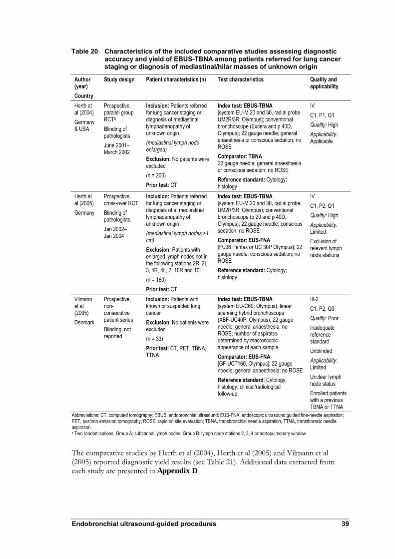
Endobronchial ultrasound-guided procedures 39
Table 20 Characteristics of the included comparative studies assessing diagnostic accuracy and yield of EBUS-TBNA among patients referred for lung cancer staging or diagnosis of mediastinal/hilar masses of unknown origin
Author (year) Country
Study design Patient characteristics (n) Test characteristics Quality and applicability
Herth et al (2004) Germany & USA
Prospective, parallel group RCTa Blinding of pathologists June 2001– March 2002
Inclusion: Patients referred for lung cancer staging or diagnosis of mediastinal lymphadenopathy of unknown origin (mediastinal lymph node enlarged) Exclusion: No patients were excluded (n = 200) Prior test: CT
Index test: EBUS-TBNA [system EU-M 20 and 30, radial probe UM2R/3R, Olympus]; conventional bronchoscope (Excera and p 40D, Olympus); 22 gauge needle; general anaesthesia or conscious sedation; no ROSE Comparator: TBNA 22 gauge needle; general anaesthesia or conscious sedation; no ROSE Reference standard: Cytology; histology
IV C1, P1, Q1 Quality: High Applicability: Applicable
Herth et al (2005) Germany
Prospective, cross-over RCT Blinding of pathologists Jan 2002– Jan 2004
Inclusion: Patients referred for lung cancer staging or diagnosis of a mediastinal lymphadenopathy of unknown origin (mediastinal lymph nodes >1 cm) Exclusion: Patients with enlarged lymph nodes not in the following stations 2R, 2L, 3, 4R, 4L, 7, 10R and 10L (n = 160) Prior test: CT
Index test: EBUS-TBNA [system EU-M 20 and 30, radial probe UM2R/3R, Olympus); conventional bronchoscope (p 20 and p 40D, Olympus); 22 gauge needle; conscious sedation; no ROSE Comparator: EUS-FNA [FU36 Pentax or UC 30P Olympus]; 22 gauge needle; conscious sedation; no ROSE Reference standard: Cytology; histology
IV C1, P2, Q1 Quality: High Applicability: Limited Exclusion of relevant lymph node stations
Vilmann et al (2005) Denmark
Prospective, non-consecutive patient series Blinding, not reported
Inclusion: Patients with known or suspected lung cancer Exclusion: No patients were excluded (n = 33) Prior test: CT, PET, TBNA, TTNA
Index test: EBUS-TBNA [system EU-C60, Olympus); linear scanning hybrid bronchoscope (XBF-UC40P, Olympus); 22 gauge needle; general anaesthesia; no ROSE; number of aspirates determined by macroscopic appearance of each sample. Comparator: EUS-FNA [GF-UCT160, Olympus]; 22 gauge needle; general anaesthesia; no ROSE Reference standard: Cytology; histology; clinical/radiological follow-up
III-2 C1, P2, Q3 Quality: Poor Inadequate reference standard Unblinded Applicability: Limited Unclear lymph node status Enrolled patients with a previous TBNA or TTNA
Abbreviations: CT, computed tomography; EBUS, endobronchial ultrasound; EUS-FNA, endoscopic ultrasound guided fine-needle aspiration; PET, positron emission tomography; ROSE, rapid on site evaluation; TBNA, transbronchial needle aspiration; TTNA, transthoracic needle aspiration a Two randomisations, Group A: subcarinal lymph nodes, Group B: lymph node stations 2, 3, 4 or aortopulmonary window
The comparative studies by Herth et al (2004), Herth et al (2005) and Vilmann et al (2005) reported diagnostic yield results (see Table 21). Additional data extracted from each study are presented in Appendix D.

40 Endobronchial ultrasound-guided procedures
Herth et al (2004) reported that EBUS-TBNA had a higher diagnostic yield than TBNA (77% vs 63%, respectively). This study also assessed the yield of these tests in two specific sub-groups—Group A: enlarged subcarinal lymph nodes, and Group B: enlarged lymph nodes at stations 2, 3, 4 or the aortopulmonary window. Herth et al (2004) observed that the relative difference in yields was increased in Group B (74% vs 54%) and maintained by Group A (80% vs 72%). When the diagnostic criteria for benign lymph nodes were expanded to include patients with non-malignant lymphocytes, but without a specific benign diagnosis, the yield of both techniques increased, but the relative difference remained similar (85% vs 66%). It is noteworthy that the yield calculated from the tabulated results was different from the yield reported in the text of the paper (EBUS-TBNA 80% vs TBNA 71%); regardless, the yield of EBUS-TBNA remained higher than TBNA.
Herth et al (2005) and Vilmann et al (2005) examined the comparative yield of EBUS-TBNA and EUS-FNA. These studies indicated that EBUS-TBNA obtained equivalent or moderately higher diagnostic yields than EUS-FNA in their respective patient populations. The results of both studies also suggest that EBUS-TBNA combined with EUS-FNA may obtain higher yields than either test alone. The relative yield of the different techniques remained similar in the study by Herth et al (2005) when the diagnosis of benign pathology was expanded to include patients with non-malignant lymphocytes but without specific benign diagnoses.
Table 21 Diagnostic yields of included comparative studies assessing EBUS-TBNA among patients referred for NSCLC staging and diagnoses of mediastinal/hilar masses of unknown origin
Specific diagnostic yielda Non-specific diagnostic yieldb
Level of evidence
Author (year)
n/N (%)
Diagnostic test
n/N % (95% CI) n/N % (95% CI)
EBUS-TBNA 77/100 77.00 (68.75, 85.25)
85/100 85.00 (78.00, 92.00) Herth et al
(2004) NR TBNA 63/100 63.00
(53.54, 72.63) 66/100 66.00
(56.72, 75.28)
IV C1 P1 Q1
EBUS-TBNA 137/160 85.63 (80.19, 91.06)
142/160 88.75 (83.85, 93.65)
EUS-FNA 121/160 75.63 (68.97, 82.28)
126/160 78.75 (72.41, 85.09)
Herth et al (2005) NR
EBUS-TBNA/ EUS-FNA
151/160 94.38 (90.80, 97.95)
155/160 96.88 (94.18, 99.57)
IV C1 P2 Q1
EBUS-TBNA NR NR 32/33 96.97 (91.92, 100.00)
EUS-FNA NR NR 32/33 96.97 (91.92, 100.00)
Vilmann et al (2005) NR
EBUS-TBNA/ EUS-FNA
NR NR 33/33 100.00
III-2 C1 P2 Q3
Abbreviations: CI, confidence interval; EBUS, endobronchial ultrasound; EUS-FNA, endoscopic ultrasound guided fine-needle aspiration; NR, not reported; TBNA, transbronchial needle aspiration a Per patient diagnostic yield when the diagnosis of a benign lymph node was restricted to requiring a specific diagnosis b Per patient diagnostic yield when diagnosis of a benign lymph nodes was expanded to patients with non malignant lymphocytes but without specific benign diagnoses
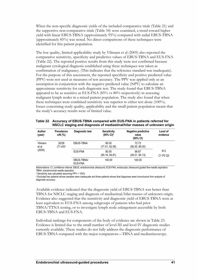
Endobronchial ultrasound-guided procedures 41
When the non-specific diagnostic yields of the included comparative trials (Table 21) and the supportive non-comparative trials (Table 54) were examined, a trend toward higher yield with linear EBUS-TBNA (approximately 95%) compared with radial EBUS-TBNA (approximately 85%) was noted. No direct comparisons of these techniques were identified for this patient population.
The low quality, limited applicability study by Vilmann et al (2005) also reported the comparative sensitivity, specificity and predictive values of EBUS-TBNA and EUS-FNA (Table 22). The reported positive results from this study were not confirmed because malignant cytological diagnosis established using these techniques was taken as confirmation of malignancy. (This indicates that the reference standard was inadequate). For the purpose of this assessment, the reported specificity and positive predicted value (PPV) were not used as measures of test accuracy. The PPV was applied only as an assumption in conjunction with the negative predicted value (NPV) to calculate an approximate sensitivity for each diagnostic test. The study found that EBUS-TBNA appeared to be as sensitive as EUS-FNA (85% vs 80% respectively) in assessing malignant lymph nodes in a mixed patient population. The study also found that when these techniques were combined sensitivity was superior to either test alone (100%). Issues concerning study quality, applicability and the small patient population meant that the study’s accuracy results were of limited value.
Table 22 Accuracy of EBUS-TBNA compared with EUS-FNA in patients referred for NSCLC staging and diagnosis of mediastinal/hilar masses of unknown origin
Author (year)
Prevalence n/N (%)
Diagnostic test Sensitivity (95% CI)a
Negative predictive value
(95% CI)
Level of evidence
EBUS-TBNA 85.00 (77.41, 92.59)
72.73 (56.35, 89.30)
EUS-FNA 80.00 (65.18, 94.81)
66.67 (49.21, 84.13)
Vilmann et al (2005)
20/28 (71.43)b
EBUS-TBNA/ EUS-FNA
100.00 100.00
III-2 C1 P2 Q3
Abbreviations: CI, confidence interval; EBUS, endobronchial ultrasound; EUS-FNA, endoscopic ultrasound guided fine-needle aspiration; TBNA, transbronchial needle aspiration a Sensitivity was calculated assuming PPV = 100% b Excluded two patients whose samples were inadequate and three patients whose final diagnoses were inconclusive from analysis of diagnostic accuracy
Available evidence indicated that the diagnostic yield of EBUS-TBNA was better than TBNA for NSCLC staging and diagnosis of mediastinal/hilar masses of unknown origin. Evidence also suggested that the sensitivity and diagnostic yield of EBUS-TBNA were at least equivalent to EUS-FNA among subgroups of patients who had prior TBNA/TTNA testing, or to investigate lymph node enlargement accessible by both EBUS-TBNA and EUS-FNA.
Individual rankings for components of the body of evidence are shown in Table 23. Evidence is limited due to the small number of level III and level IV diagnostic studies currently available. These studies do not fully address the diagnostic performance of EBUS-TBNA compared with the major comparators—TBNA and mediastinoscopy.

42 Endobronchial ultrasound-guided procedures
Table 23 Assessment of the comparative body of evidence for EBUS-TBNA in NSCLC staging and diagnosis of mediastinal/hilar masses of unknown origin
Component Rank Reason Volume of evidence D A small number of comparative diagnostic yield (IV) and low-quality diagnostic
accuracy studies (III) were identified Consistency B The reported outcomes were generally consistent in comparative trials when radial
EBUS-TBNA and linear EBUS-TBNA were considered separately Clinical impact D The reported diagnostic yield of EBUS-TBNA was greater than TBNA. No studies
reported diagnostic accuracy of EBUS-TBNA compared with TBNA No studies were identified that compared the diagnostic performance of EBUS-TBNA with mediastinoscopy The reported sensitivity and diagnostic yield of EBUS-TBNA was at least equivalent to EUS-FNA in selected patient subgroups
Generalisability B The study populations did not correspond to the research questions, however it was presumed that the reported populations were representative of clinical practice and therefore relevant to the current assessment
Applicability C The diagnostic yield evidence of EBUS-TBNA compared with TBNA was applicable to the Australian healthcare setting The evidence of EBUS-TBNA compared with EUS-FNA had limited applicability to the Australian healthcare setting
Abbreviations: EBUS, endobronchial ultrasound; EUS-FNA, endoscopic ultrasound guided fine-needle aspiration; TBNA, transbronchial needle aspiration
Endobronchial cancer depth diagnosis
No trials were identified that compared diagnostic accuracy of EBUS with or without EBBX to EBBX alone in the depth diagnosis endobronchial carcinomas for patients whose lymph nodes were not enlarged.
All nine potentially relevant studies were excluded from the assessment for a range of reasons: EBUS performance was reported per procedure not per patient (Miyazu et al 2002, Takahashi et al 2003); lymphadenopathy status was unclear (Baba et al 2002, Herth et al 2003b, Kotsianos-Hermle et al 2007, Kurimoto et al 1998, Takemoto et al 2000); and EBUS was used to diagnose benign/malignant status of central lesions (Becker et al 2000, Herth et al 2003a).
Peripheral lung lesions
There were 10 studies identified that investigated the diagnostic accuracy or diagnostic yield of EBUS-TBBX in the diagnosis of peripheral lung lesions. Of the 10 studies, five were excluded from the review because they evaluated the diagnostic performance of EBUS imaging alone without tissue sampling (Chao et al 2006, Kurimoto et al 2002, Omori et al 2002), or limited the patient population to patients with lung cancer (Yang et al 2004), or reported the performance of EBUS-TBBX per lesion not per patient (Asahina et al 2005).
Of the 10 studies, four compared radial EBUS-TBBX with other appropriate forms of guided or unguided TBBX (Eberhardt et al 2007, Herth et al 2002, Paone et al 2005, Shirakawa et al 2004); the characteristics of these studies are presented in Table 25. Of the remaining six, five were non-comparative studies of radial EBUS-TBBX and one an RCT that investigated variations of the radial EBUS-TBBX procedure. Details of these studies are presented as supportive evidence in Appendix C.

Endobronchial ultrasound-guided procedures 43
Of the 10 identified studies, Shirakawa et al (2004) recruited patients who had peripheral lung lesions and normal visible airways. The EBUS-TBBX procedure reported in the study used a radial EBUS probe (with guide sheath), fluoroscopic navigation, and curette support to obtain histological and cytological biopsy samples. Curette supports are used occasionally in Australia and the reported procedure is applicable to the Australian clinical setting. Patients were enrolled on a randomised basis, but assessors were not blinded to the test results. Although patients were randomly enrolled to undergo EBUS-TBBX, their results (comparative sensitivity, specificity and predictive values) were compared with a fluoroscopy-TBBX historical control (Table 24). Confirmation of positive results was not clear in this study (the reference standard was inadequate). Reported specificity and positive predictive value (PPV) were not used as measures of test accuracy for this assessment. PPV was applied as an assumption only with NPV to calculate an approximate sensitivity for each diagnostic test. The study suggested that EBUS-TBBX may be equally as sensitive as fluoroscopy-TBBX for the diagnosis of peripheral lung lesions (71% vs 70%, respectively). The diagnostic accuracy results of this study had limited value because of quality and applicability deficits coupled with the limitations of a small patient population. This study was regarded as low quality and limited applicability providing level III-3 evidence.
Table 24 Accuracy of EBUS-TBBX compared with fluoroscopy-TBBX in patients with peripheral lesions
Author (year)
Prevalence n/N (%)
Diagnostic test
Sensitivity (95% CI)a
NPV (95% CI)
Level of evidence
24/49 (48.98) EBUS-TBBX 70.83 (58.10, 83.56) 78.13 (66.56, 89.70) Shirakawa et al (2004) 23/42 (54.76)b Fluoroscopy-
TBBX 69.57 (55.65, 83.48) 73.08 (59.67, 86.49)
III-3 C1 PX Q3
Abbreviations: CI, confidence interval; EBUS, endobronchial ultrasound; NPV, negative predictive value; TBBX, transbronchial biopsy a Sensitivity was calculated assuming 100% PPV b Historical control group
Of the 10 identified studies, three were RCTs that assessed the diagnostic yield of EBUS-TBBX compared with ENB-TBBX (Eberhardt et al 2007), fluoroscopy-TBBX (Herth et al 2002) or TBBX (Paone et al 2005). These studies were classified as providing level IV evidence and had limited applicability to Australian clinical practice because they did not report fluoroscopic navigation use in conjunction with EBUS. Differences in quality among these studies could not be accommodated by NHMRC levels of evidence. Only the study by Paone et al (2005) reported blinding the assessors to the test results.
Of the 10 studies, only two compared EBUS-TBBX with fluoroscopy-TBBX (Shirakawa et al 2004, Herth et al 2002), one of the major comparators identified by the advisory panel. No evidence was identified that compared EBUS-TBBX with TTNA, the other major comparator in this indication.
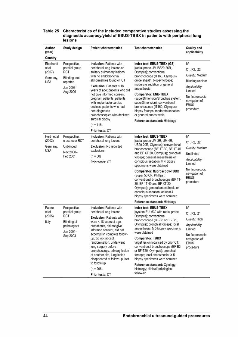
44 Endobronchial ultrasound-guided procedures
Table 25 Characteristics of the included comparative studies assessing the diagnostic accuracy/yield of EBUS-TBBX in patients with peripheral lung lesions
Author (year) Country
Study design Patient characteristics Test characteristics Quality and applicability
Eberhardt et al (2007) Germany, USA
Prospective, parallel group RCT Blinding, not reported Jan 2003– Aug 2006
Inclusion: Patients with peripheral lung lesions or solitary pulmonary lesions with no endobronchial abnormalities found on CT Exclusion: Patients < 18 years of age; patients who did not give informed consent; pregnant patients, patients with implantable cardiac devices, patients who had non-diagnostic bronchoscopies who declined surgical biopsy (n = 118) Prior tests: CT
Index test: EBUS-TBBX (GS) [radial probe UM-BS20-26R, Olympus]; conventional bronchoscope (IT160, Olympus); guide sheath; biopsy forceps; moderate sedation or general anaesthesia Comparator: ENB-TBBX (superDimension/Bronchus system, superDimension); conventional bronchoscope (IT160, Olympus); biopsy forceps; moderate sedation or general anaesthesia Reference standard: Histology
IV C1, P2, Q2 Quality: Medium Blinding unclear Applicability: Limited No fluoroscopic navigation of EBUS procedure
Herth et al (2002) Germany, USA
Prospective, cross-over RCT Unblinded Nov 2000– Feb 2001
Inclusion: Patients with peripheral lung lesions Exclusion: No reported exclusions (n = 50) Prior tests: CT
Index test: EBUS-TBBX [radial probe UM-3R, UM-4R, US20-20R, Olympus]; conventional bronchoscope (BF 1T-30, BF 1T 40 and BF XT 20, Olympus); bronchial forceps; general anaesthesia or conscious sedation; ≥ 4 biopsy specimens were obtained Comparator: fluoroscopy-TBBX (Super 50 CP, Phillips); conventional bronchoscope (BF 1T-30, BF 1T 40 and BF XT 20, Olympus); general anaesthesia or conscious sedation; at least 4 biopsy specimens were obtained Reference standard: Histology
IV C1, P2, Q2 Quality: Medium Unblinded Applicability: Limited No fluoroscopic navigation of EBUS procedure
Paone et al (2005) Italy
Prospective, parallel group RCT Blinding of pathologists Jan 2001– Sep 2003
Inclusion: Patients with peripheral lung lesions Exclusion: Patients who were < 18 years of age, outpatients, did not give informed consent, did not accomplish complete follow-up, did not accept randomisation, underwent lung surgery before bronchoscopy, primary lesion at another site, lung lesion disappeared at follow-up, lost to follow-up (n = 206) Prior tests: CT
Index test: EBUS-TBBX [system EU-M30 with radial probe, Olympus]; conventional bronchoscope (BF-B3 or BF-T20, Olympus); bronchial forceps; local anaesthesia; ≥ 5 biopsy specimens were obtained Comparator: TBBX target lesion localised by prior CT; conventional bronchoscope (BF-B3 or BF-T20, Olympus); bronchial forceps; local anaesthesia; ≥ 5 biopsy specimens were obtained Reference standard: Cytology; histology; clinical/radiological follow-up
IV C1, P2, Q1 Quality: High Applicability: Limited No fluoroscopic navigation of EBUS procedure

Endobronchial ultrasound-guided procedures 45
Author (year) Country
Study design Patient characteristics Test characteristics Quality and applicability
Shirakawa et al (2004) Japan
Randomised patient series with historical control Unblinded Jan 2001– Dec 2001
Inclusion: Patients with peripheral lung lesions and normal visible airways Exclusion: Patients who were not randomised to EBUS, patients who did not undergo EBUS, patients with spontaneous bleeding, patients with no final diagnosis (n = 49) Prior tests: X-ray
Index test: EBUS-TBBX (GS) [radial probe UM-3R, UM-4R, US-20-20R, Olympus]; conventional bronchoscope (BS 1T-240R, Olympus); guide sheath; bronchial forceps and bronchial brushing; unclear anaesthesia; fluoroscopy and curette support Fluoroscopy-TBBX; unclear details Reference standard: Cytology; histology; clinical/radiological follow-up; other examinations
III-3 C1, P2, Q3 Quality: low Inadequate reference standard Unblinded Unclear comparator details Applicability: Applicable
Abbreviations: CT, computed tomography; EBUS, endobronchial ultrasound; ENB, electromagnetic navigation bronchoscopy; GS, guide sheath; RCT, randomised controlled trial; TBBX, transbronchial biopsy
Herth et al (2002) indicated that the yield obtained using EBUS-TBBX was equivalent to fluoroscopic-TBBX (80% vs 76%, respectively). It was also reported that EBUS-TBBX yield was maintained, but fluoroscopic-TBBX decreased when a subgroup of patients with peripheral lesions less than 3 cm diameter was assessed (81% vs 57%).
Paone et al (2005) reported that EBUS-TBBX generated a considerably higher yield than TBBX alone (76% vs 52%, respectively) and that the difference was maintained regardless of lesion size.
Eberhardt et al (2007) reported individual and combined yields for EBUS-TBBX, ENB-TBBX. EBUS-TBBX demonstrated a trend toward higher diagnostic yield than ENB-TBBX (69% vs 59%), but the use of the combined techniques was superior to either alone (88%). The relative difference in yield between techniques was generally maintained when subgroups of patients based on lesion size were examined.
The primary diagnostic yield results from the RCTs conducted by Herth et al (2002), Paone et al (2005) and Eberhardt et al (2007) are presented in Table 26. Additional data extracted from these RCTs are presented in Appendix D.
Table 26 Diagnostic yield of the included comparative studies assessing EBUS-TBBX in patients with peripheral lung lesions
Diagnostic yield Author (year)
Prevalence n/N (%)
Diagnostic test n/N % (95% CI)
Level of evidence
EBUS-TBBX 40/50 80.00 (68.91, 91.09) Herth et al (2002)
45/50 (90) Fluoroscopy-TBBX 38/50 76.0 (64.16, 87.84)
IV C1 P1 Q2
EBUS-TBBX 66/87 75.86 (66.87, 84.85) Paone et al (2005)
144/206 (69.9) TBBX alone 62/119 52.1 (43.13, 61.08)
IV C1 P1 Q1
EBUS-TBBX 27/39 69.23 (54.75, 83.72) ENB-TBBX 23/39 58.97 (43.54, 74.41)
Eberhardt et al (2007)
82/118 (69.49)
ENB/EBUS-TBBX 35/40 87.5 (77.25, 97.75)
IV C1 P1 Q2
Abbreviations: CI, confidence interval; EBUS, endobronchial ultrasound; ENB, electromagnetic navigation bronchoscopy TBBX, biopsy

46 Endobronchial ultrasound-guided procedures
The available evidence suggests that EBUS-TBBX sensitivity is equivalent to fluoroscopy-TBBX for diagnosis of peripheral lung lesions. Evaluated studies also indicated that the diagnostic yield of EBUS-TBBX was greater than TBBX alone and at least equivalent to other methods (electromagnetic, fluoroscopic) of guided-TBBX. The available evidence also suggested that the diagnostic yield of EBUS-TBBX alone may be superior to other methods of guided-TBBX for diagnosis of smaller peripheral lesions.
Individual rankings for components of the body of evidence are shown in Table 27. Evidence was limited because of the low number of level III and level IV diagnostic studies, which offered limited applicability, and did not fully address diagnostic performance of EBUS-TBBX compared with fluoroscopy-TBBX and TTNA.
Table 27 Assessing the comparative body of evidence for EBUS-TBBX in the diagnosis of peripheral lung lesions
Component Rank Reason Volume of evidence D A small number of comparative diagnostic yield (IV) and low-quality diagnostic
accuracy studies (III) were identified Consistency B The reported outcomes for EBUS-TBBX were generally consistent in the
comparative trials Clinical impact D The reported sensitivity and diagnostic yield of EBUS-TBBX was equivalent to
fluoroscopy-TBBX No studies were identified that compared diagnostic performance of EBUS-TBBX with TTNA The reported diagnostic yield of EBUS-TBBX was greater than TBBX and at least equivalent to ENB-TBBX EBUS-TBBX diagnostic yield may be greater than other methods of guided-TBBX in diagnosing smaller peripheral lesions
Generalisability A The study populations corresponded with the patient population identified in the research question
Applicability D Evidence for EBUS-TBBX diagnostic yield compared with fluoroscopy-TBBX had limited applicability to the Australian healthcare setting Evidence for EBUS-TBBX diagnostic yield compared with TBBX and ENB-TBBX had limited applicability to the Australian healthcare setting
Abbreviations: EBUS, endobronchial ultrasound; ENB, electromagnetic navigation bronchoscopy; EUS-FNA, endoscopic ultrasound guided fine-needle aspiration; TBBX, transbronchial biopsy; TTNA, transthoracic needle aspiration
Patient management
No pre-test/post-test case series studies were identified that assessed the impact of EBUS-guided procedures on patient management.
Improvements in lung cancer staging and diagnosis may lead to better patient management by avoiding invasive diagnostic procedures and providing more accurate curative and palliative treatment planning leading to improved survival and quality of life.

Endobronchial ultrasound-guided procedures 47
Treatment effectiveness
It was considered that use of EBUS guided procedures for non-small cell lung cancer (NSCLC) staging, diagnosis of mediastinal/hilar masses of unknown origin, and diagnosis of peripheral lung lesions would not identify any unique patient groups that were substantially different from those currently seen in Australian clinical practice. Evidence of treatment effectiveness for these indications was therefore not presented in this assessment.
No treatment effectiveness evidence was identified among patients undergoing EBUS imaging for the depth diagnosis of endobronchial carcinomas.

48 Endobronchial ultrasound-guided procedures
Economic considerations
Summary
A decision analytic model was constructed to assess cost implications of endobronchial ultrasound (EBUS)-guided procedures when compared with current modalities. A cost analysis of EBUS-transbronchial needle aspiration (TBNA) relative to TBNA alone was performed for non-small cell lung cancer (NSCLC) staging and diagnosis of mediastinal/hilar masses of unknown origin; a cost analysis of EBUS-transbronchial biopsy (TBBX) relative to TBBX alone was conducted for diagnosis of peripheral lung lesions less than 3 cm diameter. There were insufficient clinical data available to inform a cost analysis for the depth diagnosis of endobronchial cancers.
The analysis indicated that use of EBUS-TBNA in NSCLC staging and diagnosis of mediastinal masses was estimated to generate a cost saving of $347 per patient; EBUS-TBBX for diagnosis of peripheral lung lesions less than 3 cm diameter was estimated to generate a cost saving of $364 per patient. This reflected the economic benefits associated with improved yield offered by using EBUS guidance for these procedures.
These results should be interpreted in the context of data inputs and assumptions made in the analyses. The current analysis assumed that the use of EBUS-guidance had no impact on the overall diagnostic accuracy of sampling procedures. Should EBUS use influence TBNA or TBBX diagnostic accuracy, patients’ prognoses would likely be affected, creating important health outcomes and economic implications. No relevant data were available to allow evaluation of these outcomes.
Sensitivity analyses indicated that the yield represents the most critical variable in the analysis. This result was anticipated because the most significant clinical benefit was likely to result from avoiding expensive and invasive follow-up surgical procedures.
Epidemiological data indicated that the estimated total annual cost of employing EBUS-TBNA was between $2.5 and $3.6 million for assessment of central, mediastinal and hilar tumours. The total annual cost of EBUS-TBBX for use in diagnosis of peripheral lung lesions less than 3 cm diameter was estimated to be between $1.2 and $2.2 million.
The use of EBUS-guided procedures generated cost savings compared with current procedures. The total cost saving was expected to be between $763,994 and $1.1 million for implementation of EBUS-TBNA in the assessment of NSCLC and mediastinal/hilar masses; and from $363,802 to $691,224 for implementation of EBUS-TBBX in diagnosing of peripheral lung lesions less than 3 cm diameter.
It should also be noted the cost savings of EBUS presented may represent conservative estimates. This is because EBUS-guided procedures could replace more invasive biopsy modalities, such as mediastinoscopy, for some patients as a first line therapy in the assessment of lung cancer, thereby generating greater cost offsets.
Endobronchial ultrasound-guided procedures 49
Background and approach
This section examines whether the introduction of EBUS-guided procedures under the proposed indications represent value-for-money for the Australian healthcare system.
In practice, a variety of guiding and biopsy techniques are applied in transbronchial biopsy (TBBX) sampling (see Figures 3–6). Because comparative data are limited, evaluation of all techniques was not possible. For example, mediastinoscopy is often considered to be the preferred procedure applied in non-small cell lung cancer (NSCLC) staging and for diagnosis of mediastinal/hilar masses, but no data comparing mediastinoscopy with EBUS-transbronchial needle aspiration (TBNA) were identified in the course of this evaluation. The following analysis compares EBUS-guided sampling procedures with alternative techniques for which sufficient clinical data were available.
A systematic review of the literature demonstrated a paucity of published evidence regarding potential changes in staging/diagnostic accuracies that would occur as a result of using EBUS in place of the existing alternatives. A full economic evaluation that comparatively assessed alternative strategies in terms of costs and final patient outcomes (such as life years) was not considered to be feasible.
Current evidence indicated that the diagnostic yield of EBUS-TBNA was greater than TBNA alone (77 % vs 63%; Herth et al 2004) for staging NSCLC and diagnosis of mediastinal/hilar masses. Improved diagnostic yield reduces the need for repeat or follow-up procedures. This represents a potential source of healthcare cost savings.
Herth et al (2004) conducted a randomised comparative study in which EBUS-TBNA was compared with TBNA alone––one of the major comparators identified by the advisory panel. The study’s patient and test characteristics were adequately reported and are likely to be applicable to Australian clinical practice. EBUS-TBNA was also reported to achieve equivalent or moderately higher diagnostic yield rates than EUS-fine needle aspiration (FNA) (Herth et al 2005; Vilmann et al 2005). However, the advisory panel indicated that EUS-FNA was a possible adjunct to, rather than a comparator for, EBUS-TBNA in Australia.
A cost analysis of EBUS-TBNA relative to TBNA alone in NSCLC staging and for diagnosis of mediastinal/hilar masses was performed using data from Herth et al (2004). The cost analysis aimed to quantify cost implications associated with the improved diagnostic yield offered by EBUS-guidance.
There were no trials identified that compared the diagnostic performance of EBUS with or without endobronchial biopsy (EBBX) in the depth diagnosis of endobronchial cancers. An economic analysis was therefore not conducted for this indication.
Herth et al (2002) indicated that EBUS-TBBX and fluoroscopic-TBBX offered equivalent yields for diagnosis of patients with peripheral lung lesions. Because fluoroscopy is used in both EBUS-TBBX and fluoroscopic-TBBX, the only cost difference between them is associated with EBUS guidance. This means that EBUS-TBBX procedures are expected to incur an additional cost to fluoroscopic-TBBX. From the economic perspective, EBUS-TBBX does not represent a preferred strategy––it provides no clinical benefit at higher cost when compared with fluoroscopic-TBBX.
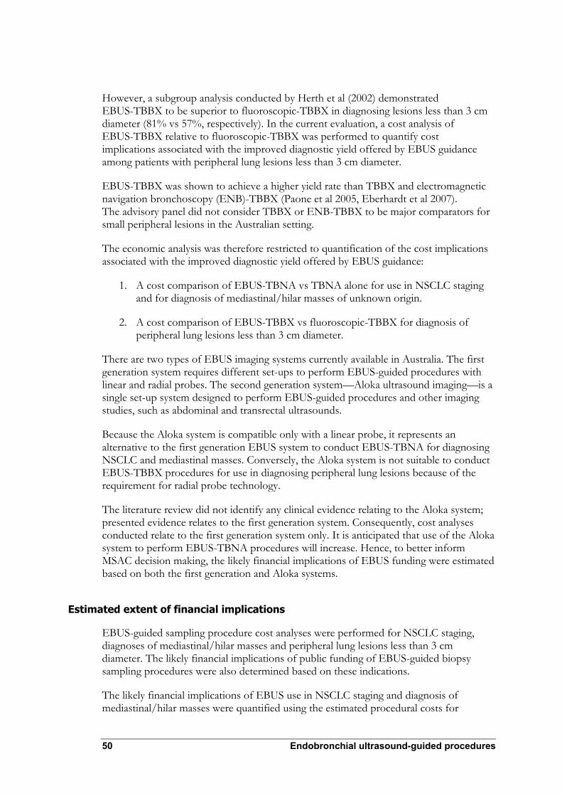
50 Endobronchial ultrasound-guided procedures
However, a subgroup analysis conducted by Herth et al (2002) demonstrated EBUS-TBBX to be superior to fluoroscopic-TBBX in diagnosing lesions less than 3 cm diameter (81% vs 57%, respectively). In the current evaluation, a cost analysis of EBUS-TBBX relative to fluoroscopic-TBBX was performed to quantify cost implications associated with the improved diagnostic yield offered by EBUS guidance among patients with peripheral lung lesions less than 3 cm diameter.
EBUS-TBBX was shown to achieve a higher yield rate than TBBX and electromagnetic navigation bronchoscopy (ENB)-TBBX (Paone et al 2005, Eberhardt et al 2007). The advisory panel did not consider TBBX or ENB-TBBX to be major comparators for small peripheral lesions in the Australian setting.
The economic analysis was therefore restricted to quantification of the cost implications associated with the improved diagnostic yield offered by EBUS guidance:
1. A cost comparison of EBUS-TBNA vs TBNA alone for use in NSCLC staging and for diagnosis of mediastinal/hilar masses of unknown origin.
2. A cost comparison of EBUS-TBBX vs fluoroscopic-TBBX for diagnosis of peripheral lung lesions less than 3 cm diameter.
There are two types of EBUS imaging systems currently available in Australia. The first generation system requires different set-ups to perform EBUS-guided procedures with linear and radial probes. The second generation system––Aloka ultrasound imaging––is a single set-up system designed to perform EBUS-guided procedures and other imaging studies, such as abdominal and transrectal ultrasounds.
Because the Aloka system is compatible only with a linear probe, it represents an alternative to the first generation EBUS system to conduct EBUS-TBNA for diagnosing NSCLC and mediastinal masses. Conversely, the Aloka system is not suitable to conduct EBUS-TBBX procedures for use in diagnosing peripheral lung lesions because of the requirement for radial probe technology.
The literature review did not identify any clinical evidence relating to the Aloka system; presented evidence relates to the first generation system. Consequently, cost analyses conducted relate to the first generation system only. It is anticipated that use of the Aloka system to perform EBUS-TBNA procedures will increase. Hence, to better inform MSAC decision making, the likely financial implications of EBUS funding were estimated based on both the first generation and Aloka systems.
Estimated extent of financial implications
EBUS-guided sampling procedure cost analyses were performed for NSCLC staging, diagnoses of mediastinal/hilar masses and peripheral lung lesions less than 3 cm diameter. The likely financial implications of public funding of EBUS-guided biopsy sampling procedures were also determined based on these indications.
The likely financial implications of EBUS use in NSCLC staging and diagnosis of mediastinal/hilar masses were quantified using the estimated procedural costs for

Endobronchial ultrasound-guided procedures 51
EBUS-TBNA. Estimated EBUS-TBBX costs were applied to quantify associated financial implications for diagnosing peripheral lung lesions less than 3 cm diameter.
Per procedure costs of EBUS-guided techniques were first estimated. These costs were then multiplied by the estimated number of eligible patients to determine the total anticipated expenditure for EBUS-guided techniques.
Cost analyses of EBUS-guided techniques (EBUS-TBNA or EBUS-TBBX) indicated that the improved yield offered by the use of EBUS guidance over current practices should lead to cost savings by reducing the need for follow-up procedures; total cost savings for implementing EBUS for the Australian health system were also determined.
Estimated cost of EBUS-guided TBNA or TBBX per procedure
There are two types of EBUS imaging systems currently available in Australia—the first generation system and the Aloka system. Because their associated costs differ, the estimated costs of conducting EBUS-guided procedures were determined for both systems. All estimates were assumed to represent average costs among private and public sectors.
Cost of EBUS guidance Table 28 presents costs of each health resource item required for both EBUS guidance systems. The total cost includes expenditure required for capital equipment, probes and professional fees. All cost information was provided by the Applicant.
Both radial and linear probes are used with first generation systems and are associated with different resource requirements and costs. Thus, separate procedural cost estimates were derived for the first generation system using both probes. A single cost for the Aloka system was determined because it incorporates use of linear probes only. Use of the Aloka system was assumed to incur the same professional fees as the first generation imaging system.
Table 28 Total cost of EBUS guidance per procedure
Costs First generation system Aloka Source via linear probe via radial probe via linear probe Equipment/consumables costs Capital equipment costsa $56.00 $76.00 $75.00 Application document
(see Appendix I) Ultrasonic probe $44.00 $58.00 $46.00 Application document Total Professional fees associated with EBUS guidance
$327.85 $327.85 $327.85 Application document (MBS item 38448)b
Total—EBUS guidance $427.85 $461.85 $448.85 Average cost $444.85 —
Abbreviation: EBUS, endobronchial ultrasound
a Assumes 12 procedures per week per centre, amortising the capital costs over a period of five years b Item 38448: Mediastinum, cervical exploration of, with or without biopsy
Note: Different capital equipment costs were suggested by the applicant for first generation EBUS equipment employing linear and radial probes ( total costs of $101,020 and $156,400, respectively, for linear and radial EBUS guidance)

52 Endobronchial ultrasound-guided procedures
Capital equipment costs for the first generation system using linear and radial probes were estimated at $56 and $76 per procedure respectively. Capital equipment costs for the Aloka system were estimated at $75 per procedure.
These estimates were based on equipment costs and the predicted number of procedures performed each year. Expert opinion indicated that each centre equipped with EBUS currently performs four to five procedures per week and this rate could increase to 10 to 12 procedures per week should public funding be approved for EBUS-guided procedures. The current analysis assumed that each site would perform 12 procedures on average per week should EBUS be publicly subsidised. Capital costs per procedure were estimated by amortising total capital costs and assuming that 624 procedures were performed per year (=12×52 weeks) over a period of five years. Further details on the derivation of this estimate are provided in Appendix I.
The costs of linear probes were estimated to be $44 and $46 per procedure for the first generation and Aloka systems, respectively. The same capital equipment costs estimating methodology was applied to derive linear probe costs, given that their corresponding utilisation life spans are duplicated.
Radial probes used with the first generation system were expected to be replaced after approximately 100 procedures. In practice, EBUS-guided techniques can be performed with two modes of radial probes, depending on physicians’ preferences and diagnostic site. The cost of the radial probe was estimated to be $5825 and was calculated by averaging the cost of two probes. The radial probe cost per procedure was therefore estimated to be $58 (=$5825 ÷ 100).
It was assumed that MBS item 38448 (Mediastinum, cervical exploration of, with or without biopsy) provides a reasonable estimate of the professional fee for EBUS-guided procedures (see Table 28). Although EBUS guided procedures are less invasive and carry lower risks compared with mediastinoscopy, they are more time-consuming and require a higher level of training, than non-EBUS bronchoscopic procedures.
The total costs for first generation EBUS guidance using linear and radial probes were estimated to be $428 and $462 per procedure, respectively. Similarly, costs for the Aloka system were estimated to be $449 per procedure.
EBUS-guided TBNA can be undertaken with either linear or radial probes for diagnosing NSCLC and mediastinal/hilar masses. The estimated costs of the first generation EBUS guidance with linear and radial probes were comparable (Table 28). For simplicity, the following analysis of first generation EBUS guided systems for TBNA was performed using the average costs of EBUS guidance for both probe types––$445 per procedure.
EBUS-guided TBBX for diagnosis of peripheral lung lesions is undertaken using radial probes only. The estimated cost of $462 for first generation EBUS guided procedures using radial probes was applied to calculate the following cost analysis of EBUS guidance for TBBX.
Cost of EBUS-guided TBNA or TBBX per procedure The total costs of EBUS-TBNA and EBUS-TBBX were estimated and presented in Table 29 and Table 31, respectively.

Endobronchial ultrasound-guided procedures 53
As well as costs associated with EBUS guidance (Table 28), total EBUS-guided TBNA procedure costs included professional fees, consumables and other medical services. Table 29 presents the costs of each identified health resource item.
Table 29 Total cost of EBUS-TBNA per procedure
Costs First generationa Aloka Source Cost of EBUS guidance per procedure $444.85 $448.85 Estimated Professional fee for TBNA $188.90 $188.90 MBS 38812b
Disposable needle $175.00 $175.00 Application document Disposable balloon $24.00 $24.00 Application document Other medical services $287.40 $287.40 MBS (see Table 30) Total $1120.15 $1124.15
Abbreviation: EBUS, endobronchial ultrasound; TBNA, transbronchial needle aspiration a The total EBUS-TBNA cost per procedure was estimated based on the health resource usage for EBUS-TBNA with linear probe b The Applicant proposed MBS Item 38412. This item was replaced by 38812 Percutaneous needle biopsy of lung (November 2007) Note: The fee for MBS Item 41892 Bronchoscopy with 1 or more endobronchial biopsies or other diagnostic or therapeutic procedures is $212.25. The fee for MBS Item 41898 Fibreoptic bronchoscopy with 1 or more transbronchial lung biopsies, with or without bronchial or bronchoalveolar lavage, with or without the use of interventional imaging is $232.05
The total costs of EBUS-TBNA per procedure were estimated to be $1120 using the first generation system and $1124 for the Aloka system. The total costs included professional fees for TBNA and other medical services, such as anaesthesia management and cytology evaluation. Costs for these services were estimated to be $287 (see Table 30).
In addition to standard diagnostic cytology, rapid on-site evaluation (ROSE) may be conducted for some patients during TBNA. The associated costs were not included in the current cost calculation.
Table 30 Other costs associated with EBUS-TBNA per procedure
MBS item number, description Fee 17610 Anaesthetist, pre-anaesthesia consultation
– a brief consultation involving a targeted history and limited examination (including the cardio-respiratory system) – and of not more than 15 minutes duration $38.80
20520 Initiation of management of anaesthesia for all closed chest procedures $107.40 23043 Anaesthesia, perfusion or assistance at anaesthesia (56 minutes to 1.00 hour) $71.60 73049 Cytology of material obtained directly from a patient by fine needle aspiration of solid tissue or tissues $69.60 Total costs per procedure $287.40
Abbreviation: MBS, Medicare Benefits Schedule Source: Medicare Benefits Schedule Book November 2007
The total cost for EBUS-TBBX was estimated based on the cost of the first generation EBUS system with radial probe (Table 31).
Based on the expert opinion of the advisory panel, MBS item 41898 Fibreoptic bronchoscopy with 1 or more transbronchial lung biopsies, with or without bronchial or bronchoalveolar lavage, with or without the use of interventional imaging was assumed to represent an appropriate professional fee for TBBX sampling procedures. The current MBS fee for fluoroscopy was also included because the EBUS-TBBX procedure requires fluoroscopic guidance.

54 Endobronchial ultrasound-guided procedures
A guide sheath set consisting of a sheath for probe, biopsy forceps and cytology brush is required for TBBX. The cost of the guide sheath kit was estimated to be $131, by averaging the costs of both available guide sheath kits.
Other medical services costs associated with TBBX were assumed to be same as TBNA ($287, see Table 30). Therefore, the total cost of EBUS-TBBX was estimated to be $1176 per procedure.
Table 31 Total cost of EBUS-TBBX per procedure
Costs Estimates Source Cost of EBUS guidance per procedure $461.85 Estimated (see Table 28) Professional fee for fluoroscopic-TBBX
– TBBX $232.05 MBS 41898a – Fluoroscopy $63.75 MBS 60506b
Disposable guide sheath kit $131 Estimated Other medical services (including anaesthesia and pathology) $287.40 MBS (see Table 30) Total $1176.05
Abbreviations: EBUS, endobronchial ultrasound; TBBX, transbronchial biopsy a Item 41898 Fibreoptic bronchoscopy with 1 or more transbronchial lung biopsies, with or without bronchial or bronchoalveolar lavage, with or without the use of interventional imaging b Item 60506 Fluoroscopy using a mobile image intensifier, in conjunction with a surgical procedure lasting less than 1 hour
The costs of EBUS-guided biopsy sampling procedures are difficult to estimate because there are no reliable cost data available for Australian practice. For example, the Applicant suggested that MBS item 38448 (Table 28) and MBS item 38812 (Table 29) were likely associated with equivalent resource requirements and skill level as EBUS-TBNA. Based on these cost estimates, the total professional fee for EBUS-TBNA was estimated to be $517. Similarly, the total professional fee for EBUS-TBBX was estimated to be $624, covering EBUS-guidance, TBBX and fluoroscopy procedures (Table 32). These estimates are assumed to appropriately represent the professional fees for EBUS-guided procedures.
In addition, MBS item 306903 and MBS item 306944 represent the professional fees associated with EBUS-guided sampling procedures at $509. If those existing items were to represent the procedure fee for EBUS-guided sampling procedures, the total costs associated with the use of EBUS guidance would be less than that estimated in this assessment. Nonetheless, as also suggested by the Applicant, EBUS professional fees should account for the additional time required by clinicians to conduct the procedure.
3 MBS item 30690 Endoscopic ultrasound-endoscopy with ultrasound imaging, with or without biopsy, with fine needle aspiration, including aspiration of the locoregional lymph nodes if performed, for the staging of 1 or more of oesophageal, gastric or pancreatic cancer, not in association with another item in this subgroup and not being a service associated with the routine monitoring of chronic pancreatitis.
4 MBS item 30694 Endoscopic ultrasound-endoscopy with ultrasound imaging, with or without biopsy, with fine needle aspiration for the diagnosis of 1 or more of pancreatic, biliary or gastric submucosal tumours, not in association with another item in this subgroup and not being a service associated with the routine monitoring of chronic pancreatitis.

Endobronchial ultrasound-guided procedures 55
Table 32 Summary of professional fees for EBUS-guided procedures
Professional fee Estimates Source Professional fee for EBUS-TBNA
– EBUS guidance $327.85 See Table 28 – TBNA $188.90 See Table 29
Total professional fee for EBUS-TBNA $516.75 Professional fee for EBUS-TBBX
– EBUS guidance $327.85 See Table 28 – TBBX $232.05 See Table 31 – Fluoroscopy $63.75 See Table 31
Total professional fee for EBUS-TBBX $623.65 Abbreviation: EBUS, endobronchial ultrasound; TBNA, transbronchial needle aspiration; TBBX, transbronchial biopsy
Estimated extent of financial implications
The total likely costs of both techniques for each indication were determined from the estimated procedural costs of EBUS-guided techniques and the estimated eligible population.
The likely financial implications for use of EBUS in NSCLC staging and diagnosis of mediastinal/hilar masses were quantified by applying the estimated procedural costs of EBUS-TBNA. The estimated costs of EBUS-TBNA using the first-generation EBUS system and the Aloka system were similar (Table 29). This signified that the likely financial implications of implementing both systems would be parallel. Thus, the expected financial implication for use of EBUS-TBNA in NSCLC staging and diagnosis of mediastinal/hilar masses were quantified by applying the cost estimates of EBUS-TBNA with the first-generation system.
Similarly, estimated EBUS-TBBX costs were applied to quantify the financial implications for diagnosis of peripheral lung lesions less than 3 cm. The likely total financial costs of implementing EBUS-TBNA and EBUS-TBBX were estimated and presented in Table 33 and Table 34, respectively.
Table 33 Estimated total costs of EBUS-TBNA for assessment of NSCLC and mediastinal/hilar masses
The first generation system Lower estimate Upper estimate
Source
Eligible population 2200 3200 See Table 1 Costs of EBUS-TBNA per procedure $1120 See Table 29 Estimated total costs of EBUS-TBNA for all eligible populations $2,464,330 $3,584,480
Abbreviations: EBUS, endobronchial ultrasound; TBNA, transbronchial needle aspiration
The current evaluation estimated that the eligible population was in the range of 2200–3200 patients annually for assessment of NSCLC and mediastinal/hilar masses (Table 1). These eligible population estimates translate to an annual cost of between $2.5 and 3.6 million for performing EBUS-guided sampling procedures.

56 Endobronchial ultrasound-guided procedures
Table 34 Estimated total costs of EBUS-TBBX for diagnosis of peripheral lung lesions
The first generation system Lower estimate Upper estimate
Source
Eligible population 1000 1900 See Table 2 Costs of EBUS-TBBX per procedure $1176 See Table 31 Total costs of EBUS-TBBX for all eligible populations $1,176,050 $2,234,495
Abbreviations: EBUS, endobronchial ultrasound; TBBX, transbronchial biopsy
Based on AIHW data for the current use of comparator procedures, it was estimated that approximately 1500–3000 procedures are being performed for patients with peripheral lung lesions annually (see Table 2). The advisory panel indicated that of these, between 1000 and 1900 patients with peripheral lung lesions less than 3 cm would potentially be eligible for EBUS-TBBX. This would generate annual total costs of between $1.2 and $2.2 million.
These cost estimates do not account for possible cost offsets arising from substitution effects due to public funding of EBUS-guided sampling procedures, or potential cost savings such as reduced need for follow-up procedures due to improved diagnostic yield from EBUS guidance.
The following cost analysis demonstrates that using TBNA without EBUS-guidance as a comparator for NSCLC staging and diagnosing mediastinal masses EBUS-TBNA would generate a cost saving of $347 per patient using first generation imaging system. This reflects the lower follow-up costs of EBUS-TBNA because of its improved yield, compared with TBNA alone, which more than offset the additional procedural costs associated with EBUS-guidance. Similarly, use of EBUS-TBBX rather than fluoroscopic-TBBX would generate a cost saving of $364 per procedure when used in the diagnosis of peripheral lung lesions less than 3 cm diameter.
The extent of total cost-savings was estimated for both indications under consideration using the estimated eligible patient populations, as shown in Table 35 and Table 36.
Table 35 Estimated total cost savings offered by EBUS-TBNA for assessment of NSCLC and mediastinal/hilar masses in the Australian health system
The first generation system Lower estimate Upper estimate
Source
Eligible population 2200 3200 See Table 1 Cost saving associated with EBUS-TBNA per procedure $347 See Table 29 Total cost savings associated with EBUS-TBNA $763,994 $1,111,264
Abbreviation: EBUS, endobronchial ultrasound; TBNA, transbronchial needle aspiration
Based on per procedure cost savings associated with EBUS-TBNA over TBNA alone, and the estimated eligible population requiring assessments for NSCLC or mediastinal/hilar masses (see Table 1), the total cost saving of implementing EBUS-TBNA was expected to fall in the range of between $763,994 and $1.1 million (see Table 35).
Similarly, based on the per procedure cost saving associated with EBUS-TBBX over fluoroscopic-TBBX, and the estimated eligible population requiring diagnosis of

Endobronchial ultrasound-guided procedures 57
peripheral lung lesions less than 3 cm diameter (Table 2), the total cost saving for implementing EBUS-TBBX was expected at between $363,802 and $691,224 (Table 36).
Table 36 Estimated total cost savings of EBUS-TBBX for diagnosis of peripheral lung lesions < 3 cm diameter for the Australian health system
First generation system Lower estimate Upper estimate
Source
Eligible population 1000 1900 See Table 2 Cost saving associated with EBUS-TBBX per procedure $364 See Table 31 Total cost savings associated with EBUS-TBBX $363,802 $691,224
Abbreviation: EBUS, endobronchial ultrasound; TBBX, transbronchial biopsy
These estimates should be interpreted with some caution due to the degree of uncertainty in estimating eligible populations and numbers of procedures performed.
It was estimated in the analysis that each centre had capacity to perform up to 12 procedures per week in practice. There are currently seven centres with EBUS equipment in Australia, for a maximum capacity of approximately 4400 EBUS procedures per annum. The capacity to perform EBUS procedures has been increasing in Australia, and this trend is likely to accelerate should public funding become available. Given the eligible patient population (Table 33 and Table 34), it is possible that some centres might not operate at full capacity, which would consequently increase capital equipment costs per procedure. However, should EBUS-guided procedures be approved for other indications, such as diagnosis of endobronchial cancers and peripheral lesions greater than 3 cm diameter, full use of services may be absorbed. Furthermore, the Aloka ultrasound imaging system is able to perform other imaging studies, such as endoscopic ultrasound (EUS). Accordingly, if the Aloka system was used to perform EBUS-TBNA and other imaging procedures, more procedures could be performed with each system, resulting in reduced capital costs per procedure. Nevertheless, capital equipment costs per procedure are unlikely to differ significantly from estimates applied in the current analysis.
It should also be noted that the presented overall cost savings for EBUS may be conservative because EBUS-guided procedures could replace more invasive and more costly biopsy modalities. That is, for some patients, EBUS-TBNA may replace procedures such as mediastinoscopy as a first line approach for staging NSCLC and diagnosis of mediastinal/hilar masses of unknown origin, thereby generating greater cost offsets attributable to the diagnostic process.
Published evidence regarding endobronchial ultrasound cost-effectiveness
There are very few published economic evaluations of EBUS for the indications considered by this assessment. The literature search did not identify any Australian studies. A cost assessment was conducted in Germany (Herth et al 2003), and a précis of findings is presented. Improvement in diagnostic yield offered by implementing EBUS-guided TBNA was demonstrated to offset associated additional costs when compared with TBNA and mediastinoscopy.

58 Endobronchial ultrasound-guided procedures
Herth et al (2003) conducted a cost comparison involving EBUS-TBNA, TBNA and mediastinoscopy for mediastinal staging of lung cancer among 100 patients in Germany. Follow-up mediastinoscopy was provided as the follow-up staging procedure when the EBUS-TBNA and TBNA yields were inadequate. Per procedure costs of €180, €57 and €1620 were reported for EBUS-TBNA, TBNA and mediastinoscopy, respectively. TBNA was associated with requirement for follow-up mediastinoscopies among 40 per cent of patients. The mediastinoscopy follow-up rate among EBUS-TBNA patients was 25 per cent. Costs per patient cost for lymph node staging were estimated to be €585 for EBUS-TBNA, €705 for the TBNA arm, and €1620 for patients undergoing mediastinoscopy.
Cost analyses of EBUS-guided techniques
A decision analytic model was developed to quantify cost implications associated with improvements in the yield rate provided by EBUS-guided procedures compared with current practice.
Two cost analyses were conducted. The first considered EBUS-TBNA relative to TBNA alone and was performed for NSCLC staging and diagnosis of mediastinal/hilar masses of unknown origin. The second analysis compared costs for diagnosis of peripheral lung lesions less than 3 cm with and without EBUS. The biopsy procedure was fluoroscopic-TBBX, as per Herth et al (2002). A cost analysis of EBUS-guided sampling was not conducted in the assessment of endobronchial carcinoma because of the absence of relevant clinical data.
In Australia, mediastinoscopy is often considered to be the preferred procedure for NSCLC staging and diagnosis of mediastinal/hilar masses, but no clinical evidence comparing mediastinoscopy with EBUS-TBNA was identified during this assessment. A cost analysis comparing EBUS-TBNA with mediastinoscopy was not conducted.
Analytical approach
A simple decision analytic model was constructed to compute the expected costs of alternative strategies. Figure 9 presents a schematic diagram of the decision tree. This was based on clinical pathway presented in Figure 3, Figure 4 and Figure 6.
The model structure illustrates two alternative strategies that are differentiated by the use of EBUS guidance during the primary diagnostic procedure. This model was used for both of the indications under consideration.
In the absence of EBUS, TBNA alone is performed in the staging of NSCLC and for diagnosis of mediastinal/hilar masses. If the primary diagnostic procedure was inconclusive due to insufficient diagnostic yield, a follow-up mediastinoscopy procedure was performed (the gold-standard diagnostic procedure) (Figure 9).
Similarly, in the absence of EBUS, fluoroscopic-TBBX is performed in the diagnosis of peripheral lung lesions less than 3 cm diameter. Expert opinion suggested that, if the primary diagnostic procedure was to be inconclusive, CT-guided TTNA would represent a preferred secondary diagnostic procedure. Any further unsatisfactory yield from CT-guided TTNA would be followed by surgical resection via video-assisted thorascopic surgery (VATS) or thoracotomy where clinical suspicion for malignancy is high and the

Endobronchial ultrasound-guided procedures 59
patient deemed fit for surgery. Surgical resection is the gold-standard diagnostic test and can often be therapeutic. VATS is usually the favoured surgical approach for diagnostic investigation; it is both less invasive and costly than thoracotomy. Consequently, the base case analysis incorporated VATS as the follow-up procedure for those who had non-diagnostic CT-guided TTNA. A scenario of performing thoracotomy instead of VATS was explored in the sensitivity analysis.
Figure 9 Structure of the current economic model
The model aims to cover only the pre-treatment algorithm which in turn determines the appropriate downstream clinical management. Given that a diagnosis would not be made unless sufficient diagnostic material were obtained, the current approach appropriately captures cost differences between the strategies.
The current model does not incorporate simulation of final patient outcomes such as life years or quality-adjusted life years (QALYs) due to the lack of relevant data. It is assumed that the final diagnostic accuracies remain equivalent across the strategies under consideration and these outcomes are unaffected by the introduction of EBUS.
Modelled population
The modelled population is represented by a hypothetical cohort of patients who would be eligible for EBUS-guided techniques if they were available.
The current model is based on two hypothetical patient groups. The first analysis relates to a mixed group of patients, including those with known NSCLC and mediastinal lesions. The second analysis relates to a patient population with peripheral lung lesions less than 3 cm diameter. Cost analysis was not possible for the patient population who were considered for EBUS-guided biopsy sampling as part of the depth assessment of bronchial carcinoma due to a lack of relevant clinical data.
Selection of these patient populations was driven by the availability of clinical data. Clinical inputs used in the first analysis were derived from Herth et al (2004) that included patients with NSCLC and mediastinal mass.
Clinical input used in the second analysis was derived from Herth et al (2002) that included patients with peripheral lung lesions. This analysis was based on a sub-group analysis of patients with lesions less than 3 cm diameter. For this reason, the model outputs should be interpreted within the context of a patient population with peripheral lung lesions less than 3 cm diameter.

60 Endobronchial ultrasound-guided procedures
The patient and test characteristics were adequately reported in these studies and are likely to be applicable to Australian clinical practice.
Variables used in the economic model
Clinical variables Clinical data relating to estimated yield rates included in the model are summarised in Table 37.
Table 37 Clinical variables included in the model
Variable Description Value Source Biopsy sample yield For NSCLC staging and diagnosis of mediastinal/hilar masses of unknown origin EBUS-TBNA 77% TBNA alone
Proportion of patients receiving final diagnosis/staging following the procedure
63% Herth et al (2004)
For diagnosis of peripheral lung lesions < 3 cm diameter EBUS-TBBX 81% Fluoroscopic-TBBX
As above 57%
Herth et al (2002)
Abbreviations: EBUS, endobronchial ultrasound; TBNA, transbronchial needle aspiration; TBBX, transbronchial biopsy
Cost inputs Relevant cost inputs are summarised in Table 38. All costs were estimated from the perspective of the Australian healthcare system and are expressed in Australian dollars.
Table 38 Cost variables included in the model
Variable Description Value Source Cost of EBUS-guidance for TBNA $444.85 See Table 28 Cost of EBUS-guidance for TBBX $461.85 See Table 28 Cost of diagnostic follow-up
Mediastinoscopy $5658.00 See Table 39 CT-guided TTNA plus VATS if necessary
Resource cost associated with one episode of diagnosis
$3440.22 See Table 42 Abbreviations: EBUS, endobronchial ultrasound; TBNA, transbronchial needle aspiration; TBBX, transbronchial biopsy; CT, computerised tomography; VATS: video-assisted thorascopic surgery
Only direct healthcare costs were included in the analysis. The introduction of EBUS guidance was likely to have negligible implications for indirect/societal costs.
As demonstrated, two EBUS imaging systems are currently available with differing EBUS guidance cost estimates. The cost estimate used in the current analysis related to the first generation system. This is because clinical data input used in the analyses extracted from Herth et al (2002) and Herth et al (2004) both reported the yield rate of EBUS-guided procedures with the use of the first generation imaging system. A sensitivity analysis was conducted by applying the cost estimate of EBUS guidance using the Aloka system.
The model includes the costs of EBUS-guidance associated with the primary diagnostic sampling procedure for NSCLC staging and diagnosis of mediastinal/hilar masses only (see Table 38). This is because it was assumed that all patients in both arms undergo

Endobronchial ultrasound-guided procedures 61
TBNA during bronchoscopy. For this reason, the only difference between the arms is appropriately represented by the additional costs of EBUS guidance.
Although this approach is a general representation of current Australian practice, as advised by the expert panel, EBUS-guidance would obviate the need for a separate bronchoscopy to perform TBNA in some cases. This creates important cost implications. To address this uncertainty, a sensitivity analysis was performed to examine a scenario in which bronchoscopy fees were added to the non-EBUS arm. (Mediastinoscopy is performed for patients who do not achieve a satisfactory diagnostic yield from the primary diagnostic procedure.)
The cost of mediastinoscopy was determined from the National Hospital Cost Data collection. It was assumed that the Australian Refined Diagnosis Related Groups (AR-DRGs) E02A, E02C and E02B appropriately represented the resource requirements associated with this procedure (Department of Health and Ageing 2006). Public sector estimates were employed. AR-DRG costs were weighted using the number of separations to derive the mean costs per procedure (Table 39).
Table 39 Estimated costs of mediastinal node biopsy per procedure
AR-DRG code, description Average cost per separation
Number of separations
E02A Other respiratory system operating procedures with catastrophic complications and comorbidities $18,966 561
E02B Other respiratory system operating procedures with severe complications and comorbidities $8660 481
E02C Other respiratory system operating procedures without catastrophic or severe complications and comorbidities $3240 3684
Weighted average per separation $5658 Source: National Hospital Cost Data Collection, Round 9 (2004–05)
The total cost of mediastinoscopy was estimated at $5658 per procedure. This is consistent with the cost estimated by Yap et al (2005) who determined costs associated with mediastinoscopy for staging or treatment of histologically proven NSCLC in Australia. This study was undertaken at the Austin Hospital, Melbourne, between 1 July 2000 and 30 June 2001. The costs of mediastinoscopy ranged from $3867 to $8597, with a mean cost of $4981 (converted to 2005 price; AIHW 2007).
The model includes only the costs of EBUS-guidance associated with the primary diagnostic sampling procedure for diagnosis of peripheral lung lesions less than 3 cm (Table 38). This reflects clinical practice after the introduction of EBUS-TBBX––EBUS-guidance is performed in addition to fluoroscopic-guidance to perform TBBX. The advisory panel noted that this represents standard practice for EBUS-TBBX.
CT-guided TTNA is performed for patients who fail to achieve a satisfactory diagnostic yield from the primary diagnostic procedure; VATS is then performed for diagnosis if CT-guided TTNA gives an unsatisfactory diagnostic yield. A systematic review conducted by Wahidi et al (2007) found that CT-guided TTNA was associated with a diagnostic yield of 79 per cent in the patient population under consideration.

62 Endobronchial ultrasound-guided procedures
The costs of CT-guided TTNA were estimated to be $670 per procedure (Table 40). A lack of relevant information meant that it was not considered feasible to estimate the CT-guided TTNA costs directly using a DRG-based approach. Consequently, the likely resource requirements were informed by expert opinion and from Gould et al (2003), and applied together with current MBS fees for benefit.
Table 40 Estimated costs of CT-guided TTNA per procedure
MBS item number, description Fee 57345 Computed tomography, in conjunction with a surgical procedure using interventional techniques $241.60 38812 Percutaneous needle biopsy of lung $188.90 73049 Cytology of material obtained directly from a patient by fine needle aspiration of solid tissue or tissues $69.60 72823 Tissue pathology level 4 – 1 tissue block $97.95 58503 Chest (lung fields) by direct radiography $47.15 Disposable needle $24.53 Total costs per procedure $669.73
Source: Medicare Benefits Schedule Book November 2007 Abbreviation: MBS, Medicare Benefits Schedule
No relevant information was available to inform direct estimation of VATS costs using a DRG-based approach. Several overseas studies compared procedural costs of VATS and thoracotomy consistently demonstrating that the costs of VATS were roughly 80 per cent of thoracotomy costs (Crisci and Coloni 1996, Gould et al 2003, Nakajima et al 2000). The mean cost of thoracotomy could be estimated as $16,491 per procedure, based on the National Hospital Cost Data Collection (Table 41). This is comparable with cost estimates reported by Yap et al (2005) that were based on data from 41 thoracotomy procedures. The estimated cost of thoracotomy in the study ranged from $8822 to $52,871, with the mean cost of $18,728 (converted to 2005 pricing; AIHW 2007). The cost of VATS can therefore be estimated as $13,192, which accounts for 80 per cent of estimated thoracotomy costs.
Table 41 Estimated costs of thoracotomy per procedure
AR–DRG code, description Average cost per separation
Number of separations
E01A Major chest procedure with catastrophic complications and comorbidities $23,476 1340 E01B Major chest procedure without catastrophic complications and comorbidities $12,273 2219
Weighted average per separation for thoracotomy $16,491 Source: National Hospital Cost Data Collection, Round 9 (2004–2005)
Overall, each patient who failed to achieve a satisfactory yield with the primary procedure would incur an average follow-up cost of $3440 (Table 42).

Endobronchial ultrasound-guided procedures 63
Table 42 Estimated costs of follow-up procedures for the diagnosis of peripheral lung lesions < 3 cm
Variable Value Source Total cost of CT-guided TTNA $669.73 See Table 40 Cost of VATS if failed with CT-guided TTNA
– Total costs of VATS $13,192 See Table 41 – % of CT-guided TTNA recipients requiring follow-up using VATS 21% Wahidi et al (2007) – Expected costs per patient who require follow-up $2770 Estimated
Total cost of diagnostic follow-upa $3440.22 Abbreviations: TTNA, transthoracic needle aspiration; CT, computerised tomography; VATS: video-assisted thorascopic surgery a Expected costs per patient who fail to achieve a satisfactory yield with the primary procedure (ie fluoroscopy-guided TBBX or EBUS-TBBX)
Results
Base case analysis
Base case results are summarised in Table 43. The use of EBUS-guidance in addition to TBNA alone/fluoroscopy-TBBX is expected to generate a moderate cost saving for the Australian healthcare system.
Table 43 Summary of costs and incremental costs
Costs Primary biopsy cost Follow-up costa Total For NSCLC staging and diagnosis of mediastinal/ hilar masses of unknown origin EBUS-TBNA $444.85 $1301.34 $1746.19 TBNA alone $0.00 $2093.46 $2093.46 Incremental costs $444.85 –$792.12 –$347.27 For diagnosis of peripheral lung lesions < 3 cm diameter EBUS-TBBX $461.85 $653.64 $1115.49 Fluoroscopic-TBBX $0.00 $1479.29 $1479.29 Incremental costs $461.85 –$825.65 –$363.80
Abbreviations: EBUS, endobronchial ultrasound; TBNA, transbronchial needle aspiration; TBBX, transbronchial biopsy a Represents the costs associated with unsuccessful yield from the primary procedure, necessitating secondary procedures
The initial staging/diagnostic costs for patients referred for NSCLC staging or diagnosis of mediastinal masses of unknown origin were $445 higher in the EBUS-TBNA group. These represent additional costs associated with use of EBUS-TBNA over TBNA alone; follow-up costs were $792 lower in the EBUS-TBNA arm. Overall, the use of EBUS-TBNA for NSCLC staging and for diagnosis of mediastinal masses was estimated to generate a cost saving of $347 per patient.
The initial diagnostic costs associated with peripheral lung lesion less than 3 cm diameter were $462 higher in the EBUS group. However, follow-up costs were more than $800 lower in the EBUS-TBBX arm. Overall, the use of EBUS-TBBX for diagnosis of peripheral lung lesions less than 3 cm diameter was estimated to generate a cost saving of $364 per patient.

64 Endobronchial ultrasound-guided procedures
Sensitivity analysis
A series of one-way sensitivity analyses was performed to assess the impact of uncertainty on all input variables and robustness of results.
The following sensitivity analyses were conducted to compare costs of EBUS-TBNA relative to TBNA alone (Table 44):
• It is possible that the yield rates included in the current model may not reflect the actual rates observed in Australia. A sensitivity analysis was conducted by varying the yield rate of EBUS-TBNA, while maintaining a constant rate for TBNA alone (sensitivity analysis 1, Table 44). The 95 per cent confidence interval (CI) values reported Herth et al (2004) (Table 21) were used to explore this uncertainty.
• In the base case analysis, mediastinoscopy was used to represent the secondary biopsy procedure in the event of insufficient primary yield for this indication. The associated costs were estimated using AR-DRGs and verified by comparison with findings from an Australian observational study by Yap et al (2005). Resource requirements associated with mediastinoscopies may vary so the analysis was conducted using the reported cost range for mediastinoscopy ($3867–$8597) from Yap et al (2005) (sensitivity analysis 2, Table 44).
• The current capital costs of EBUS-guidance per procedure were estimated by amortising the total capital costs on the assumption of 624 procedures being conducted annually (=12 procedures per week ×52 weeks) over a period of five years. With the increasing number of centres performing the EBUS-guided procedures, some centres may not operate at maximum capacity (ie 12 procedures per week). This would lead to higher capital cost of EBUS-guidance per procedure and therefore higher total costs of EBUS-guidance. A sensitivity analysis was conducted by assuming that each centre performs eight EBUS procedures per week (sensitivity analysis 3, Table 44).
• The cost of bronchoscopy was excluded from the base case analysis because it was assumed to affect both arms. However, the advisory panel indicated that in some circumstances EBUS-TBNA does not generate a separate bronchoscopy fee. A sensitivity analysis was therefore conducted by including the bronchoscopy fee in the non-EBUS arm (sensitivity analysis 4, Table 44).

Endobronchial ultrasound-guided procedures 65
Table 44 Results of sensitivity analyses for EBUS-TBNA vs TBNA alone
Patients with NSCLC and mediastinal/hilar masses Analysis Expected costs EBUS-TBNA TBNA
Incremental costs
Base case analysis $1746.19 $2093.46 –$347.27 1.Varying the yield rate of EBUS-guided procedure Reduced to 68.75% $2122.98 $2093.46 $119.52 Increased to 85.25% $1279.41 $2093.46 –$814.06 2. Varying the cost of mediastinoscopy Reduced to $3867 $1334.26 $1430.79 –$96.53 Increased to $8597 $2422.16 $3180.89 –$758.73 3. Varying the number of procedures performed per centre per annum 416 procedures per centre $1783.19 $2093.46 –$310.27 4. Including the cost of bronchoscopy in the non-EBUS arm Bronchoscopy cost ($160.75)a $1746.19 $2254.21 –$508.02
a Item 41889 Bronchoscopy as an independent procedure (source: MBS)
Sensitivity analysis 1 indicated that the 95 per cent confidence interval (CI) values for diagnostic yield profoundly affected the base case findings. In particular, when the lower limit of the CI estimate was applied, the cost savings associated with use of EBUS-guidance in the base case analysis were no longer observed. Under this scenario, use of EBUS-guidance was estimated to generate additional costs of $120, since the yield improvement offered by EBUS-guidance did not generate sufficient savings to offset the additional costs associated with use of EBUS.
Sensitivity analysis 2 indicated that the results are relatively sensitive to the cost of mediastinoscopy. When the cost of mediastinoscopy increased to $8597, the use of EBUS-guided procedures generated a bigger cost saving. When the cost of mediastinoscopy decreased to $3867, the cost saving associated with EBUS-TBNA use diminished.
Under sensitivity analysis 3, the procedural costs of EBUS-guidance were estimated to be slightly higher than the base case analysis. As expected, the cost savings associated with the EBUS arm declined from the base case level; however, no significant impacts were observed. Inclusion of bronchoscopy costs in the non-EBUS arm improved the cost advantage associated with the EBUS-TBNA arm (sensitivity analysis 4).
Similar sensitivity analyses were conducted for EBUS-TBBX (Table 45):
• A sensitivity analysis was also conducted by varying the yield rate of EBUS-TBBX. The yield rate of EBUS-TBBX used in the base case analysis was derived from Herth et al (2002). The number of subjects included in the sub-group analysis was considered to be too small to accurately calculate the CI. For this reason, the yield rate was arbitrarily increased and decreased by 10 per cent (see sensitivity analysis 1, Table 45). This range was comparable to the 95 per cent CIs for EBUS-TBNA.
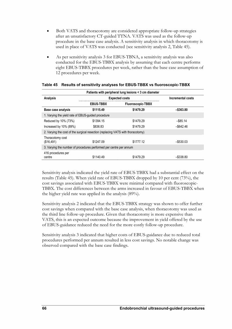
66 Endobronchial ultrasound-guided procedures
• Both VATS and thoracotomy are considered appropriate follow-up strategies after an unsatisfactory CT-guided TTNA. VATS was used as the follow-up procedure in the base case analysis. A sensitivity analysis in which thoracotomy is used in place of VATS was conducted (see sensitivity analysis 2, Table 45).
• As per sensitivity analysis 3 for EBUS-TBNA, a sensitivity analysis was also conducted for the EBUS-TBBX analysis by assuming that each centre performs eight EBUS-TBBX procedures per week, rather than the base case assumption of 12 procedures per week.
Table 45 Results of sensitivity analyses for EBUS-TBBX vs fluoroscopic-TBBX
Patients with peripheral lung lesions < 3 cm diameter Analysis Expected costs EBUS-TBBX Fluoroscopic-TBBX
Incremental costs
Base case analysis $1115.49 $1479.29 –$363.80 1. Varying the yield rate of EBUS-guided procedure Reduced by 10% (73%) $1394.15 $1479.29 –$85.14 Increased by 10% (89%) $836.83 $1479.29 –$642.46 2. Varying the cost of the surgical resection (replacing VATS with thoracotomy) Thoracotomy cost ($16,491) $1247.09 $1777.12 –$530.03 3. Varying the number of procedures performed per centre per annum 416 procedures per centre $1140.49 $1479.29 –$338.80
Sensitivity analysis indicated the yield rate of EBUS-TBBX had a substantial effect on the results (Table 45). When yield rate of EBUS-TBBX dropped by 10 per cent (73%), the cost savings associated with EBUS-TBBX were minimal compared with fluoroscopic-TBBX. The cost differences between the arms increased in favour of EBUS-TBBX when the higher yield rate was applied in the analysis (89%).
Sensitivity analysis 2 indicated that the EBUS-TBBX strategy was shown to offer further cost savings when compared with the base case analysis, when thoracotomy was used as the third line follow-up procedure. Given that thoracotomy is more expensive than VATS, this is an expected outcome because the improvement in yield offered by the use of EBUS-guidance reduced the need for the more costly follow-up procedure.
Sensitivity analysis 3 indicated that higher costs of EBUS-guidance due to reduced total procedures performed per annum resulted in less cost savings. No notable change was observed compared with the base case findings.

Endobronchial ultrasound-guided procedures 67
Discussion
Accurate NSCLC staging and diagnosis of mediastinal/hilar masses of unknown origin and peripheral lung lesions are vital for prognostic and therapeutic decision making.
The use of EBUS procedures was shown to improve the likelihood of successfully obtaining TBNA material of diagnostic quality for NSCLC staging and diagnosis of mediastinal/hilar masses (Herth et al 2004). A decision analytic model, comparing EBUS-TBNA with TBNA alone, was used to evaluate associated cost implications.
Although EBUS did not improve yield in diagnosis of peripheral lung lesions greater than 3 cm diameter, it was shown to offer improvement when performed for peripheral lung lesions less than 3 cm diameter (Herth et al 2002). To this end, a model was used to compare EBUS-TBBX with fluoroscopic-TBBX in this patient sub-group.
No analysis was carried out for EBUS procedures in the depth diagnosis of endobronchial cancers due to the lack of relevant clinical data.
The current model estimated that EBUS-TBNA would generate a mean cost saving of $347 per patient when compared with TBNA alone for NSCLC staging and diagnosis of mediastinal/hilar masses. This finding is consistent with the analysis of Herth et al (2003).
The use of EBUS-TBBX was estimated to generate a cost saving of $364 per patient in the diagnosis of peripheral lung lesions less than 3 cm diameter when compared with fluoroscopic-TBBX.
Sensitivity analyses demonstrated that only a small change in yield rates in the model significantly affected the results. This was anticipated because yield rate represents the key factor in the model, influencing the likelihood of avoiding the more costly follow-up procedure.
The break-even yield rate for EBUS-TBNA, at which the cost advantage of EBUS guidance is lost, was approximately 71 per cent according to the current model. This break-even yield rate fell within the 95 per cent confidence intervals calculated from Herth et al (2004). As such, the cost advantage demonstrated for EBUS-TBNA in the base case analysis should be interpreted with caution.
The break-even point was also estimated to be 71 per cent for EBUS-TBBX. The use of EBUS-TBBX was reported to offer a yield rate of 81 per cent in the diagnosis of peripheral lung legions less than 3 cm diameter, well over the break-even point of 71 per cent based on the current model. This indicates that EBUS-TBBX could be considered as a preferred strategy over fluoroscopic-TBBX.
Improved yield offered by EBUS guidance was the main focus of the current analyses. The use of EBUS-guidance was assumed to have no impact on the overall diagnostic accuracy of sampling procedures (TBNA and TBBX). Should the use of EBUS influence diagnostic accuracy of TBNA or TBBX, patients’ prognoses would likely be affected. This would potentially create important health outcomes and economic implications. No relevant data were available to inform evaluation of these outcomes.

68 Endobronchial ultrasound-guided procedures
A variety of biopsy techniques could be considered as appropriate comparators (Figures 3–6). The current model elected TBNA alone and fluoroscopic-TBBX as comparators for EBUS-TBNA and EBUS-TBBX, respectively. This reflected the limited availability of comparative data between EBUS-guided procedures and other modalities.
Mediastinoscopy is often used as a first-line invasive staging procedure in practice for NSCLC staging and diagnosis of mediastinal/hilar masses of unknown origin. Mediastinoscopy is a considerably more expensive procedure than EBUS-TBNA. Given the reported yield rates for EBUS-TBNA, these procedures are very likely to offer cost advantages over mediastinoscopy as the first-line staging/diagnostic procedure. At a yield rate of 77 per cent obtained by EBUS-TBNA, its expected costs, plus mediastinoscopy if necessary, were estimated at $2426 per patient. This level of cost advantage means that with the assumption of a perfect yield for mediastinoscopy, EBUS-TBNA would still represent a superior alternative even if its yield rate were far worse than the levels reported in the available evidence––as low as 20 per cent.
Mediastinoscopy has been reported to be associated with a small risk of complication (Aabakken et al 1999, Kramer et al 2004). Avoiding mediastinoscopy by using EBUS technology is likely to generate health benefits. These benefits were not captured by the current model. The current analyses are therefore likely to be conservative and to underestimate the cost advantages of EBUS procedures.
VATS or thoracotomy were employed as the final diagnostic procedures in the model for diagnosis of peripheral lung lesions less than 3 cm diameter. The model was designed to determine the overall costs associated with the diagnostic algorithm, and not the costs associated with downstream clinical management of patients following confirmed diagnosis; VATS or thoracotomy can offer both therapeutic and diagnostic interventions in some cases. This means some patients would undergo VATS or thoracotomy for treatment as well as diagnosis.
Given that a higher proportion of patients in the fluoroscopic-TBBX arm underwent VATS or thoracotomy, more people in this arm may potentially have undergone therapeutic surgical resection simultaneously with the (more invasive) diagnostic procedure than patients in the EBUS-TBBX arm in the model. Accordingly, fewer patients in the fluoroscopic-TBBX arm would have required an additional therapeutic intervention after the diagnostic procedure outside the model, potentially reducing the costs of downstream management for these patients, which was not captured in the model.
Accordingly, the cost savings demonstrated for the EBUS arm may not be fully realised in practice. However, given the small incremental difference in the proportion of patients undergoing surgical resection, this is likely to have a negligible cost implication.
Clinical observation could represent an alternative to surgical resection (ie VATS and thoracotomy) for diagnosis of peripheral lung lesions. This strategy is most appropriate for patients at very low risk of malignancy and/or those at high risk of surgical resection complication. This could however potentially delay diagnosis and treatment in patients with malignant nodules (Gould et al 2007). The current analysis did not explore the cost implications of employing clinical observation as a follow-up procedure.
Endobronchial ultrasound-guided procedures 69
It is noted that most centres equipped to provide EBUS-guided sampling services are currently located in public hospitals. Based on the available evidence (Table 46), private hospitals represented 33 per cent of the total separations relating to respiratory system procedures in 2002–2003; the remainder were in the public hospital system. This trend might also develop for EBUS-guided sampling procedures. As a result, health resource utilisation for implementing EBUS-guided sampling procedures in the public hospital system would be reduced.
Table 46 Separation statistics for respiratory system procedures by public and private hospitals
AR-DRG code, description Public patient separations
Private patient separations
E02A Other respiratory system operating procedures with catastrophic complications and comorbidities 1864 632
E02B Other respiratory system operating procedures with severe complications and comorbidities 1919 800
E02C Other respiratory system operating procedures without catastrophic or severe complications and comorbidities 8619 4765
Total number of separations 12,402 6197 Source: National Hospital Cost Data Collection, Round 7 (2002–2003). The data from Round 8 and Round 9 were not used because of the absence of the comparative private hospitals separations with those DRG codes
The introduction of EBUS-guided procedures for NSCLC staging and diagnoses of mediastinal masses and peripheral lung lesions less than 3 cm diameter may generate modest cost savings for the Australian healthcare system. A reliable assessment of the potential impact of EBUS-guided procedures in terms of patient outcomes is not possible due to lack of relevant data.

70 Endobronchial ultrasound-guided procedures
Other considerations
This section raises matters relating to endobronchial ultrasound (EBUS) that may not have been addressed by the identified evidence indicating the clinical safety, effectiveness and cost-effectiveness of EBUS procedures. Advice from the expert advisory panel and issues raised by the evaluators is presented. Information provided information is additional to evidence identified during the systematic literature review.
Generalisability of evidence
The Thoraxklinik (University of Heidelberg, Germany) is credited as a collaborating centre in most studies presenting EBUS technology and procedural information. The unique status of Thoraxklinik should be considered when reviewing this report because it may limit the generalisability of the evidence.
Endobronchial ultrasound and endoscopic ultrasound
Endobronchial ultrasound-guided transbronchial needle aspiration (EBUS-TBNA) and endoscopic ultrasound-guided fine-needle aspiration (EUS-FNA) are complementary techniques. EBUS provides better access to anterior and superior mediastinal lymph nodes, and EUS is better able to access to posterior and inferior mediastinal lymph nodes (Table 4). Combining EBUS and EUS should theoretically enable access to the whole of the mediastinum. It was assumed that a reasonable evaluation approach to confirm diagnoses of mediastinal masses would involve CT/PET scanning followed by EBUS-TBNA and/or EUS-FNA. This single procedure evaluation strategy could potentially minimise the number of procedures patients undergo, and provide accurate staging of mediastinal masses. This approach could also provide benefits for patients considered medically unfit to undergo surgical diagnostic procedures (Rintoul et al 2005). Larger randomised controlled trials that examined accuracy of combined EBUS-TBNA and EUS-FNA in staging mediastinal masses, compared with currently available studies that considered surgical techniques, are required. If combined, the resulting procedure would be more expensive because Australian clinical practice requires two different specialists to perform the procedures.
Prospective studies
A number of relevant prospective studies are under way that should be considered as part of the decision making process about reimbursement of EBUS (see Table 47). Studies being conducted by Tournoy and Annema (NCT00432640) and Rintoul (ISRCTN97311620) combine EBUS with EUS for comparison with mediastinoscopy (the reference standard) and may be particularly informative. No prior parallel group randomised controlled trial (RCT) has been conducted to ascertain the capability of the combined techniques versus mediastinoscopy.
Emerging experience indicates the possible use of EBUS-TBNA for patients who do not have abnormal lymph nodes detected by CT and/or PET (Herth et al 2006a).

Endobronchial ultrasound-guided procedures 71
Table 47 Characteristics of potentially relevant ongoing trials using EBUS-guided procedures
Trial register details
Study characteristics
ISRCTN97311620 Robert Rintoul Identified using Current Controlled Trials Register
Study design: Parallel group RCT Population: Patients with presumed or known NSCLC with mediastinal lymphadenopathy identified by prior tests (n = 142) Prior tests: CT Index test: EBUS-TBNA and EUS-FNA Comparator: Mediastinoscopy Reference standard: Negative histology samples will be confirmed with surgical resection Outcomes: Primary Sensitivity of lymph node staging; Secondary Utility; assessment of the rate of avoided surgical procedures
NCT00432640 Kurt Tournoy & Jouke Annema Identified using ClinicalTrials.gov register
Study design: Open-label parallel group RCT Population: Patients with presumed or known NSCLC with mediastinal lymphadenopathy identified by prior tests (n = 150) Prior tests: NR Index test: EBUS-TBNA/EUS-FNA Comparator: Mediastinoscopy Reference standard: NR Outcomes: Primary Sensitivity of lymph node staging; Secondary Assessment of mediastinal tumour invasion (T4); assessment of the rate of avoided surgical procedures; assessment of the negative predictive value; assessment of the difference in the cost for lymph node staging; assessment of the complication rates; assessment of the rate of futile thoracotomies
NCT00372203 Shaf Keshavjee Identified using ClinicalTrials.gov register
Study design: Prospective, open-label patient series Population: Patients with presumed or known NSCLC who require staging using mediastinoscopy or patients who have mediastinal masses of unknown origin (n = 180) Prior tests: NR Index test: EBUS-TBNA Comparator: Mediastinoscopy Reference standard: NR Outcomes: Primary sensitivity of lymph node diagnosis
NCT00415337 Han-Pin Kuo Identified using ClinicalTrials.gov register
Study design: Prospective/retrospective cross-sectional study Population: Patients with peripheral lung lesions identified by prior tests (n = NR) Prior tests: NR Index test: EBUS +/-TBBX (GS) Comparator: NR Reference standard: NR Outcomes: NR
NCT00398970 Jon Hardie Identified using ClinicalTrials.gov register
Study design: Open-label parallel group RCT Population: Patients with lesions suspected of malignancy in the lung (n = 240) Prior tests: Bronchoscopy Index test: EBUS-TBBX (GS) Comparator: TBBX Reference standard: NR Outcomes: Diagnostic yield
Abbreviations: CT, computed tomography; EBUS, endobronchial ultrasound; EUS-FNA, endoscopic fine-needle aspiration; GS, guide sheath; NR, not reported; NSCLC, non-small cell lung cancer; RCT, randomised controlled trial; TBBX, transbronchial biopsy; TBNA, transbronchial needle aspiration

72 Endobronchial ultrasound-guided procedures
Research recommendations
After reviewing the body of evidence addressing each research question the evaluators have made specific research recommendations using a modified EPICOT (evidence, population, intervention, comparison, outcome, time stamp) format (Brown et al 2006). As well as the standard EPICOT elements, the research recommendations also address the prior test element. After reviewing the current evidence it was considered appropriate to combine the NSCLC staging and diagnosis of mediastinal/hilar mass indications.
The research recommendations outlined in Table 48 were formulated to address the gaps identified in the body of evidence for use of EBUS-TBNA in NSCLC staging of and diagnosis of mediastinal/hilar masses.
A systematic review of the literature did not identify any comparative evidence of the effectiveness of EBUS-TBNA versus mediastinoscopy, which is one of the major comparators identified by the advisory panel.
The systematic review identified evidence that indicated the comparative diagnostic yield of EBUS-TBNA and TBNA. There was insufficient evidence to address the uncertainty in comparative effectiveness between procedures.
Table 48 Research recommendations for use of EBUS-TBNA in NSCLC staging and diagnosis of mediastinal/hilar masses
Element Description Evidence The reported diagnostic yield of EBUS-TBNA was greater than TBNA. There were no studies that reported
diagnostic accuracy of EBUS-TBNA compared with TBNA No studies were identified that compared the diagnostic performance of EBUS-TBNA with mediastinoscopy The reported sensitivity and diagnostic yield of EBUS-TBNA was at least equivalent to EUS-FNA in selected patient subgroups
Population Patients with presumed or known NSCLC with mediastinal/ hilar lymphadenopathy identified by prior tests Patients with mediastinal/ hilar masses of unknown origin (including lymphadenopathy) identified with CT +/- x-ray +/- symptoms Mixed patient population with presumed or known NSCLC with mediastinal/ hilar lymphadenopathy identified by prior tests or diagnosis of mediastinal lymphadenopathy of unknown origin (including lymphadenopathy) identified with CT +/- x-ray +/- symptoms
Prior tests Clinical assessment; CT +/- PET Intervention/test EBUS-TBNA; EBUS-TBNA and EUS-FNA Comparatora Mediastinoscopy; TBNA Outcome Sensitivity and specificity (compared with a histological reference standard)
Diagnostic yieldb Treatment alterations (eg assessment of the rate of avoided surgical procedures) Patient survival Quality of life Adverse events
Time stamp June 2007 Abbreviations: CT, computer tomography; EBUS, endobronchial ultrasound; EUS, endoscopic ultrasound; FNA, fine needle aspiration; NSCLC, non-small cell lung cancer; PET, positron emission tomography; TBNA, transbronchial needle aspiration a The research recommendation was formulated based on the major comparators identified by the advisory panel. Other comparators for this indication included EUS-FNA, mediastinotomy, TTNA, or VAT b Comparisons versus mediastinoscopy

Endobronchial ultrasound-guided procedures 73
The research recommendations outlined in Table 49 were formulated to address the gap identified in the body of evidence for use of EBUS with or without EBBX in depth diagnosis of endobronchial cancers.
A systematic review of the evidence did not identify any comparative evidence of EBUS with or without EBBX versus EBBX.
Table 49 Research recommendations for the use of EBUS with or without EBBX in the depth diagnosis of endobronchial cancers
Element Description Evidence No trials were identified that compared the diagnostic accuracy of EBUS +/- EBBX to EBBX in depth
diagnosis of endobronchial cancers in patients without enlarged lymph nodes Population Patients with presumed or known NSCLC without mediastinal/ hilar lymphadenopathy identified by prior
tests Prior tests Clinical assessment
CT +/- PET Intervention/test EBUS +/- EBBX Comparator EBBX Outcome Sensitivity and specificity (compared with a histological reference standard)
Diagnostic yield Treatment alterations (eg changes in planned photodynamic therapy or surgery) Patient survival Quality of life Adverse events
Time stamp June 2007 Abbreviations: CT, computer tomography; EBUS, endobronchial ultrasound; EBBX, endobronchial biopsy; NSCLC, non-small cell lung cancer; PET, positron emission tomography
The research recommendations outlined in Table 50 were formulated to address two gaps identified in the body of evidence for use of EBUS-TBBX in diagnosis of peripheral lung lesions.
A systematic review of the literature did not identify any comparative evidence of EBUS-TBBX versus TTNA, which was one of the major comparators identified by the advisory panel.
The systematic review identified evidence indicating comparative diagnostic performance of EBUS-TBBX and fluoroscopy-TBBX. The EBUS-TBBX procedures reported had limited applicability to the Australian clinical setting.

74 Endobronchial ultrasound-guided procedures
Table 50 Research recommendations for the use of EBUS-TBBX in the diagnosis of peripheral lung lesions
Element Description Evidence The reported sensitivity and diagnostic yield of EBUS-TBBX was equivalent to fluoroscopy-TBBX
No studies were identified that compared the diagnostic performance of EBUS-TBBX with TTNA The reported diagnostic yield of EBUS-TBBX was greater than TBBX and at least equivalent to ENB-TBBX The diagnostic yield of EBUS-TBBX may be greater than other methods of guided-TBBX in diagnosis of smaller peripheral lesions
Population Patients with peripheral lung lesions identified by prior tests Patients with peripheral lung lesions < 3 cm identified by prior tests
Prior tests Clinical assessment CT +/- PET
Intervention/test EBUS-TBBX (with fluoroscopic navigation) Comparator a Fluoroscopy-TBBX
TTNA Outcome Sensitivity and specificity (compared with a histological reference standard)
Diagnostic yield Treatment alterations (eg assessment of surgical procedures avoided rate) Patient survival Quality of life Adverse events
Time stamp June 2007 Abbreviations: CT, computer tomography; EBUS, endobronchial ultrasound; ENB, electromagnetic navigation bronchoscopy; TBNA, transbronchial needle aspiration; TTNA, transthoracic needle aspiration; PET, positron emission tomography a The research recommendation was formulated based on the major comparators identified by the advisory panel. Other comparators for this indication included TTNA, TBBX and electromagnetic guided TBBX

Endobronchial ultrasound-guided procedures 75
Conclusions
Safety
Endobronchial ultrasound (EBUS)-guided procedures for non-small cell lung cancer (NSCLC) staging, diagnosis of mediastinal/hilar masses, depth diagnosis of endobronchial cancers and diagnosis of peripheral lung lesions appear to be as safe as other minimally-invasive diagnostic tests. The most frequently reported adverse events were bleeding and pneumothorax. These mainly occurred among patients who underwent EBUS- or fluoroscopy-guided transbronchial biopsy.
Effectiveness
A linked evidence approach was used to evaluate use of EBUS-guided procedures for NSCLC staging, diagnosis of mediastinal/hilar masses, depth diagnosis of endobronchial cancers and diagnosis of peripheral lung lesions.
The evidence indicated that the diagnostic yield of EBUS-transbronchial needle aspiration (TBNA) was greater than TBNA alone in NSCLC staging and for diagnosis of mediastinal/hilar masses. There was limited evidence suggesting that the sensitivity and diagnostic yield of EBUS-TBNA were at least equivalent to endoscopic ultrasound fine-needle aspiration (EUS-FNA) in specific sub-groups. No evidence was available to assess EBUS-TBNA impact on patient management. Treatment effectiveness evidence was not examined because it was considered that EBUS-TBNA would not identify any unique patient groups that were substantially different from those currently seen in Australian clinical practice. There was insufficient evidence to address uncertainty concerning the clinical impact of EBUS-TBNA compared with its major comparators, TBNA and mediastinoscopy.
No trials were identified that compared the diagnostic performance of EBUS with or without endobronchial biopsy (EBBX) to EBBX alone in diagnosing the depth of endobronchial cancers. In the absence of evidence indicating diagnostic accuracy, patient management and treatment effectiveness-related evidence was not sought.
The evidence suggested that EBUS-TBBX sensitivity was equivalent to fluoroscopy-TBBX in the diagnosis of peripheral lung lesions. The evaluated studies also indicated that the diagnostic yield of EBUS-TBBX was greater than TBBX and at least equivalent to other methods (electromagnetic, fluoroscopic) of guided-TBBX. The evidence further suggested that the diagnostic yield of EBUS-TBBX may be greater than other methods of guided-TBBX in diagnosing smaller peripheral lesions (less than 3 cm diameter). There was no evidence to assess the impact of EBUS-TBBX on patient management. There was insufficient evidence to address uncertainty surrounding the clinical impact of EBUS-TBBX compared with the major comparators, fluoroscopy-TBBX and TTNA. Treatment effectiveness evidence was not examined because it was considered that EBUS-TBBX would not identify any unique patient groups that were substantially different from those currently seen in Australian clinical practice.

76 Endobronchial ultrasound-guided procedures
Economic analyses
A decision analytic model was constructed to assess cost implications of EBUS-guided procedures when compared with current procedures. A cost analysis of EBUS-TBNA relative to TBNA alone was performed for NSCLC staging and diagnosis of mediastinal/hilar masses of unknown origin. A cost analysis of EBUS-TBBX relative to TBBX was conducted for diagnosis of peripheral lung lesions less than 3 cm.
A cost analysis was not conducted for the depth diagnosis of endobronchial cancers because clinical data were limited.
The analysis indicated that use of EBUS-TBNA for NSCLC staging and diagnosis of mediastinal masses was estimated to generate a cost saving of $347 per patient. The use of EBUS-TBBX for diagnosis of peripheral lung lesions less than 3 cm diameter was estimated to generate a cost saving of $364 per patient. This reflected the economic benefits associated with improved yield offered by the use of EBUS guidance.
The presented results should be interpreted in the context of data inputs and assumptions applied in the model. A reliable assessment of the potential impact of EBUS-guided procedures in terms on patient outcomes is not possible due to a lack of relevant data.

Endobronchial ultrasound-guided procedures 77
Recommendation
MSAC has considered the safety, effectiveness and cost-effectiveness of endobronchial ultrasound (EBUS)-guided procedures for the investigation of non-small cell lung cancer, mediastinal/hilar masses, endobronchial cancer and peripheral lung lesions compared to mediastinoscopy and transbronchial needle aspiration.
The MSAC finds that the EBUS-guided procedures for the staging of non-small cell lung cancer, and the investigation of mediastinal/hilar masses and peripheral lung lesions is safer, more effective and likely to be cost saving when compared to mediastinoscopy and transbronchial needle aspiration.
MSAC finds that, though safe, there is insufficient evidence on the effectiveness and cost-effectiveness of the EBUS-guided procedure for the evaluation of endobronchial cancer.
MSAC recommends that public funding should be supported for EBUS-guided procedures for the staging of non-small cell lung cancer, and the investigation of mediastinal/hilar masses and peripheral lung lesions.
MSAC recommends that public funding should not be supported for the EBUS-guided procedure for the evaluation of endobronchial cancer.
— The Minister for Health and Ageing accepted this recommendation on 20 May 2008—

78 Endobronchial ultrasound-guided procedures
Appendix A MSAC terms of reference and membership
MSAC’s terms of reference are to:
• advise the Minister for Health and Ageing on the strength of evidence pertaining to new and emerging medical technologies and procedures in relation to their safety, effectiveness and cost-effectiveness and under what circumstances public funding should be supported
• advise the Minister for Health and Ageing on which new medical technologies and procedures should be funded on an interim basis to allow data to be assembled to determine their safety, effectiveness and cost-effectiveness
• advise the Minister for Health and Ageing on references related either to new and/or existing medical technologies and procedures, and
• undertake health technology assessment work referred by the Australian Health Ministers’ Advisory Council (AHMAC) and report its findings to AHMAC.
The membership of MSAC comprises a mix of clinical expertise covering pathology, radiology, nuclear medicine, oncology, surgery, internal medicine and general practice, plus clinical epidemiology and clinical trials, health economics, consumers and health administration and planning:
Member Expertise or affiliation Dr Stephen Blamey (Chair) General surgery
Associate Professor John Atherton Cardiology
Associate Professor Michael Cleary Emergency medicine
Associate Professor Paul Craft Clinical epidemiology and oncology
Professor Geoff Farrell Gastroenterology
Dr Kwun Fong Thoracic medicine
Professor Richard Fox Medical oncology
Dr Bill Glasson Ophthalmology
Professor Jane Hall Health economics
Professor John Horvath Chief Medical Officer Department of Health and Ageing
Associate Professor Terri Jackson Health economics
Professor Brendon Kearney Health administration and planning
Associate Professor Frederick Khafagi Nuclear medicine
Dr Ray Kirk Health research
Dr Ewa Piejko General practice
Dr Ian Prosser Haematology

Endobronchial ultrasound-guided procedures 79
Member Expertise or affiliation Ms Sheila Rimmer Consumer health issues
Dr Judy Soper Radiology
Professor Ken Thomson Radiology
Dr David Wood Orthopaedics
Mr Peter Woodley Assistant Secretary Medical Benefits Schedule (MBS) Policy Development Branch, Department of Health and Ageing

80 Endobronchial ultrasound-guided procedures
Appendix B Advisory panel
Advisory panel for MSAC application 1108
Dr Kwun Fong (Chair) Thoracic medicine
Member of MSAC
Associate Professor Frederick Khafagi (Second Chair) Nuclear medicine
Member of MSAC
Mr Phillip Antippa Cardiothoracic surgery
Nominated by the Australasian Society of Cardiovascular Surgeons
Dr Martin Phillips Respiratory medicine
Nominated by the Thoracic Society of Australia and New Zealand
Dr Marshall Plit Thoracic medicine & Lung transplation
Nominated by the Thoracic Society of Australia and New Zealand
Dr Morgan Windsor Cardio & Thoracic surgery & Upper gastrointestinal surgery
Nominated by the Australasian Society of Cardiac and Thoracic Surgeons
Ms Robin Toohey Independent consumer representative
Nominated by the Consumers’ Health Forum

Endobronchial ultrasound-guided procedures 81
Appendix C Supportive data
Nodal staging of non-small cell lung cancer and diagnosis of mediastinal/hilar masses of unknown origin
There were eight studies identified that presented supportive evidence of EBUS-TBNA use for patients primarily referred for NSCLC staging or diagnoses of mediastinal/hilar masses of unknown origin (Herth et al 2003b, Herth et al 2006b, Krasnik et al 2003, Plat et al 2006, Rintoul et al 2005, Yasufuku et al 2004, Yasufuku et al 2005, Yasufuku et al 2006). The characteristics of these studies are summarised in Table 51.
A low-quality comparative diagnostic accuracy study by Yasufuku et al (2006) assessed the diagnostic sensitivity of EBUS-TBNA, CT and PET imaging for mediastinal lymph nodes. This study did not compare EBUS with the conventional comparators and was not included in the primary analysis. Because the comparison did not involve a blinded independent comparison with a valid reference standard, it was regarded as providing level III-2 evidence. This study offers limited applicability to the Australian clinical setting because the sensitivity evaluation of CT and PET imaging included patients who did not present with mediastinal/hilar lymphadenopathy.
Of the eight identified studies, four non-comparative studies assessed sensitivity (Krasnik et al 2003; level III-2 evidence) and diagnostic yield (Herth et al 2003b, Herth et al 2006b, Plat et al 2006; level IV evidence) of EBUS-TBNA in a mixed patient population. All studies that assessed diagnostic yield of EBUS-TBNA were high quality, but the diagnostic accuracy study was regarded as low quality. All studies were applicable to Australian clinical practice, but it should be noted that Herth et al (2003b) used a larger gauge needle than is common practice in this country. Herth et al (2006b) reported diagnostic accuracy values, but a 2×2 table (Figure 8) could not be reconstructed. Diagnostic accuracy results were not reported adequately by this study so they were excluded from the current review. Only diagnostic yield results were assessed.
The identified literature included three low quality non-comparative studies that assessed the diagnostic sensitivity of EBUS-TBNA (Yasufuku et al 2004, Yasufuku et al 2005) and EBUS-TBNA/EUS-FNA (Rintoul et al 2005) for mediastinal lymph nodes. It is noteworthy that Yasufuku et al (2004) and Yasufuku et al (2005) indicated that there was a partial overlap in the enrolled patient populations. These studies did not involve blinded independent comparisons with valid reference standards and were consequently regarded as providing level III-2 evidence. These studies present evidence that is likely to be applicable to the Australian clinical setting.

82 Endobronchial ultrasound-guided procedures
Table 51 Characteristics of the included supportive studies assessing the diagnostic accuracy/yield of EBUS-TBNA in patients referred for lung cancer staging and/or diagnosis of mediastinal/hilar mass of unknown origin
Author (year) Country
Study design
Patient characteristics Test characteristics Quality and applicability
Herth et al (2003b) Germany
Prospective, consecutive patient series Jan 1999–Jan 2000
Inclusion: Patients referred for lung cancer staging or diagnosis of mediastinal lymphadenopathy of unknown origin Exclusion: No reported exclusions (n = 242) Prior tests: CT
Index test: EBUS-TBNA [system EU-M 20 and 30, radial probe UM2R/3R, Olympus]; conventional bronchoscope (p 20 and p 40D, Olympus); 19G and 22G needle; general anaesthesia or conscious sedation; no ROSE Reference standard: Cytology; histology
IV CX, P2, Q1 Quality: High Applicability: Applicable 19G tissue sampling needle
Herth et al (2006b) Germany
Prospective, consecutive patient series Jun 2002– Sep 2004
Inclusion: Patients referred for lung cancer staging or diagnosis of mediastinal lymphadenopathy of unknown origin Exclusion: No reported exclusions (n = 502) Prior tests: X-ray, CT
Index test: EBUS-TBNA [system EU-60, Olympus]; linear scanning hybrid bronchoscope (XBF-UC260F-OL8, Olympus); 22G needle; moderate sedation and local anaesthesia or general anaesthesia; no ROSE; 2 aspirates per node were obtained Reference standard: Cytology; histology; clinical/radiological follow-up
IV CX, P1, Q1 Quality: High Applicability: Applicable
Krasnik et al (2003) Denmark
Direction unclear, non-consecutive patient series Unblinded
Inclusion: Patients referred for lung cancer staging, diagnosis of mediastinal/hilar lymphadenopathy of unknown origin or assessment of mediastinal tumour recurrence Exclusion: No reported exclusions (n = 11) Prior tests: CT, bronchoscopy
Index test: EBUS-TBNA [system EU-C60, Olympus]; linear scanning hybrid bronchoscope (XBF-UC40P, Olympus); 22G needle; general anaesthesia Reference standard: Cytology; histology
III-2 CX, P2, Q3 Quality: Low Unblinded Inadequate reference standard Applicability: Applicable Patients assessed for tumour recurrence
Plat et al (2006) Belgium
Prospective, consecutive patient series Jan 2003–Jun 2004
Inclusion: Patients referred for lung cancer staging or diagnosis of mediastinal lymphadenopathy of unknown origin Exclusion: No reported exclusions (n = 33) Prior tests: CT, PET
Index test: EBUS-TBNA [system EU-M20, radial probe UM-BS20-26R, Olympus]; conventional bronchoscope (Excera, Olympus); local anaesthesia and conscious sedation; no ROSE; 4–6 punctures were obtained Reference standard: Cytology; histology; clinical/radiological follow-up
IV CX, P1, Q1 Quality: High Applicability: Applicable

Endobronchial ultrasound-guided procedures 83
Author (year) Country
Study design
Patient characteristics Test characteristics Quality and applicability
Rintoul et al (2005) UK
Direction unclear, non-consecutive patient series Unblinded
Inclusion: Patients with known or suspected lung cancer and a mediastinal lymphadenopathy of > 1 cm or paratracheal or parabronchial masses Exclusion: Patients were excluded from analysis based on imaging results (n = 18) Prior tests: CT
Index test: EBUS-TBNA/EUS-FNA [system EU-C2000, Olympus]; linear scanning hybrid bronchoscope (XBF-UC260F-OL8, Olympus); 22G needle. EUS-FNA (GF-UC240P-AL5, Olympus); 22G needle. Local anaesthesia and sedation; no ROSE; 2–3 needle passes were obtained. Reference standard: Cytology, histology, clinical/radiological follow-up
III-2 CX, P1, Q3 Quality: Low Unblinded Inadequate reference standard Applicability: Applicable
Yasufuku et al (2004) Japan
Prospective, non-consecutive patient series Unblinded Mar 2002–Sep 2003
Inclusion: Patients with known or suspected lung cancer and mediastinal and/or hilar lymphadenopathy of > 1 cm Exclusion: No reported exclusions (n = 70) Prior tests: X-ray, CT
Index test: EBUS-TBNA [system EU-C2000, Olympus]; linear scanning hybrid bronchoscope (XBF-UC260F-OL8, Olympus); 22G needle; local anaesthesia and sedation; ROSE; 1–5 needle passes were obtained Reference standard: Cytology, histology, clinical/radiological follow-up
III-2 CX, P1, Q3 Quality: Low Unblinded Inadequate reference standard Applicability: Applicable
Yasufuku et al (2005) Japan
Prospective, non-consecutive patient series Unblinded Jun 2002– Apr 2004
Inclusion: Patients with known or suspected lung cancer and mediastinal and/or hilar lymphadenopathy of > 1 cm Exclusion: Patients with a final diagnosis of a benign disease; patients with a final diagnosis of malignant disease other than NSCLC, patients with extensive N2, N3 disease on CT (n = 108) Prior tests: CT
Index test: EBUS-TBNA [system EU-C2000, Olympus]; linear scanning hybrid bronchoscope (XBF-UC260F-OL8, Olympus); 22G needle; local anaesthesia and sedation; ROSE; 1–5 needle passes were obtained Reference standard: Cytology, histology, clinical/radiological follow-up
III-2 CX, P1, Q2 Quality: Low Unblinded Inadequate reference standard Inadequate data reporting Applicability: Applicable
Yasufuku et al (2006) Japan
Prospective, consecutive patient series CT & PET blinded Dec 2003–Mar 2005
Inclusion: Patients with known or suspected lung cancer Exclusion: Patients who were not further evaluated (n = 102) Prior tests: CT, PET, other tests
Index test: EBUS-TBNA [system EU-C2000, Olympus]; linear scanning hybrid bronchoscope (XBF-UC260F-OL8, Olympus); 22G needle, conscious sedation; ROSE; maximum of 5 needle passes were obtained Comparator: CT (Light Speed, GE medical systems), multidetector row, injection of contrast material Comparator: PET (PET Advance Nxi, GE medical systems), 300 Mbq injection of FDG Reference standard: Cytology, histology, clinical/radiological follow-up
III-2 C1, P2, Q3 Quality: Low Inadequate blinding Inadequate reference standard Applicability: Limited Included patients with negative lymphadenopathy
Abbreviations: CT, computed tomography; EBUS, endobronchial ultrasound; EUS-FNA, endoscopy ultrasound-fine needle aspiration; FDG, fluorodeoxyglucose; G, gauge; PET, positron emission tomography; ROSE, rapid on site evaluation; TBBX, transbronchial biopsy; TBNA, transbronchial needle aspiration; TTNA, transthoracic needle aspiration

84 Endobronchial ultrasound-guided procedures
Yasufuku et al (2006) reported the comparative sensitivity, specificity and predictive values of EBUS-TBNA, CT and PET (Table 52). The reported positive results were not confirmed, because malignant cytological diagnoses by EBUS-TBNA were taken as final proof of malignancy (inadequate reference standard). Therefore, the reported specificity and positive predictive value (PPV) could not be used as measures of test accuracy for this review. The PPV was applied only as an assumption in conjunction with the negative predictive value (NPV) to calculate approximate sensitivities for each diagnostic test. Yasufuku and colleagues (2006) found that the sensitivity of linear EBUS-TBNA (92%) was greater than CT and PET imaging (77% and 80%, respectively) in the diagnosis of involved mediastinal lymph nodes. It is noteworthy that the prevalence reported by this study was considerably lower than the rate reported by Vilmann et al (2005). This may be indicative of differences in patient populations that could affect indirect comparisons.
Table 52 Diagnostic accuracy of included supportive studies comparing EBUS-TBNA with CT and PET imaging in patients referred for NSCLC staging and diagnoses of mediastinal/hilar masses of unknown origin
Author (year)
Prevalence n/N (%)
Diagnostic test Sensitivity (95% CI)a
NPV (95% CI)
Level of evidence
EBUS-TBNA 92.31 (87.14, 97.48) 97.44 (94.37, 100.00)
CT 76.92 (68.74, 85.10) 87.50 (81.08, 93.92)
Yasufuku et al (2006) 26/102
(25.49)
PETb 80.00 (72.24, 87.86) 91.53 (86.13, 96.93)
III-2 C1, P2, Q3
Abbreviations: CI, confidence interval; CT, computed tomography; EBUS-TBNA, endobronchial ultrasound transbronchial needle aspiration; NPV, negative predictive value; PET, positron emission tomography a Sensitivity was calculated assuming a 100% PPV b An apparent data reporting error was detected in this calculation
The reported sensitivity and NPV of the non-comparative studies are summarised in Table 53. The study by Krasnik et al (2003) reported a sensitivity of 91 per cent for linear EBUS-TBNA in the mixed patient population. This was a small study (n = 11); all enrolled participants had malignant lymph nodes. The studies by Yasufuku and colleagues (2004, 2005) reported different sensitivities for linear EBUS-TBNA—100 per cent in the 2004 study and 95 per cent in the 2005 study—in patients referred for lung cancer staging (overlapping patient population). It is noteworthy that both studies calculated the sensitivity with failed TBNA procedures contributing to the false results but it was only possible to recalculate (failed TBNA procedures excluded) the Yasufuku et al (2004) results. The non-comparative study by Rintoul et al (2005) reported a sensitivity of 85 per cent for the combination of linear EBUS-TBNA and EUS-FNA in the mixed patient population.
The reported positive results were not confirmed in any of the non-comparative diagnostic accuracy studies because a malignant cytological diagnosis made using these techniques was taken as final proof of malignancy (inadequate reference standard). Therefore, for the purpose of this review the reported specificity and PPV were not used as measures of test accuracy. The PPV was only used as an assumption in conjunction with the NPV to calculate an approximate sensitivity for each diagnostic test.

Endobronchial ultrasound-guided procedures 85
Table 53 Diagnostic accuracy of the included non-comparative supportive studies assessing EBUS-TBNA in patients referred for NSCLC staging and diagnoses of mediastinal/hilar masses of unknown origin
Author (year) Prevalence n/N (%)
Diagnostic test Sensitivity (95% CI)a
NPV (95% CI)
Level of evidence
Patients primarily referred for NSCLC staging and diagnosis of a mediastinal/hilar mass of unknown origin Krasnik et al (2003) 11/11
(100.00) EBUS-TBNA 90.91
(73.92, 100.00)
0.00 III-2 CX, PX, Q3
Patients referred for lung cancer staging Rintoul et al (2005) 13/18
(72.22) EBUS-TBNA/EUS-
FNA 84.62
(67.95, 100.00)
71.43 (50.56, 92.30)
III-2 CX, P1, Q3
Yasufuku et al (2004)
45/68 (66.18)
EBUS-TBNA 100.00 100.00 III-2 CX, P1, Q3
Yasufuku et al (2005)b 74/108
(68.52) EBUS-TBNA 94.59
(90.32, 98.86) 89.47
(83.68, 95.26)
III-2 CX, P1, Q2
Abbreviations: CI, confidence interval; EBUS, endobronchial ultrasound; TBNA, transbronchial needle aspiration a Sensitivity was calculated assuming a 100% PPV b An apparent data reporting error was detected in this calculation
The reported diagnostic yield results from five non-comparative studies are presented in Table 54. The yields obtained for patients referred for lung cancer staging (97%) and the mixed patient population (82–95%) were generally concordant with the yields observed in the comparative trials presented in Table 21.
Table 54 Diagnostic yield of the included supportive studies assessing EBUS-TBNA in a mixed patient population (including NSCLC)
Specific diagnostic yielda Non-specific diagnostic yield b
Author (year)
Prevalence n/N (%)
Diagnostic test
n/N % (95% CI) n/N % (95% CI)
Level of evidence
Patients primarily referred for NSCLC staging and diagnoses of mediastinal/hilar masses of unknown origin Herth et al (2003b)
NR EBUS-TBNA 172/242 71.07 (65.36, 76.79)
207/242 85.54 (81.11, 89.97)
IV CX, PX, Q1
Herth et al (2006b)
493/502 (98.21)
EBUS-TBNA 470/502 93.63 (91.49, 95.76)
476/502 94.82 (92.88, 96.76)
IV CX, P1, Q1
Plat et al (2006)
27/33 (81.82)
EBUS-TBNA NR NR 27/33 81.82 (68.66, 94.98)
IV CX, P1, Q1
Patients referred for lung cancer staging Yasufuku et al (2004)
NR EBUS-TBNA NR NR 68/70 97.14 (93.24, 100.00)
III-2 CX, P1, Q3
Yasufuku et al (2005)
NR EBUS-TBNA NR NR 105/108 97.22 (94.12, 100.00)
III-2 CX, P1, Q2
Abbreviations: CI, confidence interval; EBUS, endobronchial ultrasound; NR, not reported; TBNA, transbronchial needle aspiration a Per patient diagnostic yield when the diagnosis of a benign lymph node was restricted to requiring a specific diagnosis b Per patient diagnostic yield when the diagnosis of a benign lymph node was expanded to patients with negative lymphocytes but without a specific benign diagnosis

86 Endobronchial ultrasound-guided procedures
Peripheral lung lesions
Supportive evidence indicating the use of EBUS-TBBX in peripheral lung lesions diagnosis was identified in studies by Becker et al (2005), Chung et al (2007), Dooms et al (2007), Herth et al (2006a), Kikuchi et al (2004), Kurimoto et al (2004). The characteristics of these studies are summarised in Table 55.
A high-quality diagnostic yield RCT by Chung et al (2007) assessed the value of measuring the distance between the bronchial orifice and the peripheral lesion using radial EBUS. This study did not compare EBUS with the conventional comparators and was not included in the primary analysis. This study was classified as providing level IV evidence.
The high-quality non-comparative studies by Chung et al (2007), Dooms et al (2007), and Herth et al (2006a) assessed the diagnostic yield of EBUS-TBBX among patients with peripheral lung lesions. Because they did not report the use of fluoroscopic navigation in conjunction with the EBUS procedure these studies offer limited applicability to Australian clinical practice. The applicability of the study by Herth et al (2006a) was further limited because patients had been fluoroscopically examined previously. These studies were classified as providing level IV evidence.
Table 55 Characteristics of included supportive studies that assessed diagnostic yield of EBUS-TBBX among patients with peripheral lung lesions
Author (year) Country
Study design Patient characteristics Test characteristics Quality and applicability
Becker et al (2005) Germany
Direction unclear, non-consecutive patient series Jul 2003– Dec 2003
Inclusion: Patients with a peripheral lung lesions not visible using bronchoscopy Exclusion: Patients with distorted airways due to previous surgery (n = 29) Prior tests: CT
Index test: ENB/EBUS-TBBX ENB (superDimension/Bronchus system, superDimension) and EBUS-TBBX (radial probe UM-BS20-26R, Olympus), conventional bronchoscope (EXERA IT160, Olympus); bronchial forceps and bronchial brushing; general anaesthesia; fluoroscopy and curette support Reference standard: Histology, clinical/radiological follow-up
IV CX, P2, Q1 Quality: High Applicability: Applicable
Chung et al (2007) Taiwan
Prospective, parallel group RCT Blinding of pathologists Oct 2004– Jul 2005
Inclusion: Patients with a solitary pulmonary lesion not visible using bronchoscopy Exclusion: Lesion invisible using EBUS (n = 113) Prior tests: X-ray, CT
Index test: EBUS-TBBX (GS) [system EU-M30S, radial probe UM-S20-20R, Olympus]; conventional bronchoscope (P260F, Olympus); guide sheath; biopsy forceps; local anaesthesia; distance measuring; 3–5 biopsy specimens were obtained Index test: EBUS-TBBX (GS) [system EU-M30S, radial probe UM-S20-20R, Olympus]; single channel bronchoscope (P260F, Olympus); guide sheath; biopsy forceps; local anaesthesia; 3–5 biopsy specimens were obtained Reference standard: Cytology, histology
IV C1, P2, Q1 Quality: High Applicability: Limited No fluoroscopic navigation of EBUS procedure

Endobronchial ultrasound-guided procedures 87
Author (year) Country
Study design Patient characteristics Test characteristics Quality and applicability
Dooms et al (2007) Germany & Belgium
Prospective, consecutive (test-based) patient series Jan 2005– May 2005
Inclusion: Patients with a peripheral lung lesion or solitary pulmonary lesion, not visible using bronchoscopy Exclusion: Patients with a spiral CT showing a pulmonary infiltrate or a subpleural lesion lying entirely within 10 mm from the pleura (n = 50) Prior tests: CT
Index test: EBUS-TBBX [system EU-M20, radial probe UM-BS20-26R, Olympus]; conventional bronchoscope (IT160, Olympus); biopsy forceps; local anaesthesia; at least 4 biopsy specimens were obtained Reference standard: Cytology, histology
IV CX, P2, Q1 Quality: High Applicability: Limited No fluoroscopic navigation of EBUS procedure
Herth et al (2006a) Germany & USA
Prospective, consecutive patient series Jan 2003– Jan 2004
Inclusion: Patients with a solitary pulmonary lesion Exclusion: Patients with a solitary pulmonary nodule visible using fluoroscopy (n = 54) Prior tests: CT, fluoroscopy
Index test: EBUS-TBBX (GS) [radial probe UM-3R, UM-4R, US2020R, Olympus]; conventional bronchoscope (BF T160, Olympus); guide sheath; bronchial forceps; general anaesthesia or conscious sedation; 4–6 biopsy specimens were obtained Reference standard: Histology
IV CX, P2, Q1 Quality: High Applicability: Limited Enrolled patients with previous fluoroscopy No fluoroscopic navigation of EBUS procedure
Kikuchi et al (2004) Japan
Direction unclear, non-consecutive patient series Dec 2002– Jul 2003
Inclusion: Patients with a peripheral pulmonary lesion (< 30 mm in mean diameter) not visible using bronchoscopy Exclusion: No reported exclusions (n = 24) Prior tests: CT
Index test: EBUS-TBBX (GS) [system EU-M30S, radial probe XUM-S20-17R, Olympus]; conventional bronchoscope (BF-P-260F, BF-P-240, BF-P-200, Olympus); guide sheath; bronchial forceps and bronchial brushing; local anaesthesia; curette and fluoroscopy support Reference standard: Cytology, histology; clinical/radiological follow-up
IV CX, P2, Q1 Quality: High Applicability: Applicable
Kurimoto et al (2004) Japan
Prospective, consecutive patient series May 2001– Nov 2002
Inclusion: Patients with solitary pulmonary lesions Exclusion: No reported exclusions (n = 150) Prior tests: X-ray, CT
Index test: EBUS-TBBX (GS) [system EU-M30, radial probe UM-S20-20R, Olympus]; conventional bronchoscope (BF 1T-30, 40 or 240R, Olympus); guide sheath; bronchial forceps and bronchial brushing; unclear anaesthesia; curette and fluoroscopy support; at least 1 biopsy specimen was obtained Reference standard: Cytology, histology, clinical/radiological follow-up; other examinations
IV CX, P2, Q1 Quality: High Applicability: Applicable
Abbreviations: CT, computed tomography; EBUS, endobronchial ultrasound; ENB, electromagnetic navigation bronchoscopy; GS, guide sheath; RCT, randomised controlled trial; TBBX, transbronchial biopsy

88 Endobronchial ultrasound-guided procedures
The reported diagnostic yields of the six studies listed in Table 55 are summarised in Table 56. Chung et al (2007) reported that the yield of radial EBUS-TBBX can be improved by measuring the distance between the bronchial orifice and the peripheral lesion (79% with distance measuring, 57% without distance measuring).
The non-comparative EBUS-TBBX diagnostic yield studies by Dooms et al (2007), Herth et al (2006a), Kikuchi et al (2004), and Kurimoto et al (2004) reported yields ranging from 58 per cent to 77 per cent. The single non-comparative diagnostic yield study of ENB/EBUS-TBNA by Becker et al (2005) reported a yield of 69 per cent, which is considerably lower than the 88 per cent yield obtained for ENB/EBUS-TBNA by Eberhardt et al (2007).
These yields are generally concordant with observed yields in the comparative trials presented in Table 26.
Table 56 Diagnostic yield reported by the included supportive studies that assessed EBUS-TBBX among patients with peripheral lung lesions
Diagnostic yield Author (year) Prevalence n/N (%) Diagnostic test n/N % (95% CI)
Level of evidence
Becker et al (2005) 24/29 (82.76) ENB/EBUS-TBBX 20/29 68.97 (52.13, 85.80)
IV CX PX Q1
EBUS-TBBX (distance) 45/57 78.95 (68.36, 89.53)
Chung et al (2007) 82/113 (72.57)
EBUS-TBBX 32/56 57.14 (44.18, 70.10)
IV C1 P1 Q1
Dooms et al (2007) NR EBUS-TBBX 34/50 68.00 (55.07, 80.93)
IV CX P1 Q1
Herth et al (2006a) 39/54 (72.22) EBUS-TBBX 38/54 70.37 (58.19, 82.55)
IV CX P1 Q1
Kikuchi et al (2004) 18/24 (75.00) EBUS-TBBX 14/24 58.33 (38.61, 78.06)
IV CX PX Q1
Kurimoto et al (2004) 101/150 (67.33) EBUS-TBBX 116/150 77.33 (70.63, 84.03)
IV CX PX Q1
Abbreviations: CI, confidence interval; EBUS, endobronchial ultrasound; ENB, electromagnetic navigation bronchoscopy; NR, not reported; TBBX, biopsy

Endobronchial ultrasound-guided procedures 89
Appendix D Included studies

Tabl
e 57
C
hara
cter
istic
s an
d re
sults
of s
tudi
es a
sses
sing
the
diag
nost
ic a
ccur
acy
and/
or d
iagn
ostic
yie
ld o
f EB
US
guid
ed p
roce
dure
s
Auth
or (y
ear)
Coun
try
Stud
y des
ign
Popu
latio
n ch
arac
teris
tics
Test
char
acte
ristic
s St
udy o
utco
mes
St
udy q
ualit
y
Beck
er et
al
(200
5)
Germ
any
Dire
ction
uncle
ar,
non-
cons
ecuti
ve
patie
nt se
ries
Jul–D
ec 20
03
Inclu
sion:
Pati
ents
with
a per
ipher
al lun
g les
ions
not v
isible
using
bron
chos
copy
Ex
clusio
n: P
atien
ts wi
th dis
torted
airw
ays d
ue to
pr
eviou
s sur
gery
(n
= 29
) Pr
ior t
ests
: CT
Inde
x tes
t: EN
B/EB
US-T
BBX
ENB
[supe
rDim
ensio
n/Bro
nchu
s sys
tem, s
uper
Dime
nsion
] &
EBUS
-TBB
X [ra
dial p
robe
UM-
BS20
-26R
, Olym
pus],
co
nven
tiona
l bro
ncho
scop
e (EX
ERA
IT16
0, Ol
ympu
s);
bron
chial
force
ps an
d bro
nchia
l bru
shing
; gen
eral
anae
sthes
ia; flu
oros
copy
and c
urett
e sup
port
Refe
renc
e sta
ndar
d: H
istolo
gy, c
linica
l/radio
logica
l fol
low-u
p
Perip
hera
l lung
lesio
n Pr
evale
nce:
24/29
(82.7
6%)
ENB/
EBUS
-TBB
X:
Total
yield
20/29
(68.9
7%)
IV
CX, P
2, Q1
Qu
ality:
High
Ap
plica
bility
: App
licab
le
Chun
g et a
l (2
007)
Ta
iwan
Pr
ospe
ctive
, pa
ralle
l gro
up
RCT
Blind
ing of
pa
tholog
ists
Oct 2
004–
Ju
l 200
5
Inclu
sion:
Pati
ents
with
a soli
tary p
ulmon
ary l
esion
no
t visi
ble us
ing br
onch
osco
py
Exclu
sion:
Lesio
n inv
isible
using
EBU
S
(n =
113)
Pr
ior t
ests
: X-ra
y, CT
Inde
x tes
t: EB
US-T
BBX
(GS)
[sys
tem E
U-M3
0S,
radia
l pro
be U
M-S2
0-20
R, O
lympu
s]; co
nven
tiona
l br
onch
osco
pe (P
260F
, Olym
pus);
guide
shea
th; bi
opsy
for
ceps
; loca
l ana
esthe
sia; d
istan
ce m
easu
ring;
3–5
biops
y spe
cimen
s wer
e obta
ined
Index
test:
EBU
S-TB
BX (G
S) [s
ystem
EU-
M30S
, rad
ial
prob
e UM-
S20-
20R,
Olym
pus];
sing
le ch
anne
l br
onch
osco
pe (P
260F
, Olym
pus);
guide
shea
th; bi
opsy
for
ceps
; loca
l ana
esthe
sia; 3
–5 bi
opsy
spec
imen
s we
re ob
taine
d Re
fere
nce s
tand
ard:
Cyto
logy;
histol
ogy
Perip
hera
l lung
lesio
n Pr
evale
nce:
82/11
3 (72
.57%
) EB
US-T
BBX
(dist
ance
): To
tal yi
eld 45
/57 (7
8.95%
) EB
US-T
BBX:
To
tal yi
eld 32
/56 (5
7.14%
)
IV
C1, P
2, Q1
Qu
ality:
High
Ap
plica
bility
: Lim
ited
No flu
oros
copic
navig
ation
of
EBUS
proc
edur
e
Doom
s et a
l (2
007)
Ge
rman
y &
Belgi
um
Pros
pecti
ve,
cons
ecuti
ve
(test-
base
d)
patie
nt se
ries
Jan 2
005–
Ma
y 200
5
Inclu
sion:
Pati
ents
with
a per
ipher
al lun
g les
ion or
so
litary
pulm
onar
y les
ion, n
ot vis
ible u
sing
bron
chos
copy
Ex
clusio
n: P
atien
ts wi
th a s
piral
CT sh
owing
a pu
lmon
ary i
nfiltra
te or
a su
b-ple
ural
lesion
lying
en
tirely
withi
n 10 m
m fro
m the
pleu
ra
(n =
50)
Prio
r tes
ts: C
T
Inde
x tes
t: EB
US-T
BBX
[sy
stem
EU-M
20, r
adial
prob
e UM-
BS20
-26R
, Ol
ympu
s]; co
nven
tiona
l bro
ncho
scop
e (IT
160,
Olym
pus);
biop
sy fo
rceps
; loca
l ana
esthe
sia; a
t leas
t 4
biops
y spe
cimen
s wer
e obta
ined
Refe
renc
e sta
ndar
d: C
ytolog
y; his
tolog
y
Perip
hera
l lung
lesio
n Pr
evale
nce:
NR
EBUS
-TBB
X:
Total
yield
34/50
(68.0
0%)
IV
CX, P
2, Q1
Qu
ality:
High
Ap
plica
bility
: Lim
ited
No flu
oros
copic
navig
ation
of
EBUS
proc
edur
e
90 Endobronchial ultrasound-guided procedures
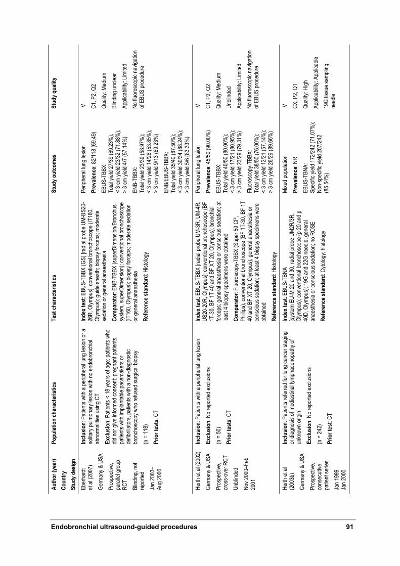
Auth
or (y
ear)
Coun
try
Stud
y des
ign
Popu
latio
n ch
arac
teris
tics
Test
char
acte
ristic
s St
udy o
utco
mes
St
udy q
ualit
y
Eber
hard
t et
al (2
007)
Ge
rman
y & U
SA
Pros
pecti
ve,
para
llel g
roup
RC
T Bl
inding
, not
repo
rted
Jan 2
003–
Au
g 200
6
Inclu
sion:
Pati
ents
with
a per
ipher
al lun
g les
ion or
a so
litary
pulm
onar
y les
ion w
ith no
endo
bron
chial
ab
norm
alitie
s usin
g CT
Exclu
sion:
Pati
ents
< 18
year
s of a
ge; p
atien
ts wh
o did
nor g
ive in
forme
d con
sent;
preg
nant
patie
nts,
patie
nts w
ith im
planta
ble pa
cema
kers
or
defib
rillato
rs, pa
tients
with
a no
n-dia
gnos
tic
bron
chos
copy
who
refus
ed su
rgica
l biop
sy
(n =
118)
Pr
ior t
ests
: CT
Inde
x tes
t: EB
US-T
BBX
(GS)
[rad
ial pr
obe U
M-BS
20-
26R,
Olym
pus];
conv
entio
nal b
ronc
hosc
ope (
IT16
0, Ol
ympu
s); gu
ide sh
eath;
biop
sy fo
rceps
; mod
erate
se
datio
n or g
ener
al an
aesth
esia
Com
para
tor:
ENB-
TBBX
(sup
erDi
mens
ion/B
ronc
hus
syste
m, su
perD
imen
sion)
; con
venti
onal
bron
chos
cope
(IT
160,
Olym
pus);
biop
sy fo
rceps
; mod
erate
seda
tion
or ge
nera
l ana
esthe
sia
Refe
renc
e sta
ndar
d: H
istolo
gy
Perip
hera
l lung
lesio
n Pr
evale
nce:
82/11
8 (69
.49)
EBUS
-TBB
X:
Total
yield
27/39
(69.2
3%);
<
3 cm
yield
23/32
(71.8
8%);
> 3 c
m yie
ld 4/7
(57.1
4%)
ENB-
TBBX
: To
tal yi
eld 23
/39 (5
8.97%
); <
3 cm
yield
14/26
(53.8
5%);
> 3 c
m yie
ld 9/1
3 (69
.23%
) EN
B/EB
US-T
BBX:
To
tal yi
eld 35
/40 (8
7.50%
); <
3 cm
yield
30/34
(88.2
4%);
> 3 c
m yie
ld 5/6
(83.3
3%)
IV
C1, P
2, Q2
Qu
ality:
Med
ium
Blind
ing un
clear
Ap
plica
bility
: Lim
ited
No flu
oros
copic
navig
ation
of
EBUS
proc
edur
e
Herth
et al
(200
2)
Germ
any &
USA
Pr
ospe
ctive
, cro
ss-o
ver R
CT
Unbli
nded
No
v 200
0–Fe
b 20
01
Inclu
sion:
Pati
ents
with
a per
ipher
al lun
g les
ion
Exclu
sion:
No r
epor
ted ex
clusio
ns
(n =
50)
Prio
r tes
ts: C
T
Inde
x tes
t: EB
US-T
BBX
[radia
l pro
be U
M-3R
, UM-
4R,
US20
-20R
, Olym
pus];
conv
entio
nal b
ronc
hosc
ope (
BF
1T-3
0, BF
1T 40
and B
F XT
20, O
lympu
s); br
onch
ial
force
ps; g
ener
al an
aesth
esia
or co
nscio
us se
datio
n; at
least
4 biop
sy sp
ecim
ens w
ere o
btaine
d Co
mpa
rato
r: Flu
oros
copy
-TBB
X (S
uper
50 C
P,
Philli
ps);
conv
entio
nal b
ronc
hosc
ope (
BF 1T
-30,
BF 1T
40
and B
F XT
20, O
lympu
s); ge
nera
l ana
esthe
sia or
co
nscio
us se
datio
n; at
least
4 biop
sy sp
ecim
ens w
ere
obtai
ned
Refe
renc
e sta
ndar
d: H
istolo
gy
Perip
hera
l lung
lesio
n Pr
evale
nce:
45/50
(90.0
0%)
EBUS
-TBB
X:
Total
yield
40/50
(80.0
0%);
< 3 c
m yie
ld 17
/21 (8
0.95%
); >
3 cm
yield
23/29
(79.3
1%)
Fluor
osco
py-T
BBX:
To
tal yi
eld 38
/50 (7
6.00%
); <
3 cm
yield
12/21
(57.1
4%);
> 3 c
m yie
ld 26
/29 (8
9.66%
)
IV
C1, P
2, Q2
Qu
ality:
Med
ium
Unbli
nded
Ap
plica
bility
: Lim
ited
No flu
oros
copic
navig
ation
of
EBUS
proc
edur
e
Herth
et al
(2
003b
) Ge
rman
y & U
SA
Pros
pecti
ve,
cons
ecuti
ve
patie
nt se
ries
Jan 1
999–
Ja
n 200
0
Inclu
sion:
Pati
ents
refer
red f
or lu
ng ca
ncer
stag
ing
or di
agno
sis of
med
iastin
al lym
phad
enop
athy o
f un
know
n orig
in Ex
clusio
n: N
o rep
orted
exclu
sions
(n
= 24
2)
Prio
r tes
t: CT
Inde
x tes
t: EB
US-T
BNA
[sy
stem
EU-M
20 an
d 30,
radia
l pro
be U
M2R/
3R,
Olym
pus);
conv
entio
nal b
ronc
hosc
ope (
p 20 a
nd p
40D,
Olym
pus);
19G
and 2
2G ne
edle;
gene
ral
anae
sthes
ia or
cons
cious
seda
tion;
no R
OSE
Refe
renc
e sta
ndar
d: C
ytolog
y; his
tolog
y
Mixe
d pop
ulatio
n Pr
evale
nce:
NR
EBUS
-TBN
A:
Spec
ific yi
eld 17
2/242
(71.0
7%);
Non-
spec
ific yi
eld 20
7/242
(8
5.54%
)
IV
CX, P
2, Q1
Qu
ality:
High
Ap
plica
bility
: App
licab
le 19
G tis
sue s
ampli
ng
need
le
Endobronchial ultrasound-guided procedures 91

Auth
or (y
ear)
Coun
try
Stud
y des
ign
Popu
latio
n ch
arac
teris
tics
Test
char
acte
ristic
s St
udy o
utco
mes
St
udy q
ualit
y
Herth
(200
4)
Germ
any &
USA
Pr
ospe
ctive
, pa
ralle
l gro
up
RCT
Blind
ing of
pa
tholog
ists
Jun 2
001–
Ma
r 200
2
Inclu
sion:
Pati
ents
refer
red f
or lu
ng ca
ncer
stag
ing
or di
agno
sis of
med
iastin
al lym
phad
enop
athy o
f un
know
n orig
in (m
edias
tinal
lymph
node
enlar
ged)
Ex
clusio
n: N
o rep
orted
exclu
sions
(n
= 20
0)
Prio
r tes
t: CT
Inde
x tes
t: EB
US-T
BNA
[syste
m EU
-M 20
and 3
0, ra
dial p
robe
UM2
R/3R
, Olym
pus];
conv
entio
nal
bron
chos
cope
(Exc
era a
nd p
40D,
Olym
pus);
22G
need
le; ge
nera
l ana
esthe
sia or
cons
cious
seda
tion;
no
ROSE
Co
mpa
rato
r: TB
NA 22
G ne
edle;
gene
ral a
naes
thesia
or
cons
cious
seda
tion;
no R
OSE
Re
fere
nce s
tand
ard:
Cyto
logy,
histol
ogy
Mixe
d pop
ulatio
n Pr
evale
nce:
NR
EBUS
-TBN
A:
Spec
ific yi
eld 77
/100 (
77.00
%);
Non-
spec
ific yi
eld 85
/100 (
85.00
%)
Conv
entio
nal T
BNA:
Sp
ecific
yield
63/10
0 (63
.00%
); No
n-sp
ecific
yield
66/10
0 (66
.00%
)
IV
C1, P
1, Q1
Qu
ality:
High
Ap
plica
bility
: App
licab
le
Herth
et al
(200
5)
Germ
any
Pros
pecti
ve,
cross
-ove
r RCT
Bl
inding
of
patho
logist
s Ja
n 200
2–
Jan 2
004
Inclu
sion:
Pati
ents
refer
red f
or lu
ng ca
ncer
stag
ing
or di
agno
sis of
a m
edias
tinal
lymph
aden
opath
y of
unkn
own o
rigin
(med
iastin
al lym
ph no
des >
1 cm
) Ex
clusio
n: P
atien
ts wi
th en
large
d lym
ph no
des
exclu
ding 2
R, 2L
, 3, 4
R, 4L
, 7, 1
0R or
10L
(n =
160)
Pr
ior t
est:
CT
Inde
x tes
t: EB
US-T
BNA
[sy
stem
EU-M
20 an
d 30,
radia
l pro
be U
M2R/
3R,
Olym
pus];
conv
entio
nal b
ronc
hosc
ope (
p 20 a
nd p
40D,
Olym
pus);
22G
need
le; co
nscio
us se
datio
n; no
RO
SE
Com
para
tor:
EUS-
FNA
[FU3
6 Pen
tax or
UC
30P
Olym
pus];
22G
need
le; co
nscio
us se
datio
n; no
ROS
E Re
fere
nce s
tand
ard:
Cyto
logy,
histol
ogy
Mixe
d pop
ulatio
n Pr
evale
nce:
NR
EBUS
-TBN
A/ E
US-F
NA:
Spec
ific yi
eld 15
1/160
(94.3
8%);
Non-
spec
ific yi
eld 15
5/160
(9
6.88%
) EB
US-T
BNA:
Sp
ecific
yield
137/1
60 (8
5.63%
); No
n-sp
ecific
yield
142/1
60
(88.7
5%)
EUS-
FNA:
Sp
ecific
yield
121/1
60 (7
5.63%
); No
n-sp
ecific
yield
126/1
60
(78.7
5%)
IV
C1, P
2, Q1
Qu
ality:
High
Ap
plica
bility
: Lim
ited
Exclu
sion o
f rele
vant
lymph
no
de st
ation
s
Herth
et al
(2
006a
) Ge
rman
y & U
SA
Pros
pecti
ve,
cons
ecuti
ve
patie
nt se
ries
Jan 2
003–
Ja
n 200
4
Inclu
sion:
Pati
ents
with
solita
ry pu
lmon
ary l
esion
, ca
ndida
tes fo
r diag
nosti
c bro
ncho
scop
y Ex
clusio
n: P
atien
ts wi
th a s
olitar
y pulm
onar
y no
dule
visibl
e usin
g fluo
rosc
opy
(n =
54)
Prio
r tes
ts: C
T, flu
oros
copy
Inde
x tes
t: EB
US-T
BBX
(GS)
[ra
dial p
robe
UM-
3R, U
M-4R
, US2
020R
, Olym
pus];
co
nven
tiona
l bro
ncho
scop
e (BF
T16
0, Ol
ympu
s);
guide
shea
th; br
onch
ial fo
rceps
; gen
eral
anae
sthes
ia or
cons
cious
seda
tion;
4–6 b
iopsy
spec
imen
s wer
e ob
taine
d Re
fere
nce s
tand
ard:
Hist
ology
Perip
hera
l lung
lesio
n Pr
evale
nce:
39/54
(72.2
2%)
EBUS
-TBB
X:
Total
yield
38/54
(70.3
7%)
IV
CX, P
2, Q1
Qu
ality:
High
Ap
plica
bility
: Lim
ited
Enro
lled p
atien
ts wi
th pr
eviou
s fluo
rosc
opy
No flu
oros
copic
navig
ation
of
EBUS
proc
edur
e
92 Endobronchial ultrasound-guided procedures

Auth
or (y
ear)
Coun
try
Stud
y des
ign
Popu
latio
n ch
arac
teris
tics
Test
char
acte
ristic
s St
udy o
utco
mes
St
udy q
ualit
y
Herth
et al
(2
006b
) Ge
rman
y &
Denm
ark
Pros
pecti
ve,
cons
ecuti
ve
patie
nt se
ries
Jun 2
002–
Se
p 200
4
Inclu
sion:
Pati
ents
refer
red f
or lu
ng ca
ncer
stag
ing
or di
agno
sis of
med
iastin
al lym
phad
enop
athy o
f un
know
n orig
in Ex
clusio
n: N
o rep
orted
exclu
sions
(n
= 50
2)
Prio
r tes
ts: X
-ray,
CT
Inde
x tes
t: EB
US-T
BNA
[sy
stem
EU-6
0, Ol
ympu
s]; lin
ear s
cann
ing hy
brid
bron
chos
cope
(XBF
-UC2
60F-
OL8,
Olym
pus);
22G
need
le; m
oder
ate se
datio
n and
loca
l ana
esthe
sia or
ge
nera
l ana
esthe
sia; n
o ROS
E; 2
aspir
ates p
er no
de
were
obtai
ned
Refe
renc
e sta
ndar
d: C
ytolog
y, his
tolog
y, cli
nical/
radio
logica
l follo
w-up
Mixe
d pop
ulatio
n Pr
evale
nce:
493/5
02 (9
8.21%
) EB
US-T
BNA:
Sp
ecific
yield
: 470
/502 (
93.63
%);
Non-
spec
ific yi
eld: 4
76/50
2 (9
4.82%
)
IV
CX, P
1, Q1
Qu
ality:
High
Ap
plica
bility
: App
licab
le
Kiku
chi e
t al
(200
4)
Japa
n Di
recti
on un
clear
, no
n-co
nsec
utive
pa
tient
serie
s De
c 200
2–
Jul 2
003
Inclu
sion:
Pati
ents
with
a per
ipher
al pu
lmon
ary
lesion
(< 30
mm
in me
an di
amete
r) no
t visi
ble us
ing
bron
chos
copy
Ex
clusio
n: N
o rep
orted
exclu
sions
(n
= 24
) Pr
ior t
ests
: CT
Inde
x tes
t: EB
US-T
BBX
(GS)
[sy
stem
EU-M
30S,
radia
l pro
be X
UM-S
20-1
7R,
Olym
pus];
conv
entio
nal b
ronc
hosc
ope (
BF-P
-260
F,
BF-P
-240
, BF-
P-20
0, Ol
ympu
s); gu
ide sh
eath;
br
onch
ial fo
rceps
and b
ronc
hial b
rush
ing; lo
cal
anae
sthes
ia; cu
rette
and f
luoro
scop
y sup
port
Refe
renc
e sta
ndar
d: C
ytolog
y, his
tolog
y, cli
nical/
radio
logica
l follo
w-up
Perip
hera
l lung
lesio
n Pr
evale
nce:
18/24
(75.0
0%)
EBUS
-TBB
X:
Total
yield
14/24
(58.3
3%);
< 3 c
m yie
ld 14
/24 (5
8.33%
)
IV
CX, P
2, Q1
Qu
ality:
High
Ap
plica
bility
: App
licab
le
Kras
nik et
al
(200
3)
Denm
ark
Dire
ction
uncle
ar,
non-
cons
ecuti
ve
patie
nt se
ries
Unbli
nded
Inclu
sion:
Pati
ents
refer
red f
or lu
ng ca
ncer
stag
ing,
diagn
osis
of me
diasti
nal/h
ilar ly
mpha
deno
pathy
of
unkn
own o
rigin
or as
sess
ment
of me
diasti
nal
recu
rrenc
e of c
arcin
oma
Exclu
sion:
No r
epor
ted ex
clusio
ns
(n =
11)
Prio
r tes
ts: C
T, br
onch
osco
py
Inde
x tes
t: EB
US-T
BNA
[sy
stem
EU-C
60, O
lympu
s); lin
ear s
cann
ing hy
brid
bron
chos
cope
(XBF
-UC4
0P, O
lympu
s); 22
G ne
edle;
ge
nera
l ana
esthe
sia
Refe
renc
e sta
ndar
d: C
ytolog
y, his
tolog
y
Mixe
d pop
ulatio
n Pr
evale
nce:
11/11
(100
.00%
) EB
US-T
BNA:
No
n-sp
ecific
yield
27/33
(81.8
2%);
TP 10
; FN
1; TN
0; F
P 0;
Sn
90.91
%; N
PV 0.
00%
III-2
CX, P
2, Q3
Qu
ality:
Low
Unbli
nded
Ina
dequ
ate re
feren
ce
stand
ard
Appli
cabil
ity: A
pplic
able
Patie
nts as
sess
ed fo
r tum
our r
ecur
renc
e
Endobronchial ultrasound-guided procedures 93

Auth
or (y
ear)
Coun
try
Stud
y des
ign
Popu
latio
n ch
arac
teris
tics
Test
char
acte
ristic
s St
udy o
utco
mes
St
udy q
ualit
y
Kurim
oto et
al
(200
4)
Japa
n Pr
ospe
ctive
, co
nsec
utive
pa
tient
serie
s Ma
y 200
1–
Nov 2
002
Inclu
sion:
Pati
ents
with
a soli
tary p
ulmon
ary l
esion
Ex
clusio
n: N
o rep
orted
exclu
sions
(n
= 15
0)
Prio
r tes
ts: X
-ray,
CT
Inde
x tes
t: EB
US-T
BBX
(GS)
[sy
stem
EU-M
30, r
adial
prob
e UM-
S20-
20R,
Olym
pus];
co
nven
tiona
l bro
ncho
scop
e (BF
1T-3
0, 40
or 24
0R,
Olym
pus);
guide
shea
th; br
onch
ial fo
rceps
and
bron
chial
brus
hing;
uncle
ar an
aesth
esia;
cure
tte an
d flu
oros
copy
supp
ort; a
t leas
t 1 bi
opsy
spec
imen
was
ob
taine
d Re
fere
nce s
tand
ard:
Cyto
logy,
histol
ogy,
,clini
cal/ra
diolog
ical fo
llow-
up, o
ther e
xami
natio
ns
Perip
hera
l lung
lesio
n Pr
evale
nce:
101/1
50 (6
7.33%
) EB
US-T
BBX:
To
tal yi
eld 11
6/150
(77.3
3%);
< 3 c
m yie
ld 92
/124 (
74.19
%);
> 3 c
m yie
ld 24
/26 (9
2.31%
)
IV
CX, P
2, Q1
Qu
ality:
High
Ap
plica
bility
: App
licab
le
Paon
e et a
l (2
005)
Ita
ly Pr
ospe
ctive
, pa
ralle
l gro
up
RCT
Blind
ing of
pa
tholog
ists
Jan 2
001–
Se
p 200
3
Inclu
sion:
Pati
ents
with
a per
ipher
al lun
g les
ion
Exclu
sion:
Pati
ents
who w
ere <
18 ye
ars o
f age
, ou
tpatie
nts, d
id no
t give
infor
med c
onse
nt, di
d not
acco
mplis
h a co
mplet
e foll
ow-u
p, did
not a
ccep
t ra
ndom
isatio
n, un
derw
ent lu
ng su
rger
y befo
re
bron
chos
copy
, prim
ary l
esion
at an
other
site,
lung
les
ion di
sapp
eare
d at fo
llow-
up, lo
st to
follow
-up
(n =
206)
Pr
ior t
ests
: CT
Inde
x tes
t: EB
US-T
BBX
[sy
stem
EU-M
30 w
ith ra
dial p
robe
, Olym
pus];
co
nven
tiona
l bro
ncho
scop
e (BF
-B3 o
r BF-
T20,
Olym
pus);
bron
chial
force
ps; lo
cal a
naes
thesia
; at
least
5 biop
sy sp
ecim
ens w
ere o
btaine
d Co
mpar
ator:
TBBX
, targ
et les
ion lo
calis
ed by
prior
CT;
co
nven
tiona
l bro
ncho
scop
e (BF
-B3 o
r BF-
T20,
Olym
pus);
bron
chial
force
ps; lo
cal a
naes
thesia
; at
least
5 biop
sy sp
ecim
ens w
ere o
btaine
d Re
fere
nce s
tand
ard:
Cyto
logy,
histol
ogy,
clinic
al/ra
diolog
ical fo
llow-
up
Perip
hera
l lung
lesio
n Pr
evale
nce:
144/2
06 (6
9.90%
) EB
US-T
BBX:
To
tal yi
eld 66
/87 (7
5.86%
); <
3 cm
yield
35/47
(74.4
7%);
> 3 c
m yie
ld 33
/40 (8
2.50%
) TB
BX:
Total
yield
62/11
9 (52
.10%
); <
3 cm
yield
18/58
(31.0
3%);
> 3 c
m yie
ld 47
/61 (7
7.05%
)
IV
C1, P
2, Q1
Qu
ality:
High
Ap
plica
bility
: Lim
ited
No flu
oros
copic
navig
ation
of
EBUS
proc
edur
e
Plat
et al
(200
6)
Belgi
um
Pros
pecti
ve,
cons
ecuti
ve
patie
nt se
ries
Jan 2
003–
Ju
n 200
4
Inclu
sion:
Pati
ents
refer
red f
or lu
ng ca
ncer
stag
ing
or di
agno
sis of
med
iastin
al lym
phad
enop
athy o
f un
know
n orig
in Ex
clusio
n: N
o rep
orted
exclu
sions
(n
= 33
) Pr
ior t
ests
: CT,
PET
Inde
x tes
t: EB
US-T
BNA
[sy
stem
EU-M
20, r
adial
prob
e UM-
BS20
-26R
, Ol
ympu
s); co
nven
tiona
l bro
ncho
scop
e (Ex
cera
, Ol
ympu
s); lo
cal a
naes
thesia
and c
onsc
ious s
edati
on;
no R
OSE;
4–6 p
unctu
res w
ere o
btaine
d Re
fere
nce s
tand
ard:
Cyto
logy,
histol
ogy,
clinic
al/ra
diolog
ical fo
llow-
up
Mixe
d pop
ulatio
n Pr
evale
nce:
27/33
(81.8
2%)
EBUS
-TBN
A:
Non-
spec
ific yi
eld
27/33
(81.8
2%)
IV
CX, P
1, Q1
Qu
ality:
High
Ap
plica
bility
: App
licab
le
94 Endobronchial ultrasound-guided procedures

Auth
or (y
ear)
Coun
try
Stud
y des
ign
Popu
latio
n ch
arac
teris
tics
Test
char
acte
ristic
s St
udy o
utco
mes
St
udy q
ualit
y
Rinto
ul et
al (2
005)
UK
Di
recti
on un
clear
, no
n-co
nsec
utive
pa
tient
serie
s Un
blind
ed
Inclu
sion:
Pati
ents
with
know
n or s
uspe
cted l
ung
canc
er an
d med
iastin
al lym
phad
enop
athy o
f > 1
cm
or pa
ratra
chea
l or p
arab
ronc
hial m
asse
s, Ex
clusio
n: P
atien
ts we
re ex
clude
d fro
m an
alysis
ba
sed o
n ima
ging r
esult
s (n
= 18
) Pr
ior t
ests
: CT
Inde
x tes
t: EB
US-T
BNA/
EUS-
FNA
[sy
stem
EU-C
2000
, Olym
pus];
linea
r sca
nning
hybr
id br
onch
osco
pe (X
BF-U
C260
F-OL
8, Ol
ympu
s); 22
G ne
edle.
EUS
-FNA
(GF-
UC24
0P-A
L5, O
lympu
s); 22
G ne
edle.
Loca
l ana
esthe
sia an
d sed
ation
; no R
OSE;
2–3 n
eedle
pass
es w
ere o
btaine
d.
Refe
renc
e sta
ndar
d: C
ytolog
y, his
tolog
y, cli
nical/
radio
logica
l follo
w-up
NSCL
C St
aging
Pr
evale
nce:
13/18
(72.2
2%)
EBUS
-TBN
A/EU
S-FN
A:
TP 11
; FN
2; TN
5; F
P 0;
Sn
84.62
%; N
PV 71
.43%
III-2
CX, P
1, Q3
Qu
ality:
Low
Unbli
nded
Ina
dequ
ate re
feren
ce
stand
ard
Appli
cabil
ity: A
pplic
able
Shira
kawa
et al
(2
004)
Ja
pan
Rand
omise
d pa
tient
serie
s wi
th his
torica
l co
ntrol
Unbli
nded
Ja
n 200
1–
Dec 2
001
Inclu
sion:
Pati
ents
with
a per
ipher
al lun
g les
ion an
d no
rmal
visibl
e airw
ays
Exclu
sion:
Pati
ents
who w
ere n
ot ra
ndom
ised t
o EB
US, p
atien
ts wh
o did
not u
nder
go E
BUS,
patie
nts
with
spon
taneo
us bl
eedin
g, pa
tients
with
no fin
al dia
gnos
is (n
= 49
) Pr
ior t
est:
X-ra
y
Inde
x tes
t: EB
US-T
BBX
(GS)
[ra
dial p
robe
UM-
3R, U
M-4R
, US-
20-2
0R, O
lympu
s];
conv
entio
nal b
ronc
hosc
ope (
BS 1T
-240
R, O
lympu
s);
guide
shea
th; br
onch
ial fo
rceps
and b
ronc
hial
brus
hing;
uncle
ar an
aesth
esia;
fluor
osco
py an
d cur
ette
supp
ort
Com
para
tor:
Fluor
osco
py-T
BBX;
uncle
ar de
tails
Refe
renc
e sta
ndar
d: C
ytolog
y, his
tolog
y, cli
nical/
radio
logica
l follo
w-up
, othe
r exa
mina
tions
Perip
hera
l lung
lesio
n Pr
evale
nce:
24/49
(48.9
8%)
EBUS
-TBB
X:
TP 17
; FN
7; TN
25; F
P 0;
Sn 70
.83%
; NPV
78.13
%
Fluor
osco
py-T
BBX:
TP
16; F
N 7;
TN 19
; FP
0; Sn
69.57
%; N
PV 73
.08%
III-3
C1, P
2, Q3
Qu
ality:
low
Inade
quate
refer
ence
sta
ndar
d Un
blind
ed
Uncle
ar co
mpar
ator d
etails
Ap
plica
bility
: App
licab
le
Vilm
ann e
t al
(200
5)
Denm
ark
Pros
pecti
ve, n
on-
cons
ecuti
ve
patie
nt se
ries
Blind
ing, n
ot re
porte
d
Inclu
sion:
Pati
ents
with
know
n or s
uspe
cted l
ung
canc
er
Exclu
sion:
No p
atien
ts we
re ex
clude
d (n
= 33
) Pr
ior t
ests
: CT,
PET
, TBN
A, T
TNA
Inde
x tes
t: EB
US-T
BNA
[sy
stem
EU-C
60, O
lympu
s]; lin
ear s
cann
ing hy
brid
bron
chos
cope
(XBF
-UC4
0P, O
lympu
s); 22
G ne
edle;
ge
nera
l ana
esthe
sia; n
o ROS
E; nu
mber
of as
pirate
s de
termi
ned b
y mac
rosc
opic
appe
aran
ce of
each
sa
mple
Com
para
tor:
EUS-
FNA
[G
F-UC
T160
, Olym
pus];
22G
need
le; ge
nera
l an
aesth
esia;
no R
OSE
Refe
renc
e sta
ndar
d: C
ytolog
y, his
tolog
y, cli
nical/
radio
logica
l follo
w-up
Mixe
d pop
ulatio
n Pr
evale
nce:
27/33
(81.8
2%)
EBUS
-TBN
A/ E
US-F
NA:
Non-
spec
ific yi
eld 33
/33
(100
.00%
);
TP 20
; FN
0; TN
8; F
P 0;
Sn
100.0
0%; N
PV 10
0.00%
EB
US-T
BNA:
No
n-sp
ecific
yield
32/33
(96.9
7%);
TN 17
; FN
3; TN
8; F
P 0;
Sn
85.00
%; N
PV 72
.73%
EU
S-FN
A:
Non-
spec
ific yi
eld
32/33
(96.9
7%);
TN 16
; FN
4; TN
8;
FP 0;
Sn 8
0.00%
; NPV
66.67
%
III-2
C1, P
2, Q3
Qu
ality:
Poo
r Ina
dequ
ate re
feren
ce
stand
ard
Unbli
nded
Ap
plica
bility
: Lim
ited
Uncle
ar ly
mph n
ode s
tatus
En
rolle
d pati
ents
who h
ad
unde
rgon
e pre
vious
TBN
A or
TTN
A
Endobronchial ultrasound-guided procedures 95

Auth
or (y
ear)
Coun
try
Stud
y des
ign
Popu
latio
n ch
arac
teris
tics
Test
char
acte
ristic
s St
udy o
utco
mes
St
udy q
ualit
y
Yasu
fuku e
t al
(200
4)
Japa
n Pr
ospe
ctive
, non
-co
nsec
utive
pa
tient
serie
s Un
blind
ed
Mar 2
002–
Se
p 200
3
Inclu
sion:
Pati
ents
with
know
n or s
uspe
cted l
ung
canc
er an
d med
iastin
al an
d/or h
ilar
lymph
aden
opath
y of >
1 cm
Exclu
sion:
No r
epor
ted ex
clusio
ns
(n =
70)
Prio
r tes
ts: X
-ray,
CT
Inde
x tes
t: EB
US-T
BNA
[sy
stem
EU-C
2000
, Olym
pus];
linea
r sca
nning
hybr
id br
onch
osco
pe (X
BF-U
C260
F-OL
8, Ol
ympu
s); 22
G ne
edle;
loca
l ana
esthe
sia an
d sed
ation
; ROS
E; 1–
5 ne
edle
pass
es w
ere o
btaine
d Re
fere
nce s
tand
ard:
Cyto
logy,
histol
ogy,
clinic
al/ra
diolog
ical fo
llow-
up
NSCL
C sta
ging
Prev
alenc
e: NR
EB
US-T
BNA:
No
n-sp
ecific
yield
68/70
(97.1
4%);
TP 45
; FN
0; TN
23; F
P 0;
Sn
100.0
0%; N
PV 10
0.00%
III-2
CX, P
1, Q3
Qu
ality:
Low
Unbli
nded
Ina
dequ
ate re
feren
ce
stand
ard
Appli
cabil
ity: A
pplic
able
Yasu
fuku e
t al
(200
5)
Japa
n Pr
ospe
ctive
, non
-co
nsec
utive
pa
tient
serie
s Un
blind
ed
Jun 2
002–
Ap
r 200
4
Inclu
sion:
Pati
ents
with
know
n or s
uspe
cted l
ung
canc
er an
d med
iastin
al an
d/or h
ilar
lymph
aden
opath
y of >
1 cm
Ex
clusio
n: P
atien
ts wi
th a f
inal d
iagno
sis of
a be
nign d
iseas
e; pa
tients
with
a fin
al dia
gnos
is of
malig
nant
disea
se ot
her t
han N
SCLC
, pati
ents
with
exten
sive N
2, N3
dise
ase o
n CT
(n =
108)
Pr
ior t
ests
: CT
Inde
x tes
t: EB
US-T
BNA
[sy
stem
EU-C
2000
, Olym
pus];
linea
r sca
nning
hybr
id br
onch
osco
pe (X
BF-U
C260
F-OL
8, Ol
ympu
s); 22
G ne
edle;
loca
l ana
esthe
sia an
d sed
ation
; ROS
E; 1–
5 ne
edle
pass
es w
ere o
btaine
d Re
fere
nce s
tand
ard:
Cyto
logy,
histol
ogy;
clinic
al/ra
diolog
ical fo
llow-
up
NSCL
C sta
ging
Prev
alenc
e: NR
EB
US-T
BNA:
No
n-sp
ecific
yield
105/1
08
(97.2
2%);
TP 70
; FN
4; TN
34; F
P 0;
Sn 94
.59%
; NPV
89.47
%
III-2
CX, P
1, Q2
Qu
ality:
Low
Unbli
nded
Ina
dequ
ate re
feren
ce
stand
ard
Inade
quate
data
repo
rting
Appli
cabil
ity: A
pplic
able
Yasu
fuku e
t al
(200
6)
Japa
n Pr
ospe
ctive
, co
nsec
utive
pa
tient
serie
s CT
& P
ET
blind
ed
Dec 2
003–
Mar
2005
Inclu
sion:
Pati
ents
with
know
n or s
uspe
cted l
ung
canc
er
Exclu
sion:
Pati
ents
who w
ere n
ot fur
ther e
valua
ted
(n =
102)
Pr
ior t
ests
: CT,
PET
, othe
r tes
ts
Inde
x tes
t: EB
US-T
BNA
[sy
stem
EU-C
2000
, Olym
pus];
linea
r sca
nning
hybr
id br
onch
osco
pe (X
BF-U
C260
F-OL
8, Ol
ympu
s); 22
G ne
edle,
con
sciou
s sed
ation
; ROS
E; m
axim
um of
5 ne
edle
pass
es w
ere o
btaine
d Co
mpar
ator:
CT (L
ight S
peed
, GE
medic
al sy
stems
), mu
ltidec
tor ro
w, in
jectio
n of c
ontra
st ma
terial
Co
mpa
rato
r: PE
T (P
ET A
dvan
ce N
xi, G
E me
dical
syste
ms),
300 M
bq in
jectio
n of F
DG
Refe
renc
e sta
ndar
d: C
ytolog
y, his
tolog
y, cli
nical/
radio
logica
l follo
w-up
Mixe
d pop
ulatio
n Pr
evale
nce:
26/10
2 (25
.49%
) EB
US-T
BNA:
TP
24; F
N 2;
TN 76
; FP
0; Sn
92
.31%
; NPV
97.44
%
CT:
TP 20
; FN
6; TN
42; F
P 34
; Sn
76.92
%; N
PV 87
.50%
PE
T:
TP 20
; FN
5; TN
54; F
P 23
; Sn
80.00
%; N
PV 91
.53%
III-2
C1, P
2, Q3
Qu
ality:
Low
Ina
dequ
ate bl
inding
Ina
dequ
ate re
feren
ce
stand
ard
Appli
cabil
ity: L
imite
d Inc
luded
patie
nts w
ith
nega
tive l
ymph
aden
opath
y
Abbr
eviat
ions:
CT, c
ompu
ted to
mogr
aphy
; EBU
S, en
dobr
onch
ial ul
traso
und;
EBUS
-TBN
A, en
dobr
onch
ial ul
traso
und t
rans
bron
chial
need
le as
pirati
on; F
N, fa
lse ne
gativ
e; FP
, false
posit
ive; N
PV, n
egati
ve pr
edict
ive va
lue; N
R,
not r
epor
ted; N
SCLC
, non
-small
cell l
ung c
ance
r; PE
T, po
sition
emiss
ion to
mogr
aphy
; ROS
E, ra
pid on
site
evalu
ation
; Sn,
sens
itivity
; TBB
X, tr
ansb
ronc
hial b
iopsy
; TBN
A, tr
ansb
ronc
hial n
eedle
aspir
ation
; TP,
true
posit
ive;
TTNA
, tran
sthor
acic
need
le as
pirati
on
96 Endobronchial ultrasound-guided procedures

Endobronchial ultrasound-guided procedures 97
Appendix E Excluded studies
Excluded: inadequate data reporting
Asahina H, Yamazaki K, Onodera Y et al. 2005. “Transbronchial biopsy using endobronchial ultrasonography with a guide sheath and virtual bronchoscopic navigation”. Chest 128: 1761–176.
Baba M, Sekine Y, Suzuki M et al. 2002. “Correlation between endobronchial ultrasonography (EBUS) images and histologic findings in normal and tumor-invaded bronchial wall”. Lung Cancer 35: 65–71.
Herth FJF, Ernst A, Schulz M et al. 2003a. “Endobronchial ultrasound reliably differentiates between airway infiltration and compression by tumor”. Chest 123: 458–462.
Kotsianos-Hermle D, Scherr M, Wirth S et al. 2007. “Visualization of bronchial lesions using multidetector CT and endobronchial ultrasound (EBUS)”. Eur J Med Res 12: 84–89.
Kurimoto N, Murayama M, Morita K et al. 1998. “Clinical applications of endobronchial ultrasonography in lung diseases”. Endosc 30 Suppl 1: A8–12.
Miyazu Y, Miyazawa T, Kurimoto N et al. 2002. “Endobronchial ultrasonography in the assessment of centrally located early-stage lung cancer before photodynamic therapy”. Am J Respir Crit Care Med 165: 832–837.
Ono R, Suemasu K, Matsunaka T 1993. “Bronchoscopic ultrasonography in the diagnosis of lung cancer”. Japan Clin Oncol 23: 34–40.
Takahashi H, Sagawa M, Sato M et al. 2003. “A prospective evaluation of transbronchial ultrasonography for assessment of depth of invasion in early bronchogenic squamous cell carcinoma”. Lung Cancer 42: 43–49.
Takemoto Y, Kawahara M, Ogawara M et al. 2000. “Ultrasound-guided flexible bronchoscopy for the diagnosis of tumor invasion to the bronchial wall and mediastinum”. J Bronchol 7: 127–132.
Wallace WAH, Monaghan HM, Salter DM et al. 2007. “Endobronchial ultrasound-guided fine-needle aspiration and liquid-based thin-layer cytology”. J Clin Pathol 60: 388–391.
Excluded: foreign language
Becker HD 1996. “Endobronchial ultrasound—A new perspective in bronchoscopy”. Ultraschall in der Medizin 17: 106–112.
Becker HD, Messerschmidt E, Ott S 1997. “Endobronchial ultrasound”. Atemwegs und Lungenkrankheiten 23: 218–224.
Faber J, Pierard P, Prigogine T et al. 2006. “Endobronchial ultrasound and PET positive mediastinal lymph nodes”. Rev malad respirat 23: 37–42.

98 Endobronchial ultrasound-guided procedures
Harada M 2005. “Recent advances in photodynamic therapy; an expanding role in non-small cell lung cancer”. Japan J Lung Cancer 45: 687–692.
Herth FJF, Hecker E, Hoffmann H et al. 2002. “Endobronchial ultrasound for local tumour and lymph node staging in patients with centrally growing lung cancer”. Ultraschall in der Medizin 23: 251–255.
Hurter T, Hanrath P 1990. “Endobronchial sonography in the diagnosis of pulmonary and mediastinal tumours”. Deutsche Medizinische Wochenschrift 115: 1899–1905.
Kurimoto N, Murayama M, Kohama Y et al. 1997. “Clinical application of endobronchial ultrasound in the diagnosis of bronchial and peri-bronchial diseases”. Japan J Clin Radiol 42: 143–150.
Kurimoto N, Hayashi K, Murayama M et al. 1999. “Endobronchial ultrasonography for lung cancer”. Nippon Geka Gakkai zasshi 100: 724–728.
Matsuo K, Watanabe Y 2002. “Endobronchial ultrasonography for diagnosis of lung neoplasms”. Nippon rinsho .Japan J Clin Med 60 Suppl 5: 191–194.
Monso E, Andreo F, Rosell A et al. 2007. “Usefulness of endobronchial ultrasonography with real-time needle aspiration for lung cancer staging”. Medicina Clinica 128: 481–485.
Morocz E, Strausz J 2002. “Endobronchial ultrasonography”. Orvosi hetilap 143: 2875–2879.
Morocz E, Strausz J 2004. “Endobronchial ultrasonography in post-intubation tracheal stenosis”. Orvosi hetilap 145: 1507–1510.
Ono R 1996. “Bronchoscopic brachytherapy: development of bronchoscopic ultrasonography and brachytherapy for early stage lung cancer of hilar type”. Nippon rinsho Japan J Clin Med 54: 1337–1342.
Postmus P 2002. “Endobronchial detection and treatment of pre-neoplastic lesions”. Revue de Pneumologie Clinique 58: 3S22–3S24.
Tsurupa DI, Sadchikova IA, Blashentseva SA et al. 1995. “The interpretation of the data from the ultrasonic examination of patients with acute and chronic suppurative lung and pleural diseases”. Vestnik Khirurgii Imeni I.I.Grekova 154: 17–21.
Votruba J, Stastny B 2002. “Endobronchial ultrasonography”. Endoskopie 11: 23–26.
Woelke K, Rausch V, Granetzny A 2006. “Mediastinal lymph node staging with endobronchial ultrasound (EBUS) and mediastinoscopy—A comparison of both methods”. Atemwegs- und Lungenkrankheiten 32: 343–344.
Excluded: wrong utilisation
Chao TY, Lie CH, Chung YH et al. 2006. “Differentiating peripheral pulmonary lesions based on images of endobronchial ultrasonography”. Chest 130: 1191–1197.

Endobronchial ultrasound-guided procedures 99
Herth FJF, Becker HD, LoCicero J et al. 2002. “Endobronchial ultrasound in therapeutic bronchoscopy”. Eur Respir J 20: 118–121.
Herth FJF, Becker HD, LoCicero III J et al. 2003. “Endobronchial ultrasound improves classification of suspicious lesions detected by autofluorescence bronchoscopy”. J Bronchol 10: 249–252.
Kurimoto N, Murayama M, Yoshioka S et al. 2002. “Analysis of the internal structure of peripheral pulmonary lesions using endobronchial ultrasonography”. Chest 122: 1887–1894.
Okamoto H, Watanabe K, Nagatomo A et al. 2002. “Endobronchial ultrasonography for mediastinal and hilar lymph node metastases of lung cancer”. Chest 121: 1498–1506.
Omori S, Takiguchi Y, Hiroshima K et al. 2002. “Peripheral pulmonary diseases: Evaluation with endobronchial US—Initial experience”. Radiol 224: 603–608.
Excluded: wrong intervention
Diacon AH, Schuurmans MM, Theron J et al. 2005. “Utility of rapid on-site evaluation of transbronchial needle aspirates” (structured abstract) Respiration 72: 182–188.
Jakob H, Lorenz J, Clement T et al. 1990. “Mediastinal lymph node staging with transesophageal echography in cancer of the lung”. Eur J Cardiothor Surg 4: 355–358.
Khoo KL, Ho KY, Nilsson B et al. 2006. “EUS-guided FNA immediately after unrevealing transbronchial needle aspiration in the evaluation of mediastinal lymphadenopathy: A prospective study”. Gastroint Endosc 63: 215–220.
Obata K, Ueki J, Dambara T et al. 1999. “Repeated ultrasonically guided needle biopsy of small subpleural nodules”. Chest 116: 1320–1324.
Singh MM, Gupta RK, Gupta RK et al. 1987. “Ultrasonically guided fine needle aspiration biopsy (FNAB) of intrathoracic lesions”. Indian J Chest Dis Allied Sci 29: 81–89.
Sortini D, Feo CV, Carrella G et al. 2003. “Thoracoscopic localization techniques for patients with a single pulmonary nodule and positive oncological anamnesis: A prospective study”. J Laparoendosc Adv Surg Tech–Part A 13: 371–375.
Yuan A, Yang PC, Chang DB et al. 1992. “Ultrasound-guided aspiration biopsy of small peripheral pulmonary nodules”. Chest 101: 926–930.
Exclude: review, letter etc
Esteva H, Becker HD 2005. “Value of fiberoptic bronchoscopy in patients undergoing surgery for solitary pulmonary nodules”. Chest 128: 474
Feller-Kopman D, Lunn W, Ernst A 2005. “Autofluorescence bronchoscopy and endobronchial ultrasound: A practical review”. Ann Thor Surg 80: 2395–2401.
Gambhir SS, Shepherd JE, Shah BD et al. 1998. “Analytical decision model for the cost-effective management of solitary pulmonary nodules”. J Clin Oncol 16: 2113–2125.

100 Endobronchial ultrasound-guided procedures
Gasparini S 2005. “It is time for this 'ROSE' to flower”. Respiration 72: 129–131.
Harewood GC, Wiersema MJ, Edell ES et al. 2002. “Cost-minimization analysis of alternative diagnostic approaches in a modeled patient with non-small cell lung cancer and subcarinal lymphadenopathy (Provisional record)”. Mayo Clin Proceed 77: 155–164.
Herth FJF, Becker HD 2000. “Endobronchial ultrasound of the airways and the mediastinum”. Monaldi Arch Chest Dis 55: 36–44.
Janssen J, Gasparini S, Mehta A 2006. “Endoscopic ultrasound-guided biopsy in the chest”. Eur Respir J 27: 238–239.
Kurimoto N, Miyazawa T 2004. “Endobronchial ultrasonography”. Sem Respirat Critical Care Med 25: 425–431.
Layfield LJ, Bentz JS, Gopez EV 2001. “Immediate on-site interpretation of fine-needle aspiration smears: A cost and compensation analysis”. Cancer 93: 319–322.
Souquet PJ 1998. “Place of transbronchial or transoesophageal endosonography guided aspiration biopsy in the initial management of lung cancer”. Acta Endoscopica 28: 1–5.
Trisolini R, Agli LL, Patelli M 2004. “Conventional vs endobronchial ultrasound-guided transbronchial needle aspiration of the mediastinum”. Chest 126: 1005–1006.
Turner JF 2002. “Endobronchial ultrasound and peripheral pulmonary lesions: Localization and histopathologic correlates using a miniature probe and the flexible bronchoscope”. Chest 122: 1874–1875.
Wang KP 2006. “EUS-guided FNA immediately after unrevealing transbronchial needle aspiration in the evaluation of mediastinal lymphadenopathy: a prospective study”. Gastrointest Endosc 64: 468–469.
Excluded: non-standard/obsolete procedure
Goldberg BB, Steiner RM, Liu JB et al. 1994. “US-assisted bronchoscopy with use of miniature transducer-containing catheters”. Radiol 190: 233–237.
Hurter T, Hanrath P 1992. “Endobronchial sonography: Feasibility and preliminary results”. Thorax 47: 565–567.
Kanoh K, Miyazawa T, Kurimoto N et al. 2005. “Endobronchial ultrasonography guidance for transbronchial needle aspiration using a double-channel bronchoscope”. Chest 128: 388–393.
Shannon JJ, Bude RO, Orens JB et al. 1996. “Endobronchial ultrasound-guided needle aspiration of mediastinal adenopathy”. Am J Respir Crit Care Med 153: 1424–1430.
Excluded: wrong outcome
Herth FJF, Becker HD, Manegold C et al. 2001. “Endobronchial ultrasound (EBUS)—Assessment of a new diagnostic tool in bronchoscopy for staging of lung cancer”. Onkologie 24: 151–154.

Endobronchial ultrasound-guided procedures 101
Excluded: wrong patient population
Herth FJF, Ernst A, Eberhardt R et al. 2006. “Endobronchial ultrasound-guided transbronchial needle aspiration of lymph nodes in the radiologically normal mediastinum”. Eur Respir J 28: 910–914.
Irani S, Hess T, Hofer M et al. 2006. “Endobronchial ultrasonography for the quantitative assessment of bronchial mural structures in lung transplant recipients”. Chest 129: 349–355.
Iwamoto Y, Miyazawa T, Kurimoto N et al. 2004. “Interventional bronchoscopy in the management of airway stenosis due to tracheobronchial tuberculosis”. Chest 126: 1344–1352.
Miyazawa T, Miyazu Y, Iwamoto Y et al. 2004. “Stenting at the flow-limiting segment in tracheobronchial stenosis due to lung cancer”. Am J Respir Crit Care Med 169: 1096–1102.
Nishimura Y, Osugi H, Inoue K et al. 2002. “Bronchoscopic ultrasonography in the diagnosis of tracheobronchial invasion of esophageal cancer”. J Ultrasound Med 21: 49–58.
Osugi H, Nishimura Y, Takemura M et al. 2003. “Bronchoscopic ultrasonography for staging supracarinal esophageal squamous cell carcinoma: Impact on outcome”. World J Surg 27: 590–594.
Wakamatsu T, Tsushima K, Yasuo M et al. 2006. “Usefulness of preoperative endobronchial ultrasound for airway invasion around the trachea: Esophageal cancer and thyroid cancer”. Respiration 73: 651–657.
Wong M, Yasufuku K, Nakajima T et al. 2007. “Endobronchial ultrasound: new insight for the diagnosis of sarcoidosis”. Eur Respir J 29: 1182–1186.
Yang MC, Liu WT, Wang CH et al. 2004. “Diagnostic value of endobronchial ultrasound-guided transbronchial lung biopsy in peripheral lung cancers”. J Formosan Med Assoc = Taiwan yi zhi 103: 124–129.
Excluded: abstract of full paper
Herth FJF, Ernst A, Becker HD 2003. “Conventional vs endobronchial ultrasound (EBUS)-guided transbronchial needle aspiration (TBNA)—a randomised trial”. Eur Respir J 22: 205s.
Exclude: endosonography case report
Herth FJF, Krasnik M, Yasufuku K et al. 2006. “Endobronchial ultrasound-guided transbronchial needle aspiration”. J Bronchol 13: 84–91.
Kramer H, Nieuwenhuis JA, Groen HJM et al. 2004. “Pulmonary tuberculosis diagnosed by esophageal endoscopic ultrasound with fine-needle aspiration”. Int J Tubercul Lung Dis 8: 272–273.

102 Endobronchial ultrasound-guided procedures
Martinez-Monge R, Subtil JC, Lopez-Picazo JM 2006. “Transoesophageal endoscopic-ultrasonography-guided 125I permanent brachytherapy for unresectable mediastinal lymphadenopathy”. Lancet Oncol 7: 781–783.
Miyazu Y, Miyazawa T, Iwamoto Y et al. 2001. “The role of endoscopic techniques, laser-induced fluorescence endoscopy and endobronchial ultrasonography in choice of appropriate therapy for bronchial cancer”. J Bronchol 8: 10–16.
Rosario F, Da Costa JD, Dionisio J et al. 2006. “Endobronchial ultrasound may reveal the presence of previously undiagnosed thyroid carcinoma”. Thyroid 16: 515–516.
Excluded: animal/in vitro study
Kurimoto N, Murayama M, Yoshioka S et al. 1999. “Assessment of usefulness of endobronchial ultrasonography in determination of depth of tracheobronchial tumor invasion”. Chest 115: 1500–1506.
Nakamura Y, Endo C, Sato M et al. 2004. “A new technique for endobronchial ultrasonography and comparison of two ultrasonic probes: Analysis with a plot profile of the image analysis software NIH image”. Chest 126: 192–197.
Yasufuku K, Chhajed PN, Sekine Y et al. 2004. “Endobronchial ultrasound using a new convex probe: a preliminary study on surgically resected specimens”. Oncol Reports 11: 293–296.

Endobronchial ultrasound-guided procedures 103
Appendix F Literature search
Search strategies were used to identify relevant studies of EBUS guided transbronchial sampling procedures for non-small cell lung cancer staging, diagnosis of peripheral lung lesions and the diagnosis of mediastinal/hilar masses. The Medline and EMBASE databases were search using the EMBASE.com interface. The PreMedline database was search using the PubMed interface. The Cochrane Database of Systematic Reviews (CDSR), Database of Abstracts of Reviews of Effects (DARE), Cochrane Central Register of Controlled Trials (CENTRAL), Cochrane Methodology Register (CMR), Health Technology Assessment (HTA), NHS Economic Evaluation Database (NHSEED databases were search using the Cochrane Library interface. The search results for EMBASE.com are presented in Table 58, the results from PubMed are presented in Table 59 and the results from the Cochrane Library search are presented in Table 60.
Table 58 EMBASE.com search results: EBUS procedures for NSCLC staging and diagnosis of peripheral lung, mediastinal and hilar masses (4 June 2007)
Keywords / search history Results 1. 'bronchoscopy'/exp 21888 2. 'bronchoscope'/exp 1069 3. #1 OR #2 22485 4. 'ultrasound'/exp 44242 5. 'echography'/exp 285250 6. #4 OR #5 322345 7. #3 AND #6 626 8. 'endobronchial echography':de 9 9. 'endobronchial ultrasonography':de 14 10. 'endobronchial ultrasound':de 15 11. 'endobronchial ultrasound driven biopsy':de 1 12. 'endobronchial ultrasonography with a guide sheath':de 1 13. 'endobronchial ultrasound guided transbronchial needle aspiration':de 1 14. 'endoscopic transbronchial real time echography guided biopsy':de 1 15. #8 OR #9 OR #10 OR #11 OR #12 OR #13 or #14 39 16. 'endobronchial *3 echography':ti,ab 2 17. 'endobronchial *3 ultrasonogram':ti,ab 2 18. 'endobronchial *3 ultrasound':ab,ti 99 19. 'endobronchial *3 ultrasonography':ab,ti 59 20. 'endobronchial *3 ultra sonography':ab,ti 1 21. 'endobronchial us':ab,ti OR ebus:ab,ti 87 22. #16 OR #17 OR #18 OR #19 OR #20 OR #21 166 23. #7 OR #15 OR #22 704

104 Endobronchial ultrasound-guided procedures
Table 59 PubMed search results for EBUS procedures for NSCLC staging and diagnosis of peripheral lung, mediastinal and hilar masses (7 June 2007)
Keywords / search history Results 1. endobronchial[tiab] AND echography[tiab] 1 2. endobronchial[tiab] AND ultrasonogram[tiab] 3 3. endobronchial[tiab] AND ultrasound[tiab] 97 4. endobronchial[tiab] AND ultrasonography[tiab] 54 5. endobronchial[tiab] AND "ultra sonography"[tiab] 0 6. "endobronchial us"[tiab] OR ebus[tiab] 67 7. #1 or #2 or #3 or #4 or #5 or #6 152 8. #1 or #2 or #3 or #4 or #5 or #6 Limits: MEDLINE 135 9. #7 NOT #8 17
Table 60 Cochrane Library search results for EBUS procedures for NSCLC staging and diagnosis of peripheral lung, mediastinal and hilar masses (4 June 2007)
Keywords / search history Results 1. MeSH descriptor Bronchoscopy explode all trees 368 2. MeSH descriptor Bronchoscopes explode all trees 41 3. #1 OR #2 389 4. MeSH descriptor Endosonography explode all trees 171 5. MeSH descriptor Ultrasonography explode all trees 4808 6. MeSH descriptor Ultrasonography, Interventional explode all trees 331 7. #4 OR #5 OR #6 4808 8. #3 AND #7 8 9. (endobronchial near echography) 0 10. (endobronchial near ultrasonogram) 0 11. (endobronchial near ultrasound) 9 12. (endobronchial near ultrasonography) 3 13. (endobronchial near "ultra sonography") 0 14. "endobronchial us" or ebus 7 15. #9 OR #10 OR #11 OR #12 OR #13 OR #14 12 16. #8 OR #15 16

Endobronchial ultrasound-guided procedures 105
Table 61 HTA websites searched in this review
Australian Safety and Efficacy Register of New Interventional Procedures—Surgical (ASERNIP-S) http://www.surgeons.org/Content/NavigationMenu/Research/ASERNIPS/default.htm Centre for Clinical Effectiveness, Monash University http://www.med.monash.edu.au/healthservices/cce/evidence/
Australia
Health Economics Unit, Monash University http://chpe.buseco.monash.edu.au Austria Institute of Technology Assessment / HTA unit http://www.oeaw.ac.at/ita/e1-3.htm
Agence d’Evaluation des Technologies et des Modes d’Intervention en Santé (AETMIS) http://www.aetmis.gouv.qc.ca/site/index.php?home Institute of Health Economics (IHE) http://www.ihe.ca/index.html Canadian Coordinating Office for Health Technology Assessment (CCHOTA) http://www.ccohta.ca/entry_e.html Canadian Health Economics Research Association (CHERA/ACRES)—Cabot database http://www.mycabot.ca Centre for Health Economics and Policy Analysis (CHEPA), McMaster University http://www.chepa.org Centre for Health Services and Policy Research (CHSPR), University of British Columbia http://www.chspr.ubc.ca Health Utilities Index (HUI) http://www.fhs.mcmaster.ca/hug/index.htm
Canada
Institute for Clinical and Evaluative Studies (ICES) http://www.ices.on.ca Danish Institute for Health Technology Assessment (DIHTA) http://www.dihta.dk/publikationer/index_uk.asp Denmark Danish Institute for Health Services Research (DSI) http://www.dsi.dk/engelsk.html
Finland FINOHTA http://finohta.stakes.fi/EN/index.htm France L’Agence Nationale d’Accréditation et d’Evaluation en Santé (ANAES) http://www.anaes.fr/ Germany German Institute for Medical Documentation and Information (DIMDI) / HTA
http://www.dimdi.de/dynamic/en/index.html The Netherlands
Health Council of the Netherlands Gezondheidsraad http://www.gr.nl/adviezen.php
New Zealand
New Zealand Health Technology Assessment (NZHTA) http://nzhta.chmeds.ac.nz/
Norway Norwegian Knowledge Centre for the Health Services http://www.kunnskapssenteret.no/index.php?show=38&expand=14,38
Spain Agencia de Evaluación de Tecnologias Sanitarias, Instituto de Salud “Carlos III”I/Health Technology Assessment Agency (AETS) http://www.isciii.es/htdocs/en/index.jsp
Catalan Agency for Health Technology Assessment (CAHTA) http://www.aatrm.net/html/en/Du8/index.html Swedish Council on Technology Assessment in Health Care (SBU) http://www.sbu.se/www/index.asp Sweden Center for Medical Health Technology Assessment (CMT) http://www.cmt.liu.se/english?l=en
Switzerland Swiss Network on Health Technology Assessment (SNHTA) http://www.snhta.ch/home/portal.php National Health Service Quality Improvement: Scotland (NHS QIS) http://www.nhshealthquality.org/nhsqis/43.0.140.html National Health Service Health Technology Assessment (UK) / National Coordinating Centre for Health Technology Assessment (NCCHTA) http://www.hta.nhsweb.nhs.uk/ University of York NHS Centre for Reviews and Dissemination (NHS CRD) http://www.york.ac.uk/inst/crd/
United Kingdom
National Institute for Clinical Excellence (NICE) http://www.nice.org.uk/ Agency for Healthcare Research and Quality (AHRQ) http://www.ahrq.gov/clinic/techix.htm Harvard School of Public Health—Cost-Utility Analysis Registry http://www.tufts-nemc.org/cearegistry/
United States
US Blue Cross/ Blue Shield Association Technology Evaluation Center http://www.bcbs.com/consumertec/index.html
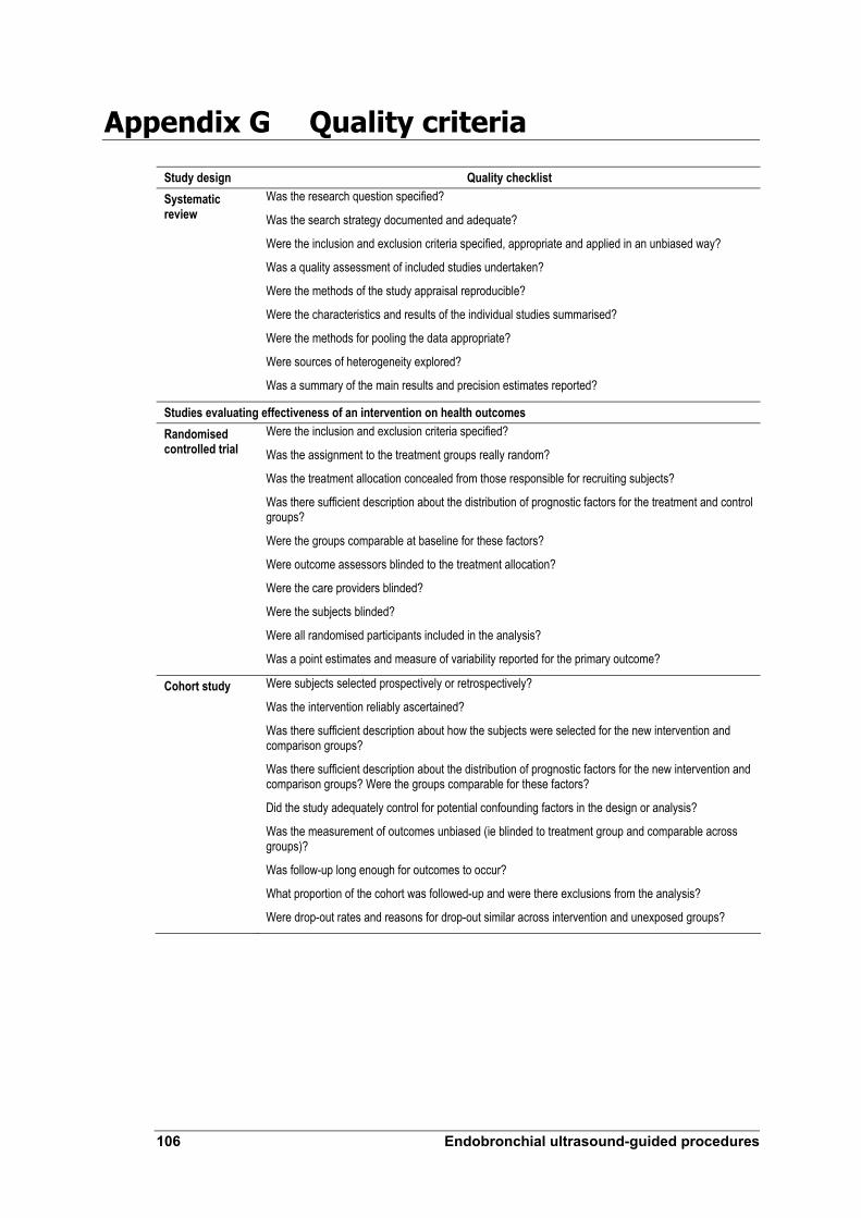
106 Endobronchial ultrasound-guided procedures
Appendix G Quality criteria
Study design Quality checklist Systematic review
Was the research question specified?
Was the search strategy documented and adequate?
Were the inclusion and exclusion criteria specified, appropriate and applied in an unbiased way?
Was a quality assessment of included studies undertaken?
Were the methods of the study appraisal reproducible?
Were the characteristics and results of the individual studies summarised?
Were the methods for pooling the data appropriate?
Were sources of heterogeneity explored?
Was a summary of the main results and precision estimates reported?
Studies evaluating effectiveness of an intervention on health outcomes Randomised controlled trial
Were the inclusion and exclusion criteria specified?
Was the assignment to the treatment groups really random?
Was the treatment allocation concealed from those responsible for recruiting subjects?
Was there sufficient description about the distribution of prognostic factors for the treatment and control groups?
Were the groups comparable at baseline for these factors?
Were outcome assessors blinded to the treatment allocation?
Were the care providers blinded?
Were the subjects blinded?
Were all randomised participants included in the analysis?
Was a point estimates and measure of variability reported for the primary outcome?
Cohort study Were subjects selected prospectively or retrospectively?
Was the intervention reliably ascertained?
Was there sufficient description about how the subjects were selected for the new intervention and comparison groups?
Was there sufficient description about the distribution of prognostic factors for the new intervention and comparison groups? Were the groups comparable for these factors?
Did the study adequately control for potential confounding factors in the design or analysis?
Was the measurement of outcomes unbiased (ie blinded to treatment group and comparable across groups)?
Was follow-up long enough for outcomes to occur?
What proportion of the cohort was followed-up and were there exclusions from the analysis?
Were drop-out rates and reasons for drop-out similar across intervention and unexposed groups?

Endobronchial ultrasound-guided procedures 107
Study design Quality checklist Case-control study
Was there sufficient description about how subjects were defined and selected for the case and control groups?
Was the disease state of the cases reliably assessed and validated?
Were the controls randomly selected from the source of population of the cases?
Was there sufficient description about the distribution of prognostic factors for the case and control groups? Were the groups comparable for these factors?
Did the study adequately control for potential confounding factors in the design or analysis?
Was the new intervention and other exposures assessed in the same way for cases and controls and kept blinded to case/control status?
How was the response rate defined?
Were the non-response rates and reasons for non-response the same in both groups?
Was an appropriate statistical analysis used?
If matching was used, is it possible that cases and controls were matched on factors related to the intervention that would compromise the analysis due to over-matching?
Case series Was the study based on a representative sample selected from a relevant population?
Were the criteria for inclusion and exclusion explicit?
Did all subjects enter the survey at a similar point in their disease progression?
Was follow-up long enough for important events to occur?
Were the techniques used adequately described?
Were outcomes assessed using objective criteria or was blinding used?
If comparisons of sub-series were made, was there sufficient description of the series and the distribution of prognostic factors?
Study of diagnostic accuracy
Was the spectrum of patients representative of the patients who will receive the test in practice?
Were selection criteria clearly described?
Is the reference standard likely to correctly classify the target condition?
Is the time period between reference standard and index test short enough to be reasonably sure that the target condition did not change between the two tests?
Did the whole sample or a random selection of the sample, receive verification using a reference standard of diagnosis?
Did patients receive the same reference standard regardless of the index test result?
Was the reference standard independent of the index test (ie the index test did not form part of the reference standard)?
Was the execution of the index test described in sufficient detail to permit replication of the test?
Was the execution of the reference standard described in sufficient detail to permit its replication?
Were the index test results interpreted without knowledge of the results of the reference standard?
Were the reference standard results interpreted without knowledge of the results of the index test?
Were the same clinical data available when test results were interpreted as would be available when the test is used in practice?
Were uninterpretable/ intermediate test results reported?
Were withdrawals from the study explained?

108 Endobronchial ultrasound-guided procedures
Appendix H Staging classification
The most widely accepted staging system for pathological staging of cancer is the TNM (tumour, node, metastasis) cancer staging system. Cancer staging involves defining the extent of spread of the primary tumour, spread to regional lymph nodes, and the presence or absence of metastases. Accurate cancer staging is essential to inform clinical management decisions. The increasing range of surgical, non-surgical and palliative treatment options has increased clinical emphasis on cancer staging.
The TNM staging for lung cancer, as described by the American Joint Committee on Cancer (AJCC), is presented in Table 62. The stage classification is presented in Table 63.
Table 62 TNM classification for lung cancer
Classification Lung cancer Tumour TX Primary tumour cannot be assessed, or tumour is proven by the presence of malignant cells in sputum or
bronchial washings but is not visualised by imaging or bronchoscopy T0 No evidence of primary tumour Tis Carcinoma in situ T1 A tumour that is ≤ 3 cm in greatest dimension, is surrounded by lung or visceral pleura and is without
bronchoscopic evidence of invasion more proximal than the lobar bronchus (ie not in the main bronchus) T2 A tumour with any of the following features of size or extent:
• > 3 cm in greatest dimension • Involves the main bronchus and is ≥ 2 cm distal to the carina • Invades the visceral pleura • Associated with atelectasis or obstructive pneumonitis that extends to the hilar region but does
not involve the entire lung T3 A tumour of any size that directly invades any of the following: chest wall (including superior sulcus
tumours), diaphragm, mediastinal pleura, parietal pericardium; or, tumour in the main bronchus < 2 cm distal to the carina but without involvement of the carina; or, associated atelectasis or obstructive pneumonitis of the entire lung
T4 A tumour of any size that invades any of the following: mediastinum, heart, great vessels, trachea, oesophagus, vertebral body, carina; or, separate tumour nodules in the same lobe; or, tumour with a malignant pleural effusiona
Node NX Regional lymph nodes cannot be assessed N0 No regional lymph node metastasis N1 Metastasis to ipsilateral peribronchial and/or ipsilateral hilar lymph nodes and intrapulmonary nodes
including involvement by direct extension of the primary tumour N2 Metastasis to ipsilateral mediastinal and/or subcarinal lymph node(s) N3 Metastasis to contralateral mediastinal, contralateral hilar, ipsilateral or contralateral scalene, or
supraclavicular lymph node(s) Metastasis MX Distant metastases cannot be assessed M0 No distant metastases M1 Distant metastasis present
a Most pleural effusions associated with lung cancer are due to tumour; multiple cytopathologic examinations of pleural fluid are negative for tumour in some patients. Pleural fluid is blood-free and is not an exudate in these circumstances. These patients may be evaluated further using video-thoracoscopy and direct pleural biopsies. When these elements and clinical judgment dictate that the effusion is not related to the tumour, the effusion should be excluded as a staging element and the patient’s disease should be staged as T1, T2, or T3. Source: Lung Cancer. In American Joint Committee on Cancer: AJCC Staging Manual. 6th ed. New York, NY: Springer, 2002, pp 167–177

Endobronchial ultrasound-guided procedures 109
Table 63 Lung cancer staging by TNM grouping
Stage TNM grouping Occult carcinoma TX, N0, M0 0 Tis, N0, M0 IA T1, N0, M0 IB T2, N0, M0 IIA T1, N1, M0 IIB T2, N1, M0
T3, N0, M0 IIIA T1, N2, M0
T2, N2, M0 T3, N1, M0 T3, N2, M0
IIIB Any T, N3, M0 T4, any N, M0
IV Any T, any N, M1 Source: Lung Cancer. In American Joint Committee on Cancer: AJCC Staging Manual. 6th ed. New York, NY: Springer, 2002, pp 167–177

110 Endobronchial ultrasound-guided procedures
Appendix I Additional information for economic evaluation
Major capital equipment costs of the first generation EBUS imaging system with radial probe
Table 64 Capital costs of EBUS with radial probe—low utilisation level
Cost of investment Year 1 Year 2 Year 3 Year 4 Year 5 Undepreciated value of equipmenta $156,400 $125,120 $93,840 $62,560 $31,280 Depreciation over a yearb $31,280 $31,280 $31,280 $31,280 $31,280 Maintenance costc $12,915 $12,915 $12,915 $12,915 $12,915 Interest costs of investment and maintenanced $25,411 $22,912 $20,412 $17,913 $15,414 Total cost per year $56,691 $54,192 $51,692 $49,193 $46,694 Present value of cost streame $56,691 $51,611 $46,886 $42,495 $38,415 Total present value of cost streamf $236,098 Return on investment Number of procedures performed annuallyg
416 416 416 416 416
Total present value of number of procedures performedh
2080
Calculated capital cost per procedurei
$114
a Cost of equipment ($156,400) supplied by applicant., including costs of a processor, probe driver, broncho-videoscope, video processor and other miscellaneous components. A cost of probe is not included because a probe has to be replaced after 100 procedures b Assumes straight-line depreciation, 5 year lifetime of equipment and $0 residual value c Proposed by applicant d Calculated by considering an interest rate of 8.0% for purchase and maintenance costs. Interest rate provided by Medfin Finance, Sydney e Present value represents the total value costs that need to be reimbursed to the investor to justify their investment f This value represents the total value of costs that needs to be reimbursed to the investor to justify their investment g Expert opinion (assuming 8 procedures per week, translating to 416 procedures per year) h Sum of the number of procedures i Total present value of cost stream divided by the total present value of procedures performed

Endobronchial ultrasound-guided procedures 111
Table 65 Capital costs of EBUS with radial probe—high utilisation level
Cost of investment Year 1 Year 2 Year 3 Year 4 Year 5 Undepreciated value of equipmenta $156,400 $125,120 $93,840 $62,560 $31,280 Depreciation over a yearb $31,280 $31,280 $31,280 $31,280 $31,280 Maintenance costc $12,915 $12,915 $12,915 $12,915 $12,915 Interest costs of investment and maintenanced $25,411 $22,912 $20,412 $17,913 $15,414 Total cost per year $56,691 $54,192 $51,692 $49,193 $46,694 Present value of cost streame $56,691 $51,611 $46,886 $42,495 $38,415 Total present value of cost streamf $236,098 Return on investment Number of procedures performed annuallyg
624 624 624 624 624
Total present value of number of procedures performedh
3120
Calculated capital cost per procedurei
$76
a Cost of equipment ($156,400) supplied by applicant., including costs of a processor, probe driver, broncho-videoscope, video processor and other miscellaneous components. A cost of probe is not included because a probe has to be replaced after 100 procedures b Assumes straight-line depreciation, 5 year lifetime of equipment and $0 residual value c Proposed by applicant d Calculated by considering an interest rate of 8.0% for purchase and maintenance costs. Interest rate provided by Medfin Finance, Sydney e Present value represents the total value costs that need to be reimbursed to the investor to justify their investment f This value represents the total value of costs that needs to be reimbursed to the investor to justify their investment g Expert opinion (assuming 12 procedures per week, translating to 624 procedures per year) h Sum of the number of procedures i Total present value of cost stream divided by the total present value of procedures performed
Major capital equipment costs of the first generation EBUS imaging system with linear probe
Table 66 Capital costs of EBUS with linear probe—low utilisation level
Cost of investment Year 1 Year 2 Year 3 Year 4 Year 5 Undepreciated value of equipmenta $101,020 $80,816 $60,612 $40,408 $20,204 Depreciation over a yearb $20,204 $20,204 $20,204 $20,204 $20,204 Maintenance costc $12,915 $12,915 $12,915 $12,915 $12,915 Interest costs of investment and maintenanced $20,986 $19,372 $17,757 $16,143 $14,529 Total cost per year $41,190 $39,576 $37,961 $36,347 $34,733 Present value of cost streame $41,190 $37,691 $34,432 $31,398 $28,575 Total present value of cost streamf $173,286 Return on investment Number of procedures performed annuallyg 416 416 416 416 416 Total present value of number of procedures performedh 2080 Calculated capital cost per procedurei $83
a Equipment cost ($101,020) supplied by Applicant (processor, light source, video processor, miscellaneous component costs) linear probe excluded b Assumes straight-line depreciation, 5 year lifetime of equipment and $0 residual value c Proposed by Applicant d Calculated by considering an interest rate of 8.0% for purchase and maintenance costs. Interest rate provided by Medfin Finance, Sydney e Present value represents the total value costs that need to be reimbursed to the investor to justify their investment f This value represents the total value of costs that needs to be reimbursed to the investor to justify their investment g Expert opinion (assuming 8 procedures per week, translating to 416 procedures per year) h Sum of the number of procedures i Total present value of cost stream divided by the total present value of procedures performed Note: The cost of linear probe per procedure was estimated using the same methodology as that for capital cost calculation

112 Endobronchial ultrasound-guided procedures
Table 67 Capital costs of EBUS with linear probe—high utilisation level
Cost of investment Year 1 Year 2 Year 3 Year 4 Year 5 Undepreciated value of equipmenta $101,020 $80,816 $60,612 $40,408 $20,204 Depreciation over a yearb $20,204 $20,204 $20,204 $20,204 $20,204 Maintenance costc $12,915 $12,915 $12,915 $12,915 $12,915 Interest costs of investment and maintenanced $20,986 $19,372 $17,757 $16,143 $14,529 Total cost per year $41,190 $39,576 $37,961 $36,347 $34,733 Present value of cost streame $41,190 $37,691 $34,432 $31,398 $28,575 Total present value of cost streamf $173,286 Return on investment Number of procedures performed annuallyg 624 624 624 624 624 Total present value of number of procedures performedh 3120 Calculated capital cost per procedurei $56
a Equipment cost ($101,020) supplied by Applicant (processor, light source, video processor, miscellaneous component costs) linear probe excluded b Assumes straight-line depreciation, 5 year lifetime of equipment and $0 residual value c Proposed by applicant d Calculated by considering an interest rate of 8.0% for purchase and maintenance costs. Interest rate provided by Medfin Finance, Sydney e Present value represents the total value costs that need to be reimbursed to the investor to justify their investment f This value represents the total value of costs that needs to be reimbursed to the investor to justify their investment g Expert opinion (assuming 12 procedures per week, translating to 624 procedures per year) h Sum of the number of procedures i Total present value of cost stream divided by the total present value of procedures performed Note: The cost of linear probe per procedure was estimated using the same methodology as that for capital cost calculation
Major capital equipment costs of Aloka EBUS imaging system
Table 68 Capital costs of Aloka EBUS—low utilisation level
Cost of investment Year 1 Year 2 Year 3 Year 4 Year 5 Undepreciated value of equipmenta $155,400 $124,320 $93,240 $62,160 $31,080 Depreciation over a yearb $31,080 $31,080 $31,080 $31,080 $31,080 Maintenance costc $12,915 $12,915 $12,915 $12,915 $12,915 Interest costs of investment and maintenanced $25,331 $22,848 $20,364 $17,881 $15,398 Total cost per year $56,411 $53,928 $51,444 $48,961 $46,478 Present value of cost streame $56,411 $51,360 $46,662 $42,294 $38,237 Total present value of cost streamf $234,964 Return on investment Number of procedures performed annuallyg 416 416 416 416 416 Total present value of number of procedures performedh 2080 Calculated capital cost per procedurei $113
a Equipment cost ($101,020) supplied by Applicant (processor, light source, video processor, miscellaneous component costs) linear probe excluded b Assumes straight-line depreciation, 5 year lifetime of equipment and $0 residual value c Proposed by applicant d Calculated by considering an interest rate of 8.0% for purchase and maintenance costs. Interest rate provided by Medfin Finance, Sydney e Present value represents the total value costs that need to be reimbursed to the investor to justify their investment f This value represents the total value of costs that needs to be reimbursed to the investor to justify their investment g Expert opinion (assuming 8 procedures per week, translating to 416 procedures per year) h Sum of the number of procedures i Total present value of cost stream divided by the total present value of procedures performed Note: The cost of linear probe per procedure was estimated using the same methodology as that for capital cost calculation
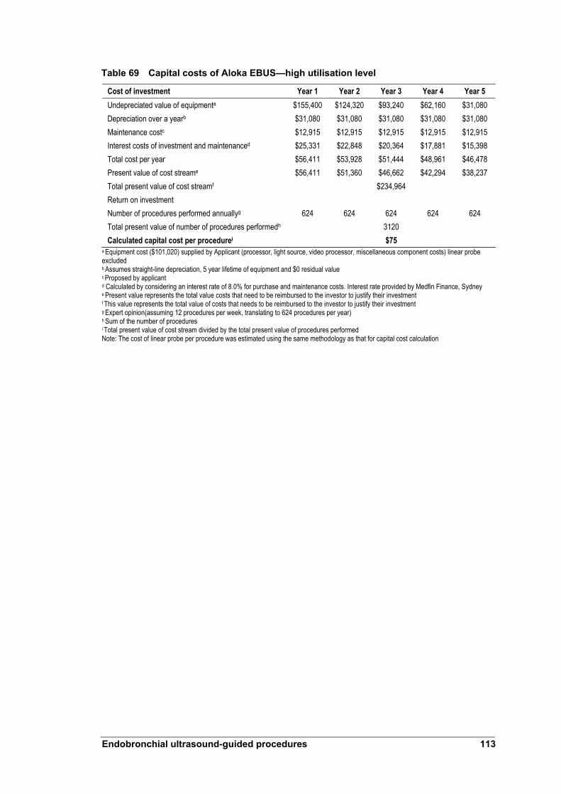
Endobronchial ultrasound-guided procedures 113
Table 69 Capital costs of Aloka EBUS—high utilisation level
Cost of investment Year 1 Year 2 Year 3 Year 4 Year 5 Undepreciated value of equipmenta $155,400 $124,320 $93,240 $62,160 $31,080 Depreciation over a yearb $31,080 $31,080 $31,080 $31,080 $31,080 Maintenance costc $12,915 $12,915 $12,915 $12,915 $12,915 Interest costs of investment and maintenanced $25,331 $22,848 $20,364 $17,881 $15,398 Total cost per year $56,411 $53,928 $51,444 $48,961 $46,478 Present value of cost streame $56,411 $51,360 $46,662 $42,294 $38,237 Total present value of cost streamf $234,964 Return on investment Number of procedures performed annuallyg 624 624 624 624 624 Total present value of number of procedures performedh 3120 Calculated capital cost per procedurei $75
a Equipment cost ($101,020) supplied by Applicant (processor, light source, video processor, miscellaneous component costs) linear probe excluded b Assumes straight-line depreciation, 5 year lifetime of equipment and $0 residual value c Proposed by applicant d Calculated by considering an interest rate of 8.0% for purchase and maintenance costs. Interest rate provided by Medfin Finance, Sydney e Present value represents the total value costs that need to be reimbursed to the investor to justify their investment f This value represents the total value of costs that needs to be reimbursed to the investor to justify their investment g Expert opinion(assuming 12 procedures per week, translating to 624 procedures per year) h Sum of the number of procedures i Total present value of cost stream divided by the total present value of procedures performed Note: The cost of linear probe per procedure was estimated using the same methodology as that for capital cost calculation

114 Endobronchial ultrasound-guided procedures
Abbreviations
AACR Australian Association of Cancer Registries
ABS Australian Bureau of Statistics
AFB autofluorescence bronchoscopy
AHMAC Australian Health Ministers’ Advisory Council
AIHW Australian Institute of Health and Welfare
AJCC American Joint Committee on Cancer
AR-DRG Australian Refined Diagnosis Related Groups
ARTG Australian Register of Therapeutic Goods
Bx biopsy
CCD charge coupled device
CT computed tomography
EBBX endobronchial biopsy
EBUS endobronchial ultrasound
ENB electromagnetic navigation bronchoscopy
EPICOT evidence, population, intervention, comparison, outcome, time stamp
EUS endoscopic ultrasound
18F-FDG 2-[18F] fluoro-2-deoxyglucose
FN false negative
FNA fine-needle aspiration
FP false positive
HTA health technology assessment
MBS Medicare Benefits Scheme
MSAC Medical Services Advisory Committee
NBI narrow band imaging
NCSG National Cancer Strategies Group

Endobronchial ultrasound-guided procedures 115
NHMRC National Health and Medical Research Council
Nd-YAG neodymium-doped yttrium aluminium garnet
NR not reported
NSCLC non-small cell lung cancer
NPV negative predictive value
PET positron emission tomography
PPICO population, prior tests, index test, comparator, outcomes
PPV positive predictive value
QALY quality-adjusted life year
QUADAS Quality Assessment of Studies of Diagnostic Accuracy included in Systematic Reviews
QUOROM quality of reporting of meta-analyses
RCT randomised controlled trials
ROSE rapid on site evaluation
TBBX transbronchial biopsy
TBNA transbronchial needle aspiration
TGA Therapeutic Goods Administration
TN true negative
TP true positive
TTBX transthoracic biopsy
TTNA transthoracic needle aspiration
VAT video-assisted thoracoscopy

116 Endobronchial ultrasound-guided procedures
References
Aabakken L, Silvestri GA, Hawes R et al. 1999. “Cost-efficacy of endoscopic ultrasonography with fine-needle aspiration vs mediastinotomy in patients with lung cancer and suspected mediastinal adenopathy”. Endoscopy 31: 707–11.
American Cancer Society. 2005 Cancer Facts and Figures 2005. Atlanta, American Cancer Society.
American Cancer Society 2007. What is non-small cell lung cancer? [Internet]. American Cancer Society, USA. Available from: http://www.cancer.org/docroot/CRI/content/CRI_2_4_1x_What_Is_Non-Small_Cell_Lung_Cancer.asp?sitearea [Accessed 20 December 2007].
Annema J, Rabe K 2006. “State of the art lecture: EUS and EBUS in pulmonary medicine”. Endoscopy 38(S1): S118–S122.
Asahina H, Yamazaki K, Onodera Y et al. 2005. “Transbronchial biopsy using endobronchial ultrasonography with a guide sheath and virtual bronchoscopic navigation”. Chest 128: 1761–1765.
Australian Bureau of Statistics 2004. 1301.0 Year Book Australia. Available from http://www.abs.gov.au/
Australian Bureau of Statistics 2002. National Health Survey: Aboriginal and Torres Strait Islanders Results, Australia 2001. Available from: http://www.abs.gov.au/Ausstats/[email protected]/e8ae5488b598839cca25682000131612/c36e019cd56ede1fca256c76007a9d36!OpenDocument [Accessed 10 August 2007]
Australian Bureau of Statistics 2006. National Health Survey: Summary of Results 2004–2005 Available from: http://www.ausstats.abs.gov.au/Ausstats/subscriber.nsf/0/3B1917236618A042CA25711F00185526/$File/43640_2004-05.pdf [Accessed: 10 August 2007]
Australian Institute of Health and Welfare and Australasian Association of Cancer Registries 2007. Cancer in Australia: an overview, 2006. AIHW cat no. CAN 32. Canberra, Australian Institute of Health and Welfare.
Australian Institute of Health and Welfare 2007. Health expenditure Australia 2005–2006. Canberra: AIHW.
Australian Institute of Health and Welfare 2006. Australia’s health No. 10 (2006) Available from: http://www.aihw.gov.au/publications/index.cfm/title/10321
Australian Institute of Health and Welfare 2005. AIHW Interactive National Hospital Morbidity Data 2005. Available from: http://www.aihw.gov.au/hospitals/datacubes/index.cfm [Accessed: 10 August 2007].

Endobronchial ultrasound-guided procedures 117
Australian Institute of Health and Welfare and Australasian Association of Cancer Registries 2004. Cancer in Australia 2001. AIHW cat no. CAN 23. Canberra, Australian Institute of Health and Welfare.
Australian Institute of Health and Welfare and Australasian Association of Cancer Registries 2005. Cancer survival in Australia 2001. Part 1: National summary statistics. AIHW cat. no. CAN 13. 2005. Cancer Series No. 18. Canberra, Australian Institute of Health and Welfare.
Australian Institute of Health and Welfare and Australasian Association of Cancer Registries & National Cancer Strategies Group: McDermid I 2005. Cancer incidence projections, Australia 2002 to 2011. Canberra: Australian Institute of Health and Welfare, Australasian Association of Cancer Registries and the National Cancer Strategies Group.
Baba M, Sekine Y, Suzuki M et al 2002. “Correlation between endobronchial ultrasonography (EBUS) images and histologic findings in normal and tumor-invaded bronchial wall”. Lung Cancer 35: 65–71.
Bard MPL, Amelink A, Skurichina M et al. 2005. “Optical spectroscopy for the classification of malignant lesions of the bronchial tree”. Chest 129: 995–1001.
Becker HD, Herth F, Mueller K 2000. “Endobronchial ultrasound for detection of early cancer”. Lung Cancer 29(Suppl 1): 244.
Becker H, Herth F, Ernst A et al. 2005. “Bronchoscopic biopsy of peripheral lung lesions under electromagnetic guidance: A pilot study”. J Bronchol 12: 9–13.
Brown P, Brunnhuber K, Chalkidou K et al. 2006. “How to formulate research recommendations”. Br Med J 333: 804–806.
Caddy G, Conron M, Wright G et al. 2005. “The accuracy of EUS-FNA in assessing mediastinal lymphadenopathy and staging patients with NSCLC”. Eur Respir J 25: 410–415.
Cancer Council Australia 2004. “Clinical practice guidelines for the prevention, diagnosis and management of lung cancer”. National Health and Medical Research Council Sydney, Australia.
Chao TY, Lie CH, Chung YH et al. 2006. “Differentiating peripheral pulmonary lesions based on images of endobronchial ultrasonography”. Chest 130: 1191–1197.
Chung YH, Lie CH, Chao TY et al. 2007. “Endobronchial ultrasonography with distance for peripheral pulmonary lesions”. Respir Med 101: 738–745.
Commonwealth of Australia 2007. Medicare Benefits Schedule, available from MBS Online at http://www.health.gov.au/mbsonline.
Crisci R, Coloni GF 1996. “Video-assisted thoracoscopic surgery versus thoracotomy for recurrent spontaneous pneumothorax”. Eur J Cardiothor Surg 10: 556–560.

118 Endobronchial ultrasound-guided procedures
De Wever W, Vandecaveye V, Lanciotti S et al. 2004. “Multidetector CT-generated virtual bronchoscopy: an illustrated review of the potential clinical indications”. Eur Respir J 23: 776–782.
Dooms CA, Verbeken EK, Becker HD et al. 2007. “Endobronchial ultrasonography in bronchoscopic occult pulmonary lesions”. J Thor Oncol 2: 121–124.
Duwe BV, Sterman DH, Musani AI 2005. “Tumors of the mediastinum”. Chest 128: 2893–2909.
Eberhardt R, Anantham D, Armin E et al 2007. “Multimodality bronchoscopic diagnosis of peripheral lung lesions: a randomized controlled trial”. Am J Respir Crit Care Med 176: 36–41.
Eggerstedt JM 2003. Lymphomas, endocrine, mesenchymal, and other rare tumors of the mediastinum. Available from: http://www.emedicine.com/med/topic3455.htm (Accessed 17 August 2007).
Ferlay J, Parkin DM, Bray F et al. 2004. GLOBOCAN 2002. Cancer Incidence, Mortality and Prevalence Worldwide. IARC CancerBase No.5, Version 1.0. Lyon, IARC.
Goldberg BB, Steiner RM, Liu JB et al. 1994. “EUS-assisted bronchoscopy with use of miniature transducer-containing catheters”. Radiol 190: 233–237.
Gould M, Sanders GD, Barnett PG et al. 2003. “Cost-effectiveness of alternative management strategies for patients with solitary pulmonary nodules”. Ann Intern Med 138: 724–735.
Govert JA, Dodd LG, Kussin PS et al. 1999. “A prospective comparison of fiberoptic transbronchial needle aspiration and bronchial biopsy for bronchoscopically visible lung carcinoma”. Cancer 87: 129–134.
Häuβinger K, Becker H, Stanzel F et al. 2005. “Autofluorescence bronchoscopy with white light bronchoscopy compared with white light bronchoscopy alone for the detection of precancerous lesions: A European randomised controlled multicentre trial”. Thorax 60: 496–503.
Herth FJF, Becker HD, Manegold C et al. 2001. “Endobronchial ultrasound: Assessment of a new diagnostic tool in bronchoscopy for staging of lung cancer”. Onkologie 24: 151–154.
Herth FJF, Ernst A, Becker HD 2002. “Endobronchial ultrasound-guided transbronchial lung biopsy in solitary pulmonary nodules and peripheral lesions”. Eur Resp J 20: 972–974.
Herth F, Ernst A, Schulz M et al. 2003a. “Endobronchial ultrasound reliably differentiates between airway infiltration and compression by tumor”. Chest 123: 458–462.
Herth FJ, Becker HD, Ernst A 2003b. “Ultrasound-guided transbronchial needle aspiration: An experience in 242 patients”. Chest 123: 604–607.

Endobronchial ultrasound-guided procedures 119
Herth FJ, Ernst A, Becker H 2003c. “Initial EBUS guided TBNA is the most cost-effective means of lymph node staging—a German experience”. Eur Respir J 22: Suppl. 45, 205s.
Herth F, Becker HD, Ernst A 2004. “Conventional vs endobronchial ultrasound-guided transbronchial needle aspiration: A randomized trial”. Chest 125: 322–325.
Herth F, Lunn W, Eberhardt F et al 2005. “Transbronchial versus transesophageal ultrasound-guided aspiration of enlarged mediastinal lymph nodes”. Am J Resp Crit Care Med 171: 1164–1167.
Herth FJF, Eberhardt R, Becker HD et al. 2006a. “Endobronchial ultrasound-guided transbronchial lung biopsy in fluoroscopically invisible solitary pulmonary nodules: A prospective trial”. Chest 129: 147–150.
Herth FJF, Ernst A, Eberhardt R et al. 2006b. “Endobronchial ultrasound-guided transbronchial needle aspiration of lymph nodes in the radiologically normal mediastinum”. Eur Resp J 28: 910–914.
Herth FJF, Eberhardt R, Vilmann P et al. 2006c. “Real-time endobronchial ultrasound guided transbronchial needle aspiration for sampling mediastinal lymph nodes”. Thorax 61: 795–798.
Hirata M, Tanaka S, Oka S et al. 2007. “Evaluation of microvessels in colorectal tumors by narrow band imaging magnification”. Gastrointest Endosc 66: 945–952.
Hirsch FR, Prindiville SA, Miller YE et al. 2001. “Fluorescence versus white-light bronchoscopy for detection of preneoplastic lesions: a randomised study”. J Natl Cancer Inst 2001; 93:1385–91.
Hurter T, Hanrath P 1992. “Endobronchial sonography: Feasibility and preliminary results”. Thorax 47: 565–567.
Kanoh K, Miyazawa T, Kurimoto N et al. 2005. “Endobronchial ultrasonography guidance for transbronchial needle aspiration using a double-channel bronchoscope”. Chest 128: 388–393.
Kikuchi E, Yamazaki K, Sukoh N et al. 2004. “Endobronchial ultrasonography with guide-sheath for peripheral pulmonary lesions”. Eur Resp J 24: 533–537.
Koh MS, Tee A, Wong P et al. 2007. “Advances in lung cancer diagnosis and staging: endobronchial ultrasound”. Intern Med J Oct 3; [Epub ahead of print].
Kotsianos-Hermle D, Scherr M, Wirth S et al. 2007. “Visualization of bronchial lesions using multidetector CT and endobronchial ultrasound”. Eur J Med Res 12: 84–89.
Kramer H, van Putten JW, Post WJ et al. 2004. “Oesophageal endoscopic ultrasound with fine needle aspiration improves and simplifies the staging of lung cancer”. Thorax 59: 596–601.

120 Endobronchial ultrasound-guided procedures
Krasnik M, Vilmann P, Larsen SS et al. 2003. “Preliminary experience with a new method of endoscopic transbronchial real time ultrasound guided biopsy for diagnosis of mediastinal and hilar lesions”. Thorax 58: 1083–1086.
Kurimoto N, Murayama M, Yoshioka S et al. 2002. “Analysis of the internal structure of peripheral pulmonary lesions using endobronchial ultrasonography”. Chest 122: 1887–1894.
Kurimoto N, Miyazawa T, Okimasa S et al. 2004. “Endobronchial ultrasonography using a guide sheath increases the ability to diagnose peripheral pulmonary lesions endoscopically”. Chest 126: 959–965.
Maghfoor I, Perry M 2005. ‘Lung cancer, non-small cell’ in Ribas A, Talavera F, Movsas B, McKenna R and MacDonald JS [eds] emedicine Available from: www.emedicine.com/med/topic1333.htm. [Accessed: 10 August 2007].
Mazzone P, Jain P, Arroliga AC et al. 2002. “Bronchoscopy and needle biopsy techniques for diagnosis and staging of lung cancer”. Lung Cancer 23: 137–157.
Medical Services Advisory Committee. Endoscopic ultrasound guided fine-needle aspiration for the staging of non-small cell lung cancer and the diagnosis of mediastinal masses: Assessment Report (MSAC Assessment 1104). Canberra; 2008
Mentzer SJ, Swanson SJ, DeCamp MM et al. 1997. “Mediastinoscopy, thoracoscopy, and video-assisted thoracic surgery in the diagnosis and staging of lung cancer”. Chest 112: 239–241.
Minna JD 2001. “Neoplasms of the Lung” in Braunwald E [ed] Harrison's Principles of Internal Medicine, New York, McGraw-Hill.
Miyazu Y, Miyazawa T, Iwamoto Y et al. 2001. “The role of endoscopic techniques, laser-induced fluorescence endoscopy and endobronchial ultrasonography in choice of appropriate therapy for bronchial cancer”. J Bronchol 8: 10–16.
Mountain CF, Dresler CM 1997. “Regional lymph node classification for lung cancer staging”. Chest 111: 1718–1723.
Mountain CF 1997. “Revisions in the International System for Staging Lung Cancer”. Chest 111:1710–1717.
Nakajima J, Takamoto S, Kohno T et al. 2000. “Costs of videothoracoscopic surgery versus open resection for patients with of lung carcinoma”. Cancer 89: 2497–501.
National Hospital Cost Data Collection. 2006. Public sector—estimated round 9 (2004–2005) AR-DRG 5.0 cost report, available online at www.health.gov.au/casemix.
National Cancer Institute. Non-small cell lung cancer: Treatment. 2007. Available from: http://www.cancer.gov/cancertopics/pdq/treatment/non-small-cell-lung/healthprofessional [Accessed 10 August 2007]
National Health and Medical Research Council. 1999. A guide to the development, implementation and evaluation of clinical practice guidelines. National Health and Medical Research Council, Canberra.

Endobronchial ultrasound-guided procedures 121
National Health and Medical Research Council. 2000. How to use the evidence: assessment and application of scientific evidence. National Health and Medical Research Council, Canberra.
Okamoto H, Watanabe K, Nagatomo A et al. 2002. “Endobronchial ultrasonography for mediastinal and hilar lymph node metastases of lung cancer”. Chest 121: 1498–1506.
Olympus–UM-BS20-26R (2008) Available from: http://www2.olympus.cz/endoscopy/427_UM-BS20-26R.htm. [Accessed: 14 January 2008].
Omori S, Takiguchi Y, Hiroshima K et al. 2002. “Peripheral pulmonary diseases: Evaluation with endobronchial US—Initial experience”. Radiol 224: 603–608.
Paone G, Nicastri E, Lucantoni G et al. 2005. “Endobronchial ultrasound-driven biopsy in the diagnosis of peripheral lung lesions”. Chest 128: 3551–3557.
Plat G, Pierard P, Haller A et al. 2006. “Endobronchial ultrasound and positron emission tomography positive mediastinal lymph nodes”. Eur Resp J 27: 276–281.
Rintoul RC, Skwarski KM, Murchison JT et al. 2005. “Endobronchial and endoscopic ultrasound-guided real-time fine-needle aspiration for mediastinal staging”. Eur Resp J 25: 416–421.
Shannon JJ, Bude RO, Orens JB et al. 1996. “Endobronchial ultrasound-guided needle aspiration of mediastinal adenopathy”. Am J Resp Crit Care Med 153: 1424–1430.
Shirakawa T, Imamura F, Hamamoto J et al. 2004. “Usefulness of endobronchial ultrasonography for transbronchial lung biopsies of peripheral lung lesions”. Respirat 71: 260–268.
Schwarz Y, Greid J, Becker HD et al. 2006. “Real-time electromagnetic navigation bronchoscopy to peripheral lung lesions using overlaid CT Images: The first human study”. Chest; 129: 988–994.
Schwarz Y, Mehta AC, Ernst A et al. 2003. “Electromagnetic navigation during flexible bronchoscopy”. Respirat 70: 516–522.
Semik M, Netz B, Schmidt C et al. 2004. “Surgical exploration of the mediastinum: mediastinoscopy and intraoperative staging”. Lung Cancer 45: S55–S61.
Strollo DC, Rosado de Christenson ML, Jett JR 1997a. “Primary mediastinal tumors. Part I: Tumors of the anterior mediastinum”. Chest 112: 511–522.
Strollo DC, Rosado-de-Christenson ML, Jett JR 1997b. “Primary mediastinal tumors: Part II. Tumors of the middle and posterior mediastinum”. Chest 112: 1344–1357.
Supramaniam R, Smith DP, Coates MS, Armstrong BK 1998. Survival from cancer in New South Wales in 1980 to 1995. NSW Cancer Council, Sydney, NSW.
Takahashi H, Sagawa M, Sato M et al. 2003. “A prospective evaluation of transbronchial ultrasonography for assessment of depth of invasion in early bronchogenic squamous cell carcinoma”. Lung Cancer 42: 43–49.

122 Endobronchial ultrasound-guided procedures
Tanaka F, Muro K, Yamasaki S et al. 2000. “Evaluation of tracheo-bronchial wall invasion using transbronchial ultrasonography”. Eur J Cardiothor Surg 17: 570–574.
Vilmann P, Krasnik M, Larsen SS et al. 2005. “Transesophageal endoscopic ultrasound-guided fine-needle aspiration (EUS-FNA) and endobronchial ultrasound-guided transbronchial needle aspiration (EBUS-TBNA) biopsy: A combined approach in the evaluation of mediastinal lesions”. Endoscopy 37: 833–839.
Vincent BD, Fraig M, Silvestri GA 2007. “A pilot study of narrow-band imaging compared to white light bronchoscopy for evaluation of normal airways and premalignant and malignant airways disease”. Chest 131: 1794–9.
Wallace WAH, Monaghan HM, Salter DM et al. 2007. “Endobronchial ultrasound-guided fine-needle aspiration and liquid-based thin-layer cytology”. J Clin Pathol 60: 388–391.
Whiting P, Rutjes AWS, Reitsma JB et al. 2003. “The development of QUADAS: a tool for the quality assessment of studies of diagnostic accuracy included in systematic reviews”. BMC Med Res Methodol 3: 25.
Yang MC, Liu WT, Wang CH et al. 2004. “Diagnostic value of endobronchial ultrasound-guided transbronchial lung biopsy in peripheral lung cancers”. J Formosan Med Assoc Taiwan yi zhi 103: 124–129.
Yap KK, Yap KS, Byrne AJ 2005. “Positron emission tomography with selected mediastinoscopy compared to routine mediastinoscopy offers cost and clinical outcome benefits for pre-operative staging of non-small cell lung cancer”. Eur J Nucl Med Mol Imaging 32: 1033–40.
Yasafuku K, Chiyo M, Sekine Y et al. 2004. “Real-time endobronchial ultrasound-guided transbronchial needle aspiration of mediastinal and hilar lymph nodes”. Chest 126: 122–128.
Yasafuku K, Chiyo M, Koh E et al. 2005. “Endobronchial ultrasound guided transbronchial needle aspiration for staging of lung cancer”. Lung Cancer 50: 347–354.
Yasufuku K, Nakajima T, Motoori K et al. 2006. “Comparison of endobronchial ultrasound, positron emission tomography and CT for lymph node staging of lung cancer”. Chest 130: 710–718.
Yasufuku K, Nakajima T, Chiyo M et al. 2007. “Endobronchial Ultrasonography: Current status and future directions”. J Thorac Oncol 2: 970–979.
Zimmermann FB 2005. “Contemporary Issues in staging of lung cancer” in Brady LW, Heilmann HP, Molls M [eds] Advances in Radiation Oncology in Lung Cancer, Berlin, Springer.