Quality Assurance Guidelines for Percutaneous Vertebroplasty
Upload
khangminh22Category
view
8download
0
diagnostics
Review
Performing an Ultrasound-Guided Percutaneous NeedleKidney Biopsy: An Up-To-Date Procedural Review
Antonio Granata 1 , Giulio Distefano 2,* , Francesco Pesce 3 , Yuri Battaglia 4 , Paola Suavo Bulzis 3,Massimo Venturini 5, Stefano Palmucci 2 , Vito Cantisani 6, Antonio Basile 2 and Loreto Gesualdo 3
�����������������
Citation: Granata, A.; Distefano, G.;
Pesce, F.; Battaglia, Y.; Suavo Bulzis,
P.; Venturini, M.; Palmucci, S.;
Cantisani, V.; Basile, A.; Gesualdo, L.
Performing an Ultrasound-Guided
Percutaneous Needle Kidney Biopsy:
An Up-To-Date Procedural Review.
Diagnostics 2021, 11, 2186. https://
doi.org/10.3390/diagnostics11122186
Academic Editor: Po-Hsiang Tsui
Received: 6 October 2021
Accepted: 23 November 2021
Published: 24 November 2021
Publisher’s Note: MDPI stays neutral
with regard to jurisdictional claims in
published maps and institutional affil-
iations.
Copyright: © 2021 by the authors.
Licensee MDPI, Basel, Switzerland.
This article is an open access article
distributed under the terms and
conditions of the Creative Commons
Attribution (CC BY) license (https://
creativecommons.org/licenses/by/
4.0/).
1 Nephrology and Dialysis Unit, “Cannizzaro” Hospital, 95126 Catania, Italy; [email protected] Radiology Unit 1, Department of Medical Surgical Sciences and Advanced Technologies “GF Ingrassia”,
University Hospital “Policlinico-San Marco”, University of Catania, 95123 Catania, Italy;[email protected] (S.P.); [email protected] (A.B.)
3 Nephrology, Dialysis and Transplantation Unit, Department of Emergency and Organ Transplantation,University of Bari “Aldo Moro”, 70121 Bari, Italy; [email protected] (F.P.);[email protected] (P.S.B.); [email protected] (L.G.)
4 Division of Nephrology and Dialysis, St. Anna University Hospital, 44121 Ferrara, Italy;[email protected]
5 Department of Diagnostic and Interventional Radiology, Ospedale di Circolo e Fondazione Macchi,University of Insubria, 21100 Varese, Italy; [email protected]
6 Department of Radiology, Policlinico Umberto I, Sapienza Rome University, 00161 Rome, Italy;[email protected]
* Correspondence: [email protected]
Abstract: Ultrasound-guided percutaneous renal biopsy (PRB) has revolutionized the clinical practiceof nephrology in the last decades. PRB remains an essential tool for the diagnosis, prognosis, andtherapeutic management of several renal diseases and for the assessment of renal involvement insystemic diseases. In this study, we examine the different applications and provide a review of thecurrent evidence on the periprocedural management of patients. PRB is recommended in patientswith significant proteinuria, hematuria, acute kidney injury, unexpected worsening of renal function,and allograft dysfunction after excluding pre- and post-renal causes. A preliminary ultrasoundexamination is needed to assess the presence of anatomic anomalies of the kidney and to identifyvessels that might be damaged by the needle during the procedure. Kidney biopsy is usuallyperformed in the prone position on the lower pole of the left kidney, whereas in patients with obesity,the supine antero-lateral position is preferred. After preparing a sterile field and the injection of localanesthetics, an automatic spring-loaded biopsy gun is used under ultrasound guidance to obtainsamples of renal parenchyma for histopathology. After the procedure, an ultrasound scan mustbe performed for the prompt identification of potential early bleeding complications. As 33% ofcomplications occur after 8 h and 91% occur within 24 h, the ideal post-procedural observation timeis 24 h. PRB is a safe procedure and should be considered a routine part of the clinical practiceof nephrology.
Keywords: kidney; ultrasound; biopsy; chronic kidney disease; acute kidney disease; proteinuria;hematuria; percutaneous; ultrasound-guided
1. Introduction
Nephrology was revolutionized in the 1950s and 1960s by the introduction of percuta-neous renal biopsy (PRB) into clinical practice. The first attempt to perform a percutaneousneedle biopsy of the kidney was described in 1951 by Iversen and Brun, who used afluoroscopically guided approach and obtained parenchyma samples after antegrade pyel-ography in a sitting position, with a success rate of only 40% [1]. The technique wasmodified by Karm, who, in 1954, proposed the performance of the biopsy in the proneposition with an ad hoc needle, reaching success in 96% of cases [2]. Since the 1980s,
Diagnostics 2021, 11, 2186. https://doi.org/10.3390/diagnostics11122186 https://www.mdpi.com/journal/diagnostics

Diagnostics 2021, 11, 2186 2 of 13
percutaneous biopsy has been performed under ultrasound guidance using spring-loadedbiopsy needles, mainly in nephrological and radiological departments. Despite its uniquerole in the management of nephrological diseases, it has been reported that about half ofall post-graduate nephrology program directors do not consider biopsies to be an essentialcomponent of the core curriculum for residents [3]. In this review, we provide an operativeguide for the safe and successful execution of ultrasound-guided renal biopsies for clinicalnephrologists and radiologists.
2. Indications
All physicians must know the indications for a renal biopsy and should be aware of thecontraindications and risks in order to estimate the risk/benefit ratio of the procedure [4].Common indications include significant proteinuria (defined as >1 g/day in the absenceof other comorbidities or rapid increases in proteinuria in patients with a diagnosis ofkidney disease, systemic immunological disease, or diabetes), acute kidney injury (AKI)when an intrinsic etiology is suspected, hematuria associated with proteinuria and/ordecreased renal function or suspected secondary glomerular disease, rapid, and unexpectedworsening of eGFR, or rapid increase in proteinuria in known chronic kidney disease (CKD).A renal biopsy is also essential to assess the extent of renal damage in systemic diseases,such as systemic lupus erythematosus or paraproteinemia, due to the impact on treatment(Table 1). In patients without already known glomerular or systemic pathologies, isolatedhematuria, or proteinuria <1 g/day does not necessarily require biopsy, as the courseof these nephropathies is generally indolent and the risk/benefit ratio of knowing thehistological diagnosis does not justify an invasive procedure [5,6].
Table 1. Indications for performing renal biopsy [5,6].
Proteinuria >1 g/day (new finding, in the absence of other comorbidities), rapid increasing ofproteinuria in a patient with known kidney disease, systemic immunological disorder or diabetes.
Rapid increase in proteinuria (in already known kidney disease, systemic immunological disease,or diabetes).
Acute kidney injury.
Rapid and unexpected worsening of eGFR in already known kidney disease.
Hematuria is associated with proteinuria and/or decreased renal function.
Systemic lupus erythematosus and paraproteinemia with any renal involvement.
3. Contraindications
Coagulation impairments are, in some cases, the main contraindication for PRB. Cur-rently, there is no consensus on the management of patients on aspirin. Some centersregularly discontinue aspirin within seven days before and two days after renal biopsy. Thediscontinuation of aspirin, however, does not appear to reduce the risk of major bleedingcomplications [6]. Other antiplatelet agents (e.g., clopidogrel) are almost always discon-tinued at least one week before the biopsy. Therefore, it is important to promptly contactnephrologists for any patient who is a candidate for renal biopsy thus that the procedure isnot delayed if these agents are not stopped in a timely manner. Non-correctable coagulopa-thy and lack of safe access are two absolute contraindications of biopsy performance, whichis considered a relatively invasive procedure; an uncooperative or unwell patient is anothercontraindication to consider [7,8]. The alterations in coagulation parameters may requirepostponing the procedure. Since renal biopsy is considered a high-risk bleeding proce-dure, INR and a platelet count are routinely recommended, while aPTT is required if thepatient undergoes unfractionated IV heparin therapy. An INR >1.5 or platelet counts below50,000/× 109/L are also contraindications to performing the procedure but are virtuallysusceptible to correction [9]. The same contraindications reported for the native kidneyare valid for the transplanted kidney. Additional contraindications include urosepsis,

Diagnostics 2021, 11, 2186 3 of 13
peri-renal abscesses, hydronephrosis, renal malformations (horseshoe kidney and nativesolitary kidney), multiple bilateral cysts, and uncontrolled high blood pressure [6] (Table 2).
Table 2. Absolute and relative contraindications for performing renal biopsy [6–8].
Absolute Relative
Non-correctable coagulopathy (INR >1.5).Therapy with antiplatelet agents, heparins or oral
anticoagulants (to be suspended before theprocedure; see main text).
Lack of safe access. Hypertension (management with oral or IV drugsbefore the procedure).
Platelet counts below 50,000/× 109/L. Abscess.
Non-correctable hypertension. Renal malformation.
4. Management of Relative Contraindications
Although the relative risk of severe bleeding is not significantly lowered by thewithdrawal of antiplatelet drugs (aspirin and clopidogrel), it is currently accepted to dis-continue the drug within five days prior to the procedure. New oral anticoagulant drugsmust be suspended at least three days before, and they can be temporarily replaced by low-molecular-weight heparin (LMWH). Dicoumarols should be discontinued approximatelyfive days prior to biopsy and replaced by low-molecular-weight heparin (LMWH) until thenext day (acenocumarolo) or two days after (warfarin). LMWH administration should bestopped 12 h before biopsy if it is administered for prophylactic purposes, while it shouldbe discontinued 24 h earlier if administered at full doses for therapeutic purposes [8–11].LMWH administration can be resumed after 12 h (prophylactic dose) or 24–72 h (fulldose) [11]. The targets for systolic and diastolic blood pressure are <140 mmHg and<90 mmHg, respectively, as it has been shown that the risk of complications (mainly bleed-ing) is 10 times higher if blood pressure exceeds these thresholds and, up to 26 times higher,if the systolic exceeds 170 mmHg [6]. All oral antihypertensive medications that the patientis currently taking should be continued regularly and may possibly be supplemented byIV therapy immediately prior to the procedure.
5. Equipment
A table covered with a sterile cloth should be set with the following tools: drapes,disinfectant sponges, sterile gauze pads, surgical scalpel blade no. 11, anesthetic syringe,spinal anesthesia needle, and an automatic or semi-automatic spring-loaded biopsy system.A 15/19 cm long needle with a penetration depth of 22 mm and a sample notch of 13–18 mmis adequate for the purpose; 14-, 16-, and 18-gauge needles can be used, and a 16-gaugeneedle appears to be associated with the best risk/benefit ratio in terms of glomeruliharvested and severe bleeding. Some companies market biopsy kits with all of the essentialtools for performing the procedure (Figure 1) [12–14]. The operator and their assistantmust be provided with sterile gloves, eye protection, surgical caps, and surgical masks.A full-size sterile drape should be placed over the patient after the access site skin hasbeen sterilized with 7.5% povidone-iodine solution. An ultrasound machine with convex-array, high-resolution, Doppler module is preferred for this procedure (Figure 2). Steriletransduction gel, an acoustically transparent sterile transducer sheath, software with guidemarks with an inclination of 20–30◦, and sterile rubber bands or clips to secure the sheatharound the transducer are also needed (Table 3).

Diagnostics 2021, 11, 2186 4 of 13
Figure 1. Commercially available renal biopsy kit, consisting of an automatic spring-loaded biopsy system with an 18 Gneedle and a 1 or 2 cm slit, sterile drapes, sterile gauze, sterile ultrasound gel, a syringe and needle for anesthesia, a scalpel,and a swab for surface disinfection.
Figure 2. The correct position of the operator with respect to the patient is demonstrated. A needleguide system was mounted on the convex probe. The whole procedure is conducted in a sterile state,and the sterile sheets surrounding the uncovered skin areas are not punctured.

Diagnostics 2021, 11, 2186 5 of 13
Table 3. Tools of the trade.
Disinfectant sponges/bufferand 7.5%
povidone-iodine solutionSterile gauze pads Sterile drapes
Surgical scalpel blade no. 11 Anesthetic syringe with spinalanesthesia needle
Personal protectiveequipment, sterile gloves,
and apron
Sterile set for ultrasoundprobe (with probe cover,sterile gel, rubber bands)
Containers with formalin forfixing the histological
samples taken
Automatic or semi-automaticspring-loaded biopsy system
The specimen should be immediately evaluated using an optical microscope beforepreparing it for the pathologist to decide how many biopsy passes are needed to obtain anadequate renal sample. In general, a sampling of 20 glomeruli is considered adequate toallow for a correct diagnosis [4].
6. Preparation
Before carrying out any invasive procedure, it is necessary to obtain informed consentfrom the patient or whoever has legal responsibility, in accordance with national regulationsand institutional guidelines. Ideally, written consent should be obtained the day beforethe procedure and possibly confirmed on the day of the procedure; however, this timingis often waived. The patient must be informed about the indication for carrying outthe procedure, the potential complications, and the benefits with respect to choosing theoptimal treatment after a correct diagnosis [15]. Patients should have a light meal onthe evening before the procedure, and the administration of a cleansing enema can beconsidered. On the day of the procedure, patients must be fasting. Any antihypertensivedrugs should be given regularly. A peripheral vein must be cannulated. Blood pressureshould be measured before performing PRB. The bladder needs to be empty. The usefulnessof antibiotic prophylaxis in procedures where one does not cross-contaminated territoriesis debated, and it is not clear which antibiotic to choose or for how long to prescribe it.However, it is considered appropriate to administer a dose of broad-spectrum antibioticsbefore starting the procedure.
7. Position of the Patient
A kidney biopsy is usually performed in the prone position on the lower pole of theleft kidney, which is preferred to reduce the risk of inadvertent injury to a major vessel. Ifthe patient is obese or has breathing difficulties, a supine anterolateral position (SALP) ismore suitable and does not increase the risk of complications [10].
8. Prebiopsy Ultrasound Study
Before performing an ultrasound-guided PRB, it is advisable to perform a completeultrasound examination of the kidneys in the same position as the procedure to evaluatethe location of the kidney, its excursion during breathing, the cortical thickness, and thedistance of the kidney from the skin surface. These data are useful to plan the procedure.Furthermore, this allows for the exclusion of anatomical variants (for example, horseshoekidney), hydronephrosis, and cystic lesions, which may be contraindications for the proce-dure [16]. The examination must be completed with an ECD assessment to rule out thepresence of vessels along the presumed path of the needle (Figure 3). Vascular abnormali-ties are reported in up to 10% of patients who undergo renal biopsy, and this may explainthe reduction in complications observed when an ECD study preceded the procedure [15].

Diagnostics 2021, 11, 2186 6 of 13
Figure 3. The dotted lines show the expected trajectory of the needle when correctly fixed to theappropriate support. At the lower pole of the kidney, the site of the biopsy, there is an abnormalvascular formation that would have been crossed by the needle if it had not been recognized.
9. Procedure9.1. Approach
An ultrasound-guided PRB can be performed either freehand or with the aid of needlesupports to force its trajectory (needle-guided insertion). Compared with the probe, theneedle can have a parallel or transverse insertion: the guided needle technique can onlybe used with parallel insertion [3]. In most cases, the prone position is preferred; inpatients who are obese or pregnant (after the 20th week), a lateral decubitus or sittingupright position can be evaluated [9]. The use of needle guides was associated with greaterreproducibility and speed of execution compared with the freehand technique, especiallyfor beginners, but there were no significant differences in terms of complications [17].
It is preferable to perform the ultrasound-guided needle biopsy on the lower pole ofthe left kidney, at the level of Brodel’s line (a relatively poorly vascularized plane betweenthe anterior and posterior branches of the renal artery). The inferior pole of the left kidneyis in fact, more accessible than the contralateral, and it is more distant from large-calibervessels [18].
9.2. Performing the Biopsy in the Prone Position
The patient is placed in a prone position, possibly with a pillow under the abdomen toreduce the physiological dorsal lordosis and to make the kidney more accessible (Figure 4).Patients must be collaborative, and this is an important aspect in the execution of anultrasound-guided needle biopsy, given the motion of the kidneys during respiration. Theuse of anxiolytic drugs before the procedure can be considered. However, in uncooper-ative or seriously ill patients, the use of hypnotic drugs might require the support of an

Diagnostics 2021, 11, 2186 7 of 13
anesthesiologist [7,19]. The whole procedure must be carried out under sterile conditions.First, it is necessary to cleanse the skin with povidone-iodine and then to apply the sterilesheets to obtain a sterile window on the access site; the probe must be covered by sterileprotection, and the ultrasound examination must be carried out using a sterile gel. Anadditional preprocedural ultrasound scan allows for the confirmation of the planned posi-tion and trajectory. The trajectory of the needle through the guide can be displayed on theultrasound monitor.
Figure 4. (a) Demonstration of the correct position of a patient undergoing a renal biopsy in prone position, the position inwhich the biopsy is performed must also be maintained during the ultrasound examination, and (b) the same patient afterthe preparation of a sterile field and application of the drapes, just before the injection of the anesthetic.
Local anesthesia is performed (Supplementary Video S1). Under ultrasound guidance,the biopsy needle is then introduced with an inclination of about 20◦ or 30◦ through thesubcutaneous tissue, the muscular planes, and the fascia until it stops right above the renalcapsule (Figure 5, Supplementary Video S2). Although puncture of the cortical region ofthe kidney is always performed in B-mode, an ECD scan is advisable to identify abnormalvessels (Supplementary Video S3). In that case, it is appropriate to perform the biopsy onthe contralateral kidney.
It is important to underline that the needle and the tip must always be visualizedon the monitor during the procedure to avoid errors (Supplementary Video S4). Usinga small amount of saline solution or by shaking the needle, it is possible to increase thevisibility of the tip [19]. The patient’s collaboration and breathing, particularly inspiratoryapnea during the execution of the biopsy, are fundamental since unexpected movements ofthe kidney can shift the trajectory of the needle with potential unexpected and unwantedoutcomes. When the optimal target is reached, with the needle resting on the renal capsule,the automatic needle is triggered, thus activating the descent, initially, of the mandrel and,subsequently, of the shirt, which locks up the sample.

Diagnostics 2021, 11, 2186 8 of 13
Figure 5. US highlights the trajectory of the needle that crosses the capsule with the tip in the internalregion of the cortex. This image shows the needle after the activation of the trigger mechanism. Theoperator must verify that the needle does not reach the region of the renal pelvis, considering both thetrajectory and the maximum possible excursion of the tip after the activation of the trigger mechanism.
The needle is then extracted from the kidney, and by opening it, the biopsy sampleis removed and then either laid down on a gauze soaked in physiological solution or in asterile container containing physiological solution. If available, an optical microscope canbe used to evaluate fresh biopsy samples for the presence of cortical parenchyma. Usually,two passes are sufficient to obtain a renal parenchyma specimen sufficient for analysis bylight microscopy, immunofluorescence, and electron microscopy, if necessary. In manyinstitutions, the samples should be fixed in formalin solution before sending them to thepathologist. In any case, it is always advisable to send an accompanying clinical report todirect the diagnosis.
Once the needle has been extracted, hemostasis by compression is applied for a few minutes.At the end of the procedure, before moving the patient, it is necessary to perform an
additional US-ECD evaluation to check the absence of hematomas, pseudoaneurysms, orarteriovenous fistulas, which are the most common periprocedural adverse events. Thepatient should be informed that a period of bed rest is necessary for at least 24 h afterthe procedure.
9.3. Performing the Biopsy in the Supine Antero-Lateral Position (SALP)
This technique can be used in patients who are obese (BMI > 30) to avoid severalinconveniences, such as poor respiratory performance due to the prone position, the depthof the inferior renal pole, and the difficulty of some patients in maintaining the positionfor a long time (Figure 6). This technique has a significantly lower complication rate thanthe classic approach, excellent diagnostic power, and better patient compliance in patientswho are obese [20]. In the SALP position, a towel is placed under the ipsilateral shoulderand the gluteus to elevate the flank by an angle of 30◦. The ipsilateral arm is placed overthe thorax while the contralateral is abducted and used for intravenous perfusion. The

Diagnostics 2021, 11, 2186 9 of 13
ipsilateral leg is slightly flexed over a pillow, while the contralateral is flexed and abducted,thus that its lateral aspect is lying on the table. This position provides full exposure ofPetit’s triangle, thus providing enough space to perform ultrasound scanning and to easilyorient the ultrasound-guided puncture toward the inferior renal pole. After shaving anddraping the flank, the kidney is ultrasound-scanned to determine the ideal puncture path.The identification of the lower kidney pole by ultrasound scanning is easy, and the qualityof image resolution is similar to the prone position. After injecting a local anesthetic, anautomatic needle is ultrasound-guided to the capsule in the lower pole of the kidney andfired into the renal parenchyma, similar to what is expected for biopsies performed in thesupine position.
Figure 6. Supine antero-lateral position (SALP) is indicated in patients who are obese (a), (b); (c) pre-bioptic study.
9.4. Percutaneous Renal Transplant Biopsy
A biopsy of transplanted kidney is usually easier to perform, since the graft is moresuperficial, more accessible, and relatively static with respiration. The graft is in the anteriorpart of the lower abdomen in the iliac region, and it lays over the iliopsoas muscle in theretroperitoneal space. The transplanted kidney is separated from the anterior abdominalwall only by the transversalis fascia, abdominal muscles, and subcutaneous tissue: thesupine position with an approach from the anterolateral surface of the abdomen generallyallows for easy access to the organ. The steps necessary to obtain an ultrasound-guidedneedle biopsy of graft are quite similar to those described for the native kidney, but it mustbe noted that the risk of complications is higher. If the biopsy is performed a few daysafter the transplant, there is a high rate of complications such as relevant hemorrhagesand lesions of the parenchyma due to the frailty of the kidney after cold ischemia. On theother hand, a long-transplanted kidney instead has pericapsular fibrosis due to the chronicdesmogenic reaction of the perirenal tissues. The fibrosis surrounding the transplantedorgan could make it difficult to penetrate the renal capsule. In these cases, it is advisableto go deeper, with the tip of the needle a few millimeters below the capsular plane, andonly after that is it advisable to activate the automatic needle system. In the case ofsuspected rejection, samples from different spots of the kidney should be obtained; asthe inflammatory lesions may have a heterogeneous distribution, the trajectory withinthe renal cortex should be slightly oblique to increase the quantity of cortical structuresremoved for analysis and, at the same time, to reduce the risk of vessel injury [16].
Diagnostics 2021, 11, 2186 10 of 13
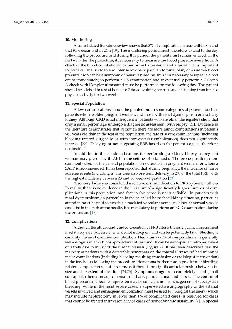
10. Monitoring
A consolidated literature review shows that 3% of complications occur within 8 h andthat 91% occur within 24 h [19]. The monitoring period must, therefore, extend to the dayfollowing the procedure, and during this period, the patient must remain enticed. In thefirst 6 h after the procedure, it is necessary to measure the blood pressure every hour. Acheck of the blood count should be performed after 4–6 h and after 24 h. It is importantto point out that sudden and intense low back pain, abdominal pain, or a sudden bloodpressure drop can be a symptom of massive bleeding, thus it is necessary to repeat a bloodcount immediately, to perform a US examination and to eventually perform a CT scan.A check with Doppler ultrasound must be performed on the following day. The patientshould be advised to rest at home for 7 days, avoiding car trips and abstaining from intensephysical activity for two weeks.
11. Special Population
A few considerations should be pointed out in some categories of patients, such aspatients who are older, pregnant women, and those with renal dysmorphism or a solitarykidney. Although CKD is not infrequent in patients who are older, the registers show thatonly a small percentage undergo a diagnostic assessment with biopsy [21]. Evidence inthe literature demonstrates that, although there are more minor complications in patients>61 years old than in the rest of the population, the rate of severe complications (includingbleeding treated surgically or with intravascular embolization) does not significantlyincrease [22]. Delaying or not suggesting PRB based on the patient’s age is, therefore,not justified.
In addition to the classic indications for performing a kidney biopsy, a pregnantwoman may present with AKI in the setting of eclampsia. The prone position, morecommonly used for the general population, is not feasible in pregnant women, for whom aSALP is recommended. It has been reported that, during pregnancy, the incidence of majoradverse events (including in this case also pre-term delivery) is 2% of the total PRB, withthe highest incidence between 23 and 26 weeks of gestation [23].
A solitary kidney is considered a relative contraindication to PRB by some authors.In reality, there is no evidence in the literature of a significantly higher number of com-plications in this population, and fear in this sense is not justifiable. In patients withrenal dysmorphism, in particular, in the so-called horseshoe kidney situation, particularattention must be paid to possible associated vascular anomalies. Since abnormal vesselscould be in the path of the needle, it is mandatory to perform an ECD examination duringthe procedure [16].
12. Complications
Although the ultrasound-guided execution of PRB after a thorough clinical assessmentis relatively safe, adverse events are not infrequent and can be potentially fatal. Bleeding iscertainly the most common complication. Hematoma (75% of complications) is generallywell-recognizable with post-procedural ultrasound. It can be subcapsular, retroperitonealor, rarely due to injury of the lumbar vessels (Figure 7). It has been described that themajority of patients with a detectable hematoma on the control ultrasound had minor ormajor complications (including bleeding requiring transfusion or radiologist intervention)in the few hours following the procedure. Hematoma is, therefore, a predictor of bleeding-related complications, but it seems as if there is no significant relationship between itssize and the extent of bleeding [24,25]. Symptoms range from completely silent (smallsubcapsular hematomas) to hematuria, flank pain, anemia, and shock. The control ofblood pressure and local compression may be sufficient in the management of subcapsularbleeding, while in the most severe cases, a super-selective angiography of the arterialvessels involved and subsequent embolization must be used [6]. Surgical treatment (whichmay include nephrectomy in fewer than 1% of complicated cases) is reserved for casesthat cannot be treated intravascularly or cases of hemodynamic instability [7]. A special

Diagnostics 2021, 11, 2186 11 of 13
case of subcapsular bleeding is the page kidney, which is caused by the accumulation ofblood in the perinephric or subcapsular space, resulting in extrinsic compression of theinvolved kidney, renal ischemia, activation of the renin–angiotensin–aldosterone system,and systemic hypertension. Arteriovenous fistula (AVF) is a relatively rare complication ofkidney biopsy detectable with ECD, which is especially common in transplanted kidneys(described in up to 10% of biopsies). The treatment can be conservative or interventional inconfirmed cases.
Figure 7. Extensive post-bioptic subcapsular hematoma; the anecogenicity of the effusion indicatesthat the bleeding is recent, but it is not possible to obtain reliable information as to whether bleedingstill exists. Size is an unreliable parameter in these cases. In the presence of post-biopsy hematoma,non-invasive dynamic contrast studies (CEUS or contrast-enhanced CT) can be very useful to evaluatea bleeding source and any rarer post-biopsy vascular complications.
13. Conclusions
Ultrasound-guided renal needle biopsy is an essential tool in daily nephrological prac-tice, necessary to provide a histological diagnosis with a possible impact on therapeuticmanagement. The technique of this invasive procedure is highly standardized, and ithas been shown to be associated with a high technical success rate and a relatively smallnumber of minor complications, in addition to major complications being quite rare. Thetechnique for performing this minimally invasive procedure is highly standardized, and inexpert hands, it has a high success rate and relatively few manageable complications. Thesafety of the procedure relies on the correct selection of patients, a good knowledge of thetools, proper management of modifiable risk factors, and subsequent patient monitoring.In our opinion, given its essential role in the management of renal parenchymal diseases,knowledge of the indications and procedural method of PRB must be clear to all clinicalnephrologists and radiologists who deal with interventional procedures, reserving the in-tervention of second-level centers for the treatment of specific populations with potentiallygreater risk of complication.

Diagnostics 2021, 11, 2186 12 of 13
Supplementary Materials: The following are available online at https://www.mdpi.com/article/10.3390/diagnostics11122186/s1, Supplementary Video S1: Inferior polar anesthesia of the left kidney; B-modeUS shows the needle as it traverses the subcutis and the muscular bundles up to the renal capsule;Supplementary Video S2: Inferior polar left kidney biopsy; US shows the tip of the needle at thecapsule and the moment of the biopsy; Supplementary Video S3: The power-doppler study integratesUS imaging and allows us to exclude the presence of abnormal vessels that may cause bleedingcomplications; Supplementary Video S4: US shows the importance of seeing the needle tip beforebiopsy; in this case, kidney, probe, and needle misalignment.
Author Contributions: Conceptualization A.G.; methodology A.G.; formal analysis A.G., G.D., L.G.and A.B.; investigation, A.G.; writing—original draft preparation A.G.; writing—review and editingG.D., F.P., and A.B.; visualization S.P., V.C., Y.B., P.S.B., M.V., and L.G.; project administration A.G. Allauthors meet the ICMJE recommendations for authorship credit. All authors have read and agreed tothe published version of the manuscript.
Funding: This research received no external funding.
Institutional Review Board Statement: According to National and Institutional regulations, a priorauthorization from the ethics committee is not necessary for an analysis of the literature and for the usefor educational purposes of anonymous diagnostic images acquired during normal clinical practice.
Informed Consent Statement: All patients provided their informed consent for the acquisition ofdiagnostic images for clinical uses and for the storage of images and other data for clinical, legal,and research purposes; informed consent and images are kept at our institute. The images used inthis review were taken in the context of normal clinical practice (not for experimental purposes), areanonymous, and do not allow for the recognition of any patient.
Conflicts of Interest: The authors declare no conflict of interest.
References1. Iversen, P.; Brun, C. Aspiration biopsy of the kidney. 1951. J. Am. Soc. Nephrol. 1997, 8, 1778–1787; discussion 1778–1786.
[CrossRef] [PubMed]2. Karm, R.M.; Muehrcke, R.C. Biopsy of kidney in prone position. Lancet 1954, 266, 1047–1049. [CrossRef] [PubMed]3. Yuan, C.M.; Nee, R.; Little, D.J.; Narayan, R.; Childs, J.M.; Prince, L.K.; Raghavan, R.; Oliver, J.D., 3rd. Nephrology Education
Research and Development Consortium (NERDC). Survey of Kidney Biopsy Clinical Practice and Training in the United States.Clin. J. Am. Soc. Nephrol. 2018, 13, 718–725. [CrossRef] [PubMed]
4. Hogan, J.J.; Mocanu, M.; Berns, J.S. The Native Kidney Biopsy: Update and Evidence for Best Practice. Clin. J. Am. Soc. Nephrol.2016, 11, 354–362. [CrossRef]
5. Madaio, M.P. Renal biopsy. Kidney Int. 1990, 38, 529–543. [CrossRef] [PubMed]6. Luciano, R.L.; Moeckel, G.W. Update on the Native Kidney Biopsy: Core Curriculum 2019. Am. J. Kidney Dis. 2019, 73, 404–415.
[CrossRef]7. Veltri, A.; Bargellini, I.; Giorgi, L.; Almeida, P.A.M.S.; Akhan, O. CIRSE Guidelines on Percutaneous Needle Biopsy (PNB).
Cardiovasc. Intervent. Radiol. 2017, 40, 1501–1513. [CrossRef]8. Patel, I.J.; Davidson, J.C.; Nikolic, B.; Salazar, G.M.; Schwartzberg, M.S.; Walker, T.G.; Saad, W.A. Standards of Practice
Committee, with Cardiovascular and Interventional Radiological Society of Europe (CIRSE) Endorsement. Consensus guidelinesfor periprocedural management of coagulation status and hemostasis risk in percutaneous image-guided interventions. J. Vasc.Interv. Radiol. 2012, 23, 727–736. [CrossRef] [PubMed]
9. O’Connor, S.D.; Taylor, A.J.; Williams, E.C.; Winter, T.C. Coagulation concepts update. AJR Am. J. Roentgenol. 2009, 193, 1656–1664.[CrossRef] [PubMed]
10. Mackinnon, B.; Fraser, E.; Simpson, K.; Fox, J.G.; Geddes, C. Is it necessary to stop antiplatelet agents before a native renal biopsy?Nephrol. Dial. Transplant. 2008, 23, 3566–3570. [CrossRef]
11. Hadi, M.; Walker, C.; Desborough, M.; Basile, A.; Tsetis, D.; Hunt, B.; Müller-Hüllsbeck, S.; Rand, T.; van Delden, O.; Uberoi, R. CIRSEStandards of Practice on Peri-operative Anticoagulation Management During Interventional Radiology Procedures. Cardiovasc.Intervent. Radiol. 2021, 44, 523–536. [CrossRef] [PubMed]
12. Burstein, D.M.; Korbet, S.M.; Schwartz, M.M. The use of the automatic core biopsy system in percutaneous renal biopsies: Acomparative study. Am. J. Kidney Dis. 1993, 22, 545–552. [CrossRef]
13. Mai, J.; Yong, J.; Dixson, H.; Makris, A.; Aravindan, A.; Suranyi, M.G.; Wong, J. Is bigger better? A retrospective analysis of nativerenal biopsies with 16 Gauge versus 18 Gauge automatic needles. Nephrology 2013, 18, 525–530. [CrossRef]
14. Roth, R.; Parikh, S.; Makey, D.; Foster, J.; Rozenblit, G.; Satoskar, A.; Nadasdy, G.; Von Visger, J.; Hebert, L.; Rovin, B.H.; et al.When size matters: Diagnostic value of kidney biopsy according to the gauge of the biopsy needle. Am. J. Nephrol. 2013, 37,249–254. [CrossRef]
Diagnostics 2021, 11, 2186 13 of 13
15. Taslakian, B.; Georges Sebaaly, M.; Al-Kutoubi, A. Patient evaluation and preparation in vascular and interventional radiology:What every interventional radiologist should know (part 1: Patient assessment and laboratory tests). Cardiovasc. Intervent. Radiol.2016, 39, 325–333. [CrossRef] [PubMed]
16. Granata, A.; Floccari, F.; Ferrantelli, A.; Rotolo, U.; Di Lullo, L.; Fiorini, F.; Logias, F.; Gallieni, M.; Fiore, C.E. Does systematicpreliminar colour Doppler study reduce kidney biopsy complication incidence? Int. J. Nephrol. 2011, 2011, 419093. [CrossRef]
17. Shabana, W.; Kielar, A.; Vermani, V.; Fernandes, D.D.; Antoniscu, R.; Schweitzer, M. Accuracy of sonographically guided biopsyusing a freehand versus needle-guided technique: Computed tomographic correlation study. J. Ultrasound. Med. 2013, 32, 535–540.[CrossRef] [PubMed]
18. Lorentzen, T.; Nolsøe, C.P.; Ewertsen, C.; Nielsen, M.B.; Leen, E.; Havre, R.F.; Gritzmann, N.; Brkljacic, B.; Nürnberg, D.;Kabaalioglu, A.; et al. EFSUMB. EFSUMB Guidelines on Interventional Ultrasound (INVUS), Part I. General aspects (longversion). Ultraschall Med. 2015, 36, E1–E14. [PubMed]
19. Gupta, S. New techniques in image-guided percutaneous biopsy. Cardiovasc. Intervent. Radiol. 2004, 27, 91–104. [CrossRef]20. Gesualdo, L.; Cormio, L.; Stallone, G.; Infante, B.; Di Palma, A.M.; Carri, P.D.; Cignarelli, M.; Lamacchia, O.; Iannaccone, S.;
Di Paolo, S.; et al. Percutaneous ultrasound-guided renal biopsy in supine antero-lateral position: A new approach for obese andnon-obese patients. Nephrol. Dial. Transplant. 2008, 23, 971–976. [CrossRef] [PubMed]
21. Moutzouris, D.A.; Herlitz, L.; Appel, G.B.; Markowitz, G.S.; Freudenthal, B.; Radhakrishnan, J.; D’Agati, V.D. Renal biopsy in thevery elderly. Clin. J. Am. Soc. Nephrol. 2009, 4, 1073–1082. [CrossRef]
22. Corapi, K.M.; Chen, J.L.; Balk, E.M.; Gordon, C.E. Bleeding complications of native kidney biopsy: Asystematic review andmeta-analysis. Am. J. Kidney Dis. 2012, 60, 62–73. [CrossRef] [PubMed]
23. Piccoli, G.B.; Daidola, G.; Attini, R.; Parisi, S.; Fassio, F.; Naretto, C.; Deagostini, M.C.; Castelluccia, N.; Ferraresi, M.; Roccatello, D.; et al.Kidney biopsy in pregnancy: Evidence for counselling? A systematicnarrative review. BJOG 2013, 120, 412–427. [CrossRef]
24. Waldo, B.; Korbet, S.M.; Freimanis, M.G.; Lewis, E.J. The value of post-biopsy ultrasound in predicting complications afterpercutaneous renal biopsy of native kidneys. Nephrol. Dial. Transplant. 2009, 24, 2433–2439. [CrossRef] [PubMed]
25. Whittier, W.L.; Korbet, S.M. Timing of complications in percutaneous renal biopsy. J. Am. Soc. Nephrol. 2004, 15, 142–147.[CrossRef] [PubMed]
Copyright © 2022 FDOKUMEN