
Practical Gynaecological Ultrasound - Poliklinika Harni
177
-
Upload
khangminh22 -
Category
Documents
-
view
0 -
download
0
Transcript of Practical Gynaecological Ultrasound - Poliklinika Harni


Practical Gynaecological Ultrasound
This user-friendly second edition provides a fullyupdated and practical introduction to gynaecolo-gical ultrasound. It describes and explainsbackground anatomy and physiology, instrumen-tation and how to make the best use of equipment.Emphasis is placed on how to maximise imagequality, and how to recognise normal and patholo-gical features. The volume also assesses otherrelevant diagnostic techniques and variousmanagement strategies, and evaluates the role ofultrasound as part of patient management. Itincludes chapters on pathology of the uterus,ovaries and adnexae, paediatric and trauma cases,together with management of infertility and othergynaecological perspectives of patient manage-ment. Illustrated throughout with numeroushigh-quality ultrasound images and line drawings,many of them new for this latest edition, this isessential reading for practitioners in training,including radiologists, gynaecologists andsonographers.
With more than 20 years’ experience in diagnosticultrasound, principally as Lead UltrasoundPractitioner at St James’s University Hospital, Leeds,Jane Bates is well qualified and well known in thisfield. She is also Past President of the BritishMedical Ultrasound Society.


PracticalGynaecologicalUltrasound2nd edition
Edited by
Jane Bates, MPhil, DMU, DCRR
Ultrasound DepartmentSt James’s University Hospital, Leeds

c a m b r i d g e u n i v e r s i t y p r e s sCambridge, New York, Melbourne, Madrid, Cape Town, Singapore, Sao Paulo
Cambridge University PressThe Edinburgh Building, Cambridge CB2 2RU, UK
Published in the United States of America by Cambridge University Press, New York
www.cambridge.orgInformation on this title: www.cambridge.org/9780521674508
C© Cambridge University Press 2006
This publication is in copyright. Subject to statutory exceptionand to the provisions of relevant collective licensing agreements,no reproduction of any part may take place withoutthe written permission of Cambridge University Press.
First published 2006
Printed in the United Kingdom at the University Press, Cambridge
A catalogue record for this book is available from the British Library
ISBN-13 978-0-521-67450-8 paperbackISBN-10 0-521-67450-6 paperback
Cambridge University Press has no responsibility for the persistence or accuracy ofURLs for external or third-party internet websites referred to in this publication,and does not guarantee that any content on such websites is, or will remain,accurate or appropriate.
Every effort has been made in preparing this publication to provide accurate andup-to-date information which is in accord with accepted standards and practice atthe time of publication. Although case histories are drawn from actual cases, everyeffort has been made to disguise the identities of the individuals involved.Nevertheless, the authors, editors and publishers can make no warranties that theinformation contained herein is totally free from error, not least because clinicalstandards are constantly changing through research and regulation. The authors,editors and publishers therefore disclaim all liability for direct or consequentialdamages resulting from the use of material contained in this book. Readers arestrongly advised to pay careful attention to information provided by themanufacturer of any drugs or equipment that they plan to use.

Contents
List of contributors page viiPreface ix
1 Equipment selection andinstrumentation 1Tony Evans
2 Practical equipment operation andtechnique 15Jane Bates
3 Anatomy, physiology and ultrasoundappearances 32Jane Bates
4 Pathology of the uterus, cervix and vagina 54Josephine M. McHugo
5 Pathology of the ovaries, fallopian tubesand adnexae 79Damian J. M. Tolan and Michael J. Weston
6 Ultrasound in the acute pelvis 103Hassan Massouh
7 Ultrasound and fertility 120Stephen Killick
8 Paediatric gynaecological ultrasound 126David W. Pilling
9 Clinical management of patients: thegynaecologist’s perspective 142Lynne Rogerson, Sean Duffy and
Chris Kremer
Index 157
v


Contributors
Jane Bates MPhil DMU DCRRUltrasound Department
St James’s University Hospital
Beckett Street
Leeds LS9 7TF
Sean Duffy MD FRCS (Glasg) FRCOGAcademic Department of Obstetrics and Gynaecology
St James’s University Hospital
Beckett Street
Leeds LS9 7TF
Tony Evans BSc MSc PhD CEng CPhysMedical Physics Department
Leeds General Infirmary
Great George Street
Leeds LS1 3EX
Stephen Killick MD FFFP FRCOGDepartment of Obstetrics and Gynaecology
Women’s and Children’s Hospital
Anlaby Road
Hull HU3 2JZ
Chris Kremer MD MRCOGPinderfields General Hospital
Aberford Road
Wakefield
WF1 4DG
Hassan Massouh FRCRDepartment of Radiology
Frimley Park Hospital
Portsmouth Road
Frimley
Surrey
vii

viii List of contributors
Josephine M. McHugo FRCR FRCP FRCPCHUltrasound Department
Birmingham Women’s Hospital
Edgbaston
Birmingham B15 2TG
David W. Pilling MB ChB DCH DMRD FRCR FRCPCHDepartment of Radiology
Royal Liverpool Children’s Hospital
Eaton Road
Alder Hey
Liverpool L12 2AP
Lynne Rogerson MD MRCOG PG Cert GynaeUltrasoundAcademic Department of Obstetrics and Gynaecology
St James’s University Hospital
Beckett Street
Leeds LS9 7TF
Damian J. M. Tolan MBChB MRCP(UK) FRCRDepartment of Radiology
St James’s University Hospital
Beckett Street
Leeds LS9 7TF
Michael J. Weston MB ChB MRCP FRCRDepartment of Radiology
St James’s University Hospital
Beckett Street
Leeds LS9 7TF

Preface
Ultrasound is one of the most important and pri-mary diagnostic tools in gynaecology. Its use con-tinues to increase, and it is now an essential partof the diagnostic process in examining the femalepelvis. The increasingly complex technology, whilstproducing images of greater detail and diagnosticvalue, requires a more comprehensive knowledge ofultrasound scanning than ever before. Practitionersmust be aware of pitfalls and diagnostic dilemmas,and must know how to produce the best images pos-sible within the capabilities and limitations of theirequipment.
Our understanding of physiology and pathologi-cal processes and the increasingly successful andminimally invasive treatment options have carved animportant niche for the gynaecological ultrasoundpractitioner. This text aims to provide both a refer-ence for more experienced ultrasound practition-ers and a guide and teaching aid for students ofultrasound. Experts from various fields of gynae-cology have contributed to the book, to achievea comprehensive, well-informed and up-to-dateproject.
The book incorporates both the normal andabnormal pelvis, illustrated with diagrams and high-quality images, together with an emphasis on therole of the scan within the patient’s management.It incorporates the latest thinking and practice invarious fields, including the acute pelvis, infertil-ity diagnosis and treatment and patient manage-ment. The special considerations of the paediatricpelvis merit a separate chapter. Students will findsections on how to make the most of equipment and
ix

x Preface
scanning techniques, in order to maximise the diag-nostic potential of their scan.
It has often been said that the greatest haz-ard of ultrasound is that of the untrained opera-tor. No mere text can be a substitute for practicalexperience and good training, but this book aimsto assist the student in understanding ultrasoundand the gynaecological patient. I hope it will alsoprovide the more experienced ultrasound practi-tioner with an easily accessible and comprehensivereference.
The nature of medical ultrasound is such thatdevelopments rapidly outstrip publications. I hopethis book will form a basic and enduring foundationwhich will foster best practice and encourage prac-titioners to develop their knowledge and skills.
Acknowledgement
My grateful thanks to all the staff of the ultrasounddepartment at St James’s Hospital, Leeds.

1
Equipment selection and instrumentation
Tony EvansLeeds General Infirmary, Leeds
Equipment selection
Introduction
The selection of equipment for gynaecological ultra-sound, as in other clinical areas, amounts to:� selecting the scanner� selecting the transducer� selecting how best to use themAlthough the operator may have little or no choiceabout the scanner to be used, it is important to recog-nise that it is the combination of all three of theabove which is critical. A proficient operator gettingthe best out of poor equipment is frequently moreeffective than a poor operator using potentially goodequipment in an uninformed, unthinking or poorlythought-out manner. It follows that whoever is usingthe equipment needs a good understanding of theultrasonic imaging process, its limitations and char-acteristics. In particular, there is a need to under-stand the many compromises that exist, how theycome about and how the operator can control thechoices being made in order to optimise the qualityof the scan. The list below summarises the main con-siderations to be taken into account before the scanbegins:� spatial resolution� temporal resolution� penetration� contrast resolution� probe shape and size� scanning ergonomics
� operating modes (e.g. pulsed and colour Doppler)� contrast agents� safety (acoustic, mechanical, electrical, biological,chemical)
Note that the transducer frequency is omitted fromthe above list. This is partly because manufacturer’sprobe labelling may be inaccurate but, more import-antly, because the probe frequency is not a goodpredictor of image quality and certainly does notdescribe it. The operator may well find that a low-frequency probe on one scanner gives a better imagethan a higher-frequency probe on another.
We will consider each of the features on the list inturn.
Spatial resolutionIt is important that small details within a structureor small objects are adequately imaged. This abil-ity may be referred to as the overall ‘sharpness’ or‘definition’ of the image and is described as its spa-tial resolution. It may be defined more strictly as theability of the system to identify correctly two targetslying close together. Thus, in Figure 1.1, the targetsare sets of pairs of wires lying in a tissue-equivalentphantom and seen in cross-section. In the first case,only the pair in the lowest row are resolved and theother two pairs are blurred or smeared together but,when the wires are imaged using a different machine,the second pair are also resolved, although neithermachine can resolve the top pair which are closesttogether.
C© Cambridge University Press 2005.
1

2 Practical Gynaecological Ultrasound
Figure 1.1 Images obtained by scanning wires in a tissue-equivalent phantom. (a) A 3.5-MHz probe is able to resolve the lowest pair
(5 mm separation) satisfactorily, the middle pair (2.5 mm) is only just resolvable and the top pair is unresolvable. (b) Using a
7.5-MHz probe all the pairs are adequately demonstrated.
One peculiarity of ultrasound is that the spatialresolution depends not only on the position ofthe targets in the imaged section but also on theorientation of the targets in that section. One wayof describing this is to use the concept of a resolu-tion cell. We can imagine the section being imaged asdivided into small volumes or cells. If two targets areso small that they fit within the same cell, then theywill not be resolved. In other words, details which aresmall enough to fit entirely within a resolution cellwill not be visualised by the scanner. The exact shapeof a resolution cell may be complex (typically a littlelike a flattened sausage!) but it can be described ashaving three dimensions: an axial length, x, a lateralwidth, l, and a slice thickness, t (Fig. 1.2). This leads tothe need to describe the resolution of an ultrasoundscanner in at least three planes and the complicationthat the three values obtained may not only be verydifferent from each other but may also vary through-out the image. The three values x, l and t are oftendescribed as three ultrasound resolutions: axial,lateral and slice thickness.
It seems obvious that smaller values of resolutionare unambiguously ‘better’ and this is so, but themeans by which smaller values are achieved mayinvolve unacceptable compromise in other features.We first need to consider more carefully what governseach of these resolutions.
Figure 1.2 The shaded area represents a single resolution cell
for the scanning system. Note that the dimensions x, l and t are
the resolution values in each direction at the position of the
specific cell. Elsewhere, the values may be different.
Axial resolutionThe axial resolution, which is the x value of the reso-lution cell (Fig. 1.2), depends primarily on the pulselength. This is normally a fixed number of cycles(typically 2–3), and so it follows that higher frequen-cies, which bring shorter wavelengths, will give bet-ter axial resolution. For frequencies between 5 and7 MHz, this will normally be between 0.5 and 1 mm.In almost all cases, it is the smallest and therefore the

Equipment selection and instrumentation 3
Figure 1.3 The effect of focusing is normally to reduce the
lateral beamwidth, l, in the region close to the focal zone (zone
A). However, away from the focus in zone B, the effect is to
degrade the beamwidth and hence also the lateral resolution.
best of the resolutions and consequently, operatorsare encouraged to make measurements in an axialdirection wherever possible.
Lateral resolutionThis is the l value in Figure 1.2 and is often referred toas the beamwidth. Manufacturers use a wide varietyof ingenious methods to minimise beamwidth sinceit manifestly has a profound effect on image qual-ity. In many cases this involves electronic focusing ofarrays, which allows the beam to be narrowed only inthe plane of the scanning slice and is the reason whythe beam cross-section is not circular. Furthermore,the focusing techniques used will often improve theresolution at some depths at the expense of degrad-ing the resolution at others and hence the resolutiondepends additionally on depth of the target (Figs. 1.3and 1.4). Manufacturers will often include a figurefor lateral resolution in their specification for a probeand with modern equipment working between 5 and7 MHz it is commonly between 2 and 8 mm. However,this will be a best case and may be quite mislead-ing: the operator is very influential here. Since thefocusing depth is normally selected from the scan-ner’s control panel, care should be taken to matchthe depth selected to that of greatest clinical sig-nificance. Many machines now offer the facility foradditional focusing on transmission which reducesthe beamwidth still further. However, this normally
Figure 1.4 Lateral resolution is normally depth-dependent.
The region nearest the probe (arrow) has significantly better
resolution than at greater depths.
incurs a frame rate penalty and it is the operator whomust decide whether the additional resolution gainis worth the price.
Slice thicknessThe third dimension of the resolution cell is known asslice thickness and is the t value in Figure 1.2. In thiscase, electronic focusing will have no effect and soit is likely that this resolution will be relatively poor.Some focusing can be achieved by including lensesin the front face of the probe, but this will be at a fixeddepth. For electronic probes, this will result in slicethickness resolution in the range 5–10 mm, althoughfor mechanical scanners, the figure will be the sameas for lateral resolution since the beam cross-sectionwill be circular. The impact of this clinically is to pro-duce slice thickness artefacts which, for example,will result in transonic areas such as cysts becom-ing partially filled with echoes which are generatedwithin surrounding tissue. Thus it is important thatthe operator is aware of the resolution characteristics

4 Practical Gynaecological Ultrasound
of the probe in use in order to avoid being misled bysuch appearances.
Spatial resolution – key points� The shorter the pulse, the better the axial reso-lution, i.e. higher frequencies are better� The narrower the beam, the better the lateralresolution, i.e. in the focal zone of the beam.(Focusing is usually worse at greater depths,with consequent inferior lateral resolution)� The narrower the slice thickness, the better theresolution. i.e. lenses or curved elements in aplane at right angles to the image
Temporal resolutionTemporal resolution is the term often used todescribe the ability of the scanner to detect and dis-play rapid movement. Clearly this is associated withthe time between samples at a given site, in otherwords, the frame rate. Here we have another com-promise involving the operator but based on a fun-damental limitation.
The frame rate can be increased by either accept-ing a reduced number of lines in the image or areduced imaged depth or both. There are two addi-tional points to note. The first is that if lateral reso-lution is improved by selecting transmit focusing,this requires more pulses to acquire each scan line.In effect, this is increasing the time per line. Thus theimproved resolution must be ‘bought’ by a reducedframe rate, a reduced depth, a reduced number oflines in the image or some combination of theseoptions. It is the operator who makes these decisionsand selects the best compromise, although the con-trol panel of the machine might obscure these starkchoices in some cases. For a machine using a sector-shaped field of view, such as a curvilinear array, thecompromise might appear as a reduced sector angle,which is a means of reducing the total number ofscan lines without sacrificing line density (Fig. 1.5b).Manufacturers of more modern equipment havedevised means by which some of these compromisesare less critical than was once the case, but the user
Figure 1.5 Practical image optimisation. (a)(i) The focal zone
has been incorrectly placed in the near field. (ii) Correct focal
zone placement at the depth of the uterus narrows the beam
at this point and results in improved resolution. (b) The
longitudinal image of this ovary (i) is improved by narrowing
the sector angle (ii), thus increasing the line density. (c)(i) This
small endometrial polyp in a patient with postmenopausal
bleeding is unclear on the transvaginal scan (arrowhead).
(ii) By reducing both the sector angle and depth, increasing
the line density, it now becomes apparent. (d)(i) Fluid
(arrow) is demonstrated in the endometrial cavity of this
postmenopausal patient. (ii) It is better emphasised by
adjusting the postprocessing options to improve the contrast
resolution.

Equipment selection and instrumentation 5
should watch the displayed value of the frame rate tocheck how this is working in practice. Gynaecologi-cal ultrasound, unlike cardiac or obstetric scanning,does not demand a high frame rate and there is astrong case for using all available means to maximiseresolution even if the frame rate drops to aroundthree or four frames per second or less. It is theinformed operator who must make this decision.
Temporal resolution – key points� Depends on the frame rate� Frame rate is faster when less time is taken toconstruct the image, i.e. when the image hasa small field (in terms of depth and/or width),or is constructed of fewer lines of information,which reduces image quality� Frame rate is usually of less importance ingynaecological scanning than spatial or con-trast resolution and is therefore often sacrificedto improve these latter considerations� The frequency label on the transducer may notnecessarily be a reliable indicator of either thepenetration or the resolution capabilities
PenetrationThe operator will want to be reassured that the equip-ment selected is capable of producing images downto a clinically acceptable depth. The maximum depthat which useful information can be obtained is deter-mined by many factors, the dominant one of whichis tissue attenuation, although it can be increased byone or more of the following:� reducing the frequency� using bigger output pulses� reducing the system noiseThe attenuation suffered by the pulse tissue in trav-elling through the tissue depends only on the fre-quency of that pulse for a given tissue type. In normalgynaecological practice, this limits 5 MHz ultrasoundto a depth range of about 7 cm and 7 MHz ultrasoundto about 5 cm. Modern transducer technology doesnow allow probes to be used well away from theirbasic resonant frequency (multifrequency probes)
(Fig. 1.5) and this allows the operator to trade offfrequency and penetration more explicitly in somecases.
Penetration – key points� Depends primarily on the attenuation of thepulse, which is less with lower frequencies� Greater penetration is achieved either by usinga lower-frequency transducer or by electron-ically manipulating the existing resonant fre-quency� It also depends on the power setting� And it depends on the level of system noiseor artefact, which can be reduced by using thecorrect time gain compensation, and is highlyoperator-dependent
Using larger pulses does provide some additionalpenetration but, because the attenuation is loga-rithmic, the effect is less than might be expected.Thus a doubling of output power will typically resultin an increased penetration at 7 MHz of roughly 5mm. As we shall see later, there is insufficient evi-dence to establish firm safety limits at present, andso doubling the power is not immediately vetoedon safety grounds. However, tissue does not behavein a way which might be expected in response tohigher outputs and the effect is often to increasenon-linear effects and harmonic generation (see sec-tion on harmonic imaging, below) which will notimprove penetration at all. We therefore concludethat, for the most part, if the probe selected willnot provide the penetration required at the high-est practical gain levels available, then the oper-ator can only change to a probe at a lower fre-quency or else find a closer approach to the target ofinterest.
The most obvious consequence of the high attenu-ation of overlying tissue has been the introductionof transvaginal (TV) probes. Instead of the conven-tional transabdominal (TA) approach which involvesthe beam traversing up to 7 cm of tissue, the TVapproach will allow many of the key structures to
6 Practical Gynaecological Ultrasound
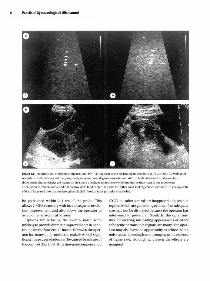
Figure 1.6 Inappropriate time gain compensation (TGC) settings can cause misleading impressions. (a)(i) Correct TGC with good
resolution of all the wires. (ii) Inappropriately increased overall gain causes deterioration of both lateral and axial resolution.
(b) Acoustic characteristics aid diagnosis: (i) a band of enhancement (arrows) behind this ovarian mass is due to reduced
attenuation within the mass, and is indicative of its fluid content (despite the rather solid-looking echoes within it). (ii) The opposite
effect of increased attenuation through a calcified fibroid causes posterior shadowing.
be positioned within 2–3 cm of the probe. Thisallows 7 MHz scanning with its consequent resolu-tion improvement and also allows the operator toavoid other anatomical barriers.
Options for reducing the system noise seemunlikely to provide dramatic improvements in pene-tration for the foreseeable future. However, the oper-ator has many opportunities to make it worse! Signi-ficant image degradation can be caused by misuse ofthe controls (Fig. 1.6a). If the time gain compensation
(TGC) and other controls are inappropriately set thenregions which are generating echoes of an adequatesize may not be displayed because the operator hasintervened to prevent it. Similarly, the opportun-ities for creating misleading appearances of eitherechogenic or transonic regions are many. The oper-ator may also have the opportunity to achieve somenoise reduction using frame averaging at the expenseof frame rate, although at present the effects aremarginal.
Equipment selection and instrumentation 7
Manufacturers may be tempted to declare thattheir probes are working at a higher frequency thanthey really are in order to impress a customer withwhat appears to be extremely high penetration withthe tacit assumption that the corresponding resolu-tion gains are available. The user needs to set morestore by the actual performance of the probe than bythe frequency label.
Contrast resolutionWhereas spatial resolution can be defined as the abil-ity of the system to distinguish two closely spacedtargets, the contrast resolution is its ability to distin-guish two targets of almost the same nature. In otherwords, the ability to identify one point or region asbeing qualitatively different from another solely fromthe grey levels of the echo displayed from the two.If the echoes generated are in fact different but areassigned the same grey levels by the machine, thenthe operator will have no way of knowing they aredifferent. In practice, this will always be true to someextent since the range of incoming echo sizes is manytimes greater than the number of available grey levelsin the machine and even when the number of greylevels within the machine is increased, the funda-mental limit is set by the number which can be mean-ingfully displayed by a television monitor and distin-guished by the eye. Manufacturers have responded tothis by providing a wide range of options for deter-mining which echo amplitudes are translated intowhich grey levels and most equipment has controlslabelled pre- or postprocessing, which allows theoperator to choose, although there remains consid-erable uncertainty about how this can be optimised.The clinical significance of this is illustrated in Fig-ure 1.5d where the same region is scanned at twodifferent grey-scale settings and the diagnostic con-sequences are clear. Operators should be aware thatthe ‘best’ setting will differ between clinical areasand most scanners are set up according to some gen-eral compromise. The more sophisticated machinesallow the operator to use dedicated set-ups if themachine is dedicated to one clinical area, e.g. gynae-cology. How this is determined and validated is prob-lematical.
Contrast resolution – key points� Depends on the perceived number of greylevels� By using different processing or set-up options,contrast resolution may be improved over cer-tain relevant regions. However, this will differaccording to the tissues under observation
Probe shape and sizeTransabdominal imagingThe modern ultrasound machine consists of a mainviewing and control console to which one or moreprobes can be attached. The operator on a day-to-day basis may have to choose between three or fourprobes but at the time of purchase or upgrade, awider choice will be available.
Linear array This is the most traditional of elec-tronic array formats. It is characterised by being rel-atively long and narrow, giving a large anterior fieldof view but requiring good acoustic contact over itswhole length. It is not ideal for most gynaecologicaluse because of its large contact area, often referredto as its footprint.
Curvilinear array The curvilinear array was devel-oped as a sector version of the linear array and isnow the workhorse of many general scanning depart-ments. It has a smaller footprint than the corres-ponding linear array but is subject to some loss ofresolution at the edges of sector towards the largerdepths.
Phased array This type of probe has a particularlysmall footprint because it uses all of the elementsin its length all of the time rather than having theactive section stepping along the array in sequence. Itis most frequently found in cardiology departmentswhere the narrow acoustic window prevents otherprobe types from being effective. Its main drawbackin imaging the pelvis is that its anterior field of view

8 Practical Gynaecological Ultrasound
Figure 1.7 Examples of transvaginal (top) and transabdominal
(bottom) probes used for gynaecological imaging.
is very limited. In addition it is particularly prone tosidelobe artefacts because of its scanning action.
Thus it is probable that some form of electronicarray will be the normal probe of choice for gynae-cological imaging. The majority of patients will besatisfactorily imaged using 5-MHz probes, althougha small number of difficult or obese cases will only beproperly imaged at a lower frequency. In some cases,a higher frequency such as 7.5 MHz will give evenbetter results.
Transvaginal imagingIt is now widely accepted that the optimal imagesfrom many gynaecological patients will be obtainedusing a TV rather than TA technique. The probesdeveloped for this purpose can almost always be fit-ted directly on to the console of standard machine.Indeed, a number of small portable scanners arenow available with TV probes as an option. Therange of probe types, shapes and sizes is surprisinglylarge and there is a marked lack of standardisation(Fig. 1.7 and see Fig. 2.7). Potential purchasers woulddo well to check the viewing angle of a TV probe andbe aware of the compromises in resolution and frame
rate which can be associated with working with wideangles.
Scanning ergonomicsThe choice of probes and consoles is not entirelyobjective and operator preferences continue to beimportant. Having all the controls within easy reachis critical but there are those who prefer more adjust-ments and those who wish to minimise the numberof knobs. There are variations in the weight of probes,the use of foot pedals, the arrangements for calipermeasurements and hard copy, the choice of slidercontrols or others for TGC and the difficulty or easewith which probes can be interchanged. In addition,consideration must be given to whether portabil-ity is important. Even the largest machines shouldbe moveable with good wheel design, but thereare many small, light-weight, inexpensive scannersavailable now which can easily be picked up and car-ried around. The compromise in this case is betweenportability and image quality and facilities.
Operating modesThe normal operating mode of a conventional diag-nostic ultrasound scanner is real-time B-mode. Inaddition, there may be an option of using a modecalled harmonic imaging, which is described below.
Harmonic imagingIt is a feature of soft tissue (and indeed many mater-ials) that as the pulse travels through them it suf-fers distortion. One aspect of this is the generationof additional frequencies which were not present inthe original pulse when it set out. It turns out thatif the original pulse was at a frequency f, then the‘extra’ frequencies will be at multiples of f. In otherwords, frequencies 2f, 3f, 4f etc. will be generatedand these are known as harmonics of f. When har-monic imaging mode is selected, the scanner ‘tunesin’ to one of these higher frequencies (usually 2f )when it is receiving rather than looking for echoes atthe same frequency as it sent out. Since the resolu-tion normally improves with increasing frequency,it might be expected that this would improve the

Equipment selection and instrumentation 9
image quality, and in many cases it does. Howeverthere is another, more important bonus. Much ofthe artefact such as reverberation which obscuresthe ultrasound image is from echoes which do notcontain a significant amount of harmonic. By tun-ing the receiver to the harmonic frequency, theseartefacts are partially suppressed. The net result isa sharper and clearer image.
Of course, this will not improve all scanning onevery occasion, but there are situations where itmakes a significant difference. Some manufactur-ers offer transducers which can be used in har-monic mode if extra software is bought and hencethe machine can be readily upgraded. In other cases,especially if the machine is relatively small andportable, this may not be an option and so purchasersneed to consider carefully what their needs really are.
DopplerFor the detection, assessment and measurement offlow, one of the various Doppler modes should beconsidered. They can be categorised as follows:� continuous-wave (CW) Doppler� pulsed Doppler� colour flow Doppler� power Doppler
CW Doppler In CW Doppler it is necessary to haveseparate transducers for transmission and recep-tion, although both can be incorporated into a singlehousing.
The main problems with CW Doppler are:� There is uncertainty about the anatomical positionof the origin of the signals� It is difficult to use since, unless the probe is posi-tioned correctly, there may be no signal at all andthe operator may not know where to look.� The angle dependence (Cos � term) implies thatif the vessel is approached at or close to 90◦, noDoppler shift will result� Other nearby moving structures, such as vesselwalls, may generate much larger Doppler signals,obscuring those of interest
Figure 1.8 The sample volume has been placed over a small
artery within this ovarian mass. The resulting spectrum from
the artery is displayed as a high-resistance waveform.
As a result of the above, the use of CW Doppler ingynaecology is virtually non-existent and will not bediscussed further.
Pulsed Doppler The main advantage of pulsedDoppler is that the operator can select the regionfrom which the Doppler information is to beobtained because the use of pulses allows the timingto be used as a marker. The commonest approach isto arrange for a line to be generated on the imagealong which Doppler signals will be received andthen for a small ‘sample volume’ to be moved alongthe line by the operator to indicate the precise depthat which the information is required (Fig. 1.8). Thedisplay then shows the Doppler spectrum at thatdepth and hence the technique is also known asspectral Doppler.
Electronic arrays can be used for this purposesince individual elements or groups of elements canbe made to generate the extended pulses or act asreceivers for the Doppler shifted signals. When theappropriate command is given, the display switches
10 Practical Gynaecological Ultrasound
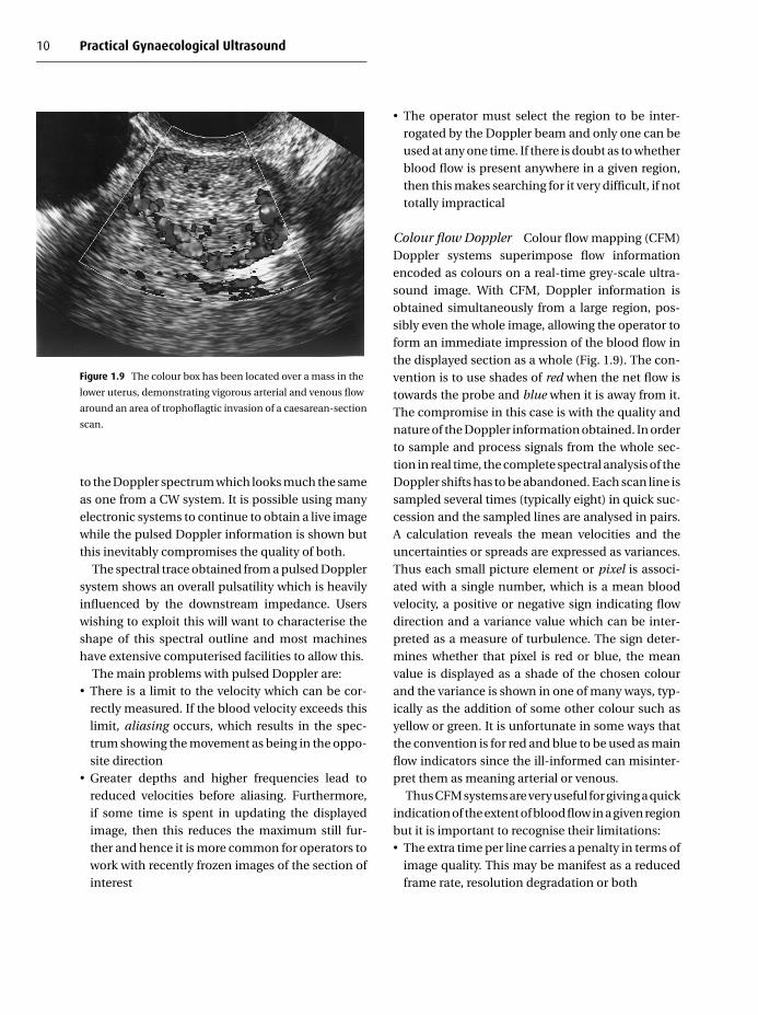
Figure 1.9 The colour box has been located over a mass in the
lower uterus, demonstrating vigorous arterial and venous flow
around an area of trophoflagtic invasion of a caesarean-section
scan.
to the Doppler spectrum which looks much the sameas one from a CW system. It is possible using manyelectronic systems to continue to obtain a live imagewhile the pulsed Doppler information is shown butthis inevitably compromises the quality of both.
The spectral trace obtained from a pulsed Dopplersystem shows an overall pulsatility which is heavilyinfluenced by the downstream impedance. Userswishing to exploit this will want to characterise theshape of this spectral outline and most machineshave extensive computerised facilities to allow this.
The main problems with pulsed Doppler are:� There is a limit to the velocity which can be cor-rectly measured. If the blood velocity exceeds thislimit, aliasing occurs, which results in the spec-trum showing the movement as being in the oppo-site direction� Greater depths and higher frequencies lead toreduced velocities before aliasing. Furthermore,if some time is spent in updating the displayedimage, then this reduces the maximum still fur-ther and hence it is more common for operators towork with recently frozen images of the section ofinterest
� The operator must select the region to be inter-rogated by the Doppler beam and only one can beused at any one time. If there is doubt as to whetherblood flow is present anywhere in a given region,then this makes searching for it very difficult, if nottotally impractical
Colour flow Doppler Colour flow mapping (CFM)Doppler systems superimpose flow informationencoded as colours on a real-time grey-scale ultra-sound image. With CFM, Doppler information isobtained simultaneously from a large region, pos-sibly even the whole image, allowing the operator toform an immediate impression of the blood flow inthe displayed section as a whole (Fig. 1.9). The con-vention is to use shades of red when the net flow istowards the probe and blue when it is away from it.The compromise in this case is with the quality andnature of the Doppler information obtained. In orderto sample and process signals from the whole sec-tion in real time, the complete spectral analysis of theDoppler shifts has to be abandoned. Each scan line issampled several times (typically eight) in quick suc-cession and the sampled lines are analysed in pairs.A calculation reveals the mean velocities and theuncertainties or spreads are expressed as variances.Thus each small picture element or pixel is associ-ated with a single number, which is a mean bloodvelocity, a positive or negative sign indicating flowdirection and a variance value which can be inter-preted as a measure of turbulence. The sign deter-mines whether that pixel is red or blue, the meanvalue is displayed as a shade of the chosen colourand the variance is shown in one of many ways, typ-ically as the addition of some other colour such asyellow or green. It is unfortunate in some ways thatthe convention is for red and blue to be used as mainflow indicators since the ill-informed can misinter-pret them as meaning arterial or venous.
Thus CFM systems are very useful for giving a quickindication of the extent of blood flow in a given regionbut it is important to recognise their limitations:� The extra time per line carries a penalty in terms of
image quality. This may be manifest as a reducedframe rate, resolution degradation or both

Equipment selection and instrumentation 11
� Doppler spectral flow information is lost and hencethe facility for evaluating downstream impedanceand characterising the waveform is unavailable. Itis often necessary to use the CFM as a crude indi-cator of where to look in more detail using pulsedDoppler� There is virtually no quantitative informationavailable from CFM systems. Manufacturers usevery different methods for determining the colourmapping and most allow the operator to modify itstill further. Thus information reported on any onemachine is very difficult to interpret and diagnosticmarkers may well vary between systems� The angle dependence which limits CW and pulsesystems still applies and may lead to confusingartefacts when flow exists but is parallel to theprobe face� The machine has to use some algorithm or ruleto determine when the colour information will beallowed to overwrite and hide the grey-scale infor-mation. This can lead to missing colour when closeto strong stationary targets.
Power Doppler More recently, some manufactur-ers have decided to display the Doppler informa-tion in a different way, often described as powerDoppler. In such systems, all of the power from allof the Doppler-shifted signals within a given regionis added together to produce a single value. In thiscase, no angle correction is needed since no attemptis being made to compute velocities and only onecolour is required since there can be no negativepower values. The benefit is that a much strongertotal signal emerges which does not have angledependence and allows the identification and dis-play of very small vessels which may be too small todetect with CFM systems. Power spectral Dopplerhas many trade names, which adds to the confu-sion, and has mistakenly been described as produc-ing a perfusion map, which is not strictly true. It iscloser to being a description of the total energy asso-ciated with moving blood in a region and seems to beskewed towards venous flow. Its clinical value, if any,remains to be proven but it is undoubtedly attractingmuch attention at the present time.
Operating modes – key points� Predominantly real-time B-mode� Continuous-wave Doppler not practical ingynaecological scanning because the positionof the vessel generating the signal is unknown� Pulsed-wave Doppler uses long pulses ofDoppler from individual elements or smallgroups within an array. It allows the operatorto select a vessel visible on the real-time imageand obtain a spectrum.
The spectrum gives quantitative and quali-tative information about the direction, veloc-ity, variance and downstream resistance of theblood film� Colour flow (CF) Doppler superimposesDoppler information on the real-time image,giving the operator the immediate impressionof an organ’s vascular ‘map’.
It needs more time per line of informationto do this and therefore there are penalties ofpoorer image quality.
It gives information on presence or absenceof flow and its direction, and an impressionof the velocity and variance, but no quantita-tive information. It is usually, therefore, used inconjunction with a pulsed-wave sepctrum� Power Doppler superimposes Doppler infor-mation on the real-time image as with CFDoppler, but displays only the amout of energy,without any of the directional or variance infor-mation. This results in a stronger signal whichmay potentially identify smaller vessels withslower velocities than CF
Contrast agentsThe use of contrast agents is rapidly increasing in allareas of ultrasound. In fact gynaecological applica-tions were among the first to be recognised. Thereis a wide variety of forms but the common elementis that they all contain small gas bubbles. The pres-ence of such bubbles, either in the blood stream orelsewhere, results in a very strong echo being sentback to the transducer. This can result in a vessel

12 Practical Gynaecological Ultrasound
or tube being visualised which would otherwise notbe seen and hence there can be benefits even in nor-mal B-mode operation. However the interaction withthe bubbles can also result in extra harmonic gen-eration and so scanners equipped with this modeof operation might be at an advantage. Further-more, if the bubble is moving then the echo signalwill be Doppler-shifted and so will have the effectof enhancing any of the various Doppler modes aswell.
SafetyThe safety issues which arise in gynaecological ultra-sound can be categorised as follows:� ultrasonic� electrical� microbiological
UltrasonicThe question of whether diagnostic ultrasound canhave harmful effects has been the subject of manypapers and discussions since it was first intro-duced. The reader is referred to the references fora fuller account but it is clear that there is a needfor ongoing vigilance in this area. Traditionally theview has been that ultrasound hazards can arisethrough three mechanisms: cavitation, heating andmicrostreaming.
Cavitation is the growth, oscillation and possiblecollapse of bubbles under the influence of the ultra-sonic field. This is unlikely in typical gynaecologi-cal applications but there is concern where gas-filledcontrast agents are employed.
Microstreaming, as the name implies, is the small-scale local circulation of free fluids both inter- andintracellularly and the consequent alterations to cellmetabolism.
However, most emphasis is now placed on thethermal effects of ultrasound and the possibilityof ultrasound-induced thermal damage. Unfortu-nately, it is extremely difficult to predict the temper-ature rise which a given ultrasonic beam would pro-duce in a given volume of tissue and the knowledgethat vasodilation would generally occur in most liv-ing systems confounds the calculation still further.It is likely that the temperature rise will be related to
the total output power from the transducer and otherfactors. The situation is complex and rapidly chang-ing. Clinical users therefore need to seek advice andreassurance from responsible expert groups. Suchadvice is available from the British Medical Ultra-sound Society (BMUS),1 the European Federationof Societies for Ultrasound in Medicine and Biology(EFSUMB) and the World Federation of Ultrasoundin Medicine and Biology (WFUMB).
Manufacturers in North America are now requiredto adopt a system of on-screen labelling2 to advise theuser about the exposure conditions created by themachine at any time and this has now becomethe de facto standard elsewhere. The system uses twoindices, the mechanical index (MI) and the thermalindex (TI) to advise users about the worst-case in-vivo conditions which might arise from their use ofthe machine in the mode of operation in use at thetime. It is for the user then to decide whether or notto proceed.
The MI is concerned about the liklehood that theremight be cavitation arising within the tissue beingexposed. It is defined as:
MI = p/√
f
where p is the pressure amplitude in MPa after allow-ing for overlying tissue attenuation and f is the fre-quency in megahertz. The BMUS advice1 is that usersshould strive to keep the MI at 0.3 or below whereverpossible.
The TI is defined as:
TI = W/Wdeg
where W is the output power and Wdeg is the powerwhich would lead to a 1◦ rise in temperature in aworst-case scenario. In other words, if the TI is equalto 1, then a worst-case temperature rise of 1 ◦C mightbe expected in vivo.
The ways in which the user might influence theMI and TI values vary between scanners and canbe complicated. BMUS and other organisations havedrawn up helpful guidance and the reader is referredto their websites for further information.1
An additional consideration arises from the pos-sibility of heating directly from the probe itself. Inpulsed Doppler mode, the transducer, which has
Equipment selection and instrumentation 13
been optimised to produce short imaging pulses, isrequired to generate and receive longer pulses andits efficiency for this purpose is relatively low. Theloss of energy due to inefficiency manifests itself asheat within the probe and there is evidence that, leftunattended, in worst-case conditions, some probescan reach up to 60 ◦C.
Most of the vast bulk of epidemiological evidencesupports the assertion that no one anywhere has everbeen shown to have been damaged by ultrasonicenergy from a diagnostic machine. The epidemiolog-ical evidence relating to the safety of ultrasound inobstetric applications has been reviewed on a num-ber of occasions3 and it is generally assumed that, ifthe exposure conditions are acceptable for obstetricuse, then they will also be reasonable for other appli-cations. Studies have been conducted relating to pos-sible associations of ultrasonic exposure of the fetuswith childhood malignancies, neurological malde-velopment, left-handedness and low birth weight.It seems clear that most of the work supports thenotion of there being no association of these out-comes with ultrasound exposure. However, they con-clude that, on some issues, such as the incidenceof left-handedness and low birth weight, no firmconclusions can yet be drawn and hence the recenttrend has been towards somewhat more guardedstatements than in the past. There is no direct evi-dence that these effects are harmful but, nonethe-less, it should be noted that the general consensus isthat most at risk is the developing embryo and thatthis risk is maximised when the embryo is imagedtransvaginally using pulsed Doppler.
It should be stressed that routine clinical scanningof every woman during pregnancy using real-timeB-mode imaging is not contraindicated by the evi-dence currently available from biological investiga-tions and its performance should be left to clinicaljudgement.
In view of the possibility of ultrasonically inducedbiological effects within tissues in the path of aDoppler beam, routine examinations of the develop-ing embryo during this particularly sensitive periodof organogenesis using pulsed Doppler devices isconsidered inadvisable at present. It is advisable tominimise output levels and exposure time in pulsed
Doppler mode during fetal examinations and par-ticularly when fetal bone structures lying within theDoppler beam may be preferentially heated.4
In this light, it seems clear that the general prin-ciple must be to avoid unnecessary ultrasonic expo-sure of anyone. The use of ultrasound should there-fore be to limit the dose to that which is needed toobtain the appropriate clinical information. In otherwords, it should be governed by the ALARA principle(as low as reasonably achievable), which applies inconventional radiography.
ElectricalUltrasonic probes in medicine are subject to thesame electrical safety requirements as any otherelectromedical equipment. In the UK, they mustsatisfy the British Standard BS5724 (or its Inter-national Electrotechnical Commission equivalent).This standard is particularly demanding of intracav-itary devices such as TV probes since they are in verygood electrical contact with the patient. In general,manufacturers are careful to ensure that their equip-ment complies with these regulations and problemsare rare. However, it is important to bear in mind thatit is the whole system that must meet the require-ments. Thus if the scanner to which a TV probe isattached is itself connected electrically to anotherpiece of equipment such as a camera, video cassetterecorder or computer, then the complete system mayfail electrical tests even though its individual compo-nents have passed. Obviously any physical damageto a probe or its cable such as a crack which mightreduce its electrical insulation must be taken seri-ously, recorded and drawn to the attention of therelevant parties.
MicrobiologicalWhile any piece of equipment which is regularlycoming into contact with patients must be kept cleanto avoid cross-infection, this is particularly import-ant for TV probes. Unlike many other devices usedin a similar way, ultrasound probes cannot be auto-claved but some of them have design shapes whichmake them more difficult to sterilise than others.The considerations include the nature of any clean-ing methods, the type of disinfectant to be used, the

14 Practical Gynaecological Ultrasound
use of probe covers and the need for an asepctictechnique. The issue has been the subject of anadvisory statement from the American Institute forUltrasound in Medicine (AIUM).5 All such methodsmust be used in the full awareness of specific advicefrom the equipment manufacturer since some probematerials will be irreparably damaged by some anti-septics such as gluteraldehyde.
Thus it is clear that this is another situation inwhich the operator plays the crucial role. By beingaware of the situation and adhering closely to pub-lished guidelines, the operator can reduce the patientexposure in an ultrasound examination manifold.It has often been claimed that: ‘In diagnostic ultra-sound, the greatest hazard to the patient is that pre-sented by the untrained or poorly trained operator’.
Practical use of ultrasound – key points� The basic gynaecological ultrasound servicerequires both transabdominal and transvagi-nal capabilities. It should be subject to regularquality control and safety checks� Choice of equipment must be informed, takinginto account its performance in terms of reso-lution, penetration and probe selection anddesign� The operator must use a combination of bothtechnical ability and clinical knowledge inorder to maximise the diagnostic capability.He/she should have undergone recognisedtraining specific to gynaecological ultrasoundand maintain regular scanning experience andcontinuing development in the field� Good, safe practice includes:� regular audit and quality control procedures� the use of current guidelines and schemes of
work� operation in accordance with the ALARAprinciple (as low as reasonably achievable)� recognition of any limitations of the equip-ment and the technique used
R E F E R E N C E S
1. British Medical Ultrasound Society http://www.bmus.org/
safety of ultrasoundNF.htm.
2. M. D. Laurel, American Institute of Ultrasound in Medicine/
National Electrical Manufacturers Association (AIUM/
NEMA), Standard for real-time display of thermal and
mechanical acoustic output indices on diagnostic ultrasound
equipment, revision 1. AIUM (1998).
3. K. A. Salvesen and S. H. Eik-Ness, Is ultrasound unsound? A
review of epidemiological studies of human exposure to ultra-
sound. Ultrasound in Obstetrics and Gynecology, 6 (1995),
293–8.
4. European Federation of Societies for Ultrasound in Medicine
and Biology (EFSUMB), Clinical safety statement 1994,
Trondheim. European Journal of Ultrasound, 2 (1995), 77.
5. S. R. Goldstein, AIUM: report for cleaning and preparation
of endocavitary ultrasound transducers between patients.
Ultrasound in Obstetrics and Gynecology, 7 (1996), 92–4.

2
Practical equipment operation and technique
Jane BatesSt James’s University Hospital, Leeds
Practical approach to image optimisation
Arguably the first consideration in choosing amachine is the quality of the image in terms of reso-lution. Equipment differs significantly, and shouldbe carefully evaluated before purchase by someonewith experience in gynaecological ultrasound. Costis not necessarily an indicator of quality of image,and in some cases it may be advisable to forgo ele-ments of advanced functionality in favour of a basic,good-quality image.
Intelligent, informed operation of even a basic sys-tem is the key to accurate diagnosis. There are anumber of controls which can be found on even themost basic systems which, if used correctly, offer sig-nificant improvements in image quality which caninform the appropriate patient management. Thepractical improvements that can be made by theoperator to the image are underpinned by an under-standing of the theoretical principles outlined inChapter 1.
As demonstrated in Chapter 1, using the tissue-equivalent phantom, the image should be optimisedusing the focal zones, frame rate and line density, fre-quency manipulation and other image-processingoptions.
Frame rate and line density
The pelvic viscera are usually stationary targets and,as such, can be examined using a relatively low frame
rate. This has the effect of increasing the line densitywith a consequent improvement in diagnostic infor-mation (Fig. 2.1).
Focal zone
The focal zone, which corresponds to the area wherethe beam is narrowest, should be aligned against thestructure under examination, e.g. the ovary. Whena single focal zone is used it is important to placeit to affect the depth of interest (Fig. 2.2). If goodresolution is required through a greater depth – forexample, when looking at a large, fibroid uterus –the number of focal zones can be increased to threeor four. This keeps the beam narrow over a greaterdepth at the expense, again, of decreasing the framerate.
Depth/sector angle/zoom
Resolution, in terms of line density, may also beimproved by choosing to scan a smaller area. The sec-tor angle may be narrowed when scanning an ovary,for example. For looking at structures in the near ormid-field, the depth may also be reduced (Fig. 2.3).This improves the line density whilst maintaining theframe rate. In addition, small areas may be zoomedfor closer examination, achieving a very good linedensity within a small area.
C© Cambridge University Press 2005.
15

16 Practical Gynaecological Ultrasound
a
b
Figure 2.1 Effect of frame rate. (a) A high frame rate reduces
the line density, losing image quality. (b) By lowering the frame
rate, the line density is increased, giving a much improved
image.
Frequency
As we have seen in Chapter 1, the higher the fre-quency the better the resolution, but the poorer thepenetration. The operator must therefore choose thehighest frequency possible whilst being able to pen-etrate to the required depth. Most modern machineswith broadband technology allow the user to changethe resonant frequency without changing transduc-ers, and the operating frequency can therefore bechanged throughout the examination as appropri-ate (Fig. 2.4).
a
b
Figure 2.2 Effect of focal zone. (a) The focal zone is
inappropriately placed in the near field, with consequent poor
resolution of the ovary. (b) Correctly placed focal zone with
good delineation of the ovarian follicles.
Tissue harmonics
The use of non-linear harmonics, if available on yourmachine, can be very useful in reducing artefacts. Inparticular, when examining cystic structures, rever-beration and noise can often be eliminated, allow-ing more accurate interpretation of the appearances.Tissue harmonics tends to produce an image whichhas a reduced dynamic range (looks more ‘contrasty’)

Practical equipment operation and technique 17
a
b
Figure 2.3 Effect of field of view. (a) Left ovary. (b) Same ovary
with a reduced sector angle and depth of field allows a higher
line density, and enables the operator to appreciate the ovarian
morphology better.
and so is usually used in conjunction with funda-mental imaging during the scan (Fig. 2.5).
Extended field of view
Many machines now have the ability to displayextended images over a greater field of view. Thisdoes not add to the image quality, but can allow theoperator to appreciate the extent of large masses,
a
b
Figure 2.4 Effect of frequency. (a) Transvaginal scan of a
normal ovary. (b) The same ovary, using the same probe but
switched to a higher resonant frequency: improved detail and
resolution, but with slightly poorer penetration.
which could not otherwise be entirely displayed onone image, and can facilitate more accurate meas-urements (Fig. 2.6).
Choice of approach
The pelvic organs are routinely visualised by twoapproaches – transabdominal (TA) and transvagi-nal (TV). Each has its own advantages and limi-tations and examinations frequently employ bothtechniques, depending on the reason for referral.Occasionally a transrectal (TR) scan may be useful,

18 Practical Gynaecological Ultrasound
a
b
c
Figure 2.5 Tissue harmonic imaging (THI). (a) The contents of
this ovarian cyst are unclear. (b) With THI peripheral blood clot
can be demonstrated clearly. (c) The endometrium is more
clearly outlined with THI on the right-hand image.
Figure 2.6 The use of extended field of view helps the operator
to appreciate the full extent of this case of endometriosis, in
which large endometriotic cysts extend well above the fundus
of the uterus.
Figure 2.7 Examples of probes suitable for gynaecological
ultrasound. Top, curved array for transabdominal scanning;
middle, transvaginal probe, bottom, high-frequency linear
array suitable for examination of the anterior abdominal wall.
for example, in a postoperative patient with a pelviccollection who is unable to tolerate a TV scan.
Most general gynaecological scanners require atleast two probes – a general curved array (around4–5 MHz) and a higher-frequency transvaginal probe(7.5 MHz) (Fig. 2.7).
The curved array probe is also suitable for scan-ning other abdominal organs where necessary, suchas the kidneys for suspected hydronephrosis, or the

Practical equipment operation and technique 19
Figure 2.8 The use of a linear array probe demonstrates
malignant plaque or ‘cake’ on the anterior abdominal wall in
the near field in a patient with ovarian carcinoma. Note that
the plaque is vascular on colour Doppler. As the vessels are
approximately perpendicular to the beam, and therefore
undetectable by Doppler, the colour box has been ‘steered’ to
create a smaller angle between the beam and the vessels.
liver, spleen and adrenals to exclude metastasesin the case of gynaecological cancer. It may alsobe useful, particularly with ovarian carcinoma, touse a high-frequency linear array probe to examinethe abdominal wall for peritoneal or omental plaquefrom disseminated cancer (Fig. 2.8). This probe hasa wide near field of view, with good line density andresolution throughout the depth of view.
Scan preparation
In symptomatic patients who have not had previousscans, it is advisable to prepare the patient for botha TA and TV scan. Women attend with a full bladder,allowing the TA scan to be performed first (see below)
Figure 2.9 The wide field of view of the Transabdominal scan
can accommodate the uterus and both ovaries in transverse
section.
with the intention of proceeding to a TV scan aftermicturition.
A careful explanation of the procedure intendedand a private scanning environment are essential. Itis also good practice to offer the services of a chap-erone where possible.
It is invariably necessary first to take a careful his-tory from the patient. This should include the cur-rent menstrual state in addition to current and pastgynaecological history.
Transabdominal (TA) scanning
The main advantage of TA ultrasound lies in its abilityto encompass a comparatively large field of view. Thisis useful for:� locating the ovaries in relation to the uterus, par-
ticularly those sited laterally (Fig. 2.9).� demonstrating large masses such as a fibroiduterus, adnexal masses or pelvic collections� demonstrating iliac fossae, bladder and any asso-ciated renal pathology� demonstrating uterine anomalies, such as bicor-nuate uterus, which may be more difficult to appre-ciate on a TV scan.
TA scanning, via a distended bladder, has been usedsince diagnostic ultrasound began. The full bladder

20 Practical Gynaecological Ultrasound
b
Figure 2.10 Bladder filling on Transabdominal scans. (a)
Anteverted uterus with an almost empty bladder. (b) Optimal
bladder filling retroflexes the uterus and displaces bowel. Note
the strong reflection from the intrauterine contraceptive
device.
displaces small bowel away from the pelvic visceraand partially retroflexes the normally anteverteduterus to maintain the endometrial echo at a moreperpendicular angle to the beam (Fig. 2.10).
Some patients find the distended bladderuncomfortable, and others are unable to reachthe required degree of filling. Associated medicalproblems, such as incontinence, renal failure or pre-vious bladder surgery, also prevent adequate filling
Figure 2.11 The presence of free fluid allows the pelvic viscera
to be scanned transabdominally with the bladder empty. The
uterus and ligaments are demonstrated.
and TV techniques should be considered for thesepatients.
The full bladder itself displaces the organs into thefar field of the image where the resolution is usuallyinferior. The bladder can also give rise to unwan-ted artefacts such as reverberation and mirror-imaging.
Occasionally, ascites may avoid the need for afull bladder, outlining the uterus, ovaries and broadligament sufficiently to obtain diagnostic informa-tion (Fig. 2.11). It is useful, however, to visualise thebladder itself, particularly when bladder pathologyis present (Fig. 2.12), or when trying to distinguishpelvic cysts from structures of vesical origin (e.g.bladder diverticula).
Transabdominal technique
The patient is usually scanned supine with the dis-tended bladder as a ‘window’ to visualise the uterusand ovaries in longitudinal and transverse sections.The uterus is best displayed with its long axis per-pendicular to the beam. This varies from patientto patient, in terms of ante- or retroversion and

Practical equipment operation and technique 21
Figure 2.12 Filling the bladder has the advantage of being able to detect other, often unsuspected pathology, such as this
ureterocele.
obliquity. Having found the uterine axis, the pelviscan be examined from side to side by a combinationof transducer movement and angulation.
The organs should always be examined in twoplanes where possible, and by turning at right anglesto the uterine axis, transverse or axial scans canthen be performed through the pelvis, maintainingthe beam perpendicular to the endometrial cavity(Figs. 2.13 and 2.14).
The position of the ovaries varies from patientto patient, and according to the degree of bladder-filling. By maintaining the bladder as an acousticwindow, the relationship of the ovaries to the uteruscan be demonstrated. It is often helpful to identifythe ovaries first in transverse section by visualisingthe uterine cornu and scanning slightly inferior andlateral to this.
It is often advisable to perform any preliminarymeasurements (of ovarian volume, masses, etc.) atthis stage in case visualisation is incomplete orunsuccessful on TV scanning. An ovarian volumeestimation requires three measurements truly per-pendicular to each other (Fig. 2.15). This is easier toachieve with a TA scan, as the planes obtained TV areslightly oblique.
Transvaginal scanning
The reduced distance between probe and organs inthe TV route allows the use of a higher frequency(7.5 MHz). The additional benefit of a lack of layersof subcutaneous tissue (which attenuate the soundin a TA scan) culminates in vastly superior resolutionby using a TV technique (Fig. 2.16).
Unfortunately, TV scanning does have somedrawbacks: the field of view is smaller, makingassessment of larger organs and masses difficult.Large masses will lie outside the transducer’s fieldof view and focal zone, and there is reduced flexibil-ity in the available planes of scan which can makemeasurements such as ovarian volumes less easy.
The bladder should be empty in order to allowthe uterus and ovaries to lie close to the transducerwithin its focal zone. This often makes the TV scanmore acceptable to women than the TA approach.
Acceptability to the patient
Patient acceptability depends almost entirely onthe approach by the sonographer and it is rare forpatients to decline. The amount of time necessary

22 Practical Gynaecological Ultrasound
Figure 2.13 (a) The endometrial cavity echo is poorly
demonstrated because of its low angle to the beam. (b) With a
cephalic angle, maximum reflection from this interface is now
obtained. (Note how the echo from the vaginal interface has
now disappeared.)
to explain the procedure and put the patient at herease is always well spent as the benefits of a TVscan in terms of improved acoustic information areenormous.1 Privacy and dignity must be maintainedat all times, and patients may sometimes feel morecomfortable with a family member present. Thereare very few contraindications to vaginal scanningbut these include paediatrics and virgi intactae.
Patients may also be offered a chaperone whois, preferably, a female member of staff familiarwith departmental practices. This has the advantageof reassuring and assisting the patient whilst also
affording a measure of protection to the operator interms of confirming the nature of the scan subse-quently if necessary.
Friendly and professional communication withthe patient cannot be stressed too highly, and themajority of litigious cases surrounding TV ultra-sound, though few, could possibly have been avoidedby employing good communication skills.
Transvaginal technique
The scan is performed with an empty bladder andusually carried out with the patient semi-recumbent,knees bent, buttocks resting on a pad or pillow. Thisis usually quite sufficient to allow the operator tomanoeuvre the probe satisfactorily. (The use of alithotomy table, with stirrups for the patient’s legs,is normally unnecessary but may be found in somespecialised departments such as assisted conceptionunits.) Alternatively, the decubitus position, partic-ularly in patients who have difficulty lying supine, isuseful.
Using a slightly reverse Trendelenburg positionencourages any free fluid to collect in the pouch ofDouglas, outlining the posterior uterine wall and, insome cases, the adnexal structures.
In the case of the patient’s first attendance, the TVscan will often follow a TA survey, which will haveallowed the operator to locate the position and lie ofthe uterus and ovaries and highlighted any masses.(Patients who attend for follow-up scans or regularscreening examinations are usually able to proceedstraight to TV scans without first filling the bladder.)
Different scan planes are achieved by a com-bination of rotation of the probe and angulation(Fig. 2.17). It is sometimes helpful for the learner toimagine the sector beam as a thin fan emerging from,and fixed to the probe, and then retain this mentalimage as the probe is turned and angled after inser-tion. As with any scanning technique, it is importantto adapt to the individual patient and not performthe procedure simply as a technical process.
The probe is inserted gently into the vagina andmay be located in the fornix or withdrawn slightlyback down the vagina, to display the uterus fully.
Practical equipment operation and technique 23
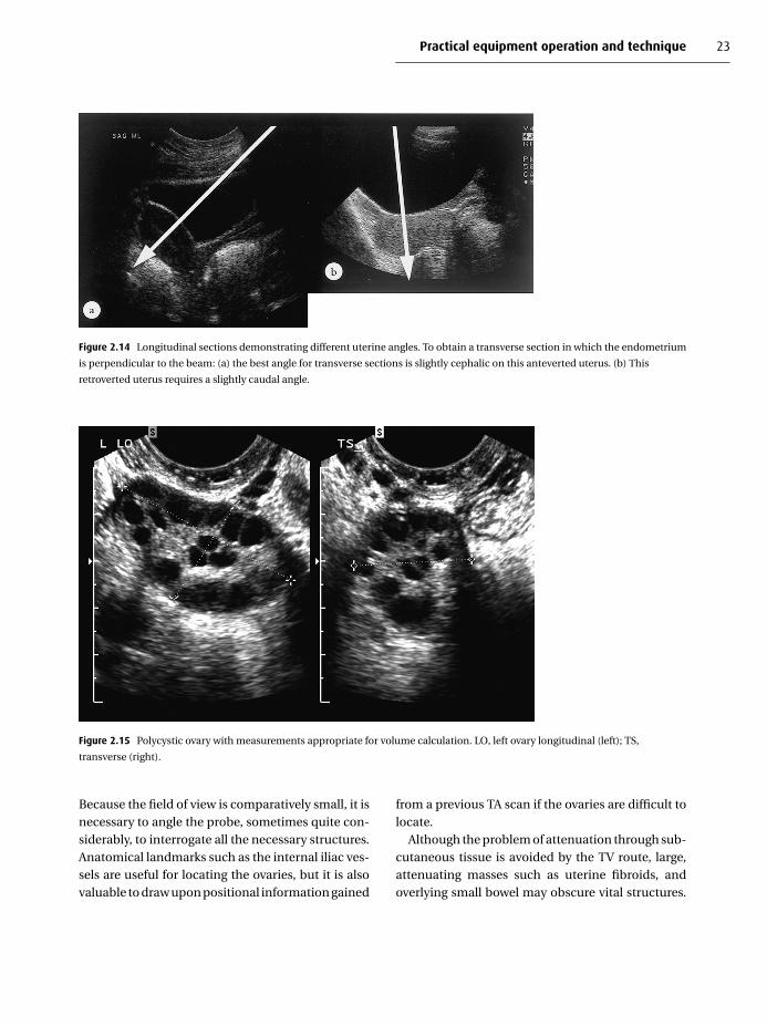
Figure 2.14 Longitudinal sections demonstrating different uterine angles. To obtain a transverse section in which the endometrium
is perpendicular to the beam: (a) the best angle for transverse sections is slightly cephalic on this anteverted uterus. (b) This
retroverted uterus requires a slightly caudal angle.
Figure 2.15 Polycystic ovary with measurements appropriate for volume calculation. LO, left ovary longitudinal (left); TS,
transverse (right).
Because the field of view is comparatively small, it isnecessary to angle the probe, sometimes quite con-siderably, to interrogate all the necessary structures.Anatomical landmarks such as the internal iliac ves-sels are useful for locating the ovaries, but it is alsovaluable to draw upon positional information gained
from a previous TA scan if the ovaries are difficult tolocate.
Although the problem of attenuation through sub-cutaneous tissue is avoided by the TV route, large,attenuating masses such as uterine fibroids, andoverlying small bowel may obscure vital structures.

24 Practical Gynaecological Ultrasound
a b
c d
Figure 2.16 (a) Transabdominal uterus. (b) Transvaginal scan of the same uterus as (a), clearly showing the ovulatory stage of the
endometrium. (c) Transabdominal scan showing both ovaries. (d) Transvaginal scan of the right ovary in the same patient as
(c), showing the corpus luteum clearly.
Figure 2.17 Basic transvaginal planes of scan.

Practical equipment operation and technique 25
This is a particular problem in postmenopausalwomen when the uterus and ovaries are atrophied.Gentle manipulation of the ovaries and bowel trans-abdominally with the free hand can overcome thisproblem and may bring superiorly placed ovariesdown into the focal zone of the transducer.
A further advantage of TV scanning is the abil-ity to use the probe to push the pelvic viscera gen-tly and establish whether they move freely over theperitoneal surfaces. Known as the ‘sliding organ’sign, this is a useful clue in the diagnosis of adhe-sions, which will prevent this free organ movementif present.
Manipulation of the transducer by angling, rotat-ing and sliding movements should obviously be gen-tle and slow to avoid tension and discomfort.
Biological safety
To minimise the risk of infection, the probe shouldalways be covered with a disposable cover. Commer-cially available condoms are adequate for the pur-pose of covering the TV probe, but those with sper-micidal lubrication should be avoided, particularlyin assisted conception units. A small amount of cou-pling gel should first be introduced into the condom,which is then rolled on to the transducer, smoothingany air bubbles away from the transducer face. Gel isthen applied to the outside to maintain contact andfacilitate insertion of the probe. Most condoms con-tain latex, making them unsuitable for use in womenwith a latex allergy. In such cases, the finger of a latex-free surgical glove makes a useful probe cover.
The sonographer is responsible for minimising therisk of cross-infection and probes should be thor-oughly cleaned after each procedure using a disin-fectant approved by the manufacturer.2
Table 2.1 gives a comparison of TA and TV tech-niques.
Recognising the acoustic characteristics
It is vital to recognise the acoustic characteristicsof organs and masses in order to interpret the scan
Table 2.1 Summary of advantages and limitations of transabdominal
and transvaginal techniques
Transabdominal (TA) Transvaginal (TV)
Field size Large: displays relationship
of ovaries to uterus
Limited; large masses may be
beyond focal zone
Accommodates large
masses within the image
May not be able to
accommodate entire uterine
section in the field
Flexibility Easy to examine upper
abdomen (e.g. kidneys)
with same transducer
Must use TA transducer for
upper abdomen
Bladder and distal ureters
can be assessed
Bladder not well seen
Invasive
nature
Perceived as non-invasive,
so is the technique of
choice for paediatrics
and others
May be perceived as invasive.
Must have good
patient–sonographer
communication
Privacy essential
Preparation Full bladder may be
uncomfortable or
impossible
No preparation required
Resolution 3.5/5 MHz 5/7.5 MHz
Limited resolution,
especially in far field
Considerably superior to TA
correctly (Fig. 2.18). Appearances to be taken intoconsideration when making a diagnosis include:� internal echo content and pattern� margins or capsule of the lesion – well- or ill-
defined, focal thickening or nodules� attenuation characteristics – posterior enhance-ment, shadowing or mixed attenuation
For example, a simple cyst or follicle should havea well-defined, thin, regular outer capsule, no inter-nal echoes and posterior acoustic enhancement. Anydeparture from these criteria implies that the lesionis not a simple cyst. Internal echoes with poster-ior enhancement suggest that the mass is predom-inantly fluid but contains other material such ashaemorrhage.
Recognition of artefact is of particular import-ance when interpreting the appearances. Reverber-ation or noise can mimic haemorrhage, pus or evenseptations. Always ensure you scan from differ-ent angles and in different planes to be confident

26 Practical Gynaecological Ultrasound
a b
c d
Figure 2.18 (a) This ovarian cyst has a well-defined capsule with a band of posterior acoustic enhancement (arrows). Note the
low-level echoes within it from blood. (b) Although this mass also contains low-level echoes, it has no posterior enhancement and is
a small, solid fibroma. (c) The fibroid on the right uterine wall is clearly solid, attenuating the beam and making it difficult to
demonstrate its posterior margins. (d) Despite the internal echoes, the enhancement posterior to this cystadenocarcinoma
demonstrates that it is predominantly fluid in nature.
that appearances represent true findings. Incorrectgain settings can obliterate important characteris-tics, such as posterior enhancement, shadowing orlow-level internal echoes, which would otherwise aiddiagnosis.
Doppler techniques
Although colour and spectral Doppler modes arecapable of giving haemodynamic information about
the pelvic viscera, Doppler often has little to con-tribute to the general gynaecological ultrasoundscan, due to the non-specific nature of the informa-tion obtained.
Displaying small vessels within the ovaries ishighly dependent on the sensitivity of the equip-ment, and many normal ovaries appear ‘avascular’,particularly in the early part of the cycle, simplybecause the machine cannot detect such small, low-velocity vessels.

Practical equipment operation and technique 27
a
b
Figure 2.19 Transvaginal section through a normal ovary.
(a) With colour Doppler tiny intraovarian blood
vessels are demonstrated. Red indicates flow towards, and blue
away from the transducer. (b) Power Doppler can be more
sensitive than colour, displaying small low-velocity vessels in
the ovary.
Power Doppler tends to be more sensitive thancolour Doppler, and has the advantage that it isnot as angle-dependent, potentially displaying a sig-nal in vessels which are perpendicular to the beam(Fig. 2.19).
The information available from using Doppler isboth qualitative (establishing whether somethingis vascular or not) (Fig. 2.20) and quantitative(measurements of resistance index, for example)(Fig. 2.21). The TV approach, because of its higher
a
b
c
d
Figure 2.20 (a) The tubular structure on the left of the uterus
could be a vessel or dilated tube. (b) Colour Doppler indicates
that it is a vein. (c) The endometrium is indistinct in a patient
with bleeding following dilatation and curettage. (d) Colour
Doppler demonstrates an atriovenous malformation not
visible on the grey-scale image.
28 Practical Gynaecological Ultrasound
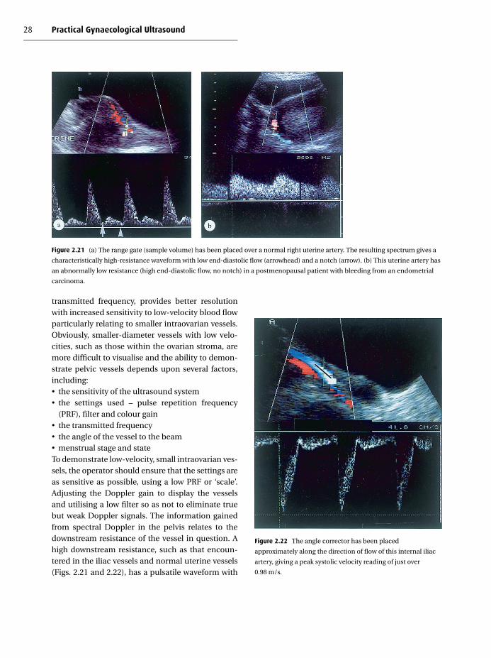
Figure 2.21 (a) The range gate (sample volume) has been placed over a normal right uterine artery. The resulting spectrum gives a
characteristically high-resistance waveform with low end-diastolic flow (arrowhead) and a notch (arrow). (b) This uterine artery has
an abnormally low resistance (high end-diastolic flow, no notch) in a postmenopausal patient with bleeding from an endometrial
carcinoma.
transmitted frequency, provides better resolutionwith increased sensitivity to low-velocity blood flowparticularly relating to smaller intraovarian vessels.Obviously, smaller-diameter vessels with low velo-cities, such as those within the ovarian stroma, aremore difficult to visualise and the ability to demon-strate pelvic vessels depends upon several factors,including:� the sensitivity of the ultrasound system� the settings used – pulse repetition frequency
(PRF), filter and colour gain� the transmitted frequency� the angle of the vessel to the beam� menstrual stage and stateTo demonstrate low-velocity, small intraovarian ves-sels, the operator should ensure that the settings areas sensitive as possible, using a low PRF or ‘scale’.Adjusting the Doppler gain to display the vesselsand utilising a low filter so as not to eliminate truebut weak Doppler signals. The information gainedfrom spectral Doppler in the pelvis relates to thedownstream resistance of the vessel in question. Ahigh downstream resistance, such as that encoun-tered in the iliac vessels and normal uterine vessels(Figs. 2.21 and 2.22), has a pulsatile waveform with
Figure 2.22 The angle corrector has been placed
approximately along the direction of flow of this internal iliac
artery, giving a peak systolic velocity reading of just over
0.98 m/s.

Practical equipment operation and technique 29
low end-diastolic flow and often a notch during earlydiastole. A measure of this resistance to flow can bemade by several indices, the most commonly usedbeing:
Resistance index − A − BA
Systolic/diastolic index − AB
Pulsatility index − A − Bmean
where A is peak systolic frequency, and B end-diastolic frequency.
If you are unable to obtain any Doppler flow,check that:� the angle of insonation of the vessel is low –
preferably less than 60◦ to the beam� the Doppler gain setting is turned up suffi-ciently� the filter is on a low setting� the system pulse repetition frequency is set forlow velocities – (i.e. low ‘range’ or ‘scale’ setting)� the transmitted power is sufficient (within theas low as reasonably achievable (ALARA) prin-ciple)� power Doppler is often more sensitive indemonstrating small, low-velocity vessels
Additional techniques
Repeating the pelvic scan after an interval of 1 or 2weeks can be a useful exercise in some cases; thephysiological nature of appearances may be con-firmed by scanning during a different stage of themenstrual cycle.
Occasionally, loaded bowel may mimic a mass, orobscure pelvic structures. In such cases a repeat scanafter bowel preparation may be necessary.
Saline contrast hysterosonography, in which asmall amount of warm saline is introduced via acatheter into the endometrial cavity, improves visu-alisation of the endometrium and is particularly
Figure 2.23 Saline infusion hysterosonography. Saline has
been infused into the endometrial cavity, outlining this small
polyp.
successful in the investigation of postmenopausalbleeding.3 This has the effect of mildly distending thecavity and increasing the contrast resolution, outlin-ing abnormalities not seen on the routine ultrasound(Fig. 2.23).
The use of contrast media in the investigation ofinfertility and tubal patency is discussed in subse-quent chapters.
Many probes have the option of a biopsy attach-ment (Fig. 2.24), which enables drainage and biopsyprocedures to be carried out with accurate place-ment of the needle and minimum discomfort to thepatient.
The role of the sonographer
Most gynaecological ultrasound in the UK is car-ried out by sonographer practitioners who haveundergone recommended training and assessmentprogrammes in medical ultrasound. It is a well-recognised fact that ultrasound is a highly operator-dependent technique, and one of the greatest haz-ards in diagnostic ultrasound is not any perceivedbiological effects on tissues, but its use by untrainedpersonnel.4

30 Practical Gynaecological Ultrasound
Figure 2.24 (a) A transvaginal probe with a needle-guide
attatchment. (The probe cover has been omitted for clarity.)
(b) Using the guide, a needle is placed into an ovarian cyst for
drainage.
The gynae sonographer is a vital part of themultidisciplinary team, providing expertise in clin-ical ultrasound techniques and making an invalu-able contribution to the management of the gynaepatient. Their role in audit and quality control, train-ing, research and development is well established inthe UK. Most good departments operate to regularlyreviewed ultrasound schemes of work, which outlinebest practice and provide a framework for monitor-ing the quality of the service.
The production of images as a record of the exam-ination can provide a useful baseline for subsequentexaminations, and can also be invaluable in pro-viding medicolegal proof that the operator has per-formed a comprehensive examination in a com-petent fashion.5 Many departments prefer to storethese electronically. Although incurring significantcapital costs, this is a subsequently cost-effectivesystem of recording, storing and retrieving patientexaminations.
A successful sonographer-based ultrasound ser-vice incorporates:� recognised training� continuing education� regular, frequent ultrasound practice� proper delegation by the medically qualified
person in charge� the use of protocols or schemes of work� good audit and quality control procedures
Summary
The practitioner should aim to maximise the diag-nostic information by:� carefully assessing the patient’s history� giving a full explanation of the procedure to facili-
tate cooperation� utilising the equipment controls properly through-out the scan� using any additional techniques necessary such assaline infusion
Both TA and TV techniques have their benefits. Thereare situations in which TA scanning alone cannot

Practical equipment operation and technique 31
deliver the required information – assessing the post-menopausal endometrium, for example, requires thefine detail of TV scanning. Alternatively, large pelvicmasses cannot be accommodated in the field ofthe TV transducer, which may underestimate theirextent, and are better appreciated transabdominally.
The strength of the ultrasound examination liesin the fact that it is dynamic. The operator must beboth technically skilled and clinically aware, with aflexible approach using a combination of techniqueswhere necessary. Recognised training together withcontinued professional development will ensure thatthe scan findings are appropriately interpreted.
Practical use of ultrasound – key points� The basic gynaecological ultrasound servicerequires both transabdominal and transvagi-nal capabilities. Equipment should be subjectto regular quality control and safety checks� Choice of equipment must be informed, takinginto account its performance in terms of reso-lution, penetration, probe selection and design� The practitioner must use a combination oftechnical ability and clinical knowledge tomaximise the diagnostic capability. He/sheshould have undergone recognised trainingspecific to gynaecological ultrasound andmaintain regular scanning experience and con-tinuing development in the field6� Good, safe practice includes:� regular audit and quality control procedures� the use of current guidelines and schemes of
work� operation in accordance with the as low asreasonably achievable (ALARA) principle� recognition of any limitations of the equip-ment and the technique used
R E F E R E N C E S
1. Royal College of Radiologists, Intimate Examinations
(London: Royal College of Radiologists, 1998).
2. S. R. Golstein, Report for cleaning and preparation of endo-
cavitary ultrasound transducers between patients. Ultra-
sound in Obstetrics and Gynecology, 7 (1996), 92–4.
3. L. Rogerson, J. Bates, M. Weston and S. Duffy, Outpatient
hysteroscopy versus saline infusion hydrosonography (SIH).
British Journal of Obstetrics and Gynaecology, 109 (2002),
800–4.
4. J. Bates, D. Lindsell and C. Deane, Extending the Provision
of Ultrasound Services in the UK (London: British Medical
Ultrasound Society, 2003).
5. H. B. Meire, Ultrasound-related litigation in obstetrics and
gynaecology: the need for defensive scanning. Ultrasound in
Obstetrics and Gynecology, 7 (1996), 233–5.
6. United Kingdom Association of Sonographers,Guidelines
for Professional Working Standards; Ultrasound Practice
(London: United Kingdom Association of Sonographers,
1996).

3
Anatomy, physiology and ultrasound appearances
Jane BatesSt James’s University Hospital, Leeds
Introduction
The female reproductive system comprises thevagina, uterus, ovaries and fallopian tubes. Theappearances of these structures on ultrasounddepend on the age of the patient and menstrualstate and stage at the time of the scan. Throughoutthe woman’s life, particularly during the functionalcycle, the organs are subject to physiological changesbrought about by the influence of the hormones.
While the main object of the scan in most situ-ations is to examine the reproductive organs, it isalso essential to know and recognise other structureswithin the pelvis, including muscles, blood vessels,ligaments, rectum and sigmoid colon and the blad-der and ureters (Figs. 3.1 and 3.2).
Vagina
The vagina is a midline, thin-walled, muscular struc-ture approximately 8–9 cm in length, extending fromthe uterus to the vestibule (Fig. 3.3). It is H-shaped incross-section, constructed of longitudinal folds andtransverse ridges (rugae) which give it the ability todistend to accommodate the fetus during parturi-tion. In its upper portion, it is contiguous with theuterine cervix and it divides into the fornices – anter-ior, posterior and right/left lateral.
During transabdominal (TA) scanning the dis-tended bladder, which acts as an acoustic window,does not affect vaginal position. The vagina cantherefore be used as an effective landmark, even if
the uterus does not occupy its familiar position inthe pelvis. The bladder also compresses the vagina,producing the hyperechoic midline echo (Fig. 3.4).
Uterus
The uterus is a pear-shaped, muscular, hollow organsituated in the true pelvis. It usually lies in the mid-line, anterior to the rectum and posterior to the urin-ary bladder and is supported by ligaments and mus-cles (Figs. 3.1 and 3.2). Typical measurements of theadult uterus are 7 cm in length, 4 cm wide and 3 cm inanteroposterior diameter. These measurements allincrease in size, on average by 1.2 cm following preg-nancy.
The uterus is generally anteverted but may beretroverted or retroflexed (Fig. 3.5). The degree ofbladder-filling also has an effect on the flexion of theuterus (see Chapter 2).
The uterus is divided into four parts: (1) fundus;(2) corpus (body); (3) isthmus; and (4) cervix(Fig. 3.6a). Confident delineation of the cervix is notalways possible on ultrasound images as there is noclear acoustic difference in the tissues of the bodyof the uterus and the cervix, unless the cervix is sur-rounded by fluid (Fig. 3.6b).
The fundus of the uterus refers to the dome-shaped uterine roof, which extends superiorly to theentrance of the fallopian tubes.
The corpus or body is composed of the upper two-thirds and the lower third is the cervix. At the junction
C© Cambridge University Press 2005.
32
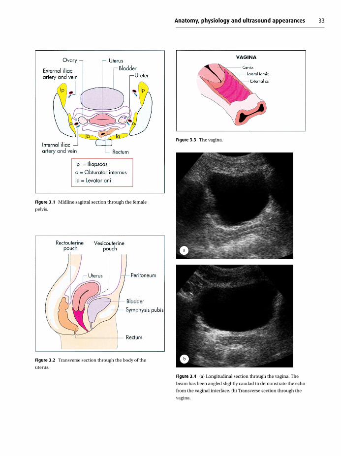
Anatomy, physiology and ultrasound appearances 33
Figure 3.1 Midline sagittal section through the female
pelvis.
Figure 3.2 Transverse section through the body of the
uterus.
Figure 3.3 The vagina.
a
b
Figure 3.4 (a) Longitudinal section through the vagina. The
beam has been angled slightly caudad to demonstrate the echo
from the vaginal interface. (b) Transverse section through the
vagina.

34 Practical Gynaecological Ultrasound
Figure 3.5 (a) Various uterine positions. (b) Normally
anteverted and (c) Retroverted uterus.
a
b
Figure 3.6 (a) The uterus. (b) The cervix is well-delineated on
ultrasound when surrounded by fluid.
Figure 3.7 The acoustic properties of the three uterine layers:
e, endometrium; m, myometrium; p, parametrium
(arrowhead).

Anatomy, physiology and ultrasound appearances 35
Figure 3.8 Uterine anomalies.
of the corpus and the cervix lies the isthmus; it is herethat, during the later stages of pregnancy, the loweruterine segment develops.
The two main parts of the uterus have separatefunctions – the corpus for gestation and the cervix toact as a sphincter.
The uterine wall is composed of three layers: (1)the parametrium – the external layer; (2) themyometrium – the thick muscular layer; and (3)the endometrium – the inner layer. The differenttissues of these layers exhibit different acousticproperties (Fig. 3.7). The parametrium, an incom-plete covering layer of peritoneum, produces ahighly reflective echo outlining the uterus. Lower-level homogeneous echoes are reflected by thethick layer of myometrium and the inner lining,the endometrium, exhibits changing ultrasoundappearances throughout the hormonal cycle, givingrise to different thicknesses and reflectivities.
These cyclical changes of the endometrium andovaries are described in more detail later in this chap-ter (see Fig. 3.27).
Figure 3.9 Contrast hysterosalpingogram demonstrating a
bicornuate uterus. Patent tubes are seen bilaterally with spill of
contrast into the peritoneal cavity.
Figure 3.10 Magnetic resonance axial T2-weighted image
demonstrating a bicornuate uterus (arrowheads).
Uterine anomaliesThe reproductive organs develop in the embryo fromtwo tubes, the Mullerian ducts. The caudal portionsof these ducts eventually fuse to form the uterus,cervix and superior part of the vagina, while the cra-nial portions remain separate, forming the fallop-ian tubes. Development takes place from the thirdor fourth week of gestation and continues into thesecond trimester. Interruption to this process causesincomplete fusion, which may result in a varietyof structural anomalies1,2 and the more commonanomalies are demonstrated in Figure 3.8.

36 Practical Gynaecological Ultrasound
a
b
Figure 3.11 (a) Ultrasound demonstration of a bicornuate
uterus in transverse section. This may often be more obvious on
transabdominal than transvaginal scanning, due to the larger
field of view. (b) A three-dimensional transvaginal ultrasound
image of a bicornuate uterus.
It may be difficult to demonstrate on ultrasoundthe more subtle anomalies; these may often be bet-ter revealed by laparotomy, laparoscopy or hystero-salpingogram during investigations for infertility orfailed pregnancies (Fig. 3.9). Magnetic resonanceimaging is also useful for demonstrating uterineanomalies (Fig. 3.10).
The bicornuate uterus is most frequently identi-fied on ultrasound in transverse section, by demon-strating two distinct cornua, each with its endome-trial echo, towards the fundus. The advent of
three-dimensional ultrasound has also played a use-ful role in evaluating uterine anomalies (Fig. 3.11)by being able to display the endometrium in coronalsection.3
Ovaries
The function of the ovaries is to produce a matureovum every 28 days, to secrete the female hormones,oestrogen and progesterone, which maintain thereproductive cycle, and to support pregnancy.

Anatomy, physiology and ultrasound appearances 37
Table 3.1 Age-related differences in size of the uterus and ovaries
Infantile Prepubertal Reproductive Postmenopausal
Uterus Length (cm) 1.5–2 2–5.4 5–12 3.5–6.5
Width 0.8–1.0 1.0–2.2 4.0 1.2–1.8
Anteroposterior diameter 1.0 3 1.5–2.0
Ovaries Volume (cm3) 0.7–3.6 1–6 6–12 <4
Figure 3.12 Anatomical relations of the ovary.
Figure 3.13 Transvaginal scan of the normal ovary
demonstrating graafian follicles.
Each ovary usually lies posterior and lateral toeither side of the uterus attached to the broad liga-ments by its own mesentery, the mesovarium andto the uterus by the ovarian ligament. Its upper sur-face lies in close proximity to the fimbrial end of thefallopian tube (Fig. 3.12).
Ovaries are oval in shape, usually about the size ofa walnut but varying with age (Table 3.1). The ovaryis made up of the central portion or medulla and theouter cortex (stroma) within which are contained anumber of graafian follicles. It is the follicle whichhouses the ovum. The blood and lymphatic vesselsand nerves are situated in the medulla.
Ultrasonically, the stroma and medulla exhibitquite different appearances: the stroma demon-strates the greater changes during the menstrualcycle. The follicles, a ‘soft-marker’ for ovarian iden-tification, are visualised in a peripheral arrange-ment as well-defined echo-free areas in the stroma(Fig. 3.13). Their size varies with the stage of the cycle.The central medulla appears as an area of relativelystrong reflectivity with no definite borders.
The mesovarium is mobile, making the exact loca-tion of the ovaries variable. The position of theovaries also varies according to adjacent bowel andthe degree of bladder-filling (Fig. 3.14), and may alterposition during scanning.4 This, together with ovar-ian atrophy following menopause, can make scan-ning a challenge. It is useful to use ultrasonic land-marks to help with identification. In most cases, theovary lies in close proximity to the internal iliac arterywhich can be visualised passing obliquely lateral tothe lower third of the uterus (Fig. 3.15). A less eas-ily recognisable but still useful landmark is the posi-tion of the levator ani muscle which marks the most

38 Practical Gynaecological Ultrasound
b
Figure 3.14 (a) Transverse transabdominal scan
demonstrating varying ovarian positions (arrows). The right
ovary lies behind the uterus (U), (b) Longitudinal section with
the left ovary lying above the fundus of the uterus.
Figure 3.15 Transvaginal longitudinal section through the
ovary demonstrating the proximity of the internal iliac artery
(A) and internal iliac vein (V).
Figure 3.16 The fallopian tube.
inferior position at which the ovary can be located(see section on musculature, below).
For practical scanning purposes, the ovaries canbe visualised in a variety of positions from the pouchof Douglas to the superolateral aspect of the uterus.
Fallopian tubes
The two fallopian tubes lie either side of the uterus inthe superior portion of the broad ligament (Fig. 3.16).Their walls are composed of the same three layers asthe uterus.
Each tube is approximately 10 cm in length andpasses laterally, posteriorly and inferiorly from theupper part of the uterus. The uterine opening isminute whilst the abdominal opening, which is inclose proximity to the ovary, is larger.
Each tube has four parts:1. the interstitial portion, which lies within the uter-
ine wall2. the isthmus – the medial third extending laterally
from the upper part of the uterus

Anatomy, physiology and ultrasound appearances 39
a
b
Figure 3.17 (a) Hystero-contrast sonography. The normal
(undilated) fallopian tube, not usually visible on ultrasound,
can be demonstrated when contrast is introduced, increasing
the echogenicity and contrast resolution. (b) The tube is
demonstrated when dilated with fluid and debris in the
hydrosalpinx.
3. the ampulla, which is the widest part of the tube4. the infundibulum, which is the dilated funnel-
shaped portion close to the ovaryThe opening of the infundibulum has tiny projec-tions, known as fimbriae, which serve to guide theovum into the tube.
The function of each tube is to convey the ovumfrom the ovary to the uterine cavity. If fertilisationtakes place, it usually occurs in the tube.
The normal fallopian tube is difficult to imagesonographically as its tiny diameter is beyond theresolution capabilities of most transducers and it isa relatively poor reflector of ultrasound. The over-
Figure 3.18 Transvaginal axial section through the lower
uterus. Fluid in the pouch of Douglas (arrow) is a normal
finding mid-cycle.
all structure may be demonstrated after the intro-duction of contrast during X-ray hysterosalpingo-graphy (Fig. 3.9) or preferably, without incurring thepotential hazard of radiation, with hystero-contrastsonograpy or HyCoSy (Fig. 3.17: see Chapter 7). Occa-sionally, pelvic fluid may be present surrounding thetubes, making at least partial visualisation possible.
Peritoneum and ligaments
The peritoneum surrounds the body of the uterus,forming recesses, or pouches, anteriorly and poster-iorly (Figs. 3.1 and 3.2). The anterior recess betweenthe uterus and posterior bladder wall is known as thevesicouterine fossa. The posterior recess lies anter-ior to the rectum and extends down to the posteriorfornix of the vagina. It is known as the rectouterinefossa or pouch of Douglas.
It is not unusual to see fluid in this recess on ultra-sound, especially during mid-cycle after ovulation,as this represents a gravity-dependent pocket whenthe patient lies supine (Fig. 3.18).
The ligaments attaching the uterus within thepelvic cavity are predominantly extensions ofthe peritoneum in anterior, posterior and lateraldirections.5 The anterior and posterior ligaments

40 Practical Gynaecological Ultrasound
Figure 3.19 The ligaments. The broad ligament is a double
fold of peritoneum in which lie the adnexal structures.
form the vesicouterine and rectouterine pouches(see above). Two further peritoneal extensionsbetween the posterior uterine surface and thesacrum form the paired uterosacral ligaments.
The paired broad ligaments are composed of dou-ble folds of peritoneum which extend laterally fromeither side of the uterus to the wall of the bonypelvis. Each ligament contains the fallopian tube inits superior portion, the round ligament, the ovarywith its ovarian ligament, blood vessels and nerves(Fig. 3.19).
These ligaments are not easy to see ultrasoni-cally. However with careful scanning in the transax-ial plane, superior to the uterine fundus, an arc oflow-level homogeneous echoes can be visualised,which represents the broad ligament and its con-tents as it arises adjacent to the horn of the uterus(Fig. 3.20).
The paired round ligaments are not extensionsof the peritoneum, but fibromuscular cords whichextend laterally from the region of the uterine cor-nua and lie within the broad ligament. Each ovary isalso attached to the lateral wall of the uterus by theovarian ligament. When free fluid is present in the
a
b
Figure 3.20 (a) Right adnexal area. The superior portion of the
broad ligament (arrowheads) containing the ovarian artery.
(b) The ovarian liagament (arrow) is demonstrated when
surrounded by fluid. A small fimbrial cyst is noted (arrowhead).
pelvis it is possible to visualise the ovarian ligamentas a highly reflective linear echo in the fluid.
Vasculature
The main uterine artery arises from the internal iliacartery. It passes medially, supplying the cervix andascending the lateral aspect of uterus within thebroad ligament. Branches from the main artery fur-ther supply the cervix and vagina (Fig. 3.21). The vagi-nal arteries eventually form the azygos arteries of thevagina as they descend along its surface. These ves-sels are tortuous and coiled to provide extra lengthduring pregnancy.
The ovary arises from the abdominal aorta, imme-diately below the renal arteries. At the margin of the

Anatomy, physiology and ultrasound appearances 41
Figure 3.21 Arterial supply to the uterus and ovary.
pelvis, the artery passes between the folds of thebroad ligament. Several of its smaller branches sup-ply the fallopian tubes and anastomose with uterinearteries.
Venous drainage from the uterus is via the inter-nal iliac vein, and from the ovaries is into the venousplexus, from which arise the ovarian veins, lying pos-terior to the uterus. The right ovarian vein drains intothe inferior vena cava, the left ovarian vein into theleft renal vein.
Musculature
The pelvic organs are supported by a ‘shelf’ of muscleon each side. There are four main muscle pairs; thelevator ani and the coccygeus make up the pelvicdiaphragm. The levator ani lies between the ischialspines and the posterior portion of the pubis and actsas the floor of the pelvis.5 It supports the pelvic organsand surrounds the rectum, vagina and urethra whichpierce it.
The piriformis lies slightly posterior and superiorto the coccygeus, and the obturator internus liesagainst the lateral wall on each side of the pelvis
Figure 3.22 The pelvic muscles. (a) Axial section at the level of
the body of the uterus. (b) Axial section at the level of the
vagina.
with the insertion point being the greater trochanter(Fig. 3.22).
The muscles can be demonstrated on ultra-sound as well-defined linear structures with low-level homogeneous echoes. Their position is prob-ably most readily appreciated transabdominally,posterior and lateral to the uterus. Occasionally theymay be mistaken for pathology, and it is importantalways to be aware of their location and appearance(Fig. 3.23).
Bladder and ureters
The bladder is a reservoir for urine and occupies thelower portion of the true pelvis anterior to the uterus.The normal full bladder on ultrasound has a thin,hyperechoic smooth wall with anechoic fluid within.It is fixed at its caudal portion (trigone) to the cervix.
The ureters are hollow, muscular tubes not usu-ally seen on ultrasound unless pathologically dilated.

42 Practical Gynaecological Ultrasound
a
b
c
d
Figure 3.23 The pelvic muscles in transverse section. (a) Piriformis muscles seen behind the body of the uterus (arrows).
(b) Iliopsoas (arrows) is demonstrated by angling slightly laterally. (c) Levator ani (arrowheads) at the level of the vagina.
(d) The obturator internuis (OI) may be demonstrated lateral to the piriformis (P).
They are approximately 30 cm in length and passanterior to the internal iliac artery and posteriorto the ovary. Their relationship to the reproductiveorgans is important in cases of pelvic pathology,which may give rise to obstructive uropathy.
It is possible to identify the vesicoureteric junctionin a transverse scan through the bladder base, and
the ureteric jet may be clearly demonstrated usingcolour Doppler as it enters the bladder (Fig. 3.24).
Anatomical changes related to age
The uterus and ovaries vary in size according to age.Neonatal and paediatric appearances are discussed

Anatomy, physiology and ultrasound appearances 43
Figure 3.24 Ultrasonic appearances of the ureters. In transverse section, the ureters may be traced down as they travel medially,
(a) and enter the bladder base (b). (c) At this level the ureteric jets may be demonstrated with colour Doppler. O.I., obturator
internus; CX, cervix; L.A., levator ani. In longitudinal section, (d) the ureter may sometimes be seen as a tiny tube (arrow) between
the ovary (o) and iliac vessels. iiv, internal iliac vein; iia, internal iliac artery; p, piriformis.
in Chapter 8 and postmenopausal pelvic organsin Chapter 4. A comparison of normal anatomicalappearances with age is given in Table 3.1.
A simple formula for estimation of ovarian volumeis:
Volume (cm3) = length × width × anteroposterior
diameter × 0.53
Later in life, at and following menopause, theuterus and ovaries atrophy. This regression in size isa gradual process and in the early postmenopausalphase the organs can appear ultrasonically very sim-ilar to those of the reproductive years. As in the pre-menarchal stage, the cervix is larger than the corpus.In the normal elderly patient, the endometrial echo,although thin, should still be demonstrated. The

44 Practical Gynaecological Ultrasound
c
e
b
d
Figure 3.25 The differing appearances of the uterus with age.
(a) Paediatric (b and c); adult uterus transabdominal and transvaginal
scan; (d and e) postmenopausal uterus transabdominal and
transvaginal scan.
normal uterus at this stage is of uniform echogenic-ity but the ovaries become increasingly difficult toimage unless cysts are present (Fig. 3.25).
Physiology of reproduction
The female reproductive years occupy an average35–40 years between puberty and menopause. The
standard hormonal cycle is 28 days, although it iswidely recognised that this is an arbitrary figure andthat there are variations to this pattern.
Throughout each normal cycle, the endometriumand ovaries undergo changes brought about bythe hormones secreted by the pituitary gland –follicle-stimulating hormone (FSH), luteinising hor-mone (LH) and prolactin. The ovaries themselves are

Anatomy, physiology and ultrasound appearances 45
Figure 3.26 The influence of hormones on the uterus and
ovaries. FSH, Follicle-stimulating hormone; LH, luteinising
hormone.
Figure 3.27 (a) The uterus in a normal cycle. POD, pouch of Douglas.
also active and secrete oestrogen and progesterone(Fig. 3.26).
The purpose of these hormones is to pro-duce a mature ovum for fertilisation, prepare theendometrium for the fertilised ovum and support thepregnancy in its early stages.
During the proliferative, or initial, phase of thecycle, FSH influences the immature follicles todevelop, resulting in a dominant follicle being pro-duced. Around day 14 of cycle, when the oestrogenlevels are high, LH stimulates ovulation. During thisperiod, the endometrium thickens and ripens. Fol-lowing ovulation, during the secretory phase, theovary secretes progesterone, FSH and oestrogen lev-els diminish and the follicular wall collapses formingthe corpus luteum. If pregnancy does not occur, theendometrium becomes thickened and menstruationtakes place on or around the 28th day.
Ultrasound appearances
Changes in the endometrium and ovaries can bewell demonstrated sonographically using a high-frequency TV transducer (Figs. 3.27 and 3.28).
It is important to remember that the endometriumis a three-dimensional structure, and to scan it fullyby angling the transducer from left to right uterinehorn in longitudinal section, and from cervix to fun-dus in transverse section. In this way the operator

b c
d
e
fg
h
Figure 3.27 (cont.) Ultrasound scans of the normal endo-
metrium through the cycle. (b) Menstruation (days 1–4). Blood
is seen within the cavity. (c) Regenerative/early proliferative
(days 1–8). Thin endometrium. (d) Late proliferative (days 9–12).
Thickening, increasingly hyperechoic endometrium. (e) Peri-
ovulatory (days 12–15). Hypoechoic with a hyperechoic rim.
(f) Secretory (day 16 to menstruation). Irregular and hypere-
choic. (g and h) The junctional zone (arrowheads) can be seen
as hypoechoic layer surrounding the endometrium, seen here
in the regenerative (g) and ovulatory (h) phases of the cycle.
This layer represents part of the myometrium and should not
be included in the endometrial thickness measurement.

b
c
d e
f
Figure 3.28 (a) The appearances of the dominant ovary. Ultra-
sound appearances of the dominant ovary through the cycle:
(b) Regenerative (days 1–8): small follicles. (c) Proliferative (days
9–13): dominant follicle growing. (d) Ovulatory (days 13–14):
cumulus oophorus seen in dominant follicle. (e) Early secretory
(day 15 onwards). Corpus luteum. (f) Late secretory. Regression
of corpus luteum.
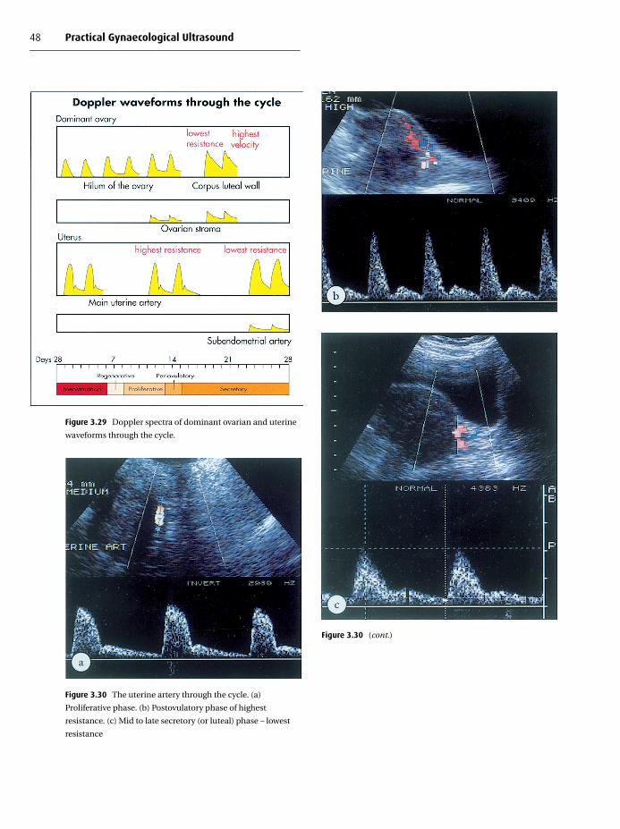
48 Practical Gynaecological Ultrasound
Figure 3.29 Doppler spectra of dominant ovarian and uterine
waveforms through the cycle.
Figure 3.30 The uterine artery through the cycle. (a)
Proliferative phase. (b) Postovulatory phase of highest
resistance. (c) Mid to late secretory (or luteal) phase – lowest
resistance
Figure 3.30 (cont.)

Anatomy, physiology and ultrasound appearances 49
may identify focal abnormalities which may not beapparent on a midline longitudinal section.
Ultrasound appearances of the normalendometrium (Fig. 3.27)During the early proliferative or regenerative stage,the endometrium appears as a thin reflective line inthe uterine cavity.
At ovulation, the endometrium is hypoechoic,surrounded by a prominent, highly reflective echo,giving rise to the term ‘triple-line’ echo sign.6
Just before menstruation, the endometrium can bevisualised as a thick hyperechoic structure passingcentrally down the uterine cavity.
During menses, it is possible to visualise theendometrial cavity separated by low-level echoesrepresenting blood and mucus.
Just below the endometrium it is often possibleto see a hypoechoic, subendometrial ‘halo’ whichrepresents the junctional zone between the endo-metrium and myometrium (Fig. 3.27g and 3.27h).This actually consists of normal myometrium7
which comprises tightly packed mucosal cells withincreased vascularity, and should not be included inthe measurement of endometrial thickness.
Peristaltic waves within the endometrium havebeen identified on ultrasound, passing towards thecervix in menses, and towards the fundus in mid-cycle. These can best be appreciated by playing avideo of the scan at a fast speed.8
The endometrium of a normal postmenopausalwoman is thin (less than 4 mm) and hyperechoic,9
mostly composed of the basalis layer. It is partic-ularly important when scanning in cases of post-menopausal bleeding to identifiy the endometrialecho, and to examine it fully. Lack of a demon-strable endometrial echo is a highly suspicious find-ing and may indicate an invasive malignant processin a small number of cases.
Appearances of the ovary (Fig. 3.28)During the regenerative and early proliferative stagethe ovaries contain several immature follicles, typi-cally less than 1 cm, within the ovarian tissue.
As the cycle progresses to ovulation, a dominantfollicle measuring up to 2.0 cm can be seen. It is
possible to have more than one dominant follicle.In 60–65% of cases, the cumulus oophorus can beimaged between 12 and 24 hours prior to ovulation.4
Ultrasonically, this appears as a small hyperechoicfocus adherent to the follicular wall (Fig. 3.28d).
The collapsed walls of the dominant follicle arethen apparent during the early secretory phase.Haemorrhagic changes also occur, demonstratedas low-level echoes within the collapsing corpusluteum, which also has a less defined outline. In somecases, fluid can also be seen in the pouch of Douglas.6
In postmenopausal women, the ovaries shrink andatrophy, often being more difficult to locate on ultra-sound. With improvements in technology it is nowcommon to see small cysts within the postmeno-pausal ovary and these are of no significanceprovided they are small (less than 5 cm) and simple(anechoic and smooth-walled).10
Haemodynamics of the uterus and ovaries andthe use of Doppler ultrasound
The blood supply to the uterus and ovaries fluctu-ates throughout the normal cycle. These physiologi-cal changes can be appreciated using Doppler ultra-sound (Fig. 3.29).
Both the ovary and endometrium exhibit angio-genesis – the formation of new blood vessels – dur-ing the course of the normal cycle. These ‘new ves-sels’ have low-resistance vasculature characterisedby increased end-diastolic flow velocities with a lowresistance index value. This phenomenon is also wellrecognised in pathological processes, such as thedevelopment of malignant tumours,11 and the useof Doppler is therefore non-specific, particularly inpremenopausal women.
Uterine arteriesThe main uterine arteries, located just laterally tothe region of the cervix, have a high resistancepattern with low end-diastolic flow and a notch(Fig. 3.30), and should not be confused with theiliac artery, which lies lateral to the uterine artery.The common and external iliac arteries exhibit

50 Practical Gynaecological Ultrasound
Figure 3.31 High-resistance, triphasic waveform from the iliac
artery.
very-high-resistance, triphasic flow with a reversecomponent; the internal iliac artery is less pulsatilebut still exhibits a high-resistance, notched wave-form with very little end-diastolic flow (Fig. 3.31).
Waveforms and resistance index measurementsare more consistently reproducible from the mainuterine artery than from its smaller, low-velocity,arcuate branches and the artery tends to bemore easily visualised in premenopausal than post-menopausal patients. Vessels situated within theuterus, just below the endometrium, are more dif-ficult to locate as they are of much lower velo-city. The Doppler waveforms from these are there-fore of low amplitude and tend to have a poorlydefined envelope, making measurements prone toerror (Fig. 3.32).
In a normal cycle, the resistance of the uter-ine artery is at its highest (lowest end-diastolicflow velocity) 1–2 days after ovulation – i.e. aroundcycle day 16.12–14 It is at its lowest (having great-est end-diastolic velocity) during the mid to lateluteal phase. Although it is more difficult to esti-mate velocity accurately in the tortuous pelvic ves-sels, an increase in velocity has also been noted dur-ing the same period. This is thought to correlateloosely with an increase in uterine perfusion, whichwould be logical at this time of possible implantation.
Figure 3.32 Low-velocity waveform from arcuate arteries
within the myometrium.
Figure 3.33 Decreased uterine artery resistance with fibroids.

Anatomy, physiology and ultrasound appearances 51
Figure 3.34 Decreasing resistance of the ovarian artery through the cycle. (a) Regenerative. (b) Early proliferative. (c) Late
proliferative. (d) Periovulatory. (e) Secretory – wall of corpus luteum.
Although the uterine artery on the dominant sidehas been noted to have a slightly lower resistanceindex than the contralateral artery,14 there is consid-erable overlap between the values, and the differenceis small.
Several factors may be responsible for alteringthe resistance to flow of the uterine arteries, andthese must be taken into account when interpreting
Doppler information. A lower resistance index is seenin late luteal phase, in women on hormone replace-ment therapy, tamoxifen, and in the presence of uter-ine fibroids (Fig. 3.33). Malignancy such as endome-trial carcinoma or uterine sarcoma is also associatedwith a lower uterine artery resistance index.
Uterine artery resistance increases with age, anda high resistance index may also be associated

52 Practical Gynaecological Ultrasound
Figure 3.35 Typically increased vascularity around the wall of
the corpus luteum.
with infertility, including tubal damage and endo-metriosis.
Ovarian arteries
As with the uterine arteries, but to an even greaterdegree, the resistance of the ovarian arteries isaffected by cyclical, physiological changes, whichare much greater in the dominant than the non-dominant ovary (Fig. 3.29). Downstream resistanceis greatest (lowest end-diastolic flow) during days 1–8 of the cycle, with maximum resistance occurringaround day 8.14 (Days 1–8, therefore, make a good‘window’ during which to examine the ovaries of pre-menopausal women, as low resistance is less likely atthis time and misinterpretation of Doppler informa-tion is minimised.) Resistance to flow then graduallydecreases from day 8 as the dominant follicle devel-ops and ruptures around day 14. Resistance is at itslowest during the luteal phase, days 16–21, with thewall of the corpus luteum displaying the most intensecolour pattern. It is at this time that the vessels aretechnically easiest to visualise. A gradual increase ofresistance is then observed through to the regenera-tive phase (Fig. 3.34).15,16
These changes are most pronounced in the corpusluteum (Fig. 3.35).
The contralateral, non-dominant ovary tends tomaintain a relatively high resistance throughout thecycle. In fact, it may be quite difficult to demonstrateany colour Doppler at all within the stroma, depend-ing on the sensitivity of the ultrasound systemused.
Identification of Doppler signals in the ovariesbecomes increasingly difficult in the post-menopausal woman and usually demonstratesa low-velocity, high-resistance pattern.
Doppler ultrasound – key points� Doppler information may be used to comple-ment morphological information and shouldnever be interpreted in isolation� Normal Doppler waveforms vary with the stageof the menstrual cycle� With less sensitive equipment, it may be diffi-cult to visualise any Doppler flow in the ovary,particularly the non-dominant one� Physiological and pathological Doppler wave-forms can be indistinguishable� False-positive Doppler results are therefore lesscommon in postmenopausal women
R E F E R E N C E S
1. J. A. Rock and S. M. Markham, Developmental anomalies
of the reproductive tract. In Fertility: Evaluation and Treat-
ment, ed. W. R. Keye, R. J. Chang, R. W. Reber and M. R. Sules.
(Philadelphia: W. B. Saunders, 1992).
2. M. Sanford, M. D. Markham and T. B. Waterhouse, Struc-
tural anomalies of the reproductive tract. Current Opinion
in Obstetrics and Gynecology 4 (1992), 867–73.
3. A. Lev-Toaff, L. Pinheiro, G. Bega et al., Three dimensional
multiplanar sonohysterography. Journal of Ultrasound in
Medicine, 20 (2001), 295–306.
4. I. E. Timor-Tritsch and S. Rottem (eds), Transvaginal Sono-
graphy (New York: Elsevier, 1988).
5. R. Warwick and P. Williams (eds), Gray’s Anatomy, 35th edn.
(Longman, 1973).
6. D. Gratton, C. Harrington, S. Holt and E. Lyons, Normal
pelvic anatomy using transvaginal ultrasound. Obstetrics
and Gynecology Clinics of North America, 18 (1991), 4.

Anatomy, physiology and ultrasound appearances 53
7. R. Tetlow, I. Richmond, J. Greenman et al., Histological ana-
lysis of the uterine junctional zone as seen by transvaginal
ultrasound. Ultrasound in Obstetrics and Gynaecology, 14
(1999), 188–93.
8. K. de Vries, E. A. Lyons, G. Ballard et al., Contractions of the
inner third of the myometrium. American Journal of Obstet-
rics and Gynecology, 162 (1990), 679–82.
9. B. Gull, B. Karlsson, I Milsom et al., Transvaginal sonogra-
phy of the endometrium in a representative sample of post-
menopausal women. Ultrasound in Obstetrics and Gynae-
cology, 7 (1996) 322–7.
10. L. Valentin, L. Skoog and E. Epstein, Frequency and type
of adnexal lesions in autopsy material from post-
menopausal women: ultrasound study with histological
correlation. Ultrasound in Obstetrics and Gynecology, 22
(2003), 284–9.
11. A. Kurjak, I. Zalud and Z. Alfirevic, Evaluation of adnexal
masses with transvaginal color ultrasound. Journal of Ultra-
sound Medicine, 10 (1991), 295–7.
12. P. Sladkevicius, L. Valentin and K. Marsal, Blood flow velo-
city in the uterine and ovarian arteries during the normal
menstrual cycle. Ultrasound in Obstetrics and Gynecology,
3 (1993), 199.
13. R. K. Goswamy and P. C. Steptoe, Doppler ultrasound studies
of the uterine artery in spontaneous ovarian cycles. Human
Reproduction, 3 (1988), 721–6.
14. M. C. W. Scholtes, J. W. Wladimiroff, H. J. M. van Rijen and
W. C. J. Hop, Uterine and ovarian flow velocity waveforms
in the normal menstrual cycle: a transvaginal study. Fertility
and Sterility, 52 (1989), 981–5.
15. L. T. Merce, D. Garces, M. J. Barco and F. de la Fuente, Intra-
ovarian Doppler velocimetry in ovulatory, dysovulatory and
anovulatory cycles. Ultrasound in Obstetrics and Gynecol-
ogy, 2 (1992), 197.
16. K. Hata, T. Hata, D. Senoh et al., Change in ovarian arterial
compliance during the human menstrual cycle assessed by
Doppler ultrasound. British Journal of Obstetrics and Gynae-
cology, 97 (1990), 163–6.

4
Pathology of the uterus, cervix and vagina
Josephine M. McHugoBirmingham Women’s Hospital, Birmingham
Introduction
Ultrasound is the appropriate first investigation forthe majority of pelvic symptoms in the female. It ishowever very operator-dependent and when usedby appropriately trained personnel with appropriateequipment is cost-effective and safe. Transvaginalscanning is essential to increase the diagnostic accu-racy of ultrasound and should be used in all casesunless there is a specific contraindication, such as inthe paediatric population. It is essential that transab-dominal ultrasound is also performed in order thatthe large fibroid uterus or other masses can be fullyassessed in addition to imaging transvaginally.
There is a need for a meticulous approach to scan-ning in all cases so that significant pathology isnot missed. The sonographer, having completed theultrasound examination, must then issue an appro-priate report. A structured report is also an essentialpart of the examination and should not only includethe biographical details of the patient but also thesalient ultrasound findings, both normal and abnor-mal, followed by either a definite diagnosis if this ispossible, or a differential diagnosis.
Further investigations or definitive managementcan then be arranged.
The sonographer performing the examinationshould be clearly identified in the report.
A report that is structured in this way will allowretrospective audit of the ultrasound findings rela-
tive to operative findings and histology. This audit ofaccuracy of the ultrasound to histological outcomeis essential for best practice.
In many situations the accuracy of transvaginalultrasound is sufficient for further management ofthe patient but in some cases additional imagingsuch as magnetic resonance imaging (MRI) will beappropriate.
This chapter aims to outline the ultrasound find-ings of the common pathologies of the uterus, cervixand vagina.
The ultrasound findings must always be inter-preted in the context of the clinical symptoms, ageand hormonal status of the patient being scanned.It is therefore essential that each request for a pelvicultrasound contains sufficient clinical information.
For clarity, this chapter with be divided intopathologies involving the myometrium, endome-trium, cervix and vagina. For the ultrasound to beclinically useful the sonographer needs to have anunderstanding of the information that the referringclinician requires in order to manage the patientappropriately. The major decision is whether theultrasound abnormality is responsible for the symp-toms or is an incidental finding.
Similarly there is a need to assess the risk of malig-nancy. To this end there is a need to work in a mul-tidisciplinary team, not only for appropriate man-agement of the individual patient but also to auditoutcome and effectiveness of ultrasound.
C© Cambridge University Press 2005.
54

Pathology of the uterus, cervix and vagina 55
The uterus
Myometrium
Fibroids (leiomyomata)Fibroids are benign tumours of the smooth mus-cle of the uterus and are the commonest gynae-cological tumour, being present in up to 40% ofwomen over 40 years. There is an increased inci-dence in women of Afro-Caribbean origin. Fibroidsmost commonly occur in the uterine body but arealso found in the cervix or broad ligament. Histologi-cally they are composed mainly of smooth muscle.The smooth-muscle fibres are arranged in denseconcentric rings which are well demarcated fromthe surrounding normal myometrium, and it is thisarrangement which is responsible for the ultrasoundappearances.
These benign tumours are hormone-dependent,being associated with relatively high levels of oestro-gen, and are frequently multiple. The incidencetherefore increases with age until the menopause,and fibroids do not occur until well after the onset ofpuberty. Following the menopause the majority offibroids reduce in size. Fibroids often enlarge in preg-nancy and if they rapidly increase in size may out-grow their blood supply, resulting in central necrosis(red degeneration.) As with any ischaemic, necroticprocess this results in considerable pain. Hor-mone replacement and tamoxifen can also stimu-late growth. Fibroids, being oestrogen-dependent,respond to medical management and anti-oestrogen therapy with gonadotrophin-releasinghormone antagonist is of benefit to aid fibroidshrinkage.
Clinical symptomsThe clinical symptoms depend on the size and site ofthe fibroids. Fibroids that impinge into the endome-trial cavity are defined as submucous. Submucousfibroids distort the uterine cavity and often result inmenstrual problems, usually menorrhagia and dys-menorrhoea. Fibroids projecting from the outer uter-ine surface are termed subserosal, as they lie underthe serosal surface of the uterus, and many of these
Figure 4.1 Diagram of fibroids.
remain asymptomatic. Fibroids within the myome-trial mass that are neither subserosal nor submucousare termed intramural. Figure 4.1 outlines the poss-ible sites of fibroids.
In some situations the fibroid may enlarge witha relatively thin stalk maintaining continuity to themyometrium. These are termed pedunculated. Itis this continuity with the myometrium which isimportant to define with ultrasound, particularlyif the fibroid projects into the broad ligament, asconfusion with adenxal masses can occur if conti-nuity with the myometrium is not identified. Sub-mucosal pedunculated fibroids can prolapse intoor through the cervical canal. These present clin-ically with pain, often dysmenorrhoea, but canalso present with intermenstrual bleeding. Pedun-culated fibroids, having a relatively narrow stalk, canundergo torsion which results in compromise of thevascular supply presenting with acute pain.
An enlarged fibroid uterus can result in pressuresymptoms on the bladder or, if the uterus is retro-verted, rectal symptoms. A markedly enlarged uteruspresents as an abdominal mass and, rarely, can resultin acute urinary retention. Following the menopauseor after a period of rapid growth and degeneration,such as in pregnancy, fibroids often regress with the

56 Practical Gynaecological Ultrasound
Figure 4.2 Posterior fibroid showing distal acoustic
shadowing.
deposition of calcium. The calcium may be eitherpunctate or circumferential.
Ultrasound appearancesFibroids may be single or multiple, subserosal, intra-mural, submucous or pedunculated. These are veryvariable but are always well demarcated from thenormal myometrium.
The dense nature of the abnormal smooth-musclefibres results in loss of the normal homogeneousultrasound appearances of the myometrium [Fig.4.2) and acts as a stronger reflector than the nor-mal myometrium. Therefore there is usually adegree of acoustic shadowing seen posterior to thefibroid.
This is more marked with a calcified fibroid, whichis a stronger reflector (Fig. 4.3).
The reflectivity relative to the surroundingmyometrium is variable; most is of lower reflectivity,but increased reflectivity is not an infrequent finding(Fig. 4.4).
Fibroids show a classical vascular supply with cir-cumferential vessels, which are easily demonstratedusing Doppler (Fig. 4.5).
The ultrasound report should describe thesite, size and number of fibroids with particular ref-erence to any distortion of the uterine cavity. It is vital
Figure 4.3 Transabdominal scan of a uterus with calcified
fibroids, demonstrating strong acoustic shadowing.
a
b
Figure 4.4 Sagittal image showing the varying reflectivity of
fibroids: (a) a relative hyperechoic pattern and (b) a
hypoechoic pattern of different fibroids.

Pathology of the uterus, cervix and vagina 57
Figure 4.5 Power Doppler of a fibroid demonstrating
circumferential vessels.
Figure 4.6 A submucosal fibroid.
to differentiate an endometrial polyp from a smallfibroid, this being apparent by a combination ofthe acoustic pattern and the vascular supply (seeendometrial disease, below) (Fig. 4.6).
In pregnancy the location and size of the fibroidrelative to the cervical canal and the presenting fetal
part are important. A relatively large fundal fibroidwill not obstruct labour whereas smaller cervicalfibroids may.1
A large fibroid uterus can cause a mass effect withpartial obstruction of the distal ureters. Ultrasoundexamination of the upper renal tract and bladdervoiding are therefore appropriate for a markedlyenlarged fibroid uterus.
Degenerating fibroids create bizarre ultrasoundappearances with central areas of necrosis showingas fluid-filled areas. In the acute phase these are verytender.
Diagnostic dilemmasA pedunculated fibroid extending into the broadligament may suggest a solid adnexal mass – care-ful examination should demonstrate the continuitywith the myometrium.2
A retroverted normal uterus may suggest a fibroidon transabdominal scanning. This confusion is read-ily avoided by transvaginal scanning.
A bicornuate uterus may be confused with a fibroidbut, by careful examination of the endometrial cav-ities, two separate uterine cavities and the overlyingmyometrium will be apparent. Examination in thelater part of the menstrual cycle is particularly help-ful in this circumstance as the endometrium will bemore readily seen.
Small submucous fibroids may cause cornualobstruction and infertility secondary to tubal occlu-sion (Fig. 4.7).
Distortion of the endometrial cavity interferes withimplantation and it is important to identify a nor-mal cavity prior to embryo transfer in infertility pro-grammes.
In pregnancy the non-gravid horn of a bicornuateuterus may appear as a fibroid if the endometrialcavity in this horn is not appreciated.
Larger fibroids will interfere with the normalenlargement of the pregnant uterus, resulting insecond-trimester fetal loss, although this is relativelyuncommon.1 Fibroids can undergo rapid growth inpregnancy, outgrowing the blood supply, resulting inred degeneration causing acute pain.
A large fibroid low in the uterus can obstructlabour.

58 Practical Gynaecological Ultrasound
a
b
Figure 4.7 (a) An intracavitary submucosal fibroid,
demonstrating some degeneration, with posterior acoustic
enhancement, in a patient with infertility. (The differential
diagnosis for these appearances would be of an endometrial
polyp.) (b) A cornual fibroid.
A Braxton Hicks contraction in pregnancy maysimulate a fibroid but if the uterus is scanned overa 20-min period the transient deformity caused by acontraction will disappear.
MRI of the uterus is extremely helpful and is essen-tial if embolisation of the fibroids is being consid-ered. T2 images define the zonal anatomy and read-ily identify the location of the fibroids in relation tothe endometrial cavity (Fig. 4.8) (Table 4.1).
LeiomyosarcomaWhether this represents a primary tumour or ismalignant change in a previously benign fibroidremains an area of debate but in most cases it isthought to result from malignant change in a benign
Table 4.1 Differential diagnosis of fibroids
Small intrauterine fibroid Adenomyoma Endometrial
polyp –
generally hyperechoic
Large/pedunculated fibroid Solid ovarian mass
Faeces-laden colon
Large-bowel tumour
Lymphoma, lymph nodes
Pelvic kidney Other adnexal
masses – especially ovarian
fibroma
Figure 4.8 Magnetic resonance imaging of fibroids
demonstrating their relationship to the endometrial cavity.
lesion. Its true incidence is unknown but it has apeak incidence at 50–60 years, being more commonin Afro-Caribbeans and in those on tamoxifen.3
Sarcoma should be suspected when there is a soli-tary, solid, vascular mass in the myometrium whichlacks the classical, well-demarcated borders of afibroid and which shows loss of the circumferentialblood vessels with central, turbulent flow within thetumour. There may be evidence of a rapid increasein size if the lesion has been scanned previously.

Pathology of the uterus, cervix and vagina 59
Ultrasound evidence of distant spread may beapparent.
The ultrasound features are non-specific and his-tological diagnosis is required whenever the suspi-cion of malignancy is raised.
Congenital abnormalities of the uterusSee Chapter 3. Fusion abnormalities should alwaysbe considered in the differential diagnosis of uterineabnormalities – many present around the time ofpuberty with obstructed features but some remainasymptomatic and result in confusing ultrasoundappearances unless the sonographer is aware of thispossibility.
Other uterine tumoursThe following are all rare.
Mixed Mullerian carcinomaHistologically this tumour is part carcinoma andpart sarcoma, arising from pluripotential cells of theMullerian duct system. There are rapidly invasivetumours with a poor prognosis and very variableultrasound and MRI appearance.4
Uterine metastasesThese are uncommon, but primary breast and stom-ach carcinomas may rarely spread to the uterus.Ovarian metastatic disease may be associated. Gen-erally, simple or multiple endometrial masses ofvarying echogenicity are found. Abnormal uterinebleeding is the usual mode of presentation.5
Associated ultrasound features of malignant dis-ease such as adenopathy should be noted.
LymphomaLymphomatous infiltration of the uterus and cervixusually presents as part of a generalised lymphoma-tous disorder. Ultrasound will normally show a focalecho-poor uterine mass or infiltrate.6 Other signs oflymphomatous involvement in the liver, spleen andlymph nodes will suggest the diagnosis.
LipomaA lipoma is a rare condition affecting mainlypostmenopausal women with a characteristic, well-
defined, highly echogenic appearance. The MRIcomputed tomography (CT) appearance is also char-acteristic of a fatty tumour.7
Endometrial diseaseIt is essential to recognise the appearances of thenormal adult (see Chapter 3) and paediatric (seeChapter 8) in order to understand the spectrum ofchanges seen in pathology.
An appreciation of the normal appearances of theendometrium with respect to the age and hormonalstatus of the patient is also essential for an under-standing of abnormal features seen on ultrasound.What may be entirely normal in the fertile patientwho has regular cycles will be pathological in thepre-or postmenopausal patient.
Endometrial disease usually presents with men-strual disorders: intermenstrual bleeding in thefertile female and per vaginam bleeding in thepostmenopausal patient. Cervical causes of bleedingshould always be excluded with this symptomatol-ogy. Direct visual examination and cervical cytologyare mandatory in all cases with menstrual disorders.Bleeding from other areas of the genital tract, e.g.the vulva, should be excluded and bleeding from thebladder and rectum should also be excluded. Thisis particularly true in paediatric and elderly patientswhere the exact site of the bleeding may be uncertainfrom the clinical history.
The aetiologies of abnormal vaginal bleeding arevaried (Table 4.2).
It is essential to have sufficient clinical informationin each patient and this must include the hormonalstatus and stage of the menstrual cycle as well as thepresenting clinical symptoms. Fortunately, in mostcases it is possible to elicit this from the patient bydirect questioning if it is not provided by the referringphysician.
Endometrial thicknessThe endometrium, regardless of the stage in themenstrual cycle, should be of regular thickness andhave a regular contour with a sharp, well-definedborder between the endometrium and myometrium.In the normal postmenopausal patient a low reflect-ive region between the endometrium and the

60 Practical Gynaecological Ultrasound
Table 4.2 Aetiology of abnormal vaginal bleeding
Uterus Fibroids
Adenomyosis
Endometrial pathology
Hyperplasia
Polyp
Carcinoma
Pelvic inflammatory disease (PID)
Chronic endometritis
Oestrogen-producing ovarian tumour
Postmenopausal atrophic endometritis
Cervix Chronic cervicitis
Polyp
Carcinoma
Vagina Atrophic vaginitis
Infection
Foreign body (retained tampon, pessary)
Trauma
Carcinoma
Vulva Squamous carcinoma
Melanoma
Other Haematuria or rectal bleeding (mistaken for vaginal bleeding)
Bleeding disorders
Medications – heparin, warfarin
myometrium is apparent (Fig. 4.9). This regular-ity and boundary are of particular importance.The endometrial echo texture changes during themenstrual cycle but the texture should be uni-form throughout the length of the endometrium.The measurement of the endometrial thickness isthe maximum double thickness measured on asagittal scan. It is vital to relate this measurementto the remainder of the endometrial thickness asminor increases can indicate significant pathology(Fig. 4.10). This is particularly true in the post-menopausal patient. Focal increases in thicknessusually indicate polyps and interrogating the vascu-lar supply with colour Doppler is useful as polyps willhave a focal feeding vessel at the base (Fig. 4.11).
In the postmenopausal patient, an endome-trial thickness greater than 4 mm is consideredabnormal.8–10
In the menstrual years a thickness of up to 14 mmis considered normal (Table 4.3).
Figure 4.9 Sagittal scan showing the hypoechoic area between
the thin, regular line of the endometrium and the
homogeneous echo pattern of the myometrium. Note the
calcification in the arcuate vessels of the uterus – a normal
finding in the postmenopausal female.
Figure 4.10 Focal endometrial thickening due to a polyp.
Abnormal focal thickening of the endometrium orfocal abnormalities suggesting a submucous fibroidcan be further investigated by the instillation of nor-mal saline via a transcervical catheter (saline infu-sion sonohysterogram)11 (Fig. 4.12). In this tech-nique a soft catheter is placed under direct visioninto the uterine cavity via the cervix. Once in posi-tion, normal saline is instilled into the uterine cavityat the same time as a transvaginal scan is being per-formed (Fig. 4.13). The fluid in the cavity accurately

Pathology of the uterus, cervix and vagina 61
Table 4.3 Endometrial thickness
Thin Premenarche
Cyclical change during menstrual cycle
Oligomenorrhoea/amenorrhoea
Oral contraceptive pill
Post transcervical endometrial resection
Thick Cyclical change during menstrual cycle
Endometrial hyperplasia
Endometrial polyp
Endometrial carcinoma
Medications hormone replacement
therapy, tamoxifen
Dysfunctional uterine bleeding
Apparent or
pseudo-thickness
Retained products of conception
Trophoblastic disease
Haemato/pyometra
Figure 4.11 Doppler scan showing the vascular supply to an
endometrial polyp.
demonstrates the endometrium.12,13 This techniqueis increasingly employed by clinicians, but for manythe advent of outpatient hysteroscopy is a procedurethat is not only diagnostic but also allows biopsyand hysteroscopic polypectomy and is therefore
Figure 4.12 Saline infusion hysterosonography, demon-
strating the balloon catheter within the lower part of the uterus
and saline outlining the endometrial cavity.
Figure 4.13 Saline within the uterine cavity outlines a small
posterior endometrial polyp.
considered more appropriate.14 However salinehystersonography is an appropriate option for mon-itoring tamoxifen if MRI is not employed.
Endometrial polypThis is a common disorder, being found in 10%of women, and is defined as overgrowths of theendometrial glands, stroma and blood vessels thatproject into the cavity (Figs. 4.13 and 4.14).
They are frequently multiple on histology. Theultrasound appearances are of focal endometrialthickening, often with some cystic changes of theendometrium with feeding vessels seen at the

62 Practical Gynaecological Ultrasound
Figure 4.14 Hysteroscopy of a polyp.
Figure 4.15 Ultrasound of a polyp with a feeding vessel
(arrowhead) on power Doppler.
base (Fig. 4.15). The echogenicity is similar tothe endometrium, which should distinguish thesepolyps from submucosal fibroids, in which theechogenicity is usually reduced.
Pedunculated endometrial polyps may protrudeinto the cervical canal.
Ultrasound appearances. Polyps may be singleor multiple, and are frequently small – usually < 1 cm.They tend to be hyperechoic, sometimes contain-ing small cystic spaces. A feeding vessel can often bedemonstrated from the base using colour or powerDoppler.
Investigation of postmenopausal bleedingThis is a common symptom, resulting in 5% of gynae-cological referrals.10
The aetiology of these symptoms is mainly froman atrophic endometrium in which the superficialblood vessels are friable and unsupported by thesurrounding connective tissue, resulting in bleed-ing. In this case the endometrium is thin on ultra-sound. Other causes of postmenopausal bleedingresult in endometrial thickening and for the major-ity this is benign disease, but in 10% of symptomaticpostmenopausal women endometrial carcinoma ispresent.
The measurement of endometrial thickness onultrasound, using > 4 mm as the cut-off for abnor-mal, has an excellent negative predictive value inexcluding endometrial pathology. However it has apoor positive predictive value for endometrial car-cinoma or hyperplasia, therefore transvaginal ultra-sound is employed as a first-line screening tool in theinvestigation of postmenopausal bleeding to excludesignificant endometrial pathology. A positive ultra-sound, when the endometrium is > 4 mm, requiresfurther diagnostic investigation. Further demonstra-tion of the endometrial–myometrial border, andirregularity of the endometrium, allows the detec-tion of significant pathology when the thickness is ≤4 mm.
When the postmenopausal endometrium isatrophic, the ultrasound findings are of a thinendometrial stripe (Fig. 4.16).
Endometrial hyperplasiaHistologically four types of hyperplasia are seen inpostmenopausal women:1. metaplasia (usually squamous)2. simple cystic hyperplasia3. complex (adenomatous) hyperplasia4. complex hyperplasia with atypia

Pathology of the uterus, cervix and vagina 63
Figure 4.16 Scan showing thin and regular atrophic
endometrium in a postmenopausal woman.
This normally results from excessive oestrogenstimulation (both endogenous and exogenous) andwith unopposed progesterone there is an increasedrisk of endometrial cancer.15 Tamoxifen, beingweakly oestrogenic in postmenopausal women,increases the risk by two to six times.16 Additionalrisk factors for endometrial carcinoma are obesity,diabetes and nulliparity.
The preinvasive nature of endometrial cancer iswell established. The malignant potential of simplehyperplasia is 1–3% over 15 years, that of complexhyperplasia is 3–4%, and that of atypical hyperplasiais 23%.17
Endometrial carcinomaThis is the commonest gynaecological malignancy,with a peak incidence between 60 and 70 years. Itis rarely seen before the menopause: only 3% ofpatients present under 40 years. The classificationis based on the histological cell type – most are ade-nocarcimomas.
The tumour is manifest early in the disease pro-cess by symptoms of postmenopausal bleeding andis present in 10% of patients with this symptom.
It is essential in patients with per vaginam bloodloss to exclude other causes of bleeding. Direct visualinspection of the cervix and cervical cytology ismandatory. Vulval causes of bleeding, e.g. squamous
Table 4.4 International Federation of Gynecology and
Obstetrics (FIGO) classification of endometrial cancer
staging
Stage I Carcinoma confined to the corpus
IA The length of the uterine cavity is 8 cm or less
IB The length of the uterine cavity is greater than 8 cm
Stage II Carcinoma has involved corpus and cervix but has
not extended outside the uterus
Stage III Carcinoma has extended outside the uterus but not
outside the true pelvis
Stage IV Carcinoma has extended outside the true pelvis or
has obviously involved the mucosa of the bladder
or rectum
IVA Carcinoma has spread to adjacent organs
IVB Carcinoma has spread to distant organs
carcinoma or melanoma, need to be considered, asdo other non-genital bleeding sites, particularly thebladder or rectum. This is particularly true in theelderly when the origin of the bleeding is less cer-tain from the clinical history.
Ultrasound findings Endometrial carcinoma hasa thickened endometrium. The endometrial–myo-metrial border is irregular and there is loss ofhomogenicity of the endometrium.
Asymmetry of the myometrial thickness (indicat-ing myometrial invasion) can also be demonstrated.
Disordered, turbulent intratumoral blood flowcan be seen on colour or power Doppler (Fig. 4.17aand b).
As the disease is clinically apparent early in thedisease process, spread to the broad ligament andlateral pelvic wall is rarely seen at presentation.
Staging, and hence prognosis and treatment,depends on myometrial invasion. Although this canbe inferred by the depth of the myometrium seen onultrasound, this is less accurate than MRI, which isthe investigation of choice for staging (Fig. 4.18).
Endometrial cancer staging The tumour is stagedclinically using the International Federation ofGynecology and Obstetrics (FIGO) classification(Table 4.4).
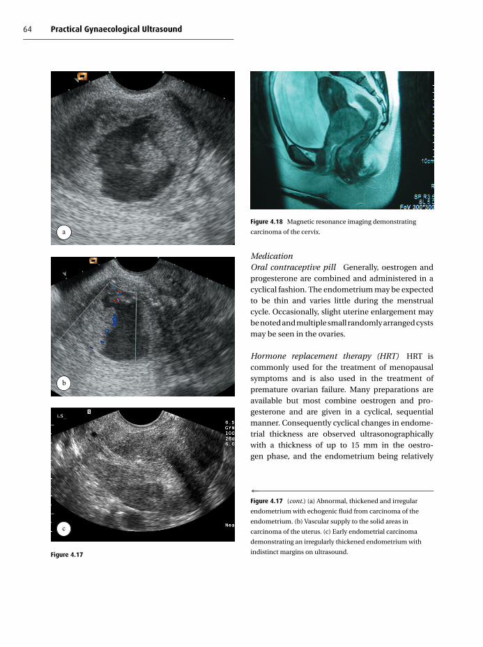
64 Practical Gynaecological Ultrasound
a
b
c
Figure 4.17
Figure 4.18 Magnetic resonance imaging demonstrating
carcinoma of the cervix.
MedicationOral contraceptive pill Generally, oestrogen andprogesterone are combined and administered in acyclical fashion. The endometrium may be expectedto be thin and varies little during the menstrualcycle. Occasionally, slight uterine enlargement maybe noted and multiple small randomly arranged cystsmay be seen in the ovaries.
Hormone replacement therapy (HRT) HRT iscommonly used for the treatment of menopausalsymptoms and is also used in the treatment ofpremature ovarian failure. Many preparations areavailable but most combine oestrogen and pro-gesterone and are given in a cyclical, sequentialmanner. Consequently cyclical changes in endome-trial thickness are observed ultrasonographicallywith a thickness of up to 15 mm in the oestro-gen phase, and the endometrium being relatively
←Figure 4.17 (cont.) (a) Abnormal, thickened and irregular
endometrium with echogenic fluid from carcinoma of the
endometrium. (b) Vascular supply to the solid areas in
carcinoma of the uterus. (c) Early endometrial carcinoma
demonstrating an irregularly thickened endometrium with
indistinct margins on ultrasound.

Pathology of the uterus, cervix and vagina 65
Figure 4.19 Sagittal scan of the uterus showing typical features
of the endometrium, which is thickened with some cystic
changes, in a patient taking tamoxifen.
less thick in the progesterone phase. Ultrasoundassessment of endometrial thickness is best per-formed immediately after the progesterone phaseof the cycle when the endometrium is likely to bethinnest. The majority of patients are scanned forunscheduled bleeding on HRT. The most importantultrasound sign in this group to suggest endometrialpathology is irregularity of the endometrium withabnormal vascularity.
Tamoxifen Tamoxifen is an oral synthetic antioe-strogen compound with mild oestrogenic effectson the uterine properties. It is used as an adju-vant chemotherapeutic agent in the treatment ofbreast cancer. Tamoxifen, due to the oestrogeniceffects, causes endometrial metaplasia, hyperpla-sia and carcinoma.18 In patients receiving tamoxifentherapy, the normal risk of developing endometrialcarcinoma is increased sixfold, with both pre- andpostmenopausal women being affected.19 Fifty percent of women taking tamoxifen will develop sometype of endometrial pathology after 6–36 months oftreatment.20
Tamoxifen has characteristic ultrasound appear-ances of endometrial thickening with multipleabnormal small cystic spaces within the abnormal
endometrium (Fig. 4.19). In addition it causes suben-dometrial cysts which can be indistinguishable fromthe endometrium on ultrasound. Therefore the trueendometrial thickness is overestimated by ultra-sound.
Ultrasound is therefore unreliable in the pre-diction of endometrial pathology in symp-tomatic women on tamoxifen. These womenrequire histological correlation, which is best per-formed under direct hysteroscopic vision. Salinehysterosonography21 (Fig. 4.20) is helpful in mon-itoring the endometrial irregularity more accuratelybut MRI is best as it differentiates the endometriumfrom the subendometrial cysts.
Ultrasound appearances The endometrium isthickened – almost always >10 mm. In addition itmay be hyperechoic, irregular in outline, with cysticareas (<5 mm) and there may be polyps.
Gestational trophoblastic diseaseThis results from abnormal trophoblastic tissue andis therefore always associated with the products ofconception, usually presenting with symptoms ofpregnancy and no discernible fetus.
Three entities are recognised:1. molar pregnancy2. invasive mole3. chorion carcinomaThese subdivisions can be considered as types withincreasing invasive propensities: choriocarcinoma isthe most invasive, with early lung metastases.22
In the majority the genetic make up of the tissue isof paternal origin. The abnormal trophoblastic tis-sue secretes high levels of beta-human choriono-gonadotrophic (�HCG) hormone, resulting in symp-toms of nausea and vomiting. The abnormal tissueis prone to bleeding and per vaginam blood loss is acommon symptom.
Ultrasound features of all types are of an endome-trial mass with cystic spaces (Fig. 4.21). The high�HCG levels are associated with thecal luteal cystsof the ovaries.23

66 Practical Gynaecological Ultrasound
a
b
c
Figure 4.20 (a) Saline infusion hysterosonography of a
tamoxifen endometrium, showing subendometrial cystic
hyperplasia. (b) Cystic change on ultrasound in the
endometrium of a patient on tamoxifen. (c) A polyp in a
patient on tamoxifen, demonstrating cystic change.
Figure 4.21 An area of trophoblastic invasion in the lower
uterus, with peripheral vascularity demonstrated on colour
Doppler.
Choriocarcinoma Choriocarcinoma is treated bycurettage and methotrexate therapy. Response andrecurrence are monitored with serum �HCG levels,which allow excellent assessment of the response,of the disease to treatment. MRI and CT are use-ful to check for distant spread, most particularlyto the lungs. Chemotherapy produces an excellentresponse, with a 90% cure rate.
Inflammatory conditionsAcute endometritis This condition is most com-monly seen in the postpartum uterus, usually asso-ciated with retained products of conception whichact as the foci for the infection. Similarly, postuter-ine instrumentation, including suction terminationof pregnancy, is a significant risk factor.
The ultrasound appearances are of an enlargedtender uterus showing loss of the endometrialmyometrial borders and echogenic retained prod-ucts if present. There is a diffuse increase in thevascularity of the uterus due to the acute infec-tion. If extensive, there will be additional signs ofpelvic infections such as free or loculated fluid andthe ultrasound signs of a tubo-ovarian abscess. Thepatient will be acutely unwell with fever and an ele-vated white cell count.24

Pathology of the uterus, cervix and vagina 67
Postoperative conditionsIt is essential to have an understanding of the type ofgynaecological surgery performed in order to under-stand the normal and abnormal postoperative fea-tures seen with ultrasound. It is normal to have asmall collection of blood at the vault of the vaginaimmediately postoperatively following a hysterec-tomy and in a subtotal hysterectomy the cervix is leftin situ. Abnormal postoperative collections are usu-ally haematomas but these may become infected.
Ultrasound signs of infection include septation,echogenic fluid and increasing tenderness. In thissituation ultrasound guided per vaginam drainagemay be appropriate.
Post-caesarean section Following a lower-segment caesarean section, the uterus will usuallyappear entirely normal after 6 weeks. But in somepatients, a small anterior deformity will be seenat the junction of the middle and lower uterinesegments at the site of the surgical incision. Thismay have the ultrasound appearance of a smallfibroid but its site and the clinical history shouldsuggest the diagnosis. This minor abnormality isclinically insignificant (Fig. 4.22).
Adenomyosis Adenomyosis, in which ectopicendometrium ‘invades’ the myometrium, may beconsidered a variant of endometriosis. The two con-ditions are known to coexist in 20% of affectedpatients. Histologically, ectopic endometrial glandsand stroma are found in the myometrium. The typ-ical patient will be a multiparous woman in her 40spresenting with menorrhagia and dysmenorrhoea.This presentation contrasts with that of endometrio-sis whereby a nulliparous, infertile woman in her 20sor 30s is most likely to be affected. Adenomyosis maybe associated with previous dilatation and curettage(D&C), caesarean section or elevated oestrogen lev-els. The disease is usually generalised but occasion-ally a localised focus, termed an adenomyoma, maybe found.25
Ultrasound appearances The ultrasound appear-ances are variable and non-specific but usually there
a
b
Figure 4.22 (a) Post-caesarean-section scar on ultrasound
(arrow). (b) This lower-segment caesarean-section scar fills
with fluid during a saline infusion hysterosonogram.
is slight enlargement of the uterus with loss of thehomogenicity of the myometrium, without specificfeatures of discrete fibroids. Occasionally tiny cys-tic areas within the myometrium can be discernedon transvaginal ultrasound, and hyperechoic areas,probably corresponding to areas of cyclic haemor-rhage, have also been reported.
MRI is more specific and is the diagnostic imagingmodality of choice (Fig. 4.23).
Asherman’s syndromeIntrauterine synechiae, or adhesions, result fromoverenthusiastic endometrial curettage during or

68 Practical Gynaecological Ultrasound
a
b
Figure 4.23 (a) The ultrasound features of adenomyosis may
be subtle. This uterus is enlarged, with a heterogeneous
myometrial texture and a slightly irregular endometrium.
(b) Magnetic resonance imaging demonstrating adenomyosis.
following pregnancy. During the pregnancy theendometrium is more friable than normal and thismay allow the complete removal of focal areas ofendometrium. As a result, single or multiple bandsof fibrous tissue completely or partially traverse theendometrial cavity. Similar adhesions may develop
Figure 4.24 Asherman’s syndrome demonstrating synechiae
on saline infusion.
post-myomectomy or post-caesarean section and inassociation with infection, i.e. endometritis, tuber-culosis and schistosomiasis.
These adhesions may be an incidental finding atultrasound but the patient may present with a his-tory of dysmenorrhoea, oligomenorrhoea, infertilityor recurrent miscarriage.
On ultrasound, the adhesions are usually seen asvery small echogenic foci related to the endome-trial cavity.26 If there is fluid in the cavity, the adhe-sions may be better and more conclusively demon-strated. It is best diagnosed following saline infusion(Fig. 4.24).
Generally the diagnosis will be confirmed at hys-terosalpingography or hysteroscopy, at which timethe adhesions may be divided.
Intrauterine contraceptive device (IUCD)These are a common form of contraception.Transvaginal ultrasound is the investigation ofchoice to locate the device. It should be situated highin the uterine cavity and placement outside of thisshould be considered abnormal and more likely tobe ineffective. It is important to be aware of the dif-fering ultrasound appearances of the various typesof devices. This is particularly true of the Mirenacoil, a device impregnated with levonorgestrel whichis widely used for the symptomatic treatment ofmenorrhagia as well as for contraception. With the

Pathology of the uterus, cervix and vagina 69
Figure 4.25 An intrauterine contraceptive device in the
endometrial cavity.
Mirena the long arm is more echogenic than the Tarm and for the unwary the ultrasound appearancessuggest a coil that is abnormally low in the cavity.
Ultrasound appearances of IUCD The IUCD isusually a strong reflector with posterior acousticshadowing (Fig. 4.25), lying within and along theendometrial cavity.
Abnormal positions There is approximately a1:2000 risk of perforation of the device, usually intothe myometrium, at insertion. In this situation theIUCD may be shown to lie outside the uterine cavity,i.e separate to the endometrium, rather than withinit (Fig. 4.26).
Coils outside the uterus are rarely seen with ultra-sound and any investigation where the coil cannotbe identified with ultrasound merit an abdominal X-ray.
It is important to remember that even with theIUCD correctly sited pregnancy can occur and in thissituation there is also an increased risk of an ectopicpregnancy.
The cervix
Investigation of cervical pathologies is by directvision and cytology, although transvaginal scan-ning in the pregnant and non-pregnant female will
a
b
Figure 4.26 (a) Sagittal transvaginal scan showing an
intrauterine contraceptive device (IUCD) penetrating the
myometrium. (b) Sagittal transabdominal scan showing a
7-week gestation sac with an IUCD located in the cervix.
demonstrate the cervix if this area is carefully exam-ined. However MRI with its excellent tissue charac-terisation is the imaging modality of choice.
Benign conditionsNabothian cystsThese are epithelial inclusion cysts which developin the endocervical canal, and are most commonlyfound in the perimenopausal period. These cystsdevelop as a result of chronic cervicitis which causesobstruction of the endocervical glands, resulting inretention of secretions. Nabothian cysts are asymp-tomatic and clinically insignificant.
Ultrasound appearances Nabothian cysts gen-erally occupy a central position relating to cervicalcanal. They may be single or multiple (Fig. 4.27) and

70 Practical Gynaecological Ultrasound
a
b
c
Figure 4.27 (a and b) Multiple nabothian cysts in the cervix.
(c) One of these nabothian cysts contains echoes and
debris.
a
b
Figure 4.28 (a) A cervical fibroid (arrowsheads). The
differential diagnosis on these appearances would be of a
cervical polyp. (b) Cervical fibroid in a pregnant patient may
potentially obstruct labour.
the size varies from a few millimetres to severalcentimetres, but they are generally < 1 cm.
They are usually echo-free, but some will containconfluent internal echoes.
Cervical fibroidCervical fibroids are uncommon, accounting for 8%of fibroids, the majority being in the uterine body.Most are small and asymptomatic with the sameultrasound features as the uterine fibroids describedearlier in this chapter. However, when large cervi-cal fibroids may be symptomatic due to their masseffect by producing either bladder compression withurinary frequency or in the pregnant patient by caus-ing obstruction to labour (Fig. 4.28). MRI may be ofvalue in clearly defining the position and size of the
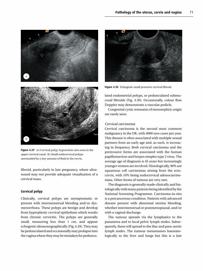
Pathology of the uterus, cervix and vagina 71
a
b
Figure 4.29 (a) Cervical polyp: hypoechoic area seen in the
upper cervical canal. (b) Small endocervical polyps
surrounded by a tiny amount of fluid in the cervix.
fibroid, particularly in late pregnancy, where ultra-sound may not provide adequate visualisation of acervical mass.
Cervical polyp
Clinically, cervical polyps are asymptomatic orpresent with intermenstrual bleeding and/or dys-menorrhoea. These polyps are benign and developfrom hyperplastic cervical epithelium which resultsfrom chronic cervicitis. The polyps are generallysmall, measuring less than 1 cm, and appearechogenic ultrasonographically (Fig. 4.29). They maybe pedunculated and occasionally may prolapse intothe vagina where they may be mistaken for peduncu-
Figure 4.30 Echogenic small posterior cervical fibroid.
lated endometrial polyps, or pedunculated submu-cosal fibroids (Fig. 4.30). Occasionally, colour flowDoppler may demonstrate a vascular pedicle.
Congenital cystic remnants of mesonephric originare rarely seen.
Cervical carcinomaCervical carcinoma is the second most commonmalignancy in the UK, with 4000 new cases per year.This disease is often associated with multiple sexualpartners from an early age and, as such, is increas-ing in frequency. Both cervical carcinoma and thepreinvasive forms are associated with the humanpapillomavirus and herpes simplex type 2 virus. Theaverage age of diagnosis is 45 years but increasinglyyounger women are involved. Histologically, 90% aresquamous cell carcinomas arising from the ecto-cervix, with 10% being endocervical adenocarcino-mata. Other forms of tumour are very rare.
The diagnosis is generally made clinically and his-tologically with many patients being identified by theNational Screening Programme. Carcinoma-in-situis a precancerous condition. Patients with advanceddisease present with abnormal uterine bleeding,whether intermenstrual or postmenopausal, and/orwith a vaginal discharge.
The tumour spreads via the lymphatics to theparametra and to local pelvic lymph nodes. Subse-quently, these will spread to the iliac and para-aorticlymph nodes. The tumour metastasises haemato-logically to the liver and lungs but this is a late
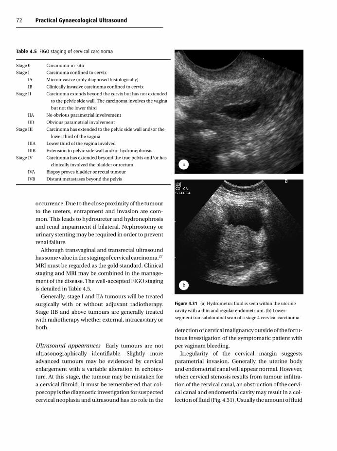
72 Practical Gynaecological Ultrasound
Table 4.5 FIGO staging of cervical carcinoma
Stage 0 Carcinoma-in-situ
Stage I Carcinoma confined to cervix
IA Microinvasive (only diagnosed histologically)
IB Clinically invasive carcinoma confined to cervix
Stage II Carcinoma extends beyond the cervix but has not extended
to the pelvic side wall. The carcinoma involves the vagina
but not the lower third
IIA No obvious parametrial involvement
IIB Obvious parametrial involvement
Stage III Carcinoma has extended to the pelvic side wall and/or the
lower third of the vagina
IIIA Lower third of the vagina involved
IIIB Extension to pelvic side wall and/or hydronephrosis
Stage IV Carcinoma has extended beyond the true pelvis and/or has
clinically involved the bladder or rectum
IVA Biopsy proves bladder or rectal tumour
IVB Distant metastases beyond the pelvis
occurrence. Due to the close proximity of the tumourto the ureters, entrapment and invasion are com-mon. This leads to hydroureter and hydronephrosisand renal impairment if bilateral. Nephrostomy orurinary stenting may be required in order to preventrenal failure.
Although transvaginal and transrectal ultrasoundhas some value in the staging of cervical carcinoma,27
MRI must be regarded as the gold standard. Clinicalstaging and MRI may be combined in the manage-ment of the disease. The well-accepted FIGO stagingis detailed in Table 4.5.
Generally, stage I and IIA tumours will be treatedsurgically with or without adjuvant radiotherapy.Stage IIB and above tumours are generally treatedwith radiotherapy whether external, intracavitary orboth.
Ultrasound appearances Early tumours are notultrasonographically identifiable. Slightly moreadvanced tumours may be evidenced by cervicalenlargement with a variable alteration in echotex-ture. At this stage, the tumour may be mistaken fora cervical fibroid. It must be remembered that col-poscopy is the diagnostic investigation for suspectedcervical neoplasia and ultrasound has no role in the
a
b
Figure 4.31 (a) Hydrometra: fluid is seen within the uterine
cavity with a thin and regular endometrium. (b) Lower-
segment transabdominal scan of a stage 4 cervical carcinoma.
detection of cervical malignancy outside of the fortu-itous investigation of the symptomatic patient withper vaginam bleeding.
Irregularity of the cervical margin suggestsparametrial invasion. Generally the uterine bodyand endometrial canal will appear normal. However,when cervical stenosis results from tumour infiltra-tion of the cervical canal, an obstruction of the cervi-cal canal and endometrial cavity may result in a col-lection of fluid (Fig. 4.31). Usually the amount of fluid

Pathology of the uterus, cervix and vagina 73
Table 4.6 Causes of cervical stenosis
Malignancy Cervical carcinoma
Endometrial carcinoma
Iatrogenic Cone biopsy
Post-radiotherapy
Obstetric trauma
Postmenopausal atrophy
Benign uterocervical pathology Polyp
Fibroid
in the cervical canal is small, although more signifi-cant collections may occur in the uterine cavity and apyometra or haematometra may develop (Fig. 4.32).Regional lymphadenopathy may be apparent withenlarged pelvic lymph nodes being best visualisedtransvaginally. The examination should be extendedto the upper abdomen in order to visualise the kid-neys to check for hydronephrosis. The para-aorticarea should be examined for lymphadenopathy andthe liver examined for metastatic disease.
Hydrometra and pyometra may be seen at the timeof diagnosis, post-treatment or, in the elderly patient,often many years after the successful treatment ofthe disease. Fluid collects in the uterine cavity dueto chronic cervical narrowing either from radia-tion fibrosis or age-related stenosis. Advanced pri-mary or recurrent disease may present with haema-turia and urinary symptoms and ultrasound mayshow evidence of bladder invasion by the tumour(Fig. 4.33).
MRI is the investigation that defines tumour bulk,spread and recurrence most effectively, althoughultrasound can be used to monitor renal dilatation.
Cervical stenosisThe causes of cervical stenosis are varied (Table 4.6)and a good clinical history is often required in orderto elucidate the cause. However, whatever the cause,the clinical picture and the ultrasound appearanceare likely to be similar. When obstruction allows
→Figure 4.32 (a) Scan showing echogenic fluid within the
uterine cavity in a tender uterus. (b) Haematometra – blood
and clots are seen in the endometrial cavity.
(c) Hydronephrosis as a result of obstruction of the ureter in
the pelvis.
a
b
c
Figure 4.32

74 Practical Gynaecological Ultrasound
Figure 4.33 Thickened, abnormal bladder mucosa in
malignant involvement of the bladder from carcinoma of the
cervix.
Figure 4.34 Hydrometra in a postmenopausal uterus – a
normal finding.
Table 4.7 Uterine fluid collections
Infancy and childhood Hydro/haematometrocolpos
Physiological Menstruation
Postmenopausal
Pregnancy Normal early pregnancy
Ectopic
Haemorrhage
Retained products of conception
Post-instrumentation
Cervical stenosis (see above)
Intrauterine pathology Endometrial carcinoma
Endometrial hyperplasia
Endometrial polyp
Pelvic inflammatory disease Endometritis
a significant amount of fluid to collect within theendometrial cavity, according to the type of fluid, theappearance is variously named as follows:� hydrometra: serous fluid� haematometra: blood� pyometra: pusIn the case of pyometra, if gas-forming organismsare the cause of infection, then gas may be seen inthe uterine cavity. In pyometra the uterus will be ten-der and the patient may have symptoms of infection,although chronic infections are often asymptomatic.
It must be remembered that a small amount offluid seen in the postmenopausal uterus is normal asa result of atrophic cervical stenosis. This fluid canbe useful in defining the endometrium (Fig. 4.34).
Uterine/endometrial fluidThe causes of fluid in the endometrial cavity may bephysiological or pathological (Table 4.7)
Ultrasound appearances When there is fluid in theuterus it may be enlarged, with a distended endome-trial cavity. The cervical canal may also be distendedwith fluid. The fluid may contain internal echoes,debris or fluid–fluid levels or gas.
An associated tumour mass may be apparent, forexample endometrial, outlined by fluid.
Dilation of the cervical canal is a relatively straight-forward procedure which will allow drainage of

Pathology of the uterus, cervix and vagina 75
Figure 4.35 Air in the vagina.
the abnormal fluid. However, where malignancy isinvolved, further investigations will be required.
The vagina
Vaginal pathology is generally recognised and inves-tigated clinically. However, vaginal abnormalities areoften diagnosed coincidentally at ultrasound exam-ination, generally transabdominally as the vagina isnot as well-visualised transvaginally due to its closeproximity to the probe. Perineal examination may bemore appropriate.
Ultrasound has a significant role to play inthe investigation of congenital abnormalities (seeChapter 8).
Ultrasound can be useful in the investigation ofretained tampons or other foreign bodies, althoughMRI is often required to assess these further, partic-ularly in the paediatric age group.
Air may be visible in the vagina following instru-mentation or sexual intercourse. This has the typicalappearance of gas and is seen as flashing brightlyechogenic foci in the upper vaginal vault (Fig. 4.35)and may indeed outline the cervix as seen in axialsection. Similarly, an air-containing tampon may bevisualised in the vagina transabdominally at ultra-
Figure 4.36 Sagittal image showing the strong reflection from
a tampon in the vagina.
Figure 4.37 Transverse scan through the vagina showing a
vaginal ring pessary, with strong acoustic shadowing.
sound as a strongly reflective lower structure pro-ducing acoustic shadowing (Fig. 4.36).
A vaginal pessary used in the treatment of uter-ine prolapse will be readily identified transabdom-inally. The superior and inferior margins of the ringare seen in the upper vagina on a sagittal section andthe two lateral margins of the ring are seen in an axialplane. The ring will produce strong acoustic shadow-ing (Fig. 4.37).

76 Practical Gynaecological Ultrasound
Vaginal fluid collectionsA small collection of fluid is not an uncommon find-ing at transabdominal ultrasound. This is most com-monly seen at the time of menstruation. In a smallchild, small amounts of urine may occasionally bevisualised following micturition. In the adult, urin-ary incontinence, a urinary fistula or an ectopicallysited ureter may result in urine pooling in the vagina.Occasionally, extravaginal fluid collections such asparavaginal haematoma or urethral cyst may com-press the vagina and give the erroneous impressionof a vaginal fluid collection.
Haematocolpos Obstruction of the vagina willallow collection of fluid within. This usually occurs inyoung women post-menarche whereby the monthlymenstrual blood collects in the vagina due to the lackof exit. The obstruction may lie distally at the level ofthe introitus, due to an imperforate hymen, or mayresult from a more proximal vaginal atresia, stenosisor the presence of a vaginal septum. These patientsgenerally present with cyclical low abdomimal painand amenorrhoea, lasting for some years after theexpected date of the menarche. A lower abdom-inal mass may also be present. A similar conditionis sometimes seen in infants or young children and,indeed, has been diagnosed antenatally. In this groupof patients, the influence of the maternal hormonesmay be partially responsible for the condition (seeChapter 8).
When haematocolpos is associated with ananomaly of the genital tract, a thorough examinationof the urinary tract is essential as in approximately athird of cases renal anomalies may be present. Uni-lateral renal agenesis or renal ectopia is the mostlikely anomaly to be found.
Ultrasound appearances A medium to large-sizedcystic mass may be seen lying centrally in the lowerabdomen. It should generally be possible to demon-strate continuity with the uterus and, indeed, thefluid may outline the ectocervix (Fig. 4.38). The masshas a thin wall and may contain fine internal echoesdue to presence of altered blood. The appearance of
Figure 4.38 Fluid and blood clots distend the vagina in
haematocolpos.
Figure 4.39 Magnetic resonance imaging scan showing
haematocolpos.
the fluid may be very similar to that of an endometri-otic cyst. Translabial ultrasound may be of valuein demonstrating the length of the stenotic seg-ment from the introitus.28 MRI is also of value inthis situation, with the information obtained allow-ing for accurate planning of reconstructive surgery(Fig. 4.39).

Pathology of the uterus, cervix and vagina 77
Figure 4.40 Three-dimensional ultrasound image
demonstrating the endometrium.
Three-dimensional (3D) ultrasoundThe role of 3D in gynaecology has yet to be estab-lished but is increasingly used, in particular to definethe shape and volume of the endometrium in threedimensions (Fig. 4.40).29 However, MRI remainssuperior and therefore the use of 3D is more likelyin situations when access to MRI is limited.
R E F E R E N C E S
1. F. Hasan, K. Arumugam and V. Sivanesaratnam, Uterine
leiomyomota in pregnancy. International Journal of Gynae-
cology and Obstetrics, 34 (1991), 45–8.
2. R. E. Pelsang, J. Sorosky and T. Woods, Sonographic evalua-
tion of the broad ligament of the uterus. Journal of Clinical
Ultrasound, 27 (1999), 402–4.
3. W. M. Christopherson, E. O. Williamson and L. O.
Gray, Leiomyosarcoma of the uterus. Cancer, 29 (1972),
1512–17.
4. L. G. Shapeero and H. Hrick, Mixed Mullerian sarcoma of the
uterus: MR imaging findings. American Journal of Radiology,
153 (1989), 317–19.
5. S. H. Kim, H. Y. Hwang and B. I. Choi, Case report: uterine
metastasis from stomach cancer: radiological findings. Clin-
ical Radiology, 42 (1990), 285–6.
6. A. Malatsky, K. L. Reuter and B. Woda, Sonographic findings
in primary uterine cervical lymphoma. Journal of Clinical
Ultrasound, 19 (1991), 62–4.
7. W. H. Su, P. H. Wang, S. P. Chang and M. C. Su, Preoperative
diagnosis of uterine lipoleiomyomata using ultrasound and
computed tomography a case report. European Journal of
Gynaecology and Oncology, 22 (2001), 439–40.
8. J. K. Gupta, P. F. W. Chien, D. Voit et al., Ultrasonographic
endometrial thickness for diagnosing endometrial pathol-
ogy in women with postmenopausal bleeding: a meta ana-
lysis. Acta Obstetrica Gynecologica Scandinavica, 81 (2002),
799–816.
9. E. Epstein and L. Valentin, Managing women with post
menopausal bleeding. Best Practice in Research Clinic in
Obstetrics and Gynaecology, 18 (2004), 125–43.
10. Scottish Intercollegiate Guidelines Network Investigation of
Post-menopausal Bleeding (2002).
11. American College of Obstetrics and Gynecologist Tech-
nology Assessment, Saline infusion sonography. Journal of
Gynaecology and Obstetrics, 84 (2004), 95–8.
12. T. J. Dubinsky, K. Stroehlein, Y. Abu-ghazzeh, H. R. Parvey
and N. Maklad, Prediction of benign and malignant disease:
hysterosonographic-pathologic correlation. Radiology, 210
(1999), 393–7.
13. S. Laifer-Narin, N. Ragavendra, E. K. Parmenter and E. G.
Grant, False-normal appearance of the endometrium on
conventional transvaginal sonography: comparison with
saline hyterosonography. American Journal of Roentgenol-
ogy, 178 (2002), 129–33.
14. A. M. Darwish, A. M. Makhlouf, A. A. Youssof and
H. A. Gadalla, Hysteroscopic myometrial biopsy in unex-
plained abnormal uterine bleeding. European Journal of
Obstetrics, Gynecology and Reproductive Biology, 86 (1999),
139–43.
15. M. C. Anderson, E. B. Butler and G. V. P. Chamberlain, Oestro-
gen replacement and endometrial cancer. A statement by the
British Cancer Group, Lancet, I (1981), 1359–60.
16. L. E. Rutqvist, H. Johansson, T. Signomklao et al., Adjuvant
tamoxifen therapy for early stage breast cancer and second
primary malignancies. Journal of the National Cancer Insti-
tute, 87 (1995), 645–51.
17. A. Ayiomamitis, The epidemiology of cancer of the uterine
corpus in Canada 1950–85. International Journal of Gynae-
cology and Obstetrics, 27 (1988), 205–11.
18. M. Berliere, A. Charles, C. Galant and J. Donnez, Uter-
ine side effects of tamoxifen: a need for systematic pre-
treatment screening. Obstetrics and Gynaecology, 91 (1998),
40–4.

78 Practical Gynaecological Ultrasound
19. S. Cecchini, S. Ciatto, R. Bonardi et al., Risk of endometrial
cancer in breast cancer patients under long term adjuvant
treatment with Tamoxifen. Tumori, 84 (1998), 21–3.
20. X. De Muylder, P. Neven, M. De Somer et al., Endometrial
lesions in patients undergoing tamoxifen therapy. Interna-
tional Journal of Gynecology and Obstetrics, 36 (1991), 127–
30.
21. L. E. Hann, C. M. Kim, M. Gonen et al., Sonohysterogra-
phy compared with endometrial biopsy for evaluation of
endoemtrium in tamoxifen treated women. Obstetrics and
Gynaecology Survey, 59 (2004), 440–1.
22. N. J. Sebire, G. Makrydimaas, N. J. Agnantis, N. Zagori-
anakou and H. Rees, Updated diagnostic criteria for partial
and complete hydatidiform mole in early pregnancy. Anti-
cancer Research, 23 (2003), 1723–8.
23. B. W. Hancock and J. A. Tidy, Current management of molar
pregnancy. Journal of Reproductive Medicine, 47 (2002),
347–54.
24. L. Savelli, G. Pilu, B. Valeri and L. Bovicelli, Transvaginal
sonographic appearances of anaerobic endometris. Ultra-
sound in Obstetrics and Gynaecology, 21 (2003), 624–5.
25. R. Devlieger, T. D’Hoogle and D. Timmerman, Uterine ade-
nomyosis in the infertility clinic. Human Reproduction
Update, 9 (2003), 139–47.
26. E. Confino, J. Friberg, R. V. Giglia and N. Fleicher, Sono-
graphic imaging of intrauterine adhesions. Obstetrics and
Gynecology, 66 (1985), 596–8.
27. M. Cobby, J. Browning, A. Jones, E. Whipp and P. Goddard,
Magnetic resonance imaging, computed tomography and
endosonography in the local staging of carcinoma of the
cervix. British Journal of Radiology, 63 (1990), 673–9.
28. K. A. Scanlon, M. A. Pozniak, M. Fagerholm and S. Shapiro,
Value of transperineal sonography in the assessment of
vaginal atresia. American Journal of Roentgenology, 154
(1990), 545–8.
29. R. Salim, B. Woelfer, M. Backos, L. Regan and D. Jurkovic,
Reproducibility of three dimensional ultrasound diagnosis
of congenital uterine anomalies. Ultrasound in Obstetrics
and Gynecology, 21 (2003), 578–82.
B I B L I O G R A P H Y
J. Anderson,Gynaecologic Imaging (Churchill Livingstone, 1999).
S. Robby, M. C. Anderson and P. Russell, Pathology of the Female
Reproductive Tract (Churchill Liuingstone, 2002).
A. C. Fleischer, F. A. Manning, P. Jeanty and R. Romero, Sonogra-
phy in Obstetric and Gynaecology (Prentice Hall International)

5
Pathology of the ovaries, fallopian tubes and adnexae
Damian J. M. Tolan and Michael J. WestonSt James’s University Hospital, Leeds
Introduction
Ultrasound has become an essential part of thegynaecological evaluation of the adnexae (whichcomprise the ovaries, fallopian tubes and broad liga-ment). In this chapter we will discuss the pathology,ultrasound imaging features and aspects of manage-ment of a range of conditions affecting the adnexaeand conditions that may mimic adnexal pathology.We will concentrate on important or common con-ditions, with particular emphasis on the ovaries.
Clinical presentation
Patients referred for ultrasound examination ofthe pelvis may present with a range of symp-toms. Pain is common and may be related toovulation (mittelschmerz), menstruation (partic-ularly in endometriosis) or intercourse (dyspareu-nia). Chronic pain may indicate pelvic inflammatorydisease (PID) and acute severe pain may herald anemergency in the form of ovarian torsion or ectopicpregnancy. However some women complain of pres-sure rather than pain, often related to masses caus-ing compression and displacement of pelvic organs.The close proximity of the adnexae to other organsmay produce gastrointestinal or renal tract symp-toms, which may misdirect the clinical evaluationaway from the real cause for the complaint. Bloating
and distension, though non-specific, are symptomsthat may suggest ascites or massive ovarian tumours.
Alteration to the normal menstrual cycle, withirregular bleeding or amenorrhoea, and infertility orsubfertility are further indicators of ovarian pathol-ogy ranging from polycystic ovary syndrome (PCOS)to hormone-secreting tumours.
Increasingly, with improvement in technology andincrease in the demand for examinations, pathologyis being discovered incidentally on imaging exam-inations for unrelated complaints. This emphasisesthe point that important ovarian pathology is oftenasymptomatic and helps to explain why ovarian can-cer is known as the silent killer, resulting from the latepresentation of many cases and the apparent lack ofsymptoms in the early phase of disease.
Essential patient details
Prior to scanning the patient should be interviewedto discuss her presenting symptoms and to obtaininformation essential to the correct interpretation ofthe scan. This should include the following details:� patient’s age – as adnexal masses in premenarchal
or older postmenopausal women are more likelyto be malignant� date of the first day of the last period and normallength of cycle� menstrual status (premenarchal, pre- or post-menopausal)
C© Cambridge University Press 2005.
79

80 Practical Gynaecological Ultrasound
� medications (including hormone replacement,oral or depot contraceptives and tamoxifen)� gynaecological history, including previous opera-tions
Acceptability of transvaginal scanning should beensured in an appropriate setting prior to scanning.
Benign physiological (functional) cysts
Cyst formation in the ovary is a normal consequenceof ovarian function. These physiological cysts arisefrom the hormone-regulated development of folli-cles within the ovary. Larger cysts result from physi-ological over-stimulation of a dominant graafian fol-licle and are a cause of concern because they maymimic more serious pathology.
Follicular cysts
These are the commonest clinically detectable ovar-ian masses and one of the most common causes ofan ovarian mass on ultrasound. They arise within anovary because of a failure of the mature Graafian fol-licle to rupture and release the ovum. Typically, rup-ture occurs when the follicles reach 2.5 cm in diame-ter. However follicular cysts continue to grow underhormone mediation and may reach up to 8–10 cm indiameter in some cases.
The typical appearance is of a unilocular cystover 3 cm in diameter with a thin, smooth walland echo-free fluid within it (Fig. 5.1). Some normalcysts may undergo haemorrhagic change, producingechogenic fluid.1 The normal history is of sponta-neous regression within 4–6 weeks. A repeat exam-ination is recommended in 5–7 weeks to ensure reso-lution of the follicle – a malignant cyst will not regressin size.
Corpus luteum cysts
The corpus luteum is formed by rupture of themature graafian follicle. Its function is to produceprogesterone and oestrogens to support early preg-nancy in the period before the placenta takes over
a
b
Figure 5.1 (a) Normal ovary showing a few small follicular
structures. (b) A simple follicular cyst of the ovary. There are no
internal echoes.
this role. When these follicles fail to involute, cystsare formed.
The clinical picture may be pain with delayedmenses or amenorrhoea. In this setting the mostimportant differential is ectopic pregnancy: this maymimic the imaging findings of a haemorrhagic cor-pus luteum, particularly as there is often associ-ated endometrial proliferation. Knowledge of theresult of a pregnancy test is advantageous. Clearlya well-demonstrated intrauterine pregnancy wouldsupport an adnexal lesion being a corpus luteum,but an important exception is in the setting of in

Pathology of the adnexae 81
a
b
Figure 5.2 (a) A small luteal cyst of the ovary showing uniform
internal echoes due to recent haemorrhage. (b) A large luteal
cyst showing internal echoes due to haemorrhage as well as a
focal lump due to clot retraction.
vitro fertilisation (IVF) where multiple pregnancyand ectopic pregnancy are commoner than in thegeneral population and rarely both may coexist (i.e.heterotopic pregnancy).
The sonographic findings of a corpus luteum cystare variable. Typically these cysts are hypoechoicwith thick walls and may vary in size from 2 to10 cm, although most are less than 3 cm in diameter.Haemorrhage is often present, resulting in theappearance of either a complex cystic mass contain-ing fluid with internal echoes or a solid adnexal struc-ture (Fig. 5.2). Occasionally layering of contents mayoccur. The Doppler and colour flow characteristics
are non-specific and vary as the corpus luteummatures and involutes but peripheral diastolic flowis normal in these cysts during the luteal phase of thecycle. Corpus luteum cysts can cause complicationsthrough rupture and haemorrhage, obstructed deliv-ery and by undergoing torsion. A persistent largecorpus in the second half of pregnancy may requiresurgical excision to prevent later complications.Similarly, a complex haemorrhagic luteal cyst maysimulate malignancy. In this setting follow-up scan-ning is needed to ensure reduction or resolution.
Parovarian cysts (fimbral cysts, peritonealinclusion cysts)
While these are not truly ovarian, they may mimicovarian cysts and so are dealt with in this section ofthe chapter. Parovarian cysts arise in the broad liga-ment, originating either from the peritoneum cover-ing the ligament or embryological remnants of thewolffian ducts that run from the vaginal vestibuleto the ovary within the ligament. They comprise upto 10% of adnexal masses, tending to affect pre-menopausal women, and are seen separate fromthe neighbouring ovary. Cysts are fluid-filled andthin-walled with no septations or internal soft-tissueprojections.2 In common with ovarian cysts they maybe complicated by haemorrhage, torsion and rup-ture and can reach several centimetres in diame-ter, extending out of the pelvis.3 Peritoneal inclu-sion cysts are more likely to be seen if there has beenprior surgery. The cyst walls will conform to adjacentstructures and often show acute angular marginsas a characteristic feature. Not all pelvic cysts arisefrom gynaecological structures. Sometimes intesti-nal lesions may mimic an ovarian lesion. Carefullyassessing if the lesion is attached to bowel will avoidthis pitfall, though a precise diagnosis may only bemade confidently following surgery (Fig. 5.3).
Postmenopausal cysts
Simple cysts of less than 5 cm in size are a verycommon finding in postmenopausal women dur-ing pelvic ultrasound examination. It is important to
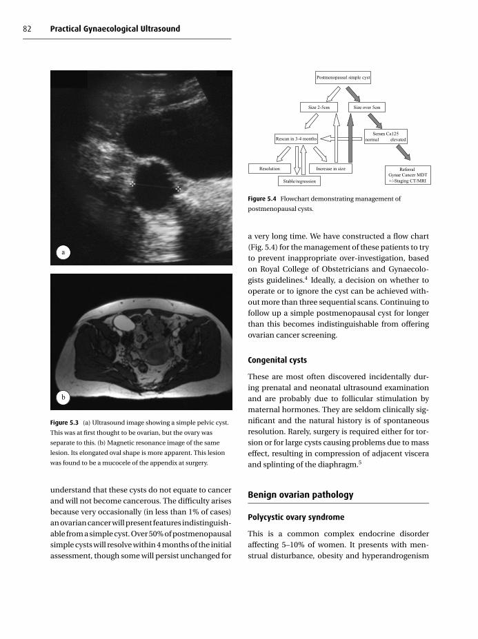
82 Practical Gynaecological Ultrasound
a
b
Figure 5.3 (a) Ultrasound image showing a simple pelvic cyst.
This was at first thought to be ovarian, but the ovary was
separate to this. (b) Magnetic resonance image of the same
lesion. Its elongated oval shape is more apparent. This lesion
was found to be a mucocele of the appendix at surgery.
understand that these cysts do not equate to cancerand will not become cancerous. The difficulty arisesbecause very occasionally (in less than 1% of cases)an ovarian cancer will present features indistinguish-able from a simple cyst. Over 50% of postmenopausalsimple cysts will resolve within 4 months of the initialassessment, though some will persist unchanged for
Postmenopausal simple cyst
Size over 5cm
Referral Gynae Cancer MDT+/-Staging CT/MRI
Size 2-5cm
Rescan in 3-4 monthsSerum Ca125
normal elevated
Resolution
Stable/regression
Increase in size
Figure 5.4 Flowchart demonstrating management of
postmenopausal cysts.
a very long time. We have constructed a flow chart(Fig. 5.4) for the management of these patients to tryto prevent inappropriate over-investigation, basedon Royal College of Obstetricians and Gynaecolo-gists guidelines.4 Ideally, a decision on whether tooperate or to ignore the cyst can be achieved with-out more than three sequential scans. Continuing tofollow up a simple postmenopausal cyst for longerthan this becomes indistinguishable from offeringovarian cancer screening.
Congenital cysts
These are most often discovered incidentally dur-ing prenatal and neonatal ultrasound examinationand are probably due to follicular stimulation bymaternal hormones. They are seldom clinically sig-nificant and the natural history is of spontaneousresolution. Rarely, surgery is required either for tor-sion or for large cysts causing problems due to masseffect, resulting in compression of adjacent visceraand splinting of the diaphragm.5
Benign ovarian pathology
Polycystic ovary syndrome
This is a common complex endocrine disorderaffecting 5–10% of women. It presents with men-strual disturbance, obesity and hyperandrogenism

Pathology of the adnexae 83
a
b
Figure 5.5 (a) Histological specimen of a polycystic ovary. This
shows the typical features one would expect to see on
ultrasound of numerous small follicles and an increase in
stroma. (b) An ultrasound image of a polycystic ovary showing
the same features.
resulting from testosterone and androstenedioneproduction, which leads to increased hair growth(hirsutism). The criteria for ultrasound diagnosisof PCOS have recently been revised in the light ofimproved ultrasound technology and better under-standing of the condition.6 The diagnosis can be sup-ported when one or more of the following features aredemonstrated (Fig. 5.5):� 12 or more follicles (3–12 mm diameter) are present
in an ovary (either peripheral or diffusely arranged)
� ovarian volume is over 10 ml/s (when no folliclesmeasuring over 10 mm in diameter are present)
Only a single ovary need be affected to make the diag-nosis. If a large follicle is present (over 10 mm), thenthe volume should be calculated on a repeat scanwhen the ovary is quiescent to prevent overestima-tion of ovarian volume. Bright ovarian stroma rela-tive to the myometrium is no longer an essential partof the diagnosis but is specific to the condition andprovides supporting evidence in the presence of theessential features above. This appearance is thoughtby some authorities to be caused by increased soundtransmission through follicles in the ovaries.
The appearance of a polycystic ovary is muchcommoner than the diagnosis of the syndrome.An important point emphasised in these newdiagnostic guidelines is that imaging findings aloneshould not diagnose PCOS in an asymptomaticpatient. In this situation the possibility may beraised but further supporting evidence in termsof the patient’s history, clinical examination andblood tests (elevated androgens and luteinisinghormone with low/normal follicle-stimulatinghormone level) should be obtained before a firmdiagnosis is made.6
Endometriosis
This is a relatively common condition, affectingaround 5–10% of all women but accounting for ahigh proportion of patients presenting with infertil-ity problems and chronic pelvic pain. It is thoughtto arise from deposits of endometrial tissue seedingoutside the uterus during retrograde menstruation(i.e. along the fallopian tubes into the peritoneum).Classically symptoms are cyclical, occurring duringmenstruation when ectopic endometrial depositsbleed, but intermenstrual bleeding and menorrha-gia may occur. Interestingly, the bulk of diseasedoes not appear to correlate closely with symptoms;those with large-volume disease are sometimesasymptomatic, while those with relatively minordeposits on imaging and laparoscopy may havesevere intractable symptoms.

84 Practical Gynaecological Ultrasound
a
b
c
While all imaging techniques struggle to demon-strate the scattered small endometriosis depositsseen at times during laparoscopy, ultrasound is goodat demonstrating focal masses called endometri-omas. They are usually thin-walled ovoid or roundhypoechoic masses, and may contain fluid with scat-tered internal echoes or gravity-dependent fluid–fluid levels from layering of haemorrhage. In mostcases there is acoustic enhancement through thelesion.7 The size (1–20 cm), wall thickness and con-tour and the degree of internal septation are variableand differentiation from other pelvic pathology, par-ticularly ovarian malignancy, is often impossible. Inthis setting magnetic resonance imaging (MRI) is anextremely useful tool; the unique signal character-istics of endometrioma produce high signal (bright)on T1-weighted images, with no loss of signal on fat-saturated T1-weighted sequences and intermediateor low signal areas on T2, due to the blood prod-ucts within it8 (Fig. 5.6). This allows differentiation inmost cases from dermoid tumours, which normallycontain fat, and ovarian carcinoma, and helps avoidunnecessary surgery.
An important consideration in cases ofendometriosis is the distribution of disease. Mostoften deposits are found in gravity-dependent areaswithin the pelvis, involving the ovaries, rectosigmoidcolon9 and urinary bladder.10 The disease is alsorecognised to occur at the site of caesarean-sectionscar.11 Transvaginal and transrectal scans are verysensitive methods of assessing plaque-like depositsof endometriosis along the uterosacral ligamentsby demonstrating thickening of the rectovaginalseptum and uterosacral ligaments.12 Disease mayalso disseminate to other sites in the abdomen,spreading along peritoneal recesses to involve other
←Figure 5.6 Endometriosis (a) A large endometrioma showing
the typical uniform low-level internal echoes on ultrasound.
(b) Tiny cystic endometriotic deposits around the surface
of an ovary. There is a fleck of associated calcification.
(c) Magnetic resonance of an endometriotic cyst. Layering
of the signal intensity is seen from the blood within the cyst
(arrow).

Pathology of the adnexae 85
sections of bowel and even across the diaphragm toinvolve the pleural space.
Torsion
Ovarian torsion normally presents in the first threedecades of life with a short history of acute-onsetlower abdominal pain, often localised to one side,and accompanied by nausea and vomiting. Feverand elevated white cell counts are not commonlypresent.13,14 Intermittent pain may precede theacute event, weeks or months before diagnosis, dueto intermittent torsion of the ovary.
Patients are predisposed to torsion by coexist-ing ovarian pathology, such as follicular cysts, butin many cases there is no underlying abnormalitypresent. The process of torsion of the vascular pedi-cle causes venous outflow obstruction and engorge-ment of the ovary. Eventually gangrene ensues,as a result of either venous infarction or arterialocclusion.
The diagnosis is suggested by unilateral enlarge-ment of a rounded or oval-shaped ovary containingmultiple enlarged follicles, although these folliclesare not always present. The ovarian stroma is oftenbrighter than adjacent myometrium and free fluidmay be present in the pelvis. The mass may be solidwith posterior acoustic enhancement or cystic.13,14
Free fluid is present in the majority of cases. Rarelypatients may present with massive ovarian enlarge-ment, which can be bilateral, mimicking neoplasia15
(Fig. 5.7). A further indicator of torsion is that theovary may lie in an abnormal position in the mid-line behind the uterus. A peripheral blood flow pat-tern may be present but colour flow can be absent ininfarction. Importantly, normal colour flow does notexclude the diagnosis, which may occur in cases ofintermittent torsion.
This is often an underdiagnosed condition sono-graphically and clinicians with a high index of suspi-cion should not be falsely reassured by a normal scan.In this setting laparoscopy is required to ensure thatthe ovaries are healthy and, if torted, detorsion andfixation performed or oophorectomy in the presenceof gangrene.
Figure 5.7 Massive ovarian oedema as a result of ovarian
torsion.
Ovarian hyperstimulation
This arises most often as a result of ovarian hor-mone stimulation in infertility techniques such asprior to egg-harvesting in IVF. It may also occur insingle or multiple pregnancies, hydatidiform moleand choriocarcinoma.16,17 Briefly, the findings con-sist of enlarged ovaries where increasing diametercorrelates with disease severity (from 5 cm in grade2 to over 12 cm in grades 5 and 6). Multiple largecysts are present and there may be associated ascitesand pleural effusions. This is dealt with more fully inChapter 7.
Absent ovaries and ovarian remnant syndrome
Either or both ovaries are sometimes not visu-alised during assessment of the pelvis, particularlywhen only a transabdominal scan is performed. It isimportant to remember that some patients may haveno ovaries at all. This should be considered partic-ularly in cases of amenorrhoea. Turner’s syndrome,where only one X chromosome is present, may resultin absent ovaries. Patients have short stature and

86 Practical Gynaecological Ultrasound
may have associated renal and skeletal developmen-tal anomalies. Swyer’s syndrome results in isolatedabsence of the ovaries without other abnormality.
Ovarian remnant syndrome presents in patientswho have had previous incomplete oophorectomywith abdominal pain and sometimes a palpablemass. The residual functioning ovarian tissue hyper-trophies to become an enlarged multicystic structureand haemorrhage is often present. The mass maycause ureteric obstruction and hydronephrosis.18 Anovarian mass can be present when the ovaries areexpected to be absent.
Multimodality and multidisciplinaryassessment of ovarian tumours
Any case of possible ovarian malignancy requiresmultidisciplinary assessment by a team with experi-enced gynaecologists, pathologists, radiologists andoncologists. This serves two purposes: to decide,first, on what further investigations are required, andsecond, what treatments should be offered with aview to cure or palliation of symptoms.
Preoperative biopsy should not be done to con-firm the diagnosis of localised ovarian malignancy.It is contraindicated because of the risk of seed-ing tumour into the peritoneum and upstaging thepatient from early stage I to stage II or III with asubsequent poorer prognosis. Likewise, drainage ofascites is best avoided if surgery is planned in thenear future anyway. Occasionally there are reports ofunwary clinicians inserting drainage catheters intoovarian cysts in the mistaken belief they are drain-ing either ascites or the bladder. Tumour markers arenot always helpful, with CA125 elevated in only 50%of stage I tumours. MRI scanning has a useful rolein the evaluation of indeterminate lesions on ultra-sound. Many benign lesions have characteristic fea-tures on MR that allow differentiation from ovariancarcinoma. In the case of ovarian cancers, an assess-ment of the stage of local disease can be made withgreater confidence on MRI and the ultrasound fea-tures of the tumour confirmed. This is particularlyhelpful in borderline cases on ultrasound, where MRI
allows a more complete characterisation of the lesionin question. Computed tomography (CT) scanninghas a role in the staging assessment of disseminateddisease, where there is no doubt about the malig-nant features on ultrasound or where metastatic dis-ease has been demonstrated on the ultrasound scan.Neither technique can assess the peritoneum wellfor small-volume metastases, although delayed MRIfollowing gadolinium is producing some promis-ing results. Laparoscopy gives the best evaluation inthese circumstances.
Imaging allows preoperative planning of the typeof surgery required, with radical hysterectomy, bilat-eral oophorectomy and omentectomy as the stan-dard curative technique for ovarian cancer (the bigoperation). This compares with much more lim-ited surgery in benign or some borderline malignantcases, where unilateral oophorectomy or cystectomymay be considered (the small operation). In cases ofdisseminated disease where radical surgical debulk-ing is inappropriate, imaging-guided biopsy of peri-toneal deposits is a simple and effective method ofobtaining histological confirmation of the diagno-sis, and can assist in planning palliative chemother-apy for symptom control. In some cases, where thereis inoperable malignancy on initial scanning, somepatients may proceed, after a confirmatory biopsy,to chemotherapy to down-stage the ovarian tumourbefore interval debulking surgery. It is important tohave confirmation of disseminated ovarian malig-nancy, rather than disseminated metastatic breastcancer, for example, as the treatment options aredifferent and prognosis far more grave in the caseof breast metastasis in this situation. Ultrasoundhas an additional role in facilitating periodic guideddrainage for palliation of malignant ascites. This isvery well tolerated by patients and provides excel-lent symptom relief.
Benign tumours – epithelial neoplasms
Serous cystadenoma
These are more common than mucinous cys-tadenomas and tend to affect younger women of

Pathology of the adnexae 87
Figure 5.8 An extended field of view ultrasound used to show
the full extent of this ovarian serous cystadenoma. There are a
few fine septations present.
reproductive age and account for between 20 and50% of all ovarian neoplasms. They are often smallerthan benign mucinous tumours and bilateral lesionsare much more common. Cysts have thin walls andare either unilocular or contain multiple thin sep-tations (Fig. 5.8). Small (< 3 mm) internal papillaryprojections may be present. Internal fluid is usu-ally echo-free but may be echogenic where haem-orrhage has occurred. Once again, if there is ruptureof the cyst, free fluid may be present and Dopplerstudies are not very helpful at distinguishing benignfrom malignant disease. Surgery is therefore oftenperformed as primary treatment.
Mucinous cystadenomas
These are classically unilateral ovarian tumours pre-sumed to arise from metaplasia of normal epithelialtissue. Women aged from 20 to 50 are the group mostcommonly affected but these tumours may occurat any age and they account for around 15% of allbenign tumours. They consist of large and some-times massive cysts (up to 50 cm) with thin walls andmultiple thin-walled locules. They contain mucin, athick proteinaceous fluid, which has an echogenicappearance on ultrasound, and distinct fluid–fluidlevels are sometimes present (Fig. 5.9). Sonographicmorphological criteria and Doppler interrogationalone cannot accurately predict benignity, and these
a
b
Figure 5.9 Mucinous cystadenoma. (a) A large unilocular cyst
containing uniform low-level echoes. This can mimic the
appearance of an endometrioma but the size and the clinical
history should aid in the differentiation. Magnetic resonance
would be able to distinguish these lesions if required. (b) A
large multilocular cyst, with the locules displaying different
levels of internal echo. The septa are thickened, increasing the
level of concern for underlying malignancy.
lesions are removed to assess for malignant changeand to prevent torsion and rupture.
Rupture of the mucinous contents of thesetumours into the peritoneal cavity results in pseu-domyxoma peritonei, a condition where mucinouscollections are formed in the abdomen leading todistension, peritoneal thickening and mass effect onadjacent organs. This carries a poor prognosis due to

88 Practical Gynaecological Ultrasound
a b
c d
e
Figure 5.10 Line diagram indicating the differing features of
dermoid tumours. (a) Hyperechoic mass with all the borders
visible. (b) The distal borders are visible. (c) Only the proximal
border is visible (‘tip of the iceburg’). (d) Dense shadow behind
mass. (e) Fat–fluid level.
its propensity to recur after surgery, which is the onlyeffective treatment presently available. Pseudomyx-oma pertonei is also associated with perforation ofappendiceal epithelial tumours resulting in muci-nous tumour dissemination on peritoneal surfaces.
Germ cell tumours – dermoid/teratoma
These are complex germ cell tumours most com-monly presenting in younger women of reproductiveage, but all age groups may be affected. They accountfor around 15% of all ovarian tumours and arise fromectodermal (hair, skin, sweat glands), mesodermal(bone, muscle and cartilage) and endodermal tissue
elements (neural tissue and vascular) in variableamounts. They are most commonly cystic but solidteratomas, although rare, are recognised. The differ-ing proportion of fat, calcium, hair and soft-tissuecomponents results in a highly variable appearanceon ultrasound. A simple classification system, witha high predictive value, and based on sonographicfeatures has been formulated as follows:19� I: an echogenic mass of varying density and shad-
owing subdivided into:� A: all borders visible� B: distal border visible� C: only proximal border visible: ‘tip of theiceberg’� II: echogenic particles in a hypoechoic medium
‘dermoid mesh’� III: Cyst with fat–fluid level, where the uppermostoil layer is echo-free (Fig. 5.10)
A pathognomonic distinct hyperechoic mural nod-ule or ‘dermoid plug’ may also be present, represent-ing an ingrowth of solid tissue, such as hair or teeth,from the tumour wall20 (Fig. 5.11). Possible pitfallsin the diagnosis are, firstly, the potential for failingto recognise the presence of a dermoid because ofsimilarity to the echogenic contents of bowel loopswithin the pelvis or, secondly, mistaking the hyper-echogenicity of a small dermoid for fresh haemor-rhage within a cyst.
Torsion is the commonest complication of thesetumours but rupture may occur, usually relatedto pregnancy. Dermoids rarely undergo malignantdegeneration (around 3%) in older women, butimaging cannot differentiate non-metastatic malig-nant from benign tumours. MRI is the best test toconfirm the presence of fat and other mesenchymalcomponents within the tumour; enhancing nodulesare a cause for concern, suggesting malignancy.
Ovarian stromal tumours
Solid ovarian masses are relatively uncommon,accounting for about 5% of ovarian tumours. Thedifferential of a solid adnexal mass, after a pedun-culated uterine fibroid has been considered, should

a b
c d
e f
Figure 5.11 Several examples of dermoid tumours. (a) A small bright focus within an ovary. This could be either a small dermoid
tumour or an area of haemorrhage. A follow-up ultrasound scan will tell the difference as an area of haemorrhage will resolve. (b) A
plain radiograph of the pelvis. The air-filled tampon is seen to be displaced to the right by a fat-density left adnexal lesion. This
lesion contains a tooth-like structure in its inferior aspect. (c) A dermoid showing the characteristic feature of an echogenic plug
casting a dense echo shadow. (d) There is a collection of fine parallel lines within this dermoid. Another very typical feature. (e and f)
An incidental adnexal mass found during pregnancy is confirmed on magnetic resonance imaging to be a dermoid. Magnetic
resonance is particularly useful for characterising adnexal masses in pregnancy.
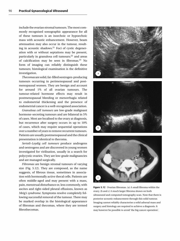
90 Practical Gynaecological Ultrasound
include the ovarian stromal tumours. The most com-monly recognised sonographic appearance for allof these tumours is an isoechoic or hypoechoicmass with acoustic enhancement. However, beamattenuation may also occur in the tumour, result-ing in acoustic shadows.21 Foci of cystic degener-ation with or without septations may be present,particularly in granulosa cell tumours;22 and areasof calcification may be seen in fibromas.23 Noform of imaging can reliably distinguish thesetumours; histological examination is the definitiveinvestigation.
Thecomas are solid, fat-filled oestrogen-producingtumours occurring in perimenopausal and post-menopausal women. They are benign and accountfor around 1% of all ovarian tumours. Thetumour-related hormone effects may result inpostmenopausal bleeding or menorrhagia relatedto endometrial thickening and the presence ofendometrial cancer is a well-recognised association.
Granulosa cell tumours are low-grade malignanthormone-secreting tumours and are bilateral in 5%of cases. Most are localised to the ovary at diagnosis,but recurrence after surgery occurs in up to 10%of cases, which may require sequential operationsover a number of years to remove recurrent tumours.Patients are usually postmenopausal and the clinicalpresentation is identical to thecoma.
Sertoli–Leydig cell tumours produce androgensand oestrogens and are discovered in young womeninvestigated for virilisation, usually in a search forpolycystic ovaries. They are low-grade malignanciesand are managed surgically.
Fibromas are benign stromal tumours of varyingsize (Fig. 5.12). They are composed, as the namesuggests, of fibrous tissue, sometimes in associa-tion with hormonally active thecal cells. Patients areoften middle-aged and may present with a mass,pain, menstrual disturbance or, less commonly, withascites and right-sided pleural effusions, known asMeig’s syndrome. Symptoms resolve completely fol-lowing successful removal of the tumour. There maybe marked overlap in the histological appearanceof fibromas and thecomas, where they are termedfibrothecomas.
a
b
c
Figure 5.12 Ovarian fibromas. (a) A small fibroma within the
ovary. (b and c) A much larger fibroma shown on both
ultrasound and computed tomography scan. Note that there is
posterior acoustic enhancement through this solid tumour.
Imaging cannot reliably characterise a solid adnexal mass and
surgery and histology are required to achieve a diagnosis. It
may however be possible to avoid ‘the big cancer operation’.

Pathology of the adnexae 91
Brenner tumours may arise from ovarian stromaltissue or epithelium and are composed of fibroustissue, similar to fibroma. They are rare and usu-ally benign, although malignant forms are recog-nised. While most cases are not hormonally active,oestrogen-related side-effects have been seen insome cases and tumours can reach several centime-tres in size.
Subserosal or pedunculated uterine fibroids
Although clearly not an ovarian neoplasm, uterinefibroids need to be mentioned in this section becauseof the problems they can pose in the diagnosis ofovarian neoplasms. Pedunculated fibroids can varygreatly in size and position within the pelvis, and typ-ical imaging features of a homogeneous or heteroge-neous solid mass with tissue characteristics rangingfrom hypo- to hyperechoic can be altered in the pres-ence of necrosis of larger lesions, resulting in a morecomplex cystic appearance on ultrasound (Fig. 5.13).In this setting, a younger patient may unwittinglybe diagnosed as having ovarian carcinoma, partic-ularly when CT confirms the localised mass with-out the further tissue characterisation or detailedmultiplanar examination available with MR scan-ning. Similarly, in older women, in whom fibroids areuncommon due to involution after the menopause,an ovarian tumour may be mistaken for a necroticpedunculated fibroid. Clearly the management andcounselling of the patient are very different for thetwo conditions. We would like to raise the readers’awareness of this potential pitfall and advise them tobe extremely cautious in dismissing complex massesas pedunculated fibroids, particularly in the post-menopausal female, without full assessment, includ-ing MRI where appropriate.
Malignant ovarian tumours
Ovarian carcinoma
Epidemiology of ovarian cancerOvarian cancer is a significant health problem, beingthe fifth most common cancer in women and also the
leading cause of death from gynaecological malig-nancies. In 1992, 4360 women died of ovarian can-cer in the UK24 and in 1994 in the USA there wereapproximately 13 600 deaths.25 However, to placethese large figures in perspective, the disease preva-lence is between 30 and 50 cases per 100 000 popu-lation and the lifetime incidence is one in 70 women(in comparison, the lifetime risk of breast cancer isone in 12). It is uncommon in black women.26 Mostcases occur after the age of 45.
Survival depends on the stage of the disease atdiagnosis. The early stages of the disease do nothave any specific symptoms and so nearly 60% ofwomen present with disease that has already spreadwith abdominal metastases such as in the omentum(stage III) or with distant metastases outside the peri-toneal cavity (stage IV) to lung, liver, bone and dis-tant lymph nodes. These women only have a one in10 chance of surviving for 5 years. Conversely thosewomen (25%) who present with the disease confinedto the ovary (stage I) have a six or seven in 10 chanceof surviving for the same period. Stage II disease withovarian spread confined to the pelvis has an interme-diate survival (Table 5.1).
Risk factors for ovarian cancerThe cause of ovarian cancer is not known, thoughthere are women who are at higher risk than others.Risk factors include greater age, nulliparity, northernEuropean or North American descent, and a familyhistory of ovarian cancer. There is a theory that ovu-lation itself is a causative factor, as the risk of ovar-ian cancer appears to be decreased by pregnancy,breast-feeding and oral contraceptives, all of whichsuppress ovulation.
Women who have one close relative with ovar-ian cancer have a threefold increase in lifetime risk,whereas those with two close relatives have a dra-matic increase to a 40% lifetime risk. As many as5% of ovarian cancers occurring before the age of50 may be due to familial cancer.26 Mutations inthe genes BRCA1 and 2 may account for most ofthese families.27 This raises the intriguing prospectof genetic testing to enable individuals to learn ifthey carry a cancer-predisposing mutation. Not all

92 Practical Gynaecological Ultrasound
a
b
c
d
e
Figure 5.13 (a) Degenerating pedunculated fibroid. An adnexal lesion with central cavitation was identified during early pregnancy.
(b and c) The pregnancy miscarried. The woman was seen several months later to have a large abdominal mass. Taken in isolation,
these ultrasound images would be of concern because of the apparently nodular margins and because the lesion appears separate
from the uterus. (d) Magnetic resonance shows the full extent of this adnexal mass. (e) A large, centrally cavitated fibroid is removed
at surgery. This sequence of images shows the value of obtaining a history and comparing old images with new. The progression of
the cavitation in a benign fibroid becomes apparent.
Pathology of the adnexae 93
Table 5.1 Abbreviated International Federation of Gynecology and Obstetrics (FIGO) staging
of ovarian carcinoma
Stage Location Symptoms
I Confined to ovary Asymptomatic
Good prognosis after surgery
Diagnosed by screening or during investigation for other
symptomatic conditions
II Spread confined to the pelvis Usually asymptomatic, diagnosed as above
III Abdominal metastases – omentum,
lymphadenopathy – peritoneal implants,
e.g. liver surface
Presents with abdominal enlargement or symptoms from
distant metastases
Poor 5-year survival
IV Distant metastases – liver parenchyma, lungs
those at risk want such testing, though those froma higher economic class and those with more rel-atives affected are more likely to request the test.28
The implications of a positive test need to be exploredfully before testing, including factors such as difficul-ties in obtaining a mortgage or life insurance. Fur-thermore, whether a positive genetic predispositionshould lead to active ovary and breast screening oreven prophylactic bilateral oophorectomy (and sub-cutaneous mastectomy) needs to be discussed.
Imaging features and scoring systemsUltrasound has a very valuable role to play in thecharacterisation of adnexal masses. There are pro-ponents for the use of various morphological, spec-tral and colour Doppler criteria both transabdom-inally and transvaginally in the differentiation ofmalignant from benign ovarian disease. Morphologi-cal scoring systems have been proposed by severalauthors.29,30 They all have in common the associ-ation of malignancy with the presence of a largesize (over 10 cm), a solid or partly solid content andpapillae (Fig. 5.14). Fine septations and daughtercysts are associated with benignity. A review of thesescoring systems found their sensitivities to rangefrom 62 to 100%, the specificities from 73 to 95% andthe positive predictive values from 31 to 83%. Usingtheir own criteria they could obtain 100% sensitiv-ity, but only at the cost of a low specificity of 37%.
This was caused by abscesses, teratomas and haem-orrhagic cysts being indistinguishable from malig-nant tumours on appearance alone.
The low specificity of morphology prompted asearch for Doppler criteria of malignancy. Initialreports of the use of resistance and pulsatility indiceswere very encouraging, but a series of later reportsshowed that spectral Doppler is not helpful becausethere is too great an overlap in the indices betweenbenign and malignant disease.31–34 With time, how-ever, Doppler’s true role, particularly colour Doppler,is becoming clearer. Buy and colleagues,35 in theirstudy of 132 adnexal masses in 115 women, usedthe presence or absence of colour flow in sec-tions of a mass thought morphologically indeter-minate or malignant to predict whether or not themass was truly malignant. If a mass was thoughtto be morphologically benign then the presence orabsence of colour flow was disregarded. This tech-nique improved the confidence level, specificity andpositive predictive value of grey-scale sonography;specificity rose from 82 to 97% and the positivepredictive value from 63 to 91%. The same studyalso confirmed the limited value of spectral Dopplerindices.
Our experience with a cohort of patients in Leeds36
concurs with the poor results obtainable from spec-tral Doppler indices, but also indicates that an ex-perienced observer with access to all the ultrasound

94 Practical Gynaecological Ultrasound
a b
c
d
Figure 5.14 Typical patterns of malignant ovarian morphology. (a) Large complex cystic and solid lesion on transabdominal
scanning in a 52-year-old woman. (b) Thick-walled cyst with internal nodules or papillae lying behind the bladder in a 56-year-old
woman. (c) Relatively simple cyst with a fine septum but also with a small solid component in an 80-year-old woman. All these
lesions were malignant. (d) A large ovarian malignancy showing the typical mixed solid and cystic morphology.

Pathology of the adnexae 95
e f
Figure 5.14 (cont.) (e) A smaller cyst showing an irregular mass arising out of one wall. There is free fluid. The mass is typical for
malignancy. (f) Omental ‘cake’ surrounded by ascites. The ultrasound operator should always examine the rest of the abdomen
when a suspected ovarian malignancy is found. Ultrasound is not sensitive to the presence of tumour spread but it is highly specific.
Hence if tumour spread is found, then an advanced stage of malignant disease can be diagnosed.
information (i.e. morphology and Doppler indices)can outperform any of the individual scoring systemscurrently available. This multiparameter analysis byexperienced observers is also now advocated byKurjak and Kupesic,37 who go on to describe the needfor ‘artistic rendering’ in obtaining and interpretingresults (Table 5.2).
Ultrasound contrast agents have been shown intwo small studies to improve the diagnostic accu-racy of power Doppler ultrasound in the diagno-sis of early ovarian cancer.38,39 These data need tobe confirmed with larger-scale trials to ensure thatthis is not another false dawn in ovarian ultrasoundimaging.
Metastases to ovaryMetastatic involvement of the ovaries from a dis-tant primary tumour represents 10% of all ovarianmalignancies. The primary tumour is sometimesonly detected during investigation of the ovar-ian lesion. A Krukenberg tumour describes thespread of mucinous breast or gastrointestinal tractmalignancy (from the colon, stomach and pan-creas) to the ovaries. They may spread by directinvasion, intraperitoneal seeding, lymphatic orvascular routes. Tumours are most often bilateralsolid lesions with a cystic component; bilaterality is
the first indicator to alert the sonographer that theovaries may be involved in malignant disease froma distant site. This should then prompt a thorough
Table 5.2 Ultrasound features used in scoring systems to
differentiate benign from malignant ovarian masses
Morphology
Size Larger masses more likely to be
malignant
Wall thickness Thick-walled masses score more highly
for malignancy than those with thin
walls
Composition Complex, mainly fluid masses score
more highly for malignancy than
simple cysts
Papillae and thickened septa score highly
Doppler
Presence/absence Non-vascular masses more likely to be
benign
Distribution Masses confined to the wall or with a
regular pattern are more likely to be
benign than masses with irregular
vascular distribution throughout
Resistance Low-resistance (high end-diastolic flow
velocity) scores more highly for
malignancy

96 Practical Gynaecological Ultrasound
review of the rest of the abdomen for a primarysite of disease. Often a past history of malignancyis present, sometimes many years after the initialtreatment, and particularly so in patients with breastcancer. In such cases CT scanning is advised toassess other sites of disease and biopsy is essen-tial to confirm the source of the disease and to planthe most appropriate treatment. Lymphoma andleukaemia may also involve the ovaries, producingsolid lesions, as a part of a more generalised diseaseprocess.40
Ovarian cancer screeeningThe aim of ultrasound screening of the ovaries isto detect malignant disease without resulting in alarge number of operations for conditions that even-tually turn out to be benign. Early studies withtransabdominal scanning on asymptomatic womenachieved a poor rate of 67 operations for everyovarian cancer found. This can be improved by nar-rowing the cohort of women to be scanned, by select-ing a higher-risk subgroup, or by using transvagi-nal scans and sequential testing with follow-upscans and CA125. A family history of ovarian cancerreduces the number of operations for benign dis-ease for each cancer detected from around 1 in 50to 1 in 12. Transvaginal scans in postmenopausalwomen also reduce this rate to around 1 in 10,although some operators, for example Kurjak andcolleagues, appear to do much better than others.Using CA125 as the primary screening tool beforeultrasound reduces the benign operation rate fur-ther, but also has the potential to miss up to 50%of early-stage tumours. It is apparent that an ill-conceived programme has a huge potential to causeharm without necessarily improving ovarian cancersurvival.
Several major trials are currently under way todetermine whether population screening is a viableand effective use of health care resources. TheUK Collaborative Trial of Ovarian Cancer Screen-ing Study (UKCTOCSS) aims to screen 200 000 post-menopausal women (with a combination of CA125,followed by transvaginal ultrasound where this is
elevated). The UK Familial Ovarian Cancer Screen-ing Study (UKFOCSS) will follow up 5000 womenwith a strong family history of ovarian cancer. Theresults of these studies should be available in the nearfuture.
Summary of main points: screening forovarian cancer� Lifetime risk is approximately 1 in 70 women� Screening tools include bimanual palpation,
genetic testing, CA125 levels and ultrasound� Screening currently suffers from low sensitivity(cancers are missed) and low specificity (too manyfalse-positive results) which cause unnecessaryintervention and distress� Sensitivity and specificity are improved whenthe screened population is restricted to high-riskwomen > 45 years or with a family history of ovar-ian or breast cancer� False-positive results due to physiological massesare minimised by scanning in the early part ofthe cycle (days 1–8) or by selecting only post-menopausal women for the screening programme� The role of ultrasound is enhanced by usingtransvaginal and colour Doppler techniques� Although scoring systems have had some success,the best results are obtained using knowledgeableand experienced interpretation� There is no evidence to support screening of thegeneral population
Fallopian tube and adnexal pathology
Pelvic inflammatory disease
PID is an infective condition, usually caused byascending lower genital tract infection by Chlamy-dia, gonorrhoea, commensal organisms or, rarely,from tuberculosis. Up to 10% of women are affectedduring their lives and the incidence may be rising.Risk factors for infection include previous episodesof sexually transmitted disease, multiple partners,
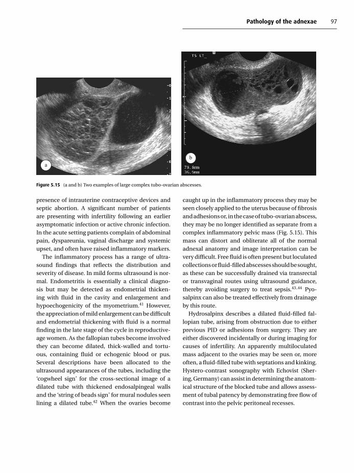
Pathology of the adnexae 97
ab
Figure 5.15 (a and b) Two examples of large complex tubo-ovarian abscesses.
presence of intrauterine contraceptive devices andseptic abortion. A significant number of patientsare presenting with infertility following an earlierasymptomatic infection or active chronic infection.In the acute setting patients complain of abdominalpain, dyspareunia, vaginal discharge and systemicupset, and often have raised inflammatory markers.
The inflammatory process has a range of ultra-sound findings that reflects the distribution andseverity of disease. In mild forms ultrasound is nor-mal. Endometritis is essentially a clinical diagno-sis but may be detected as endometrial thicken-ing with fluid in the cavity and enlargement andhypoechogenicity of the myometrium.41 However,the appreciation of mild enlargement can be difficultand endometrial thickening with fluid is a normalfinding in the late stage of the cycle in reproductive-age women. As the fallopian tubes become involvedthey can become dilated, thick-walled and tortu-ous, containing fluid or echogenic blood or pus.Several descriptions have been allocated to theultrasound appearances of the tubes, including the‘cogwheel sign’ for the cross-sectional image of adilated tube with thickened endosalpingeal wallsand the ‘string of beads sign’ for mural nodules seenlining a dilated tube.42 When the ovaries become
caught up in the inflammatory process they may beseen closely applied to the uterus because of fibrosisand adhesions or, in the case of tubo-ovarian abscess,they may be no longer identified as separate from acomplex inflammatory pelvic mass (Fig. 5.15). Thismass can distort and obliterate all of the normaladnexal anatomy and image interpretation can bevery difficult. Free fluid is often present but loculatedcollections or fluid-filled abscesses should be sought,as these can be successfully drained via transrectalor transvaginal routes using ultrasound guidance,thereby avoiding surgery to treat sepsis.43,44 Pyo-salpinx can also be treated effectively from drainageby this route.
Hydrosalpinx describes a dilated fluid-filled fal-lopian tube, arising from obstruction due to eitherprevious PID or adhesions from surgery. They areeither discovered incidentally or during imaging forcauses of infertility. An apparently multiloculatedmass adjacent to the ovaries may be seen or, moreoften, a fluid-filled tube with septations and kinking.Hystero-contrast sonography with Echovist (Sher-ing, Germany) can assist in determining the anatom-ical structure of the blocked tube and allows assess-ment of tubal patency by demonstrating free flow ofcontrast into the pelvic peritoneal recesses.

98 Practical Gynaecological Ultrasound
a
b
Figure 5.16 Ectopic pregnancy. (a) A typical adnexal ring
structure is identified, together with surrounding echogenic
free fluid. The echoes in the free fluid indicate the presence of a
haemoperitoneum. The ring structure is the tubal pregnancy.
(b) An example of a very rare cervical ectopic pregnancy. This
can produce life-threatening haemorrhage.
Ectopic pregnancy
While this may present unexpectedly in a patientunder investigation for infertility with an adnexalmass (Fig. 5.16), it more usually occurs in a patientwith acute abdominal pain, vaginal bleeding andwith a positive pregnancy test. The pregnancytest is the single most important investigation:a positive test makes the diagnosis highly likely.Serial serum beta-human chorionic gonadotrophic(�-HCG) measurements are a useful method of dis-tinguishing early healthy pregnancy from ectopicpregnancy. In an ectopic pregnancy the normal dou-bling of �-HCG every 48 hours does not occur; an
increase of less than 66% over 2 days is predictive ofectopic pregnancy. The imaging features and man-agement of this condition are dealt with more fully inChapter 6.
Broad-ligament fibroids
Occasionally uterine fibroids may extend into thebroad ligament, simulating a solid adnexal mass. Thediagnosis may be suggested by the presence of otheruterine fibroids. Compression of adjacent structurescan lead to fallopian tube and ureteric obstruction.Where there is doubt about the nature of the mass,MRI can be useful in further assessment. Fibroids arediscussed more fully in Chapter 4.
Fallopian-tube carcinoma
This is a rare tumour, usually affecting post-menopausal women with a history of infertility.Patients present with bleeding, pain and, in somecases, with watery vaginal discharge. The ultrasoundfeatures are non-specific, but may include a cystic/solid adnexal mass in association with a unilaterallydilated fluid-filled tube. Clarification of the diagnosismay be possible with MRI before surgery. Standardsurgical treatment is the same as for ovarian can-cer, with hysterectomy, bilateral oophorectomy andomentectomy, as the tumour is bilateral in up to aquarter of patients and peritoneal dissemination iscommon.
Non-gynaecological pelvic pathology
Appendicitis
Appendicitis may produce non-specific symptomsin a significant number of patients, but most suf-fer abdominal pain with raised white cell count andinflammatory markers on blood-testing. The clinicalpicture in young women can mimic ectopic preg-nancy, PID and torsion of the ovaries. In any femalepatient with abdominal pain the appendix may rea-sonably feature on the clinical differential diagnosis.

Pathology of the adnexae 99
Figure 5.17 Appendicitis. There is a tubular, non-compressible
structure containing an echogenic focus – an appendicolith.
The walls of the appendix demonstrate the typical layered
appearance seen in all bowel wall.
The imaging characteristics of the appendix wheninflamed are of a blind-ending intestinal loop arisingfrom the caecum, which is non-compressible, notperistalsing and measuring >6 mm in diameter(Fig. 5.17). A hyperechoic appendicolith maybe present. Associated free fluid and increasedechogenicity of the adjacent mesenteric fat may bepresent.
Bowel-related pathology
Diverticulitis is another common condition, whichresults in herniation of the colonic mucosa throughits outer muscular wall. These outpouchings maybecome obstructed and inflamed, resulting in anepisode of diverticulitis. The left colon is most com-monly affected, particularly the sigmoid. Ultrasoundof the bowel shows transmural thickening centredon a hypoechoic area (the diverticulum) protrud-ing through the bowel wall. Once again, hyperechoicinflammatory change in adjacent mesenteric fat is auseful sign (Fig. 5.18). Inflammatory bowel disease,which incorporates Crohn’s disease and ulcerativecolitis, may be detected on ultrasonography as thecause of abdominal pain in some patients. A history
Figure 5.18 Diverticulitis. Note the bright inflamed mesenteric
fat surrounding the outpouched diverticulum.
of prior altered bowel habit, blood or mucus instools or weight loss is typically present. There iswall-thickening with increased blood flow in activedisease. The colon and distal small bowel may beinflamed in either condition but when fistulae andabscesses related to affected loops are present, amore specific diagnosis of Crohn’s disease is sug-gested. Colorectal cancer may be demonstrated as abowel-based soft-tissue mass with associated lym-phadenopathy. Invasion of neighbouring viscera,such as the bladder, may be seen.
Renal tract and vascular pathology
A range of incidental abnormalities may be seen dur-ing pelvic ultrasound examination including uretericdilatation, with accompanying ureteric calculi orureteroceles and bladder tumours (Fig. 5.19). Some-times it may not be clear from the history whetherblood loss is per vaginam or per urethram. Bladdertumours are relatively common in the elderly pop-ulation and the ultrasound operator needs to bealert to this possibility in someone presenting with
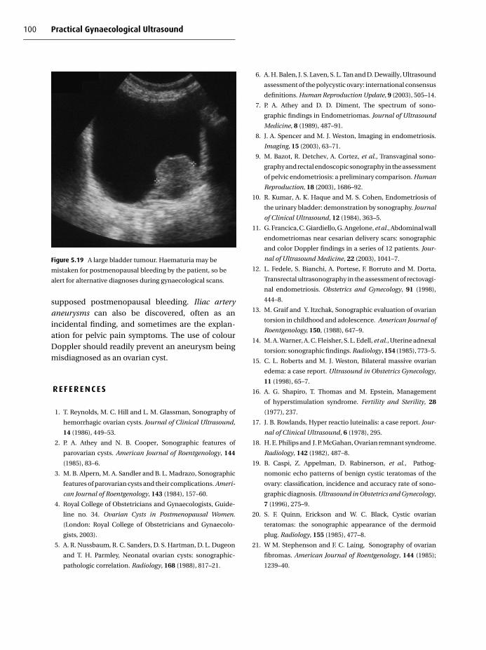
100 Practical Gynaecological Ultrasound
Figure 5.19 A large bladder tumour. Haematuria may be
mistaken for postmenopausal bleeding by the patient, so be
alert for alternative diagnoses during gynaecological scans.
supposed postmenopausal bleeding. Iliac arteryaneurysms can also be discovered, often as anincidental finding, and sometimes are the explan-ation for pelvic pain symptoms. The use of colourDoppler should readily prevent an aneurysm beingmisdiagnosed as an ovarian cyst.
R E F E R E N C E S
1. T. Reynolds, M. C. Hill and L. M. Glassman, Sonography of
hemorrhagic ovarian cysts. Journal of Clinical Ultrasound,
14 (1986), 449–53.
2. P. A. Athey and N. B. Cooper, Sonographic features of
parovarian cysts. American Journal of Roentgenology, 144
(1985), 83–6.
3. M. B. Alpern, M. A. Sandler and B. L. Madrazo, Sonographic
features of parovarian cysts and their complications. Ameri-
can Journal of Roentgenology, 143 (1984), 157–60.
4. Royal College of Obstetricians and Gynaecologists, Guide-
line no. 34. Ovarian Cysts in Postmenopausal Women.
(London: Royal College of Obstetricians and Gynaecolo-
gists, 2003).
5. A. R. Nussbaum, R. C. Sanders, D. S. Hartman, D. L. Dugeon
and T. H. Parmley, Neonatal ovarian cysts: sonographic-
pathologic correlation. Radiology, 168 (1988), 817–21.
6. A. H. Balen, J. S. Laven, S. L. Tan and D. Dewailly, Ultrasound
assessment of the polycystic ovary: international consensus
definitions. Human Reproduction Update, 9 (2003), 505–14.
7. P. A. Athey and D. D. Diment, The spectrum of sono-
graphic findings in Endometriomas. Journal of Ultrasound
Medicine, 8 (1989), 487–91.
8. J. A. Spencer and M. J. Weston, Imaging in endometriosis.
Imaging, 15 (2003), 63–71.
9. M. Bazot, R. Detchev, A. Cortez, et al., Transvaginal sono-
graphy and rectal endoscopic sonography in the assessment
of pelvic endometriosis: a preliminary comparison. Human
Reproduction, 18 (2003), 1686–92.
10. R. Kumar, A. K. Haque and M. S. Cohen, Endometriosis of
the urinary bladder: demonstration by sonography. Journal
of Clinical Ultrasound, 12 (1984), 363–5.
11. G. Francica, C. Giardiello, G. Angelone, et al., Abdominal wall
endometriomas near cesarian delivery scars: sonographic
and color Doppler findings in a series of 12 patients. Jour-
nal of Ultrasound Medicine, 22 (2003), 1041–7.
12. L. Fedele, S. Bianchi, A. Portese, F. Borruto and M. Dorta,
Transrectal ultrasonography in the assessment of rectovagi-
nal endometriosis. Obstetrics and Gynecology, 91 (1998),
444–8.
13. M. Graif and Y. Itzchak, Sonographic evaluation of ovarian
torsion in childhood and adolescence. American Journal of
Roentgenology, 150, (1988), 647–9.
14. M. A. Warner, A. C. Fleisher, S. L. Edell, et al., Uterine adnexal
torsion: sonographic findings. Radiology, 154 (1985), 773–5.
15. C. L. Roberts and M. J. Weston, Bilateral massive ovarian
edema: a case report. Ultrasound in Obstetrics Gynecology,
11 (1998), 65–7.
16. A. G. Shapiro, T. Thomas and M. Epstein, Management
of hyperstimulation syndrome. Fertility and Sterility, 28
(1977), 237.
17. J. B. Rowlands, Hyper reactio luteinalis: a case report. Jour-
nal of Clinical Ultrasound, 6 (1978), 295.
18. H. E. Philips and J. P. McGahan, Ovarian remnant syndrome.
Radiology, 142 (1982), 487–8.
19. B. Caspi, Z. Appelman, D. Rabinerson, et al., Pathog-
nomonic echo patterns of benign cystic teratomas of the
ovary: classification, incidence and accuracy rate of sono-
graphic diagnosis. Ultrasound in Obstetrics and Gynecology,
7 (1996), 275–9.
20. S. F. Quinn, Erickson and W. C. Black, Cystic ovarian
teratomas: the sonographic appearance of the dermoid
plug. Radiology, 155 (1985), 477–8.
21. W M. Stephenson and F. C. Laing, Sonography of ovarian
fibromas. American Journal of Roentgenology, 144 (1985);
1239–40.

Pathology of the adnexae 101
22. E. K. Outwater, B. Marchetto and B. J. Wagner, Virilis-
ing tumours of the ovary: imaging features. Ultrasound in
Obstetrics Gynecology, 15 (2000), 365–71.
23. P. A. Athey and R. S. Malone, Sonography of ovarian fibro-
mas/thecomas. Journal of Ultrasound in Medicine, 6 (1987),
431–6.
24. J. Austoker, Screening for ovarian, prostatic, and testicular
cancers. British Medical Journal, 309 (1994), 315–20.
25. NIH consensus development panel on ovarian cancer, Ovar-
ian cancer: screening, treatment and follow up. Journal of
the American Medical Association, 273 (1995), 491–7.
26. K. J. W. Taylor and P. E. Schwartz, Screening for early ovarian
cancer. Radiology, 192 (1994), 1–10.
27. D. Ford, D. F. Easton and J. Peto, Estimates of the gene fre-
quency of BRCAl and its contribution to breast and ovar-
ian cancer. American Journal of Human Genetics, 57 (1995),
1457–62.
28. C. Lerman, S. Narod, K. Schulman et al., BRCAl testing in
families with hereditary breast–ovarian cancer. A prospect-
ive study of patient decision making and outcomes. Journal
of the American Medical Association, 275 (1996), 1885–92.
29. A. Sassone, I. Timor-Tritsch, A. Artner, W. Carolyn and
W. B. Warren, Transvaginal sonographic characterisation of
disease: evaluation of a new scoring system to predict ovar-
ian malignancy. Obstetrics and Gynecology, 78 (1991), 70–6.
30. S. Rottem, N. Levit, I. Thraler et al. Classification of ovarian
lesions by high frequency transvaginal sonography. Journal
of Clinical Ultrasound, 18 (1990), 359–63.
31. D. L. Brown, M. C. Frates, F. C. Laing et al., Ovarian masses:
can benign and malignant lesions be differentiated with
color and pulsed Doppler US? Radiology, 190 (1994), 333–6.
32. L. Valentin, P. Sladkevicius and J. K. Marsa, Limited
contribution of Doppler velocimetry to the differential
diagnosis of extrauterine tumors. Obstetrics and Gynecol-
ogy, 83 (1994), 425–33.
33. B. Bromley, H. Goodman and B. R. Benacerraf, Compari-
son between sonographic morphology and Doppler wave-
form for the diagnosis of ovarian malignancy. Obstetrics and
Gynecology, 83 (1994), 434–7.
34. S. M. Stein, S. Laifer-Narin, M. B. Johnson et al., Differen-
tiation of benign and malignant adnexal masses: relative
value of gray-scale, color Doppler, and spectral Doppler
sonography. American Journal of Roentgenology, 164 (1995),
381–6.
35. J.-N. Buy, M. A. Ghossain, D. Hugol et al., Characterization of
adnexal masses: combination of color Doppler and conven-
tional sonography compared with spectral Doppler analysis
alone and conventional sonography alone. American Jour-
nal of Roentgenology, 166 (1996), 385–93.
36. L. I. G. Haigh, G. Lane and M. Weston, The role of Doppler
ultrasound in the assessment of ovarian masses. British
Journal of Radiology, 68, (1995), 809.
37. A. Kurjak and S. Kupesic, Color Doppler imaging for
the detection of ovarian malignancy is reliable (let-
ter). Ultrasound in Obstetrics and Gynecology, 7 (1996),
380–3.
38. T. J. D’Arcy, V. Jayaram, M. Lynch et al., Ovarian can-
cer detected non-invasively by contrast-enhanced power
Doppler ultrasound. British Journal of Obstetrics are Gynae-
cology, 111 (2004), 619–22.
39. V. Sparac, S. Kupesic and A. Kurjak, What do contrast media
add to three-dimensional power Doppler evaluation of
adnexal masses? Croatian Medical Journal, 41 (2000), 257–
61.
40. G. H. Bickers, J. J. Siebert, J. C. Anderson, S. Golladay and
D. L. Berry, Sonography of ovarian involvement in childhood
lymphocytic leukemia. American Journal of Roentgenology,
137 (1981), 399–401.
41. L. C. Swayne, M. B. Love and S. R. Karasick, Pelvic inflam-
matory disease: sonographic-pathologic correlation. Radi-
ology, 151 (1984), 751–5.
42. I. E. Timor-Tritsch, J. P. Lerner, A. Monteagudo, K. E. Mur-
phy and D. S. Heller, Transvaginal sonographic markers of
tubal inflammatory disease. Ultrasound in Obstetrics and
Gynecology, 12 (1998), 56–66.
43. J. L. Nosher, G. S. Needell, J. K. Amorosa and I. H. Krasna,
Transrectal pelvic abscess drainage with sonographic guid-
ance. American Journal of Roentgenology, 146 (1986),
1047–8.
44. J. L. Nosher, H. K. Winchman and G. S. Needell, Transvaginal
pelvic abscess drainage with US guidance. Radiology, 165
(1987), 872–3.


6
Ultrasound in the acute pelvis
Hassan MassouhFrimley Park Hospital, Frimley, Surrey
Introduction
Ultrasound is the first examination of choice inpatients with acute pelvic pain, regardless of age, pre-sentation or gender, and a more common requestin females than males. Pelvic pain represents 10–15% of all urgent ultrasound examinations per-formed in a district general hospital (local audit)and results from a large variety of underlying causes,many of which may be gynaecological. The causesof pain vary and will depend upon the location,organ involved and underlying pathology such asinfection, haemorrhage, infarct or ectopic pregnancy(EP).
The examination of choice in these circum-stances is a combination of transabdominal (TA) andtransvaginal (TV) ultrasound except in young girlsand those with an intact hymen. The use of high-frequency vaginal probes and the proximity of theprobe to the pelvic organs allow clear visualisationof the pelvic organs. The ability of the vaginal probeto touch the pelvic organs allows the sonographer totest for pain and detect the mobility of these organs,which are two important and useful signs in evalu-ating the cause of pelvic pain.
TA ultrasound is advantageous because of its abil-ity to visualise large pelvic masses, detect the pres-ence of ascites and examine other structures such asthe kidneys and the other upper abdominal organs,which may be associated with the pain. However,TA ultrasound may be technically difficult in acute
pelvic pain due to the presence of ileus, abdominalwall tenderness/rigidity and the lack of filling of theurinary bladder (which is frequently associated inparticular with pelvic inflammation).
Prior to the ultrasound examination, the sonogra-pher should obtain a full clinical history of the pain:its onset, frequency and relationship to menstrualcycle, its location, any associated fever, evidence ofvaginal discharge, date of last menstrual period, pastmedical history and knowledge of previous surgery.This is essential information in helping the interpre-tation of the ultrasound findings.
Ectopic pregnancy
EP is defined as ‘implantation of a fertilised ovumoutside the uterine cavity’. Its incidence in the devel-oped world is increasing1,2 and it is associated withthe use of intrauterine contraceptive devices, pelvicinflammatory disease, adhesions from previous EPand past pelvic surgery or corrective surgery fortreating infertility (Table 6.1). Endometriosis, pelvicscarring and adhesions are also associated with anincreased incidence of EP.
Patients with EP are usually women of child-bearing age, presenting with lower abdominal pain(more to the side of pregnancy) and tenderness, vagi-nal bleeding and history of missed or delayed period.It is possible for EP to pass undiagnosed in patientswith minimal symptoms, in whom the implants die
C© Cambridge University Press 2005.
103

104 Practical Gynaecological Ultrasound
Table 6.1 Factors associated with increased incidence
of ectopic pregnancy
Use of contraceptive devices
Pelvic inflammatory disease
Previous pelvic surgery
Previous surgery for infertility
Endometriosis
Other causes of pelvic adhesions
and the products resorb spontaneously. Confirmingthe presence of EP is usually by the history, clin-ical presentation, positive pregnancy test and pos-itive hormone markers.
Ultrasound (particularly transvaginal) plays amajor role in the early diagnosis of EP and its contri-bution to an accurate early diagnosis has reduced themortality rate of EP. Over 95% of EP are within the fal-lopian tube (tubal) and the rest are intra-abdominalor intraperitoneal, ovarian, cervical or heterotopic.3
Ultrasound findings
The presence of a normal intrauterine pregnancyusually excludes EP except in very rare cases of het-erotopic pregnancy, where there is an ectopic gesta-tion with an intrauterine one.3
There is usually an adnexal or tubal mass con-sisting of an echogenic ring, representing thetrophoblastic tissue, surrounding chorionic fluid.Depending on the age of the ectopic, it may be possi-ble to see a yolk sac, fetal pole and also to detect a fetalheart beat, in which case the diagnosis is confirmed(Fig. 6.1). However, it is more common to find a com-plex mass on the side of the suspected EP and thecontact of the vaginal probe with this mass is usuallytender and painful. Care should be taken to differen-tiate between an ectopic sac and corpus luteal cyst,which is usually on the same side as the EP, by clearlyidentifying the ovary separately from the ectopic sac.
In the uterus, the endometrium is thickenedas a result of decidual reaction, secondary to theeffect of hormones. Fluid can accumulate with thisdecidual reaction, leading to what is described as
Figure 6.1 Transvaginal ultrasound showing right-sided
ectopic pregnancy in the distal right tube (arrowheads) with
decidual reaction of the endometrium (long arrows).
Figure 6.2 Transvaginal ultrasound of the uterus in a patient
with an ectopic pregnancy showing fluid within the
endometrial cavity with some thickening (decidual reaction) of
the endometrium. Note the irregular margin of this fluid,
which should not be mistaken for intrauterine pregnancy.
‘pseudo-gestational sac’ (Fig. 6.2). Compared withthe true intrauterine sac, the pseudo-sac has an irre-gular margin, containing echogenic (bloody) fluidwith absence of the ‘double-ring border’ of the nor-mal intrauterine sac. Colour Doppler ultrasound candifferentiate the hypervascular trophoblastic ringfrom the less vascular wall of the pseudo-sac. It isabsolutely essential to examine both adnexae in all

Ultrasound in the acute pelvis 105
cases of an empty intrauterine sac in case it repre-sents a pseudo-sac with an EP (Fig. 6.3).
Excess fluid in the pouch of Douglas, more than isusually present following ovulation, is also a positivesign of EP. The fluid may be bloody and echogenic(swirling motion of bloody fluid).
A pregnancy test is usually positive in both intra-and extrauterine gestation. The use of the dis-criminatory level of serum b human chorionicgonadotrophin (�hCG) has become widely used. It isnow accepted that a normal intrauterine pregnancyshould be detectable on transvaginal ultrasound ifthe serum �hCG is 1000 IU/l (second InternationalStandard) or more. Failure to visualise intrauterinesac at this level of �hCG highly indicates either an EPor recent abortion.4,5
The value of serum progesterone for diagnosing EPremains equivocal, due to the overlap between nor-mal and EP. Progesterone of ≥ 80 nmol/l indicatesa normal intrauterine pregnancy in 98% of cases.Progesterone of < 16 nml/l indicates a non-viablepregnancy regardless of location. Most EPs will havea progesterone level of 32–60 nmol/l at presentation,significantly limiting the clinical usefulness of pro-gesterone measurement in EP.6
It is important to remember that a negative ultra-sound finding does not exclude EP. If there is clinicaldoubt about the diagnosis, follow-up scans and serialmeasurements are recommended.7
Ovarian and intra-abdominal EPs are rare and dif-ficult to diagnose on ultrasound. They are usuallyfound on laparoscopy following continuing clinicalsuspicion after a negative ultrasound.
Cervical pregnancy is also rare but can be diag-nosed on TV ultrasound as a gestational sac in thecervical canal. It is a very painful type of EP and alsodangerous, as it can bleed heavily.
Heterotopic pregnancy is very rare but its inci-dence is rising with the increasing use of repro-ductive or infertility procedures.3 It consists of anintrauterine gestational sac in addition to an ectopicone (Fig. 6.4). It is good practice always to scanboth adnexae during ultrasound in early pregnancy,particularly in those patients who have had fertilityprocedures.
Figure 6.3 A young woman who had a dilatation and curettage
for a suspected 7-week blighted ovum. The histological
examination of the curetted tissue showed no fetal or
trophoblastic tissue. Repeated transvaginal colour Doppler
ultrasound showed right-sided ectopic pregnancy (arrowed).
Note the increased vascularity of the trophoblastic tissue. What
was thought to be a blighted ovum was, in retrospect, a
pseudo-sac.
Figure 6.4 Heterotopic pregnancy: intrauterine gestational sac
(long arrows) as well as a right-side ectopic pregnancy
(arrowheads) in a 34-year-old woman who had fertility
treatment using Clomid.
Surgery, whether open or laparoscopic, is usuallythe treatment for EP. However, non-surgical treat-ment is gathering popularity, particularly the useof methotrexate as a single dose.8,9 Only selectedpatients meet the criteria for this type of treatment.

106 Practical Gynaecological Ultrasound
Figure 6.5 Fluid within the endometrial cavity (arrows) in a
woman who developed acute endometritis and pelvic
inflammatory disease following a difficult removal of an
intrauterine contraceptive device.
Pelvic inflammatory disease
Pelvic inflammatory disease (PID) is the result ofan infection by an organism spreading from thevagina to the fallopian tubes and ovaries via theendometrial cavity. The commonest organisms areChlamydia trachomatis and Neisseria gonorrhoeae,although other organisms have been found to causethis infection, such as Mycoplasma genitalium andanaerobes.10
Various factors have been associated with therisk of developing PID, such as instrumentationof the cervix, abortion, hysteroscopy, contraceptivedevices and other already existing pelvic infectionssuch as appendicitis and diverticulitis.
PID markedly increases the risk of infertility, EPand chronic pelvic pain. UK national guidelines onsexually transmitted infection11 were published in2002, highlighting the increasing rate of gonorrhoeaand the likelihood of increased risk of gonorrhoeaPID in future. The Royal College of Obstetriciansand Gynaecologists issued guidelines12 stressing theneed for good management of acute PID.
A further possible complication in patients witha history of PID is Fitz-Hugh and Curtis syn-drome, an inflammation of the space around theliver capsule secondary to repeated Chlamydiainfection.
The clinical symptoms on presentation of PID vary.In low-grade cases patients present with mild lowerabdominal pain and tenderness. In moderate tosevere cases, patients experience severe pelvic pain,fever, deep dyspareunia, cervical discharge, elevatedwhite blood count, high C-reactive protein and ery-throcyte sedimentation rate, although blood cultureis rarely positive.
PID is primarily a clinical diagnosis. However,ultrasound, particularly TV ultrasound, remains theexamination of choice in diagnosing any complica-tion of PID.13 Other imaging, such as magnetic reson-ance imaging (MRI) of the pelvis, has also been usedfor diagnosing PID.14 Doppler ultrasound has beenshown to be useful during TV ultrasound for the diag-nosis of PID and inflammatory masses,15,16 showingincreased vascularity, hyperaemia and reduced levelof pulsatility and resistance indices.
Ultrasound findings
Pain and tenderness are experienced during TVultrasound when the probe is in contact with thecervix or the adnexae, reflecting the degree of under-lying inflammation. This is the most common andpossibly the only sign observed during TV ultra-sound.
Fluid within the endometrial cavity in response tolocal endometritis is also seen, usually in small quan-tities (Figs. 6.5 and 6.6). This retained fluid may beechogenic or echo-poor, distending the endometrialcavity and outlining the surrounding endometrium.Vaginal discharge is commonly associated with thepresence of retained fluid in the endometrial cavity.
Free fluid in the pelvis is a common finding andrepresents a reaction to pelvic inflammation.
Pure inflammation of the fallopian tube may notbe confidently diagnosed on TV ultrasound unlessthe wall of the tube is thickened. Timor-Tritschet al.17 concluded that a tube wall of 5 mm or moredetermines pathological thickness. The wall thick-ness is seen along the length of the tube from cornuato ovary or on a transverse section through the thick-ened part of this tube. Retained infected fluid can beseen in the fallopian tube in acute cases, representingpyosalpinx. This appears as a tubal, oval-shaped

Ultrasound in the acute pelvis 107
Figure 6.6 Transvaginal ultrasound in a 51-year-old woman
with acute endometritis and pelvic inflammatory disease
following hysteroscopy. Note the fluid within the endometrial
cavity (arrowheads) and the small air bubble, presumably
retained from the previous procedure (long arrow), which was
observed to be mobile during scanning.
structure extending from the cornua to the ovary. Theamount of fluid and distension of the tube’s lumenvary. The wall of the tube becomes noticeably thick-ened and easily measurable and the fluid within thetube may be echogenic (see section on hydrosalpinx,below).
When the inflammation extends into the adnexalarea, adhesions occur between the nearby struc-tures, mainly the tube, ovary and bowel, leadingto the formation of an inflammatory tubo-ovariancomplex. In acute cases, this complex is referred to asa tubo-ovarian abscess because of the acute nature ofthe underlying infection (Fig. 6.7). Ultrasonically, thistubo-ovarian abscess appears as an ill-defined, rela-tively hypoechoic tender mass with a lack of anatom-ical differentiation of the normal structures of thispart of the pelvis. A small collection of fluid may beseen within this mass or nearby, representing abscessformation.17
Chronic tubo-ovarian complex mass can also becaused by adhesions from previously treated PIDand/or tubo-ovarian abscess. Although its ultrasonicappearance may be similar to the acute phase, thereis usually less tenderness on TV ultrasound andless vascular hyperaemia. The history of previouslytreated PID, absence of inflammatory markers, plusthe different clinical presentation at the time of
Figure 6.7 Tubo-ovarian abscess seen during a
transabdominal ultrasound on a 28-year-old woman known to
have had pelvic inflammatory disease, and who presented with
right pelvic pain and signs of infection. Note the inflammatory
complex mass (arrowed) consists of areas of increased
reflectivity as well as pockets of echo-free fluid. Similar
appearances can be seen in other causes of inflammation such
as in Crohn’s disease or following appendicitis.
Figure 6.8 A 52-year-old woman with right pelvic pain.
Transabdominal ultrasound showed a heterogeneous mass
(arrowed) of mixed echogenicity which was mildly tender and
was thought to be an adnexal inflammatory mass. At
operation, it was found to be an inflammatory mass
originating from an inflamed appendix.
attending for the ultrasound scan, usually help toconfirm the chronic nature of this complex mass.Chronic inflammatory tubo-ovarian abscess can besimilar to masses caused by other inflammation suchas appendicitis or Crohn’s disease (Fig. 6.8).

108 Practical Gynaecological Ultrasound
Figure 6.9 Transvaginal colour Doppler ultrasound of a young sexually active girl who was also known to have pelvic inflammatory
disease. Note the fluid collection representing an abscess (A) near the vaginal vault and also the hypervascularity of the wall and
nearby tissue, representing hyperaemia as a reaction to the inflammation.
Occasionally a large abscess may be seen in theadnexal area as a thick-walled fluid-filled cavity(Fig. 6.9). In this case the patient will have all theclinical signs and symptoms of the presence of anabscess.
Management of PID involves treatment withbroad-spectrum antibiotics, either oral or intra-venous, depending on the severity of the disease.18 Incase of the presence of pelvic abscess, TV ultrasound-guided drainage may be required.19 Intrauterinedevices must be removed in severe cases.
Hydrosalpinx
Hydrosalpinx is a Greek word, meaning fallopiantube filled with fluid. Although a normal tube is notusually visualised on routine TV ultrasound unlesssurrounded by adjacent fluid, it becomes noticeableand obvious when it is filled with fluid or when its wallbecomes thickened. Hydrosalpinx is commonly theresult of previous acute salpingitis and PID but hasalso been reported following pelvic surgery such ashysterectomy. It is usually unilateral but can also bebilateral. It is one of the common causes of infertility
and the presence of hydrosalpinx adversely affectsthe clinical pregnancy rate achieved with in vitrofertilisation20 due to the direct toxic effect of the tubalfluid on the endometrium and the embryo.21
Pyosalpinx is when the fluid within this distendedtube becomes infected. It is found either as partof acute PID or as an infection developed on anexisting hydrosalpinx. The clinical symptoms of pyo-salpinx are similar to acute PID. The fluid within thetube is usually echogenic (Fig. 6.10). Timor-Tritschet al.17 have described a ‘Beads-on-a-string’ signwhich represents hyperechoic mural nodules arisingfrom the wall of tube, projecting into the fluid-filledlumen and measuring 2–3 mm, indicating under-lying inflammation. The appearance of the cross-section of the fluid-filled, inflamed, thickened tubehas also been described by the same authors as ‘cog-wheel sign’.
Non-complicated hydrosalpinx is usually seen asan incidental finding and the fluid within it is usuallyecho-free (Fig. 6.11). It appears as a tortuous/coiledwell-defined fluid-filled structure, oval-shaped andextending from the cornua to the ovaries. Its widthvaries from a few millimetres to a few centimetresdepending on the amount of retained fluid within it.

Ultrasound in the acute pelvis 109
Figure 6.10 Transvaginal ultrasound of a young woman with
acute pelvic pain and clinical picture of pelvic inflammation.
Note the dilated tubular structure representing a hydrosalpinx
(black arrows). Note that the fluid within the dilated tube is
echogenic. White arrows are pointing to the folds which do not
cross the entire lumen, as usually seen in septated ovarian cyst.
Often it is mistaken for a multicystic adnexal mass(Fig. 6.12) or septated ovarian cyst. The apparentsepta, in reality the folded wall of the tube, do notcross the lumen completely (Figs. 6.10 and 6.11) and,by rotating the vaginal probe round in a fixed posi-tion, the operator will realise that all these ‘apparentcysts’ are communicating and part of one lumen. It isimportant to visualise the ovary on the same side asthe tortuous hydrosalpinx, which is often mistakenfor multiple large follicles.
Torsion of hydrosalpinx is a well-known complica-tion, particularly when it is large, due to the weightof the fluid within it. The fluid becomes echogenicrather than echo-free, due to the underlying haemor-rhage (see adnexal torsion below). A twisted hydro-salpinx can resemble ovarian torsion on ultrasound,making the definitive diagnosis difficult.
Other imaging modalities have been describedand used in diagnosing hydrosalpinx such as MRI22
and colour Doppler sonography. Guerriero et al.23
have found that adding colour Doppler energy tech-nique does not increase the diagnostic accuracy ofTV ultrasound and found that TV ultrasound alonehas a sensitivity and specificity of 84% and 99%respectively, with negative predictive value of 99%.
a
b
Figure 6.11 (a and b) Two examples of hydrosalpinx (H) seen
on transvaginal ultrasound in two different women who had
vague pelvic symptoms, which were probably unrelated to the
findings. The folds (arrows) do not fully cross the lumen. U,
uterus.
Haemorrhagic ovarian cyst
Haemorrhagic ovarian cyst (HOC) is usually seen inadults and premenopausal women, although it isalso seen and reported in prepubertal children. Itmay also be seen in postmenopausal women whoare on hormonal treatment.
Haemorrhagic corpus luteum is the commonesttype of HOC, although haemorrhage into an existingcyst can also occur.
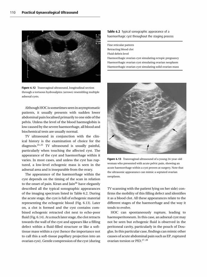
110 Practical Gynaecological Ultrasound
Figure 6.12 Transvaginal ultrasound, longitudinal section
through a tortuous hydrosalpinx (arrows) resembling multiple
adnexal cysts.
Although HOC is sometimes seen in asymptomaticpatients, it usually presents with sudden lowerabdominal pain localised primarily to one side of thepelvis. Unless the level of the blood haemoglobin islow caused by the severe haemorrhage, all blood andbiochemical tests are usually normal.
TV ultrasound in conjunction with the clin-ical history is the examination of choice for thediagnosis.24,25 TV ultrasound is usually painful,particularly when touching the affected cyst. Theappearance of the cyst and haemorrhage within itvaries. In most cases, and unless the cyst has rup-tured, a low-level echogenic mass is seen in theadnexal area and is inseparable from the ovary.
The appearance of the haemorrhage within thecyst depends on the timing of the scan in relationto the onset of pain. Kiran and Jain26 have elegantlydescribed all the typical sonographic appearancesof the imaging spectrum listed in Table 6.2. Duringthe acute stage, the cyst is full of echogenic materialrepresenting the echogenic blood (Fig. 6.13). Lateron, a clot is formed and the cyst contains com-bined echogenic retracted clot next to echo-poorfluid (Fig. 6.14). At a much later stage, the clot retractstowards the wall of the cyst and appears like a fillingdefect within a fluid-filled structure or like a soft-tissue mass within a cyst (hence the importance notto call this a soft-tissue papillary projection into anovarian cyst). Gentle compression of the cyst (during
Table 6.2 Typical sonographic appearance of a
haemorrhagic cyst throughout the staging process
Fine reticular pattern
Retracting blood clot
Fluid debris level
Haemorrhagic ovarian cyst simulating ectopic pregnancy
Haemorrhagic ovarian cyst simulating ovarian neoplasm
Haemorrhagic ovarian cyst simulating solid ovarian mass
Figure 6.13 Transvaginal ultrasound of a young 24-year-old
woman who presented with acute pelvic pain, showing an
acute haemorrhage within a cyst proven at surgery. Note that
the ultrasonic appearance can mimic a septated ovarian
neoplasm.
TV scanning with the patient lying on her side) con-firms the mobility of this filling defect and identifiesit as a blood clot. All these appearances relate to thedifferent stages of the haemorrhage and the way ittends to evolve.
HOC can spontaneously rupture, leading tohaemoperitoneum. In this case, an adnexal cyst maynot be seen but echogenic fluid is observed in theperitoneal cavity, particularly in the pouch of Dou-glas. In this particular case, findings can mimic othercauses of acute abdominal pain such as EP, rupturedovarian torsion or PID.27,28

Ultrasound in the acute pelvis 111
a
b
Figure 6.14 (a and b) Examples of haemorrhagic cysts
(arrows). Note the retracted clots (C ) within these cysts.
Adnexal torsion
Adnexal torsion is an acute medical condition wherethe ovary, ipsilateral fallopian tube or both rotatearound its vascular pedicle, leading to vascularcompromise.29 It is commonly seen in young andmiddle-aged women but also reported in child-hood and adolescents.30,31 Adnexal torsion in elderlywomen is almost always associated with underlyingadnexal mass.
Figure 6.15 Ovarian torsion: 24-year-old woman with sudden
severe right-sided pelvic pain. Transvaginal ultrasound showed
torsion of the right ovary (arrows) next to the uterus (U).
Clinical presentation and symptoms can be mis-leading, making the diagnosis challenging and dif-ficult. Symptoms vary according to the degree oftwist (180–360◦) and the underlying associated vas-cular insult. In mild rotation, lymphatic congestion isnoted but both arterial and venous flow are present.Further torsion can compromise the venous returnand more complete torsion will compromise thearterial blood supply and cause total necrosis ofthe ovary. Because the clinical symptoms are non-specific, presentation may resemble other causes ofacute pelvic pain such as HOC, PID, EP and others.Therefore the diagnosis should be considered in theappropriate clinical picture. Often, ovarian torsionis associated with ipsilateral ovarian mass29,32 andis more common on the right side than on the left,33
probably due to the fact that the sigmoid colon occu-pies the left side of the pelvis.
The ultrasound findings vary but the commonestfinding is an ovarian enlargement which is primarilydue to lymphatic or lymphatic plus venous conges-tion of the ovary. Enlargement of the remaining fol-licle at the periphery of the ovary has been noted30
(Fig. 6.15). The presence of an ovarian mass, whetherit is cystic or solid, is common (Figs 6.16–6.18). Likeother causes of acute pelvic pain, fluid in the pouchof Douglas is also a common finding and may be
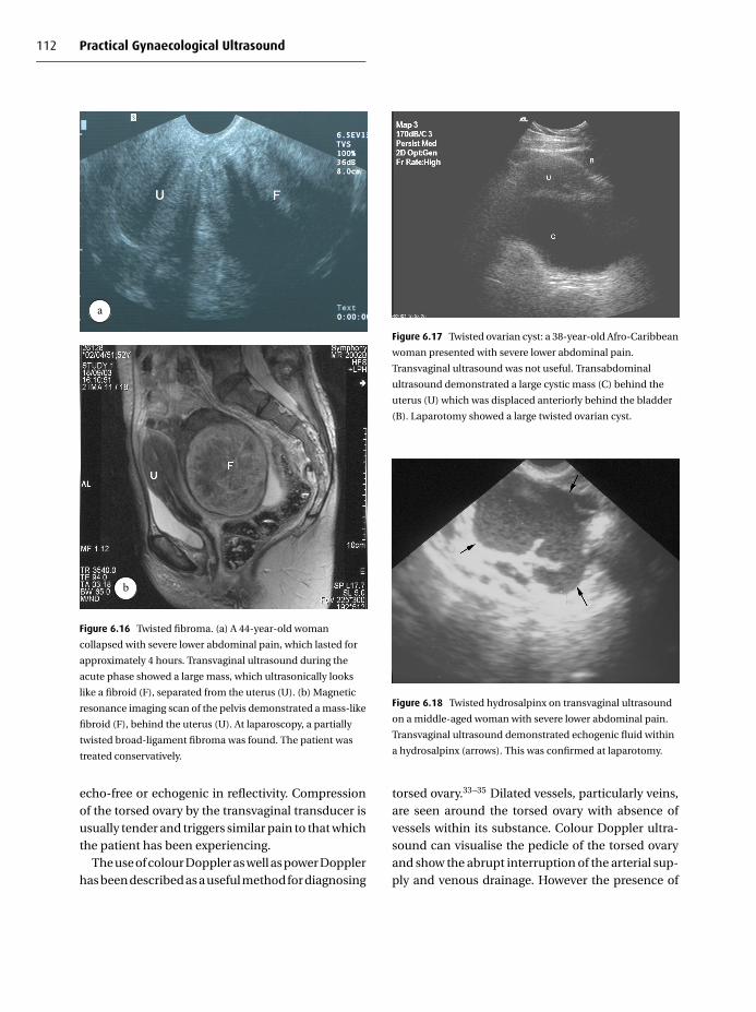
112 Practical Gynaecological Ultrasound
a
b
Figure 6.16 Twisted fibroma. (a) A 44-year-old woman
collapsed with severe lower abdominal pain, which lasted for
approximately 4 hours. Transvaginal ultrasound during the
acute phase showed a large mass, which ultrasonically looks
like a fibroid (F), separated from the uterus (U). (b) Magnetic
resonance imaging scan of the pelvis demonstrated a mass-like
fibroid (F), behind the uterus (U). At laparoscopy, a partially
twisted broad-ligament fibroma was found. The patient was
treated conservatively.
echo-free or echogenic in reflectivity. Compressionof the torsed ovary by the transvaginal transducer isusually tender and triggers similar pain to that whichthe patient has been experiencing.
The use of colour Doppler as well as power Dopplerhas been described as a useful method for diagnosing
Figure 6.17 Twisted ovarian cyst: a 38-year-old Afro-Caribbean
woman presented with severe lower abdominal pain.
Transvaginal ultrasound was not useful. Transabdominal
ultrasound demonstrated a large cystic mass (C) behind the
uterus (U) which was displaced anteriorly behind the bladder
(B). Laparotomy showed a large twisted ovarian cyst.
Figure 6.18 Twisted hydrosalpinx on transvaginal ultrasound
on a middle-aged woman with severe lower abdominal pain.
Transvaginal ultrasound demonstrated echogenic fluid within
a hydrosalpinx (arrows). This was confirmed at laparotomy.
torsed ovary.33–35 Dilated vessels, particularly veins,are seen around the torsed ovary with absence ofvessels within its substance. Colour Doppler ultra-sound can visualise the pedicle of the torsed ovaryand show the abrupt interruption of the arterial sup-ply and venous drainage. However the presence of

Ultrasound in the acute pelvis 113
arterial and venous flow does not exclude the diag-nosis of ovarian torsion.
Shalev et al.35 have published 14 cases of what theydescribed as subtorsion, which is a twist of the ovar-ian pedicle less than 360◦. They found that torsedovary may not be significantly enlarged, particularlyif compared with the normal side, and that bloodflow, both arterial and venous, can be still detectedin the ovary. Congested and dilated periovarian ves-sels were also a common finding. They also foundthat fluid in the pouch of Douglas is not common inthe subtorsion type.
Although sonography, particularly TV ultrasoundwith colour Doppler, is usually the most readily avail-able and used modality for diagnosing torsion, otherimaging techniques like CT and MRI have been usedfor this diagnosis and play a role, particularly in thesubacute cases.36
The active diagnosis of this condition is veryimportant, particularly as early diagnosis and sur-gical treatment can save the ovary from developingirreversible ischaemia and necrosis.
Non-gynaecological pelvic inflammation
Appendicitis
Appendicitis is a common cause of right iliac fossapain, frequently occurring in children and adoles-cents but also seen in patients of any age. Diagnosisis usually made on clinical grounds based on the pre-senting symptoms in conjunction with the elevatedwhite blood cell count and inflammatory markers. Inmost cases, the entire appendix is usually inflamedand all layers are affected and thickened (Fig. 6.19).However, inflammation of part of the appendix, usu-ally the tip, can also occur.37 The underlying inflam-mation may lead to local perforation and the forma-tion of an abscess or inflammatory complex mass.
Imaging is commonly used, particularly in youngfemales, to exclude any other cause of the painsuch as ovarian/adnexal. Ultrasound is the mostreadily available and commonly used modality fordiagnosing this condition. Ultrasonic visualisation
Figure 6.19 Acute appendicitis in a middle-aged woman: the
appendix has a sausage shape, and is non-compressible with
thickening of all layers (arrows).
of the inflamed appendix or the inflammatory massis often hampered by the presence of local ileus anddilated loop of bowel and also by the local pain andabdominal wall rigidity.
Graded compression technique38 has beendescribed as a useful method of visualising theinflamed appendix which involves scanning at adifferent degree of compression over the site ofappendix.
The transverse section of the normal appendixappears either rounded or ovoid. Rettenbacheret al.,39 in a large prospective study, showed that arounded appendix raises the suspicion of an under-lying inflammation but only with specificity and pos-itive predictive value of 37% and 50% respectively.They also concluded that an ovoid-shape transversesection over the entire appendiceal length reliablyrules out acute appendix.
Poortman et al.40 found that an incompressibleappendix with a transverse diameter of 6 mm ormore, with and without appendicolith, indicatedappendicitis. The inflamed appendix appears as ablind-ended sausage shape on longitudinal scan(Fig. 6.19). Appendicolith may be seen within itslumen as a hyperreflective structure (Fig. 6.20a). Theinflamed appendix may be part of an inflammatory

114 Practical Gynaecological Ultrasound
a
b
Figure 6.20 Acute appendicitis. (a) Transabdominal
ultrasound using a high-frequency linear transducer:
sausage-shape inflamed appendix (arrows) with appendicolith
(A) at its base. (b) Inflammatory appendicular mass:
transvaginal ultrasound on a patient with clinical signs of
appendicitis showed a relatively hypoechoic inflammatory
mass (long arrows) surrounding an inflamed appendix with
appendicolith (A) within it, proven at surgery.
Figure 6.21 Crohn’s colitis: transvaginal ultrasound on a
24-year-old female with severe lower abdominal pain and
diarrhoea showing thickening of the wall and mucosa of the
pelvic colon (arrows). Colonoscopic-guided biopsy confirmed
the diagnosis of Crohn’s. Ulcerative colitis can give similar
appearances.
mass or an abscess (Fig. 6.20b). In such a case, andwithout the presence of an appendicolith, the ultra-sonic appearance may be similar to other causessuch as PID and endometriosis. Fujii et al.,41 in a largeprospective study, found that the use of ultrasoundin patients with suspected appendicitis increasesthe accuracy of the diagnosis, reduces unnecessaryoperations and provides more appropriate selectionof patients’ treatment.
Colitis
This is an inflammation of the bowel occurring inyoung patients, commonly in the form of ulcer-ative colitis and Crohn’s disease. Ulcerative coli-tis exclusively affects the colon and Crohn’s affectsboth small and large bowel. When the colon isaffected, its wall becomes oedematous and thick-ened with thickening of the mucosa. The ultra-sonic appearance reflects exactly these histologicalchanges, showing thickened echo-poor muscularisand echogenic mucosa42 (Fig. 6.21). Local abscesses
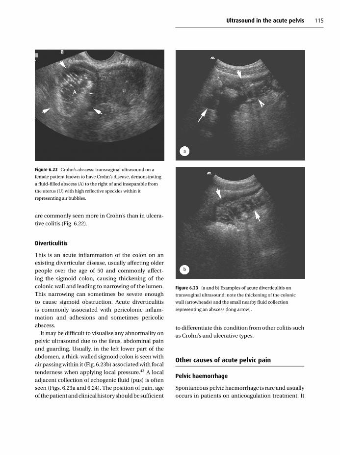
Ultrasound in the acute pelvis 115
Figure 6.22 Crohn’s abscess: transvaginal ultrasound on a
female patient known to have Crohn’s disease, demonstrating
a fluid-filled abscess (A) to the right of and inseparable from
the uterus (U) with high reflective speckles within it
representing air bubbles.
are commonly seen more in Crohn’s than in ulcera-tive colitis (Fig. 6.22).
Diverticulitis
This is an acute inflammation of the colon on anexisting diverticular disease, usually affecting olderpeople over the age of 50 and commonly affect-ing the sigmoid colon, causing thickening of thecolonic wall and leading to narrowing of the lumen.This narrowing can sometimes be severe enoughto cause sigmoid obstruction. Acute diverticulitisis commonly associated with pericolonic inflam-mation and adhesions and sometimes pericolicabscess.
It may be difficult to visualise any abnormality onpelvic ultrasound due to the ileus, abdominal painand guarding. Usually, in the left lower part of theabdomen, a thick-walled sigmoid colon is seen withair passing within it (Fig. 6.23b) associated with focaltenderness when applying local pressure.43 A localadjacent collection of echogenic fluid (pus) is oftenseen (Figs. 6.23a and 6.24). The position of pain, ageof the patient and clinical history should be sufficient
a
b
Figure 6.23 (a and b) Examples of acute diverticulitis on
transvaginal ultrasound: note the thickening of the colonic
wall (arrowheads) and the small nearby fluid collection
representing an abscess (long arrow).
to differentiate this condition from other colitis suchas Crohn’s and ulcerative types.
Other causes of acute pelvic pain
Pelvic haemorrhage
Spontaneous pelvic haemorrhage is rare and usuallyoccurs in patients on anticoagulation treatment. It

116 Practical Gynaecological Ultrasound
Figure 6.24 Acute diverticulitis with pericolic abscess:
transvaginal ultrasound on an elderly lady showing thickened
pelvic colon wall (short arrows) plus a nearby fluid
collection/pus (long arrows) close to the vaginal vault.
is commonly localised to the abdominal wall morethan the intraperitoneal space.
Postoperative haemorrhage/haematoma is a com-mon complication. The haematoma is usuallylocalised to the site of surgery and develops withinthe first few days following the operation. Post-hysterectomy (TA or TV) haematoma is common fol-lowing gynaecological surgery. The haematoma issituated in the uterine bed or near the vaginal vault.
Ultrasonically, haematomas are hypoechoic, haveill-defined margins in the acute phase and canbecome localised and liquefied with time (Fig. 6.25).Colour Doppler scanning shows no vessels withinit and this helps to differentiate haematomas fromother echo-poor tumours such as lymphomas wherevessels are seen within the tissue.
Haemorrhage within an ovarian cyst is already cov-ered above. However, haemorrhage within a fibroidis rare but well-known. It occurs during pregnancyas a reaction of the fibroid to the maternal hormone(Fig. 6.26).
Endometriosis
This is covered more fully in Chapter 4 but is a well-known cause of acute pelvic pain. In most cases, the
a
b
Figure 6.25 (a) Transvaginal ultrasound on a 64-year-old
woman who developed urinary frequency and dysuria
following pelvic surgery showing a large non-vascular
echo-poor mass (H) in the left side of the pelvis classical of
a haematoma. Note the displacement of the bladder (B).
(b) T2-weighted spin-echo axial magnetic resonance imaging
scan of the pelvis on the same patient 5 weeks after surgery
showed the haematoma (H) as a high-signal mass due to the
liquefication displacing the bladder (B).
patient gives a clear history of the disease and TVultrasound is commonly the examination of choicein helping with the diagnosis (Fig. 6.27).
Imperforated hymen
This is a cause of an acute on chronic pelvic paindue to retention of the menstrual blood within thevagina.44 TA ultrasound often demonstrates a fluid

Ultrasound in the acute pelvis 117
Figure 6.26 Haemorrhage within a fibroid: a 9-week pregnant
woman presented to casualty with sudden severe lower pelvic
pain. Previous pelvic ultrasound, before the pregnancy, had
already identified a 4-cm uterine fibroid. Transvaginal
ultrasound showed a viable fetus (long arrow) in a normally
positioned gestational sac (S) with adjacent mass of mixed
echogenicity representing haemorrhage within the fibroid
(arrowed). This was proven at laparoscopy. The patient later
miscarried.
Figure 6.27 Endometriosis: transvaginal ultrasound on a
female with a known history of endometriosis. Note the
bicornuate uterus (arrowheads). The endometriotic cysts
( E ) are seen containing echogenic fluid (chocolate cysts).
Note the lack of separation and tissue planes between
the uterus and the surrounding structures due to
adhesions.
Figure 6.28 Imperforate hymen: a 14-year-old girl presented
with lower abdominal pain which, on careful questioning, she
had had intermittently for over a year. She had not had a
period before. Transvaginal ultrasound showed the uterus
displaced cranially by a large amount of echogenic fluid in the
region of the vagina representing haematocolpos (H). On
examination she was found to have an intact hymen.
collection in the region of the vagina with the uterusdisplaced cranially (Fig. 6.28). See also Chapter 8.
R E F E R E N C E S
1. F. E. Skjeldestad, J. S. Kendrick, H. K. Atrash and A. K. Daltveit,
Increasing incidence of ectopic pregnancy in one Norwegian
county – a population based study, 1970–1983. Acta Obstet-
rica Gynecologica Scandinavica, 76 (1997), 159–65.
2. W. H. Chow, J. R. Daling, W. Cates et al., Epidemiology of
ectopic pregnancy. Epidemiological Review, 9 (1987), 71–
94.
3. S. Ikeda, M. Sumiyoshi, M. Nakae et al., Heterotopic preg-
nancy after in vitro fertilization and embryo transfer. Acta
Obstetrica Gynecologica Scandinavica, 77 (1998), 463–4.
4. T. S. Mehta, D. Levine and B. Beekwith, Treatment of ectopic
pregnancy: is a hCG level of 2000 mLU/ml a reasonable
threshold? Radiology, 205 (1997), 569–73.
5. N. Kadar, M. Bohrer, E. Kemmann et al., The discriminatory
chorionic gonadotropin zone for endovaginal sonography:
a prospective randomised study. Fertility and Sterility, 61
(1994), 1016–20.
6. R. Dart, P. Ramanujam and L. Dart, Progesterone as a
predictor of ectopic pregnancy when the ultrasound is
118 Practical Gynaecological Ultrasound
indeterminate. American Journal of Emergency Medicine, 20
(2002), 575–9.
7. M. C. Frate and F. C. Laing, Sonographic evaluation of ectopic
pregnancy: an update. American Journal of Roentgenology,
165 (1995), 251–9.
8. T. G. Stoval and F. W. Ling, Single dose methotrexate: an
expanded clinical trial. American Journal of Obstetrics and
Gynecology, 168 (1993), 1759–65.
9. M. Yao and T. Tulandi, Current status of surgical and nonsur-
gical management of ectopic pregnancy. Fertility and Steril-
ity, 67 (1997), 421–33.
10. C. R. Cohen, L. E. Manhart, E. A. Bukusi et al., Association
between Mycoplasma genitalium and acute endometritis.
Lancet, 359 (2002), 765–6.
11. Clinical Effectiveness Group, UK National Guidelines
on Sexually Transmitted Infections. 2002. Pelvic Inflam-
matory Disease. Available online at: www.agum.org.uk/
ceg2002/pid0601.htm.2002.
12. Royal College of Obstetricians and Gynaecologists, Guide-
lines no. 32. Acute Pelvic Inflammatory Disease, Manage-
ment of (London: Royal College of Obstetricians and Gynae-
cologists, 2003).
13. D. I. Bulas, P. A. Ahlstrom, C. J. Sivit et al., Pelvic inflamma-
tory disease in the adolescent: comparison of transabdom-
inal and transvaginal sonographic evaluation. Radiology,
183 (1992), 435–9.
14. T. A. Tukeva, H. J. Aronen, P. T. Karjalainen et al., MR
imaging in pelvic inflammatory disease: comparison with
laparoscopy and US. Radiology, 210 (1999), 209–16.
15. H. Tikanen and E. Kujansuu, Doppler ultrasound findings in
tubo-ovarian infectious complex. Journal of Clinical Ultra-
sound, 21 (1993), 175–8.
16. P. Molander, J. Sjoberg, J. Paavonen and B. Cacciatore,
Transvaginal power Doppler findings in laparoscopically
proven acute pelvic inflammatory disease. Ultrasound in
Obstetrics and Gynecology, 17 (2002), 233–8.
17. I. E. Timor-Tritsch, J. P. Lerner, A. Monteagudo et al.,
Transvaginal sonographic markers of tubal inflammatory
disease. Ultrasound in Obstetrics and Gynecology, 12 (1998),
56–66.
18. J. D. C. Ross, Pelvic inflammatory disease: how should it be
managed? Current Opinion in Infectious Diseases, 16 (2003),
37–41.
19. E. Van Sonnenberg, H. B. D’Agostino, G. Casola et al., US-
guided transvaginal drainage of pelvic abscesses and fluid
collections. Radiology, 181 (1991), 53–6.
20. W. De Wit, C. J. Gowrising, D. J. Kuik et al., Only hydro-
salpinges visible on ultrasound are associated with reduced
implantation and pregnancy rate after IVF. Human Repro-
duction, 13 (1998), 1696–701.
21. T. Mukherjee, A. B. Copperman, C. McCaffrey et al.,
Hydrosalpinx fluid has embryotoxic effect on murine
embryogenesis: a case for prophylactic salpingectomy.
Fertility and Sterility, 66 (1996), 851–3.
22. E. K. Outwater, E. S. Siegelman, P. Chiowanich et al., Dilated
fallopian tubes: MRI imaging characteristics. Radiology, 208
(1998), 463–9.
23. S. Guerriero, S. Ajossa, M. P. Lai et al., Transvaginal ultra-
sonography associated with colour Doppler energy in the
diagnosis of hydrosalpinx. Human Reproduction, 15 (2000),
1568–72.
24. T. Okai, K. Kobayashi, E. Ryo et al., Transvaginal sonographic
appearance of hemorrhagic functional ovarian cyst and their
spontaneous regression. International Journal of Gynaecol-
ogy and Obstetrics, 44 (1994), 47–52.
25. O. H. Baltarowich, A. B. Kurtz, M. E. Pasto et al., The spec-
trum of sonographic findings in hemorrhagic ovarian cyst.
American Journal of Roentgenology, 148 (1987), 901–5.
26. A. Kiran and M. D. Jain, Sonographic spectrum of hem-
orrhagic ovarian cyst. Journal of Ultrasound Medicine, 21
(2002), 879–86.
27. J. G. Hallatt, C. H. Steele and M. Snyder, Ruptured corpus
luteum with hemoperitoneum: a study of 173 surgical cases.
American Journal of Obstetrics and Gynecology, 149 (1984),
5–9.
28. B. S. Hertzberg, M. A. Kliewer and E. K. Paaulson, Ovarian cyst
rupture causing hemoperitoneum: imaging features and the
potential for misdiagnosis. Abdominal Imaging, 24 (1999),
304–8.
29. L. T. Hibbard, Adnexal torsion. American Journal of Obstetrics
and Gynecology, 152 (1985), 456–61.
30. M. Graif and Y. Itzchak, Sonographic evaluation of ovarian
torsion in childhood and adolescence. American Journal of
Roentgenology, 150 (1988), 647–9.
31. J. E. Stark and M. J. Siegel, Ovarian torsion in prepubertal
and pubertal girls: sonographic findings. American Journal
of Roentgenology, 163 (1994), 1479–82.
32. M. A. Warner, A. C. Fleischer, S. L. Edell et al., Uterine adnexal
torsion: sonographic findings. Radiology, 154 (1985), 773–5.
33. F. Albayram and U. M. Hamper, Ovarian and adnexal torsion:
spectrum of sonographic findings with pathologic correla-
tion. Journal of Ultrasound Medicine, 20 (2001), 1083–9.
34. E. J. Lee, H. C. Kwon, H. J. Joo et al., Diagnosis of ovarian
torsion with color Doppler sonography: depiction of twisted
vascular pedicle. Journal of Ultrasound Medicine, 17 (1998),
83–9.

Ultrasound in the acute pelvis 119
35. J. Shalev, R. Mashiach, I. Bar-Hava et al., Subtorsion of
the ovary: sonographic features and clinical management.
Journal of Ultrasound Medicine, 20 (2001), 849–54.
36. S. E. Rha, J. Y. Byun, S. E. Jung et al., CT and MR imag-
ing features of adnexal torsion. Radiographics, 22 (2002),
283–94.
37. H. V. Nghiem and R. B. Jeffry, Jr., Acute appendicitis confined
to the appendiceal tip: evaluation with graded compres-
sion sonography. Journal of Ultrasound Medicine, 11 (1992),
205–7.
38. J. B. C. M. Puylaert, Acute appendicitis: US evaluation using
graded compression. Radiology, 158 (1986), 355–60.
39. T. Rettenbacher, A. Hollerweger, P. Macheiner et al., Ovoid
shape of the vermiform appendix: a criterion to exclude
acute appendicitis – evaluation with US. Radiology, 226
(2003), 95–100.
40. P. Poortman, P. N. M. Lohle, C. M. C. Schoemaker et al.,
Comparison of CT and sonography in the diagnosis of
acute appendicitis: a blind prospective study. American
Journal of Roentgenology, 181 (2003), 1355–9.
41. Y. Fujii, J. Hata, K. Futagami et al., Ultrasonography improves
diagnostic accuracy of acute appendicitis and provide cost
saving to hospitals in Japan. Journal of Ultrasound Medicine,
19 (2000), 409–14.
42. M. E. O’Malley and S. R. Wilson, US of gastrointestinal tract
abnormalities with CT correlation. Radiographics, 23 (2003),
59–72.
43. S. G. Parulekar, Sonography of colonic diverticulitis. Journal
of Ultrasound in Medicine, 12 (1985), 659–66.
44. M. R. Spevak and H. L. Cohen, Ultrasonography of the
adolescent female pelvis. Ultrasound Quarterly, 18 (2002),
275–88.

7
Ultrasound and fertility
Stephen KillickWomen and Children’s Hospital, Hull
Introduction
Ultrasound is the single most useful agent in evaluat-ing human fertility. Not only do the fluid–tissue inter-faces in the female pelvic organs allow precise meas-urements of structure but the low cost and safetyof ultrasound scanning allow repeated examinationsand hence, most importantly, an understanding offunction (Table 7.1).
Although transabdominal ultrasound is usedoccasionally, for example when surgery has movedovaries from their normal location, the vast majorityof scans are performed vaginally. The close proxim-ity of the ovaries and uterus to the vaginal fornicesallows for the use of higher frequencies (∼10 MHz)and hence higher resolution.
Subfertility is common. Some 19% of pregnanciestake more than a year to conceive1 and one in six ofall couples are referred for fertility investigations atsome time during their lives.2
Ovarian function
A human ovary contains many thousands of germcells distributed throughout its stroma, each ofwhich is surrounded by a small number of special-ist cells to create a primordial follicle. These folli-cle complexes begin to develop one by one through-out reproductive life in response to gonadotrophin
hormones secreted by the pituitary gland but theyonly become visible to ultrasound in their later stagesof development when they develop a fluid-filledantrum. These fluid-filled follicles give the ovary itscharacteristic appearance during the reproductiveyears. When visualising the ovary it is important toremember that its structure is constantly changingas part of a dynamic system involving many otherorgans.
During an ovulatory cycle antral follicles increasein diameter by up to 2 mm a day in response tofollicle-secreting hormone (FSH) secreted by thepituitary gland. The first follicle to begin develop-ment in any one month is the largest and is termedthe first-order (graafian) follicle, with second- andthird-order follicles following on behind. At mid-cycle the pituitary releases a surge of gonadotrophins(FSH and especially luteinising hormone (LH)) andthe first-order follicle (and sometimes the second-order follicle as well) ovulates. Follicles are usuallybetween 18 and 22 mm diameter when they ovu-late but this is variable and they can be smaller orlarger. Lower-order follicles are not mature enoughto respond and slowly resolve. The release of follicu-lar fluid at the time of ovulation can be seen by ultra-sound to take several hours; it is not a sudden burst.The follicle shrinks to at least half its previous size andthen grows into a corpus luteum, which is irregularlycystic and up to 3 cm in diameter. Whereas the firsthalf of the ovarian cycle before ovulation can be of
C© Cambridge University Press 2005.
120

Ultrasound and fertility 121
Table 7.1 Advantages and disadvantages of ultrasound
in evaluating fertility
Advantages Disadvantages
Precise measurements of pelvic
structures
Initial cost of
machinery
Instant result Operator-dependent
Viewed by the woman herself Vaginal scan is
personally invasiveAllows for simultaneous
counselling
Safe
Inexpensive repeat measurements
Used for oocyte capture
Gives idea of function, not just
structure
Permanent record can be retained
variable duration, menstruation takes place 14 daysafter ovulation if conception does not occur.
A single view of a normal functioning ovary may,therefore, demonstrate a complex picture of severalsmall newly developing follicles, some old degener-ating follicles and a corpus luteum, all embedded ina solid vascular stroma. More useful information isgained by observing how the picture changes day byday (Fig. 7.1).
Premenarchal ovaries often contain antralfollicles; in fact the ovaries of adolescent girls havemultiple follicles, indicating that there is enoughFSH to induce follicular growth but the pituitaryhas yet to mature and develop its feedback mech-anism with the ovary in order to induce ovulation.This picture is also seen in other situations wherepituitary function is compromised, such as weight-related amenorrhoea or anorexia nervosa. It can beconfused with polycystic ovaries (PCO; see below)but in PCO there is an increased amount of ovarianstroma.
Towards the end of reproductive life as themenopause encroaches, multiple ovulationsbecome more common, as does anovulation, andso the ovary may contain several quite large folli-cles. For this reason non-identical twins are morecommon in older women.
Fol
licl
e si
ze in
mm
Scan here shows 4developing folliclesand 1 degenerating
follicle
Scan here shows 4degenerating folliclesand a corpus luteum
Day of cycle
Ovulation
10
20
14 281
Figure 7.1 A schematic representation of how follicles develop
and degenerate in the normal ovulating ovary, thus altering its
ultrasound appearance.
The endometrial cycle
The routine growth and shedding of the endometriallining can also be monitored with ultrasound. Duringmenstruation the endometrium appears somewhatirregular and highly reflective. The endometriumincreases in thickness to about 1 cm in the first halfof the cycle. At this stage, just prior to ovulation, it isof a lower echo intensity than the myometrium andis surrounded by a narrower layer of even lower echointensity, the junctional zone. The junctional zonerepresents the innermost cells of the myometrium,which are more tightly packed than in the outer layer.After ovulation, under the influence of the hormoneprogesterone, the endometrial layer becomes moreecho-intense than the myometrium (Fig. 7.2).
The ultrasonographer needs to remember func-tional interrelationships. Antral follicles secretealmost all of the body’s oestrogen and so, for exam-ple, if an ovary is seen to contain a 2-cm-diameterecho-free cystic structure, then the woman is prob-ably mid-cycle and the uterine endometrium will beseveral millimetres thick but still of low echo inten-sity. If this is not the case and the endometrium is

122 Practical Gynaecological Ultrasound
Oestradiol from Graafianfollicle causes
endometrial growth Progesterone from corpusluteum causes secretorychanges in endometrium
Oestradiol
Progesterone
Ovu
lati
on
Ovarianappearance
Uterineappearance
Bloodhormone
levels
Day of cycle14 28
Junctional zone
Figure 7.2 Diagram to illustrate the interrelationships
between blood hormone levels and ovarian and uterine
appearances throughout the ovulatory cycle.
thin (∼4 mm) the cystic structure is not a graafianfollicle producing oestrogen.
Management of subfertility
Subfertile couples need to be managed with sym-pathy. Information counselling and support coun-selling do not necessarily mean the intervention ofa trained counsellor. An ultrasonographer is in anideal situation to provide information and supportwhilst viewing the screen with the subfertile cou-ple. To maximise this advantage the ultrasonogra-pher must be part of the fertility team and should beaware of the clinical situation of each specific couple.
Each subfertile couple needs to have an estimateof their chances of natural conception before theydecide whether to opt for treatment. In practice thisneeds a minimum of three assessments: an analy-sis of the male partner’s ejaculate, an assessment ofovulation and some test of fallopian tube patency.
Ultrasound could be used for the diagnosis of ovula-tion but usually a more convenient way is to take asingle blood sample for progesterone 7 days beforethe first day of the next period, when the value shouldbe at its peak and greater than 30 nmol/l.
Tubal patency testing
Fallopian-tube patency can be visualised ultrasono-graphically if a positive contrast medium is used.The procedure is termed hystero-contrast sonogra-phy, or HyCoSy for short, and is recommended bythe National Institute for Clinical Excellence (NICE)as the procedure of choice for initial determinationof tubal patency.3 A catheter is introduced throughthe cervix and held in place by inflating a small bal-loon at its tip. The commercially available materialEchovist (Schering, UK) is then injected in volumesof less than 1 ml at a time whilst a vaginal scan isperformed.4
Procedures are usually performed in the firsthalf of the menstrual cycle, preferably before theendometrium has attained its maximum thicknessand before any chance of pregnancy, although goodresults can be obtained at any stage of the cycle. Con-siderable experience of vaginal scanning is neces-sary for consistent results. The use of Doppler doesnot help in the assessment. HyCoSy may be com-bined with an initial assessment of the uterine cavityto exclude such things as uterine polyps, submu-cous fibroids or septa. In this case a negative con-trast is much better and saline is usually used beforeany positive contrast is injected. This procedure istermed saline infusion sonography.
It has been estimated that 50 HyCoSy proceduresneed to be performed before operators can be rea-sonably sure of their findings. A national teachingcourse is currently available.
Testicular structure and function
Ultrasound can be useful for locating ectopic testi-cles or in the diagnosis of testicular or epididymaltumours, but there is a poor correlation between
Ultrasound and fertility 123
testicular function and structural findings withinthe testicle. Hence it is not used routinely in theinvestigation of male subfertility. Small varicoceles,although detectable with ultrasound, do not lead toimproved fertility when they are treated surgically, soa routine search for them is unjustified.
Ovulation induction
Ultrasound is ideal for monitoring the response ofthe ovary to stimulatory drugs for all the reasonspreviously mentioned. Frequent blood sampling foroestradiol levels used to be used but this is now rarelynecessary.
For women who do not ovulate spontaneously,drugs such as clomifene stimulate FSH from the pitu-itary gland and therefore stimulate follicle growth inthe ovary. Alternatively FSH, which is available com-mercially in a variety of preparations, usually in com-bination with LH, can be given directly by injection.In these circumstances ultrasound is used to checkthe numbers of follicles developing on each ovary. Ifmore than two preovulatory-sized follicles (≥16 mmdiameter) develop, there is the chance of a multiplegestation of triplets or more and conception needsto be avoided. Follicles are usually measured in twoplanes, their longest diameter and the diameter atright angles to this. It is possible to rotate the vaginaltransducer through 90% and measure a third diam-eter but this adds little to precision. What really mat-ters is how quickly the follicles are growing and howmany of them there are.
Ultrasound is often used to decide on the timingof an injection of human chorionic gonadotrophinin order to trigger ovulation. Depending on proto-col and circumstances this may be when the leadingfollicle is 20 mm diameter. If the scan is repeated 48hours later to confirm ovulation, the follicle shouldhave ruptured and a small amount of fluid may bevisible behind the uterus in the pouch of Douglas.
In vitro fertilisation
The first step in in vitro fertilisation (IVF) isto inhibit pharmacologically the secretion of the
gonadotrophins FSH and LH from the pituitarygland. This is termed downregulation. Ultrasoundis used to check that adequate downregulation hasbeen obtained by checking that the ovaries havebecome inactive with no follicles greater than 9 mmdiameter and that the endometrium is thin (< 4 mm),i.e. that the serum oestadiol is low.
Follicle growth is then monitored in much thesame way as in ovulation induction but there is norisk of supermultiple gestations with IVF becauseall the oocytes are extracted from their follicles andonly one or two embryos subsequently transferredto the uterus. Hence higher doses of drugs can beused and many more follicles develop. Multiple ovar-ian follicles often push together to give a cartwheelappearance.
Vaginal ultrasound is the method of choice forharvesting the oocytes unless an ovary is situatedin an unusual intra-abdominal position. A needle ispassed through a guide on the transducer, throughthe vaginal fornix and into the ovary on each side.Follicular fluid is aspirated and examined micro-scopically to find each oocyte. This is a relativelysafe procedure, although there are instances of pelvicinfection or damage to the internal iliac vein. Thevein is immediately adjacent to the ovary and canlook very much like a follicle in cross-section, so ifthere is any doubt a follicle should be scanned fromwall to wall before puncture.
If the oocytes fertilise successfully in vitro they areusually transferred to the uterus 2 or 3 days aftertheir collection. Ultrasound can be useful in assist-ing embryo transfer.5 The objective here is to trans-fer the embryos through the cervical canal into theupper uterine cavity as quickly as possible and withminimal trauma. Ultrasound can record the lengthand direction of the cervical canal and uterine cavityprior to attempts at transfer so as to make the proce-dure easier.
Ovarian hyperstimulation syndrome
Ovarian hyperstimulation syndrome (OHSS) maydevelop in the first few days and weeks afteroocyte capture if the ovaries have been inadvertently

124 Practical Gynaecological Ultrasound
overstimulated. It is more common in women whohave PCO syndrome (PCOS) and when more than 15or 20 oocytes have been harvested. It is rare to seethe syndrome except in IVF cycles, although it canoccur after ovulation induction or even after spon-taneous ovulation. Women complain of abdominalpain and distension, nausea, diarrhoea and some-times breathlessness. They retain fluid within theperitoneal and thoracic cavities and hence urine out-put falls, blood viscosity increases and there is a riskof venous thromboembolism. Ultrasound appear-ances help to differentiate the syndrome into mild,moderate and severe cases. Ovaries are less than5 cm in diameter in mild cases, between 5 and12 cm in moderate cases and greater than 12 cm insevere cases. There is also an increasing amount ofintraperitoneal fluid in the more severe forms, mostnotably adjacent to the kidneys.
OHSS can be avoided by abandoning treatmentprior to the human chorionic gonadotrophin ovu-latory trigger in cases where a large number of fol-licles are seen to be developing. Alternatively, ifoocyte capture has already taken place, all result-ing embryos can be cryopreserved and not trans-ferred to the uterus until the condition has sub-sided. This avoids pregnancy, which potentiates thecondition.
Of course abdominal pain after embryo transferhas many other causes. Apart from OHSS the mostserious of these is ectopic pregnancy. This can bea difficult diagnosis after IVF for a number of rea-sons. Human chorionic gonadotrophin is given asan ovulatory trigger so all women will have a pos-itive pregnancy test for the first week or two afterembryo transfer. The diagnosis of early pregnancy isoften made before an intrauterine embryo is largeenough to be seen. OHSS can occur at the same timeas an ectopic. Even if an intrauterine embryo is seen,there may be a coincident tubal gestation (hetero-topic pregnancy) as both the transferred embryosmay have implanted. Suspicious cases may need tobe followed by serial scans and serum beta – humanchorionic gonadotrophin estimations. Miscarriagesand molar pregnancies occur at least as commonlyafter IVF as after natural conception.
Doppler studies of follicles andendometrium
Advanced ultrasound techniques, although rarelyused in clinical practice, have been used to enhanceour knowledge of reproductive physiology. Dopplerstudies have shown increased blood flow around fol-licles with mature oocytes6 and the pulsatility indexof the endometrium correlates inversely with theprobability of implantation.7
Uterine contractions
The uterine endometrium is in a constant stateof movement throughout most of the menstrualcycle. However movements are slow and can onlybe demonstrated by speeding up video recordingsof vaginal ultrasound images. Using this techniqueperistaltic-like waves can be seen to run along thelength of the uterus in either direction and occa-sionally laterally towards the fallopian-tube orifices.A typical wave progresses along the uterus at aspeed of about 3 cm/min. Contractions are morefrequent at the time of ovulation in natural cyclesor at the time of oocyte collection in IVF cycles.These contractions are thought to have a functionin both sperm transport8 and in implantation9 andin the future their monitoring may have importantimplications for the management of subfertile cou-ples. We know, for example, that the high incidenceof ectopic pregnancy (4%) after IVF treatment isprobably caused by uterine contractions relocatingthe embryos from the uterine cavity to the fallop-ian tubes.10 A traumatic embryo transfer increasesendometrial contractions and reduces the chances ofpregnancy.11
Polycystic ovary syndrome
PCO are a common finding. The original ultra-sonic definition calls for an enlarged ovary withmore than 10 follicles up to 8 mm in diameter situ-ated peripherally around an echo-dense, thickened

Ultrasound and fertility 125
central stroma.12 Using this definition some 23%of ovaries were said to appear polycystic,13 but thegreater resolving power of modern vaginal transduc-ers detects more follicles and therefore includes evenmore women in the diagnosis. A PCO, as an iso-lated ultrasound finding, has no relevance for fer-tility.14 However, when combined with other symp-toms of the PCOS, such as obesity, hirsutism orirregular periods, women with PCO are increasinglysubfertile.
Women with PCOS may respond to ovulationinduction by producing multiple preovulatory folli-cles and there is therefore a greater risk of supermul-tiple gestations. The diagnosis of PCOS is, therefore,an important one for ultrasonographers to note. Itis also important to note, from a physiological pointof view, that these ovaries are constantly changing.The follicles seen one day become atretic and arereplaced by others by the time a scan is repeated aweek later, even though the overall ovarian appear-ance is unaltered.
Pelvic pathology
It is not uncommon for pelvic pathology to be com-pletely asymptomatic apart from its effect on fer-tility and hence to present for the first time dur-ing fertility investigations. Examples would be uter-ine malformations, endometriosis, uterine fibroidsor hydrosalpinges. The latter three conditions maybe hormone-dependent and increase in size inresponse to superovulation during IVF treatment.Occasionally they seem to appear for the first timeduring treatment, having been missed at a baselinescan when they were smaller.
R E F E R E N C E S
1. M. A. M. Hassan and S. R. Killick, Effect of male age on fertility:
evidence for the decline in male fertility with increasing age.
Fertility and Sterility, 79 (suppl. 3) (2003), 1520–7.
2. M. G. R. Hull, C. M. Glazener, N. J. Kelly, et al. Population
study of causes, treatment, and outcome of infertility. British
Medical Journal, 291 (1985), 1693–7.
3. NICE Clinical Guideline. Fertility: Assessment and Treatment
for People with Fertility Problems (NICE, 2004).
4. S. R. Killick, Hysterosalpingo contrast sonography as a
screening test for tubal patency in infertile women. Journal
of the Royal Society of Medicine, 92 (1999), 628–31.
5. F. P. Biervliet, P. Lesny, S. D. Maguiness and S. R. Kil-
lick, Ultrasound-guided embryo transfer maximizes the IVF
results on day 3 and day 4 embryo transfer but has no impact
on day 5. Human Reproduction, 17 (2002), 1131.
6. G. Nargund, T. Bourne, P. Doyle et al., Associations between
ultrasound indices of follicular blood flow, oocyte recovery
and preimplantation oocyte quality. Human Reproduction,
11 (1996), 109–13.
7. J. Zaidi, R. Pittrof, A. Shaker et al., Assessment of uter-
ine artery blood flow on the day of human chorionic
gonadotropin administration by transvaginal color Doppler
ultrasound in an in vitro fertilization program. Fertility and
Sterility, 65 (1996), 377–81.
8. M. Fukuda and K. Fukuda, Uterine endometrial cavity move-
ment and cervical mucus. Human Reproduction, 9 (1994),
1013–16.
9. M. M. Ijland, J. L. H. Evers, G. A. J. Dunselman et al., Relation
between endometrial wavelike activity and fecundability in
spontaneous cycles. Fertility and Sterility, 67 (1997), 492–5.
10. F. P. Biervliet, P. Lesny, S. D. Maguiness and S. R. Killick, Mech-
anisms for bilateral ectopics after embryo transfer? Fertility
and Sterility, 76 (2001), 212–13.
11. P. Lesny, S. R. Killick, J. Robinson, G. Raven and S. D. Magui-
ness, Junctional zone contractions and embryo transfer: is
it safe to use a tenaculum? Human Reproduction, 14 (1999),
2367–70.
12. J. Adams, S. Franks, D. W. Polson et al., Multifollicular
ovaries: clinical and endocrine features and response to pul-
satile gonadotropin releasing hormone. Lancet, ii (1985),
1375–8.
13. D. W. Polson, J. Wadsworth, J. Adams and S. Franks, Polycys-
tic ovaries: a common finding in normal women. Lancet, i
(1988), 870–2.
14. M. A. Hassan and S. R. Killick, Ultrasound diagnosis of poly-
cystic ovaries in women who have no symptoms of polycystic
ovary syndrome is not associated with subfecundity or sub-
fertility. Fertility and Sterility, 80 (2003), 966–97.

8
Paediatric gynaecological ultrasound
David W. Pilling MB ChB DCH DMRD FRCR FRCPCHRoyal Liverpool Children’s Hospital, Alder Hey, Liverpool
Ultrasound is clearly established as the imagingmodality of choice for the evaluation of paediatricgynaecological conditions. In certain circumstancesmagnetic resonance imaging (MRI) will also have arole, as will specialised techniques such as genitogra-phy. Computed tomography (CT) has a very limitedplace and should be avoided, if possible, becauseof the significant dose of ionising radiation to theovaries.
Ultrasound technique
Children of all ages have many special needs and allultrasound examinations should be undertaken bythose with experience of managing children who arealso aware of the very different pathology likely tobe encountered. Particularly for smaller children, afriendly environment is essential with appropriatetoys and books to distract especially the preschoolchild.
As scans must be carried out transabdominally, afull bladder is necessary, which most girls over theage of 3 or 4 years can readily achieve. In youngerchildren and babies it is pure chance as to whetherthe bladder is full or not. If it is not, then the infantshould be given a drink and rescanned at half-hourlyintervals until the bladder is full enough for an ade-quate examination. In neonates the bladder tends tofill and empty even more quickly so the time intervalcan be shorter.
Modern broadband curvilinear transducers aremost appropriate, with a frequency range of 5–8 MHzin the neonate, 4–7 MHz in the older child and 3–5MHz reserved for adolescents. In the neonate a lin-ear 5–10 MHz probe (perhaps surprisingly) can occa-sionally be very useful, as the structures of interestare always within 1–2 cm of the probe and the linearconfiguration is not as big a handicap as may at firstbe thought.
In the USA transvaginal sonography in the appro-priate clinical situations is undertaken in sexuallyactive adolescents. This is not generally the practicein the UK.
Normal anatomy
The uterus and ovaries undergo significant changesbetween birth and puberty and these must be appre-ciated in order to avoid misinterpreting normalchanges as pathological processes.1 At birth both theuterus and ovaries are affected by maternal and pla-cental gonadotrophins and as this stimulus disap-pears, so their appearances change over the first fewmonths. Further significant changes take place at thetime of puberty. A chart of normal measurementswith age is essential.
Neonates and young infants
At birth the fundal region of the uterus is proportion-ately larger than at a slightly older age. The length of
C© Cambridge University Press 2005.
126

Paediatric gynaecological ultrasound 127
Figure 8.1 Neonatal uterus showing prominent endometrium
with multiple layers due to maternal hormone stimulus.
Figure 8.2 Normal ovary at 1 month with volume of 0.9 cm3
and small microcysts.
the uterus is between 2.3 and 4.0 cm and the widthbetween 0.8 and 2.2 cm. The endometrium may bevisualised as an echogenic stripe and there may be alittle fluid within the endometrial cavity (Fig. 8.1).
By birth the ovaries have usually descended to lie atthe superior margin of the broad ligament, althoughrarely they may be as high as the lower pole of thekidneys. On ultrasound they appear homogeneousapart from the frequent presence of small microcysts(Fig. 8.2). Mean ovarian volume has been shown tobe 1.06 cm3 (range 0.7–3.6 cm3) up to 3 months ofage; 1.05 cm3 (range 0.2–2.7 cm3) in girls aged 4–12months; and 0.67 cm3 (range 0.1–1.7 cm3) in girlsbetween 13 and 24 months old.1 The uterus in par-ticular is very easy to demonstrate at this age – more
Table 8.1 Paediatric uterine and ovarian growth
Uterine length (mm)a Ovarian volume (cm3)
Age Mean sd Mean sd
2 33.1 ± 4.4 0.75 ± 0.41
4 32.9 ± 3.3 0.82 ± 0.36
6 33.2 ± 4.1 1.19 ± 0.36
8 35.8 ± 7.3 1.05 ± 0.50
10 40.3 ± 6.4 2.22 ± 0.69
12 54.3 ± 8.4 3.80 ± 1.40
13 53.8 ± 11.4 4.18 ± 2.30
a Total uterine length from fundus to cervix.
Data from Orsini et al.40
so than at any stage between 1 month and puberty.For this reason it is imperative to scan babies withambiguous genitalia at this age rather than later ininfancy. The ovaries are easily identified if folliclesare present but more difficult if not.
Premenarchal girls
Beyond the neonatal period the uterus assumesa more tubular or teardrop shape with the cervixaccounting for two-thirds of the total length(Fig. 8.3). The length of the uterus and the size ofthe ovaries change very little in the first 6 years of lifebut then gradually increase (Table 8.1). Throughoutthis time small microcysts within the ovaries may beseen on ultrasound scanning (Fig. 8.4) but it may bedifficult to demonstrate normal ovaries at this age.By the age of 7 years the uterus has achieved a staticphase which it maintains until the onset of puberty(Fig. 8.5).
Puberty
The uterus increases in size and changes shape at thetime of puberty (Table 8.1), assuming a pear shapeand starting to undergo cyclical endometrial changesidentical to the adult. With gonadotrophin stimulusthe ovaries enlarge and lie more deeply in the pelviseither laterally or posterolaterally to the uterus.

128 Practical Gynaecological Ultrasound
Figure 8.3 (a) At 6 months the cervix is more prominent than
the fundus and body of the uterus. (b) By 1 year the uterus has
assumed a more tubular appearance.
Follicular development
Small (less than 0.9 cm) microcysts or follicles arefrequently seen in the ovaries at any time betweenbirth and puberty. They have been shown to occurin 84% of ovaries between birth and 2 years ofage.2 In the same study larger macrocysts (1–1.4 cm)were noted in 18% of normal ovaries. From 6 or 7years onwards the number and size of cysts increase,although it is unusual for the cysts to be larger than1.5 cm in diameter. In girls aged between 2 and10 years, cysts up to 1.7 cm in diameter have beenidentified in 68%.3
At the time of puberty cyclical follicular devel-opment begins with a number of small primordialfollicles being present early in the cycle. Between
Figure 8.4 Normal ovary containing microcysts in a
3.5-year-old.
Figure 8.5 Normal uterus at 7 years of age with cervix more
prominent than body.
days 8 and 12 of the cycle a dominant folliclebecomes apparent with ovulation occurring at mid-cycle when the follicle is between 1.7 and 2.7 cm insize. With follicular rupture the follicle decreases insize and a corpus luteum forms. This appears as a 1.6–2.4-cm cystic structure with internal echoes, whichdegenerates at the end of the cycle if fertilisation doesnot occur.
Neonatal pathology
Ovarian cysts
With the widespread use of both antenatal and post-natal ultrasound scanning, ovarian cysts of greaterthan 1.5 cm in diameter have been diagnosed

Paediatric gynaecological ultrasound 129
Figure 8.6 A 5-cm unilocular neonatal ovarian cyst with debris
(arrow) in its base.
Figure 8.7 Large neonatal ovarian cyst about 9 cm in diameter.
This resolved in infancy following partial aspiration.
with increasing frequency. Occasionally larger cystspresent as abdominal masses or as bowel obstructionor respiratory embarrassment (Figs. 8.6 and 8.7). Inthese cases absolute differentiation from other cysticstructures such as mesenteric, duplication or urachalcysts can be extremely difficult, although the pres-
ence of a daughter cyst is virtually pathognomonic.4
All ovarian cysts can theoretically undergo torsion orcyst rupture, although this is unusual with the smallones.
Most neonatal ovarian cysts are of follicular ori-gin. The stimulus to cyst formation is thought tobe maternal gonadotrophins leading to aberrantfollicular development. It is therefore reasonableto assume that after birth, when this stimulus isremoved, at least some of these cysts will resolve. Asa result of this a conservative approach to their man-agement has been advocated. There is no agreementon the frequency of scanning but it seems reason-able to scan weekly in the first few weeks of life withless frequent scans as the baby gets older and thecyst reduces in size. Residual cysts can be seen up to9 months of age and possibly longer. If a cystincreases in size or fails to involute, the diagnosisof follicular cyst should be questioned. Early studiessuggested that only uncomplicated cysts which weretotally anechoic and thin-walled should be man-aged in this way and that other complicated cystsshould be removed. More recent studies have shownthat most asymptomatic cysts can be managedconservatively with serial ultrasound monitoring.5
Symptomatic cysts can be treated by percutaneousaspiration or surgery. Minilaparotomy has beensuggested,6 as has laparoscopy.7 It has always beenthe surgical practice to conserve the ovaries in therare situation of bilateral ovarian cysts but ovar-ian conservation is encouraged even with unilat-eral cysts wherever possible. The ultrasound appear-ances of complicated cysts include the presence offluid/debris levels, retracting clot, septa or calcifica-tion in the cyst wall, but neither these features nor thesize reliably predict the clinical outcome (Fig. 8.6).8
Vaginal bleeding
Neonatal vaginal bleeding is an uncommon event,usually caused by shedding of the endometriumwhich has become hypertrophied in utero sec-ondary to maternal hormone stimulus and followingremoval of the stimulus behaves in the same way thatthe adult endometrium does. Ultrasound will show a

130 Practical Gynaecological Ultrasound
prominent midline echo sometimes with a low echoelement. Reassurance can be given in the presence ofa normal-size uterus and lack of demonstrable ovar-ian pathology. Follow-up scans are only justified ifthe bleeding continues.
Hydrocolpos or hydrometrocolpos
Vaginal obstruction in the neonate may lead to fluidsecretions distending the vagina (hydrocolpos) orthe vagina and uterus (hydrometrocolpos).9 Thismay be due to imperforate hymen, complete vaginalmembrane, vaginal stenosis or atresia. More rarelyit may be associated with a urogenital sinus or cloa-cal malformation. Urogenital sinus is a condition inwhich there is a single opening for the bladder andvagina whereas with a cloacal malformation there isa single perineal opening for the bladder, vagina andrectum. These types of abnormality are often alsoassociated with other congenital anomalies such asbicornuate uterus, imperforate anus, oesophageal orduodenal atresia, congenital heart disease or renalabnormalities.
The fluid distension of the vagina and/or uterusleads to a predominantly cystic tubular mid-line mass lying between the bladder and rectum(Fig. 8.8). The fluid within this mass often containslow-level echoes due to the presence of debris. Occa-sionally the mass may be very large, containing upto 1 litre of fluid and, if very large, may displacethe bladder, causing ureteric obstruction and evenhydronephrosis.
Ambiguous genitalia
Rapid and accurate assessment of a newborn withambiguous genitalia is required so that a decision canbe made as to whether the child should be broughtup as a boy or a girl.10,11 The main role of ultrasoundis to determine whether a uterus is present. As theuterus is most easily seen in the neonatal period theexamination should be undertaken as soon as pos-sible after birth. Whilst the changes on ultrasoundwill be clearly visible for several weeks after birth theexamination should be undertaken with a degree ofurgency because of the inevitable parental anxiety
Figure 8.8 Transverse section of bladder with fluid-filled
vagina behind. This is usually of no pathological significance.
this distressing situation causes. In later childhoodtransrectal ultrasound may play a role in the detailedassessment of the pelvis.12 As well as ultrasound, gen-itography (the introduction of a contrast mediuminto the presumed vagina) is often also required todefine the presence or absence of a vagina or uro-genital sinus. Sex assignment is based on a combi-nation of chromosomal analysis, gonadal biopsy andthe knowledge of genital anatomy. Surgical manage-ment and its timing remain controversial.13
Ambiguous genitalia may be classified intofour main groups: (1) female intersex (femalepseudohermaphroditism); (2) true hermaphrodi-tism; (3) mixed gonadal dysgenesis; and (4) maleintersex (male pseudohermaphroditism).14
Female intersex is seen in females with normalchromosomes (46 XX). These babies have mas-culinised external genitalia with an enlarged clitoris,prominent fused labia and an elongated male-typeurethra. The usual cause is excessive androgenicstimulus and this is often the result of congen-ital adrenal hyperplasia. These babies have normalfemale internal genital anatomy.
True hermaphrodites usually have an ovary on oneside and a testis on the other side or the gonads maybe fused as ovotestes. A uterus is often present butmay be hypoplastic.
In mixed gonadal dysgenesis there is asymmetricgonadal differentiation, often with both a testis anda streak gonad. A uterus is usually present.

Paediatric gynaecological ultrasound 131
Figure 8.9 Solid pelvic tumour superior to the bladder.
Histologically this was a rhabdomyosarcoma.
In male intersex (male pseudohermaphroditism)there are testes with feminisation or ambiguousexternal genitalia with an XY karyotype. Ultrasoundis required to evaluate the presence or absence ofgonads and a uterus. Congenital androgen insensi-tivity is probably the commonest cause of this raresituation.
Abnormalities in premenarchal girls
Vaginal bleeding/discharge
In the absence of evidence of pubertal developmentvaginal bleeding and/or discharge will often jus-tify an ultrasound of the pelvis. It is however alsoimportant to exclude a foreign body as the causeof this symptom by examination which will prob-ably need to be undertaken under anaesthetic. Ultra-sound rarely demonstrates vaginal foreign bodiesbut has a role in excluding tumours such as rhab-domyosarcoma which, although rare, can present inthis way (Fig. 8.9).
A rhabdomyosarcoma usually arises from theanterior vaginal wall adjacent to the cervix. It canoccasionally infiltrate the uterus or arise primarilyfrom within the uterus itself. These tumours appearon ultrasound as inhomogeneous solid masses lyingbehind the bladder in the region of the vagina andcervix (Fig. 8.10). They usually present in children
Figure 8.10 Vaginal rhabdomyosarcoma (arrow) abutting the
uterus (arrowhead) in a 5-month-old girl with vaginal bleeding.
under the age of 5 with vaginal bleeding or polypoidprolapse of the tumour through the vagina. Theyneed to be differentiated from vaginal adenocarci-noma, which is extremely rare.
Occasionally vaginal discharge is due to urinedraining into the vagina from an ectopic ureter sothat it may be helpful to assess the kidneys by ultra-sound in this group of girls.
Precocious puberty
Precocious puberty is the appearance of gonadalmaturation or secondary sexual characteristicsbefore 8 years of age in girls and 9.5 years in boys. Iso-sexual precocious puberty refers to pubertal mani-festations which are appropriate to the sex of thechild. For example, premature breast developmentin a female is considered isosexual. If the pubertalmanifestation is inappropriate for the child’s sex (e.g.virilisation occurring in a girl) then this is considereda heterosexual disorder.
Central precocious puberty
In girls the majority of cases are idiopathic with nocause found. MRI can however be useful where thereis a demonstrable cause such as hydrocephalus andhamartomas producing gonadotrophins. Hamar-tomas can produce precocious puberty at a very earlyage even in the neonatal period. Precocious puberty

132 Practical Gynaecological Ultrasound
can also follow severe brain injury or infection as wellas being associated with neurofibromatosis, tuber-ous sclerosis and primary hypothyroidism.
The role of ultrasound including uterine arteryDoppler15 in the investigation of precocious pubertyis to determine the size and degree of developmentof the uterus and ovaries16, 17 as well as assessing thepresence of ovarian cysts or masses. It may also beappropriate to evaluate the adrenal glands by ultra-sound, CT or MRI and, in true precocious puberty,MRI of the brain is necessary.18–20
True precocious puberty is seen in associationwith increased oestrogen and gonadotrophin lev-els. There is enlargement of the uterus and ovarieswith ovulation before the age of 8 years and thedevelopment of secondary sexual characteristics.Some 60–80% are due to idiopathic activation of thehypothalamic–pituitary–gonadal axis but in the oth-ers there may be a demonstrable abnormality of thecentral nervous system and hence the need for MRIscanning.
Pseudosexual precocity
This is more unusual and is characterised by find-ing raised sex hormones with normal gonadotrophinlevels. The most frequent cause is a hormone-secreting ovarian tumour, although rarely an adrenaltumour may secrete these hormones and cause sim-ilar findings.
McCune–Albright syndrome can cause pubertaldevelopment independent of gonadotrophins butthe mechanism is not well understood.
Premature thelarche and premature adrenarche
Premature thelarche is used to describe isolatedbreast development and, in its early stages, is dif-ficult to differentiate from precocious puberty. Hor-mone profile is usually normal for early puberty andtreatment is not usually necessary.
Premature adrenarche is the growth of pubic andaxillary hair before the age of 8. It is due to elevationin adrenal androgens which are in the pubertal rangebut other investigations are normal. If late onset of
congenital adrenal hyperplasia is excluded no treat-ment is necessary.
Ultrasound of the pelvis and adrenal is sometimesrequested in these situations and can be reassuringif normal, prepubertal appearances are seen.
A recent study looking at the usefulness of assess-ing ovarian volume and the presence of cysts infemale isosexual precocious puberty has providedsome helpful guidelines.21 Bilateral ovarian enlarge-ment (mean ovarian volume 4.6 cm3) appears to be areliable indicator of true precocious puberty whereasunilateral ovarian enlargement (mean volume 4.1cm3) in combination with macrocysts (greater than9 mm in size) is suggestive of incomplete precociouspuberty. In the same study looking at girls under theage of 8 years, mean ovarian volume in the controlgroup and in a small group with premature adrenar-che was less than 1 cm3. Although microcysts (lessthan 9 mm in diameter) can be seen in normal ovariesat all ages it has been suggested that these microcystsare seen more frequently and are more numerous ingirls with isolated premature thelarche.22
Hirsutism
This is an embarrassing condition which is age-and race-dependent. Hair growth is androgen-dependent and is therefore associated with excessandrogens or excessive response to normal levels. Inthe paediatric age group it is associated with polycys-tic ovary syndrome, late-onset adrenal hyperplasia,obesity, ovarian or adrenal tumours, Cushing’s syn-drome or drugs. A proportion are idiopathic.
Gynaecological pelvic masses inpremenarchal girls
These account for 3–4% of all abdominopelvicmasses in children. Many are simple benign ovar-ian cysts which fulfil the ultrasound criteria for acyst, usually being unilocular, thin-walled and totallyanechoic. Occasionally, haemorrhage within a sim-ple cyst may alter the appearances so that they aredifficult to differentiate from solid ovarian masses.Serous cystadenomas or cystadenocarcinomas may

Paediatric gynaecological ultrasound 133
Figure 8.11 Transverse section of the lower abdomen showing
a mixed echo mass which histologically proved to be a
neuroblastoma.
Figure 8.12 (a) Multi-loculated semicystic semisolid lesion in
a 12-year-old girl with back pain. (b) Computed tomographic
examination of the pelvis shows that this arises from the
sacrum and is an aneurysmal bone cyst.
rarely occur in the ovaries of adolescents but arevery rare before puberty. In the investigation of solidpelvic masses the primary role of ultrasound is to
Figure 8.13 Predominantly solid ovarian dysgerminoma lying
in the midline of the pelvis in a 10-year-old girl.
identify the organ of origin. Many of the solid massesthat can occur in the pelvis have similar appearancesand can be very difficult to differentiate. Althoughmany of the masses will arise from the ovaries,it is important to remember that others such asneuroblastoma, sacrococcygeal teratoma, lymph-adenopathy, bowel-related lesions, abscesses (par-ticularly related to the appendix) and pelvic inflam-matory disease may occur (Fig. 8.11). Some of thesemay mimic cystic ovarian masses (Fig. 8.12). Otherimaging modalities such as MRI and CT may aid dif-ferentiation. Rhabdomyosarcomas may also presentas a pelvic mass throughout childhood. These arisefrom the vagina in this age group. They may alsopresent as a mass at the introitus.
Ovarian tumours
About a third of these are malignant. The most com-mon ovarian tumour is a teratoma (or dermoid cyst)which is usually, but not always, benign, with themost common malignant tumour being a dysgermi-noma (Figs. 8.13 and 8.14). The most commontumour to be seen in association with precociouspuberty is a granulosa thecal cell tumour (Fig.8.15). Other rare causes of ovarian masses are fibro-mas which can be associated with pleural effusions(Meigs syndrome) and leukaemic infiltration.23,24
Teratomas can occur in both pre- and postpubertalgirls but are more common after puberty. They may

134 Practical Gynaecological Ultrasound
Figure 8.14 Mixed cystic and solid tumour. Histologically this
proved to be a malignant teratoma.
Figure 8.15 A 6.5-cm solid fairly homogeneous mass lying
behind the uterus (arrow) in a 6-year-old and shown at surgery
to be an ovarian juvenile granulosa cell tumour.
present due to the presence of a mass or as a resultof torsion. Their appearances are varied. They oftenhave hyper- and hypoechoic elements and may con-tain sebum, fat, hair and teeth. If the fat content issignificant they may be overlooked on ultrasound asthe echogenicity of the fat may be misinterpreted asadjacent bowel. The most characteristic findings arethe presence of mural nodules and acoustic shadow-ing from teeth or calcification within a complex, solidand cystic mass (Fig. 8.16).25 Clinically, recurrence ofthese tumours, if mature histologically, is unlikely
Figure 8.16 (a) An 11-year-old girl with a huge
abdominopelvic teratoma shown on ultrasound to be a
combination of cystic and solid areas with focal areas of
calcification. (b) Computed tomography confirms the
semisolid semicystic nature of the tumour as well as showing
the extensive focal calcification within.
and regimens for follow-up used in adults can safelybe used in children.26 From the clinical viewpoint it iscrucial that measurements in two planes are given toallow the clinician to appreciate the overall size of thelesion. It is impossible to give a confident histologi-cal diagnosis but a gynaecologist is likely to observe asmall otherwise innocent-looking lesion but removea larger one.
Pelvic pain
This is a common problem in girls; a gynaecologi-cal cause is unusual prepuberty. Adnexal torsion,

Paediatric gynaecological ultrasound 135
Figure 8.17 Longitudinal section showing a haemorrhagic
ovarian cyst which is of uniform echogenicity.
which is more common after puberty, can also occurbefore.
Adnexal torsion presents with acute or intermit-tent abdominal or pelvic pain, which may be con-fused with conditions such as appendicitis and othercommon causes of abdominal pain in children,such as mesenteric adenitis. Normal adnexal struc-tures, which in young girls are unusually mobile,may undergo torsion or there may be torsion ofan ovarian mass. The degree of torsion is vari-able, ranging from lymphatic obstruction throughvenous obstruction to arterial occlusion. This leadsto a markedly enlarged and oedematous ovarywhich, if completely infarcted, will show no bloodflow on colour Doppler. The appearances maybe misinterpreted as those of an ovarian tumour(Fig. 8.17). Occasionally a characteristic appearanceof distended follicles on the surface of an abnormallyenlarged ovary may give the clue to the diagnosis.A study of 20 girls, 11 of whom were prepubertal,showed a variety of appearances.27 Neonates andyoung children with torsion were more likely to haveextrapelvic cystic or complex cystic masses, whereaspubertal girls had predominantly solid masses in anadnexal location. Colour Doppler signals were fre-
quently detected in the torted ovaries, with absentflow in about a third of cases.
Ultrasound imaging of adolescentgynaecological problems
Problems with menstruation
Problems with menstruation are very common inadolescence.
Primary amenorrhoea is defined as the failureto menstruate by the age of 16. Gonadotrophinhormones (luteinising and follicle-stimulating hor-mone) stimulate the graafian follicle within the ovary,leading to ovulation. Following ovulation the corpusluteum produces oestrogen and progesterone, whichprepares the endometrium for implantation. If fertil-isation does not occur the blood levels of oestrogenand progesterone fall and the endometrium breaksdown and menstruation ensues. Any interruption inthis pathway may cause problems with menstrua-tion. Abnormalities in the region of the pituitary orhypothalamus may be detected by MRI and rarely thecause may relate to androgen-producing tumoursof the adrenal gland, which may be visualised withultrasound or CT. Primary amenorrhoea may be fur-ther subdivided into those with otherwise normalsecondary sex development and those who remainprepubertal. The role of ultrasound is to evaluate thepresence of the uterus, ovaries and vagina and toassess whether they appear prepubertal or pubertalin size and shape in addition to detecting genital tractobstruction.18
Genital tract obstruction
Uterovaginal obstruction usually becomes apparentat puberty when the onset of menstruation leadsto an accumulation of menstrual blood and sec-ondary distension of the vagina (haematocolpos)(Fig. 8.18). In addition the uterus may also be dis-tended (haematometrocolpos) (Fig. 8.19) or, rarely,the uterus alone is distended (haematometra). Thisdistension may lead to cyclical lower abdominal painwithout menstruation occurring.

136 Practical Gynaecological Ultrasound
Figure 8.18 Soft-tissue mass of uniform density typical of
haematometrocolpos. The uterus distended with blood
products is seen at the left of the image.
Figure 8.19 Echogenic fluid distension of the vagina with less
distension of the uterus (arrow) in a 13-year-old girl with
haematometrocolpos.
Three main types of uterovaginal abnormalitieshave been described28 and the corresponding ultra-sound findings documented.29
1. disorders of vertical fusion such as imperforatehymen, transverse vaginal septum and agenesisof the cervix
2. agenesis of the uterus and vagina (Mayer–Rokin-tansky–Kuster–Hauser syndrome). An activeMullerian duct remnant with functioning
Figure 8.20 A pelvic abscess from a perforated appendix lying
behind the bladder (arrow) in an 11-year-old girl mimicking
haematometrocolpos.
endometrial tissue may lead to unilateralhaematometra
3. disorders of lateral fusion, usually leading to uni-lateral obstruction
The ultrasound appearances of haematocolpos andhaematometrocolpos are of a tubular semifluid col-lection lying in the midline of the pelvis behind thebladder. The degree of internal echogenicity is vari-able and on occasions the mass may appear solid.
Confusion can arise with other pelvic fluid col-lections such as abscesses (Fig. 8.20). Infrequentlya single horn of a bicornuate uterus or one part ofa duplicated vagina may become obstructed and inthis situation a similar pelvic mass is present but withnormal menstruation (Fig. 8.21).
It must be remembered that in some casesof unilateral obstruction a confusing clinical pic-ture presents with the ultrasound appearances ofobstruction but with apparently normal periods. It isimportant to consider this diagnosis with any pelvicmass in this age group.30–32
Primary amenorrhoea with absence of sexualcharacteristic developmentAny girl without secondary sex characteristics at theage of 14 warrants investigation. The commonercauses are shown in Table 8.2.

Paediatric gynaecological ultrasound 137
Table 8.2 Common causes of primary amenorrhoea
Constitutional delay
Chronic illness
Absence of ovarian function
Gonadal dysgenesis
Ovarian failure
Hypothalamic–pituitary
dysfunction
Figure 8.21 Unilateral haematocolpos (arrow) in a duplicated
genital tract in a teenage girl with lower abdominal pain but
normal menstruation. The normal right moiety of the genital
tract can also be seen (arrowhead).
In girls with constitutional delay there is delay inmaturation of the hypothalamic–pituitary–ovarianaxis. Growth, bone age and sexual characteristics areall delayed. There is sometimes a history of suchdelay in other family members. Any child who isaffected by a chronic illness may suffer a delay inpuberty which often responds when the illness istreated.
Gonadal dysgenesis
The most common form of gonadal dysgenesis isTurner’s syndrome, an abnormality of sex chromo-somes (XO). In Turner’s syndrome the uterus is nor-mal although it may remain prepubertal in size andshape. About two-thirds of patients will have no visi-ble ovarian tissue or simply ‘streak’ ovaries. On ultra-sound, streak ovaries are visualised as very small
Figure 8.22 Longitudinal section of the ovary with multiple
small peripheral cysts. These features, together with clinical
findings are consistent with polycystic ovary syndrome.
confluent streaks of tissue in the expected regionof the ovaries. One-third of patients will have ‘non-streak’ ovaries which have variable appearances onultrasound. These range from small ovaries, some-times containing minute cysts, to normal-lookingovaries.33 The importance of this differentiation isthat non-streak ovaries may retain a degree of func-tion and in some cases spontaneous breast devel-opment and uterine enlargement may occur. Artifi-cial induction of puberty and treatment with growthhormone34 in these young women cause uterinegrowth which can be monitored by ultrasound.35
Other causes of ovarian failureThese are all rare and include chromosome abnor-malities other than Turner’s syndrome, iatrogeniccauses, including surgery and radiotherapy of thepelvis, and, more rarely still, infection. Ultrasoundappearances are those of a prepubertal uterus andovaries which are small or impossible to identify.
Congenital androgen insensitivity syndrome(CAIS)
This syndrome, which is also known as testicularfeminisation syndrome, is caused by internal organinsensitivity to androgens.

138 Practical Gynaecological Ultrasound
Figure 8.23 Dilated fluid-filled fallopian tube (arrow) in a
teenage girl with pelvic inflammatory disease.
Figure 8.24 Transverse section of pelvis showing an
inflammatory mass.
These patients have no uterus, a rudimentaryvagina and testes lying somewhere between theretroperitoneum and the labia. They have a malekaryotype and female phenotype.
Polycystic ovaries in childhood
Polycystic ovaries are a well-recognised cause ofdelayed puberty or menarche in teenage girls andthis condition is considered later.
Secondary amenorrhoea
In teenage girls the most common cause of sec-ondary amenorrhoea is pregnancy. Other causesinclude functional cysts, polycystic ovary syndromeor ovarian failure. Much rarer causes would be ovar-ian tumours or central nervous system lesions.
Polycystic ovary syndromeThis is a common cause of menstrual disturb-ance in adolescence. Polycystic ovary syndrome ischaracterised by luteinising-hormone oversecretionand hyperandrogenism. Disordered folliculogenesisresults and this is seen in as many as 23% of thepopulation.35 The syndrome should only be diag-nosed where menstrual disturbance, obesity, acneand hirsutism are found together with the appropri-ate hormone profile.36, 37
Menstrual disturbances include secondary amen-orrhoea or menorrhagia (heavy periods). On ultra-sound scanning these ovaries must be differenti-ated from the normally appearing multicystic ovariesthat are seen before or during puberty. Multicysticovaries are normal-sized ovaries containing morethan six cysts measuring greater than 4 mm in diam-eter, but with a normal stromal pattern. Polycys-tic ovaries may be of normal or increased size andtend to have an excessive number of small, periph-erally based cysts and an increased stromal pattern(Fig. 8.22). However, not all girls with polycysticovaries will have polycystic ovary syndrome. Evenin the absence of manifestations of the polycysticovary syndrome there does appear to be an associ-ation between polycystic ovaries, obesity and a laterreduction in fertility.38
Congenital abnormalities of the uterus
It may only be possible to detect these with ultra-sound at or after puberty when the uterus hasenlarged. Even then some of the minor abnormalitiessuch as uterine and vaginal septa may be impossibleto visualise.
The most common congenital abnormality is abicornuate uterus where there is fusion which is
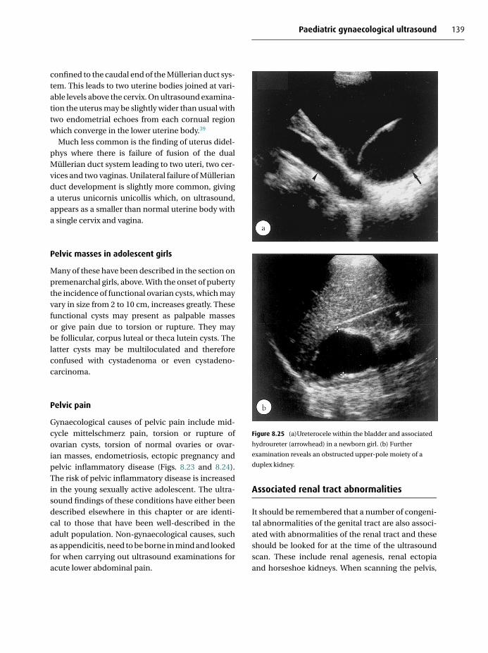
Paediatric gynaecological ultrasound 139
confined to the caudal end of the Mullerian duct sys-tem. This leads to two uterine bodies joined at vari-able levels above the cervix. On ultrasound examina-tion the uterus may be slightly wider than usual withtwo endometrial echoes from each cornual regionwhich converge in the lower uterine body.39
Much less common is the finding of uterus didel-phys where there is failure of fusion of the dualMullerian duct system leading to two uteri, two cer-vices and two vaginas. Unilateral failure of Mullerianduct development is slightly more common, givinga uterus unicornis unicollis which, on ultrasound,appears as a smaller than normal uterine body witha single cervix and vagina.
Pelvic masses in adolescent girls
Many of these have been described in the section onpremenarchal girls, above. With the onset of pubertythe incidence of functional ovarian cysts, which mayvary in size from 2 to 10 cm, increases greatly. Thesefunctional cysts may present as palpable massesor give pain due to torsion or rupture. They maybe follicular, corpus luteal or theca lutein cysts. Thelatter cysts may be multiloculated and thereforeconfused with cystadenoma or even cystadeno-carcinoma.
Pelvic pain
Gynaecological causes of pelvic pain include mid-cycle mittelschmerz pain, torsion or rupture ofovarian cysts, torsion of normal ovaries or ovar-ian masses, endometriosis, ectopic pregnancy andpelvic inflammatory disease (Figs. 8.23 and 8.24).The risk of pelvic inflammatory disease is increasedin the young sexually active adolescent. The ultra-sound findings of these conditions have either beendescribed elsewhere in this chapter or are identi-cal to those that have been well-described in theadult population. Non-gynaecological causes, suchas appendicitis, need to be borne in mind and lookedfor when carrying out ultrasound examinations foracute lower abdominal pain.
Figure 8.25 (a)Ureterocele within the bladder and associated
hydroureter (arrowhead) in a newborn girl. (b) Further
examination reveals an obstructed upper-pole moiety of a
duplex kidney.
Associated renal tract abnormalities
It should be remembered that a number of congeni-tal abnormalities of the genital tract are also associ-ated with abnormalities of the renal tract and theseshould be looked for at the time of the ultrasoundscan. These include renal agenesis, renal ectopiaand horseshoe kidneys. When scanning the pelvis,

140 Practical Gynaecological Ultrasound
abnormalities of the renal tract may cause confu-sion. Dilated tubular structures behind the bladdermay be due to dilated ureters and cyst-like struc-tures within the bladder may be due to ureteroceles.Both of these findings should lead to an examina-tion of the kidneys to look for hydronephrosis andthe presence of duplex collecting systems (Fig. 8.25).A bladder diverticulum may be misinterpreted as anadnexal cyst and a close look for a communicationwith the bladder should be made.
Conclusion
Ultrasound examination of the paediatric femalepelvis requires a thorough knowledge of the devel-opmental changes that take place in the uterus andovaries between birth and puberty. It also requiresknowledge of the various congenital abnormalitiesthat may occur and their associations with abnor-malities elsewhere within the abdomen. The mainrole of ultrasound in the evaluation of a pelvic massis to define whether the mass is cystic or solid andits organ of origin. Many of the solid pelvic masseshave similar ultrasound appearances and a specificpathological diagnosis cannot be made until the timeof surgery or biopsy.
R E F E R E N C E S
1. L. Garel, J. Dubois, A. Grignon, D. Filiatrault and G. Van Vliet,
US of the paediatric female pelvis: a clinical perspective.
Radiographics, 21 (2001) 193–407.
2. H. L. Cohen, M. A. Shapiro, F. S. Mandel and M. L. Shapiro,
Normal ovaries in neonates and infants: a sonographic study
of 77 patients 1 day to 24 months old. American Journal of
Roentgenology, 160 (1993), 583–6.
3. H. L. Cohen, P. Eisenberg, F. Mandel and J. O. Haller, Ovar-
ian cysts are common in premenarchal girls: a sonographic
study of 101 children 2–12 years old. American Journal of
Roentgenology, 159 (1992), 89–91.
4. H. J. Lee, S. K. Woo, J. S. Kim and S. J. Suh, “Daughter cyst”
sign: a sonographic finding of ovarian cysts in neonates,
infants and young children. American Journal of Roentgenol-
ogy, 174 (2000), 1013–15.
5. C. Muller-Leisse, U. Bick, K. Paulussen et al., Ovarian cysts in
the fetus and neonate – changes in sonographic pattern in
the follow-up and their management. Pediatric Radiology,
22 (1992), 395–400.
6. F. Ferro, D. Iacobelli, A. Zaccara et al., Exteriorisation – aspir-
ation mini laparotomy for treatment of neonatal ovarian
cyst. Journal of Pediatric and Adolescent Gynecology, 15
(2002), 205–7.
7. D. Tseng, T. J. Curran and M. L. Silen, Minimally invasive
management of the pre natally torted ovarian cyst. Journal
of Pediatric Surgery, 37 (2002), 1467–9.
8. C. Mittermayer, W. Blaicher, D. Grassauer et al., Fetal ovar-
ian cysts: development and neonatal outcome. Ultraschall
Medicine, 24 (2003), 21–6.
9. A. Nussbaum, A. R. Blask, R. C. Sanders and J. P. Gearhart,
Obstructed uterovaginal anomalies: demonstration with
sonography. Part I. Neonates and infants. Radiology, 179
(1991), 79–83.
10. Y. Low and J. M. Hutson, Rules for clinical diagnosis in babies
with ambiguous genitalia. Journal of Paediatric Child Health,
39 (2003), 406–13.
11. C. Sultan, F. Paris, C. Jeandel, S. Lumbroso and R. B.
Galifer, Ambiguous genitalia in the new born. Seminars in
Reproductive Medicine, 20 (2002), 181–8.
12. J. A. Blanco, C. Perez, M. Jimenez et al., Usefulness of trans-
rectal ultrasonography in the diagnosis of anomalies of
intersexual conditions. Cir Pediatr 16 (2003), 86–9.
13. L. Rangecroft, Surgical management of ambiguous genitalia.
Archives of Disease in Childhood, 88 (2003), 799–801.
14. C. Battaglia, G. Regnani, F. Mancini et al., Pelvic sonography
and uterine color Doppler analysis in the diagnosis of female
precocious puberty. Ultrasound in Obstetrics and Gynaecol-
ogy, 19 (2002), 386–91.
15. T. D. Allen, Disorders of sexual differentiation. In Clinical
Pediatric Urology, ed. P. P., Kelalis, L. R. King and A. B. Belman
(Philadelphia: Saunders, 1985), pp. 904–21.
16. C. Battaglia, F. Mancini, G. Regnani et al., Pelvic ultrasound
and color Doppler findings in different isosexual precoci-
ties. Ultrasound in Obstetrics and Gynaecology, 22 (2003),
277–83.
17. L. D. Herter, E. Golendziner, J. A. Flores et al., Ovarian and
uterine findings in pelvic sonography: comparison between
prepubertal girls, girls with isolated thelarche, and girls with
central precocious puberty. Journal of Ultrasound Medicine,
21 (2002), 1237–46.
18. W. J. Zwiebel and K. A. Murray, Imaging assessment of puber-
tal disorders. Seminars in Ultrasound, CT and MRI, 16 (1995),
296–303.

Paediatric gynaecological ultrasound 141
19. S. M. Ng, Y. Kumar, D. Cody, C. S. Smith and M. Didi, Cra-
nial MRI scans are indicated in all girls with central preco-
cious puberty. Archives of Disease in Childhood, 88 (2003),
414–18.
20. A. Cassio, E. Cacciari, S. Zucchini et al., Central preco-
cious puberty: clinical and imaging aspects. Journal of Pedi-
atric Endocrinological Metabolism, 13 (suppl. 1), (2000)
703–6.
21. L. R. King, M. J. Siegel and A. L. Solomon, Usefulness of ovar-
ian volume and cysts in female isosexual precocious puberty.
Journal of Ultrasound Medicine, 12 (1993), 577–81.
22. S. M. Freedman, P. M. Kreitzer, S. S. Elkowitz et al., Ovarian
microcysts in girls with isolated premature thelarche. Journal
of Pediatrics, 122 (1993), 246–9.
23. J. L. Breen and W. S. Maxson, Ovarian tumours in children
and adolescents. Clinics in Obstetrics and Gynecology, 20
(1977), 607–23.
24. A. Wu and M. Siegel, Sonography of pelvic masses in children:
diagnostic predictability. American Journal of Roentgenology,
148 (1987), 1199.
25. C. L. Sisler and M. J. Siegel, Ovarian teratomas: a comparison
of the sonographic Appearance in prepubertal and postpu-
bertal girls. American Journal of Roentgenology, 154 (1990),
139–41.
26. C. L. Templeman, S. P. Hertweck, J. P. Scheetz, S. E. Pearlman
and M. E. Fallat, The management of mature cystic teratomas
in children and adolescents: a retrospective analysis. Human
Reproduction, 15 (2000), 266–72.
27. J. E. Stark and M. J. Siegel, Ovarian torsion in prepubertal
and pubertal girls: sonographic findings. American Journal
of Roentgenology, 163 (1994), 1479–82.
28. J. A. Rock, Anomalous development of the vagina. Seminars
in Reproductive Endocrinology, 4 (1986), 13–31.
29. A. R. Nussbaum Blask, R. C. Sanders and J. A. Rock,
Obstructed uterovaginal anomalies: demonstration with
sonography. part II. Teenagers. Radiology, 91, 84–8.
30. L. Ballesio, C. Andreoli, M. L. De Cicco, Angeli and L.
Mangananro, Haematocolpos in double vagina associated
with uterus didelphus: US and MR findings. European
Journal of Radiology, 45 (2003), 150–3.
31. S. Leurie, M. Feinstein and Y. Mamet, Unusual presentation
of acute abdomen in a syndrome of double uterus, unilater-
ally imperforated double vagina and ipsilateral renal agen-
esis. Acta Obstetrica Gynaecologica Scandinavica, 79 (2000),
152–3.
32. C. Peironi, D. L. Rosenfeld and M. L. Mokrzycki, Uterus didel-
phys with obstructed hemivagina and ipspateral renal age-
nesis. A case report. Journal of Reproductive Medicine, 46
(2001), 133–6.
33. A. A. Massarono, J. A. Adams, M. A. Preece and C. G. D. Brook,
Ovarian ultrasound appearances in Turner syndrome. Jour-
nal of Pediatrics, 114 (1989), 568–73.
34. P. Sampaolo, V. Calcaterra, C. Klersy et al., Pelvic ultrasound
evaluation in patients with Turner’s syndrome during treat-
ment with growth hormone. Ultrasound in Obstetrics and
Gynaecology, 22 (2003), 172–7.
35. C. M. McDonnell, L. Coleman and M. R. Zacharin, A three
year prospective study to assess uterine growth in girls
with Turner’s syndrome by pelvic ultrasound. Clinics in
Endocrinology (Oxford), 58 (2003), 446–50.
36. R. J. Norman, R. W. and M. T. Stankiewicz, Polycystic ovary
syndrome. Med J Aust, 180 (2004) 132–7.
37. P. Sampaolo, C. Livieri, L. Montanari, et al., Precocious signs
of polycystic ovaries in obese girls. Ultrasound in Obstetrics
and Gynaecology, 4 (1994), 3130–5.
38. C. G. D. Brook, H. S. Jacobs and R. Stanhope, Polycystic
ovaries in childhood. British Medical Journal, 296 (1988), 878.
39. A. L. Deutch and B. B. Gosink, Non neoplastic gynecologic
disorders. Seminars in Roentgenology, 17 (1982), 269–83.
40. L. F. Orisini, S. Salardi, G. Pilu, L. Bovicelli and E. Calliari,
Pelvic organs in premenarchal girls real time ultrasonogra-
phy. Radiology, 153 (1984), 113–16.

9
Clinical management of patients:the gynaecologist’s perspective
Lynne Rogerson1, Sean Duffy1 and Chris Kremer2
1St James’s University Hospital, Leeds2Pinderfields General Hospital, Wakefield
Introduction
Probably the most significant contribution in thehistory of ultrasound within the field of obstetricsand gynaecology came from Professor Ian Donald atGlasgow in the early 1960s.1 The types of pathologyfirst identified in the uterus by pelvic ultrasonic scan-ning included hydatidiform moles2 and retainedproducts of conception.3 Kratochwil4 first describedtransvaginal ultrasonography in 1969, but it was onlyafter its evaluation of infertility in the 1980s thatits full potential was realised.5 It is now also usedin the investigation and management of infertilityand assisted reproduction, early pregnancy,6 tubalpregnancy7 and first-trimester pregnancy-relatedconditions.
Clinical use of ultrasound in gynaecology
Why do gynaecologists request ultrasound scans? Asin all clinical areas, examination skills are being lostor open to marked interobserver variation, result-ing in greater reliance on investigations as a sub-stitute. This opens up the abuse of such investi-gations with requests made with little thought asto how the investigation will help in diagnosis ormanagement. Ultrasound should in no way replacephysical examination; it should assist in building theclinical picture to contribute to appropriate patientmanagement.
There are occasions, nevertheless, where thebimanual examination is just not enough. Obesitymakes it more difficult to feel pelvic organs, andin such circumstances a transvaginal scan makesthe pelvic organs more accessible. The patient maybe virgo intacta and so pelvic examination is obvi-ously inappropriate, or in a postmenopausal womanwith atrophic tissues, examination may be very dif-ficult or sometimes impossible due to discomfort.Ultrasound can also be used to reduce the use ofmore invasive investigations such as hysteroscopyor laparoscopy and may also reduce the chance ofinvestigative major abdominal surgery.
From a clinical viewpoint the use of ultrasoundcan be roughly divided into pregnancy-related andnon-pregnancy-related. In gynaecology, pregnancy-related topics usually refer to miscarriage (moreproperly referred to as early pregnancy loss) andectopic pregnancy. Non-pregnancy-related clinicalconditions range from lost intrauterine contracept-ive devices through to postmenopausal bleeding.This chapter will concentrate on non-pregnancy-related clinical situations and helps to put into a clin-ical context the use of ultrasound in daily practice.
Non-pregnancy-related gynaecologicalconditions
It is perhaps easier to consider the need and prac-tical use of ultrasound in gynaecology when one
C© Cambridge University Press 2005.
142
Clinical management: gynaecologist’s perspective 143
Table 9.1 Causes of abnormal uterine bleeding
Organic Iatrogenic
Uterine Intrauterine contraceptive
device
Anticoagulants
Progesterones
Obesity
Fibroids
Endometriosis
Adenomyosis
Endometrial/cervical polyps
Pelvic inflammatory disease
Endometrial hyperplasia/
malignancy
Coagulopathies
Congenital deficiencies
Thrombocytopenia
Leukaemia
Systemic disorders
Hypothyroidism
Systemic lupus
erythematosis
Chronic liver failure
considers the symptom groups that are encountered.In broad terms, gynaecological patients requiringultrasound fall into four main categories: (1) thosewith symptoms relating to ‘period problems’ (abnor-mal uterine bleeding: AUB); (2) symptoms relating toinfertility; (3) symptoms or signs relating to a pelvicmass; and (4) pelvic pain. By far the commonest con-dition seen by gynaecologists is menstrual disorders.
Abnormal uterine bleeding
AUB can be divided into three main groups:(1) regular or irregular heavy bleeding (menor-rhagia/metrorrhagia); (2) intermenstrual bleeding(IMB); and (3) postmenopausal bleeding (PMB),which includes abnormal bleeding on hormonereplacement therapy (HRT).
There are many causes of AUB, ranging fromorganic to iatrogenic (Table 9.1). Once these areexcluded in the premenopausal woman, a diagno-sis of dysfunctional uterine bleeding (DUB) remains.In the postmenopausal group, 96% will have agynaecological cause for the PMB and the remain-der are non-gynaecological8 (Table 9.2).
Table 9.2 Causes of postmenopausal bleeding
Gynaecological (96%) Non-gynaecological (4%)
Malignant (12%)
Primary tumours
Secondary tumours
Per rectum or per urethram
bleeding, from
gastrointestinal or
genitourinary tract
pathology, may be mistaken
for per vaginam bleeding in
a minority of cases
Benign (88%)
Atrophic genital changes
Exogenous/endogenous
Oestrogen
Benign tumours
Infection
Injuries
Premenopausal patientsChanges in women’s lifestyles through the genera-tions have meant that they are having fewer chil-dren, are breast-feeding for shorter intervals and fre-quently have busier lives trying to balance a familylife and a career. Women therefore have more periodsthan previous generations, are less tolerant of themand seek advice more often. AUB, occurring beforethe menopause, accounts for nearly a third of refer-rals to a gynaecologist. The pattern of bleeding maybe excessive heaviness, losing clots, with or withoutflooding each month, or a totally chaotic cycle withno pattern at all.
In premenopausal women the need to investi-gate the uterus is because of the likelihood of find-ing significant intrauterine pathology which maybe the cause of the bleeding. Once found, fibroidsand polyps can be treated in the hope of resolvingthe symptoms. Women who do not have an organiccause for the bleeding, that is, no pathological basis,are classified as having DUB.
The traditional method of investigating suchwomen was by dilatation and curettage (D&C), ablind sampling technique performed under generalanaesthesia with a sharp metal curette. Hys-teroscopy, the direct visualisation of the uterine cav-ity with an endoscope, has superseded D&C9 and willidentify intrauterine pathologies in approximately45% of this group of women. In other words, 55%of women investigated by hysteroscopy will have anapparently normal uterus (DUB).

144 Practical Gynaecological Ultrasound
Figure 9.1 A typical outpatient hysteroscopy setting.
With the advances in lens technology, hystero-scopes have become smaller and more versatile andare now routinely used in the outpatient settingwith no analgesia required.10–12 Figure 9.1 shows astandard outpatient hysteroscopy set-up with thechair, and camera stack allowing the patient to watchthe hysteroscopy if she wishes. Figure 9.2 shows astandard flexible hysteroscope routinely used in theoutpatient setting.
As so many women were found to have normaluterine cavities (Fig. 9.3), this led to the recent inter-est in transvaginal ultrasound as a screening toolfor the diagnosis of normality in such women. Theadvantages are that it is a non-invasive procedureand more realistic use of resources.
Figure 9.2 A typical flexible hysteroscope.
Figure 9.3 Hysteroscopic view of a normal uterine cavity.
If normality can be used as the basis for screeningwith ultrasound, why can ultrasound not be used forall cases of AUB in premenopausal women? The limi-tation of ultrasound in the presence of pathologyis its inability to define the presence, location andcharacter of any given abnormality reliably. The sen-sitivity of transvaginal ultrasound in this situationis at best 75%.13,14 Studies that have assessed therole of ultrasound in such a group of women havefailed to justify its place in clinical practice because ofthe need for the clinician to be able to map theabnormality accurately, so that appropriate treat-ment may be provided.15

Clinical management: gynaecologist’s perspective 145
At present, therefore, ultrasound has a potentialrole in acting as a triage mechanism in womenwith DUB by screening out those patients who donot require subsequent hysteroscopy. There may beadditional benefit in the use of ultrasound in thisgroup of women because of the ability to visualisethe ovaries. Whether additional information on ovar-ian pathology, gathered during the ultrasound, is abonus, has yet to be clarified.
Management of dysfunctional uterine bleedingThe management of patients in whom no intrauter-ine pathology is discovered at investigation can bethrough either medical or surgical therapy. Med-ical therapy consists of hormonal-based or non-hormonally based drugs. If the menstrual cycle ischaotic, progesterone is often used to stabilise themenstrual loss. Alternatively, if there are no con-traindications, the combined oral contraceptive pillis excellent in controlling menstrual loss, especiallyin a younger woman who also requires contracep-tion. In a non-smoking patient the pill can be usedbeyond the age of 35. Non-hormonal therapy actsby controlling the haemostatic mechanisms in theendometrium, for example, tranexamic acid, or theprostaglandin production in the uterus, for example,mefenamic acid. Mefenamic acid is used in womenwho have a regular but heavy menstrual loss and istaken only whilst the woman is menstruating.
The Mirena intrauterine systemA more recent development, the Mirena intrauterinesystem (IUS), licensed in the UK in May 1995, was ini-tially marketed as a contraceptive device but over thelast decade it has become a very versatile treatmentoption for a variety of clinical indications and it isnow used widely throughout the UK. Potential usesof the Mirena IUS include:� contraception� fibroids� premens trial syndrome� hormone replacement therapy� endometrial hyperplasia� endometriosis� adenomyosis
16mm 32mm
Figure 9.4 The Mirena intrauterine system.
� tamoxifen users� endometrial preparation� menorrhagiaThe Mirena IUS (Fig. 9.4) comprises a T-shaped
polyethylene frame carrying a white hormone cylin-der 19 mm in length around the vertical arm, cre-ating an outer diameter of 2.8 mm. The cylindercontains a mixture of polydimethylsiloxane (50%)and levonorgestrel (LNG) (50%) and is covered bya polydimethylsiloxane membrane, which regulatesthe release of the LNG. The total amount of LNG inthe system is 52 mg. After insertion into the uterus,LNG is released from the reservoir at an initial rate of20 �g/24 hours directly to the endometrium. The T-shaped frame is impregnated with barium sulphateto make the system X-ray-detectable. Dark monofila-ment polyethylene threads are attached to the lowerend of the vertical arm.
The Mirena may be left in situ for 5 years. The thera-peutic effects of the Mirena IUS are based on localeffects in the uterus. The progesterone release has acontraceptive action that complements the mechan-ical effect of the device but it also has a direct effect onthe function of the adjacent endometrium. Endome-trial proliferation has been shown to be prevented bythe local administration of LNG, which inhibits the

146 Practical Gynaecological Ultrasound
function of oestradiol in the endometrium;16 alsoLNG thickens the cervical mucus, thus inhibitingsperm motility and function.17 Suppression of ovu-lation may occur in some women using the MirenaIUS, but on average 75% of cycles have been shownto be ovulatory.17,18 The device may also have a weakforeign-body effect.16
Patients presenting with menorrhagia whosesymptoms are refractory to conventional medicaltherapy traditionally face the choice of either under-going a hysterectomy or, more recently, alterna-tive, less invasive surgical treatment. Endometrialresection is the alternative to hysterectomy thathas been formally assessed in clinical trials.19–22
This involves a general anaesthetic, insertion ofan operative hysteroscope and the removal of theendometrial lining using a loop through whichdiathermy is passed cutting through and destroy-ing the endometrium. The benefits to the patientinclude shorter hospital stay, less postoperative mor-bidity, quicker return to work and speedier overallrecovery.
More recently, newer second-generation ablativetechniques23 have increased in both number andpopularity, having been developed to equal or exceedthe efficacy of the first-generation ablative methods,whilst simultaneously aiming to reduce the com-plications by removing the requirement for hys-teroscopic skills. The intention is for the newertechnologies to be suitable for use by general gynae-cologists who will not have undergone the lengthytraining necessary for endometrial resection. Thesetechniques use ablative methods such as microwaveand circulation of hot water in balloons. These tech-niques, unlike the traditional resection method, areblind and therefore a request for ultrasound may bemade to assess the thinnest part of the uterine wallto evaluate the risk of uterine perforation during theprocedure.
Hysterectomy is the most traditional surgicalmethod and can be performed by the abdominalroute (usually with a transverse scar in the lowerabdomen) or by the vaginal route. The reason forusing the abdominal route as opposed to the vagi-nal route was often because of operator preference,
uterine size or the presence of other pelvic pathol-ogy, such as adhesions and ovarian cysts. With theintroduction of laparoscopic-assisted vaginal hys-terectomy (LAVH), the ability to perform a vaginalhysterectomy in more cases is now possible.
Premenopausal abnormal uterine bleeding –key points� Hysteroscopy identifies pathology in about
45% of women� 55% have dysfunctional uterine bleeding withno evident pathology� Ultrasound:� is a useful screening tool, identifying pathol-
ogy such as polyps or fibroids� screens out those with a normal uteruswho would otherwise have gone on to hys-teroscopy� is limited in its ability to map the location ofabnormalities accurately and reliably char-acterise them� may give useful additional informationregarding the ovaries� Treatment options for dysfunctional uterine
bleeding include:� medical therapy� Mirena intrauterine system� endometrial resection – traditional orsecond-generation� hysterectomy
Postmenopausal patientsWomen with PMB are often anxious about the possi-bility of cancer and frequently concerned about theneed for an operation. Investigations in this group ofwomen should aim to exclude serious pathology butshould also use the investigation to give the womanthe maximum amount of information to help makedecisions when choosing management.
Women with abnormal bleeding after themenopause are a group more at risk of endometrialcancer than their premenopausal counterparts. Therisk of cancer increases with age; therefore there is

Clinical management: gynaecologist’s perspective 147
a need for more urgent investigation in this groupof patients. Once more, the traditional approach toinvestigation was by performing a D&C but latterlyhysteroscopy has taken over as the gold standard.If ultrasound is used as the screening tool in thisgroup it is essential that its accuracy in excludingpathology matches that of direct visualisation athysteroscopy, especially as hysteroscopy, as anoutpatient procedure, has become more patient-acceptable.24 Attempts at utilising ultrasound inPMB patients have centred on the measurement ofendometrial thickness. It has been suggested thatan endometrial thickness of less than 5 mm is auseful screening end-point for the determination ofnormality.25 Many studies have taken other meas-urements (< 3 and < 4 mm).26 There are a numberof problems with this approach: firstly, endometrialcancers have been identified in women whoseendometrium is less than 5 mm.27 This may meanthat a lower cut-off measurement should be used,but this would then generate an increased numberof hysteroscopies. Secondly, there is considerableintra- and interobserver variation in ultrasoundmeasurements, which would have to be taken intoaccount if universal screening becomes employed.28
The same arguments concerning the identifica-tion of abnormalities in premenopausal womenapply to postmenopausal women. The latter arestill likely to have endometrial polyps (22%) andfibroids (15%). In addition, the presence of endome-trial hyperplasia (a pathological diagnosis with arisk of coincident endometrial cancer which may befound as a localised area of thickened endometrium)may be missed.
In both these categories of patients there arecontinuing developments in ultrasound technologythat show promise in improving diagnostic accu-racy. The use of a contrast medium, in the form ofsaline, appears to enhance the view obtained, allow-ing accurate visualisation of polyps and fibroids.In addition, the saline may make the definition ofa thin endometrium easier. Doppler studies of theendometrial–myometrial interface to identify vas-cular angiogenesis have looked promising29 but havenot been widely adopted in routine practice. How-
ever the detection of feeding vessels with Dopplerhas been useful in identifying polyps.
Postmenopausal bleeding and tamoxifenA specific group of postmenopausal patients isattracting an increasing number of new referrals togynaecologists and ultrasonographers. This groupconsists of patients with breast cancer who are onlong-term tamoxifen therapy. Tamoxifen, which is apartial oestrogen antagonist, is extremely effectivein controlling breast disease. Its antagonistic action,however, is in the endometrium. This can lead toexcessive oestrogen stimulation in the endometriumand endometrial polyps, endometrial proliferationand, in some cases, endometrial cancer. The exactrelationship between tamoxifen and its endometrialeffects is currently being investigated. Tamoxifen-related polyps and endometrial changes may be dif-ficult to detect by ultrasound. Because of the tissueeffects of tamoxifen, the endometrium can simplyseem thickened, but when subsequent hysteroscopyis carried out the true nature of the lesion is seen.The polyps are often very soft and vascular withstromal thickening and oedema, and their removal isessential.
Postmenopausal abnormal uterine bleeding –key points� This group has an increased risk of endometrial
carcinoma� Hysteroscopy is the gold standard for investi-gation� Ultrasound:� can be used as a screening tool� enables measurement of the endometrial
thickness, which correlates with the presenceof pathology� the cut-off point, in millimetres, is a balancebetween generating too many hysteroscopies(if set too low) and missing abnormalities (ifset too high)� Tamoxifen may be associated with endome-trial proliferation, polyps and endometrialcancer

148 Practical Gynaecological Ultrasound
Figure 9.5 Hysteroscopic view of an endometrial polyp.
Figure 9.6 Hysteroscopic view of a fibroid (type 0).
Fibroids and polypsIn both groups of patients with AUB, fibroids andpolyps can be found at hysteroscopy and ultrasound.From the clinical viewpoint, the exact site and char-acter of these lesions are becoming important issues.There is a growing body of evidence that the removalof the lesion alone, rather than the total removalof the uterus, has a successful chance of improv-ing the patient’s symptoms.30,31 With advances inhysteroscopic surgery it is now possible to removethese lesions with the minimum of trauma and underdirect vision.
Figure 9.7 Classification of submucous fibroids. Courtesy of
the European Society of Hysteroscopy.
Polyps (Fig. 9.5) are exuberant endometrial pro-liferation and, more often than not, are benign innature. However, they can be the site of endome-trial carcinoma and as such should be removed inpostmenopausal women.32 Removal of polyps insymptomatic women is more likely to result in cessa-tion of symptoms in the postmenopausal group thanthe premenopausal group. The difficulty ultrasoundhas in the detection of polyps is in differentiatingbetween a thick endometrium and that containing apolyp.
Fibroids can be found in three places in theuterus. They may be subserosal, on the outer sur-face of the uterus, intramural, within the myome-trial wall, or submucous, impinging on the endome-trial cavity (Fig. 9.6). The former is not amenable tohysteroscopic surgery and is also unlikely to beinvolved in the evolution of the patient’s bleedingsymptoms. Fibroids in the intramural portion of theuterus may be removed by hysteroscopic resection,but it is difficult. The final site, impinging on theendometrial cavity, is the site most amenable to hys-teroscopic surgery.30 For the patient and the clini-cian, it is important to know the exact site of the sub-mucous fibroid (Fig. 9.7). How much of the fibroid

Clinical management: gynaecologist’s perspective 149
is present in the endometrial cavity as opposed tothe wall of the uterus and the size of the fibroid areimportant pieces of information. Both these issuesare taken into account during patient counsellingand are indices used to establish the ability to treat.The success rate of the procedure is also determined,preoperatively, using these indices. Patients need tobe warned of the possibility of a two-stage procedureto treat the fibroid and this can only be done if theinformation is available. To date, hysteroscopy is thebest way of obtaining this information but contrastsonography may prove useful in the future.
Fibroids and polyps – key points� Treatment options for fibroids include hystero-scopic resection or hysterectomy� Submucous fibroids are more amenable to hys-teroscopic removal than intramural fibroids.The exact location and size of the lesion areimportant in treatment planning, counsellingand in predicting the success of treatment� Polyps may be the site of endometrial carci-noma� Removal of polyps alleviates symptoms inpostmenopausal women more successfullythan in premenopausal women
Pelvic masses and gynaecological cancer
Patients are often referred specifically to investigatea pelvic mass. The most serious concern is that ofovarian carcinoma. Other reasons for a pelvic massare large fibroids, benign ovarian cysts, inflamma-tory masses and non-gynaecological causes (suchas inflammatory bowel disease, diverticular diseaseand colonic carcinoma). Usually, the patient’s symp-toms are not overt but, occasionally, there are vaguesymptoms of abdominal bloatedness or pressuresymptoms relating to bladder function.
Ultrasound plays a key role in the differentiationof pelvic masses. Large fibroids are usually readilyidentifiable. If not removed surgically by the clini-cian, subsequent monitoring of the fibroid can be
performed with further ultrasound scans in order toassess pressure on the renal tract and subsequenthydronephrosis. Ovarian cysts can be easily iden-tified by ultrasound. Features suggestive, but notindicative, of malignancy are solid and cystic compo-nents and vascular neogenesis as seen with Dopplerexamination.33 Usually, laparotomy is carried out inthe presence of a suspicious pelvic mass and the finalhistology is obtained thereafter. Subsequent follow-up of patients with malignant disease of the ovarymay occasionally be carried out by ultrasound, butCT is superior in its ability to stage and monitorspread of disease.
Malignant ovarian tumoursMalignant ovarian cysts all need to be removed atlaparotomy. The nature of ovarian cancer is that ittends to present late in the stage of the disease, andthis often means that the disease has spread beyondthe pelvis. Computed tomography (CT) is superior toultrasound in its ability to stage the disease, detect-ing spread into adjacent organs, omentum and lym-phadenopathy. It provides a good baseline for moni-toring the results of debulking surgery and the effectsof subsequent chemotherapy.
Surgery for ovarian cancer necessitates removalof as much of the malignancy as possible and usu-ally means a hysterectomy, removal of both ovariesand, in addition, removal of the omentum andappendix. The ability of postoperative chemother-apy, as needed in nearly all cases, to be effective, isto some extent determined by the volume of diseaseremaining after surgery. The smaller the volume oftissue, the better the chance of a response. The prog-nosis for ovarian cancer also depends on the stage ofthe disease; the more confined the cancer, the betterthe outcome. Despite this, because of the late pre-sentation of the disease in many women, the overallsurvival for ovarian cancer is poor. The cumulativelifetime risk of developing ovarian cancer is 1.5% andthe risk of dying from cancer is similar, 1.3%.
Benign ovarian tumoursOvarian masses that appear benign and are persist-ently greater than 4 cm in diameter may need to

150 Practical Gynaecological Ultrasound
be explored surgically with subsequent removal. Ifleft in place ovarian cysts may bleed or tort and cantherefore result in an acute episode with emergencyadmission. It is better to have the cyst removed elect-ively. Benign ovarian cysts can often be removedlaparoscopically, but sometimes there is no normalremaining tissue and in these cases the whole ovarymay need to be removed together with the cyst.
Benign ovarian cysts are more likely to be unilocu-lar and completely cystic. Physiological benign cysts,such as ovulatory cysts and corpus luteal cysts,are well recognised and, if doubt exists, may berescanned at a different stage in the cycle to ensureresolution. Usually, cysts fewer than 4 cm in diameterresolve spontaneously.
Dermoid cysts are benign cystic teratomas andhave solid features within the cyst. As there is thechance of bilateral dermoid cysts in a proportionof patients, screening of the non-affected ovary isimportant. Occult cysts, seen at ultrasound in thepresumed unaffected ovary, would warrant a carefulexamination at the time of removal of the affectedside.
Ovarian fibromas are benign, hard, solid tumourswhich generally carry no risk of malignancy. Thesemay be found incidentally, most frequently around50 years of age, and fewer than 10% of cases are bilat-eral. Most can be managed conservatively. However,surgical removal may be necessary if the lesions arelarge, causing symptoms, or in rare cases of Meigs’syndrome (1% of cases) when they cause ascites andpleural effusions.
Ovarian screeningScreening for ovarian cancer by ultrasound hasbeen investigated.34 Pelvic ultrasound in combina-tion with CA125 (a tumour marker) has been themethod used (see chapter 6). However, if this screen-ing procedure was introduced in the general pop-ulation, a huge number of people would have to bescreened to detect a very small number of malignan-cies. In addition, a substantial number of patientswould have benign cysts and would undergo laparo-tomy for a non-malignant condition. Overall, thisstrategy for screening has not been pursued because
of the above considerations. In high-risk families,who have a first-degree or close relative with ovariancancer, limited screening is available. It is importantthat this screening is done in a facility where geneticcounselling is available.
Cervical cancerCervical cancer tends to be diagnosed clinically oras a result of cervical cytology. However ultrasound,may be important in the preliminary investigation ofpatients. If ureteral involvement is suspected identi-fication of ureteral dilatation or pelvicalyceal dilata-tion would then lead to more extensive investigationusing CT.
Pelvic masses – key points� Pelvic masses may present with abdominalbloating and/or bladder symptoms� Ultrasound is the first-line investigation� Treatment options for ovarian carcinomainclude:� computed tomography for staging of carci-
noma and postoperative follow-up� laparotomy-hysterectomy, bilateral sal-pingo-oophorectomy, omentectomy andlymphadenectomy� postoperative chemotherapy.� Diagnosis of cervical cancer is usually made
clinically and with cervical cytology. Ultra-sound may identify renal tract involvement
Infertility
This is comprehensively discussed in Chapter 7.
Investigation of the infertile coupleIn routine clinical practice there are three areascausing problems with conception: (1) male factor;(2) tubal factor; and (3) anovulation. A semen ana-lysis should be obtained from all couples before anyinvasive investigations are performed on the femalepartner to establish or exclude male-factor infertil-ity. Tubal disease is best assessed by laparoscopy and

Clinical management: gynaecologist’s perspective 151
dye test, hysterosalpingography or hystero-contrastsonography.35 The diagnosis of anovulation is usu-ally made by repeat progesterone assays around day21 of a 28-day cycle. Causes of anovulation, such aspolycystic ovaries, can be readily identified by ultra-sound and characteristic features recognised.36
In women with long menstrual cycles, timing bydates may be impossible, and in these cases follicularmapping, performed serially, can be very useful indetermining the correct time to carry out the testand to time intercourse.
Monitoring of assisted conceptionUltrasound monitoring of ovarian and endometrialresponse to stimulation regimes is now establishedpractice in fertility centres. The ability to time cycles,harvest eggs and to prevent multiple pregnancies hasbeen made possible by the concomitant use of ultra-sound and hormonal monitoring, and it is essentialfor the guidance of harvest procedures, obviating theneed for laparoscopic egg retrieval.37
Ultrasound is also useful in reducing the complica-tions associated with therapy. Ovarian hyperstimu-lation, following the maturation of a large numberof follicles, is one of the most serious complicationsfor women undergoing gonadotrophin therapy, andis responsible for major maternal morbidity. Theunderlying problem of capillary leakage from thestimulated ovary can lead to ascites, pleural effu-sions, hypovolaemia and venous thrombosis. Theproblem can be avoided or reduced by withhold-ing administration of the ovulation induction agent(human chorionic gonadotrophin) when too manyfollicles are present.38
EndometriosisEndometriosis is defined as the presence of tissuethat is histologically similar to endometrium outsidethe uterine cavity and the myometrium. It is a rela-tively common disease with estimates from 2 to 50%of all women who undergo laparoscopy.
The relationship between endometrosis and infer-tility is complex and poorly understood. The preva-lence within the infertile population is between 20and 40%. The main symptoms are dysmenorrhoea,pelvic pain, dyspareunia and infertility.
The use of transvaginal ultrasound has improvedthe diagnosis of clinically undetectable ovarian cystsbut it has a poor sensitivity.
The wide range of presentations of endometriosisallows a variety of treatment options to be considereddepending on age, need for contraception, repro-ductive status and so on. The main goals of treatmentare to relieve symptoms and restore fertility wherenecessary. Treatment includes medical options suchas the combined oral contraceptive pill, progesto-gens, danazol and gonadotrophin-releasing hor-mone analogues. Surgical options include laparo-scopic diathermy to endometriosis, excision, ovariancystectomy or hysterectomy and oophorectomy.
The acute pelvis
Female patients admitted via casualty with a historyof acute lower abdominal pain are often assessedby gynaecologists. The first diagnosis to exclude orconsider is ectopic pregnancy, which is discussedunder pregnancy-related gynaecological conditions,below.
Pelvic inflammatory diseaseThe second most common condition to consider ispelvic inflammatory disease. The symptoms mostoften described in pelvic infection are bilateral lowabdominal pain and a vaginal discharge. Patientsare not always pyrexial. Examination usually revealsbilateral iliac fossa tenderness, sometimes withguarding, with both adnexal regions usually tenderto palpation. Vaginal examination may reveal a cervi-cal discharge, but not always. The diagnosis is basedon the clinical symptoms and confirmatory bacter-ial swabs; however, ultrasound can be used to cor-roborate the findings and, more usefully, to excludeother pathology. The finding of fluid in the pouchof Douglas has been used to help confirm a clin-ical suspicion of pelvic inflammatory disease39 butmay also be found in a ruptured ectopic pregnancy.A pelvic abscess has characteristic appearances onultrasound and the patient is often quite toxic.
Patients with a suspected diagnosis of pelvicinflammatory disease are usually treated with antibi-

152 Practical Gynaecological Ultrasound
otics, after bacterial swabs have been taken fromthe endocervix and the high vagina. Registrationwith a genitourinary clinic with contact tracing isalso an important part of the patient’s manage-ment. Occasionally, if the patient’s symptoms do notrespond to conservative antibiotic therapy, a diag-nostic laparoscopy is carried out to ensure there isno pelvic collection or abscess formation. In patientswith a pelvic abscess, intravenous antibiotics are alsoimportant but the abscess should be drained sur-gically at laparoscopy or via ultrasound guidancewhere amenable, in order for symptoms to resolve.
Ovarian cyst accidentsPain low in the pelvis may also be associated withovarian cysts. The commonest cysts to cause suchsymptoms are corpus luteal cysts, which have rup-tured, usually postcoitally. Ovarian cysts may alsobecome twisted on their pedicle (torted) or bleedinternally into the substance of the cyst itself.
Patients who are admitted with a history of acutepelvic pain and who have a cyst diagnosed at subse-quent ultrasound may need to undergo laparoscopyto assess the nature of the cyst and treat it accordinglyif the pain does not settle with conservative man-agement. Often a haemorrhagic cyst may requireremoval, together with the ovary, if it is affected. Atorted ovarian cyst will need to be removed but theovary may be conserved if it has not been affected bythe interruption in its blood supply; in other words,if it has not become ischaemic. Laparoscopic treat-ment rather than laparotomy should be possible inthe majority of cases.
Fibroid red degenerationPedunculated fibroids on the surface of the uteruscan also be a source of pain because of torsion orred degeneration. The latter is most often associatedwith pregnancy and occurs as a result of the fibroidoutgrowing its blood supply, with subsequent haem-orrhage into itself. Fibroid red degeneration can bevery difficult to manage, as the only option is painrelief until the pregnancy reaches a suitable gesta-tion to deliver the baby.
The acute pelvis – key points� High index of suspicion for ectopic pregnancy� Pelvic inflammatory disease (PID) is a com-mon cause of lower abdominal pain and maybe accompanied by vaginal discharge� Diagnosis of PID clinically is confirmed by bac-terial swabs and ultrasound scan� Treatment is with antibiotics and subsequentdrainage of abscess if symptoms persist� Ovarian cysts are assessed laparoscopicallyand may be removed if haemorrhagic or torted
Pregnancy-related gynaecologicalconditions
In most gynaecology units throughout the UK, theintroduction of early-pregnancy assessment unitshas advanced the early diagnosis and managementof complications of early pregnancy. Such unitsincorporate a dedicated team of nursing staff, famil-iar with early-pregnancy protocols, along with qual-ified ultrasonographers to perform the scanning. Amultidisciplinary approach is recommended for thecare of these women as it can obviously be a verydifficult time, with uncertainty about the viability oftheir pregnancy.
Ectopic pregnancy
The incidence of ectopic pregnancy has shown a con-sistent increase during the last two or three decades,in particular with the increase in assisted concep-tion techniques. However, it is difficult to comparethe various publications because ectopic pregnancyis seldom described in absolute numbers. Most oftenit is expressed as an overall rate, such as ectopicpregnancy per 1000 live births. The current incidencerate for ectopic pregnancy in western societies is 11.5per 1000 pregnancies. In the UK, ectopic pregnancyaccounts for about 10% of maternal deaths and is thefifth commonest cause of direct maternal death.40
In the western world as a whole, ectopic pregnancy

Clinical management: gynaecologist’s perspective 153
is the major cause of maternal mortality in the firsttrimester.
Ectopic pregnancy is often difficult to diagnoseand a high index of suspicion should be maintainedwhen a woman of reproductive age complains ofabdominal pain. In current practice, in patients withabdominal pain, the diagnosis is only definitive infewer than 50% of cases. Ectopic pregnancy maymimic the symptoms of other gynaecological con-ditions, such as pelvic inflammatory disease, rup-tured corpus luteum, DUB and incomplete abor-tion. Abdominal pain is the commonest symptomand is frequently present even before rupture. Amen-orrhoea is reported in 75–95% of patients. The lastmenstrual period is often described as lighter thannormal and may occur earlier or later than expected.The most common physical sign is abdominal ten-derness, often associated with rebound tenderness.
The role of ultrasonography in suspected ectopicgestation is to attempt to localise the pregnancy. If thepregnancy is found to be intrauterine, ectopic preg-nancy is virtually excluded because the combinationof intrauterine and ectopic pregnancy is extremelyrare in normal conceptions (1 in 30 000), although instimulated in vitro fertilisation pregnancies the het-erotopic pregnancy rate is higher. The interpretationof ultrasound findings is dependent on the accurateknowledge of the duration of amenorrhoea.
It is well known that more than a third of patientswith ectopic pregnancy do not know the date oftheir last menstrual period, and in others irregularbleeding may lead to confusion. If no gestationalsac or pregnancy is seen on transabdominal ultra-sound and a pregnancy test is positive, an ectopicpregnancy is highly likely.
A major advantage of transvaginal sonography isthat it can diagnose normal or failed intrauterinepregnancy at least 1 week earlier than by transab-dominal sonography.41 In particular, when tryingto differentiate between a pseudogestational sac, asoccurs with ectopic pregnancy, and a normal earlyintrauterine gestational sac, the transvaginal probeis more useful than a transabdominal scan.
Ectopic pregnancy is a potentially dangerous con-dition that should be managed with the utmost
of care. Initial expectant management with beta-human chorionic gonadotrophin monitoring is pos-sible in women who are haemodynamically stableand where there is the possibility that the pregnancyis situated in the uterus. There are guidelines for thisapproach; when a suboptimal rise in the pregnancyhormone occurs the patient should undergo a diag-nostic laparoscopy.
At laparoscopy the ectopic pregnancy can eitherbe removed on its own after opening the fallopiantube (salpingostomy) or by removing of the ectopicand the fallopian tube (salpingectomy). Some cen-tres are injecting the ectopic with methotrexate orhyperosmolar glucose to kill the pregnancy, and thismay prove useful in the future. Most surgery shouldnow be carried out laparoscopically with a reductionin postoperative pain and earlier recovery rates thanwhen surgery is performed by laparotomy.
Cystic adnexal masses and free fluid in the pouchof Douglas are other sonographic signs which shouldraise the index of suspicion of the presence of a pos-sible ectopic gestation. However, it is very difficultto differentiate between the other numerous possi-ble underlying conditions such as pelvic inflamma-tory disease, ovarian cysts and endometriosis, all ofwhich may mimic an ectopic pregnancy. In addition,it has been reported that the adnexal mass, as seenultrasonically, can be on the contralateral side of theectopic pregnancy in about 30% of cases, and nor-mal adnexal findings on ultrasound have been seenin 20% of women with laparoscopically confirmedectopic pregnancy. Overall, the use of sonographyas a diagnostic test in suspected ectopic pregnancyallows an accurate diagnosis as to the presence orabsence of an ectopic gestation in approximately70–90% of affected cases.
Miscarriage
Bleeding in early pregnancy is a considerable sourceof distress to the patient. The concerns regardingthe continuing viability of a pregnancy are very realand should be allayed as soon as possible. In thepast, bleeding, was often a reason for immediateadmission to a gynaecological ward but, with the

154 Practical Gynaecological Ultrasound
introduction of early-pregnancy assessment units(EPAU) throughout the UK, the management is nowcarried out in outpatients. Ultrasound availabilityhas played a critical role in the development of theseunits. Patients and general practitioners can now, atshort notice, have an assessment of the uterus in theevent of bleeding. Ultrasound helps clarify whethera pregnancy is ongoing or not.
Types of miscarriage vary, and can be categorisedon history, examination and ultrasound findings.A complete miscarriage means the conception hasbeen lost completely. The patient will often havea history of heavy bleeding with clots followed bycrampy abdominal pain with subsequent reductionin both the vaginal loss and pain. On ultrasound theuterus should be empty. In cases of incomplete mis-carriage, ultrasound will identify remaining prod-ucts of conception. A missed miscarriage, wherethe pregnancy has stopped developing and only thegestational sac remains, also has a characteristicappearance.
Trophoblastic diseaseOccasionally a pregnancy which has become com-plicated by excessive trophoblastic proliferation ispicked up at a routine pregnancy scan in the firsttrimester. These molar pregnancies can be easilytreated by evacuation of the uterus and, occasion-ally, methotrexate therapy. The classical appear-ances on ultrasound are of an enlarged uterusfilled with echogenic material in the early stages,developing easily visible cystic vesicles as the moleprogresses, which considerably increases throughtransmission of the beam. There are a handful of cen-tres throughout the UK that follow up all patientswith molar pregnancies and maintain a nationaldatabase. They monitor the urinary beta-humanchorionic gonadotrophin levels to ensure they returnto normal levels and do not proceed to chorionic car-cinoma.
In women who miscarry recurrently (defined asmore than three times) the early ultrasound findingsof a normally situated and viable pregnancy pro-vide the woman with a 95% chance of an eventual
good outcome. The reassurance value of such a non-invasive intervention early in pregnancy is signifi-cant. A randomised trial of early scanning and activereassurance found a significant benefit, in terms ofpregnancy outcome, to those women who receivedsupportive care by scanning and reassurance com-pared with those who did not.42
The future
The quality and definition of ultrasound technologyare improving all the time. As our understandingincreases, the scope for incorporation into clinicalpractice will inevitably improve. Saline infusion hys-terosonography (SIH) is a concept attracting increas-ingly innovative practice with obvious clinical use-fulness. Saline is infused into the uterine cavityto enhance visualisation of its contents during atransvaginal ultrasound examination.43–50 Althoughinitially used to investigate tubal patency,51 SIHrapidly evolved into an investigation of the uterinecavity. The advantage of SIH is that it is relatively non-invasive compared to hysteroscopy, it is performedduring transvaginal ultrasound and it can thereforealso identify ovarian pathology.52
The use of ultrasound is also likely to becomeincreasingly established as a guidance mech-anism for minimally invasive therapies, increasingpatient choices and improving gynaecologicalmanagement.
R E F E R E N C E S
1. I. Donald, Use of ultrasonics in the diagnosis of abdominal
swelling. British Medical Journal, 2 (1963), 1154–5.
2. J. MacVicar and I. Donald, Sonar in the diagnosis of
early pregnancy and its complications. Journal of Obstet-
rics and Gynaecology of the British Commwealth, 70 (1963),
387–95.
3. H. P. Robinson, Sonar in the puerperium, a means of diag-
nosing retained products of conception. Scottish Medical
Journal, 17 (1972), 364–6.
4. A. Kratochwil, Ein neues vaginales Schnittbildverfahren.
Geburtshilfe Frauenheilkd, 29 (1969), 379–85.

Clinical management: gynaecologist’s perspective 155
5. S. R. Schwimer and J. Lebovic, Transvaginal pelvic
ultrasonography: accuracy in follicle and cyst size determin-
ation. Journal of Ultrasound in Medicine, 4 (1985), 61–3.
6. S. R. Goldstein, J. R. Snyder, C. Watson and M. Danon, Very
early pregnancy detection with endovaginal ultrasound.
Obstetrics and Gynecology, 72 (1988), 200–4.
7. A. Rempen, Vaginal sonography in ectopic pregnancy: a
prospective evaluation. Journal of Ultrasound in Medicine,
7 (1987), 381–7.
8. Y. C. Choo, K. C. Mak, C. Hsu, T. S. Wong and H. K. Ma, Post-
menopausal uterine bleeding of non organic cause. Obstet-
rics and Gynecology, 66 (1985), 225–8.
9. M. H. Goldrath and A. I. Sherman, Office hysteroscopy and
suction curettage: can we eliminate the hospital diagnostic
dilatation and curettage? American Journal of Obstetrics and
Gynecology, 152 (1985), 220–9.
10. L Rogerson and S. Duffy, National survey of outpatient hys-
teroscopy. Gynaecology and Endoscopy, 10 (2001), 343–7.
11. C. Kremer, S. Barik and S. Duffy, Flexible outpatient hys-
teroscopy without anaesthesia: a safe, successful and well
tolerated procedure. British Journal of Obstetrics and Gynae-
cology, 105 (1998), 672–6.
12. C. Kremer, S. Duffy and M. Moroney, Patient satisfaction
with outpatient hysteroscopy versus day case hysteroscopy:
randomised controlled trial. British Medical Journal, 320
(2000), 279–82.
13. B. Cacciatore, T. Ramsay, P. Lehtovirta and P. Ylostalo,
Transvaginal sonography and hysteroscopy in post-
menopausal bleeding. Acta Obstetrica Gynecologica Scan-
dinavica, 73 (1994), 413–16.
14. Z. Weiner, D. Beck, S. Rottem, J. Brandes and I. Thaler, Uter-
ine artery flow velocity waveforms and color flow imaging in
women with perimenopausal and postmenopausal bleed-
ing. Acta Obstetrica Gynecologica Scandinavica, 72 (1993),
162–6.
15. E. Cicinelli, F. Romano, P. S. Anastasio, et al. Transabdomi-
nal sonohysterography, transvaginal sonography, and hys-
teroscopy in the evaluation of submucous myomas. Obstet-
rica Gynecologica, 85 (1995), 42–7.
16. S. G. Silverberg, M. Haukkamaa, H. Arko, C. G. Nilsson
and T. Luukkainen, Endometrial morphology during long-
term use of levonorgestrel-releasing intrauterine devices.
International Journal of Gynecological Pathology, 5 (1986),
235–41.
17. I. Barbosa, O. Bakos, S. E. Olsson, V. Odlind and E. D. B.
Johansson, Ovarian function during use of a levonorgestrel-
releasing IUD. Contraception, 42 (1990), 51–66.
18. C. G. Nilsson, P. Lahteenmaki and T. Luukkainen, Ovar-
ian function in amenorrhoeic and menstruating users of
a levonorgestrel-releasing intrauterine device. Fertility and
Sterility, 41 (1984), 52–5.
19. M. J. Gannon, E. M. Holt, J. Fairbank et al., A ran-
domised controlled trial comparing endometrial resection
and abdominal hysterectomy for the treatment of menor-
rhagia. British Medical Journal, 303 (1991), 1362–4.
20. N. Dwyer, J. Hutton and G. M. Stirrat, Randomised con-
trolled trial comparing endometrial resection with abdomi-
nal hysterectomy for the surgical treatment of menorrhagia.
British Journal of Obstetrics and Gynaecology, 100 (1993),
237–43
21. S. B. Pinion, D. E. Parkin, D. R. Abramovich et al., Ran-
domised controlled trial of hysterectomy, endometrial laser
ablation, and transcervical resection for dysfunctional uter-
ine bleeding. British Medical Journal, 309 (1994), 979–83.
22. H. O’Connor, J. A. M. Broadbent, A. Magos and K. McPher-
son, Medical Research Council randomised controlled trial
of endometrial resection versus hysterectomy in manage-
ment of menorrhagia. Lancet, 349 (1997), 897–901.
23. C. Kremer and S. D. Duffy, Endometrial ablation: the next
generation. British Journal of Obstetrics and Gynaecology,
107 (2000), 1443–52.
24. F. Nagele, H. O’Connor, A. Davies, et al., 2500 outpatient
diagnostic hysteroscopies. Obstetrics and Gynecology, 88
(1996), 87–92.
25. M. N. Nasri, J.H. Shepherd, M.E. Setchell, D.G. Lowe and
T. Chard, The role of vaginal scan in measurement of
endometrial thickness in postmenopausal women. British
Journal of Obstetrics and Gynaecology, 98 (1991), 470–5.
26. S. Granberg, M. Wikland, B. Karlsson, A. Norstrom and
L. Friberg, Endometrial thickness as measured by endovagi-
nal ultrasonography for identifying endometrial abnormal-
ity. American Journal of Obstetrics and Gynecology, 164
(1991), 47–52.
27. A. Dorum, B. Kristensen, A. Langebrekke, T. Sornes and
O. Skaar, Evaluation of endometrial thickness measured by
endovaginal ultrasound in women with postmenopausal
bleeding. Acta Obstetrica Gynecologica Scandinavica, 72
(1993), 116–19.
28. B. Karlsson, S. Granberg, B. Ridell and M. Wikland, Endome-
trial thickness as measured by transvaginal sonography:
interobserver variation. Ultrasound in Obstetrics and Gyne-
cology, 4 (1994), 320–5.
29. T. H. Bourne, S. Campbell, C. V. Steer et al., Detection of
endometrial cancer by transvaginal ultrasonography with
color flow imaging and blood flow analysis: a preliminary
report. Gynecology and Oncology, 40 (1991), 253–9.
30. S. G. Derman, J. Rehnstrom and R. S. Neuwirth, The long-
term effectiveness of hysteroscopic treatment of menorrha-
156 Practical Gynaecological Ultrasound
gia and leiomyomas. Obstetrics and Gynecology, 4 (1991),
591–4.
31. K. Wamsteker, M. H. Emanuel and J. H. De Kruif, Trans-
cervical hysteroscopic resection of submucous fibroids for
abnormal uterine bleeding: results regarding the degree of
intramural extension. Obstetrics and Gynecology, 82 (1993),
736–40.
32. B. Petterson, H. O. Adami, A. Lindgren and I. Hesselius,
Endometrial polyps and hyperplasia as risk factors for
endometrial carcinoma. Acta Obstetrica Gynecologica Scan-
dinavica, 64 (1985), 653–9.
33. U. J. Herrmann, G. W. Locher and A. Goldhirsch, Sono-
graphic patterns of ovarian tumours. Prediction of malig-
nancy. Obstetrics and Gynecology, 69 (1987), 777–81.
34. M. J. Webb, Screening for ovarian cancer. Still a long way to
go. British Medical Journal, 306 (1993), 1015–16.
35. S. Campbell, T. H. Bourne, S. L. Tan and W. P. Collins,
Hysterosalpingo contrast sonography (hycosy) and its
future role within the investigation of infertility in
Europe. Ultrasound in Obstetrics and Gynecology, 4 (1994),
245–53.
36. A. H. Balen, J. S. E. Laven, S. L. Tan and D. Dewailly,
Ultrasound assessment of the polycystic ovary: interna-
tional consensus definitions. Human Reproduction Update,
9 (2003), 1–10.
37. P. Dellenbach, P. Nisand, L. Moreau et al., Transvaginal
sonographically controlled ovarian follicle puncture for egg
retrieval. Lancet, 1 (1984), 1467.
38. P. R. Brinsden, I. Wada, S. L. Tan, A. Balen and H. S. Jacobs,
Diagnosis, prevention and management of ovarian hyper-
stimulation syndrome. British Journal of Obstetrics and
Gynaecology, 102 (1995), 767–72.
39. S. Kupesic, A. Kurjak, L. Pasalic, S. Benic and M. Ilijas, The
value of transvaginal color Doppler in the assessment of
pelvic inflammatory disease. Ultrasound Medicine and Biol-
ogy, 21 (1995), 733–8.
40. Department of Health, Why Mothers Die. Report on Confi-
dential Enquiries into Maternal Deaths in the United King-
dom 1994–1996 (London: TSO, 1998).
41. I. Stabile and J. G. Grudzinskas, Ectopic pregnancy: what’s
new? In J. Studd, ed. Progress in Obstetrics and Gynaecology,
vol. 11 (Edinburgh: Churchill Livingstone, 1995), pp. 281–
309.
42. H. S. Liddell, N. S. Pattison and A. Zanderigo, Recurrent mis-
carriage – outcome after supportive care in early pregnancy.
Australia and New Zealand Journal of Obstetrics and Gyne-
cology, 31 (1991), 320–2.
43. J. Van Roessel, K. Wamstaker and N. Exalto, Sonographic
investigation of the uterus during artificial uterine cavity
distension. Journal of Clinical Ultrasound, 15 (1987), 439–
50.
44. C. H. Syrop and V. Sahakain, Transvaginal sonographic
detection of endometrial polyps with fluid contrast aug-
mentation. Obstetrics and Gynecology, 79 (1992), 1041–3.
45. F. Bonilla-Munsoles, C. Simon, V. Serra, M. Sampaio and
A. Pellicer, An assessment of hysterosalpingosonography
(HSSG) as a diagnostic tool for uterine cavity defects and
tubal patency. Journal of Clinical Ultrasound, 20 (1992),
175–81.
46. A. K. Parsons and J. J. Lense, Sonohysterography for
endometrial abnormalities: preliminary results. Journal of
Clinical Ultrasound, 21 (1993), 87–95.
47. J. R. Cohen, D. Luxman, J. Sagi et al., Sonohysterography for
distinguishing endometrial thickening from endometrial
polyps in postmenopausal bleeding. Ultrasound in Obstet-
rics and Gynecology, 4 (1994), 227–80.
48. S. R. Goldstein, Use of ultrasonohysterography for triage of
perimenopausal patients with unexplained uterine bleed-
ing. American Journal of Obstetrics and Gynecology, 170
(1994), 565–70.
49. T. J. Dubinsky, H. R. Parvey, G. Gormaz, M. Curtis and
N. Maklad, Transvaginal hysterosonography: comparison
with biopsy in the evaluation of post-menopausal bleeding.
Journal of Ultrasound in Medicine, 14 (1995), 887–93.
50. T. J. Dubinsky, H. R. Parvey, G. Gormaz and N. Maklad,
Transvaginal hysterosonography in the evaluation of small
endoluminal masses. Journal of Ultrasound in Medicine, 14
(1995), 1–6.
51. T. S. Richman, G. N. Viscomi, A. de Cherney, M. L. Polan
and L. O. Alcebo, Fallopian tubal patency assessed by ultra-
sound following fluid injection. Radiology, 152 (1984), 507–
10.
52. L. Rogerson, J. Bates, M. Weston and S. Duffy, Outpa-
tient hysteroscopy versus saline infusion hydrosonography
(SIH). British Journal of Obstetrics and Gynaecology, 109
(2002), 800–4.

Index
Note: page numbers in italics refer to figures and tables
ablative techniques, second-generation 146
abscess
pelvic 136, 151
pericolic 115
tubo-ovarian 97, 107
acoustic characteristics 25–6
adenomyosis 67, 68
adnexal mass
cystic 153
pedunculated fibroid 92
adnexal pathology
clinical presentation 79
fallopian tubes 96–8
patient details 79–80
adnexal torsion 111–13
premenarchal girls 134–5
adolescence
gynaecological problems 135–9
pelvic masses 139
polycystic ovary syndrome 138
adrenal hyperplasia, late-onset 132
adrenal tumours, hirsutism 132
adrenarche, premature 132
age, ovary/uterus changes 42–4
aliasing 10
amenorrhoea
absent ovaries 85–6
primary 135, 137
absence of sexual character development 136–7
secondary 138
androgen insensitivity 137–8
angiogenesis 49
anovulation, diagnosis 151
antibiotics 151–2
157

158 Index
anti-oestrogen therapy 55
appendicitis 99, 98–9, 113–14, 139
acute 113, 114
appendicolith 113
arcuate arteries 50
artefacts 25
Asherman’s syndrome 68, 67–8
axial resolution 2–3
beamwidth 3
focusing 3
biopsy probe attachment 29
bladder 41–2, 130
cervical carcinoma involvement 74
distended 19–20
diverticulum 139–40
full 21
tumours 100, 99–100
bone cyst, aneurysmal 133
bowel-related pathology 99
BRCA1 and 2 genes 91
breast cancer, metastatic 86, 95–6
breasts, isolated development 132
Brenner tumours 91
broad ligaments 40, 40
fibroids 98
CA125 96, 150
caesarean section
scar 67
uterus postoperative condition 67
calculi, ureteric 99–100
cervical carcinoma 64, 71–3, 150
bladder involvement 74
diagnosis 71
staging 72
ultrasound appearance 72–3
cervical polyp 71
cervical stenosis 73–4
cervix 69–75
infant 128
Chlamydia infection 106
choriocarcinoma 65, 66, 154
ovarian hyperstimulation 85
cloaca malformation 130
clomifene 123
coccygeus muscle 41
cogwheel sign, pelvic inflammatory disease 97,
108
colitis 99, 114–15
see also Crohn’s disease
colon inflammation 115
colorectal cancer 99
colour Doppler, ovarian torsion 112
colour flow mapping (CFM) Doppler systems 10–11, 12
conception, assisted 151
congenital androgen insensitivity syndrome (CAIS) 137–8
contrast agents 11–12
ovarian carcinoma 93–5
saline 147
contrast resolution 7
corpus luteum 52
cysts 80–1
Crohn’s disease 99, 114, 114–15, 116
curved array probe 17–19
curvilinear array 7
paediatric ultrasound 126
Cushing’s syndrome
hirsutism 132
cystadenocarcinoma, serous 132
cystadenoma
mucinous 87–8
serous 86–7, 132
3D ultrasound of endometrium 77
depth of image 15
dermoid cysts 150
dermoid plug 88
dermoid tumours 88, 89
ovarian 88
diverticulitis 99, 99, 115, 115
pericolic abscess 115
Doppler ultrasound 9
colour 112
colour flow mapping (CFM) systems 10–11, 12
endometrium 124
follicles 124
ovarian arteries 52
techniques 26–9, 49–52
uterine arteries 49–52, 132
vascular angiogenesis 147
downregulation 123
dysgerminoma 133–4
Early Pregnancy Assessment Units (EPAU) 153
echo heights 7
embryo transfer 123
endometrial carcinoma 63, 64, 65

Index 159
endometrial thickening 64
polyp progression 148
risk 63, 146–7
staging 63
tamoxifen 147
ultrasound findings 63
endometrial cavity
fluid 74–5, 106–8
saline contrast hysterosonography 61
endometrial cycle 121–2
endometrial polyps 61–2
with feeding vessel 62
fibroid differential diagnosis 56
hysteroscopy 148
vascular supply 61
endometrial resection 146
endometrioma 84
endometriosis 84, 83–5, 117, 125
distribution 84–5
infertility 151
pelvic pain 116, 139
presentation 151
treatment 151
endometritis 106
acute 66
hysteroscopy 107
pelvic inflammatory disease 106–8
endometrium 35, 44, 77
acoustic properties 34
adenomyosis 67
3D ultrasound 77
disease 59–69
Doppler studies 124
echo 49, 60
cavity 22
texture 59–61
ectopic pregnancy 104
hormone replacement therapy effects 64–5
hyperplasia 62–3
inflammatory conditions 66
menstruation 49
metaplasia 65
MRI 77
normal appearance 59
oral contraceptive effects 64–5
ovulatory stage 24
polyps 147
postmenopausal 63
proliferation 147
tamoxifen effects 63, 65, 66
thickening 65
due to polyp 60
ectopic pregnancy 104
thickness 59–61, 121–2
measurement 62
ultrasound appearance 45–9, 65
epididymal tumours 122–3
equipment selection 1–14
fallopian tubes 38, 38–9, 96–8
carcinoma 98
HyCoSy 39
inflammation 106
patency testing 122
pelvic inflammatory disease 138
fertility 120–5
fibroids
broad ligament 98
cervical 70, 70–71, 71
haemorrhage 116
hysteroscopy 148
uterine 55, 56, 55–8, 125
calcified 56
circumferential vessels 57
clinical symptoms 55–6
cornual 58
degenerating 57
differential diagnosis 57–8
endometrial cavity relationship 58
haemorrhage 117
large 149
location 148–9
myometrial 148–9
pedunculated 55, 57, 92, 91
postmenopausal women 147, 148
red degeneration 55, 57, 152
reflectivity 56
submucous 57, 58, 148, 148–9
subserosal 91, 148–9
ultrasound appearance 56–7
ultrasound report 56
uterine artery resistance 50
fibroma, ovarian 90, 90, 150
twisted 112
field of view 17, 19
extended 17, 18
fimbral cysts 81
Fitz-Hugh Curtis syndrome 106

160 Index
focal zone 15, 16
focusing 3
follicles 49
antral 120–1
oestrogen secretion 121–2
development 121, 128
in puberty 128
Doppler studies 124
growth 123
number in ovaries 123
primordial 120
development 128–9
rupture 128–9
size 123
follicle-stimulating hormone (FSH) 44
antral follicle size 120–1
clomifene stimulation 123
inhibition 123
folliculogenesis, disordered 138
footprint 7
frame rate 4, 15, 16
frequency 16, 17
gastrointestinal tract, metastases from malignancy 95–6
genital tract
anomalies 76
obstruction 135–7
genitalia, ambiguous 130–1
germ cell tumours, ovarian 88
gestational trophoblastic disease 65–6
gonadal dysgenesis 130, 137
gonadotrophin-releasing hormone (GnRH) antagonists 55
gonadotrophins, ovarian hyperstimulation 151
granulosa cell tumours, ovarian 90, 133, 134
grey levels 7
gynaecological cancer 149–50
gynaecological conditions
non-pregnancy-related 142–52
pregnancy-related 152–4
haematocolpos 76, 76, 135–6, 137
haematometrocolpos 136, 135–6
haemoperitoneum 110
harmonic imaging 8–9
hermaphrodites, true 130
herpes simplex virus type 2 (HSV2) 71
hirsutism 132
hormone replacement therapy, endometrial effects
64–5
beta-human chorionic gonadotrophic hormone (�hCG) 65, 66,
98
ectopic pregnancy 105, 153
trophoblastic disease 154
human chorionic gonadotrophin (hCG)
ovulation triggering 123
human papillomavirus (HPV) 71
hydatiform mole, ovarian hyperstimulation 85
hydrocolpos 130
hydrometra 72, 73, 74
hydrometrocolpos 130
hydronephrosis 139–40, 149
hydrosalpinx 97, 109, 108–9, 125
torsion 109
tortuous 110
twisted 112
hydroureter, neonatal 139
hymen, imperforated 117
pelvic pain 116–17
hyperandrogenism 138
hysterectomy
menorrhagia 146
ovarian tumours 86
postoperative haemorrhage 115–16
uterus postoperative condition 67
hystero-contrast sonography (HyCoSy) 39, 39, 122
hysterosalpingography 67–8
contrast 35
X-ray 39
hysteroscope 144
hysteroscopic polypectomy 60–1
hysteroscopy 67–8
dysfunctional uterine bleeding 143–4, 146–7, 148
endometrial polyps 62, 148
endometritis 107
fibroids 148
outpatient 144
pelvic inflammatory disease 107
uterus 144
hysterosonography, saline contrast 29, 29, 29, 65
endometrial cavity 61
iliac artery 50
aneurysms 99–100
internal 28, 38
iliac vein, internal 38
image optimisation 4, 15–17
infants, young, normal anatomy 126–7
infertility 150–1

Index 161
adnexal pathology 79
endometriosis 83–5, 151
investigations 150–1
male factor 150
pelvic inflammatory disease 97
programmes 57
techniques using ovarian hyperstimulation 85
inflammatory bowel disease 100
inflammatory conditions
endometrial 66
fallopian tube 106
pelvic inflammatory disease 97, 106
interpretation of results 54–63
intersex
female 130
male 130–1
intrauterine contraceptive device (IUCD) 68–9, 69
removal 106
in vitro fertilisation (IVF) 123
junctional zone 121–2
kidney anomalies 139–40
Krukenberg tumour 95–6
laparoscopic-assisted vaginal hysterectomy (LAVH) 146
laparoscopy, neonatal ovarian cysts 129
lateral resolution 3
leiomyomata see fibroids
leiomyosarcoma 58–9
leukaemia, ovarian involvement 96, 133
levator ani muscle 41
levonorgestrel 145
ligaments, uterine 39–40
line density 15
linear array 7, 19
lipoma 59
luteinizing hormone (LH) 44
inhibition 123
oversecretion 138
lymphoma
ovarian involvement 96
uterine 59
McCune–Albright syndrome 132
magnetic resonance imaging (MRI)
bicornuate uterus 35
cervical carcinoma staging 72
endometrium 77
fibroids 58
ovarian tumours 86
pelvic inflammatory disease 106
mefenamic acid 145
Meigs syndrome 90, 133
ovarian fibroma 150
menorrhagia 138, 145, 146
menstrual cycle 44–5
adnexal pathology 79
menstruation 44
endometrial disease 59
endometriosis 83–5
endometrium 49
problems 135
mesovarium 37
metastases
breast cancer 86, 95–6
GI tract malignancy 95–6
ovaries 95–6
uterine 59
methotrexate 154
minilaparotomy 129
Mirena intrauterine system 68, 145, 145–6
miscarriage 153–4
categories 154
recurrent 154
mittelschmerz, pelvic pain 139
molar pregnancy 65, 154
mole, invasive 65
Mullerian carcinoma, mixed 59
Mullerian duct, unilateral failure of development
139
musculature, pelvic 41
myometrium
acoustic properties 34
adenomyosis 67
echo texture 60
endometrial carcinoma invasion 63
scanning 55–9
thickness 63
nabothian cysts 70, 69–70
neonates
hydroureter 139
normal anatomy 126–7
ovarian cysts 129
ovaries 127
pathology 128–31
ureterocele 139

162 Index
neonates (cont.)
uterus 127
anatomy 126–7
neuroblastoma 133
obesity, hirsutism 132
obturator internus muscle 41
oestrogen
fibroids 55
menstrual cycle 44
secretion by antral follicles 121–2
omentectomy, ovarian tumours 86
oocyte harvesting 123
oophorectomy, bilateral 86
operating modes of scanners 8–11
oral contraceptives
dysfunctional uterine bleeding 145
endometrial effects 64–5
ovarian arteries 40–1
Doppler ultrasound 52
resistance index 51, 52
ovarian carcinoma 91–6
BRCA1 and 2 genes 91
contrast agents 93–5
Doppler criteria 93–5
epidemiology 91
imaging 93–5
morphological scoring systems 93
risk factors 91–3
scoring systems 93–5
screening 96
staging 91
survival 91
ovarian cysts 24, 26
accidents 152
benign physiological 80–2
congenital 82
corpus luteum 80–1
follicular 80
haemorrhagic 110, 109–10, 111, 135
rupture 110
identification 149
neonatal 129
pelvic pain 110, 139
postmenopausal 81–2
rupture 139
twisted 112
ovarian hyperstimulation syndrome (OHSS) 123–4, 151
ovarian ligament 40
ovarian masses, removal 149–50
ovarian remnant syndrome 86
ovarian tumours 86
benign 149–50
epithelial neoplasms 86–8
classification of germ cell 88
cystadenoma
mucinous 87
serous 87
disseminated disease 86
dysgerminoma 133
fibroma 90
germ cell 88
granulosa cell 90, 133, 134
hirsutism 132
hormone-secreting 132
malignant 91–6, 149
mixed cystic and solid 134
precocious puberty 133–4
premenarchal girls 133–4
stromal 88–91
surgery 86
torsion 88
ovaries 36–8, 80
absent 85–6
anatomy 37
arterial supply 41
benign pathology 82–6
blood vessels 27
changes with age 42–4
cysts 128
dominant 47
waveforms 48
follicle number 123
follicular cyst 80
function 120
growth 127
haemodynamics 49–52
hormonal influence 45
hyperstimulation 85
iliac vessels 38
infants 127
luteal cyst 81
malignancy 94
disseminated 86
menstrual cycle 44
metastases 95–6
microcysts 127, 127, 128, 132
multicystic 138

Index 163
polycystic 23, 83, 125, 138
positions 38
premenarchal 121
puberty 127
screening 150
subtorsion 113
torsion 85, 85, 111, 111–13
diagnosis 113
pelvic pain 139
transvaginal scan 37
ultrasound appearance 49
volume 43, 127, 132
see also fibroma, ovarian; polycystic ovary syndrome
ovulation 44
endometrium ultrasound appearance 49
follicles 49
follicular development 128–9
induction 123, 125
triggering 123
ovulatory cycle 122
ovum, blighted 105
paediatric ultrasound 126–40
normal anatomy 126–7, 128
technique 126
pain
adnexal pathology 79
see also pelvic pain
parametrium, acoustic properties 34
parovarian cysts 81
patient acceptability 21–2
pelvic inflammation 109
non-gynaecological 113
pelvic inflammatory disease (PID) 96–7, 108, 106–8,
151–2
fallopian tube 138
hydrosalpinx 97
hysteroscopy 107
IUCD removal 106
pelvic pain 139
treatment 151–2
ultrasound findings 106
pelvic masses 138, 149–50
adolescence 139
gynaecological in premenarchal girls 132–3
pelvic muscles 41, 42
pelvic pain 107, 109, 139, 151–2
acute 110
endometriosis 83–5
ovarian cyst accidents 152
premenarchal girls 134–5
pelvic scan
repeated 29
transabdominal 20
pelvis
abscess 136, 151
acute 103, 151–2
cyst 82
haematoma surgery 116
haemorrhage 115–16
musculature 41
pathology 125
non-gynaecological 98–100
section 33
penetration 5–7
peritoneal inclusion cysts 81
peritoneum 39–40
phased array 7–8
piriformis muscle 41
polycystic ovary syndrome 82–3, 124–5, 137
adolescence 138
hirsutism 132
polydimethylsiloxane 145
polypectomy, hysteroscopic 60–1
postmenopausal bleeding 62
abnormal uterine 146–7
endometrial carcinoma 63
postoperative haemorrhage 115–16
pouch of Douglas 39–40, 49
fluid 39, 151
excess 105
free 153
power Doppler 11, 26–7
ovarian torsion 112
precocious puberty 131
central 131–2
ovarian tumours 133–4
pregnancy
bicornuate uterus 57
ectopic 98, 98, 103–5, 152–3
cervical 105
colour Doppler 105
diagnosis 153
endometrial thickening 104
incidence 104, 152–3
incidence with IVF treatment 124
intra-abdominal 105
IUCD 68

164 Index
pregnancy (cont.)
localisation 153
management 153
maternal deaths 152–3
ovarian 105
ovarian hyperstimulation syndrome 124
pelvic pain 139
tubal 104
ultrasound findings 104–5
fibroids 57
haemorrhage 116
heterotopic 105, 105
IUCD 68
molar 65, 154
ovarian hyperstimulation 85
pregnancy test, ectopic pregnancy 105
premenarchal girls 127
abnormalities 131–5
gynaecological pelvic masses 132–3
premenopausal patients, abnormal uterine bleeding
143–9
probes 7–8, 18
cleaning 13–14
safety 13
progesterone
dysfunctional uterine bleeding 145
ectopic pregnancy 105
prolactin 44
pseudogestational sac 104–5
pseudohermaphroditism, male 130–1
pseudomyxoma peritonei 87
pseudosexual precocity 132
puberty
anatomy 127
follicular development 128
ovaries 127
uterus 127
see also precocious puberty
pulsatility index 28–9
pulsed Doppler 9–10, 12
pyometra 73, 74
pyosalpinx 97, 106, 108
rectouterine fossa see pouch of Douglas
renal agenesis 139–40
renal ectopia 139–40
renal tract 99–100
abnormalities 139–40
reports 54
reproduction, physiology 44–5
resistance index 28–9, 49
ovarian arteries 52
uterine arteries 50–2
resistance to flow 28–9
resolution 15
lateral 3
resolution cell 2, 2
rhabdomyosarcoma, premenarchal girls 131, 131,
133
round ligaments 40
safety 12–13, 14, 25
saline contrast agent 147
sarcoma, myometrial 58
scan preparation 19
scanners, operating modes 8–11
scanning ergonomics 8
scanning wires 2
sector angle 15
semen analysis 150
Sertoli–Leydig cell tumours 90
slice thickness 3–4
sonographer role 29–30, 54
sonography, saline infusion 122
sonohysterogram, saline infusion 60–1
spatial peak temporal average intensity (ISPTA) 12
spatial resolution 1–3
spectral Doppler see pulsed Doppler
string-of-beads sign 108
pelvic inflammatory disease 97
subfertility 120
adnexal pathology 79
management 122
Swyer’s syndrome, absent ovaries 85–6
systolic/diastolic index 28–9
tamoxifen
endometrial cancer risk 63
endometrial effects 64–5, 66
postmenopausal bleeding 147
temporal resolution 4–5
teratoma 133–4
abdominopelvic 134
ovarian 88
testes 122–3
testicular feminisation syndrome see congenital androgen
insensitivity syndrome (CAIS)
thecoma 90

Index 165
thelarche, premature 132
tissue
attenuation 5–7
harmonics 16–17
tranexamic acid 145
transabdominal probes 8, 7–8
transabdominal scanning 5, 17–21
acute pelvis 103
advantages 25
biological safety 25
field of view 19
free fluid 20
limitations 25
paediatric ultrasound 126
vagina 32
transvaginal probes 5, 8, 8, 17–19
cleaning 13–14
covers 25
needle-guide attachment 30
transvaginal scanning 17–19, 21–5, 25
acute pelvis 103
advantages 25
biological safety 25
diagnostic accuracy 54
Doppler technique 27–8
dysfunctional uterine bleeding 144
ectopic pregnancy 104, 153
fertility evaluation 120
limitations 25
ovaries 37
pelvic inflammatory disease 106
sensitivity 144
sexually active adolescents 126
triple-line echo sign 49
trophoblastic disease 154
tubo-ovarian abscess 107
tubo-ovarian complex 107
Turner’s syndrome
absent ovaries 85–6
gonadal dysgenesis 137
ulcerative colitis 99, 114–15
ultrasound
clinical use 142
safety 12–13, 14, 25
ultrasound resolutions 2
axial 2–3
lateral 3
temporal 4–5
ureterocele 99–100, 139–40
neonatal 139
ureters 41–2, 43
dilatation 99–100, 139–40
ectopic 131
urogenital sinus 130
uterine arteries 28, 40
Doppler ultrasound 49–52, 132
resistance 50
index 50–2
waveform 50–2
uterine fluid 74–5
uterosacral ligaments 40
uterovaginal abnormalities 136
uterus 34, 32–5
acoustic properties of layers 34
adhesions 67–8
age changes 44
angles 23
anomalies 35–6
anteverted 34
arterial supply 41
arteriovenous malformation 27
bicornuate 35, 36, 36, 57, 138–9
at birth 126–7
changes with age 42–4
children 128
congenital abnormalities 59, 139
contractions 124
didelphys 139
dysfunctional bleeding 143–5, 149
management 145
postmenopausal 146–7
premenopausal 143–9
tamoxifen 147
endometrial thickening 65
flexion 32
growth 127
haemodynamics 49–52
hormonal influence 45
hysteroscopy 144
lymphoma 59
malformations 125
metastases 59
mixed Mullerian carcinoma 59
neonatal 127
normal cycle 45
parts 32–5
pathology 143–5

166 Index
uterus (cont.)
polyps
postmenopausal women 148
removal 148
positions 34, 57
postoperative conditions 67
presence in neonates 130
prolapse 75–7
puberty 127
retroverted 34, 57
scanning 55–69
synechiae 67–8
tender 73
transvaginal axial section 39
transverse section 33
unicornis unicollis 139
venous drainage 41
wall 35
waveforms 48
see also fibroids, uterine
vagina 32, 33, 75
air 75
bleeding
neonatal 129–30
premenarchal girls 131
discharge in premenarchal girls
131
fluid collections 76
longitudinal section 33
tampon 75
vaginal arteries 40
vaginal pessary 75–7
ring 75
varicoceles 122–3
vascular angiogenesis 147
vasculature 40–1
vesicoureteric junction 42
vesicouterine fossa 39–40
zooming 15