Tomographic Ultrasound Imaging
17
J. Perinat. Med. 34 (2006) 39–55 • Copyright by Walter de Gruyter • Berlin • New York. DOI 10.1515/JPM.2006.006 Article in press - uncorrected proof Original articles – Fetus Four-dimensional ultrasonography of the fetal heart using a novel Tomographic Ultrasound Imaging display Luı ´s F. Goncalves 1,2 , Jimmy Espinoza 1,2 , ¸ Roberto Romero 1, *, Juan Pedro Kusanovic 1,2 , Betsy Swope 1,2 , Jyh Kae Nien 1 , Offer Erez 1,2 , Eleazar Soto 1,2 and Marjorie C. Treadwell 2 1 Perinatology Research Branch, National Institute of Child Health and Human Development, NIH/DHHS, Bethesda, Maryland, and Detroit, Michigan, USA 2 Department of Obstetrics and Gynecology, Wayne State University/Hutzel Women’s Hospital, Detroit, Michigan, USA Abstract Objective: The objective of this study was to investigate the feasibility of examining the fetal heart with Tomogra- phic Ultrasound Imaging (TUI) using four-dimensional (4D) volume datasets acquired with spatiotemporal image correlation (STIC). Material and methods: One hundred and ninety-five fetuses underwent 4D ultrasonography (US) of the fetal heart with STIC. Volume datasets were acquired with B-mode (ns195) and color Doppler imaging (CDI) (ns168), and were reviewed offline using TUI, a new dis- play modality that automatically slices 3D/4D volume datasets, providing simultaneous visualization of up to eight parallel planes in a single screen. Visualization rates for standard transverse planes used to examine the fetal heart were calculated and compared for volumes acquired with B-mode or CDI. Diagnoses by TUI were compared to postnatal diagnoses. Results: (1) The four- and five-chamber views and the three-vessel and trachea view were visualized in 97.4% (190/195), 88.2% (172/195), and 79.5% (142/195), respectively, of the volume datasets acquired with B- mode; (2) these views were visualized in 98.2% (165/ 168), 97.0% (163/168), and 83.6% (145/168), respectively, of the volume datasets acquired with CDI; (3) CDI contributed additional diagnostic information to 12.5% (21/168), 14.2% (24/168) and 10.1% (17/168) of *Corresponding author: Roberto Romero, MD, Perinatology Research Branch, NICHD, NIH, DHHS, Wayne State University/Hutzel Women’s Hospital, 3990 John R, Box 4, Detroit, MI 48201, USA Tel.: q1 (313) 993-2700 Fax: q1 (313) 993-2694 E-mail: [email protected] the four- and five-chamber and the three-vessel and tra- chea views; (4) cardiac anomalies other than isolated ventricular septal defects were identified by TUI in 16 of 195 fetuses (8.2%) and, among these, CDI provided additional diagnostic information in 5 (31.3%); (5) the sensitivity, specificity, positive- and negative-predictive values of TUI to diagnose congenital heart disease in cases where both B-mode and CDI volume datasets were acquired prenatally were 92.9%, 98.8%, 92.9% and 98.8%, respectively. Conclusion: Standard transverse planes commonly used to examine the fetal heart can be automatically displayed with TUI in the majority of fetuses undergoing 4D US with STIC. Due to the retrospective nature of this study, the results should be interpreted with caution and indepen- dently confirmed before this methodology is introduced into clinical practice. Keywords: Congenital heart disease; fetal echocardio- graphy; four-dimensional; prenatal diagnosis; spatio- temporal; spatiotemporal image correlation; three- dimensional; 3D; 4D. Introduction Severe congenital heart disease is present in approxi- mately 6 of every 1,000 live births and is the leading cause of death among infants with congenital anomalies w 38, 39x . When trivial defects with minor or no clinical significance such as small muscular ventricular septal defects (VSDs) present at birth are included in the anal- ysis, the incidence may be as high as 75/1,000 live births w 38, 57, 59x . Prenatal diagnosis of congenital heart dis- ease w e.g. transposition of the great arteries, hypoplastic left heart syndrome, and coarctation of the aortax is asso- ciated with a decrease in neonatal morbidity and mor- tality w 9, 26, 47, 70x . However, prenatal diagnosis of congenital heart disease by two-dimensional (2D) ultra- sound relies heavily on operator skills, and the detection rates in population based studies range from 6 to 35% w 11, 15, 25, 27, 34, 40, 42, 58, 66, 67, 69x . Since the dependency on operator skills is considered by many as the limiting factor in improving the detection rates for congenital heart disease w 3, 68x , several investigators have explored the use of three-dimensional (3D) and four-dimensional (4D) ultrasound for the examination of the fetal heart w 1, 2, 4, 5, 7, 8, 10, 12, 13, 16–24, 28–30, 32, 33, 35–37, 41, 45, 46, 48–56, 60–65, 71, 72, 74, 76x .
-
Upload
independent -
Category
Documents
-
view
1 -
download
0
Transcript of Tomographic Ultrasound Imaging
J. Perinat. Med. 34 (2006) 39–55 • Copyright ! by Walter de Gruyter • Berlin • New York. DOI 10.1515/JPM.2006.006
Article in press - uncorrected proof
Original articles – Fetus
Four-dimensional ultrasonography of the fetal heart using anovel Tomographic Ultrasound Imaging display
Luıs F. Goncalves1,2, Jimmy Espinoza1,2,¸Roberto Romero1,*, Juan Pedro Kusanovic1,2,Betsy Swope1,2, Jyh Kae Nien1, Offer Erez1,2,Eleazar Soto1,2 and Marjorie C. Treadwell2
1 Perinatology Research Branch, National Institute ofChild Health and Human Development, NIH/DHHS,Bethesda, Maryland, and Detroit, Michigan, USA
2 Department of Obstetrics and Gynecology, WayneState University/Hutzel Women’s Hospital, Detroit,Michigan, USA
Abstract
Objective: The objective of this study was to investigatethe feasibility of examining the fetal heart with Tomogra-phic Ultrasound Imaging (TUI) using four-dimensional(4D) volume datasets acquired with spatiotemporalimage correlation (STIC).Material and methods: One hundred and ninety-fivefetuses underwent 4D ultrasonography (US) of the fetalheart with STIC. Volume datasets were acquired withB-mode (ns195) and color Doppler imaging (CDI)(ns168), and were reviewed offline using TUI, a new dis-play modality that automatically slices 3D/4D volumedatasets, providing simultaneous visualization of up toeight parallel planes in a single screen. Visualization ratesfor standard transverse planes used to examine the fetalheart were calculated and compared for volumesacquired with B-mode or CDI. Diagnoses by TUI werecompared to postnatal diagnoses.Results: (1) The four- and five-chamber views and thethree-vessel and trachea view were visualized in 97.4%(190/195), 88.2% (172/195), and 79.5% (142/195),respectively, of the volume datasets acquired with B-mode; (2) these views were visualized in 98.2% (165/168), 97.0% (163/168), and 83.6% (145/168),respectively, of the volume datasets acquired with CDI;(3) CDI contributed additional diagnostic information to12.5% (21/168), 14.2% (24/168) and 10.1% (17/168) of
*Corresponding author:Roberto Romero, MD,Perinatology Research Branch, NICHD, NIH, DHHS,Wayne State University/Hutzel Women’s Hospital,3990 John R, Box 4,Detroit,MI 48201, USATel.: q1 (313) 993-2700Fax: q1 (313) 993-2694E-mail: [email protected]
the four- and five-chamber and the three-vessel and tra-chea views; (4) cardiac anomalies other than isolatedventricular septal defects were identified by TUI in 16 of195 fetuses (8.2%) and, among these, CDI providedadditional diagnostic information in 5 (31.3%); (5) thesensitivity, specificity, positive- and negative-predictivevalues of TUI to diagnose congenital heart disease incases where both B-mode and CDI volume datasetswere acquired prenatally were 92.9%, 98.8%, 92.9% and98.8%, respectively.Conclusion: Standard transverse planes commonly usedto examine the fetal heart can be automatically displayedwith TUI in the majority of fetuses undergoing 4D US withSTIC. Due to the retrospective nature of this study, theresults should be interpreted with caution and indepen-dently confirmed before this methodology is introducedinto clinical practice.
Keywords: Congenital heart disease; fetal echocardio-graphy; four-dimensional; prenatal diagnosis; spatio-temporal; spatiotemporal image correlation; three-dimensional; 3D; 4D.
Introduction
Severe congenital heart disease is present in approxi-mately 6 of every 1,000 live births and is the leadingcause of death among infants with congenital anomaliesw38, 39x. When trivial defects with minor or no clinicalsignificance such as small muscular ventricular septaldefects (VSDs) present at birth are included in the anal-ysis, the incidence may be as high as 75/1,000 live birthsw38, 57, 59x. Prenatal diagnosis of congenital heart dis-ease we.g. transposition of the great arteries, hypoplasticleft heart syndrome, and coarctation of the aortax is asso-ciated with a decrease in neonatal morbidity and mor-tality w9, 26, 47, 70x. However, prenatal diagnosis ofcongenital heart disease by two-dimensional (2D) ultra-sound relies heavily on operator skills, and the detectionrates in population based studies range from 6 to 35%w11, 15, 25, 27, 34, 40, 42, 58, 66, 67, 69x. Since thedependency on operator skills is considered by many asthe limiting factor in improving the detection rates forcongenital heart disease w3, 68x, several investigatorshave explored the use of three-dimensional (3D) andfour-dimensional (4D) ultrasound for the examination ofthe fetal heart w1, 2, 4, 5, 7, 8, 10, 12, 13, 16–24, 28–30,32, 33, 35–37, 41, 45, 46, 48–56, 60–65, 71, 72, 74, 76x.

40 Goncalves et al., 4D Echocardiography with Tomographic Ultrasound Imaging¸
Article in press - uncorrected proof
Figure 1 Tomographic Ultrasound Imaging (TUI) of a normal fetal heart in systole (A) and diastole (B). The overview image on theleft upper panel of each figure shows the orthogonal sagittal plane to the sections that are being displayed. Each line represents aslice. The center slice is marked with an asterisk (*) and each subsequent plane to the right or left is marked with numbers rangingfrom y4 to q4. The plane marked by the dotted line is not displayed. In this volume dataset, the five transverse planes of sectionproposed by Yagel et al. (Ultrasound Obstet Gynecol 17 (2001) 367–369) for the examination of the fetal heart are visualized. Pleasenote that the five-chamber view was better visualized during systole. Legends: PA: pulmonary artery; Ao: aorta; SVC: superior venacava; LPA: left pulmonary artery; RV: right ventricle; LV: left ventricle; RA: right atrium; LA: left atrium; FO: foramen ovale; IVC: inferiorvena cava; IVS: interventricular septum; 4CH: four-chamber; 5CH: five-chamber; 3VT: three-vessel and trachea; Trv: transverse. Videoclip 1 for Figure 1 available across the reference of this presented article w77x online version: view references or acrosshttp://dx.doi.org/10.1515/JPM.2006.006_supp_1
Goncalves et al., 4D Echocardiography with Tomographic Ultrasound Imaging 41¸
Article in press - uncorrected proof
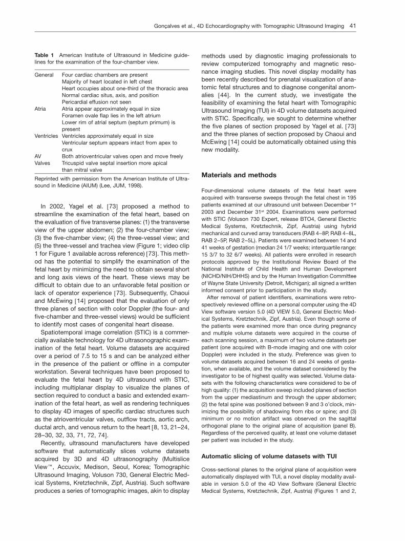
Table 1 American Institute of Ultrasound in Medicine guide-lines for the examination of the four-chamber view.
General Four cardiac chambers are presentMajority of heart located in left chestHeart occupies about one-third of the thoracic areaNormal cardiac situs, axis, and positionPericardial effusion not seen
Atria Atria appear approximately equal in sizeForamen ovale flap lies in the left atriumLower rim of atrial septum (septum primum) ispresent
Ventricles Ventricles approximately equal in sizeVentricular septum appears intact from apex tocrux
AV Both atrioventricular valves open and move freelyValves Tricuspid valve septal insertion more apical
than mitral valve
Reprinted with permission from the American Institute of Ultra-sound in Medicine (AIUM) (Lee, JUM, 1998).
In 2002, Yagel et al. w73x proposed a method tostreamline the examination of the fetal heart, based onthe evaluation of five transverse planes: (1) the transverseview of the upper abdomen; (2) the four-chamber view;(3) the five-chamber view; (4) the three-vessel view; and(5) the three-vessel and trachea view (Figure 1; video clip1 for Figure 1 available across reference) w73x. This meth-od has the potential to simplify the examination of thefetal heart by minimizing the need to obtain several shortand long axis views of the heart. These views may bedifficult to obtain due to an unfavorable fetal position orlack of operator experience w73x. Subsequently, Chaouiand McEwing w14x proposed that the evaluation of onlythree planes of section with color Doppler (the four- andfive-chamber and three-vessel views) would be sufficientto identify most cases of congenital heart disease.
Spatiotemporal image correlation (STIC) is a commer-cially available technology for 4D ultrasonographic exam-ination of the fetal heart. Volume datasets are acquiredover a period of 7.5 to 15 s and can be analyzed eitherin the presence of the patient or offline in a computerworkstation. Several techniques have been proposed toevaluate the fetal heart by 4D ultrasound with STIC,including multiplanar display to visualize the planes ofsection required to conduct a basic and extended exam-ination of the fetal heart, as well as rendering techniquesto display 4D images of specific cardiac structures suchas the atrioventricular valves, outflow tracts, aortic arch,ductal arch, and venous return to the heart w8, 13, 21–24,28–30, 32, 33, 71, 72, 74x.
Recently, ultrasound manufacturers have developedsoftware that automatically slices volume datasetsacquired by 3D and 4D ultrasonography (MultisliceView", Accuvix, Medison, Seoul, Korea; TomographicUltrasound Imaging, Voluson 730, General Electric Med-ical Systems, Kretztechnik, Zipf, Austria). Such softwareproduces a series of tomographic images, akin to display
methods used by diagnostic imaging professionals toreview computerized tomography and magnetic reso-nance imaging studies. This novel display modality hasbeen recently described for prenatal visualization of ana-tomic fetal structures and to diagnose congenital anom-alies w44x. In the current study, we investigate thefeasibility of examining the fetal heart with TomographicUltrasound Imaging (TUI) in 4D volume datasets acquiredwith STIC. Specifically, we sought to determine whetherthe five planes of section proposed by Yagel et al. w73xand the three planes of section proposed by Chaoui andMcEwing w14x could be automatically obtained using thisnew modality.
Materials and methods
Four-dimensional volume datasets of the fetal heart wereacquired with transverse sweeps through the fetal chest in 195patients examined at our ultrasound unit between December 1st
2003 and December 31st 2004. Examinations were performedwith STIC (Voluson 730 Expert, release BTO4, General ElectricMedical Systems, Kretztechnik, Zipf, Austria) using hybridmechanical and curved array transducers (RAB 4–8P, RAB 4–8L,RAB 2–5P, RAB 2–5L). Patients were examined between 14 and41 weeks of gestation (median 24 1/7 weeks; interquartile range:15 3/7 to 32 6/7 weeks). All patients were enrolled in researchprotocols approved by the Institutional Review Board of theNational Institute of Child Health and Human Development(NICHD/NIH/DHHS) and by the Human Investigation Committeeof Wayne State University (Detroit, Michigan); all signed a writteninformed consent prior to participation in the study.
After removal of patient identifiers, examinations were retro-spectively reviewed offline on a personal computer using the 4DView software version 5.0 (4D VIEW 5.0, General Electric Med-ical Systems, Kretztechnik, Zipf, Austria). Even though some ofthe patients were examined more than once during pregnancyand multiple volume datasets were acquired in the course ofeach scanning session, a maximum of two volume datasets perpatient (one acquired with B-mode imaging and one with colorDoppler) were included in the study. Preference was given tovolume datasets acquired between 16 and 24 weeks of gesta-tion, when available, and the volume dataset considered by theinvestigator to be of highest quality was selected. Volume data-sets with the following characteristics were considered to be ofhigh quality: (1) the acquisition sweep included planes of sectionfrom the upper mediastinum and through the upper abdomen;(2) the fetal spine was positioned between 9 and 3 o’clock, min-imizing the possibility of shadowing from ribs or spine; and (3)minimum or no motion artifact was observed on the sagittalorthogonal plane to the original plane of acquisition (panel B).Regardless of the perceived quality, at least one volume datasetper patient was included in the study.
Automatic slicing of volume datasets with TUI
Cross-sectional planes to the original plane of acquisition wereautomatically displayed with TUI, a novel display modality avail-able in version 5.0 of the 4D View Software (General ElectricMedical Systems, Kretztechnik, Zipf, Austria) (Figures 1 and 2,

42 Goncalves et al., 4D Echocardiography with Tomographic Ultrasound Imaging¸
Article in press - uncorrected proof
Figure 2 Tomographic Ultrasound Imaging (TUI) of a normal fetal heart in systole (A) and diastole (B). The volume datasets wereacquired using B-mode and color Doppler imaging. The overview image on the left upper panel of each figure shows the orthogonalsagittal plane to the sections that are being displayed. Each line represents a slice. The center slice is marked with an asterisk (*)and each subsequent plane to the right or left are marked with numbers ranging from y4 to q4. The plane marked by the dottedline is not displayed. In this volume dataset, the three planes of section proposed by Chaoui et al. (Ultrasound Obstet Gynecol 21(2003) 81–93) for the examination of the fetal heart are visualized. Please note that the five chamber view was better visualized duringsystole. Legends: PA: pulmonary artery; Ao: aorta; RV: right ventricle; LV: left ventricle; IVS: interventricular septum 4CH: four-chamber;5CH: five-chamber; 3V: three-vessel; Trv: transverse.Video clip 2 for Figure 2 available across the reference of this presented article w77x online version: view references or acrosshttp://dx.doi.org/10.1515/JPM.2006.006_supp_1

Goncalves et al., 4D Echocardiography with Tomographic Ultrasound Imaging 43¸
Article in press - uncorrected proof
Videos 1 and 2). This display modality allows examiners to auto-matically slice a volume dataset and simultaneously visualize upto eight parallel planes of section on the same screen. Impor-tantly, motion information in volume datasets acquired using 4Dultrasonography techniques is not lost and, therefore, multiplesections of a beating heart can be analyzed at the same time.An ‘‘overview image’’ is shown on the upper left corner. This viewdepicts a plane orthogonal to the slices, and parallel linesdemarcate the position of the slices within the volume dataset(Figure 1; video clip 1 for Figure 1 available across reference).The user can adjust the number and position of the slices withspecific software controls. Hue, brightness and contrast controlscan also be adjusted to optimize image quality.
Analysis of volume datasets displayed with TUI
All volume datasets were analyzed by a single investigator (LG).Those acquired with B-mode imaging were analyzed first, andvisualization rates for cardiac structures in each of the fiveplanes of section proposed by Yagel et al. w73x were determinedas follows: (1) abnormal; (2) normal; (3) present in the volumedataset but inadequate for diagnosis; and (4) not present in thevolume dataset. To be considered adequate for diagnosis, thefollowing structures and relationships should have been visual-ized in each scanning plane:
1. Transverse view of the upper abdomen (Figure 1A; video clip1 for Figure 1 available across reference): (1) stomach on theleft side of the abdomen; (2) transverse section of thedescending aorta in front and to the left of the spine; (3)inferior vena cava located on the right side of the spine.
2. Four-chamber view: evaluated according to the criteria pro-posed by the American Institute of Ultrasound in Medicinefor the performance of the basic fetal cardiac screeningexamination (Table 1) w43x.
3. Five-chamber view: (1) aortic root visualized and connectedto the left ventricle; (2) the anterior wall of the aorta shouldbe in continuity with the ventricular septum w43x.
4. Pulmonary artery view: pulmonary artery visualized leavingthe right ventricle and at least one of the branches bifurcatingat its distal end w43, 73, 75x.
5. Three-vessel and trachea view: (1) main pulmonary trunk indirect communication with the ductus arteriosus; (2) trans-verse section of the aortic arch between the main pulmonarytrunk and superior vena cava; (3) cross section of the supe-rior vena cava visualized on the right side of the chest; (4)trachea visualized posterior to the superior vena cava w73x.
Following the evaluation of B-mode imaging volume datasets,visualization rates for volume datasets acquired with colorDoppler were determined according to the criteria proposed byChaoui and McEwing w14x:
1. Four-chamber view: (1) diastolic perfusion across the atrio-ventricular valves documented with color Doppler, with noevidence of valve regurgitation; (2) no color flow observedcrossing the ventricular septum.
2. Five-chamber view: (1) aortic root emerging from the left ven-tricle; (2) interventricular septum in continuity with the ante-rior wall of the ascending aorta. Abnormalities in this viewincluded: (a) turbulent flow across the aortic valve; (b) shunt-ing through a perimembranous VSD with a normal aorticconnection; (c) visualization of the origin of the pulmonary
trunk emerging from the left ventricle identified by its bifur-cation into the main pulmonary arteries; and (d) overriding ofthe aorta over both ventricles, connected by a VSD.
3. Three-vessel view: (1) aorta and pulmonary trunk convergingtoward the left thorax with the trachea to their right; (2) pul-monary trunk with a slightly greater caliber than the aorta(ratio 1.2:1); (3) straight course of the vessels; and (4) ante-grade flow through both great vessels throughout the cardiaccycle.
Volume datasets acquired with B-mode imaging and colorDoppler were compared to determine if color Doppler providedmore information than that obtained by examination with B-mode imaging alone. Visualization rates for the four- and five-chamber and three-vessel views were compared using theMcNemar’s test. Agreement between diagnoses by the two-modalities was tested using the Kappa test.
Following determination of the visualization rates for eachview, the diagnoses established by TUI were compared to post-natal diagnoses by neonatal imaging modalities, cardiac surgeryor autopsy. Sensitivity, specificity, as well as positive- and neg-ative-predictive values for the diagnosis of congenital heart dis-ease were calculated first for volume datasets acquired withB-mode imaging and then for those acquired with the additionof color Doppler. Cases of isolated VSDs (either suspected pre-natally or detected in the neonatal period) were excluded fromthis analysis because neonatal echocardiograms are not per-formed in every neonate in our institution in the absence ofsymptoms.
Statistical analysis was performed with SPSS 12.0 for Win-dows (SPSS, Chicago, IL).
Results
STIC volume datasets of the fetal heart were acquiredwith B-mode or color Doppler imaging in 195 and 168cases, respectively.
Table 2 shows visualization rates for anatomical struc-tures expected to be present in the five transverse scan-ning planes proposed by Yagel et al. w73x, after automaticslicing with TUI (Figure 1, video clip 1 available acrossthe reference). The four- and five-chamber and the three-vessel and trachea views were visualized in 97.4% (190/195), 88.2% (172/195), and 79.5% (142/195) of the cas-es, respectively. A three-vessel view showing bifurcationof the pulmonary artery was visualized in only 51.8%(101/195) of the cases. Transverse views of the upperfetal abdomen were present in 64.1% of the volumedatasets (125/195).
In volume datasets acquired with color Doppler (Figure2, video clip 2 available across the reference), the four-and five-chamber and three-vessel views were visualizedin 98.2% (165/168), 97.0% (163/168), and 83.6% (145/168) of the cases, respectively (Table 3). Table 4 com-pares the visualization rates for the four-chamber,five-chamber and three-vessel views between volumedatasets acquired with B-mode or color Doppler imaging.The additional contribution of color Doppler imaging was

44 Goncalves et al., 4D Echocardiography with Tomographic Ultrasound Imaging¸
Article in press - uncorrected proof
statistically significant for the five-chamber view only(McNemar tests13.5, Ps0.004).
Isolated VSDs were suspected in 19 cases with B-mode imaging and 23 cases when the examination wasperformed with color Doppler. When isolated VSDs wereexcluded from the analysis, congenital heart anomalieswere identified by TUI in 16 of the 195 fetuses (8.2%)(Table 5). Figures 3, 4, 5 and 6 (video clips for Figures 3,4, 5 and 6 are available across the references) illustrateabnormal findings in the various planes of sections dis-played by TUI in cases of coarctation of the aorta, hypo-plastic left heart, pulmonary atresia and transposition ofthe great arteries. Color Doppler provided additionaldiagnostic information in 5 of the 16 cases (31.3%). Incase 5, retrograde perfusion of the pulmonary arteryhelped to correctly diagnose pulmonary atresia in a caseof hypoplastic right ventricle (Figure 5; video clip 5 forFigure 5 available across the reference). In case 6, a VSD
was demonstrated in association with coarctation of theaorta. In case 8, disproportion in size between the rightand left ventricles observed by B-mode imaging was cor-rectly diagnosed as coarctation of the aorta (Figure 3;video clip for Figure 3 is available across the reference).In case 13, color Doppler allowed the visuali-zation of an interrupted IVC with azygous continuation ina case where volume datasets acquired by B-modeimaging were considered non-diagnostic. In case 16,what was initially suspected as transposition of the greatarteries was diagnosed as tetralogy of Fallot.
Table 6 compares diagnoses established by the reviewof volume datasets displayed with TUI and postnataldiagnoses (isolated VSDs were excluded). Among the 17cases with a cardiac anomaly suspected by TUI or iden-tified in the neonatal period, two were lost to follow-up.In one case, only a fetal echocardiogram performed byan independent pediatric cardiologist was available for

Goncalves et al., 4D Echocardiography with Tomographic Ultrasound Imaging 45¸
Article in press - uncorrected proof
Table 4 Comparison of diagnostic information in TUI volume datasets acquired with B-mode imaging only or B-mode plus colorDoppler imaging (ns168).
Plane of section Perfect agreement between B- Color Doppler modified the No contribution Kappa P value bymode and color Doppler initial diagnostic of color Doppler McNemarimaging in classifying views as impression by B-mode to B-mode testnormal, abnormal, or not imaging only imagingdiagnostic
Four-chamber view 85.7% (145/168) 12.5% (21/168)* 1.8% (3/168) 0.537 0.135Five-chamber view 83.9% (141/168) 14.2% (24/168)† 1.8% (3/168) 0.431 0.004Three-vessel view 75% (126/168) 10.1% (17/168)‡ 14.8% (25/168) 0.370 0.172
* In 3 cases, non-diagnostic four-chamber views were adequately visualized with color Doppler; 4 false-positive and 14 false-negativediagnoses by B-mode imaging were corrected by color Doppler.† In 17 cases, a five-chamber view considered to be of no diagnostic value was adequately imaged by color Doppler; 6 false-positiveand 1 false-negative diagnoses by B-mode imaging were corrected by color Doppler.‡ In 13 cases, a three-vessel view considered to be of no diagnostic value was adequately imaged by color Doppler; 1 false-positiveand 3 false-negative diagnoses by B-mode imaging were corrected by color Doppler.TUI: Tomographic Ultrasound Imaging.
comparison. Among the 15 patients for whom follow-upwas available, there was perfect agreement between pre-natal and postnatal diagnoses in 73.3% of the cases (11/15). The four discrepant diagnoses were: (1) in case 3, afetus diagnosed with tetralogy of Fallot by TUI had apostnatal diagnosis of TGA associated with a subaorticVSD; (2) in case 7, coarctation of the aorta was sus-pected by both B-mode and color Doppler but was notconfirmed in the neonate; (3) in case 10, a rhabdomiomawas not visualized by both B-mode and color Dopplervolume datasets displayed with TUI; and (4) in case 14,
severe tricuspid regurgitation was detected in a fetuswhose final diagnosis was Ebstein anomaly.
Sensitivity, specificity, positive- and negative-predic-tive values of TUI for the diagnosis of congenital heartdisease, excluding cases of isolated VSDs, are displayedin Table 7. For this analysis, cases 3 and 14 (Table 6)were considered as true positive diagnoses because,although a specific diagnosis was not made, a significantcardiac anomaly was suspected by TUI.
Among fetuses with a VSD suspected by B-modeimaging and color Doppler (ns23), two cases (8.7%)

46 Goncalves et al., 4D Echocardiography with Tomographic Ultrasound Imaging¸
Article in press - uncorrected proof

Goncalves et al., 4D Echocardiography with Tomographic Ultrasound Imaging 47¸
Article in press - uncorrected proof
were confirmed after delivery, one case (4.3%) was diag-nosed as an AV canal, and another (4.3%) case wasdiagnosed as double inlet single ventricle. One of thethree (33.3%) isolated VSDs detected after delivery wasmissed by prenatal examination of STIC volume datasetsdisplayed with TUI.
Discussion
The results of this study indicate that: (1) the four- andfive-chamber, and the three-vessel and trachea viewscan be visualized in the majority of volume datasets auto-matically sliced with TUI; (2) color Doppler provides addi-tional diagnostic information in 10% of the three-vesselviews, 12% of the four-chamber views, and 14% of thefive-chamber views displayed with this modality; (3) theadditional information provided by color Doppler helpedto establish a correct diagnosis in 31% of the fetuseswith cardiac anomalies other than isolated VSDs; and (4)the diagnoses established by TUI agreed with the post-natal diagnoses in 73.3% of the fetuses, with good sen-sitivity, specificity, positive- and negative-predictivevalues for the diagnosis of congenital heart disease.
Technical aspects of 4D ultrasonography with STIChave been described by several investigators w8, 13,21–24, 28–30, 32, 33, 71, 72, 74x. However, a systematicevaluation of the capability of obtaining specific planesof section with this diagnostic modality has been report-ed once w72x. Vinals et al. w72x studied 100 fetuses whowere examined by 4D ultrasonography with STIC, andwhose volume datasets were evaluated by a specialist infetal echocardiography who was not involved in volumeacquisition. Standard cardiac planes were obtained byscrolling through the volume datasets from the upperabdomen to the mediastinum. Visualization rates for thefour-chamber view, left and right ventricular outflowtracts, three-vessel view, and three-vessel and tracheaview ranged from 81 to 100%, with the lowest visuali-zation rates observed for structures located in the abdo-men or upper mediastinum. TUI provides an alternativeapproach to manually scrolling through the volume data-sets to obtain these cardiac views. The differencebetween the two approaches is that the volume datasetis automatically sliced and, thus, it comes as no surpriseto us that the visualization rates for cardiac structuresreported by Vinals et al. w72x are similar to those reportedin the current study.
The view with the poorest visualization rates in ourstudy was the three-vessel view demonstrating bifurca-tion of the main pulmonary artery, the fourth plane of sec-tion proposed by Yagel et al. w73x. This could reflect theshort distance between this plane and the three-vesseland trachea view. Therefore, visualization of both planesusing automatic slicing techniques with a fixed distancebetween the planes of section may have forced the dis-

48 Goncalves et al., 4D Echocardiography with Tomographic Ultrasound Imaging¸
Article in press - uncorrected proof
Figure 3 Tomographic Ultrasound Imaging of a fetus with coarctation of the aorta. A. Volume dataset acquired with B-mode: (1)the three-vessel and trachea view shows a narrow transverse section of the aortic arch; (2) the four-chamber view shows disproportionbetween the right and left ventricles. B. Volume dataset acquired with color Doppler: (1) the three-vessel view confirms the narrowtransverse aortic arch and shows aliasing; (2) disproportion between the right and left ventricles is confirmed in the four-chamberview. Legends: PA: pulmonary artery; Ao: aorta; RV: right ventricle; LV: left ventricle; 3V: three-vessel; 3VT: three-vessel and trachea;4CH: four-chamber; 5CH: five-chamber; Trv: transverse. Video clip 3 for Figure 3 available across the reference of this presentedarticle� w�77x� online� version:� view� references� or� across� http://dx.doi.org/10.1515/JPM.2006.006_supp_1

Goncalves et al., 4D Echocardiography with Tomographic Ultrasound Imaging 49¸
Article in press - uncorrected proof
Figure 4 Tomographic Ultrasound Imaging of a fetus with hypoplastic left heart syndrome, double outlet right ventricle and trans-position of the great arteries. A. Volume dataset acquired with B-mode: (1) the three-vessel and trachea view shows the aorta leavingthe right ventricle; (2) the five-chamber view shows the pulmonary artery leaving the right ventricle as well; (3) the four-chamber viewshows the right ventricle and atrium only – the left ventricle is not visualized; (4) the transverse view of the fetal abdomen showsthat the stomach is located on the right. B. Volume dataset acquired with color Doppler: (1) the three-vessel view confirms that theaorta leaves the right ventricle; (2) the five-chamber view shows the pulmonary artery leaving the right ventricle as well. Legends:PA: pulmonary artery; Ao: aorta; RV: right ventricle; LV: left ventricle; 3V: three-vessel; 3VT: three-vessel and trachea; 4CH: four-chamber; 5CH: five-chamber; Trv: transverse. Video clip 4 for Figure 4 available across the reference of this presented article w77xonline� version:� view� references� or� across� http://dx.doi.org/10.1515/JPM.2006.006_supp_1

50 Goncalves et al., 4D Echocardiography with Tomographic Ultrasound Imaging¸
Article in press - uncorrected proof
Figure 5 Tomographic Ultrasound Imaging in a fetus with pulmonary atresia. A. Systole. (1) In the three-vessel view, retrogradeperfusion (in red) of a narrow pulmonary artery is observed; (2) severe tricuspid regurgitation is observed in the four-chamber view.B. Diastole: (1) the 5-chamber view shows the aorta connecting normally to the left ventricle; (2) the four-chamber view shows normalventricular filling only on the left ventricle, with tricuspid regurgitation still observed in blue. Legends: PA: pulmonary artery; Ao: aorta;RV: right ventricle; LV: left ventricle; 3V: three-vessel; 4CH: four-chamber; 5CH: five-chamber; Trv: transverse; TR: tricuspid regurgi-tation; RA: right atrium. Video clip 5 for Figure 5 is available across the references of this presented article w77x online version: viewreferences� or� across� http://dx.doi.org/10.1515/JPM.2006.006_supp_1

Goncalves et al., 4D Echocardiography with Tomographic Ultrasound Imaging 51¸
Article in press - uncorrected proof
Figure 6 Tomographic Ultrasound Imaging in a fetus with transposition of the great arteries. A. B-mode imaging. (1) In the three-vessel and trachea view, only the aorta is visualized, leaving the right ventricle; (2) the five-chamber view demonstrates a vessel thatbifurcates (pulmonary artery) connected to the left ventricle; (3) the four-chamber view is normal. B. Color Doppler imaging. (1) thethree-vessel view shows the aorta connecting to the right ventricle; (2) the pulmonary artery leaves the left ventricle and bifurcates.Legends: PA: pulmonary artery; Ao: aorta; RV: right ventricle; LV: left ventricle; 3V: three-vessel; 3VT: three-vessel and trachea; SVC:superior vena cava; 4CH: four-chamber; 5CH: five-chamber; Trv: transverse; TR: tricuspid regurgitation.Video clip 6 for Figure 6 is available across the references of this presented article w77x online version: view references or acrosshttp://dx.doi.org/10.1515/JPM.2006.006_supp_1

52 Goncalves et al., 4D Echocardiography with Tomographic Ultrasound Imaging¸
Article in press - uncorrected proof
Table 7 Sensitivity, specificity, positive- and negative-predictive values of TUI to detect congenital heart disease.
# of patients Prevalence Sensitivity Specificity Positive Negativeavailable for predictive value predictive valueanalysis
B-mode imaging 115† 12.2% (14/115) 85.7% (12/14) 100.0% (101/101) 100.0% (12/12) 98.0% (101/103)Color Doppler 98‡ 14.3% (14/98) 92.9% (13/14) 98.8% (83/84) 92.9% (13/14) 98.8% (83/84)†Complete follow-up was available in 131/172 patients scanned by B-mode imaging (76.2%) (excluding cases of isolated VSDs).Sixteen patients were excluded from this portion of the analysis: one patient whose images were interpreted as tetralogy of Fallotbut who was found at the time of surgery to have a subaortic VSD associated with transposition of the great arteries, and 15 patientswhose ultrasonographic images were considered to be of poor diagnostic quality.‡ Complete follow-up was available in 105/142 patients scanned by color Doppler (61.0%) (excluding cases of isolated VSDs). Sevenpatients were excluded from this portion of the analysis: in all cases, the ultrasonographic images were considered to be of poordiagnostic quality. TUI: Tomographic Ultrasound Imaging; VSD: ventricular septal defects.
play of one plane to the detriment of the other. Althoughthe distance between each slice can be adjusted by thesoftware and thus potentially correct this problem, wewanted to evaluate how this new technique would per-form with minimum operator interference. It is important,however, to be aware of this limitation, since if this viewis not automatically displayed, the examiner may need touse other techniques to explore the volume dataset inorder to visualize the pulmonary artery and its bifurcation.Among these techniques, the examiner may choose touse a previously reported systematic approach for eval-uation of the outflow tracts with STIC w31, 32x, wherebyboth the long axis view of the left ventricular outflow tractand the short axis view of the right outflow tract are dis-played side-by-side on the same image, the ‘‘spin’’ tech-nique reported by DeVore et al. w22x, or rendering
techniques to visualize the relationship between bothgreat arteries on the same image w13, 24, 28, 29, 33x.
The results of this study also corroborate the proposalof Chaoui and McEwing w14x that the examination ofthree-planes of section (four- and five-chamber andthree-vessel views) with color Doppler may be sufficientto detect most cases of congenital heart disease. Indeed,these investigators subsequently reported that thesethree planes could be demonstrated in 31/35 healthyfetuses and in 24/27 fetuses with CHD in volume data-sets acquired with color Doppler STIC w13x. In the currentstudy, the four- and five-chamber and three-vessel viewsacquired with color Doppler were visualized in 98.2%,97.0%, and 83.6% of the cases, respectively, and addi-tional diagnostic information was observed in 12%, 14%and 10% of these views when compared to B-mode

Goncalves et al., 4D Echocardiography with Tomographic Ultrasound Imaging 53¸
Article in press - uncorrected proof
imaging only. More importantly, color Doppler helped toestablish the correct diagnosis of pulmonary atresia andcoarctation of the aorta in two cases in which the pre-vious findings by B-mode imaging had shown only dis-proportional ventricular size.
The role of sonographic tomography in clinical practiceremains to be determined. Benacerraf et al. w6x reportedpreliminary findings in 25 pregnancies scanned duringthe second trimester, in which five volume datasetsencompassing the fetal head, face, chest, abdomen, andlimbs were acquired for offline later analysis. Volumedatasets were examined by physicians who were notinvolved in volume acquisition, and the visualization ratesfor fetal anatomical structures and time to complete theexamination (including volume acquisition and review)were calculated. Complete structural surveys wereobtained in 20 of the 25 fetuses. In one of the 5 incom-plete surveys, a face was visualized by neither 3D nor 2Dultrasound. Portions of the hands and feet were not visu-alized in the other four cases. Importantly, the timerequired to complete the anatomical surveys wasdecreased by half using 3D ultrasonography (13.9 minvs. 6.6 min, P-0.001). With the availability of softwareto automatically slice the volume datasets, busy clinicalpractices may welcome this approach.
Leung et al. w44x have recently reported the use ofautomatic slicing of 3D volume datasets (Multi-SliceView, Accuvix, Medison, Seoul, Korea) for the examina-tion of the fetal spine, face, brain and heart in a group of35 fetuses scanned during the second trimester of preg-nancy, and demonstrated the feasibility of diagnosingneural tube defects, facial clefts, and holoprosencephaly.The current study extends these observations to prenataldiagnosis of congenital heart disease. Although mostcases of congenital heart disease other than VSDs werecorrectly diagnosed by exploring the volume datasetswith the novel TUI display, this portion of our study needsto be interpreted with caution. Despite removing patientidentifiers and including only cases examined at least 6months before the conduction of this study, the possi-bility of recall bias exists. Therefore, our results need tobe independently confirmed.
Acknowledgements
This research was supported by the Intramural Research Pro-gram of the National Institute of Child Health and Human Devel-opment, NIH, DHHS.
References
w1x Abuhamad A: Automated multiplanar imaging: a novelapproach to ultrasonography. J Ultrasound Med 23 (2004)573
w2x Acar P, Y Dulac, A Taktak, M Villaceque: wReal time 3Dechocardiography in congenital heart diseasex. Arch MalCoeur Vaiss 97 (2004) 472
w3x Allan L, B Benacerraf, JA Copel, JS Carvalho, R Chaoui,SH Eik-Nes, et al.: Isolated major congenital heart disease.Ultrasound Obstet Gynecol 17 (2001) 370
w4x Arzt W, G Tulzer, M Aigner: wReal time 3D sonography ofthe normal fetal heart – clinical evaluationx. UltraschallMed 23 (2002) 388
w5x Bega G, K Kuhlman, A Lev-Toaff, A Kurtz, R Wapner:Application of three-dimensional ultrasonography in theevaluation of the fetal heart. J Ultrasound Med 20 (2001)307
w6x Benacerraf BR, TD Shipp, B Bromley: How sonographictomography will change the face of obstetric sonography:a pilot study. J Ultrasound Med 24 (2005) 371
w7x Bhat AH, V Corbett, N Carpenter, N Liu, R Liu, A Wu, etal.: Fetal ventricular mass determination on three-dimen-sional echocardiography: studies in normal fetuses andvalidation experiments. Circulation 110 (2004) 1054
w8x Bhat AH, VN Corbett, R Liu, ND Carpenter, NW Liu, AMWu, et al.: Validation of volume and mass assessments forhuman fetal heart imaging by 4-dimensional spatiotem-poral image correlation echocardiography: in vitro balloonmodel experiments. J Ultrasound Med 23 (2004) 1151
w9x Bonnet D, A Coltri, G Butera, L Fermont, J Le Bidois, JKachaner, et al.: Detection of transposition of the greatarteries in fetuses reduces neonatal morbidity and mor-tality. Circulation 99 (1999) 916
w10x Brekke S, E Tegnander, HG Torp, SH Eik-Nes: TissueDoppler gated (TDOG) dynamic three-dimensional ultra-sound imaging of the fetal heart. Ultrasound Obstet Gyne-col 24 (2004) 192
w11x Buskens E, DE Grobbee, IME Frohn-Mulder, PA Stewart,RE Juttmann, JW Wladimiroff, et al.: Efficacy of RoutineFetal Ultrasound Screening for Congenital Heart Diseasein Normal Pregnancy. Circulation 94 (1996) 67
w12x Chang FM, KF Hsu, HC Ko, BL Yao, CH Chang, CH Yu,et al.: Fetal heart volume assessment by three-dimension-al ultrasound. Ultrasound Obstet Gynecol 9 (1997) 42
w13x Chaoui R, J Hoffmann, KS Heling: Three-dimensional (3D)and 4D color Doppler fetal echocardiography using spatio-temporal image correlation (STIC). Ultrasound ObstetGynecol 23 (2004) 535
w14x Chaoui R, R McEwing: Three cross-sectional planes forfetal color Doppler echocardiography. Ultrasound ObstetGynecol 21 (2003) 81
w15x Crane JP, ML LeFevre, RC Winborn, JK Evans, BG Ewig-man, RP Bain, et al.: A randomized trial of prenatal ultra-sonographic screening: impact on the detection,management, and outcome of anomalous fetuses. TheRADIUS Study Group. Am J Obstet Gynecol 171 (1994)392
w16x Deng J, JE Gardener, CH Rodeck, WR Lees: Fetal echo-cardiography in three and four dimensions. UltrasoundMed Biol 22 (1996) 979
w17x Deng J, R Richards: Dynamic three-dimensional gray-scale and color Doppler ultrasound of the fetal heart fordynamic diagnosis. Ultrasound Obstet Gynecol 20 (2002)209
w18x Deng J, ID Sullivan, R Yates, M Vogel, D Mcdonald, ADLinney, et al.: Real-time three-dimensional fetal echocar-diography – optimal imaging windows. Ultrasound MedBiol 28 (2002) 1099
w19x Deng J, R Yates, AG Birkett, CF Ruff, AD Linney, WR Lees,et al.: Online motion-gated dynamic three-dimensionalechocardiography in the fetus – preliminary results. Ultra-sound Med Biol 27 (2001) 43
54 Goncalves et al., 4D Echocardiography with Tomographic Ultrasound Imaging¸
Article in press - uncorrected proof
w20x Deng J, R Yates, ID Sullivan, D Mcdonald, AD Linney, LeesWR, et al.: Dynamic three-dimensional color Doppler ultra-sound of human fetal intracardiac flow. Ultrasound ObstetGynecol 20 (2002) 131
w21x DeVore GR, P Falkensammer, MS Sklansky, LD Platt: Spa-tio-temporal image correlation (STIC): new technology forevaluation of the fetal heart. Ultrasound Obstet Gynecol22 (2003) 380
w22x DeVore GR, B Polanco, MS Sklansky, LD Platt: The ‘spin’technique: a new method for examination of the fetal out-flow tracts using three-dimensional ultrasound. UltrasoundObstet Gynecol 24 (2004) 72
w23x Espinoza J, LF Goncalves, W Lee, T Chaiworapongsa, MCTreadwell, S Stites, et al.: The use of the minimum projec-tion mode in 4-dimensional examination of the fetal heartwith spatiotemporal image correlation. J Ultrasound Med23 (2004) 1337
w24x Espinoza J, LF Goncalves, W Lee, M Mazor, R Romero: Anovel method to improve prenatal diagnosis of abnormalsystemic venous connections using three- and four-dimensional ultrasonography and ‘inversion mode’. Ultra-sound Obstet Gynecol 25 (2005) 428
w25x Fernandez CO, C Ramaciotti, LB Martin, DM Twickler: Thefour-chamber view and its sensitivity in detecting congen-ital heart defects. Cardiology 90 (1998) 202
w26x Franklin O, M Burch, N Manning, K Sleeman, S Gould, NArcher: Prenatal diagnosis of coarctation of the aortaimproves survival and reduces morbidity. Heart 87 (2002)67
w27x Garne E, C Stoll, M Clementi: Evaluation of prenatal diag-nosis of congenital heart diseases by ultrasound: experi-ence from 20 European registries. Ultrasound ObstetGynecol 17 (2001) 386
w28x Goncalves LF, J Espinoza, W Lee, M Mazor, R Romero:Three- and four-dimensional reconstruction of the aorticand ductal arches using inversion mode: a new renderingalgorithm for visualization of fluid-filled anatomical struc-tures. Ultrasound Obstet Gynecol 24 (2004) 696
w29x Goncalves LF, J Espinoza, W Lee, JK Nien, JS Hong, JSantolaya-Forgas, et al.: A new approach to fetal echo-cardiography: digital casts of the fetal cardiac chambersand great vessels for detection of congenital heart dis-ease. J Ultrasound Med 24 (2005) 415
w30x Goncalves LF, J Espinoza, R Romero, W Lee, B Beyer, MCTreadwell, et al.: A systematic approach to prenataldiagnosis of transposition of the great arteries using 4-dimensional ultrasonography with spatiotemporal imagecorrelation. J Ultrasound Med 23 (2004) 1225
w31x Goncalves LF, J Espinoza, R Romero, W Lee, M Treadwell,SE Huang, et al.: Four-dimensional fetal echocardiographywith spatiotemporal image correlation (STIC): a systematicstudy of standard cardiac views assessed by differentobservers. J Matern Fetal Neonatal Med 18 (2005) in press
w32x Goncalves LF, W Lee, T Chaiworapongsa, J Espinoza, MLSchoen, P Falkensammer, et al.: Four-dimensional ultra-sonography of the fetal heart with spatiotemporal imagecorrelation. Am J Obstet Gynecol 189 (2003) 1792
w33x Goncalves LF, R Romero, J Espinoza, W Lee, M Treadwell,K Chintala, et al.: Four-dimensional ultrasonography of thefetal heart using color Doppler spatiotemporal image cor-relation. J Ultrasound Med 23 (2004) 473
w34x Grandjean H, D Larroque, S Levi: The performance of rou-tine ultrasonographic screening of pregnancies in theEurofetus Study. Am J Obstet Gynecol 181 (1999) 446
w35x Guerra FA, AI Isla, RC Aguilar, EG Fritz: Use of free-handthree-dimensional ultrasound software in the study of thefetal heart. Ultrasound Obstet Gynecol 16 (2000) 329
w36x Herberg U, H Goldberg, J Breuer: Dynamic free-handthree-dimensional fetal echocardiography gated by cardio-tocography. Ultrasound Obstet Gynecol 22 (2003) 493
w37x Herberg U, H Goldberg, J Breuer: Three- and four-dimen-sional freehand fetal echocardiography: a feasibility studyusing a hand-held Doppler probe for cardiac gating. Ultra-sound Obstet Gynecol 25 (2005) 362
w38x Hoffman JI, S Kaplan: The incidence of congenital heartdisease. J Am Coll Cardiol 39 (2002) 1890
w39x Hoffman JIE, R Christianson: Congenital heart disease ina cohort of 19,502 births with long-term follow-up. Am JCardiol 42 (1978) 641
w40x Jaeggi ET, GF Sholler, OD Jones, SG Cooper: Compara-tive analysis of pattern, management and outcome of pre-versus postnatally diagnosed major congenital heartdisease: a population-based study. Ultrasound ObstetGynecol 17 (2001) 380
w41x Jurgens J, R Chaoui: Three-dimensional multiplanar time-motion ultrasound or anatomical M-mode of the fetalheart: a new technique in fetal echocardiography. Ultra-sound Obstet Gynecol 21 (2003) 119
w42x Klein SK, C Cans, E Robert, PS Jouk: Efficacy of routinefetal ultrasound screening for congenital heart disease inIsere County. France Prenat Diagn 19 (1999) 318
w43x Lee W: Performance of the basic fetal cardiac ultrasoundexamination. J Ultrasound Med 17 (1998) 601
w44x Leung KY, CS Ngai, BC Chan, WC Leung, CP Lee, MHTang: Three-dimensional extended imaging: a new displaymodality for three-dimensional ultrasound examination.Ultrasound Obstet Gynecol 26 (2005) 244
w45x Levental M, DH Pretorius, MS Sklansky, NE Budorick, TRNelson, K Lou: Three-dimensional ultrasonography of nor-mal fetal heart: comparison with two-dimensional imaging.J Ultrasound Med 17 (1998) 341
w46x Levi S, T Cos-Sanchez: wFetal echocardiography volumemode (3 dimensions)x. J Gynecol Obstet Biol Reprod (Par-is) 29 (2000) 261
w47x Mahle WT, RR Clancy, SP McGaurn, JE Goin, BJ Clark:Impact of prenatal diagnosis on survival and early neuro-logic morbidity in neonates with the hypoplastic left heartsyndrome. Pediatrics 107 (2001) 1277
w48x Maulik D, NC Nanda, V Singh, H Dod, S Vengala, A Sinha,et al.: Live three-dimensional echocardiography of thehuman fetus. Echocardiography 20 (2003) 715
w49x Meyer-Wittkopf M, A Cole, SG Cooper, S Schmidt, GFSholler: Three-dimensional quantitative echocardiographicassessment of ventricular volume in healthy human fetus-es and in fetuses with congenital heart disease. J Ultra-sound Med 20 (2001) 317
w50x Meyer-Wittkopf M, A Cook, A McLennan, P Summers, GKSharland, DJ Maxwell: Evaluation of three-dimensionalultrasonography and magnetic resonance imaging inassessment of congenital heart anomalies in fetal cardiacspecimens. Ultrasound Obstet Gynecol 8 (1996) 303
w51x Meyer-Wittkopf M, S Cooper, J Vaughan, G Sholler: Three-dimensional (3D) echocardiographic analysis of congenitalheart disease in the fetus: comparison with cross-sectional(2D) fetal echocardiography. Ultrasound Obstet Gynecol17 (2001) 485
w52x Meyer-Wittkopf M, M Hofbeck: wTwo- and three-dimen-sional echocardiographic analysis of congenital heart dis-ease in the fetusx. Herz 28 (2003) 240

Goncalves et al., 4D Echocardiography with Tomographic Ultrasound Imaging 55¸
Article in press - uncorrected proof
w53x Meyer-Wittkopf M, N Rappe, F Sierra, H Barth, S Schmidt:Three-dimensional (3-D) ultrasonography for obtaining thefour and five-chamber view: comparison with cross-sec-tional (2-D) fetal sonographic screening. Ultrasound Obs-tet Gynecol 15 (2000) 397
w54x Michailidis GD, JM Simpson, C Karidas, DL Economides:Detailed three-dimensional fetal echocardiography facili-tated by an Internet link. Ultrasound Obstet Gynecol 18(2001) 325
w55x Nelson TR: Three-dimensional fetal echocardiography.Prog Biophys Mol Biol 69 (1998) 257
w56x Nelson TR, DH Pretorius, M Sklansky, S Hagen-Ansert:Three-dimensional echocardiographic evaluation of fetalheart anatomy and function: acquisition, analysis, and dis-play. J Ultrasound Med 15 (1996) 1
w57x Roguin N, ZD Du, M Barak, N Nasser, S Hershkowitz, EMilgram: High prevalence of muscular ventricular septaldefect in neonates. J Am College Cardiol 26 (1995) 1545
w58x Rustico MA, A Benettoni, G D’Ottavio, A Maieron, IFischer-Tamaro, G Conoscenti, et al.: Fetal heart screeningin low-risk pregnancies. Ultrasound Obstet Gynecol 6(1995) 313
w59x Sands AJ, FA Casey, BG Craig, JC Dornan, J Rogers, HCMulholland: Incidence and risk factors for ventricular sep-tal defect in ‘‘low risk’’ neonates. Arch Dis Child Fetal Neo-natal Ed 81 (1999) F61
w60x Scharf A, F Geka, A Steinborn, H Frey, A Schlemmer, CSohn: 3D real-time imaging of the fetal heart. Fetal DiagnTher 15 (2000) 267
w61x Sklansky M: New dimensions and directions in fetal car-diology. Curr Opin Pediatr 15 (2003) 463
w62x Sklansky MS, GR DeVore, PC Wong: Real-time 3-dimen-sional fetal echocardiography with an instantaneous vol-ume-rendered display: early description and pictorialessay. J Ultrasound Med 23 (2004) 283
w63x Sklansky MS, T Nelson, M Strachan, D Pretorius: Real-time three-dimensional fetal echocardiography: initial fea-sibility study. J Ultrasound Med 18 (1999) 745
w64x Sklansky MS, TR Nelson, DH Pretorius: Usefulness of gat-ed three-dimensional fetal echocardiography to recon-struct and display structures not visualized withtwo-dimensional imaging. Am J Cardiol 80 (1997) 665
w65x Sklansky MS, TR Nelson, DH Pretorius: Three-dimensionalfetal echocardiography: gated versus nongated tech-niques. J Ultrasound Med 17 (1998) 451
w66x Stoll C, Y Alembik, B Dott, MJ Meyer, A Pennerath, MOPeter, et al.: Evaluation of prenatal diagnosis of congenitalheart disease. Prenat Diagn 18 (1998) 801
w67x Stoll C, Y Alembik, B Dott, PM Roth, B De Geeter: Eval-uation of prenatal diagnosis of congenital heart disease.Prenat Diagn 13 (1993) 453
w68x Tegnander E, SH Eik-Nes: The examiner’s ultrasound ex-perience has a significant impact on the detection rate ofcongenital heart defects at the second trimester fetalexamination wabstractx. Ultrasound Obstet Gynecol 24(2004) 217
w69x Todros T, F Faggiano, E Chiappa, P Gaglioti, B Mitola, ASciarrone: Accuracy of routine ultrasonography in screen-ing heart disease prenatally. Gruppo Piemontese for Pre-natal Screening of Congenital Heart Disease. Prenat Diagn17 (1997) 901
w70x Tworetzky W, DB McElhinney, VM Reddy, MM Brook, FLHanley, NH Silverman: Improved surgical outcome afterfetal diagnosis of hypoplastic left heart syndrome. Circu-lation 103 (2001) 1269
w71x Vinals F, L Mandujano, G Vargas, A Giuliano: Prenatal diag-nosis of congenital heart disease using four-dimensionalspatio-temporal image correlation (STIC) telemedicine viaan Internet link: a pilot study. Ultrasound Obstet Gynecol25 (2005) 25
w72x Vinals F, P Poblete, A Giuliano: Spatio-temporal image cor-relation (STIC): a new tool for the prenatal screening ofcongenital heart defects. Ultrasound Obstet Gynecol 22(2003) 388
w73x Yagel S, R Arbel, EY Anteby, D Raveh, R Achiron: Thethree vessels and trachea view (3VT) in fetal cardiac scan-ning. Ultrasound Obstet Gynecol 20 (2002) 340
w74x Yagel S, DV Valsky, B Messing: Detailed assessment offetal ventricular septal defect with 4D color Doppler ultra-sound using spatio-temporal image correlation technolo-gy. Ultrasound Obstet Gynecol 25 (2005) 97
w75x Yoo SJ, YH Lee, ES Kim, HM Ryu, MY Kim, HK Choi, etal.: Three-vessel view of the fetal upper mediastinum: aneasy means of detecting abnormalities of the ventricularoutflow tracts and great arteries during obstetric screen-ing. Ultrasound Obstet Gynecol 9 (1997) 173
w76x Zosmer N, D Jurkovic, E Jauniaux, K Gruboeck, C Lees,S Campbell: Selection and identification of standard car-diac views from three-dimensional volume scans of thefetal thorax. J Ultrasound Med 15 (1996) 25
Video clips of the presented article available across the fol-lowing reference:w77x Goncalves LF, Espinoza J, Romero R, Kusanovic JP,
Swope B, Nien JK, Erez O, Soto E, Treadwell MC: Four-dimensional ultrasonography of the fetal heart using a nov-el Tomographic Ultrasound Imaging display. J Perinat Med34 (2006)http://dx.doi.org/10.1515/JPM.2006.006_supp_1
Received September 21, 2005. Accepted October 13, 2005.