Hematoporphyrin encapsulated PLGA microbubble for contrast enhanced ultrasound imaging and...
9
Journal of Microencapsulation, 2012; 29(5): 437–444 ß 2012 Informa UK Ltd. ISSN 0265-2048 print/ISSN 1464-5246 online DOI: 10.3109/02652048.2012.655333 Hematoporphyrin encapsulated PLGA microbubble for contrast enhanced ultrasound imaging and sonodynamic therapy Yuanyi Zheng 1 , Yaping Zhang 1 , Meng Ao 1 , Ping Zhang 1 , Hui Zhang 1 , Pan Li 1 , Lang Qing 2 , Zhigang Wang 2 and Haitao Ran 1 1 Second Affiliated Hospital of Chongqing Medical University, Chongqing 400010, China and 2 Institute of Ultrasound Imaging of Chongqing Medical University, Chongqing 400010, China Abstract The purpose of this study was to develop a sonosensitizer-loaded multi-functional ultrasound (US) con- trast agent for both tumour therapy and imaging. The hematoporphyrin (HP)-encapsulated poly(lactic- co-glycolic acid) microbubbles (HP-PLGA-MBs) were prepared and filled with perfluorocarbon gases. The enhancement of US imaging and its sonodynamically induced anti-tumour effect were evaluated by both in vitro and in vivo experiments. The HP-PLGA-MBs have a narrow size distribution and smooth surface with a mean diameter of 702.6 56.8 nm and HP encapsulation efficiency of 63.50 1.26% and drug-loading efficiency of 2.15 0.13%. The HP-PLGA-MBs could well enhance the ultrasound imaging both in vitro and in vivo. A significant anti-tumour effect was obtained by HP-PLGA-MBs mediated sonodynamic therapy. The tumour growth rate and the tumour proliferation index were the lowest in the HP-PLGA-MBs plus sonica- tion group. And the tumour cell apoptotic index was the biggest in the HP-PLGA-MBs plus sonication group. In conclusion, a sonosensitizer-loaded multi-functional contrast agent was constructed and the feasibility was demonstrated, which might provide a novel strategy for tumour imaging and therapy. Keywords: hematoporphyrin, PLGA, ultrasound contrast agent, sonodynamic therapy, tumor Introduction Sonodynamic therapy (SDT) is a promising approach to the treatment of tumours, which has been clinically applied to cancer, such as breast cancer (Wang et al., 2009; Barati and Mokhtari-Dizaji, 2010). The sonosensitizers, mainly hema- toporphyrin (HP) or hematoporphyrin derivatives (HPDs), are accumulated in tumour tissues and excited by exposure to the appropriate frequency and energy of ultrasound, resulting in tumour cell killing by activated oxygen pro- duced by the sonosensitizers (Kuroki et al., 2007). While like other chemotherapy drugs, sonosensitizers have signif- icant side effects, such as blister formation, skin hyperpig- mentation, ocular discomfort, pruritus, pain at injection site and urticaria (Wooten et al., 1988). The rapid development of high molecular chemistry and nanomedicine holds promise for solving the problems of current chemotherapy, such as side effects of chemotherapeutic drugs (Rapoport et al., 2009). Various high molecular drug delivery modalities have been sug- gested for decreasing the side effects caused by chemother- apeutic drugs, which are commonly based on drug encapsulation in nano or microparticles, etc. The encapsu- lation may dramatically increases drug concentration in an aqueous environment and offers new life to bioactive com- pounds previously abandoned due to low aqueous solubil- ity (Rapoport et al., 2009). And in order to further improve the outcome of tumour therapy, the stimuli-responsive drug delivery modalities have been developed. The pay- load will be released in response to environmental or phys- ical stimuli, such as pH, hyperthermia, light, or ultrasound, which had important implications for tumour therapy. Among the physical stimuli, ultrasound irradiation has been proved to be a good tool for the controlled drug release study. It has been demonstrated that ultrasonic irradiation could efficiently trigger drug release from the Address for correspondence: Haitao Ran, Second Affiliated Hospital of Chongqing Medical University, 76 Linjiang Road, Chongqing 400010, China. Tel: 86-23-63693709. Fax: 86-23-63809074. E-mail: [email protected] (Received 22 Aug 2011; accepted 19 Dec 2011) http://www.informahealthcare.com/mnc 437
Transcript of Hematoporphyrin encapsulated PLGA microbubble for contrast enhanced ultrasound imaging and...

Journal of Microencapsulation, 2012; 29(5): 437–444� 2012 Informa UK Ltd.ISSN 0265-2048 print/ISSN 1464-5246 onlineDOI: 10.3109/02652048.2012.655333
Hematoporphyrin encapsulated PLGA microbubble for contrastenhanced ultrasound imaging and sonodynamic therapy
Yuanyi Zheng1, Yaping Zhang1, Meng Ao1, Ping Zhang1, Hui Zhang1, Pan Li1, Lang Qing2,Zhigang Wang2 and Haitao Ran1
1Second Affiliated Hospital of Chongqing Medical University, Chongqing 400010, China and 2Institute of UltrasoundImaging of Chongqing Medical University, Chongqing 400010, China
AbstractThe purpose of this study was to develop a sonosensitizer-loaded multi-functional ultrasound (US) con-trast agent for both tumour therapy and imaging. The hematoporphyrin (HP)-encapsulated poly(lactic-co-glycolic acid) microbubbles (HP-PLGA-MBs) were prepared and filled with perfluorocarbon gases. Theenhancement of US imaging and its sonodynamically induced anti-tumour effect were evaluated by bothin vitro and in vivo experiments. The HP-PLGA-MBs have a narrow size distribution and smooth surface witha mean diameter of 702.6� 56.8 nm and HP encapsulation efficiency of 63.50� 1.26% and drug-loadingefficiency of 2.15� 0.13%. The HP-PLGA-MBs could well enhance the ultrasound imaging both in vitro andin vivo. A significant anti-tumour effect was obtained by HP-PLGA-MBs mediated sonodynamic therapy. Thetumour growth rate and the tumour proliferation index were the lowest in the HP-PLGA-MBs plus sonica-tion group. And the tumour cell apoptotic index was the biggest in the HP-PLGA-MBs plus sonicationgroup. In conclusion, a sonosensitizer-loaded multi-functional contrast agent was constructed and thefeasibility was demonstrated, which might provide a novel strategy for tumour imaging and therapy.
Keywords: hematoporphyrin, PLGA, ultrasound contrast agent, sonodynamic therapy, tumor
Introduction
Sonodynamic therapy (SDT) is a promising approach to the
treatment of tumours, which has been clinically applied to
cancer, such as breast cancer (Wang et al., 2009; Barati and
Mokhtari-Dizaji, 2010). The sonosensitizers, mainly hema-
toporphyrin (HP) or hematoporphyrin derivatives (HPDs),
are accumulated in tumour tissues and excited by exposure
to the appropriate frequency and energy of ultrasound,
resulting in tumour cell killing by activated oxygen pro-
duced by the sonosensitizers (Kuroki et al., 2007). While
like other chemotherapy drugs, sonosensitizers have signif-
icant side effects, such as blister formation, skin hyperpig-
mentation, ocular discomfort, pruritus, pain at injection
site and urticaria (Wooten et al., 1988).
The rapid development of high molecular chemistry and
nanomedicine holds promise for solving the problems
of current chemotherapy, such as side effects of
chemotherapeutic drugs (Rapoport et al., 2009). Various
high molecular drug delivery modalities have been sug-
gested for decreasing the side effects caused by chemother-
apeutic drugs, which are commonly based on drug
encapsulation in nano or microparticles, etc. The encapsu-
lation may dramatically increases drug concentration in an
aqueous environment and offers new life to bioactive com-
pounds previously abandoned due to low aqueous solubil-
ity (Rapoport et al., 2009). And in order to further improve
the outcome of tumour therapy, the stimuli-responsive
drug delivery modalities have been developed. The pay-
load will be released in response to environmental or phys-
ical stimuli, such as pH, hyperthermia, light, or ultrasound,
which had important implications for tumour therapy.
Among the physical stimuli, ultrasound irradiation has
been proved to be a good tool for the controlled drug
release study. It has been demonstrated that ultrasonic
irradiation could efficiently trigger drug release from the
Address for correspondence: Haitao Ran, Second Affiliated Hospital of Chongqing Medical University, 76 Linjiang Road, Chongqing 400010, China.Tel: 86-23-63693709. Fax: 86-23-63809074. E-mail: [email protected]
(Received 22 Aug 2011; accepted 19 Dec 2011)http://www.informahealthcare.com/mnc
437

drug carriers and could transiently alter cell membrane
permeability, resulting in effective anti-tumor effect
(Watanabe et al., 2008; Xing et al., 2008).
The encapsulation of sonosensitizers inside the high
molecular particles might have several benefits. First, the
encapsulation should be able to alleviate the side effects of
sonosensitizers. Second, the application of ultrasound
could not only activate the sonosensitizers but also locally
(also called passive targeting (Liu et al., 2010)) release the
sonosensitizers, which was considered be able to improve
the efficiency of chemotherapy and further alleviate the
side effects. Third, the high molecular particles could be
fabricated as a drug carrier and an ultrasound contrast
agent at the same time. The combination of the contrast
enhanced diagnostic ultrasound imaging and ultrasound
mediated therapy could be especially attractive for reasons
of simplicity and cost-effectiveness.
Recently, the term of theranostics has become a hot
topic in both chemistry and medicine research field,
among which the drug-loaded ultrasound microbubbles
were explored (Ke et al., 2011). Ultrasound contrast
agents (UCAs) are microbubbles composed of a thin albu-
min, galactose, lipid, or polymers, Shell filled with a heavy
gas, such as perfluorocarbon or nitrogen. UCAs have a high
degree of echogenicity, which is the ability of an object to
reflect the ultrasound waves. The echogenicity difference
between the gas in the microbubbles and the soft tissue
surroundings of the body is immense. Thus, ultrasonic
imaging using UCAs enhances the ultrasound backscatter,
or reflection of the ultrasound waves, to produce a unique
sonogram with increased contrast due to the high echo-
genicity difference. With the use of UCAs, the resolution
and sensitivity of clinical ultrasound imaging have been
greatly improved. Nowdays, UCAs have been widely used
in clinical practice in Europe and China as ultrasound con-
trast agents for tumour imaging. And UCAs also have been
widely reported as effective drug carriers and enhancers of
drug and gene delivery in experimental studies (Ren et al.,
2009). The use of ultrasound microbubbles as drug carriers
is very attractive. It would combine cost-effective ultra-
sound imaging with ultrasound-mediated therapy, and
ultrasound is the only clinical used imaging modality
for real time guidance and monitoring of radiofrequency
therapy of tumours (Solbiati et al., 2004). However,
these commonly used microbubbles are not a good drug
carrier because their drug-loading efficiency is low and
they are made from either lipid or albumin, such as
Sonovue or Optison, which have very short circulation
time (minutes).
The way to solve the side effects problem of sonosensi-
tizers and to develop an effective theranostic at the same
time may consist in developing drug-loaded high molecu-
lar ultrasound microbubbles, such as poly(lactic-
co-glycolic acid) (PLGA) or chistan microbubble, etc.
Their important properties combine drug carrying and
enhancing the ultrasound contrast of tumours. While to
best of our knowledge, high molecular microbubbles car-
rying sonosensitizers for both ultrasound imaging and
sonodynamic therapy have never been reported yet.
Here, we describe therapeutic and ultrasound-imaging
properties in these high molecular microbubbles that were
explored using the mouse subcutaneous tumour model.
Materials and methods
Fabrication of hematoporphyrin (HP) encapsulated
poly(lactic-co-glycolic acid) (PLGA) Microbubbles
(HP-PLGA-MBs)
HP-PLGA-MBs were fabricated using a modified double
emulsion (water/oil/water) method followed by solvent
evaporation (Zhou et al., 2008; Ao et al., 2010). Briefly,
100 mg of PLGA (75:25; Sigma) was dissolved in 2 mL meth-
ylene chloride, and 200mL of 10 mg/mL Hematoporphyrin
(HP) in Milli-Q water was added to the organic solution
and subjected to strong vibration for 45 s using an ultra-
sound microbubble vibrator (Chonqging Anbijie Inc,
China), to create the first emulsion (W/O). The resulting
(W/O) emulsion was then poured into cold (4�C), 5% poly-
vinyl alcohol solution and homogenized using homoge-
nizer (FJ-2000,Shanghai sample model Inc, China) for
5 min at 9000 rpm, to create the second emulsion
((W/O)/W). The double emulsion was then poured into a
2% isopropanol solution and stirred at room temperature
for 1 h. The capsules were then collected by centrifugation
and washed three times with deionised water. The capsules
were mixed with 3 mL deionised water and frozen in a
�80�C freezer, lyophilized for 48 h. The dried samples
were filled with perFuorocarbon gases and stored in a free-
zer at �20�C until used.
Characterization of HP-PLGA-MBs
Particle size analysis
Particle size, size distribution and concentration were
determined using a Malvern Zetasizer Nano ZS (Malvern
Instrument, UK) and a coulter counter (Multisizer III;
Beckman Coulter, USA). BrieFy, 1 mg/mL of HP-PLGA-
MBs solution was prepared in double distilled water. Size
measurements were performed in triplicates following a
1/100 (v/v) dilution of the HP-PLGA-MBs suspension in
MilliQ water at 25�C.
Scanning electron microscope (SEM) studies
The surface morphology of microbubbles was character-
ized by using a scanning electron microscope (KYKY2000).
Assessing the encapsulation efficiency
and loading efficiency
Drug encapsulation efficiency in the HP-PLGA-MB micro-
bubbles was assessed by high-performance liquid
438 Y. Zheng et al.

chromatography. The concentration of hematoporphyrin
in the solution was obtained from the standard curve of
hematoporphyrin in dimethyl sulfoxide (DMSO), which
relates absorbance and concentrations. Briefly, 4 mg of
freeze-dried HP-PLGA-MB microbubbles were dissolved
in 2 mL DMSO in a tube, and 4 mg of dry blank PLGA
microbubbles were taken as the control group. HP content
in the solution was determined by high-performance liquid
chromatography. The encapsulation efficiency of HP in
microbubbles was calculated using the following
equation (Ran et al., 2005). Drug encapsulation efficiency
(EE):
EE½%� ¼ ðW1=WTHPÞ � 100%;
where: W1 is the total drug amount in the HP-PLGA-MB
microbubbles; WTHP is the total weight of HP used in the
preparation of the HP-PLGA-MB microbubbles. Loading
efficiency (LE):
LE½%� ¼ ðW1=WTMBÞ � 100%;
where: W1 is the total drug amount in the HP-PLGA-MB
microbubbles; WTMB is the total weight of HP-PLGA-MB
microbubbles.
In vitro drug release experiment
In vitro sonication experiment was performed in order to
assess the drug release behaviour in the sound field, and
hence its suitability as a platform for triggered drug delivery
(Janoria and Mitra, 2007). The parameters of the sonication
was the similar to our custom-used parameter for cell gene
transfection in vitro (Ren et al., 2009). Sample measuring
10 mg was added to 50 mL of 37�C PBS within the sample
holder. The agent was then insonated using the 1 MHz
transducer at 2 W/cm2 (Model UTG 1025, Institute of
Ultrasound Imaging of Chongqing Medical Sciences,
Chongqing, China). The release medium was withdrawn
at predetermined time intervals, and replaced with fresh
soaking medium (1 mL) each time. HP content in the aque-
ous solution was analyzed using the high-performance
liquid chromatography method. Controls were performed
using the same setup and time scale, but without
sonication.
In vitro ultrasound imaging experiment
Imaging phantom overview
Figure 1 shows the experimental setup consisting of poly-
ethylene tubing (internal diameter (ID): 1 mm. Western
Analytical Co., CA) embedded into a 3% gelatine phantom
in a 20 cm� 10 cm� 5 cm tank, a pump and a rack which
was used to hold the ultrasound probe on the gel phantom.
Flow was generated by the pump (Watson-Marlow Sci400,
Watson-Marlow Alitea, England). The signal produced by
the microbubbles at the concentration of 108/mL was cap-
tured by the harmonic ultrasound probe.
Ultrasound image acquisition
Contrast-enhanced harmonic images were obtained using
a 5–12 MHz linear-array ultrasound transducer (IU22;
Philips Medical Systems, France). The default harmonic
imaging mode (mechanic index (MI): 0.08, frame rate
(FR): 45 fps, depth: 3.5 cm) was used for the imaging. The
microbubbles were administered with the probe fixed by
means of a custom-designed clamp to maintain position
above the tube. The image intensity (gray value) of region
of interest (ROI) was measured using an ultrasound image
analysis software (Model: DFY, Chongqing Medical
University, China).
In vivo ultrasound imaging and therapy of mouse tumour
Cell culture and subcutaneous tumour model
(Zhang et al., 2010)
The mouse H22 hepatocellular carcinoma cells (H22 cells)
were maintained at 37�C with 5% CO2 in completed RPMI
supplemented with 100 mg/mL penicillin, 100 mg/mL
streptomycin, and 10% fetal bovine serum. Thirty-
six-week-old C57BL/6J mice were bred in house and were
kept under standard housing conditions. For tumour injec-
tion, cells were harvested by trypsinization, centrifuged,
and resuspended at a density of 2� 106 cells/mL.
Tumours were inoculated subcutaneously on the left
flank with a 20 mL of cell mixture by using a 25-gauge
needle. Tumours were monitored weekly for growth prog-
ress via ultrasound. The tumours were used for the exper-
iment once they reached a diameter of approximately
0.5 cm. All experiments were conducted under protocols
approved by the Institutional Animal Care and Use
Committee at Chongqing Medical University.
In vivo ultrasound imaging and ultrasonic exposure
Thirty mice were randomized into five groups (six rats each
group) for therapy experiment. All groups were subjected
to ultrasonic exposure except the first group. The first con-
trol animal group (Saline group) was taken as a blank con-
trol by the injection of saline only. The second group
(Saline-US group) was injected the saline and subjected
to ultrasonic exposure. The third group (PLGA-MBs-US
Figure 1. Imaging phantom overview.
Hematoporphyrin encapsulated PLGA microbubble 439

group) was injected the bubbles without hematoporphyrin
inclusion and subjected to ultrasonic exposure. The fourth
group (HP-US group) was injected hematoporphyrin
(20 mg/kg) and subjected to ultrasonic exposure. The fifth
group (HP-PLGA-MBs-US group) was injected hematopor-
phyrin inclusion (20 mg/kg) bubbles and subjected to
ultrasonic exposure. The mice were anesthetized with iso-
flurane (1.5% vol. at 2 L/min) via a nose cone. Body tem-
perature was maintained at 37�C.
The same ultrasound image acquisition method as in
the in vitro experiment was employed for the in vivo exper-
iment except the probe was fixed on the tumours instead of
the gel phantom. The ultrasound microbubbles had a final
concentration HP at 20 mg/kg. For each single injection, a
syringe with 23 gauge needles was used to draw the bubble
solution according to each mouse’s body weight. The sus-
pension was then injected through tail vein of mice.
Immediately after administration, the injection port was
flushed with 0.3 mL of saline solution. The procedure of
the enhancement of ultrasound imaging was recorded as
DICOM clips. And when the microbubbles arrive at the
tumour observed by the ultrasound imaging, the ultra-
sound imaging probe was moved away and ultrasonic
probe (frequency: 1 MHz, intensity: 2 W/cm2) was transder-
mally applied on the tumours and continuously sonicated
for 2 min. The imaging and therapy procedures were
repeated every other day for a total three times.
Evaluation of anti-tumour effect
The curative effect was evaluated by measuring the tumour
growth and comparing it to the control groups after treat-
ment. The long (a) and short (b) diameters of the tumours
and the weight of mice were measured every day after
treatment. Tumour size was calculated as: volume¼
�/6[(aþ b)/2]3 and the volume inhibition ratios were cal-
culated as: (1-average tumour volume of treated group/
average tumour volume of the control group)� 100%.
Fifteen days after the treatment, the mice were killed and
the tumours were dissected out and weighted. The tumour
inhibition ratios of the tumour weight were calculated as:
(1-average tumour weight of treated group/average tumour
weight of the control group)� 100%.
Histopathologic examination
Tumours were harvested on the fifteenth day of treatment.
The tissue was fixed in 10% formalin solution and embed-
ded in paraffin. Slides of 5mm thickness were cut and
stained with hematoxylin and eosin (H&E). The TdT-
mediated dUTP-biotin nick-end labelling (TUNEL) assay
was performed to evaluate the apoptotic cell death. The
TUNEL-positive cells were counted in 10 randomly
selected Eelds using a light microscope at 200�magniEcat-
ion. The apoptotic index (AI) of a tumour was deEned as
the number of apoptotic cells in 100 tumour cells. The pro-
liferative cell nuclear antigen (PCNA) was measured by
immunohistochemical staining method using a mouse
monoclonal antibody to PCNA (dilution 1:50) (Cormedica
Corp., CA, USA). The PCNA-positive cells were counted in
10 randomly selected Eelds using a light microscope at
200� magniEcation. The Proliferating index (PI) of a
tumour was deEned as the number of PCNA positive cell
nuclei in 100 tumour cells. All histological processing was
carried out by the Histology Core facility of the Chongqing
Medical University. Histopathological examination of the
tumours was conducted by blinded analysis.
Statistical analysis
All data were expressed as means� SD. The differences
among the groups were analyzed of variance (one-way
ANOVA), the difference of intensity between the two
groups in the in vitro ultrasound imaging study was ana-
lyzed of t-test, p5 0.05 was considered to be significant
and p5 0.01 was considered to be extremely significant.
Results and discussion
Characterization of morphology of the
hematoporphyrin-inclusion HP-PLGA-MBs
In this study, we prepared the PLGA microbubbles carrying
hematoporphyrin (HP-PLGA-MBs) as an imaging agent as
well as a controlled released sonodynamic therapeutic
agent. We used SEM to characterize the morphological
properties of these particles. Figure 2 shows the SEM
image of HP-PLGA-MBs. The SEM analysis showed that
the PLGA particles were spherical and had a smooth sur-
face. The SEM image reveals HP-PLGA-MBs to be com-
prised of essentially spherical microparticles.
The mean diameter of the PLGA microbubbles was
702.6� 56.8 nm and the mean concentration was 108
microbubbles/mL. The particle size of HP-PLGA-MBs is
important for both safety and efficacy. Most of the micro-
particles must be smaller than the size of a red blood cell
(8 mm). The particle size data demonstrate that the most of
the HP-PLGA-MBs were sub-micron particles, which is
appropriate for intravenous delivery.
Figure 2. Scanning electron microscope picture of HP-PLGA: �10k (left),
�45k (right).
440 Y. Zheng et al.
Assessing the encapsulation efficiency of hematoporphyrin
The corresponding encapsulation efficiency of HP in the
HP-PLGA-MBs was 63.50%� 1.26%. The HP loading effi-
ciency of the HP-PLGA-MBs was 2.15%� 0.13% (w/w).
In vitro release properties of HP-PLGA-MBs
Figure 3 compares the release profiles of HP-PLGA-MBs
with/without low-frequency ultrasound sonication. The
release profiles of HP-PLGA-MBs with/without sonication
Figure 5. Contrast enhanced tumor at 16, 18, 20, 22, 24 and 26 s after the injection of HP-PLGA-MBs, and the Time–intensity curve showed the contrast-
enhanced ultrasound imaging of H22 hepatocellular carcinoma.
Figure 6. H22 tumor growth curves for control mice treated by Saline only
and mice treated by HP-PLGA-MBs-US, PLGA-MBs-US, HP-US, Saline-
US. It showed that the tumor regression rate in the HP-PLGA-MBs-US
group was faster than that in the HP-US group (p5 0.05) and in the
control group (p5 0.01) (*p5 0.05; **p5 0.01).
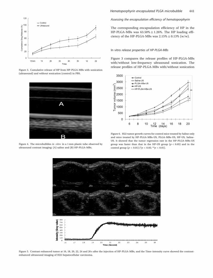
Figure 4. The microbubbles in vitro in a 1 mm plastic tube observed by
ultrasound contrast imaging: (A) saline and (B) HP-PLGA-MBs.
Figure 3. Cumulative release of HP from HP-PLGA-MBs with sonication
(ultrasound) and without sonication (control) in PBS.
Hematoporphyrin encapsulated PLGA microbubble 441

were measured and compared to evaluate the effects of
sonication on their release properties. Each release profile
is represented by the percentage of released hemotopor-
phyrin as a function of the release time. The results from
Figure 3 show that the release rate of hematoporphyrin
with sonication is faster than that without sonication.
Approximately 90% of the hematoporphyrin was released
after 2 days, but for the HP-PLGA-MBs without sonication
less than 65% was released. We also determined the values
of the time for 50% of the hematoporphyrin to be released
(t1/2) for the HP-PLGA-MBs. The average t1/2 values for
HP-PLGA-MBs with/without sonication were 3 and 24 h,
respectively. This result implied that the HP-loaded bub-
bles can be controllably disrupted with US, and the HP can
be released and deposited at the target sites under US
image guidance. The mechanism for the ultrasound-trig-
gered drug release from PLGA microbubbles might be the
degradation of PLGA caused by the sonication (El-Sherif
et al., 2004). El-Sherif et al. found that the rate of degrada-
tion is dependent not only on exposure to ultrasound, but
also on the frequency at which the microcapsules were
insonated, and the method by which the capsules were
fabricated (El-Sherif et al., 2004). This information is critical
in understanding future work on the ultrasound triggered
drug delivery pattern. It is hypothesized that the fre-
quency that causes the fastest degradation rate of cap-
sules will also produce an increase in the release rate
of the adsorbed drug. Further studies are required to be
done in order to find the best ultrasound frequency and
preparation method for the encapsulation of the
sonosentilizers.
Figure 7. (a) The apoptosis index (AI) of H22 tumor cells in each group.
There is no significant difference for the apoptotic index (AI) between the
control group and the Saline-US group (p4 0.05). In contrast, the AI for
the sonicated tumor in the HP-PLGA-MBs-US group and HP-US group is
significantly bigger than in the saline group (p5 0.01). And the AI for the
tumor in the HP-PLGA-MBs-US group is bigger than that in the HP-US
group (p5 0.05). (*p5 0.05; **p5 0.01; �p5 0.05 comparing with HP-
US). (b) The sonosensitizer caused H22 tumor cells apoptosis in the tumor
bearing mice (Tunel method, �400). The nucleus of the apoptosis cells
were dyed brown. Much more apoptosis cells were observed in the HP-US
and HP-PLGA-MBs-US group: (A): control; (B): Saline-US; (C): PLGA-
MBs-US; (D): HP-US; (E): HP-PLGA-MBs-US.
Table 3. The proliferating index of H22 tumor cell in each group.
Control Saline-
US
PLGA-
MBs-US
HP-US HP-PLGA-
MBs-US
PI (%) 90.09� 3.95 86.94� 4.92 84.79� 6.28 54.33� 7.80* 37.83� 6.34**
Note: *p5 0.01; **p5 0.05.
Table 1. Tumor inhibition ratios of the tumor weight (%).
Control Saline-US PLGA-MBs-US HP-US HP-PLGA-MBs-US
Tumor weight (g) 1.48� 0.23 1.38� 0.20 1.36� 0.30 0.97� 0.19 0.68� 0.23
Tumor inhibition ratios (%) 0 6.51 7.89 34.36* 53.86**
Note: *p5 0.01; **p5 0.05.
Table 2. The apoptosis index of H22 tumor cells in each group.
Control Saline-US PLGA-MBs-US HP-US HP-PLGA-MBs-US
AI (%) 16.14� 3.93 19.22� 5.06 21.02� 5.87 36.71� 7.28* 52.82� 5.98**
Note: *p5 0.01; **p5 0.05.
442 Y. Zheng et al.

In vitro ultrasound contrast enhancement
The HP-PLGA-MBs bubbles could well enhance ultra-
sound imaging under default settings for clinical harmonic
imaging (Figure 4). The contrast-enhanced image intensity
after the injection of HP-PLGA-MBs was significantly
bigger than that of water, which was 112.20� 6.58 and
0.80� 0.45, respectively (p5 0.01). This result implied
that the HP-PLGA-MBs could be used for clinical ultra-
sound contrast imaging with proper development.
In vivo ultrasound contrast enhancement of mouse tumour
Tumour accumulation of HP-PLGA-MBs after systemic
injections of HP-PLGA-MBs was clearly manifested by
increased echogenicity of tumour ultrasound images
(Figure 5). This implies that the ultrasonic exposure
could be guided by the HP-PLGA-MBs enhanced ultra-
sound tumour imaging. As soon as the HP-PLGA-MBs
arrive at the tumour place, the ultrasonic exposure
should be applied. The HP was released locally, which
was considered be able to improve the efficiency of che-
motherapy and alleviate the side effects.
In vivo tumour therapy
The unsonicated tumour in the Saline group grew with the
same rate as the tumours in Saline-US group (p4 0.05).
In contrast, the sonicated tumour appeared resolved after
three treatments in the HP-PLGA-MBs-US group and HP-
US group. While the tumour regression rate in the HP-
PLGA-MBs-US group is faster than that in the HP-US
group (p5 0.05) and in the control group (p5 0.01)
(Figure 6). The results of the tumour inhibition ratios of
the tumour weight are presented in Table 1. These data
indicate that without HP, the simple ultrasound or simple
PLGA-MBs had no significant affect on the tumour growth.
However, the HP-PLGA-MBs plus US is more effective than
that of pure HP plus US.
There is no significant difference for the AI between the
control group and the Saline-US group (p4 0.05). In con-
trast, the AI for the sonicated tumour in the HP-PLGA-MBs-
US group and HP-US group is significantly bigger than in
the saline group (p5 0.01). And the AI for the tumour in the
HP-PLGA-MBs-US group is bigger than that in the HP-US
group (p5 0.05) (Table 2, Figure 7(a) and (b)).
There is no significant difference for the he proliferating
index (PI) between the control group and the Saline-US
group (p4 0.05). In contrast, the PI for the sonicated
tumour in the HP-PLGA-MBs-US group and HP-US group
is significantly lower than in the saline group (p5 0.05).
And the PI for the tumour in the HP-PLGA-MBs-US group
is lower than that in the HP-US group (p5 0.05) (Table 3,
Figure 8(a) and (b)).
Since the AI of the tumours is positively related to the
tumour regression rate and the proliferating index is neg-
atively related to the tumour regression rate, these data
indicated that the encapsulation of the sonosensitizer
inside the high molecular microbubbles, the HP-PLGA-
MBs, could significantly improve the therapeutic effects
of the sonosensitizer in tumour-bearing mice model.
Conclusion
In conclusion, we successfully constructed HP-PLGA-MBs
composed of ultrasound-responsive polymeric microbub-
bles for systemic contrast-enhanced ultrasound imaging
diagnosis, and sonosensitizers inside the microbubbles
for sonodynamic therapy. The application of HP-PLGA-
MBs in vivo can significantly inhibit the growth of mice
Figure 8. (a) The proliferating index (PI) of H22 tumor cells in each group.
There is no significant difference for the he Proliferating index (PI)
between the control group and the Saline-US group (p4 0.05). In con-
trast, the PI for the sonicated tumor in the HP-PLGA-MBs-US group and
HP-US group is significantly lower than in the saline group (p5 0.05).
And the PI for the tumor in the HP-PLGA-MBs-US group is lower than
that in the HP-US group (p5 0.05). (*p5 0.05; **p5 0.01; �p5 0.05
comparing with HP-US). (b) The sonosensitizer inhibited the proliferating
of the H22 tumor bearing mice (�400). The nucleus of the proliferating
cells were dyed brown. (PCNA stain, �400). Much less proliferating cells
were observed in the HP-US and HP-PLGA-MBs-US group: (A): control
(Saline); (B): Saline-US; (C): PLGA-MBs-US; (D): HP-US; (E): HP-PLGA-
MBs-US.
Hematoporphyrin encapsulated PLGA microbubble 443

tumours. Meanwhile, HP-PLGA-MBs can still maintain
adequate acoustic properties that are required to act as
an ultrasound contrast agent. Thus, the dual-functional
ultrasound microbubbles hold a great potential for ultra-
sound-guided sonodynamic tumour therapy. This simple
and highly efficient theranostic agent would provide a
novel strategy for the sonodynamic therapy of tumour.
Limitation
In this study, we focused on the demonstration of the fea-
sibility of a sonosensitizer loaded multi-functional contrast
agent, while we did not do the examination of the drug
distribution and the side effect, which is a limitation of
this study. This should be done in our future work.
Acknowledgements
This research is partialy supported by National Natural
Science Foundation of China (No. 81130025, 30900371,
81071158, 81071157), National Key Basic Research
Program (No. 2011CB707900), Foundation for Sci and
Tech Research Project of Chongqing (2010AB50952) and
Scientific Research Foundation for Returned Scholars
(No. 2010609).
Declaration of interest
The authors report no conflicts of interest. The authors alone
are responsible for the content and writing of the article.
References
Ao M, Wang Z, Ran H, Guo D, Yu J, Li A, Chen W, Wu W, Zheng Y.Gd-DTPA-loaded PLGA microbubbles as both ultrasound contrast
agent and MRI contrast agent – A feasibility research. J Biomed MaterRes B Appl Biomater, 2010;93:551–6.
Barati AH, Mokhtari-Dizaji M. Ultrasound dose fractionation in sonody-namic therapy. Ultrasound Med Biol, 2010;36:880–7.
El-Sherif DM, Lathia JD, Le NT, Wheatley MA. Ultrasound degradation ofnovel polymer contrast agents. J Biomed Mater Res A, 2004;68:71–8.
Janoria KG, Mitra AK. Effect of lactide/glycolide ratio on the in vitro releaseof ganciclovir and its lipophilic prodrug (GCV-monobutyrate) fromPLGA microspheres. Int J Pharm, 2007;338:133–41.
Ke H, Wang J, Dai Z, Jin Y, Qu E, Xing Z, Guo C, Yue X, Liu J. Gold-nanoshelled microcapsules: A theranostic agent for ultrasound contrastimaging and photothermal therapy. Angew Chem Int Ed Engl,2011;50:3017–21.
Kuroki M, Hachimine K, Abe H, Shibaguchi H, Maekawa S,Yanagisawa J, Kinugasa T, Tanaka T, Yamashita Y. Sonodynamictherapy of cancer using novel sonosensitizers. Anticancer Res,2007;27:3673–7.
Liu HZ, Wang YJ, Xu L, Li SM. Investigation into the potential of electro-poration facilitated topical delivery of cyclosporin a. PDA J Pharm SciTechnol, 2010;64:191–9.
Ran H, Ren H, Wang Z, Zheng Y, Zhang Q, Li X, Xu C. Research on fabrica-tion of high molecular polymer ultrasound contrast agent for drug deliv-ery. J Ultrasound Clin Med, 2005;7;217–22.
Rapoport NY, Kennedy AM, Shea JE, Scaife CL, Nam KH. Controlledand targeted tumor chemotherapy by ultrasound-activated nanoe-mulsions/microbubbles. J Control Release, 2009;138:268–76.
Ren J, Xu C, Zhou Z, Zhang Y, Li X, Zheng Y, Ran H, Wang Z. A novelultrasound microbubble carrying gene and Tat peptide: Preparation andcharacterization. Acad Radiol, 2009;16:1457–65.
Solbiati L, Ierace T, Tonolini M, Cova L. Guidance and monitoring of radio-frequency liver tumor ablation with contrast-enhanced ultrasound. EurJ Radiol, 2004;51:S19–23.
Wang X, Zhang W, Xu Z, Luo Y, Mitchell D, Moss RW. Sonodynamic andphotodynamic therapy in advanced breast carcinoma: A report of3 cases. Integr Cancer Ther, 2009;8:283–7.
Watanabe Y, Aoi A, Horie S, Tomita N, Mori S, Morikawa H, Matsumura Y,Vassaux G, Kodama T. Low-intensity ultrasound andmicrobubbles enhance the antitumor effect of cisplatin. Cancer Sci,2008;99:2525–31.
Wooten RS, Smith KC, Ahlquist DA, Muller SA, Balm RK. Prospective studyof cutaneous phototoxicity after systemic hematoporphyrin derivative.Lasers Surg Med, 1988;8:294–300.
Xing W, Gang WZ, Yong Z, Yi ZY, Shan XC, Tao RH. Treatment of xeno-grafted ovarian carcinoma using paclitaxel-loaded ultrasound micro-bubbles. Acad Radiol, 2008;15:1574–9.
Zhang Y, Deng J, Feng J, Wu F. Enhancement of antitumor vaccine inablated hepatocellular carcinoma by high-intensity focused ultrasound.World J Gastroenterol, 2010;16:3584–91.
Zhou S, Sun J, Sun L, Dai Y, Liu L, Li X, Wang J, Weng J, Jia W, Zhang Z.Preparation and characterization of interferon-loaded magnetic biode-gradable microspheres. J Biomed Mater Res B Appl Biomater, 2008;87:189–96.
444 Y. Zheng et al.

Copyright of Journal of Microencapsulation is the property of Taylor & Francis Ltd and its content may not be
copied or emailed to multiple sites or posted to a listserv without the copyright holder's express written
permission. However, users may print, download, or email articles for individual use.