Transmission of video telephony images over wireless channels

Report no 1003Authors Wolfgang Leister (NR)
Per Røe (NR)Ilangko Balasingham (RH)Halfdan Ihlen (RH)Håvard Roterud (RH)
Kjell-Rune Haugland (RH)Ola-Morten Hauen (RH)Morten Kaland (SSiA)Ronald Bosgraaf (SSiA)
Date 6th November 2012ISBN 82-539-0511-4
Transmission of DigitalUltrasound Images
Wolfgang Leister (NR) Per Røe (NR) Ilangko Balasingham (RH) Halfdan Ihlen (RH) Håvard Roterud (RH) Kjell-Rune Haugland (RH) Ola-Morten Hauen (RH) Morten Kaland (SSiA) Ronald Bosgraaf (SSiA)
Norwegian Computing CenterNorsk Regnesentral (Norwegian Computing Center, NR) is a private, independent, non-profitfoundation established in 1952. NR carries out contract research and development projects ininformation and communication technology and applied statistical-mathematical modelling. Theclients include a broad range of industrial, commercial and public service organisations in thenational as well as the international market. Our scientific and technical capabilities are furtherdeveloped in co-operation with The Research Council of Norway and key customers. The resultsof our projects may take the form of reports, software, prototypes, and short courses. A proof ofthe confidence and appreciation our clients have in us is given by the fact that most of our newcontracts are signed with previous customers.

Title Transmission of Digital Ultrasound Images
Authors Wolfgang Leister (NR) , Per Røe (NR) , Ilangko Balasingham(RH) , Halfdan Ihlen (RH) , Håvard Roterud (RH) , Kjell-RuneHaugland (RH) , Ola-Morten Hauen (RH) , Morten Kaland(SSiA) , Ronald Bosgraaf (SSiA)
Date 6th November 2012
ISBN 82-539-0511-4
Publication number 1003
AbstractThe DISSH project is a project of the Høykom program in Norway in 2003 and 2004, in whicha prototype service for the transfer of cardiology images between hospitals in Norway is im-plemented. This report describes the design, implementation and evaluation of the PACSflow-application, which controls the entire process, and provides a user interface for the medical per-sonnel. As much as possible of the existing infrastructure of the hospitals is used by PACSflow,which uses information from RIS and PACS systems. PACSflow was tested in a medical settingbetween Rikshospitalet (RH) and Sørlanded Sykehus in Arendal (SSiA), using broadband infras-tructure. The report also gives an overview of relevant standards and procedures used in healthcare.
Keywords medical images transfer, DICOM, PACS, RIS, cardiologicultrasound images
Target group Hospitals, Høykom program
Availability
Project DISSH
Project number 320306
Research field distributed systems, medical image transfer,multimedia-multichannel
Number of pages 37
© Copyright Norwegian Computing Center
3
Foreword
This document describes the results of the project DISSH, which was funded, in part, by theResearch Council of Norway under the Høykom program in 2003 and 2004. The participatinginstitutions in the project include Interventional Center at Rikshospitalet, Norsk Regnesentral,and the Sørlandet Sykehus i Arendal. Commercial participants were GE Healthcare (Vingmed),Sectra Norge AS, and Communicate Norge AS, who deliver and maintain technical equipmentsand servers used in the project.
The main objective of the DISSH project was to enable for transfering medical data, e.g., cardi-ologic ultrasound images, between hospitals, while at the same time sending patient data andinformation on the medical case. At the same time the strict information security policies of thehospitals must be obeyed, and the medical personnel is given a user-friendly interface for send-ing and accessing the data. The project resulted in the implementation of the prototype PACSflow,which now will be taken into rigorous clinical testing.
The participants at the institutions were: Prof. Halfdan Ihlen at Rikshospitalet is the owner of theidea from the medical perspective. Ilangko Balasingham at Rikshospitalet is the project leader.From technical research and development Wolfgang Leister and Per Røe from Norsk Regnesen-tral participated. Other participants include Håvard Roterud from the department of radiology atRikshospitalet, and Kjell Rune Haugland and Ola Morten Hauen from the department of informa-tion technology at Rikshospitalet. From the Sørlandet Sykehus i Arendal, Harald Brunvand hadthe medical supervision, while Morten Kaland and Ronald Bosgraaf contributed from the techni-cal perspective. Participations of Olaug Råd from Rikshospitalet and Gunnar Gisleberg, AndersHanke, and Bjørn Svalastoga from GE Vingmed Ultrasound AS are acknowledged.
4 Transmission of Digital Ultrasound Images
Contents
1 Objectives . . . . . . . . . . . . . . . . . . . . . . . . . . . 71.1 Technology and Application . . . . . . . . . . . . . . . . . . . 71.2 The Clinical Application and its Key Benefits . . . . . . . . . . . . . 71.3 Project Objectives . . . . . . . . . . . . . . . . . . . . . . 9
2 Technical Background and Requirements . . . . . . . . . . . . . . . 112.1 Present Solutions. . . . . . . . . . . . . . . . . . . . . . . 112.2 Standards and Formats . . . . . . . . . . . . . . . . . . . . 132.3 Security and Integrity Requirements . . . . . . . . . . . . . . . . 152.4 System Integration and Tool Selection . . . . . . . . . . . . . . . 16
3 The PACSflow Application . . . . . . . . . . . . . . . . . . . . . 173.1 The Use-Scenario . . . . . . . . . . . . . . . . . . . . . . 173.2 System Architecture . . . . . . . . . . . . . . . . . . . . . . 173.3 Platform Choices . . . . . . . . . . . . . . . . . . . . . . . 173.4 PACS Functionality . . . . . . . . . . . . . . . . . . . . . . 183.5 Notification and Messaging Functionality . . . . . . . . . . . . . . 193.6 Web Application . . . . . . . . . . . . . . . . . . . . . . . 21
4 Medical Tryout and Conclusions . . . . . . . . . . . . . . . . . . . 264.1 Experiences. . . . . . . . . . . . . . . . . . . . . . . . . 26
A Glossary . . . . . . . . . . . . . . . . . . . . . . . . . . . . 27
B Resources – Software and Links . . . . . . . . . . . . . . . . . . . 28B.1 Software for Image Coding . . . . . . . . . . . . . . . . . . . 28B.2 Open Source Software for DICOM handling . . . . . . . . . . . . . 29B.3 Dicom viewers . . . . . . . . . . . . . . . . . . . . . . . . 30B.4 Commercial Software . . . . . . . . . . . . . . . . . . . . . 31B.5 Sites for DICOM Example Images . . . . . . . . . . . . . . . . . 32B.6 Web Pages of General Interest . . . . . . . . . . . . . . . . . . 32
C Comparison of Compression Methods. . . . . . . . . . . . . . . . . 33
References . . . . . . . . . . . . . . . . . . . . . . . . . . . . . 35
Index . . . . . . . . . . . . . . . . . . . . . . . . . . . . . 36
Transmission of Digital Ultrasound Images 5

1 Objectives
Ultrasound images are used for many different diagnostic purposes. One area of interest is diag-nosis of heart diseases. Rikshospitalet (RH) has an expert team under Prof. Ihlen and serves as anational centre for diagnosis of difficult heart cases. The patients are often from remote regions,and most of them are small children. Most of the local hospitals have ultrasound scanners and canproduce digitally encoded ultrasound images. On-line transfer of high quality ultrasound imagesfrom the local hospitals to Rikshospitalet will provide quick and better diagnosis and will be agreat help for very sick patients.
1.1 Technology and ApplicationThe new health reforms adopted by the Norwegian Government will probably increase the ten-dency for hospitals specialising in certain types of clinical procedures. It is expected that resourceswill be pooled and yet be geographically dispersed. Examples of such resources are specialisedpersonnel and medical equipment. In practice, general hospitals today use more and more ad-vanced diagnostic imaging of different organs in the body. Due to this there is an increasing needfor discussing the images with highly specialised personnel. The advent of broadband communi-cation facilities have opened for communication of such resource pools between hospitals despitethe geographical distances involved. This has already been tested with success for example in ra-diology. By using this communication the correct diagnosis may be obtained in the local hospitaland a decision can be made about treatment and where to do it. It is obvious that multimediacommunication between hospitals has important impact on efficiency and quality in health care.We believe that there will be an increasing demand for this form of communication in the future.
Until now the data volume needed in the transfer of images between the hospitals have been rel-atively small. Recent diagnostic imaging in medicine is more and more advanced creating muchhigher data volume. A typical example is ultrasonic examination of heart disease. Ultrasonic scan-ners are present in most hospitals in the country, but it is not possible to transfer the digital ul-trasonic data between hospitals today due to the current capacity and architecture of the datacommunication infrastructure. The aim of the DISSH project is to develop a technique that allowstransfer of large amounts of digital data through the Internet without a reduction in the quality ofthe data. Ultrasonic digital data obtained during imaging of the heart contain high volumes, andare therefore chosen for the project.
The Internet and the World Wide Web are just a special cases of the ability for communities toshare resources as they tackle common goals. Science today is increasingly collaborative and mul-tidisciplinary, and it is not unusual for teams to span institutions, states, countries and continents.Grid technologies [1] envision that such groups connect their data, computers, sensors and otherresources into a single virtual laboratory or organisation.
Practical use of new communication technologies will enable the health sector to utilise broad-band in a cost-effective manner. In essence, large computing facilities will be made available to allrelevant health personnel. This will enable access to geographically dispersed facilities, providesupport for distributed research collaboration, and provide an infrastructure to support commu-nication and learning – all this at reasonable cost.
1.2 The Clinical Application and its Key Benefits1.2.1 Current situation and experienceThe Echocardiographic Laboratory, Department of Cardiology, Rikshospitalet has, in cooperationwith GE Vingmed Ultrasound, developed a system for complete digitisation of ultrasonic exami-
Transmission of Digital Ultrasound Images 7

nation of the heart. Rikshospitalet has a high capacity broadband network, which is a prerequisitefor delivering high volume data. Each cineloop of a heartbeat makes data amounts 2-10 MB anda complete examination of the patient produces 50-150 MB. The network of Rikshospitalet hasshown able to handle such large amounts of data. Cardiac ultrasonic data obtained by an ultra-sonic scanner are sent through the network from all sites in the hospital including the operatingtheatres and are saved on central servers together with other hospital data. Older patient data canvery quickly be taken out from the servers for comparison with a new examination.
Our experience during the last five years is that this internal broadband communication of ultra-sonic data between different parts of the hospital is an immense success. The efficacy of the ul-trasonic main laboratory has increased significantly. Ultrasonic examinations are now performedroutinely during heart surgery to evaluate the quality of the surgical treatment. Frequently theultrasonic examinations reveal problems coming up during the operation procedure leading toresumption of the operation or changes in the procedure. We have several cases where the life ofthe patient was saved due to this ultrasonic monitoring of the surgery.
An important aspect of this monitoring is that we can compare ultrasonic registrations done dur-ing surgery with the preoperative registrations that are brought up from the central server to theoperating room for direct comparison. Ultrasonic examination is now the most important tech-nique treatment in our hospital for the diagnosis and surgical treatment of valvar heart disease.It is important that the network communication enables us as a daily routine to present the ex-aminations for the cardiologists and surgeons. Generally, the doctors have now immediate anddirect access to the images, which have improved their decision making in how to treat the pa-tient. Because we do not lose quality of the images when they are saved digitally, we also got avery successful new tool for scientific work and education.
1.2.2 Future Situation and Key BenefitsUltrasonic scanners for diagnosis of heart disease are now present in almost all hospitals in Nor-way. The next natural step now is to develop a system for communicating ultrasonic examinationbetween hospitals. We believe that in adult cardiology it is very important to see the recordingswith the same quality as obtained in the local hospital because poor acoustic conditions are oftenpresent in adults and reduce the diagnostic precision.
Today on-line communication between different hospitals is only possible using video technique.We consider high quality of the recordings to have higher priority than seeing on-line recordingsthat must have lower quality. Quick export of ultrasonic recordings from one echocardiographiclaboratory to another will be of great clinical help particularly when treating very sick patients,but also when findings of uncertain significance need discussion with other experts. In addition,digital communication between hospitals will reduce the need for repeating the ultrasonic exam-ination when patients are transferred to our hospital for further evaluation. This is particularlyimportant for the examinations through the throat, which is a painful procedure for the patient,but extremely important in many very sick patients.
1.2.3 A clinical exampleThe following example illustrates the importance of digital networks between hospitals in thetreatment of heart diseases.
A patient was admitted to a local hospital severely sick with high fever, shortness of breath andbacteria in the blood. He had previously been operated with implantation of an artificial valvebetween the left cardiac chamber and the main artery (aorta) due to a large leakage. Ultrasonicexamination now showed a leakage in this artificial valve, but because the heart rhythm was very
8 Transmission of Digital Ultrasound Images
high it was difficult to assess how large the leakage was. A probe was therefore introduced intothe throat and far better images of the valve and the heart were obtained. Abnormal structuresaround the valve were found around the valve and the examiner suspected an infection of thevalve, but the images were difficult to interpret. Video recording of the whole examination wastherefore sent to Rikshospitalet for review. When we reviewed the video recordings two dayslater we found the examination not optimal, but some images showed definite infection of thevalve and a severe leakage, which explained why the patient was short in breath. The patient wastherefore immediately transferred by ambulance plane to Rikshospitalet where a new ultrasonicexamination through the throat was performed. This examination showed that the infection hadalmost loosened the valve from the heart with a severe leakage of blood into the heart chamber.The patient was sent directly to the operating theatre and operated with implantation of a newvalve. He recovered completely.
This patient was close to death when the correct diagnosis was made. Ultrasonic images may bevery difficult to interpret and may often need expert assessment. The two days time delay beforeexperts evaluated the images was dangerously long. An immediate transfer of digital images toour hospital would have lead to a correct diagnosis two days earlier. We could also have guidedthe local doctors in how they should obtain additional images to assure the diagnosis even notbeing on-line. This example is not exceptional. Frequently we are in need of knowing exactly whatthe admitting hospitals have found. This is particularly important with critically ill patients, butthe images can also lead to the conclusion that no severe heart disease is present and transfer ofthe patient is not necessary. A digital network between hospitals for transfer of ultrasonic imagesof the heart will be therefore of great clinical importance.
1.3 Project ObjectivesThe DISSH project deploys a a pilot service for digital ultrasound multimedia exchange basedon existing broadband communication links in the health sector in Norway, implemented inthe PACSflow-application. Other objectives include genericity. PACSflow is initially designed forcommunicating digital ultrasound data. However, the development is sufficiently generic to al-low communication of other diagnostic data as well. The added value of PACSflow is along thesethree axes:
• Technological added value: The ability to provide and support a complex service for healthcare workers across hospitals.
• Organisational added value: The ability to provide cost-effective services to several organi-sational units in the face of dispersion of resources.
• Economic and social added value: The ability to handle higher volumes of patients or toperform medical treatment and analysis with higher quality.
The PACSflow-application implements the necessary user interfaces for the medical personnel,and the interfaces to the technical infrastructure of the hospitals, i.e., the PACS, the HIS and thenetworking infrastructure. The requirement scopes for PACSflow include:
• PACSflow implements a user interface for sending and retrieving images and medical data.The user interface is suitable to be used by medical personnel without profound knowledgeof ICT-systems.
• PACSflow interfaces the PACS of the hospitals for retrieving data, and for initiating DICOMtransfer between PACS.
• PACSflow interfaces the HIS for retrieveing administrative data. PACSflow also ensures theintegrity of the patient data with respect of all data elements being correctly associated to the
Transmission of Digital Ultrasound Images 9

patient.• PACSflow provides a messaging facility for informing medical personnel on newly arrived
data, and for the transfer of descriptions on medical cases.• PACSflow uses current standards and procedures within health care as far as possible. The
existing infrastructure of the hospitals must be used without introducing new services thatneed special attention of the IT departments.
• PACSflow obeys the security policies used within health care in Norway. This includes avail-ability in operating hours, confidentiality and integrity of the data.
Since this is the first attempt to achieve this kind of functionality in the hospitals in Norway, andsince we have limited resources with regard to the infrastructure, we cannot address every wishfrom the potential users. We do not address preview facilities of the remote medical images, real-time transfer of the images, video conferencing facilities, and 3D-tracking of the ultrasound probeusing a virtual probe at the expert side.
The result of the DISSH project is the implementation the PACSflow-application, which providesa service that exploits broadband communication between hospitals for distributing echocardio-graphic digital ultrasound data. With PACSflow it should be possible for a remote hospital to sendmedical images to Rikshospitalet and within an hour obtain a critical evaluation of the images andadvices how to further examine and treat the patient.
10 Transmission of Digital Ultrasound Images
Reference/ Order
Work list tomodalities
Ultrasoundexamination
Images toEchopac
RegistrationDicomID from RIS
DicomID / patientinfo to broker
Images toPACS
Patient infofrom HIS
Report storedin HIS/RIS
Report retrievedfrom HIS/RIS
Cardiology RadiologyHIS
control
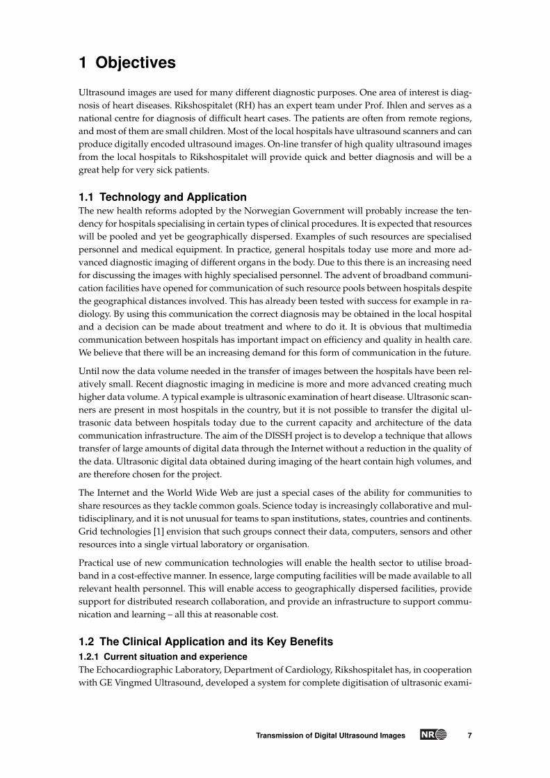
Figure 1. Work flow at cardiology department.
2 Technical Background and Requirements
We investigated a number of issues such as inter- and intra hospital network infrastructure, USscanner export and import functionalities, US data format and transmission, standards and rou-tines at departments and hospitals. Subsequently, in the course of these investigations we notedthat routines of handling messages and data from other hospitals lack standardised practise. Inthe following we outline systems and practise used at the departments of cardiology and radiol-ogy.
2.1 Present SolutionsPresent solution at the Department of Cardiology The department of cardiology at Rikshos-pitalet uses US scanners from GE Vingmed Ultrasound AS, Horten, Norway. The scanners areconnected in a separate private network called Echo-net. It has options for storage and disk back-up facilities. Workstations and PCs, operating on Microsoft Windows XP, are connected in theEcho-net. The EchoPAC software, delivered by GE Vingmed Ultrasound, is used for accessingUS data from databases, viewing and analysing images and cine-loops, and creating examina-tion reports. It provides also the possibility to export and import US raw format data (a propri-etary format by GE Vingmed Ultrasound) to DICOM1 in JPEG2 compressed format. This optionmakes it possible to export and import US data from DICOM servers, e.g., a PACS. However,in the present configuration this option is not active. Images that are exported to a PACS musthave valid DICOM fields for UID (unique ID), name, personnummer, date of birth, gender, etc.preferably obtained from the hospital information system (HIS). In the present configuration ofEchoPAC, worklist is not active.
Present solution at the Department of Radiology. The department of radiology at Rikshospi-talet implemented PACS in 2002. The PACS is delivered by Sectra AB, Linköping, Sweden, whilethe radiology information system (RIS) is delivered by Kodak Inc, USA.
The routines at the department of radiology are somewhat different from that in the departmentof cardiology, since they have different requirements and raw image formats for analysis andviewing purposes. The department of radiology has a streamlined system where PACS, RIS, and
1. Digital Imaging and Communications in Medicine.2. Joint Picture Expert Group.
Transmission of Digital Ultrasound Images 11
MITRA
WORKLIST
Modalities
RIS PACS
HIS
Images
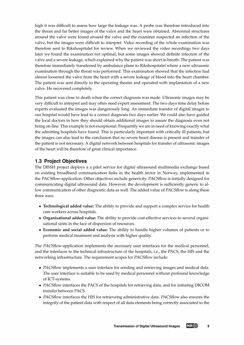
Figure 2. Setup of RIS and PACS at Sørlandet Sykehus i Arendal.
PIMS (patient information management system, aka HIS) are interconnected.
Present solution at Sørlandet Sykehus i Arendal (SSiA). We show the setup at SørlandetSykehus i Arendal (SSiA) in Figure 2, where all communication between RIS and PACS is con-trolled by the MITRA broker. SSiA uses the DIPS as HIS system. Both PACS and HIS are availableat the entire hospital, while the RIS parts are only accessible by the departments due to securityreasons. This means that each department (radiology, cardiology, etc.) has their own RIS imple-mented as a closed area of the HIS3.
General routines. When images of a patient are taken, the medical personnel needs to enterthe patient information, including a unique personal ID, e.g., in Norway the person identificationnumber (personnummer). The correct relationship between patient, medical data, reports, andimages is achieved using the HIS. The RIS, which is connected to / a part of the HIS generates aDicomID and the data for the work lists. Additionally the technical parameters, e.g., compressionmethods, must be entered.
When medical images arrive from another hospital by other means than PACS to PACS, the im-ages are not attached to a DicomID. The DicomID must be generated manually. The routinesinclude that a cardiologist writes a referral, the radiology department registers the patient datain the RIS, manually attaches the data to the DicomID, fills out forms4, and acknowledges thedescription in the RIS. For each examination, approximately 5-10 minutes are used to perform theprocedure.
The routines for teleradiology included several manual steps both on sender and receiver side, asshown in Figure 3. The steps include writing the referral. In some cases anonymizing the referral,answers, journal documents, descriptions, type of examination, number of images, etc. The nextsteps include calling the recipient by phone, faxing the data, and sending the images PACS toPACS. The recipient must perform a quality check, register the examination in the RIS, attach theDicomID, scan documents, etc. For this procedure each side uses ca. 30 minutes, since most of thesteps are performed manually. Note, that this procedure also can result in a possible quality loss,since some documents are sent by fax and scanned afterwards.
The number of manual steps on both sides must be reduced, which includes that the documentsare transferred electronically, the quality check must be automated, and smart calling should beintroduced. An integration between PACS and RIS must be considered.
3. Note, there are different routines in radiology and in cardiology.4. At SSiA the form “Underssøkelsen overført Kardiologisk Avdeling 241103” is used.
12 Transmission of Digital Ultrasound Images

Order
print order
and RIS answer
print documents
from HIS/EPR
write letter
Telephone
Fax
PACS
Telephone
Fax
PACS Quality
Check
Register
in RIS
relation
PACS-RIS
document
scan
notify
recipient
view
documents
second
opinion
new
report
OK
ERR
SENDER
RECEIVER
Figure 3. Sketch of cardiology workflow at DISSH project start. Note that PACS and RIS are very recently in-troduced (independent from the DISSH project), and data transport on CD, and manual routines for retrievingother information were employed just at project start.
2.2 Standards and Formats2.2.1 Image Data FormatUltrasound scanners produce different image formats depending of the application requirements,and the underlying system. GE Vingmed Ultrasound scanners produce image data in a propri-etary image format, which can be exported to DICOM supporting supporting lossy and losslessimage compression.
DICOM [2] is used for exchanging, storing, and retrieving medical images, including imagerywithin radiology and ultrasound cardiology. DICOM is a standard by the American College ofRadiology (ACR), and the National Electrical Manufactures Association (NEMA). The DICOMstandardisation documents are available at http://medical.nema.org/dicom. The first versionof DICOM was released in 1985, the second in 1988, and the third in 1993.
The DICOM standard defines the semantics of commands and associated data, the semanticsof file services, file formats, and information directories, and operation in a networked environ-ment. It consists of sixteen parts5, covering various subjects including information objects, ser-vice classes, data structure encoding, data dictionary, message exchange, network communicationsupport, media storage, file formats, and security profiles.
DICOM defines information objects for images, and for administrative data, e.g., patients, stud-ies, reports, and other data groupings. DICOM specifies data formats, a network protocol utilisingTCP/IP, defining the operation of service classes beyond the simple transfer of data, and a mech-anism for uniquely identifying information objects.
2.2.2 Image CodingImage coding standards provide solutions to compress, decompress, and error concealment foreffective storage and transmission of raw image data. Currently, medical image data can have
5. including two parts which are retired.
Transmission of Digital Ultrasound Images 13

resolution up to 16 bits per pixel. Within radiology the only acceptable compression standard hasbeen lossless scheme, with a typical file size reduction by a factor of 2–3. The coding standardssupported by DICOM include raw raster image, JPEG6, JPEG 2000 [3], and JPEG-LS [4, 5]. Thefollowing image coding packages were investigated as candidates:
Sharpbyte. The Irish company, Sharpbyte Ltd (www.sharpbyte.com) offers a product, whichuses a patent claimed technology for medical applications. The Sharpbyte solution was evaluatedby Rikshospitalet within the frame of the project (See Appendix C). The product seems suitablefor our project with respect to encoding requirements. However, the price tag seems too high.This solution would be non-standard with respect to PACS.
Huffyuv. Huffyuv is an implementation for a lossless video encoder. See Appendix B.1.1.
JPEG 2000. The JPEG 2000 standard (ISO/IEC 15444-1) uses wavelets and arithmetic codingfor providing lossy and lossless compression methods for images. The list of implementationsof the JPEG 2000 codec includes an implementation by Norsk Regnesentral for JPEG 2000 andMJPEG 2000, J2000, JJ2000, Kakadu, and Jasper. See Appendix B.1.2 for a description of the soft-ware packages.
JPEG-LS. Developed originally by HP for lossless compression in medical imaging applica-tions. The codec uses a combination of prediction codes and Huffman entropy coding. The ref-erence implementation is from Hewlett-Packard (see Appendix B.1.3). Other implementationsinclude the codec from the University of British Columbia, and the implementation by D. Clunie(see Appendix B.1.4).
2.2.3 Message and report formatsWhen investigating a medical case images and associated examination reports containing variousinformation about the patient are required. Presently, DICOM does not support such examinationreports to be included. Although using private tags in DICOM might be an option, this has so farnot been used, since the use of these features need changes in the setup at every site they areused. Recognising this problem, the DICOM committee has started working on a solution calledDICOM structural reporting (DICOM S/R), which still is an ongoing work.
Within one hospital there might be many different electronic patient record (EPR) systems avail-able. The most used major EPR systems in Norway are DIPS, InfoMedix, and DocuLive [6]. Thedocuments and messages created by proprietary systems are difficult to incorporate in applica-tions delivered by other vendors. To address this issue, IHE (Integrating the Health care Enter-prise) was established in the US (http://www.rsna.org/IHE), and is being considered in othercountries, e.g., in Norway.
EDI (Electronic Data Interchange) works by providing a collection of standard message formatsand element dictionary in a simple way for businesses to exchange data via any electronic messag-ing service. XML (extensible markup language) together with EDI provides a standard frameworkto exchange different types of data – for example, an invoice, health care claim, project status – sothat the information be it in a transaction, exchanged via an Application Program Interface (API),web automation, database portal, catalogue, a workflow document or message can be searched,
6. ISO/IEC IS 10918-1, ITU-T Recommendation T.81.
14 Transmission of Digital Ultrasound Images

decoded, manipulated, and displayed consistently and correctly. The supporting standards in-clude DICOM [2], HL7 (see http://www.hl7.org), and XML. KITH (http://www.kith.no) pro-vides reports and recommendations that health care system vendors in Norway use as guidelinesfor respective systems.
2.2.4 Data TransferA typical transfocal echo US examination can produce several hundreds of Mbytes of data. Aframe rate of 100–150 fps is common. Taking into account of image resolution, bits per pixel, fullcolour (RGB), and frame rate, it is evident that images need to be compressed prior to transferringon a bandwidth limited network.
DICOM [2] is used to transfer the images between PACS. DICOM uses TCP/IP as the underlyingnetworking protocol. The IT departments of the hospitals provide secure data transport betweenthe PACS. Whether security is achieved by using secure lines (e.g., ssh-tunnels, VPN), or by usingDICOM Part 15 [7] is beyond the scope of our project. In the DISSH-Project a VPN-solution isused.
Besides the medical images, administrative data and notifications must be transferred. These datacontain information on the patient, and descriptions of the medical case including diagnosis in-formation. This type of information is confidential and sensitive, and security policies apply forthe transfer between hospitals. The policies of the hospitals require that sensitive medical datacannot be sent by email.
The networks of the hospitals in Norway where the clinical data can be accessed are separatedfrom publicly accessible networks. Clinical and sensitive data cannot be accessed from the outsideof these networks. Strict policies, controlled by the Norwegian Data Inspectorate (Datatilsynet),apply to data exchange between hospitals. The closed networks of the hospitals are intercon-nected by the national health care network (nasjonalt helsenett).
The hospitals of Norway are using the AMTrix message broker system for communication bothinside the hospitals and between each other7. The AMTrix installation defines policies and rulesapproved by the Norwegian health authorities for different types of information.
2.3 Security and Integrity RequirementsThe standard ISO/IEC 17799 [8] is relevant for security and integrity requirements. The key as-pects of Information Security are to preserve the confidentiality, integrity and availability of anorganisation’s information. All three properties are relevant for the DISSH-project, since loss ofone or more of these attributes can endanger the performance of a hospital.
Within the DISSH-project most of the security issues are covered by the infrastructure of the hospi-tals. Within the hospitals the security requirements are moderate, while the security requirementsbetween hospitals are very strong. Confidentiality and integrity are achieved by using securelines, VPN, and other suitable middleware, which are beyond the scope of our project.
Patient data and medical data are considered confidential. Since the SMTP protocol is consid-ered non-secure by the health authorities, these data are not allowed to be sent over email. Fortransfer of confidential messages the AMTrix message broker must be used, which is part of theinfrastructure of/between the hospitals.
7. See the entry on AMTrix in the glossary in Section A
Transmission of Digital Ultrasound Images 15

2.4 System Integration and Tool SelectionThe DISSH project to used standards, tools and routines acknowledged for the use in health carein Norway. The introduction of new routines must be done carefully, and in full accordance withthe policies of the involved personnel and the health care authorities. Wherever possible existinginfrastructure with a perspective in the future was used. We also intended to use technologieswhich will be mostly compatible with future standards in health care, where they are not yetdecided (e.g., HL7, XML-based patient records, DICOM S/R). The following policies are used:
• The DISSH project uses DICOM for storing images, and transfer of medical images betweenPACS of the involved hospitals. Where implemented, PACS of the hospital installation willbe used; For hospitals without a PACS implementation a PACS must be chosen.8
• The DISSH project builds on hardware and operating system policies of the hospitals. Thisincludes that PACSflow is implemented as independent from these issues as possible. Weprovide both a solution for both Linux and a Windows environments.
• For messaging the use of the networking infrastructure is done according to the policies ofthe hospital. This includes the use of the AMTrix message broker.
• Since the capacity of the interconnection between the hospitals is large enough, there is noneed for using other coding than the PACS and the DICOM standard offer.
• Upon arrival of medical data the medical personnel must be notified. Since security policiesin hospitals do not allow for sending email containing patient data, we considered (a) to useDICOM structured reporting, which contains the messages in the receiver headers9, or (b) touse the message broker middleware when sending the messages from the sender-side.
8. The choice of a PACS is beyond the scope of this project. However, for open source alternatives we provide a shortoverview in Section B. The DICOM Toolkit (see Section B.2.1) seems to be a suitable alternative, possibly in connectionwith the CDmedic distribution (see Section B.2.4).9. DICOM gives the possibility to define new tags, which could be used in our case. However, this would result in anon-standard implementation, resulting in potential compatibility problems.
16 Transmission of Digital Ultrasound Images

Patient
Doctor #1
Camera
future scenarioVideo Conference
Doctor #2
PC#2
DISSHserver #1
DISSHserver #2
Scanner / PC#1
Middleware
PACS PACS
Figure 4. Illustration of the DISSH use scenario.
3 The PACSflow Application
3.1 The Use-ScenarioThe scenario illustrated in Figure 4 is basis for our decisions on system architecture. The actorsand facilities at the sending hospital are marked with #1, while the actors and facilities at thereceiving hospital are marked with #2. The scenario in more detail is as follows:
1. The medical image data are taken from a scanner and stored in the PACS database in DICOMformat at Hospital #1.
2. Doctor #1 of Hospital #1 requests medical image data to be sent to Doctor #2 / Departmentat Hospital #2 for medical analysis. Doctor #1 fills out a form on PC #1, and the form is sentto DISSH server #1.
3. DISSH server #1 retrieves medical image data from PACS database, transfers it to DISSHserver #2, which stores the data in the PACS of Hospital #2.
4. The DISSH software creates a notification message, containing the medical case, that is sentto the mailbox of Doctor #2.
5. Upon receipt of the message Doctor #2 can access the medical image data from the localnetwork of Hospital #2.
3.2 System ArchitectureThe DISSH-software consists of the distinct parts which are explained in the following. Addition-ally, the infrastructure of the hospitals, and between the hospitals are used, which include thePACS, the message broker middleware, and the communication infrastructure.
• The PACSflow-web application is used to provide a user interface to the medical personnel,using a standard web browser for interaction. The doctor at Hospital #1 enters the patientand study, including medical information on the case.
• The web application interfaces the PACS by using commands of a toolkit accessing DICOM.• The DISSH application generates messages for the medical personnel, containing the ID for
the study, and medical information on the case.• The doctors at the receiving hospital access the data by using a browser. The application is
controlled by the web application. The viewing functionality of the images itself is providedby software of the PACS.
3.3 Platform ChoicesSince the hospitals in Norway use some flavour of the Windows operating system as the defaultplatform, it is essential that the PACSflow-application runs on a Windows platform. However,
Transmission of Digital Ultrasound Images 17

imagectn imagectn
storescu
findscu
movescu
findscu
+ retrieve
PACS PACS
Figure 5. Illustration of DCMTK used in DISSH.
since Linux is used to an increasing extent, we implemented the application to be interoperablewith Linux as far as possible. Most parts of the development were performed on a Linux platform,before they were implemented in the real environment at the hospitals.
It was decided to implement a web application, which implies that most parts of the user interfaceare handled in a web browser, while a web-server handles the requests, and interfaces the PACSand the other infrastructure. The web-application can later be integrated in a portal solution.
For the implementation of a web application several possible technologies can be used. We consid-ered using technologies based on CGI-scripts using programming languages like Perl and PHP,which are platform-independent, and relatively simple to implement. We also considered usingframeworks like .NET and J2EE, which use web-services. These already contain support for XML,which would simplify the implementation of messaging over XML, as suggested by some of theinitiatives for information exchange in health services. However these frameworks require thesetup of a special application server, and the implementation would be platform-dependent.
We decided to base the implementation of PACSflow on PHP, whose run-time environment canbe easily installed on both Windows and Linux platforms. PHP applications can be run on bothApache and MS IIS web servers.
3.4 PACS FunctionalityThe medical images are stored in a PACS, and the transfer of images is implemented as a transferof data between PACS. Rikshospitalet and SSiA both use the Sectra PACS within radiology, whichsupports transfer and decoding of JPEG-encoded DICOM files according to their conformancestatement.
Partner hospitals which do not already have a PACS in use, can implement this functionalityusing open source software. Suitable choices are: the MIR CTN, (see Section B.2.2), the imageCTNof DCMTK (see Section B.2.1), and the PACS of the Java DICOM Tools (see Section B.2.7). MIRCTN implements most functionality; while imageCTN and DCMTK already contain JPEG-relatedcode.
Since the involved hospitals are using the GE VingMed ultrasound scanner PACSflow must beable to store and process files generated by this equipment, which produces compressed imagesusing the the JPEG baseline transfer syntax. The Sectra PACS is capable of storing, sending andreceiving this type of images. However, the imageCTN does not support the JPEG Baseline trans-fer syntax for the compressed images. The MIR CTN does not accept data via the JPEG baselinetransfer syntax. The Java DICOM Tools support the JPEG baseline transfer syntax.
Interaction with the PACS involves DICOM commands, e.g., C-FIND, C-MOVE, or C-STORE.10
10. In the software DCMTK the programs findscu, storescu, and movescu can be used to call these functions from a shell
18 Transmission of Digital Ultrasound Images

In order to demonstrate the transfer functionality outside the protected areas of a hospital, weused the DICOM toolkit (DCMTK), which runs on Linux and Windows platforms, as follows (seealso Figure 5):
• On two machines each we set up an image base (CTN) with the application imagectn, whichimplements the C-FIND, C-MOVE, C-GET and C-STORE service classes.
• We implemented a program to list all available DICOM files in a image base, using the appli-cation findscu. The resulting list of files can be used to select files for transfer later.
• We implemented a program to initiate transfer of a given DICOM file to a remote CTN.
3.5 Notification and Messaging FunctionalityMedical personnel are notified by PACSflow upon arrival of a new medical case, i.e. the ultra-sound images have been transferred. The doctors at the receiving hospital can access the descrip-tion of the medical case, and diagnostic data attached. Due to security reasons we cannot useemail for transport of these messages, but make use of other message handlers supported in theinfrastructure of the hospitals.
The implementation of messaging within the health-care domain is subject to several national[9] and international projects, e.g., HL7 and IHE. Since these efforts have not yet concluded, weimplement our own scheme.
3.5.1 Messaging using DICOM headersA solution using solely DICOM mechanisms would include: The (email) address of the receivermust be included into the DICOM file. This solution includes to add information to the DICOMfiles, extracting this information, and use this information in order to notify the receipt of a file.
One way to accomplish notification is to use tags in the DICOM header for addressing. For thispurpose private tags and existing tags could be used. Several fields in DICOM refer to physi-cians. The most relevant is the field “Referring Physician”, which is the physician asked for a sec-ond opinion. Other relevant fields include “Physician approving interpretation”, “Physician(s) ofrecord”, and “Name of Physician(s) reading study”.
This approach is non-standard causing possible problems, since the receiving physician will notalways have the same role, and methods for notification based on the fields in the DICOM-headermust be built into the receiving PACS. The use of DICOM S/R (which uses standardised DICOMheaders) has been considered. However, since the working groups have not concluded their workyet, we did not implement this approach.
3.5.2 Messaging outside DICOMAs an alternative to the above, we use a separate communication protocol, where the message isput together, either as an XML message, or as a pure text message. The message must be assem-bled on the sender side, since the message content is not available on the receiver side.
Since the DICOM protocol ensures that the status of the transfer of the images is returned to thesender when the transfer is completed, it is not necessary to synchronise with the reception of theimages. Therefore, the notification can be issued either on the sender side when the message issent, or on the receiver side when the message is received.
command interface.
Transmission of Digital Ultrasound Images 19

PACS
PACS
report(text+images)
EPR
second opinionDecision
Production System
second opinion
Examinationof images
report on examinationtreatment decision
PACS
Hospital A Hospital B
Examinationof images
EPR
Figure 6. Components and control flow (straight lines) and data flow (dashed lines) in PACSflow.
3.5.3 Implementation of MessagingThe messages transfered to the receiving department use the AMTrix message broker system. Itsuse is approved with regard to the security requirements for sending sensitive patient informa-tion.
After the transfer of the image series has succeeded the web-application stores a message in adedicated folder on the web server. This folder is checked regularly by AMTrix whether a newfile has arrived. If so AMTrix fetches this message using FTP. AMTrix then forwards this mes-sage according to header informations to the installation that serves the recipient. The receivingAMTrix installation then uploads the message to the receiving web-server, again using FTP. Thereceiving AMTrix also sends an email notification locally to the email addresses specified in theheaders.
Several formats for the message are possible, including several XML formats. For the sake ofsimplicity we use an ASCII text representation for the message, since the XML formats have notyet been standardised. Defining XML formats for messaging is implemented elsewhere [9].
Each message contains a header formatted in ASCII text, the message, and optionally an attach-ment. The ^-character is used as a separator between the different fields, which are interpreted byAMTrix. A message has the following fields, which are explained in Table 1:
SENDER^RECEIVER^RECV_EMAIL^EXTRA_RECV_EMAIL^TIMESTAMP^ID^TITLE^MESSAGE[^ATTACHMENT]
3.5.4 NotificationOn basis of the contents of the message a notification is assembled by the receiving AMTrix. Cur-rently in PACSflow this notification is sent through e-mail, and therefore may not contain sensi-tive information. The notification contains the timestamp, sender department, message title, andmessage ID. Using the message ID it is easy to retrieve the correct message in the web-application.
20 Transmission of Digital Ultrasound Images

Field DescriptionSENDER AMTrix-code denoting the sending department.RECEIVER AMTrix-code for the receiving department.RECV_EMAIL Email-adress for notification.EXTRA_RECV_EMAIL Extra email-address for notification. This field can be blank.TIMESTAMP UNIX timestamp of time when the message was sent.ID Message ID. Format: DATE-TIME-PATIENT INITIALS. Example:
100504-101133-TH.TITLE Message Title.MESSAGE The main message.ATTACHMENT Attachment encoded in BASE64.
Table 1. Description of message fields
3.6 Web ApplicationThe Web Application implements the user interface, and the control of the commands to thePACS, e.g., initiation of data transfer, and messaging. The Web Application also contains function-ality for lookup-tables, in order to hide unnecessary technical details from the medical personnel.These lookup-tables implement user data, departments, location of data, CTN name, ports, etc.
3.6.1 ImplementationThe PHP scripts of the web-application generate the HTML code that is viewed in the browser.Functionality that makes the application behave more dynamically, is implemented using JavaScript.The application contains of three modules, one module for sending image series and messages,one module for viewing received messages, and one module for the administration of user data.
The most important windows of the user interface are shown on the front page (login window),and in Figures 8–12. The main window for sending medical images is shown in Figure 11 In-formation about sender and receiver is retrieved from the user database. From this window it ispossible to open a dialogue for searching and retrieving image data from PACS. The sender fillsout the form, which is converted into the message sent to the recipients. Additional documentscan be attached to the message, e.g., a report exported from EchoPAC or data exported from apatient information system.
The details of the message transfer are described in Section 3.5.3. After the message has arrivedat the receiving department, the web-application offers an interface for reading the received mes-sages, and viewing the attached files.
3.6.2 SecurityThe web-application is secured by that the user has to log in using a username and password.This is handled in PHP, and it makes sure that no non-authenticated user can access any partof the web-application. Further security issues can easily be added, since standard software andstandard protocols are employed.
3.6.3 Work flow and Information flow in PACSflowIn Figure 6 we show the overall data flow and control flow for the PACSflow application. Notethat the production system (e.g., ultrasound scanner) is beyond the scope of the DISSH project.However, it is required that PACSflow can use the images produced by the production system,which are inserted into a PACS using DICOM. When a doctor uses the PACSflow an electronicform will be filled out including diagnosis data. After the doctor at the receiving hospital is no-
Transmission of Digital Ultrasound Images 21

PACS
Messaging
MessagingEPR
MessagingEPR
Messaging
PACS
Decisionsecond opinion
Web page− image id− text (email)− address of physician
DICOM image
image id
text ?
Mail/Web/Dicom Browser− browse DICOM by image id
Create diagnosis− text, images
Hospital A Hospital B
further diagnosis
address of receiver
Figure 7. Detailed flow graph for PACSflow.
Figure 8. User interface for searching after patients in PACSflow.
tified, he can access the images and perform a second opinion, which is sent to the requestinghospital.
Figure 7 shows in more detail how the information flows are designed. PACS, EPR, and messagingmodules in both hospitals are important parts in this diagram.
After the decision of requesting a second opinion is taken, an electronic form completed and theinformation is sent to the PACSflow web application. The electronic form contains the followinginformation, which partially are collected from the HIS/EPR:
• study ID of the DICOM images;• sender (including phone numbers, department, etc.);• address of receiver or receiving department;• details about the case, and what the receiving physician is requested to do.
22 Transmission of Digital Ultrasound Images

Figure 9. Results from the search after patients are presented. When several candidates are available theright patient can be selected.
Figure 10. Show the medical images available in the PACS for a patient. The Case(s) to be transferred canbe selected.
After the data are transferred to the Hospital #2, the doctor #2 will receive the following data inorder to perform the second opinion:
• a notification about a new case arriving;• the DICOM object(s) transferred to PACS at Hospital #2;• a message containing ID of the DICOM object, together with with patient information, and
information about the case #1.
After the second opinion is performed the result of the examination is sent back as a message toHospital #1, where the information is added to the HIS/EPR of Hospital #1.
Transmission of Digital Ultrasound Images 23

Figure 11. User interface of PACSflow for sending medical images. For some fields sub-windows pop up.
Figure 12. User interface for chosing departments that can receive medical images.
24 Transmission of Digital Ultrasound Images

SenderWeb Client
WebApplication
PACS 1 PACS 2 Doctor #2(Receiving images)
Patient idinformation
search patient id C-Query
Patient id searchstudies
patient id
C-Query
study list
study id
add receiveradd data onmedical case
press send call C-MOVEC-MOVEimage transfer
image transfer
finishedok?errormessage
error msgemail
receipt tosender
nei
ja
sendmed data
med descr.arrives
AMTRIX
notificationto receiver
read notificationMESSAGING Anon sensitive information
open dicom image
read med descr.
messaging B
note: messaging A sends non-sensitive information (image identification) between hospitals.instead messaging B could be implemented.
work flow ofdoctor #2 starts here
Figure 13. Work flow for implementation of PACSflow.
Transmission of Digital Ultrasound Images 25

4 Medical Tryout and Conclusions
The PACSflow-application is now implemented at Rikshospitalet and SSiA. Since the medicaltryout phase only has been started recently, we cannot give yet comments on the effectivity of oursolution.
The work in the DISSH-project resulted in the implementation of the PACSflow-application, whichprovides a user interface for doctors at hospitals to transfer medical images to another hospitalusing a web browser. The application has the potential to simplify the image transfer, shortendown response time for medical answers, and to reduce treatment costs in general.
While working with the project we recognised that the manifold of standards, applications, sys-tems, providers, and routines in all domains of health care was much more complex than firstassumed. Many of the systems in health care are not compatible with each other. This observationincludes image formats, messaging, modalities, information systems, etc. The application PACS-flow provides a simple interface that hides this type of complexity from the user in the domainfor medical second opinions within ultrasound cardiology and radiology in general.
4.1 ExperiencesA cardiac US scanner (Vivid 7, GE Vingmed Ultrasound AS, Horten, Norway), which producesdigitally encoded images and image sequences, was used to acquire US images. The images werecompatible with the digital image communication in medicine (DICOM) format. A visualizationsoftware, EchoPAC (GE Vingmed Ultrasound AS, Horten, Norway), was used to push US imagesto patient archive communication system (PACS) (Sectra AB, Linkøping, Sweden) using DICOMclasses.
The prototype system has been tested from a technical perspective. The department of cardiol-ogy at Rikshospitalet and the department of internal medicine at Sørlandet sykehus in Arendal,Norway are performing clinical use of the system. Cardiac US image sequences were compressedusing lossy JPEG standard. This reduced the file size by a factor of 3. Initial test indicates that anexamination containing image sequences of 184 MB data required 8.3 minutes to transfer fromArendal to Oslo using PACS to PACS communication on an effective 3 Mbits/s channel.
A clinical evaluation study of the system will be presented in a paper shortly. After the clini-cal evaluation phase the PACSflow-application will be offered to other hospitals, and undergo acommercialisation.
26 Transmission of Digital Ultrasound Images

A Glossary
We present a list of relevant terms, acronyms, systems, standards and procedures employed inhospitals in Norway. Reports on some of the subject are available from KITH (http://www.kith.no).
HIS: Hospital Information System; information system used in hospitals for administrative patient-data and/or EPR. At Rikshospitalet the system DocuLive EPR (Siemens) is used, while Sør-landet Sykehus i Arendal uses DIPS. In the hospital-sector in Norway the systems DoculiveEPR, DIPS, InfoMedix are used; see http://kvalis.ntnu.no/index.htm for an overview.
EPR: Acronym for Electronic Patient Record; identical to the term EPJ used in Norway.
EPJ: Acronym for Elektronisk Pasient Journal; norwegian word of EPR.
DIPS: An implementation of RIS/EPJ; used at Sørlandet Sykehus i Arendal.
PAS: Acronym for Patient Administrative System.
PACS: Acronym for Picture Archiving and Communication System.
DICOM: Digital Image Communication in Medicine; standard for exchange of image informa-tion between modalities11 and systems from different producers. A European branch calledMEDICOM exists as a pre-standard.
HL7: Health Level 7, american initiative; see www.hl7.org. HL7 is a medical information systemsstandard.
IHE: Acronym for Integrating the Healthcare Enterprise, an initiative by the Radiological Societyof North America (RSNA) and the Healthcare Information and Management Systems Soci-ety (HIMSS) (see http://www.rsna.com/practice/dicom/index.html). The IHE technicalframework defines a common information model and a common vocabulary for systems touse in communicating medical information. Ith specifies how DICOM and HL7 are to beused by information systems to complete a set of well-defined transactions that accomplisha particular task. See also the articles series by Siegel, Channin et.al. [10].
RIS: Acronym for Radiology Information System which is used to support information handlingin a radiology department.
EDI: Acronym for Electronic Data Interchange.; uses EDIFACT standard.
AMTrix: Communication middleware and integration broker for business transactions; used inNorwegian hospitals for messaging, etc. AMTrix is produced by Axway (www.axway.com)maintained by Communicate AS (www.communicate.no) in Norway.
Helsenett: Norway is divided into five health regions (Helse Nord RHF, Helse Midt-Norge RHF,Helse Vest RHF, Helse Sør RHF, Helse Øst RHF), which form together Norsk Helsenett (seewww.norsk-helsenett.no). The network ties together hospitals, doctors and other enter-prises in the health sector.
SECTRA PACS: PACS system delivered from the swedish company Sectra AB (See www.sectra.se). used at both Rikshospitalet and Sørlandet Sykehus i Arendal.
EchoPAC: System from the company GE Medical Systems (see www.gehealthcare.com) to store
11. A modality is defined as a group or type of imaging units, e.g., CT or MR.
Transmission of Digital Ultrasound Images 27

medical images, especially in echocardiographic applications. The system is used at Rikshos-pitalet.
DicomID: Identificator to join patient identity, images and reports. Other names used are: Di-comNR, Accessesionsnr, Tilgangsnr, and HenvisningsID.
MITRA: Broker system; data base program which facilitates communication between RIS, PACSand modalities.
B Resources – Software and Links
B.1 Software for Image CodingB.1.1 HuffyuvHuffyuv is an open source project by Ben Rudiak-Gould for implementing a lossless video en-coder. After having vanished for a while, the source code of Huffyuv is now available again atneuron2.net/www.math.berkeley.edu/benrg/huffyuv.html.
B.1.2 JPEG 2000The JPEG 2000 standard (ISO/IEC 15444-1) uses wavelets and arithmetic coding for providinglossy and lossless compression methods for images. The list of implementations of the JPEG 2000codec includes:
• Norsk Regnesentral have implemented an encoder for JPEG 2000 and MJPEG 2000.• The J2000 codec (http://www.j2000.org/) is an open source codec for JPEG 2000, written in
C and running on several platforms (Windows, Linux, Unix, Plan9).• JJ2000 (http://jj2000.epfl.ch/) is the official reference implementation, written in Java. It
supports features that did not become a part of the standard, and is suitable for small devicesand small pictures.
• Kakadu (http://www.kakadusoftware.com/) is a complete implementation of the JPEG2000standard with good performance. Licenses are available for ca. US$150 (single user non-commercial), ca. US$ 700 (multi-user non commercial), and US$ 7000-14000 (commercial li-cense).
• JasPer (http://www.ece.uvic.ca/~mdadams/jasper/) is an open source implementation inC.
B.1.3 HP LOCO-I/JPEG-LSLOCO-I, available at http://www.hpl.hp.com/loco/, is a reference implementation of JPEG-LSstandard by HP for lossless compression in medical imaging applications. Only binaries available.However, source code for JPEG-LS is available from http://www.ece.ubc.ca/spmg/research/
jpeg/jpeg_ls/jpegls.html.
B.1.4 JPEG-LS by D. ClunieJPEG-LS, available from http://www.dclunie.com/jpegls.html, is an open source implementa-tion of the proposed JPEG-LS standard, DIS 14495; see http://www.jpeg.org/public/jpegnew.
htm. The implementation is part of the dicom3tools package for medical images. The implemen-tation handles one component (i.e., grayscale images) in the current version (dated 2002).
28 Transmission of Digital Ultrasound Images

B.2 Open Source Software for DICOM handlingSince open source software (OSS) [11] is available in source code which can be altered, this typeof software is suitable for experimenting without causing great costs for licenses and purchase.We give an overview of some relevant packages, most of them implementing some aspects ofDICOM.
B.2.1 Dicom toolkit (DCMTK)DCMTK, available from Offis at http://dicom.offis.de, is a collection of libraries that imple-ment large parts of the DICOM standard, written in a mixture of ANSI C and C++. DCMTK isreleased under a BSD-style license by Offis (a German research institute), and is used often forresearch, and as a basis for commercial software. DCMTK works on Windows, Linux, Solaris,OSF/1, IRIX, FreeBSD and MacOS X. DCMTK includes also an implementation of lossless JPEG,and a module for transfer of DICOM data via TLS using OpenSSL.
B.2.2 MIR CTN (Central Test Node)The MIR DICOM Central Test Node Software (CTN) from ERL at Washington University avail-able at http://www.erl.wustl.edu/, implements aspects of DICOM for cooperative demonstra-tions at RSNA annual meetings. The goal was to provide an implementation that facilitated ven-dor participation based on the evolving DICOM standard. CTN is implemented in C, and workson Windows, and (possibly) Linux.
B.2.3 David Clunie’s dicom3toolsThe dicom3tools, available from http://www.dclunie.com/dicom3tools.html implement toolsand libraries for handling offline files of DICOM 3 attributes, and conversion of proprietary for-mats to DICOM 3. Can handle older ACR/NEMA format data, and some proprietary versions ofthat such as SPI. Works on Mac, and Linux. (And maybe windows.)
B.2.4 CDmedicCDmedic, available from http://cdmedicpacsweb.sourceforge.net, implements a full featuredfree PACS based on MIR CTN, DCMTK and mysql, with remote administration using apachemod perl and imaging processing capabilities using ImageMagick, Grevera’s dcm2pgm DICOMconverter and AFNI. CDmedic is based on the Linux-distribution Knoppix. Released with theGPL license.
B.2.5 OpenEMROpenEMR, available from http://stack.onlyic.org/openemr/, is a modular, HIPAA compli-ant, Open Source, cross-platform Electronic Medical Records system (EMRS). It facilitates effi-cient office management through automated patient record journaling and billing integration, andhas been integrated with third-party technologies including speech recognition, secure wirelessaccess, touch screen portables, and biometric authentication. Interface screens are customizableand optimized for consistency, simplicity, speed of access to patient information, and minimumeye strain. OpenEMR works on Linux and Windows platforms, and is released under an OSI-approved license.
B.2.6 Conquest DICOM softwareWithin the EC Conquest project, (see http://www.xs4all.nl/~ingenium/dicom.html) a full fea-tured DICOM server has been developed based on the public domain UCDMC DICOM code de-veloped by Mark Oskin at the Medical Center of the University of California at Davis. Releasedunder a BSD-style license. Works on Windows platforms.
Transmission of Digital Ultrasound Images 29

B.2.7 Java DICOM ToolsThe Java DICOM Tools, available at http://www.tiani.com/JDicom/ implement a collection oftools for DICOM in Java. Built partially on Softlink’s javadicomtoolkit. The use is free of charge,but only for special use. Source-code is not available. The PACS of the Java DICOM tools supportsthe transfer of images using the JPEG baseline transfer syntax.
B.2.8 dcm4chedcm4che, available at http://dcm4che.sourceforge.net/, is an implementation of DICOM inJava. The sample applications may be useful on its own. It also includes an IHE compliant ImageArchive application, based on J2EE.12 Released under the LGPL.
B.2.9 MRIConvertMRIConvert, available at http://lcni.uoregon.edu/~jolinda/MRIConvert.html, is a medicalimage file conversion utility that converts DICOM files to SPM99/Analyze, BrainVoyager, andMetaImage volume formats. It runs on newer Windows platforms. MRIConvert was written usingwxWindows, an open-source library for cross-platform GUI development. Free use, but source-code not available. Binary for windows available.
B.3 Dicom viewersWe present DICOM viewers in a separate section. In the DISSH project it was considered to useDICOM viewers that can be started directly from the web application.
B.3.1 DicomviewerDICOM Viewer, available at http://mars.elcom.nitech.ac.jp/dicom/index-e.html, is usedto access DICOM data that are stored on a server. It is written in Java, by Nagoya Institute ofTechnology, Iwata laboratory and Takahiro Katoji. The software is under the GPL; however thereis an additional claim that the URL of the web page, or the reference to the article [12] are in-cluded. Dicomviewer does not seem to be capable of showing the images from VingMed, neitherin compressed nor uncompressed format.
B.3.2 DicomworksDicomworks is a DICOM viewer for Windows, available at http://dicom.online.fr/, devel-oped by radiologists. Can open most kinds of DICOM-images, also compressed images, and se-ries of images. The program is rather slow, and uses much of system resources. The software canbe downloaded for free. A registration is required to get full functionality. Support for DICOMquery/retrieve is planned.
B.3.3 MedviewMedview for Windows, available at http://www.viewtec.ch/meddiv/medview_e.html, was nottested by us, but it seems to offer much functionality, and support for most DICOM files. Licensecosts ca. 300 Euro.
B.3.4 TomovisionTomovision for Windows, available at http://www.tomovision.com/download/tomovision.htm,offers a simple user interface with not so much functionality. Can open all sorts of DICOM images.Freeware.
12. Software is of alpha quality.
30 Transmission of Digital Ultrasound Images

B.3.5 ezDicomexDicom for Windows, available at http://www.psychology.nottingham.ac.uk/staff/cr1/ezdicom.html has quite good functionality. Can open all sorts of images, but gives wrong colours on someimages. Open source software.
B.3.6 Rubo Medical Imaging DicomviewerRuboMed for Windows, available at http://www.rubomed.com, offers not so much functionality.Can open all sorts of files. Costs ca. 1000 Euro per license.
B.3.7 AccuLiteAccuLite for Windows, available from Accuimage at http://www.accuimage.com/ can open mostsorts of images, but has troubles with some. Freely downloadable.
B.3.8 Dicomviewer (Nagoya Institute of Technology)The Dicomviewer by Nagoya Institute of Technology (http://mars.elcom.nitech.ac.jp/dicom/index-e.html) is Java-based, which can be used on all platforms. It is specially designed for usein a web-server with a suitable user interface, and can open all kinds of DICOM files. However, itis rather slow. GPL license.
B.3.9 Imread Dicom viewerThe Imread DICOM viewer (http://www.uchsc.edu/sm/neuroimaging/download/imread/imread1.htm) is Java-based. Works with simple DICOM images, but does not support compressed images,or series of images. Freely downloadable.
B.3.10 DicomScopeDicomScope for Windows from Offis, availabe at http://dicom.offis.de/dscope.php.en, offers amessy user interface. Does not support compressed DICOM images. Freeware.
B.3.11 OsirisOsiris, available at http://www.expasy.org/www/UIN/html1/projects/osiris/osiris.html, isa DICOM-viewer for Windows and UNIX. Does not support compressed images. Freeware.
B.3.12 Dicom Image Viewing SoftwareThe DICOM Image Viewing Software (http://www.ee.bilkent.edu.tr/~cetin/dvs.html) is aDICOM-viewer for Windows. The software did not work in our environment.
B.4 Commercial SoftwareThe following enterprises deliver commercial software for image handling, including commercialPACS implementations, etc.:
• Sectra PACS (http://www.sectra.se).• InfoMediQ (http://www.infomediq.com)• Medview http://www.viewtec.ch/meddiv/medview_e.html
• Digit Médic http://users.skynet.be/digitmedic/• http://www.siliconmindset.com
• http://www.tomovision.com
• http://www.mallinckrodt.com/
• http://www.medicalconnections.co.uk/
• http://www.softlink.be/javadicomtoolkit.htm
Transmission of Digital Ultrasound Images 31
• http://www.thecriswells.net/YourDICOM/
• http://www.leadtools.com
• http://www.accusoft.com
• http://www.intelerad.com/
• http://www.alitech.com/
B.5 Sites for DICOM Example Images• http://www.excel-medical.com/Waveform.htm
• ftp://medical.nema.org/medical/dicom/MRMultiframe/20030404
B.6 Web Pages of General Interest• http://www.pixelmed.com/ – General site, that contains software samples, and a book by
David A. Clunie.• http://medical.nema.org/ – The DICOM homepage.• http://www.erl.wustl.edu – Homepage of ERL-MIR-WUSTL.• http://rsna.org/ihe – The IHE homepage.• http://www.rsna.org/practice/dicom/ – Survey of software for DICOM.• http://www.idoimaging.com/index.shtml – A database of open-source medical software,
test images, etc.
32 Transmission of Digital Ultrasound Images
C Comparison of Compression Methods
Ultrasound images are used for many different diagnostic purposes. One area of interest is di-agnosis of heart diseases. Rikshospitalet has an expert team under Prof. Ihlen and serves as anational center for diagnosis of difficult heart cases. The patients are often from remote regions,and most of them are small children. Most of the local hospitals have ultrasound scanners and canproduce digitally encoded ultrasound images. On-line transfer of high quality ultrasound imagesfrom the local hospitals to Rikshospitalet will provide quick and better diagnosis and will be agreat help for very sick patients.
Sharpbyte Ltd (www.sharpbyte.com) is an Irish company that claims to have a patent, proprietaryimage compression solution. Their image compression software can operate in lossy as well aslossless modes.
Comparison of lossless image compression algorithms. File sizes of images compressed us-ing Sharpbyte solution are compared with the recent image compression standard, JPEG 2000. Theresults are shown in Table C.1. We did not focus on how the files transferred from one computerto another. In JPEG 2000, VM 7.2 is used. We used ISO standard images having 8-bit resolutions.In Sharpbyte upload client, we chose lossless only/lossless.
The following commands were used for the series of test images:
JPEG 2000 script: ./vm7_compress_32 -i inimage.pgm -o outinmage.cmp -Frev
JPEG-LS script: ./locoe -i inimage.pgm -ooutimage.cmp
In another test with the results in Table C.2 we used ultrasound images from a GE VingMedscanner, which have a resolution of 8 bits.
Conclusions.
• The Sharpbyte software looks easy to use and runs reasonably fast on standard personalcomputers.
• Ultrasound images have high correlation among frames. Such temporal correlation can bestbe exploited using a video compression algorithm than a still image compression scheme.This may provide smaller file sizes and may lead to faster transmission on bandwidth limitednetworks.
Transmission of Digital Ultrasound Images 33

Image Image dim. Orig. file size Sharpbyte 1a Sharpbyte 2b JPEG-LS JPEG 2000
Aerial2 2048× 2048 4.194.321 3.156.978 2.771.594 2.772.925 2.889.770Barbara 512× 512 262.159 235.207 157.584 159.561 162.825Beach 512× 512 262.159 197.213 157.145 156.775 168.394Bike 2048× 2560 5.242.89 3.766.631 2.812.403 2.856.623 3.039.775Cafe 2048× 2560 5.242.897 4.421.226 3.302.204 3.337.277 3.556.043Goldhill 512× 512 262.159 216.400 154.967 154.389 161.257Lena 512× 512 262.159 222.409 138.743 138.809 145.900Tools 1524× 1200 1.828.817 1.570.106 1.203.964 1.213.616 1.257.340Texture_1 1024× 1024 1.048.593 612.991 844.425 845.207 896.401Texture_2 1024× 1024 1.048.593 899.601 701.897 702.557 740.953Woman 2048× 2560 5.242.897 4.024.109 2.899.303 2.917.089 3.012.130Total size 24.897.651 19.322.871 15.144.229 15.254.828 16.030.788% of comp. 100 % 77.6 % 60,8 % 61.3 % 64.4 %
a. Sharpbyte1 = Conventional lossless compression.b. Sharpbyte2 = Sharpbyte Medical lossless compression.
Table C.1. Comparison of file size after compression for a series of test images.
Image Image dim File size Sharpbyte JPEG-LS JPEG 2000
Im1 416× 332 414387 103567 129946Im2 416× 332 414387 101837 127816Im3 416× 332 414387 98844 125946Im4 416× 332 414387 97212 125457Im5 416× 332 414387 97743 125670Im6 416× 332 414387 99761 128392Im7 416× 332 414387 99133 126319Im8 416× 332 414387 98955 126230Im9 416× 332 414387 99063 126237Im10 416× 332 414387 97830 125473Im11 416× 332 414387 97052 124056Im12 416× 332 414387 98864 125538Im13 416× 332 414387 98210 127017Im14 416× 332 414387 96269 122420Im15 416× 332 414387 103027 129201Im16 416× 332 414387 105195 130811Im17 416× 332 414387 102042 128003Im18 416× 332 414387 102690 128864Im19 416× 332 414387 103202 129117Im20 416× 332 414387 102259 129159Im21 416× 332 414387 101510 128638Im22 416× 332 414387 103909 130412Im23 416× 332 414387 105167 130522Im24 416× 332 414387 105671 130507Im25 416× 332 414387 105412 130542Im26 416× 332 414387 105523 130084Im27 416× 332 414387 105979 130528Im28 416× 332 414387 106474 130787Im29 416× 332 414387 106811 131143Total size 12017223 2949211 3714835% of comp 100 % 24.5 % 31.0 %
Table C.2. File size of a series of ultrasound images from GE VingMed scanner with a resolution of 8 bits.
34 Transmission of Digital Ultrasound Images

References
[1] W. Leister, Shahrzade Mazaher, Jørn Inge Vestgården, Bent Østebø Johansen, and BjørnNordlund. Grid and related technologies. Report No. 1000, Norsk Regnesentral, Oslo, 2004.
[2] DICOM. Digital Imaging and Communications in Medicine. National Electrical ManufacturersAssociation, 2003. http://medical.nema.org/dicom/.
[3] ISO/IEC JTC 1/SC 29/WG 1 (ITU-T SG8). ISO 15444-1: Coding of Still Pictures, JPEG 2000,Part 1. ISO, 2001.
[4] HP. LOCO-I: A Low Complexity, Context-Based, Lossless Image Compression Algorithm.Technical report, Hewlett Packard Laboratories, 1999. http://www.hpl.hp.com/loco/.
[5] M. Weinberger, G. Seroussi, and G. Sapiro. The LOCO-I Lossless Image Compression Al-gorithm: Principles and Standardization into JPEG-LS. Technical Report No.HPL-98-193R1,Hewlett Packard Laboratories, 1998. http://www.hpl.hp.com/loco/HPL-98-193R1.pdf.
[6] H. Lærum, G. Ellingsen, and A. Faxvaag. Doctor’s use of electronic medical records systemin hospitals: cross sectional survey. BMJ, 323(7325), 2001. available from http://www.ntnu.
no/~hallvard/id20.htm.
[7] DICOM. Digital Imaging and Communications in Medicine, Part 15: Security Profiles, PS 3.15-2003. National Electrical Manufacturers Association, 2003. http://medical.nema.org/
dicom/.
[8] NS-ISO/IED 17799. Informasjonsteknologi: Administrasjon av informasjonssikkerhet (ISO/IEC17799:2000). Norsk Standard, 2000.
[9] Den norske lægeforening. Elin-prosjektet. www.legeforeningen.no/index.db2?id=10462,last updated 2003.
[10] Eliot Siegel and David Channin. Integrating the healthcare enterprise: A primer. RadioGraph-ics 2001, 21, 2001. available from http://www.rsna.com/practice/dicom/index.html.
[11] W. Leister, Per Røe, Jørn Inge Vestgården, and Ole Aamot. mediAkit and the NR Open SourceSoftware Center. Report No. 996, Norsk Regnesentral, Oslo, 2003.
[12] K. Muto, Y. Emoto, T. Katohji, H. Nagashima, A. Iwata, and S. Koga. Pc-based web-oriented dicom server – the “diy” dicom server: cost effective, high performance and easyto customize. RSNA 2000 info-RAD, 9612, 2000. http://mars.elcom.nitech.ac.jp/dicom/index-e.html.
Transmission of Digital Ultrasound Images 35

Index
.NET, 17
Accessesionsnr, 27
AccuLite, 30
ACR, 12
AMTrix, 14, 15, 19, 20, 26
Apache, 17
Axway, 26
C-FIND, 17, 18
C-GET, 18
C-MOVE, 17, 18
C-STORE, 17, 18
CDmedic, 28
Clunie, David, 27, 28
Communicate AS, 26
Datatilsynet, 14
dcm4che, 29
DCMTK, 17, 18, 28
dcmtk, 28
DICOM, 8, 12–15, 17, 28, 29
DICOM structured reporting, 15
DICOM S/R, 13, 15
DICOM Viewer, 29
dicom3tools, 28
DicomID, 11, 27
DicomNR, 27
DicomScope, 30
Dicomworks, 29
DIPS, 11, 13, 26
DocuLive, 13
EchoPAC, 20, 25
EDI, 26
EPJ, 26
EPR, 26
ezDicom, 30
fax, 11
findscu, 17
GE VingMed ultrasound scanner, 17
Helsenett, 26
HenvisningsID, 27
HIMSS, 26
HIS, 8, 11, 26
HL7, 14, 15, 26
Huffyuv, 13, 27
IHE, 13, 26
imageCTN, 17
imagectn, 18
Imread DICOM viewer, 30
InfoMedix, 13
ISO 17799, 14
ISO/IEC 15444-1, 13, 27
J2EE, 17
JasPer, 27
Java DICOM Tools, 17, 29
JJ2000, 27
JPEG, 13
JPEG 2000, 13
JPEG baseline transfer syntax, 17
JPEG-LS, 13, 27
Kakadu, 27
Knoppix, 28
Linux, 17
LOCO-I, 13, 27
36 Transmission of Digital Ultrasound Images

mallinckrodt, 30
MEDICOM, 26
Medview, 29
MIR CTN, 17, 28
MITRA, 11, 27
movescu, 17
MRIConvert, 29
MS IIS, 17
NEMA, 12
OpenEMR, 28
Osiris, 30
PACS, 8, 11, 15, 17, 25, 26, 29
PACSflow, 3, 8, 9, 15–21, 23–25
PAS, 26
Perl, 17
personnummer, 11
PHP, 17, 20
Rikshospitalet, 25
RIS, 11, 26
RSNA, 26
RuboMed, 30
Sectra, 17, 25, 26
Sharpbyte, 13
smart calling, 11
SSiA, 25
storescu, 17
tiani, 29
Tilgangsnr, 27
Tomovision, 29
VPN, 14
web-services, 17
Windows, 16
XML, 17
Transmission of Digital Ultrasound Images 37
Copyright © 2022 FDOKUMEN