Role of Four-Chamber Heart Ultrasound Images in Automatic ...
19
Citation: Gudigar, A.; U., R.; Samanth, J.; Vasudeva, A.; J., A.A.; Nayak, K.; Tan, R.-S.; Ciaccio, E.J.; Ooi, C.P.; Barua, P.D.; et al. Role of Four-Chamber Heart Ultrasound Images in Automatic Assessment of Fetal Heart: A Systematic Understanding. Informatics 2022, 9, 34. https://doi.org/10.3390/ informatics9020034 Academic Editor: Jiang Bian Received: 15 January 2022 Accepted: 11 April 2022 Published: 18 April 2022 Publisher’s Note: MDPI stays neutral with regard to jurisdictional claims in published maps and institutional affil- iations. Copyright: © 2022 by the authors. Licensee MDPI, Basel, Switzerland. This article is an open access article distributed under the terms and conditions of the Creative Commons Attribution (CC BY) license (https:// creativecommons.org/licenses/by/ 4.0/). informatics Review Role of Four-Chamber Heart Ultrasound Images in Automatic Assessment of Fetal Heart: A Systematic Understanding Anjan Gudigar 1 , Raghavendra U. 1, * , Jyothi Samanth 2 , Akhila Vasudeva 3 , Ashwal A. J. 4 , Krishnananda Nayak 2 , Ru-San Tan 5 , Edward J. Ciaccio 6 , Chui Ping Ooi 7 , Prabal Datta Barua 8,9,10 , Filippo Molinari 11 and U. Rajendra Acharya 7,12,13,14 1 Department of Instrumentation and Control Engineering, Manipal Institute of Technology, Manipal Academy of Higher Education, Manipal 576104, India; [email protected] 2 Department of Cardiovascular Technology, Manipal College of Health Professions, Manipal Academy of Higher Education, Manipal 576104, India; [email protected] (J.S.); [email protected] (K.N.) 3 Department of Obstetrics and Gynecology, Kasturba Medical College, Manipal Academy of Higher Education, Manipal 576104, India; [email protected] 4 Department of Cardiology, Kasturba Medical College, Manipal Academy of Higher Education, Manipal 576104, India; [email protected] 5 Department of Cardiology, National Heart Centre Singapore, Singapore 169609, Singapore; [email protected] 6 Department of Medicine—Cardiology, Columbia University Medical Center, New York, NY 10032, USA; [email protected] 7 School of Science and Technology, Singapore University of Social Sciences, Singapore 599494, Singapore; [email protected] (C.P.O.); [email protected] (U.R.A.) 8 Cogninet Brain Team, Cogninet Australia, Sydney, NSW 2010, Australia; [email protected] 9 School of Business (Information Systems), Faculty of Business, Education, Law & Arts, University of Southern Queensland, Toowoomba, QLD 4350, Australia 10 Faculty of Engineering and Information Technology, University of Technology Sydney, Sydney, NSW 2007, Australia 11 Department of Electronics and Telecommunications, Politecnico di Torino, 10129 Torino, Italy; fi[email protected] 12 School of Engineering, Ngee Ann Polytechnic, Singapore 599489, Singapore 13 Department of Biomedical Informatics and Medical Engineering, Asia University, Taichung 41354, Taiwan 14 International Research Organization for Advanced Science and Technology (IROAST), Kumamoto University, Kumamoto 8608555, Japan * Correspondence: [email protected] Abstract: The fetal echocardiogram is useful for monitoring and diagnosing cardiovascular diseases in the fetus in utero. Importantly, it can be used for assessing prenatal congenital heart disease, for which timely intervention can improve the unborn child’s outcomes. In this regard, artificial intelligence (AI) can be used for the automatic analysis of fetal heart ultrasound images. This study reviews nondeep and deep learning approaches for assessing the fetal heart using standard four- chamber ultrasound images. The state-of-the-art techniques in the field are described and discussed. The compendium demonstrates the capability of automatic assessment of the fetal heart using AI technology. This work can serve as a resource for research in the field. Keywords: computer-based diagnostic tool; congenital heart disease; deep learning approaches; ultrasound imagery 1. Introduction Congenital heart disease (CHD) has a prevalence ranging from 2 to 6.5 per 1000 live births [1,2] and is a major cause of neonatal morbidity and mortality. The incidence of CHD is not related to the maternal risk status [3]. More than half of all CHD cases require early medical and/or surgical intervention at or soon after birth, which underscores the Informatics 2022, 9, 34. https://doi.org/10.3390/informatics9020034 https://www.mdpi.com/journal/informatics
-
Upload
khangminh22 -
Category
Documents
-
view
1 -
download
0
Transcript of Role of Four-Chamber Heart Ultrasound Images in Automatic ...
�����������������
Citation: Gudigar, A.; U., R.;
Samanth, J.; Vasudeva, A.; J., A.A.;
Nayak, K.; Tan, R.-S.; Ciaccio, E.J.;
Ooi, C.P.; Barua, P.D.; et al. Role of
Four-Chamber Heart Ultrasound
Images in Automatic Assessment of
Fetal Heart: A Systematic
Understanding. Informatics 2022, 9,
34. https://doi.org/10.3390/
informatics9020034
Academic Editor: Jiang Bian
Received: 15 January 2022
Accepted: 11 April 2022
Published: 18 April 2022
Publisher’s Note: MDPI stays neutral
with regard to jurisdictional claims in
published maps and institutional affil-
iations.
Copyright: © 2022 by the authors.
Licensee MDPI, Basel, Switzerland.
This article is an open access article
distributed under the terms and
conditions of the Creative Commons
Attribution (CC BY) license (https://
creativecommons.org/licenses/by/
4.0/).
informatics
Review
Role of Four-Chamber Heart Ultrasound Images in AutomaticAssessment of Fetal Heart: A Systematic UnderstandingAnjan Gudigar 1, Raghavendra U. 1,* , Jyothi Samanth 2 , Akhila Vasudeva 3, Ashwal A. J. 4,Krishnananda Nayak 2, Ru-San Tan 5, Edward J. Ciaccio 6, Chui Ping Ooi 7 , Prabal Datta Barua 8,9,10 ,Filippo Molinari 11 and U. Rajendra Acharya 7,12,13,14
1 Department of Instrumentation and Control Engineering, Manipal Institute of Technology, Manipal Academyof Higher Education, Manipal 576104, India; [email protected]
2 Department of Cardiovascular Technology, Manipal College of Health Professions, Manipal Academy ofHigher Education, Manipal 576104, India; [email protected] (J.S.);[email protected] (K.N.)
3 Department of Obstetrics and Gynecology, Kasturba Medical College, Manipal Academy of Higher Education,Manipal 576104, India; [email protected]
4 Department of Cardiology, Kasturba Medical College, Manipal Academy of Higher Education,Manipal 576104, India; [email protected]
5 Department of Cardiology, National Heart Centre Singapore, Singapore 169609, Singapore;[email protected]
6 Department of Medicine—Cardiology, Columbia University Medical Center, New York, NY 10032, USA;[email protected]
7 School of Science and Technology, Singapore University of Social Sciences, Singapore 599494, Singapore;[email protected] (C.P.O.); [email protected] (U.R.A.)
8 Cogninet Brain Team, Cogninet Australia, Sydney, NSW 2010, Australia; [email protected] School of Business (Information Systems), Faculty of Business, Education, Law & Arts, University of Southern
Queensland, Toowoomba, QLD 4350, Australia10 Faculty of Engineering and Information Technology, University of Technology Sydney,
Sydney, NSW 2007, Australia11 Department of Electronics and Telecommunications, Politecnico di Torino, 10129 Torino, Italy;
[email protected] School of Engineering, Ngee Ann Polytechnic, Singapore 599489, Singapore13 Department of Biomedical Informatics and Medical Engineering, Asia University, Taichung 41354, Taiwan14 International Research Organization for Advanced Science and Technology (IROAST), Kumamoto University,
Kumamoto 8608555, Japan* Correspondence: [email protected]
Abstract: The fetal echocardiogram is useful for monitoring and diagnosing cardiovascular diseasesin the fetus in utero. Importantly, it can be used for assessing prenatal congenital heart disease,for which timely intervention can improve the unborn child’s outcomes. In this regard, artificialintelligence (AI) can be used for the automatic analysis of fetal heart ultrasound images. This studyreviews nondeep and deep learning approaches for assessing the fetal heart using standard four-chamber ultrasound images. The state-of-the-art techniques in the field are described and discussed.The compendium demonstrates the capability of automatic assessment of the fetal heart using AItechnology. This work can serve as a resource for research in the field.
Keywords: computer-based diagnostic tool; congenital heart disease; deep learning approaches;ultrasound imagery
1. Introduction
Congenital heart disease (CHD) has a prevalence ranging from 2 to 6.5 per 1000 livebirths [1,2] and is a major cause of neonatal morbidity and mortality. The incidence ofCHD is not related to the maternal risk status [3]. More than half of all CHD cases requireearly medical and/or surgical intervention at or soon after birth, which underscores the
Informatics 2022, 9, 34. https://doi.org/10.3390/informatics9020034 https://www.mdpi.com/journal/informatics

Informatics 2022, 9, 34 2 of 19
importance of antenatal screening [4]. Fetal echocardiography is a specialized detailedultrasound (US) examination that evaluates cardiac structure and function in utero [5] forprenatal diagnosis of CHD as well as myocardial dysfunction [6]. Fetal echocardiography istypically performed between 18 and 22 weeks of gestation when the fetal heart has maturedsufficiently to be feasibly imaged using transabdominal US. Prior to that, transvaginal USwas utilized to detect the developing fetal heart with less granularity at 12 to 13 weeks ofgestation, but the findings would usually have to be confirmed later with detailed fetalechocardiography. Fetal echocardiography is indicated in pregnancies where maternal orfetal factors are present that may increase the risk of cardiac anomaly. Prenatal diagnosis ofCHD or myocardial dysfunction significantly impacts pregnancy course, the decision fortermination, fetal therapy, delivery mode, and the need for tertiary care [4].
US imaging of the fetal heart is challenging, as the position of the fetus relative to themother is highly variable, and the operator must be familiar with normal and variant car-diovascular anatomy to orient the US probe to image the heart in the correct view [7]. Thestandard four-chamber view is the most basic (Figure 1), on which 43 to 96% of fetal anoma-lies can be detected [5,8]. Test sensitivity can be improved by adding comprehensive viewsof the left and right ventricular outflow tracts [9]. Sequential segmental analysis [10,11] is asystematic approach to identify the morphologic atria, ventricles [12], and great vessels todeconstruct the veno-atrial, atrioventricular, and ventriculo-arterial connections to arrive atan anatomical diagnosis in CHD.
Informatics 2022, 9, x FOR PEER REVIEW 2 of 20
early medical and/or surgical intervention at or soon after birth, which underscores the importance of antenatal screening [4]. Fetal echocardiography is a specialized detailed ul-trasound (US) examination that evaluates cardiac structure and function in utero [5] for prenatal diagnosis of CHD as well as myocardial dysfunction [6]. Fetal echocardiography is typically performed between 18 and 22 weeks of gestation when the fetal heart has ma-tured sufficiently to be feasibly imaged using transabdominal US. Prior to that, transvag-inal US was utilized to detect the developing fetal heart with less granularity at 12 to 13 weeks of gestation, but the findings would usually have to be confirmed later with de-tailed fetal echocardiography. Fetal echocardiography is indicated in pregnancies where maternal or fetal factors are present that may increase the risk of cardiac anomaly. Prenatal diagnosis of CHD or myocardial dysfunction significantly impacts pregnancy course, the decision for termination, fetal therapy, delivery mode, and the need for tertiary care [4].
US imaging of the fetal heart is challenging, as the position of the fetus relative to the mother is highly variable, and the operator must be familiar with normal and variant car-diovascular anatomy to orient the US probe to image the heart in the correct view [7]. The standard four-chamber view is the most basic (Figure 1), on which 43 to 96% of fetal anom-alies can be detected [5,8]. Test sensitivity can be improved by adding comprehensive views of the left and right ventricular outflow tracts [9]. Sequential segmental analysis [10,11] is a systematic approach to identify the morphologic atria, ventricles [12], and great vessels to deconstruct the veno-atrial, atrioventricular, and ventriculo-arterial connections to arrive at an anatomical diagnosis in CHD.
Figure 1. Standard four-chamber view showing normal appearance and spatial relationships of the four heart chambers and aorta. This image was acquired from the subcostal window with an ultra-sound beam perpendicular to the interventricular septum. A similar image can be acquired from the cardiac apex; in which case, the ultrasound beam would be parallel to the interventricular sep-tum.
In addition to cardiac structure, fetal echocardiography allows for the assessment of heart function, including the quantification of chamber dimensions and ventricular ejec-tion fraction. However, the latter is load-dependent and may not represent intrinsic myo-cardial contractility. Advanced techniques such as tissue Doppler and speckle tracking imaging may be used to detect subclinical systolic and diastolic myocardial dysfunction, which is especially relevant in high-risk pregnancies with intrauterine growth restriction or gestational diabetes mellitus [13]. On a cautionary note, impaired myocardial function or cardiac output in utero may be a sign of compromised umbilical venous return from placental insufficiency [14,15], which puts the fetus at risk of developing hypoxia and ac-idosis during parturition.
Figure 1. Standard four-chamber view showing normal appearance and spatial relationships ofthe four heart chambers and aorta. This image was acquired from the subcostal window with anultrasound beam perpendicular to the interventricular septum. A similar image can be acquired fromthe cardiac apex; in which case, the ultrasound beam would be parallel to the interventricular septum.
In addition to cardiac structure, fetal echocardiography allows for the assessmentof heart function, including the quantification of chamber dimensions and ventricularejection fraction. However, the latter is load-dependent and may not represent intrinsicmyocardial contractility. Advanced techniques such as tissue Doppler and speckle trackingimaging may be used to detect subclinical systolic and diastolic myocardial dysfunction,which is especially relevant in high-risk pregnancies with intrauterine growth restrictionor gestational diabetes mellitus [13]. On a cautionary note, impaired myocardial functionor cardiac output in utero may be a sign of compromised umbilical venous return fromplacental insufficiency [14,15], which puts the fetus at risk of developing hypoxia andacidosis during parturition.
The interpretation of fetal echocardiographic images demands expertise and is time-consuming. Subtle myocardial dysfunction may be missed, even by skilled operators.Computer-aided diagnostic systems can assist physicians in screening out abnormalitiesfor review to save time and potentially enhance diagnostic accuracy. In this regard, manyartificial intelligence (AI) approaches have been developed for medical US image analy-
Informatics 2022, 9, 34 3 of 19
sis [16–20]. This review aims to systematically assess AI methods for automated analysisof fetal US images. In particular, we focused on studies that utilized the standard four-chamber view, as it is the most basic, has the highest diagnostic yield, and will allow us tocompare the different AI techniques using a uniform standard.
The remainder of the paper is structured as follows: Section 2 details our searchmethodology, Section 3 mentions the studies included, and Section 4 analyzes four-chamber(4Ch) heart images. In Section 5, results and discussion are stated, and in Section 6, theconclusion is provided.
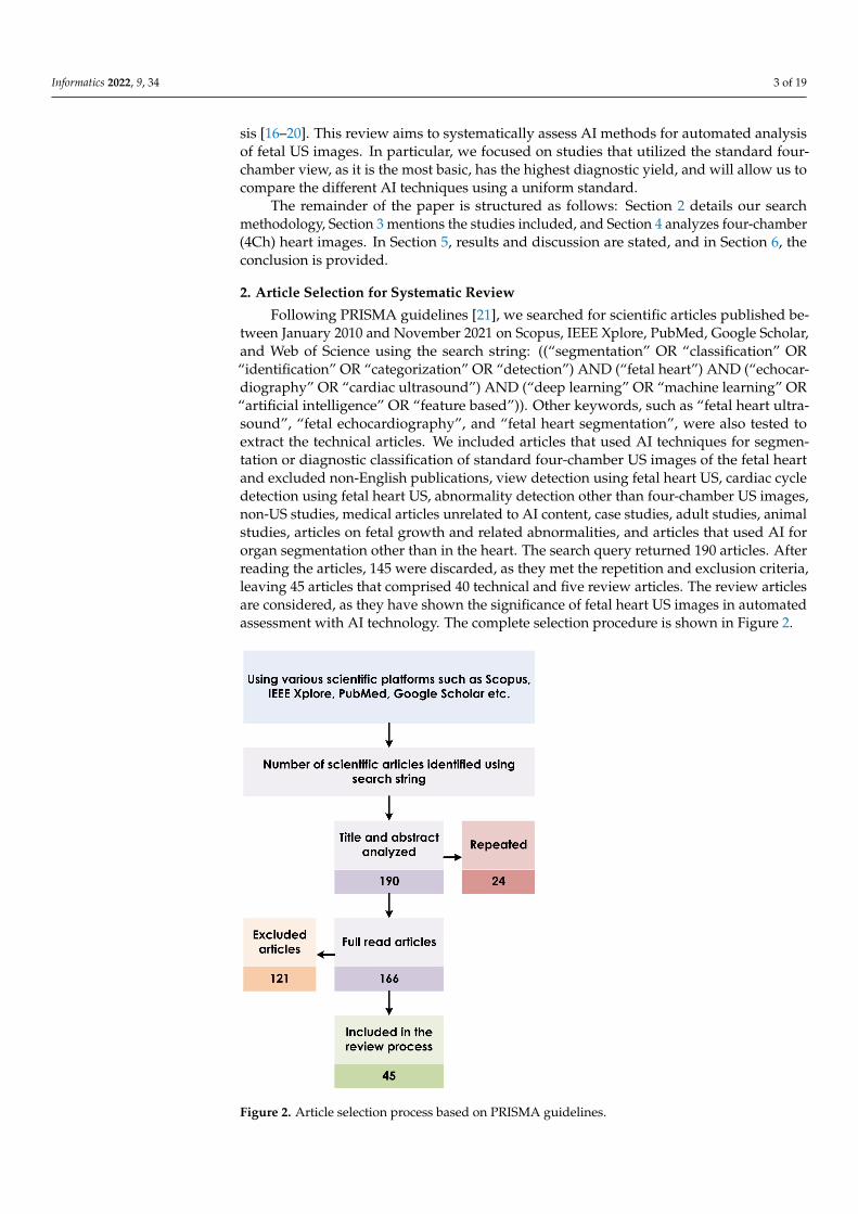
2. Article Selection for Systematic Review
Following PRISMA guidelines [21], we searched for scientific articles published be-tween January 2010 and November 2021 on Scopus, IEEE Xplore, PubMed, Google Scholar,and Web of Science using the search string: ((“segmentation” OR “classification” OR“identification” OR “categorization” OR “detection”) AND (“fetal heart”) AND (“echocar-diography” OR “cardiac ultrasound”) AND (“deep learning” OR “machine learning” OR“artificial intelligence” OR “feature based”)). Other keywords, such as “fetal heart ultra-sound”, “fetal echocardiography”, and “fetal heart segmentation”, were also tested toextract the technical articles. We included articles that used AI techniques for segmen-tation or diagnostic classification of standard four-chamber US images of the fetal heartand excluded non-English publications, view detection using fetal heart US, cardiac cycledetection using fetal heart US, abnormality detection other than four-chamber US images,non-US studies, medical articles unrelated to AI content, case studies, adult studies, animalstudies, articles on fetal growth and related abnormalities, and articles that used AI fororgan segmentation other than in the heart. The search query returned 190 articles. Afterreading the articles, 145 were discarded, as they met the repetition and exclusion criteria,leaving 45 articles that comprised 40 technical and five review articles. The review articlesare considered, as they have shown the significance of fetal heart US images in automatedassessment with AI technology. The complete selection procedure is shown in Figure 2.
Informatics 2022, 9, x FOR PEER REVIEW 4 of 20
Figure 2. Article selection process based on PRISMA guidelines.
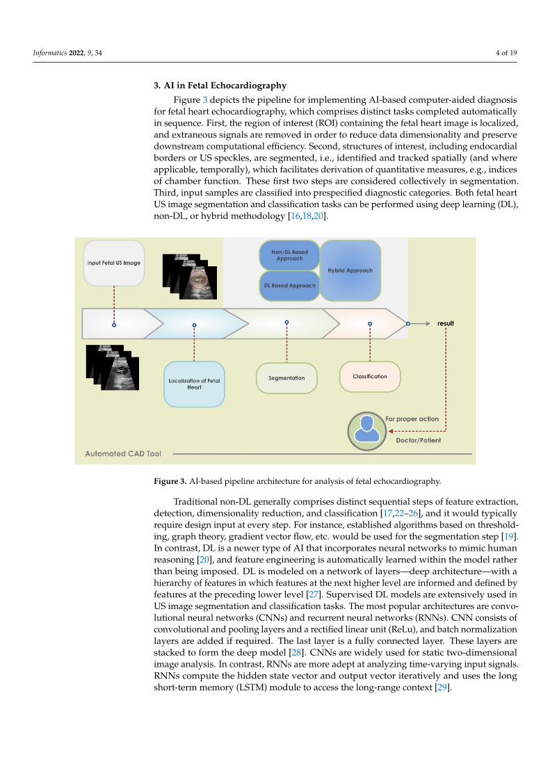
3. AI in Fetal Echocardiography Figure 3 depicts the pipeline for implementing AI-based computer-aided diagnosis
for fetal heart echocardiography, which comprises distinct tasks completed automatically in sequence. First, the region of interest (ROI) containing the fetal heart image is localized, and extraneous signals are removed in order to reduce data dimensionality and preserve downstream computational efficiency. Second, structures of interest, including endocar-dial borders or US speckles, are segmented, i.e., identified and tracked spatially (and where applicable, temporally), which facilitates derivation of quantitative measures, e.g., indices of chamber function. These first two steps are considered collectively in segmen-tation. Third, input samples are classified into prespecified diagnostic categories. Both fe-tal heart US image segmentation and classification tasks can be performed using deep learning (DL), non-DL, or hybrid methodology [16,18,20].
Figure 2. Article selection process based on PRISMA guidelines.
Informatics 2022, 9, 34 4 of 19
3. AI in Fetal Echocardiography
Figure 3 depicts the pipeline for implementing AI-based computer-aided diagnosisfor fetal heart echocardiography, which comprises distinct tasks completed automaticallyin sequence. First, the region of interest (ROI) containing the fetal heart image is localized,and extraneous signals are removed in order to reduce data dimensionality and preservedownstream computational efficiency. Second, structures of interest, including endocardialborders or US speckles, are segmented, i.e., identified and tracked spatially (and whereapplicable, temporally), which facilitates derivation of quantitative measures, e.g., indicesof chamber function. These first two steps are considered collectively in segmentation.Third, input samples are classified into prespecified diagnostic categories. Both fetal heartUS image segmentation and classification tasks can be performed using deep learning (DL),non-DL, or hybrid methodology [16,18,20].
Informatics 2022, 9, x FOR PEER REVIEW 5 of 20
Figure 3. AI-based pipeline architecture for analysis of fetal echocardiography.
Traditional non-DL generally comprises distinct sequential steps of feature extrac-tion, detection, dimensionality reduction, and classification [17,22–26], and it would typi-cally require design input at every step. For instance, established algorithms based on thresholding, graph theory, gradient vector flow, etc. would be used for the segmentation step [19]. In contrast, DL is a newer type of AI that incorporates neural networks to mimic human reasoning [20], and feature engineering is automatically learned within the model rather than being imposed. DL is modeled on a network of layers—deep architecture—with a hierarchy of features in which features at the next higher level are informed and defined by features at the preceding lower level [27]. Supervised DL models are exten-sively used in US image segmentation and classification tasks. The most popular architec-tures are convolutional neural networks (CNNs) and recurrent neural networks (RNNs). CNN consists of convolutional and pooling layers and a rectified linear unit (ReLu), and batch normalization layers are added if required. The last layer is a fully connected layer. These layers are stacked to form the deep model [28]. CNNs are widely used for static two-dimensional image analysis. In contrast, RNNs are more adept at analyzing time-varying input signals. RNNs compute the hidden state vector and output vector itera-tively and uses the long short-term memory (LSTM) module to access the long-range con-text [29].
3.1. Segmentation of Fetal Heart Structures To replace manual cropping, automatic identification of the ROI containing the fetal
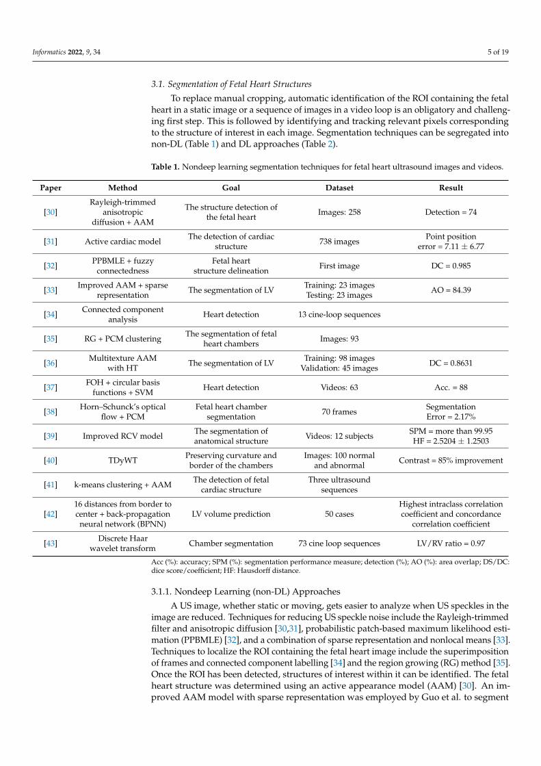
heart in a static image or a sequence of images in a video loop is an obligatory and chal-lenging first step. This is followed by identifying and tracking relevant pixels correspond-ing to the structure of interest in each image. Segmentation techniques can be segregated into non-DL (Table 1) and DL approaches (Table 2).
3.1.1. Nondeep Learning (non-DL) Approaches A US image, whether static or moving, gets easier to analyze when US speckles in
the image are reduced. Techniques for reducing US speckle noise include the Rayleigh-trimmed filter and anisotropic diffusion [30,31], probabilistic patch-based maximum like-lihood estimation (PPBMLE) [32], and a combination of sparse representation and non-local means [33]. Techniques to localize the ROI containing the fetal heart image include the superimposition of frames and connected component labelling [34] and the region growing (RG) method [35]. Once the ROI has been detected, structures of interest within
Figure 3. AI-based pipeline architecture for analysis of fetal echocardiography.
Traditional non-DL generally comprises distinct sequential steps of feature extraction,detection, dimensionality reduction, and classification [17,22–26], and it would typicallyrequire design input at every step. For instance, established algorithms based on threshold-ing, graph theory, gradient vector flow, etc. would be used for the segmentation step [19].In contrast, DL is a newer type of AI that incorporates neural networks to mimic humanreasoning [20], and feature engineering is automatically learned within the model ratherthan being imposed. DL is modeled on a network of layers—deep architecture—with ahierarchy of features in which features at the next higher level are informed and defined byfeatures at the preceding lower level [27]. Supervised DL models are extensively used inUS image segmentation and classification tasks. The most popular architectures are convo-lutional neural networks (CNNs) and recurrent neural networks (RNNs). CNN consists ofconvolutional and pooling layers and a rectified linear unit (ReLu), and batch normalizationlayers are added if required. The last layer is a fully connected layer. These layers arestacked to form the deep model [28]. CNNs are widely used for static two-dimensionalimage analysis. In contrast, RNNs are more adept at analyzing time-varying input signals.RNNs compute the hidden state vector and output vector iteratively and uses the longshort-term memory (LSTM) module to access the long-range context [29].
Informatics 2022, 9, 34 5 of 19
3.1. Segmentation of Fetal Heart Structures
To replace manual cropping, automatic identification of the ROI containing the fetalheart in a static image or a sequence of images in a video loop is an obligatory and challeng-ing first step. This is followed by identifying and tracking relevant pixels correspondingto the structure of interest in each image. Segmentation techniques can be segregated intonon-DL (Table 1) and DL approaches (Table 2).
Table 1. Nondeep learning segmentation techniques for fetal heart ultrasound images and videos.
Paper Method Goal Dataset Result
[30]Rayleigh-trimmed
anisotropicdiffusion + AAM
The structure detection ofthe fetal heart Images: 258 Detection = 74
[31] Active cardiac model The detection of cardiacstructure 738 images Point position
error = 7.11 ± 6.77
[32] PPBMLE + fuzzyconnectedness
Fetal heartstructure delineation First image DC = 0.985
[33] Improved AAM + sparserepresentation The segmentation of LV Training: 23 images
Testing: 23 images AO = 84.39
[34] Connected componentanalysis Heart detection 13 cine-loop sequences
[35] RG + PCM clustering The segmentation of fetalheart chambers Images: 93
[36] Multitexture AAMwith HT The segmentation of LV Training: 98 images
Validation: 45 images DC = 0.8631
[37] FOH + circular basisfunctions + SVM Heart detection Videos: 63 Acc. = 88
[38] Horn–Schunck’s opticalflow + PCM
Fetal heart chambersegmentation 70 frames Segmentation
Error = 2.17%
[39] Improved RCV model The segmentation ofanatomical structure Videos: 12 subjects SPM = more than 99.95
HF = 2.5204 ± 1.2503
[40] TDyWT Preserving curvature andborder of the chambers
Images: 100 normaland abnormal Contrast = 85% improvement
[41] k-means clustering + AAM The detection of fetalcardiac structure
Three ultrasoundsequences
[42]16 distances from border tocenter + back-propagation
neural network (BPNN)LV volume prediction 50 cases
Highest intraclass correlationcoefficient and concordance
correlation coefficient
[43] Discrete Haarwavelet transform Chamber segmentation 73 cine loop sequences LV/RV ratio = 0.97
Acc (%): accuracy; SPM (%): segmentation performance measure; detection (%); AO (%): area overlap; DS/DC:dice score/coefficient; HF: Hausdorff distance.
3.1.1. Nondeep Learning (non-DL) Approaches
A US image, whether static or moving, gets easier to analyze when US speckles in theimage are reduced. Techniques for reducing US speckle noise include the Rayleigh-trimmedfilter and anisotropic diffusion [30,31], probabilistic patch-based maximum likelihood esti-mation (PPBMLE) [32], and a combination of sparse representation and nonlocal means [33].Techniques to localize the ROI containing the fetal heart image include the superimpositionof frames and connected component labelling [34] and the region growing (RG) method [35].Once the ROI has been detected, structures of interest within it can be identified. The fetalheart structure was determined using an active appearance model (AAM) [30]. An im-proved AAM model with sparse representation was employed by Guo et al. to segment
Informatics 2022, 9, 34 6 of 19
the left ventricle (LV) [33]. In addition, simultaneous tracking of motion and structuralinformation can improve the detection of the fetal heart [31]. It is observed that multi-texture AAM was combined with the Hermite transform (HT) to segment the LV [36]. Afeature-based approach using Fourier orientation histograms (FOH) and a support vectormachine (SVM) classifier was used to detect the fetal heart [37]. However, optical flowwas also implemented to find the heart region [38]. An improved region-based Chan–Vese(RCV) model was proposed [39], wherein energy minimization was carried out usingthe global-pollination-based CAT swarm optimizer with the flower pollination algorithm.Furthermore, possibilistic c-means (PCM) clustering was used to detect the LV and rightventricle (RV) [35]. The curvatures and borders of all four fetal heart chambers were pre-served using the transverse dyadic wavelet transform (TDyWT) algorithm [40]. Table 1lists the non-DL methods reviewed along with their performance for the segmentation ofUS fetal heart images.
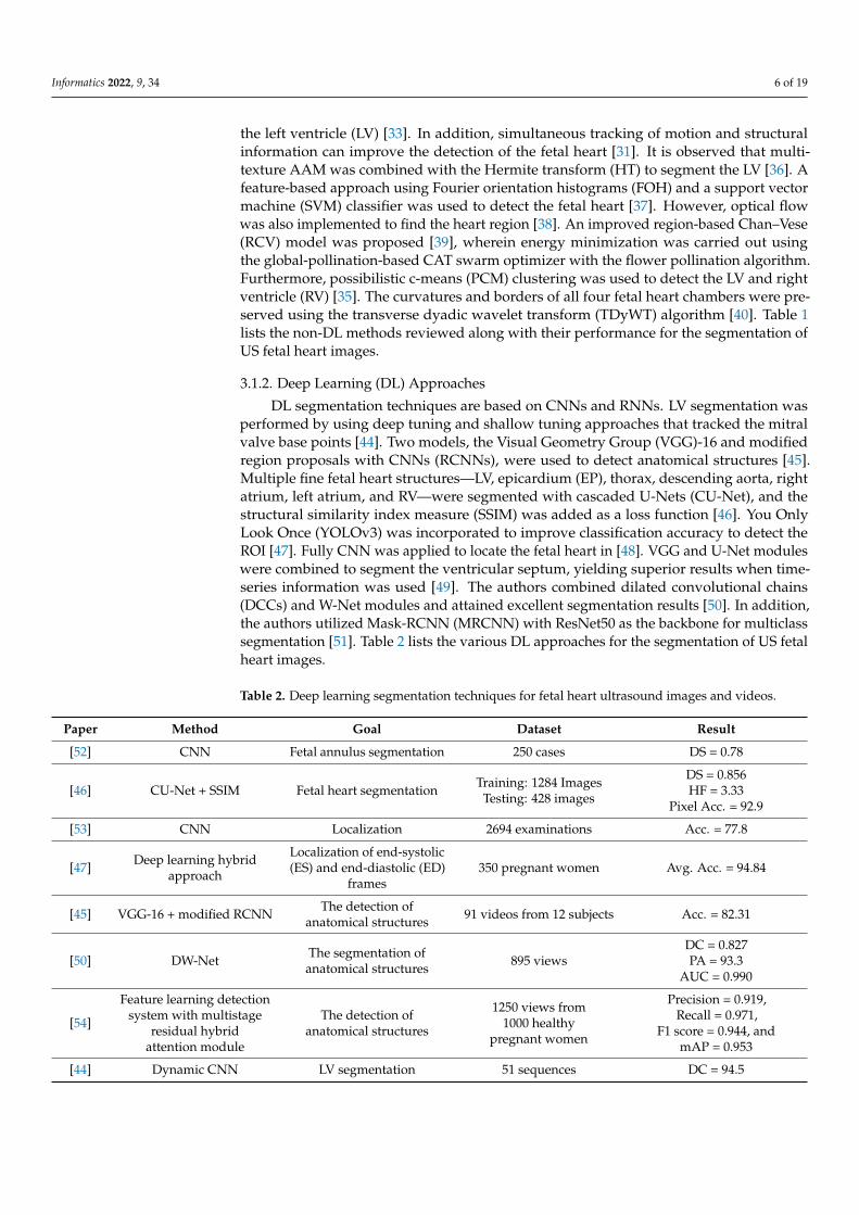
3.1.2. Deep Learning (DL) Approaches
DL segmentation techniques are based on CNNs and RNNs. LV segmentation wasperformed by using deep tuning and shallow tuning approaches that tracked the mitralvalve base points [44]. Two models, the Visual Geometry Group (VGG)-16 and modifiedregion proposals with CNNs (RCNNs), were used to detect anatomical structures [45].Multiple fine fetal heart structures—LV, epicardium (EP), thorax, descending aorta, rightatrium, left atrium, and RV—were segmented with cascaded U-Nets (CU-Net), and thestructural similarity index measure (SSIM) was added as a loss function [46]. You OnlyLook Once (YOLOv3) was incorporated to improve classification accuracy to detect theROI [47]. Fully CNN was applied to locate the fetal heart in [48]. VGG and U-Net moduleswere combined to segment the ventricular septum, yielding superior results when time-series information was used [49]. The authors combined dilated convolutional chains(DCCs) and W-Net modules and attained excellent segmentation results [50]. In addition,the authors utilized Mask-RCNN (MRCNN) with ResNet50 as the backbone for multiclasssegmentation [51]. Table 2 lists the various DL approaches for the segmentation of US fetalheart images.
Table 2. Deep learning segmentation techniques for fetal heart ultrasound images and videos.
Paper Method Goal Dataset Result
[52] CNN Fetal annulus segmentation 250 cases DS = 0.78
[46] CU-Net + SSIM Fetal heart segmentation Training: 1284 ImagesTesting: 428 images
DS = 0.856HF = 3.33
Pixel Acc. = 92.9
[53] CNN Localization 2694 examinations Acc. = 77.8
[47] Deep learning hybridapproach
Localization of end-systolic(ES) and end-diastolic (ED)
frames350 pregnant women Avg. Acc. = 94.84
[45] VGG-16 + modified RCNN The detection ofanatomical structures 91 videos from 12 subjects Acc. = 82.31
[50] DW-Net The segmentation ofanatomical structures 895 views
DC = 0.827PA = 93.3
AUC = 0.990
[54]
Feature learning detectionsystem with multistage
residual hybridattention module
The detection ofanatomical structures
1250 views from1000 healthy
pregnant women
Precision = 0.919,Recall = 0.971,
F1 score = 0.944, andmAP = 0.953
[44] Dynamic CNN LV segmentation 51 sequences DC = 94.5

Informatics 2022, 9, 34 7 of 19
Table 2. Cont.
Paper Method Goal Dataset Result
[49] Cropping–segmentation–calibration
Ventricular septumsegmentation
615 images from211 pregnant women
mIoU = 0.5543mDC = 0.6891
[55]Multiframe + cylinder
based on ensemblelearning
Thoracic wall segmentation 538 frames from256 normal cases mIoU = 0.493
[56]Supervised object
detection with normal dataonly based on CNN
The detection of structureabnormalities
349 normal cases14 CHD cases
Area under ROCHeart = 0.787Vessel = 0.891
[57] DeeplabV3 + U-net Multidisease segmentation 602 Frames from301 patients
mIoU = 0.768 ± 0.035DC = 0.926 ± 0.020
for Ebstein’s anomaly
[58] CNN-based U-NetThe segmentation of
atrioventricular septaldefect
AVSD: 337 imagesNormal: 332 images DC = 96.02%
[51] MRCNN Multiclass segmentation Images: 764
Hole detectionmIoU = 76
mAP = 99.48DC = 87.78
[59] CNNs–U-Net andOtsu threshold Fetal heart segmentation Images: 519 Mean Accuracy = 96.73
Error rate = 0.21%
Acc (%): accuracy; SPM (%): segmentation performance measure; detection (%); DS/DC: dice score/coefficient; HF:Hausdorff distance; PA (%): pixel accuracy; mean average precision (mAP); mean intersection over union (IoU).
3.2. Classification of Fetal Abnormality
Fetal heart pathologies were classified using deep learning and other conventionalapproaches. The three main steps for image analysis using non-DL approaches were seg-mentation, feature extraction and reduction, and classification. First, noise and undesirabledistortions from US images were removed using a patch-based Wiener filter (WF) [60] andPPBMLE [61]. Further, morphological operation [60], fuzzy connectedness [61], image-and-spatial transformer networks (Atlas-ISTN) [62], and Faster-RCNN [63] were used tosegment the ROI. Next, texture features of US images were extracted using the gray-levelco-occurrence matrix (GLCM) [60,61,64]. The generated features were further reduced byapplying the Fisher discriminant ratio (FDR) [61] and local preserving class separation(LPCS) [64]. Moreover, scale invariant feature transform (SIFT) descriptors and histogramof optical flow (HOF) descriptors were used, and a codebook was constructed using abag of words (BoW) [65]. Finally, classifiers such as BPNN [60], the adaptive neuro fuzzyinference system classifier (ANFIS) [61], SVM [64,65], and the Gaussian process [62] weredeployed to categorize the normal versus diseased fetal heart. DL models were used toclassify the US images in [66]. The DANomaly and GACNN (Wgan-GP and CNN) werecombined to form a DGACNN architecture [63]. An ensemble of neural networks achievedpromising results in [67]. Table 3 summarizes the various state-of-the-art approaches tofetal heart disease categorization.

Informatics 2022, 9, 34 8 of 19
Table 3. State-of-the-art decision support systems using fetal heart ultrasound images and videos.
Paper Method Dataset Result Classes
[60]
Patch-basedWF + morphological
operation + features fromGLCM + BPNN
From fetal USimage gallery
Correctly classified:30 images
Not correctly classified:9 images
3 (normal, hole in the heart,and defect in the valve.)
[61]
PPBMLE + fuzzyconnectedness + statistical
and texturefeatures + FDR + ANFIS
Normal: 185 imagesTA-CHD heart: 39 images
ROC: 0.8954F-score: 0.9673
2 (normal and truncusarteriosus (TA))
[65] SIFT + HOF + BoW + SVM Normal: 240 casesAbnormal: 60 cases Acc. (Avg.): 95.1% 2 (normal and abnormal)
[66] Deep learning modelNormal: 493
TOF: 87HLHS: 105
Normal heart vs. TOF:Sen: 75, Spe: 76
Normal vs. HLHS:Sen: 100, Spe: 90
3 (normal heart, tetralogy ofFallot (TOF), and hypoplasticleft heart syndrome (HLHS))
[64] Texture features based onshearlet + LPCS + SVM
Normal: 221 imagesPre-GDM/GDM:
212 images
Acc.: 98.15PPV: 97.22Sen: 99.05Spe: 97.28
2 (normal and pre-GDM/gestational diabetes
mellitus (GDM))
[63] DGACNN 3596 images andvideo slices
Acc.: 85AUC: 0.881 2 (normal and diseased)
[62] Atlas-ISTN + area ratios +Gaussian process
Normal: 1560 imagesHLHS: 68 images AUC-ROC: 0.978 2 (normal and HLHS)
[68] Auto-encoding generativeadversarial network
Normal: 2224 casesAbnormal: 93 cases AUC (avg.): 0.81 2 (normal and HLHS)
[67] Ensemble ofneural networks 107,823 images
AUC: 0.99Sen: 95Spe: 96
NPV: 100
2 (normal and abnormal)
PPV (%): positive predictive value; Sen. (%): sensitivity; Spe. (%): specificity; NPV (%): negative predictive value.
4. Analysis of CHD Using Four-Chamber US Images
To understand CHD, we analyzed the 4Ch US heart images and the different ap-proaches used, as described in the subsequent sections.
4.1. Data Description
The fetal cardiac ultrasound images were acquired using the Vivid 7 GE healthcareechocardiographic machine with the transducer being a linear convex probe set at 3 to4 MHz. The 4Ch view helps in the identification of cardiac chamber anatomy and thepresence of any intracardiac shunt lesions. Structural anomalies of the cardiac chambersare well-diagnosed using lateral or apical four-chamber views of the fetal heart. Thecollected images are shown in Figure 4. In the healthy normal fetus, the apical 4Ch viewdemonstrates four well-developed chambers, a concordant atrioventricular (AV) connection,unobstructed AV valves (mitral and tricuspid valves), the foramen ovale flap opening intoleft atrium (LA), and an intact interventricular septum. Additionally, the pulmonary venousopening can be visualized at the LA wall. Any structural deviations from normal anatomymay lead to congenital heart disease that can be determined by the 4Ch view of the fetalheart’s ultrasound imaging. We illustrate a few such examples that can be diagnosed by the4Ch view of the fetal heart alone. Figure 4a displays the normal cardiac structural anatomy,where all four chambers are well-developed without any dilatation/hypoplasia of anychamber and nonobstructive AV valves. Figure 4b shows an atrioventricular septal defect,commonly termed as an endocardial cushion defect, mainly affecting AV valve formation
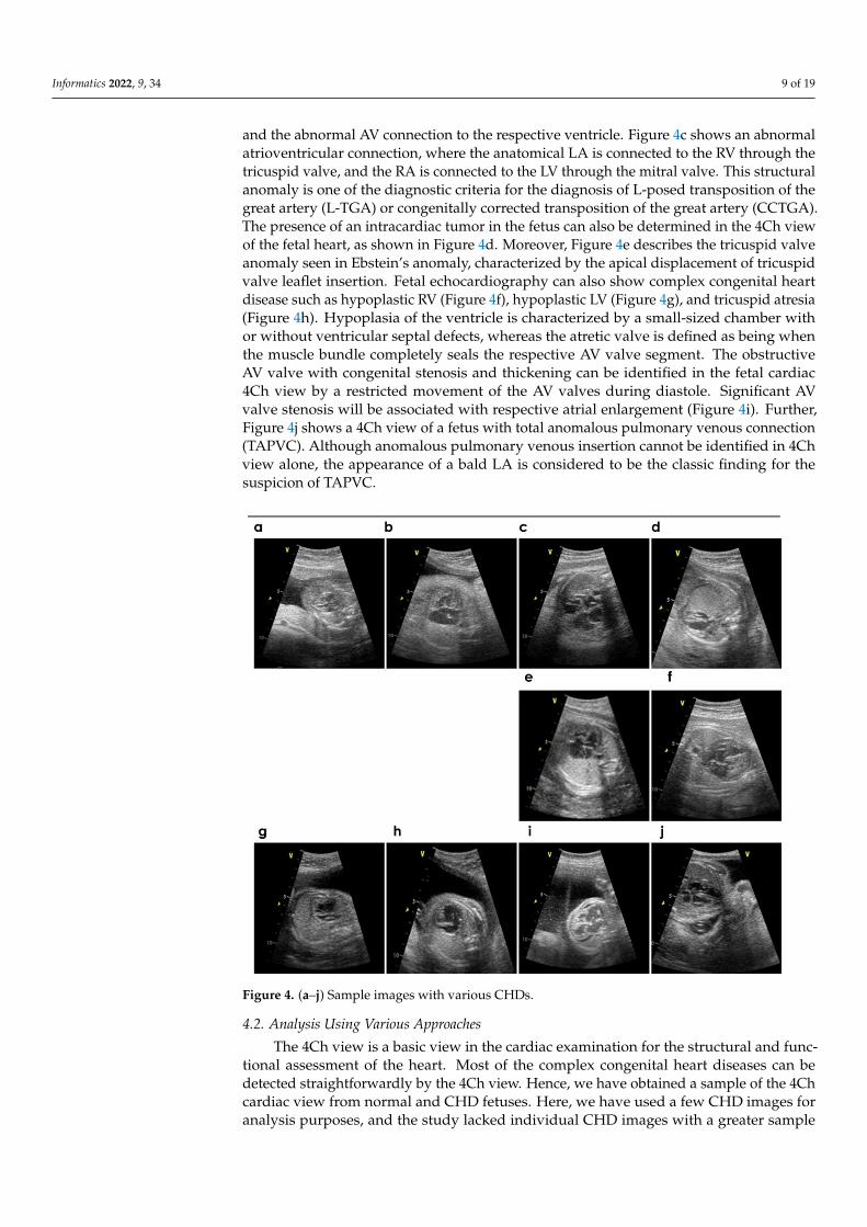
Informatics 2022, 9, 34 9 of 19
and the abnormal AV connection to the respective ventricle. Figure 4c shows an abnormalatrioventricular connection, where the anatomical LA is connected to the RV through thetricuspid valve, and the RA is connected to the LV through the mitral valve. This structuralanomaly is one of the diagnostic criteria for the diagnosis of L-posed transposition of thegreat artery (L-TGA) or congenitally corrected transposition of the great artery (CCTGA).The presence of an intracardiac tumor in the fetus can also be determined in the 4Ch viewof the fetal heart, as shown in Figure 4d. Moreover, Figure 4e describes the tricuspid valveanomaly seen in Ebstein’s anomaly, characterized by the apical displacement of tricuspidvalve leaflet insertion. Fetal echocardiography can also show complex congenital heartdisease such as hypoplastic RV (Figure 4f), hypoplastic LV (Figure 4g), and tricuspid atresia(Figure 4h). Hypoplasia of the ventricle is characterized by a small-sized chamber withor without ventricular septal defects, whereas the atretic valve is defined as being whenthe muscle bundle completely seals the respective AV valve segment. The obstructiveAV valve with congenital stenosis and thickening can be identified in the fetal cardiac4Ch view by a restricted movement of the AV valves during diastole. Significant AVvalve stenosis will be associated with respective atrial enlargement (Figure 4i). Further,Figure 4j shows a 4Ch view of a fetus with total anomalous pulmonary venous connection(TAPVC). Although anomalous pulmonary venous insertion cannot be identified in 4Chview alone, the appearance of a bald LA is considered to be the classic finding for thesuspicion of TAPVC.
Informatics 2022, 9, x FOR PEER REVIEW 10 of 20
LV (Figure 4g), and tricuspid atresia (Figure 4h). Hypoplasia of the ventricle is character-ized by a small-sized chamber with or without ventricular septal defects, whereas the atretic valve is defined as being when the muscle bundle completely seals the respective AV valve segment. The obstructive AV valve with congenital stenosis and thickening can be identified in the fetal cardiac 4Ch view by a restricted movement of the AV valves during diastole. Significant AV valve stenosis will be associated with respective atrial en-largement (Figure 4i). Further, Figure 4j shows a 4Ch view of a fetus with total anomalous pulmonary venous connection (TAPVC). Although anomalous pulmonary venous inser-tion cannot be identified in 4Ch view alone, the appearance of a bald LA is considered to be the classic finding for the suspicion of TAPVC.
Figure 4. (a–j) Sample images with various CHDs.
4.2. Analysis Using Various Approaches The 4Ch view is a basic view in the cardiac examination for the structural and func-
tional assessment of the heart. Most of the complex congenital heart diseases can be de-tected straightforwardly by the 4Ch view. Hence, we have obtained a sample of the 4Ch cardiac view from normal and CHD fetuses. Here, we have used a few CHD images for analysis purposes, and the study lacked individual CHD images with a greater sample size. However, we strategize automated individual CHD detection using more of such data in the future. In the present study, we have used methods such as steerable filters [69,70], gist [71], and higher-order spectra (HOS) cumulants [72] to analyze the structure of the heart. The application of these methods to characterize image uncertainties and their analysis to achieve efficient results have motivated us to analyze the 4Ch fetal heart images [73–81]. These approaches can be further utilized with non-DL and DL approaches to efficiently characterize CHDs.
Steerable filters are used to enhance the various clinical features in different orienta-tions. A set of basis filters is used to produce an arbitrary orientation [69,70]. Moreover, a linear combination of the oriented functions is used to generate the steered filters. The
Figure 4. (a–j) Sample images with various CHDs.
4.2. Analysis Using Various Approaches
The 4Ch view is a basic view in the cardiac examination for the structural and func-tional assessment of the heart. Most of the complex congenital heart diseases can bedetected straightforwardly by the 4Ch view. Hence, we have obtained a sample of the 4Chcardiac view from normal and CHD fetuses. Here, we have used a few CHD images foranalysis purposes, and the study lacked individual CHD images with a greater sample
Informatics 2022, 9, 34 10 of 19
size. However, we strategize automated individual CHD detection using more of such datain the future. In the present study, we have used methods such as steerable filters [69,70],gist [71], and higher-order spectra (HOS) cumulants [72] to analyze the structure of theheart. The application of these methods to characterize image uncertainties and theiranalysis to achieve efficient results have motivated us to analyze the 4Ch fetal heart im-ages [73–81]. These approaches can be further utilized with non-DL and DL approaches toefficiently characterize CHDs.
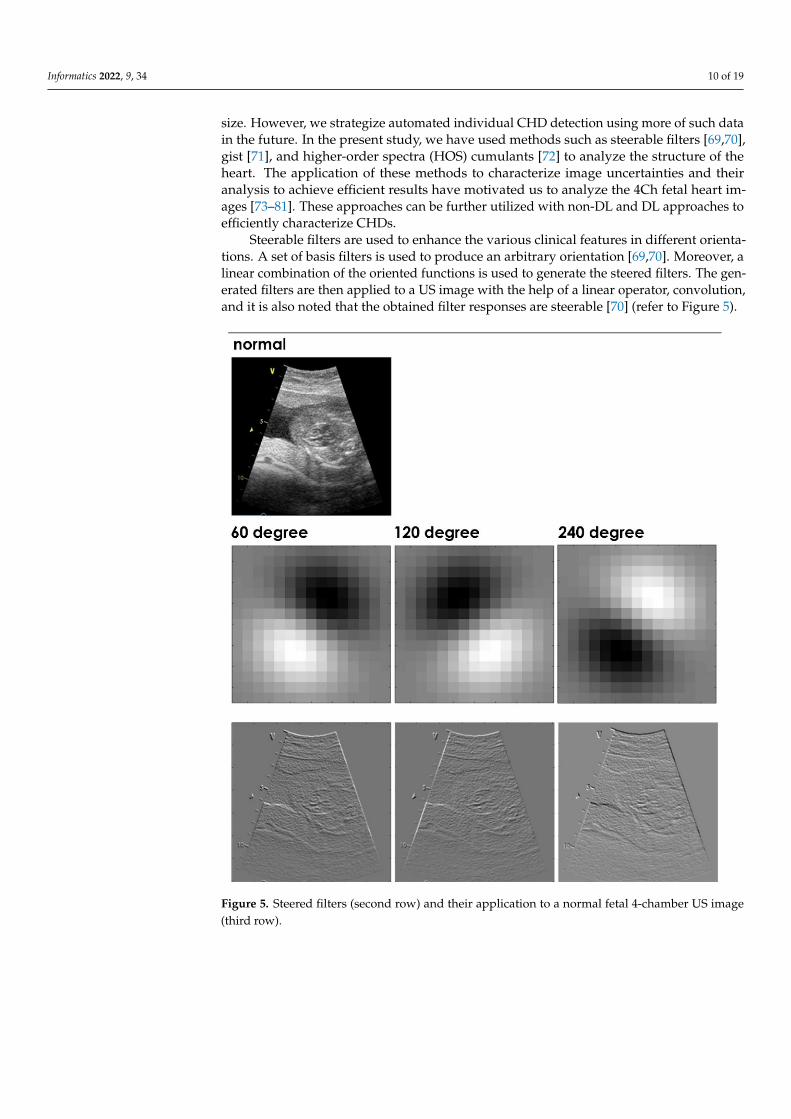
Steerable filters are used to enhance the various clinical features in different orienta-tions. A set of basis filters is used to produce an arbitrary orientation [69,70]. Moreover, alinear combination of the oriented functions is used to generate the steered filters. The gen-erated filters are then applied to a US image with the help of a linear operator, convolution,and it is also noted that the obtained filter responses are steerable [70] (refer to Figure 5).
Informatics 2022, 9, x FOR PEER REVIEW 11 of 20
generated filters are then applied to a US image with the help of a linear operator, convo-lution, and it is also noted that the obtained filter responses are steerable [70] (refer to Figure 5).
Figure 5. Steered filters (second row) and their application to a normal fetal 4-chamber US image (third row).
Gist is an efficient, holistic lower-dimension representation of an image. It investi-gates the image on a spatial four-by-four nonoverlapping grid. Initially, the power spec-trum of an image is computed and a set of Gabor filters are then used [71,73]. Then, the feature maps are generated to accumulate the information over subregions of an image. Figure 6 shows the computed gist descriptor using four-chamber US images [74].
Figure 5. Steered filters (second row) and their application to a normal fetal 4-chamber US image(third row).
Informatics 2022, 9, 34 11 of 19
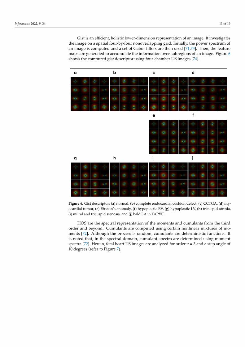
Gist is an efficient, holistic lower-dimension representation of an image. It investigatesthe image on a spatial four-by-four nonoverlapping grid. Initially, the power spectrum ofan image is computed and a set of Gabor filters are then used [71,73]. Then, the featuremaps are generated to accumulate the information over subregions of an image. Figure 6shows the computed gist descriptor using four-chamber US images [74].
Informatics 2022, 9, x FOR PEER REVIEW 12 of 20
Figure 6. Gist descriptor: (a) normal, (b) complete endocardial cushion defect, (c) CCTGA, (d) my-ocardial tumor, (e) Ebstein’s anomaly, (f) hypoplastic RV, (g) hypoplastic LV, (h) tricuspid atresia, (i) mitral and tricuspid stenosis, and (j) bald LA in TAPVC.
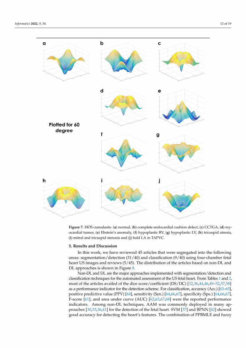
HOS are the spectral representation of the moments and cumulants from the third order and beyond. Cumulants are computed using certain nonlinear mixtures of moments [72]. Although the process is random, cumulants are deterministic functions. It is noted that, in the spectral domain, cumulant spectra are determined using moment spectra [72]. Herein, fetal heart US images are analyzed for order n = 3 and a step angle of 10 degrees (refer to Figure 7).
Figure 6. Gist descriptor: (a) normal, (b) complete endocardial cushion defect, (c) CCTGA, (d) my-ocardial tumor, (e) Ebstein’s anomaly, (f) hypoplastic RV, (g) hypoplastic LV, (h) tricuspid atresia,(i) mitral and tricuspid stenosis, and (j) bald LA in TAPVC.
HOS are the spectral representation of the moments and cumulants from the thirdorder and beyond. Cumulants are computed using certain nonlinear mixtures of mo-ments [72]. Although the process is random, cumulants are deterministic functions. Itis noted that, in the spectral domain, cumulant spectra are determined using momentspectra [72]. Herein, fetal heart US images are analyzed for order n = 3 and a step angle of10 degrees (refer to Figure 7).
Informatics 2022, 9, 34 12 of 19Informatics 2022, 9, x FOR PEER REVIEW 13 of 20
Figure 7. HOS cumulants: (a) normal, (b) complete endocardial cushion defect, (c) CCTGA, (d) my-ocardial tumor, (e) Ebstein’s anomaly, (f) hypoplastic RV, (g) hypoplastic LV, (h) tricuspid atresia, (i) mitral and tricuspid stenosis and (j) bald LA in TAPVC.
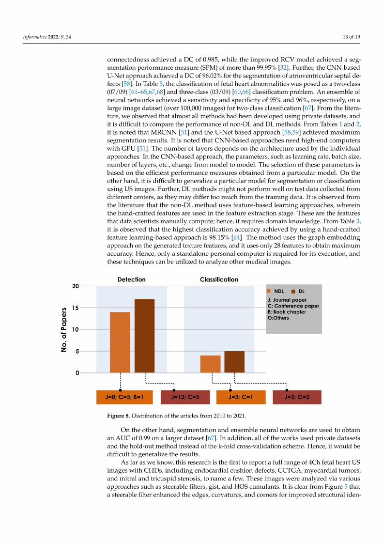
5. Results and Discussion In this work, we have reviewed 45 articles that were segregated into the following
areas: segmentation/detection (31/40) and classification (9/40) using four-chamber fetal heart US images and reviews (5/45). The distribution of the articles based on non-DL and DL approaches is shown in Figure 8.
Figure 7. HOS cumulants: (a) normal, (b) complete endocardial cushion defect, (c) CCTGA, (d) my-ocardial tumor, (e) Ebstein’s anomaly, (f) hypoplastic RV, (g) hypoplastic LV, (h) tricuspid atresia,(i) mitral and tricuspid stenosis and (j) bald LA in TAPVC.
5. Results and Discussion
In this work, we have reviewed 45 articles that were segregated into the followingareas: segmentation/detection (31/40) and classification (9/40) using four-chamber fetalheart US images and reviews (5/45). The distribution of the articles based on non-DL andDL approaches is shown in Figure 8.
Non-DL and DL are the major approaches implemented with segmentation/detection andclassification techniques for the automated assessment of the US fetal heart. From Tables 1 and 2,most of the articles availed of the dice score/coefficient (DS/DC) [32,36,44,46,49–52,57,58]as a performance indicator for the detection scheme. For classification, accuracy (Acc.) [63–65],positive predictive value (PPV) [64], sensitivity (Sen.) [64,66,67], specificity (Spe.) [64,66,67],F-score [61], and area under curve (AUC) [62,63,67,68] were the reported performanceindicators. Among non-DL techniques, AAM was commonly deployed in many ap-proaches [30,33,36,41] for the detection of the fetal heart. SVM [37] and BPNN [42] showedgood accuracy for detecting the heart’s features. The combination of PPBMLE and fuzzy
Informatics 2022, 9, 34 13 of 19
connectedness achieved a DC of 0.985, while the improved RCV model achieved a seg-mentation performance measure (SPM) of more than 99.95% [32]. Further, the CNN-basedU-Net approach achieved a DC of 96.02% for the segmentation of atrioventricular septal de-fects [58]. In Table 3, the classification of fetal heart abnormalities was posed as a two-class(07/09) [61–65,67,68] and three-class (03/09) [60,66] classification problem. An ensemble ofneural networks achieved a sensitivity and specificity of 95% and 96%, respectively, on alarge image dataset (over 100,000 images) for two-class classification [67]. From the litera-ture, we observed that almost all methods had been developed using private datasets, andit is difficult to compare the performance of non-DL and DL methods. From Tables 1 and 2,it is noted that MRCNN [51] and the U-Net based approach [58,59] achieved maximumsegmentation results. It is noted that CNN-based approaches need high-end computerswith GPU [51]. The number of layers depends on the architecture used by the individualapproaches. In the CNN-based approach, the parameters, such as learning rate, batch size,number of layers, etc., change from model to model. The selection of these parameters isbased on the efficient performance measures obtained from a particular model. On theother hand, it is difficult to generalize a particular model for segmentation or classificationusing US images. Further, DL methods might not perform well on test data collected fromdifferent centers, as they may differ too much from the training data. It is observed fromthe literature that the non-DL method uses feature-based learning approaches, whereinthe hand-crafted features are used in the feature extraction stage. These are the featuresthat data scientists manually compute; hence, it requires domain knowledge. From Table 3,it is observed that the highest classification accuracy achieved by using a hand-craftedfeature learning-based approach is 98.15% [64]. The method uses the graph embeddingapproach on the generated texture features, and it uses only 28 features to obtain maximumaccuracy. Hence, only a standalone personal computer is required for its execution, andthese techniques can be utilized to analyze other medical images.
Informatics 2022, 9, x FOR PEER REVIEW 14 of 20
Figure 8. Distribution of the articles from 2010 to 2021.
Non-DL and DL are the major approaches implemented with segmentation/detection and classification techniques for the automated assessment of the US fetal heart. From Tables 1 and 2, most of the articles availed of the dice score/coefficient (DS/DC) [32,36,44,46,49–52,57,58] as a performance indicator for the detection scheme. For classifi-cation, accuracy (Acc.) [63–65], positive predictive value (PPV) [64], sensitivity (Sen.) [64,66,67], specificity (Spe.) [64,66,67], F-score [61], and area under curve (AUC) [62,63,67,68] were the reported performance indicators. Among non-DL techniques, AAM was commonly deployed in many approaches [30,33,36,41] for the detection of the fetal heart. SVM [37] and BPNN [42] showed good accuracy for detecting the heart’s features. The combination of PPBMLE and fuzzy connectedness achieved a DC of 0.985, while the improved RCV model achieved a segmentation performance measure (SPM) of more than 99.95% [32]. Further, the CNN-based U-Net approach achieved a DC of 96.02% for the segmentation of atrioventricular septal defects [58]. In Table 3, the classification of fetal heart abnormalities was posed as a two-class (07/09) [61–65,67,68] and three-class (03/09) [60,66] classification problem. An ensemble of neural networks achieved a sensitivity and specificity of 95% and 96%, respectively, on a large image dataset (over 100,000 images) for two-class classification [67]. From the literature, we observed that almost all methods had been developed using private datasets, and it is difficult to compare the performance of non-DL and DL methods. From Tables 1 and 2, it is noted that MRCNN [51] and the U-Net based approach [58,59] achieved maximum segmentation results. It is noted that CNN-based approaches need high-end computers with GPU [51]. The number of layers depends on the architecture used by the individual approaches. In the CNN-based ap-proach, the parameters, such as learning rate, batch size, number of layers, etc., change from model to model. The selection of these parameters is based on the efficient perfor-mance measures obtained from a particular model. On the other hand, it is difficult to generalize a particular model for segmentation or classification using US images. Further, DL methods might not perform well on test data collected from different centers, as they may differ too much from the training data. It is observed from the literature that the non-DL method uses feature-based learning approaches, wherein the hand-crafted features are used in the feature extraction stage. These are the features that data scientists manually compute; hence, it requires domain knowledge. From Table 3, it is observed that the high-est classification accuracy achieved by using a hand-crafted feature learning-based ap-proach is 98.15% [64]. The method uses the graph embedding approach on the generated texture features, and it uses only 28 features to obtain maximum accuracy. Hence, only a standalone personal computer is required for its execution, and these techniques can be utilized to analyze other medical images.
Figure 8. Distribution of the articles from 2010 to 2021.
On the other hand, segmentation and ensemble neural networks are used to obtainan AUC of 0.99 on a larger dataset [67]. In addition, all of the works used private datasetsand the hold-out method instead of the k-fold cross-validation scheme. Hence, it would bedifficult to generalize the results.
As far as we know, this research is the first to report a full range of 4Ch fetal heart USimages with CHDs, including endocardial cushion defects, CCTGA, myocardial tumors,and mitral and tricuspid stenosis, to name a few. These images were analyzed via variousapproaches such as steerable filters, gist, and HOS cumulants. It is clear from Figure 5 thata steerable filter enhanced the edges, curvatures, and corners for improved structural iden-
Informatics 2022, 9, 34 14 of 19
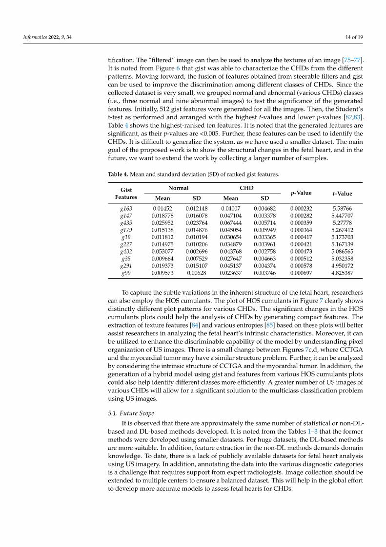
tification. The “filtered” image can then be used to analyze the textures of an image [75–77].It is noted from Figure 6 that gist was able to characterize the CHDs from the differentpatterns. Moving forward, the fusion of features obtained from steerable filters and gistcan be used to improve the discrimination among different classes of CHDs. Since thecollected dataset is very small, we grouped normal and abnormal (various CHDs) classes(i.e., three normal and nine abnormal images) to test the significance of the generatedfeatures. Initially, 512 gist features were generated for all the images. Then, the Student’st-test as performed and arranged with the highest t-values and lower p-values [82,83].Table 4 shows the highest-ranked ten features. It is noted that the generated features aresignificant, as their p-values are <0.005. Further, these features can be used to identify theCHDs. It is difficult to generalize the system, as we have used a smaller dataset. The maingoal of the proposed work is to show the structural changes in the fetal heart, and in thefuture, we want to extend the work by collecting a larger number of samples.
Table 4. Mean and standard deviation (SD) of ranked gist features.
GistFeatures
Normal CHDp-Value t-Value
Mean SD Mean SD
g163 0.01452 0.012148 0.04007 0.004682 0.000232 5.58766g147 0.018778 0.016078 0.047104 0.003378 0.000282 5.447707g435 0.025952 0.023764 0.067444 0.005714 0.000359 5.27778g179 0.015138 0.014876 0.045054 0.005949 0.000364 5.267412g19 0.011812 0.010194 0.030654 0.003365 0.000417 5.173703
g227 0.014975 0.010206 0.034879 0.003961 0.000421 5.167139g432 0.053077 0.002696 0.043768 0.002758 0.000473 5.086565g35 0.009664 0.007529 0.027647 0.004663 0.000512 5.032358
g291 0.019373 0.015107 0.045137 0.004374 0.000578 4.950172g99 0.009573 0.00628 0.023637 0.003746 0.000697 4.825387
To capture the subtle variations in the inherent structure of the fetal heart, researcherscan also employ the HOS cumulants. The plot of HOS cumulants in Figure 7 clearly showsdistinctly different plot patterns for various CHDs. The significant changes in the HOScumulants plots could help the analysis of CHDs by generating compact features. Theextraction of texture features [84] and various entropies [85] based on these plots will betterassist researchers in analyzing the fetal heart’s intrinsic characteristics. Moreover, it canbe utilized to enhance the discriminable capability of the model by understanding pixelorganization of US images. There is a small change between Figures 7c,d, where CCTGAand the myocardial tumor may have a similar structure problem. Further, it can be analyzedby considering the intrinsic structure of CCTGA and the myocardial tumor. In addition, thegeneration of a hybrid model using gist and features from various HOS cumulants plotscould also help identify different classes more efficiently. A greater number of US images ofvarious CHDs will allow for a significant solution to the multiclass classification problemusing US images.
5.1. Future Scope
It is observed that there are approximately the same number of statistical or non-DL-based and DL-based methods developed. It is noted from the Tables 1–3 that the formermethods were developed using smaller datasets. For huge datasets, the DL-based methodsare more suitable. In addition, feature extraction in the non-DL methods demands domainknowledge. To date, there is a lack of publicly available datasets for fetal heart analysisusing US imagery. In addition, annotating the data into the various diagnostic categoriesis a challenge that requires support from expert radiologists. Image collection should beextended to multiple centers to ensure a balanced dataset. This will help in the global effortto develop more accurate models to assess fetal hearts for CHDs.

Informatics 2022, 9, 34 15 of 19
In future, the development of the smart healthcare system using the internet of things(IoT) is essential for early diagnosis and treatment of CHDs in expectant mothers livingin rural areas of developing countries (refer Figure 9). Here, the collected US images areanalyzed using a cloud-based system. The obtained reports can then be sent to doctorsand specialists in multispecialty hospitals in the city. Based on the specialists’ advice,the obstetricians in the rural areas can make appropriate decisions regarding the baby’sbirth. In addition, the proper medication can be provided to the expectant mother for abetter outcome.
Informatics 2022, 9, x FOR PEER REVIEW 16 of 20
5.1. Future Scope It is observed that there are approximately the same number of statistical or non-DL-
based and DL-based methods developed. It is noted from the Tables 1–3 that the former methods were developed using smaller datasets. For huge datasets, the DL-based meth-ods are more suitable. In addition, feature extraction in the non-DL methods demands domain knowledge. To date, there is a lack of publicly available datasets for fetal heart analysis using US imagery. In addition, annotating the data into the various diagnostic categories is a challenge that requires support from expert radiologists. Image collection should be extended to multiple centers to ensure a balanced dataset. This will help in the global effort to develop more accurate models to assess fetal hearts for CHDs.
In future, the development of the smart healthcare system using the internet of things (IoT) is essential for early diagnosis and treatment of CHDs in expectant mothers living in rural areas of developing countries (refer Figure 9). Here, the collected US images are analyzed using a cloud-based system. The obtained reports can then be sent to doctors and specialists in multispecialty hospitals in the city. Based on the specialists’ advice, the obstetricians in the rural areas can make appropriate decisions regarding the baby’s birth. In addition, the proper medication can be provided to the expectant mother for a better outcome.
Figure 9. Smart healthcare system.
5.2. Limitations of the Current Study Some limitations of the proposed study include:
1. This review considers only manuscripts written in English. 2. The articles are based on specific keywords used. We may have overlooked potential
studies based on non-DL and DL approaches. 3. The study targets AI-based techniques for fetal heart assessment using only four-
chamber US images and did not consider other views or other imaging modalities.
6. Conclusions AI techniques have the potential to make a huge impact in the field of medical image
processing and analysis. Computer-based diagnostic tools have shown significant growth in clinical and medical applications. In this study, 45 articles were selected from 190. These articles were thoroughly reviewed so as to provide a comprehensive insight into the AI techniques utilized for the characterization of fetal heart US images. These studies were
Figure 9. Smart healthcare system.
5.2. Limitations of the Current Study
Some limitations of the proposed study include:
1. This review considers only manuscripts written in English.2. The articles are based on specific keywords used. We may have overlooked potential
studies based on non-DL and DL approaches.3. The study targets AI-based techniques for fetal heart assessment using only four-
chamber US images and did not consider other views or other imaging modalities.
6. Conclusions
AI techniques have the potential to make a huge impact in the field of medical imageprocessing and analysis. Computer-based diagnostic tools have shown significant growthin clinical and medical applications. In this study, 45 articles were selected from 190. Thesearticles were thoroughly reviewed so as to provide a comprehensive insight into the AItechniques utilized for the characterization of fetal heart US images. These studies weresummarized and analyzed in terms of different cutting-edge approaches. The surveyshowed that AI techniques are able to improve the assessment of the fetal heart and arelikely to be valuable resources for medical decision support.
Author Contributions: Conceptualization, A.G. and R.U.; Methodology, A.G. and R.U.; Software,A.G. and R.U.; Validation, J.S., A.V., A.A.J. and K.N.; Writing—review and editing, A.G., R.U., J.S.,R.-S.T., C.P.O., E.J.C., F.M. and U.R.A.; Visualization, U.R.A., A.G. and P.D.B. All authors have readand agreed to the published version of the manuscript.
Funding: This research received no external funding.
Institutional Review Board Statement: Not applicable.
Informed Consent Statement: Not applicable.
Informatics 2022, 9, 34 16 of 19
Data Availability Statement: Not applicable.
Acknowledgments: The authors would like to thank the Manipal Academy of Higher Education(MAHE) for providing the required facility to carry out this research.
Conflicts of Interest: The authors declare no conflict of interest.
References1. Hoffman, J.I. The global burden of congenital heart disease. Cardiovasc. J. Afr. 2013, 24, 141–145. [CrossRef] [PubMed]2. Dolk, H.; Loane, M.; Garne, E. European Surveillance of Congenital Anomalies Working Group: Congenital heart defects in
Europe: Prevalence and perinatal mortality, 2000 to 2005. Circulation 2011, 123, 841–849. [CrossRef]3. Nayak, K.; Chandra, G.S.N.; Shetty, R.; Narayan, P.K. Evaluation of fetal echocardiography as a routine antenatal screening tool
for detection of congenital heart disease. Cardiovasc. Diagn. Ther. 2016, 6, 4. [CrossRef]4. Rajiah, P.; Mak, C.; Dubinksy, T.J.; Dighe, M. Ultrasound of fetal cardiac anomalies. Am. J. Roentgenol. 2011, 197, W747–W760.
[CrossRef] [PubMed]5. Stamm, E.R. ; Drose JAThe fetal heart In Diagnostic Ultrasound, 2nd ed.; Rumack, C.A., Wilson, S.R., Charboneau, W.J., Eds.; Mosby:
St. Louis, MO, USA, 1998; pp. 1123–1159.6. Small, M.; Copel, J.A. Indications for fetal echocardiography. Pediatr. Cardiol. 2004, 25, 210–222. [CrossRef] [PubMed]7. International Society of Ultrasound in Obstetric and Gynecology. Cardiac screening examination of the fetus: Guidelines for
performing the “basic” and “extended basic” cardiac scan. Ultrasound Obstet. Gynecol. 2006, 27, 107–113.8. Dudnikov, O.; Quinton, A.E.; Alphonse, J. The detection rate of first trimester ultrasound in the diagnosis of congenital heart
defects: A narrative review. Sonography 2021, 8, 36–42. [CrossRef]9. Charafeddine, F.; Hachem, A.; Kibbi, N.; AbuTaqa, M.; Bitar, F.; Bulbul, Z.; El-Rassi, I.; Arabi, M. The first fetal echocardiography
experience for prenatal diagnosis of congenital heart disease in lebanon: Successes and challenges. J. Saudi Heart Assoc. 2019, 31,125–129. [CrossRef]
10. Sriraam, N. A Primitive Survey on Ultrasonic Imaging-Oriented Segmentation Techniques for Detection of Fetal CardiacChambers. Int. J. Biomed. Clin. Eng. 2019, 8, 69–79.
11. Carvalho, J.S.; Ho, S.Y.; Shinebourne, E.A. Sequential segmental analysis in complex fetal cardiac abnormalities: A logicalapproach to diagnosis. Ultrasound Obstet. Gynecol. 2005, 26, 105–111. [CrossRef]
12. Naderi, S.; McGahan, J.P. A primer for fetal cardiac imaging: A stepwise approach for 2-D imaging. Ultrasound Q. 2008, 24,195–206. [CrossRef] [PubMed]
13. Donofrio, M.T.; Moon-Grady, A.J.; Hornberger, L.K.; Copel, J.A.; Sklansky, M.S.; Abuhamad, A.; Cuneo, B.F.; Huhta, J.C.; Jonas,R.A.; Krishnan, A.; et al. American Heart Association Adults With Congenital Heart Disease Joint Committee of the Council onCardiovascular Disease in the Young and Council on Clinical Cardiology, Council on Cardiovascular Surgery and Anesthesia,and Council on Cardiovascular and Stroke Nursing. Diagnosis and treatment of fetal cardiac disease: A scientific statement fromthe American Heart Association. Circulation 2014, 129, 2183–2242. [PubMed]
14. Makikallio, K.; Rasanen, J.; Makikallio, T.; Vuolteenaho, O.; Huhta, J.C. Human fetal cardiovascular profile score and neonataloutcome in intrauterine growth restriction. Ultrasound Obstet. Gynecol. 2008, 31, 48–54. [CrossRef]
15. Bahtiyar, M.O.; Copel, J.A. Cardiac changes in the intrauterine growth restricted fetus. Semin Perinatol. 2008, 32, 190–193.[CrossRef] [PubMed]
16. Liu, S.; Wang, Y.; Yang, X.; Lei, B.; Liu, L.; Li, S.X.; Ni, D.; Wang, T. Deep Learning in Medical Ultrasound Analysis: A Review.Engineering 2019, 5, 261–275. [CrossRef]
17. Garcia-Canadilla, P.; Sanchez-Martinez, S.; Crispi, F.; Bijnens, B. Machine learning in fetal cardiology: What to expect. Fetal Diagn.Ther. 2020, 47, 363–372. [CrossRef]
18. de Siqueira, V.S.; Borges, M.M.; Furtado, R.G.; Dourado, C.N.; da Costa, R.M. Artificial intelligence applied to support medicaldecisions for the automatic analysis of echocardiogram images: A systematic review. Artif. Intell. Med. 2021, 120, 102165.[CrossRef] [PubMed]
19. Rawat, V.; Jain, A.; Shrimali, V. Automated techniques for the interpretation of fetal abnormalities: A review. Appl. Bionics Biomech.2018, 2018, 6452050. [CrossRef]
20. Day, T.G.; Kainz, B.; Hajnal, J.; Razavi, R.; Simpson, J.M. Artificial intelligence, fetal echocardiography, and congenital heartdisease. Prenat. Diagn. 2021, 41, 733–742. [CrossRef]
21. Moher, D.; Liberati, A.; Tetzlaff, J.; Altman, D.G. The PRISMA Group. Preferred reporting items for systematic reviews andmeta—analyses: The PRISMA statement. Int. J. Surg. 2010, 8, 336–341. [CrossRef]
22. Sudarshan, V.; Acharya, U.R.; Ng, E.Y.-K.; Meng, C.S.; Tan, R.S.; Ghista, D.N. Automated Identification of Infarcted MyocardiumTissue Characterization Using Ultrasound Images: A Review. IEEE Rev. Biomed. Eng. 2015, 8, 86–97. [CrossRef]
23. Raghavendra, U.; Acharya, U.R.; Gudigar, A.; Shetty, R.; Krishnananda, N.; Pai, U.; Samanth, J.; Nayak, C. Automated screeningof congestive heart failure using variational mode decomposition and texture features extracted from ultrasound images. NeuralComput. Appl. 2017, 28, 2869–2878. [CrossRef]

Informatics 2022, 9, 34 17 of 19
24. Raghavendra, U.; Fujita, H.; Gudigar, A.; Shetty, R.; Nayak, K.; Pai, U.; Samanth, J.; Acharya, U. Automated technique forcoronary artery disease characterization and classification using DD-DTDWT in ultrasound images. Biomed. Signal ProcessingControl. 2018, 40, 324–334. [CrossRef]
25. Gudigar, A.; Raghavendra, U.; Devasia, T.; Nayak, K.; Danish, S.M.; Kamath, G.; Samanth, J.; Pai, U.M.; Nayak, V.; Tan, R.S.; et al.Global weighted LBP based entropy features for the assessment of pulmonary hypertension. Pattern Recognit. Lett. 2019, 125,35–41. [CrossRef]
26. Gudigar, A.; Raghavendra, U.; Samanth, J.; Gangavarapu, M.R.; Kudva, A.; Paramasivam, G.; Nayak, K.; Tan, R.-S.; Molinari, F.;Ciaccio, E.J.; et al. Automated detection of chronic kidney disease using image fusion and graph embedding techniques withultrasound images. Biomed. Signal Process. Control 2021, 68, 102733. [CrossRef]
27. Deng, L.; Yu, D. Deep learning: Methods and applications. Found. Trends Signal Process. 2014, 7, 197–387. [CrossRef]28. LeCun, Y.; Bengio, Y.; Hinton, G. Deep learning. Nature 2015, 521, 436–444. [CrossRef] [PubMed]29. Yang, X.; Yu, L.; Wu, L.; Wang, Y.; Ni, D.; Qin, J.; Heng, P.-A. Fine-grained recurrent neural networks for automatic prostate
segmentation in ultrasound images. In Proceedings of the 31st AAAI Conference on Artificial Intelligence, San Francisco, CA,USA, 4–9 February 2017; AAAI Press: San Francisco, California USA, 2017; pp. 1633–1639.
30. Deng, Y.; Wang, Y.; Chen, P. Automated detection of fetal cardiac structure from first-trimester ultrasound sequences. InProceedings of the 2010 3rd International Conference on Biomedical Engineering and Informatics, Yantai, China, 16–18 October2010; pp. 127–131. [CrossRef]
31. Deng, Y.; Wang, Y.; Shen, Y.; Chen, P. Active cardiac model and its application on structure detection from early fetal ultrasoundsequences. Comput. Med. Imaging Graph. 2012, 36, 239–247. [CrossRef] [PubMed]
32. Sampath, S.; Sivaraj, N. Fuzzy Connectedness Based Segmentation of Fetal Heart from Clinical Ultrasound Images. In AdvancedComputing, Networking and Informatics—Volume 1. Smart Innovation, Systems and Technologies; Kumar Kundu, M., Mohapatra, D.,Konar, A., Chakraborty, A., Eds.; Springer: Cham, Switzerland, 2014; Volume 27. [CrossRef]
33. Guo, Y.; Wang, Y.; Nie, S.; Yu, J.; Chen, P. Automatic segmentation of a fetal echocardiogram using modified active appearancemodels and sparse representation. IEEE Trans. Biomed. Eng. 2013, 61, 1121–1133. [CrossRef] [PubMed]
34. Vijayalakshmi, S.; Sriraam, N.; Suresh, S.; Muttan, S. Automated region mask for four-chamber fetal heart biometry. J. Clin. Monit.Comput. 2013, 27, 205–209. [CrossRef] [PubMed]
35. Punya Prabha, V.; Sriraam, N.; Suresh, S. Hybrid Segmentation Approach to Segment Fetal Cardiac Chambers of Ultrasoundimages. In Proceedings of the 2019 1st International Conference on Advanced Technologies in Intelligent Control, Environment,Computing & Communication Engineering (ICATIECE), Bangalore, India, 19–20 March 2019; pp. 331–334. [CrossRef]
36. Vargas-Quintero, L.; Escalante-Ramírez, B.; Camargo Marín, L.; Guzmán Huerta, M.; Arámbula Cosio, F.; Borboa Olivares, H. Leftventricle segmentation in fetal echocardiography using a multi-texture active appearance model based on the steered Hermitetransform. Comput. Methods Programs Biomed. 2016, 137, 231–245. [CrossRef]
37. Bridge, C.P.; Noble, J.A. Object localisation in fetal ultrasound images using invariant features. In Proceedings of the 2015 IEEE12th International Symposium on Biomedical Imaging (ISBI), Brooklyn, NY, USA, 16–19 April 2015; pp. 156–159. [CrossRef]
38. Sardsud; Auephanwiriyakul, S.; Theera-Umpon, N.; Tongsong, T. Sardsud; Auephanwiriyakul, S.; Theera-Umpon, N.; Tongsong,T.Patch-Based Fetal Heart Chamber Segmentation in Ultrasound Sequences Using Possibilistic Clustering. In Proceedings of the2015 Seventh International Conference on Computational Intelligence, Modelling and Simulation (CIMSim), Kuantan, Malaysia„27–29 July 2015; pp. 43–48. [CrossRef]
39. Femina, M.A.; Raajagopalan, S.P. Anatomical structure segmentation from early fetal ultrasound sequences using globalpollination CAT swarm optimizer–based Chan–Vese model. Med. Biol. Eng. Comput. 2019, 57, 1763–1782. [CrossRef] [PubMed]
40. Nageswari, C.S.; Prabha, K.H. Preserving the border and curvature of fetal heart chambers through TDyWT perspective geometrywrap segmentation. Multimed. Tools Appl. 2018, 77, 10235–10250. [CrossRef]
41. Jacop, R.M.R.; Prabakar, S.; Porkumaran, D.R.K. Fetal cardiac structure detection from ultrasound sequences. Int. J. Instrum.Control Autom. 2013, 2, 12–16. [CrossRef]
42. Yu, L.; Guo, Y.; Wang, Y.; Yu, J.; Chen, P. Determination of fetal left ventricular volume based on two-dimensional echocardiogra-phy. J. Healthc. Eng. 2017, 9, 4797315. [CrossRef] [PubMed]
43. Prabha, V.P.; Sriraam, N.; Suresh, S. Ultrasonic imaging based fetal cardiac chambers segmentation using discrete wavelettransform. In Proceedings of the 2016 International Conference on Circuits, Controls, Communications and Computing (I4C),Bangalore, India, 4–6 October 2016; pp. 1–4. [CrossRef]
44. Yu, L.; Guo, Y.; Wang, Y.; Yu, J.; Chen, P. Segmentation of Fetal Left Ventricle in Echocardiographic Sequences Based on DynamicConvolutional Neural Networks. IEEE Trans. Biomed. Eng. 2017, 64, 1886–1895. [CrossRef] [PubMed]
45. Patra, A.; Noble, J.A. Multi-anatomy localization in fetal echocardiography videos. In Proceedings of the 2019 IEEE 16thInternational Symposium on Biomedical Imaging (ISBI 2019), Venice, Italy, 8–11 April 2019; pp. 1761–1764.
46. Xu, L.; Liu, M.; Zhang, J.; He, Y. Convolutional-neural-network-based approach for segmentation of apical four-chamber viewfrom fetal echocardiography. IEEE Access 2020, 8, 80437–80446. [CrossRef]
47. Pu, B.; Zhu, N.; Li, K.; Li, S. Fetal cardiac cycle detection in multi-resource echocardiograms using hybrid classification framework.Future Gener. Comput. Syst. 2021, 115, 825–836. [CrossRef]
Informatics 2022, 9, 34 18 of 19
48. Sundaresan, V.; Bridge, C.P.; Ioannou, C.; Noble, J.A. Automated characterization of the fetal heart in ultrasound images usingfully convolutional neural networks. In Proceedings of the 2017 IEEE 14th International Symposium on Biomedical Imaging,Melbourne, Australia, 18–21 April 2017; pp. 671–674. [CrossRef]
49. Dozen, A.; Komatsu, M.; Sakai, A.; Komatsu, R.; Shozu, K.; Machino, H.; Yasutomi, S.; Arakaki, T.; Asada, K.; Kaneko, S.; et al.Image Segmentation of the Ventricular Septum in Fetal Cardiac Ultrasound Videos Based on Deep Learning Using Time-SeriesInformation. Biomolecules 2020, 10, 1526. [CrossRef]
50. Xu, L.; Liu, M.; Shen, Z.; Wang, H.; Liu, X.; Wang, X.; Wang, S.; Li, T.; Yu, S.; Hou, M.; et al. DW-Net: A cascaded convolutionalneural network for apical four-chamber view segmentation in fetal echocardiography. Comput. Med. Imaging Graph. 2020, 80,101690. [CrossRef]
51. Nurmaini, S.; Rachmatullah, M.N.; Sapitri, A.I.; Darmawahyuni, A.; Jovandy, A.; Firdaus, F.; Tutuko, B.; Passarella, R. Accuratedetection of septal defects with fetal ultrasonography images using deep learning-based multiclass instance segmentation. IEEEAccess 2020, 8, 196160–196174. [CrossRef]
52. Philip, M.E.; Sowmya, A.; Avnet, H.; Ferreira, A.; Stevenson, G.; Welsh, A. Convolutional Neural Networks for Automated FetalCardiac Assessment using 4D B-Mode Ultrasound. In Proceedings of the 2019 IEEE 16th International Symposium on BiomedicalImaging (ISBI 2019), Venice, Italy, 8–11 April 2019; pp. 824–828. [CrossRef]
53. Baumgartner, C.F.; Kamnitsas, K.; Matthew, J.; Fletcher, T.P.; Smith, S.; Koch, L.M.; Kainz, B.; Rueckert, D. SonoNet: Real-timedetection and localisation of fetal standard scan planes in freehand ultrasound. IEEE Trans. Med. Imaging 2017, 36, 2204–2215.[CrossRef] [PubMed]
54. Qiao, S.; Pang, S.; Luo, G.; Pan, S.; Chen, T.; Lv, Z. FLDS: An Intelligent Feature Learning Detection System for VisualizingMedical Images Supporting Fetal Four-chamber Views. IEEE J. Biomed. Health Informatics 2021. [CrossRef] [PubMed]
55. Shozu, K.; Komatsu, M.; Sakai, A.; Komatsu, R.; Dozen, A.; Machino, H.; Yasutomi, S.; Arakaki, T.; Asada, K.; Kaneko, S.;et al. Model-agnostic method for thoracic wall segmentation in fetal ultrasound videos. Biomolecules 2020, 10, 1691. [CrossRef][PubMed]
56. Komatsu, M.; Sakai, A.; Komatsu, R.; Matsuoka, R.; Yasutomi, S.; Shozu, K.; Dozen, A.; Machino, H.; Hidaka, H.; Arakaki, T.;et al. Detection of Cardiac Structural Abnormalities in Fetal Ultrasound Videos Using Deep Learning. Appl. Sci. 2021, 11, 371.[CrossRef]
57. Yang, T.; Han, J.; Zhu, H.; Li, T.; Liu, X.; Gu, X.; Liu, X.; An, S.; Zhang, Y.; Zhang, Y.; et al. Segmentation of five components in fourchamber view of fetal echocardiography. In Proceedings of the 2020 IEEE 17th International Symposium on Biomedical Imaging(ISBI), Iowa City, IA, USA, 3–7 April 2020; pp. 1962–1965. [CrossRef]
58. Sapitri, A.I.; Nurmaini, S.; Sukemi, M.; Rachmatullah, M.N.; Darmawahyuni, A. Segmentation atrioventricular septal defect byusing convolutional neural networks based on U-NET architecture. IAES Int. J. Artif. Intell. 2021, 10, 553–562. [CrossRef]
59. Rachmatullah, M.N.; Nurmaini, S.; Sapitri, A.I.; Darmawahyuni, A.; Tutuko, B.; Firdaus, F. Convolutional neural network forsemantic segmentation of fetal echocardiography based on four-chamber view. Bull. Electr. Eng. Inform. 2021, 10, 1987–1996.[CrossRef]
60. Athira, P.; Mathew, L. Fetal anomaly detection in ultrasound image. Int. J. Comput. Appl. 2015, 129, 8887.61. Sridevi, S.; Nirmala, S. ANFIS based decision support system for prenatal detection of Truncus Arteriosus congenital heart defect.
Appl. Soft Comput. 2016, 46, 577–587. [CrossRef]62. Budd, S.; Sinclair, M.; Day, T.; Vlontzos, A.; Tan, J.; Liu, T.; Matthew, J.; Skelton, E.; Simpson, J.; Razavi, R.; et al. Detecting
Hypo-plastic Left Heart Syndrome in Fetal Ultrasound via Disease-Specific Atlas Maps. In International Conference on MedicalImage Computing and Computer-Assisted Intervention; Springer: Strasbourg, France, 2021; pp. 207–217.
63. Gong, Y.; Zhang, Y.; Zhu, H.; Lv, J.; Cheng, Q.; Zhang, H.; He, Y.; Wang, S. Fetal Congenital Heart Disease EchocardiogramScreening Based on DGACNN: Adversarial One-Class Classification Combined with Video Transfer Learning. IEEE Trans. Med.Imaging. 2020, 39, 1206–1222. [CrossRef] [PubMed]
64. Gudigar, A.; Samanth, J.; Raghavendra, U.; Dharmik, C.; Vasudeva, A.; Padmakumar, R.; Tan, R.-S.; Ciaccio, E.J.; Molinari, F.;Acharya, U.R. Local preserving class separation framework to identify gestational diabetes mellitus mother using ultrasoundfetal cardiac image. IEEE Access 2020, 8, 229043–229051. [CrossRef]
65. Ji, L.; Gu, Y.; Sun, K.; Yang, J.; Qiao, Y. Congenital heart disease (CHD) discrimination in fetal echocardiogram based on 3Dfeature fusion. In Proceedings of the 2016 IEEE International Conference on Image Processing (ICIP), Phoenix, AZ, USA, 25–28September 2016; pp. 3419–3423. [CrossRef]
66. Arnaout, R.; Curran, L.; Chinn, E.; Zhao, Y.; Moon-Grady, A.J. Deep-learning models improve on community-level diagnosis forcommon congenital heart disease lesions. arXiv 2018, arXiv:1809.06993v1.
67. Arnaout, R.; Curran, L.; Zhao, Y.; Levine, J.C.; Chinn, E.; Moon-Grady, A.J. An ensemble of neural networks provides expert-levelprenatal detection of complex congenital heart disease. Nat. Med. 2021, 27, 882–891. [CrossRef] [PubMed]
68. Chotzoglou, E.; Day, T.; Tan, J.; Matthew, J.; Lloyd, D.; Razavi, R.; Simpson, J.; Kainz, B. Learning normal appearance for fetalanomaly screening: Application to the unsupervised detection of Hypoplastic Left Heart Syndrome. J. Mach. Learn. Biomed.Imaging 2021, 12, 1–25.
69. Freeman, W.T.; Adelson, E.H. The design and use of steerable filters. IEEE Trans. Pattern Anal. Mach. Intell. 1991, 13, 891–906.[CrossRef]
70. Beil, W. Steerable filters and invariance theory. Pattern Recognit. Lett. 1994, 15, 453–460. [CrossRef]

Informatics 2022, 9, 34 19 of 19
71. Oliva, A.; Torralba, A. Modeling the shape of the scene: A holistic representation of the spatial envelope. Int. J. Compu.t Vis. 2001,42, 145–175. [CrossRef]
72. Chua, K.C.; Chandran, V.; Acharya, U.R.; Lim, C.M. Application of higher order statistics/spectra in biomedical signals—Areview. Med. Eng. Phys. 2010, 32, 679–689. [CrossRef]
73. Oliva, A.; Torralba, A.B.; Guerin-Dugue, A.; Herault, J. Global semantic classification of scenes using power spectrum templates.In Proceedings of the 1999 International Conference on Challenge of Image retrieval, Swindon, UK, 25–26 February 1999; pp. 1–12.
74. Siagian, C.; Itti, L. Rapid biologically-inspired scene classification using features shared with visual attention. IEEE Trans. PatternAnal. Mach. Intell. 2007, 29, 300–312. [CrossRef]
75. Raghavendra, U.; Acharya, U.R.; Fujita, H.; Gudigar, A.; Tan, J.H.; Chokkadi, S. Application of Gabor wavelet and localitysensitive discriminant analysis for automated identification of breast cancer using digitized mammogram images. Appl. SoftComput. 2016, 46, 151–161. [CrossRef]
76. Fujita, H.; Raghavendra, U.; Gudigar, A.; Vadakkepat, V.V.; Acharya, U.R. Automated Characterization of Breast Cancer UsingSteerable Filters. In New Trends in Intelligent Software Methodologies, Tools and Techniques; Frontiers in Artificial Intelligence andApplications; IOS Press: Amsterdam, The Netherlands, 2017; Volume 297, pp. 321–327. [CrossRef]
77. Ghasemzadeh, A.; Azad, S.S.; Esmaeili, E. Breast cancer detection based on Gabor-wavelet transform and machine learningmethods. Int. J. Mach. Learn. Cyber. 2019, 10, 1603–1612. [CrossRef]
78. Gudigar, A.; Raghavendra, U.; Samanth, J.; Dharmik, C.; Gangavarapu, M.R.; Nayak, K.; Ciaccio, E.J.; Tan, R.; Molinari,F.; Acharya, U.R. Novel Hypertrophic Cardiomyopathy Diagnosis Index Using Deep Features and Local Directional PatternTechniques. J. Imaging 2022, 8, 102. [CrossRef]
79. Raghavendra, U.; Bhandary, S.V.; Gudigar, A.; Acharya, U.R. Novel expert system for glaucoma identification using non-parametric spatial envelope energy spectrum with fundus images. Biocybern. Biomed. Eng. 2018, 38, 170–180. [CrossRef]
80. Molinari, F.; Raghavendra, U.; Gudigar, A.; Meiburger, K.M.; Rajendra Acharya, U. An efficient data mining framework for thecharacterization of symptomatic and asymptomatic carotid plaque using bidimensional empirical mode decomposition technique.Med. Biol. Eng. Comput. 2018, 56, 1579–1593. [CrossRef] [PubMed]
81. Gudigar, A.; Raghavendra, U.; Ciaccio, E.J.; Arunkumar, N.; Abdulhay, E.; Acharya, U.R. Automated categorization of multi-classbrain abnormalities using decomposition techniques with MRI images: A comparative study. IEEE Access 2019, 7, 28498–28509.[CrossRef]
82. Zhou, N.; Wang, L. A modified T-test feature selection method and its application on the HapMap genotype data. Genom. Proteom.Bioinform. 2007, 5, 242–249. [CrossRef]
83. Glen, S. “T Test (Student’s T-Test): Definition and Examples” From StatisticsHowTo.com: Elementary Statistics for the Rest of Us!2017. Available online: https://www.statisticshowto.com/probability-and-statistics/t-test/ (accessed on 31 January 2021).
84. Haralick, R.M.; Shanmugam, K.; Dinstein, I.H. Textural features for image classification. IEEE Trans. Syst. Man Cybern. 1973, 3,610–621. [CrossRef]
85. Gudigar, A.; Chokkadi, S.; Raghavendra, U.; Acharya, U.R. Local texture patterns for traffic sign recognition using higher orderspectra. Pattern Recognit. Lett. 2017, 94, 202–210. [CrossRef]