Detection of paradoxical cerebral echo contrast embolization by transcranial Doppler ultrasound
Upload
khangminh22Category
view
3download
0
ME7
Color Doppler Ultrasound System
Datasheet

Mindray Confidential 1
1 System Overview
1.1 Application General Abdomen Gynecology Obstetrics Cardiac Small Parts Urology Vascular Nerve Pediatric EM
1.2 Transducer types Curved array Linear array Phased array
1.3 Imaging modes B-Mode THI and PSHTM (Phase Shift Harmonic
Imaging) M-Mode/Color M-mode Free Xros MTM (Anatomical M-mode) Free Xros CMTM (Curved Anatomical M-
mode) Color Doppler Imaging Power Doppler Imaging/Directional PDI Pulsed Wave Doppler Continuous Wave Doppler TDI UWN+(Ultra-Wideband Non-linear Plus)
Contrast ImagingTM Tissue Tracking QA Stress Echo Elastography iScapeTM View (Panoramic Imaging) Smart 3D
1.4 Standard features B-Mode THI and PSHTM M-Mode Color Doppler Imaging
Power Doppler Imaging and Directional PDI
Pulsed Wave Doppler iBeamTM (Spatial Compound Imaging) iClearTM (Speckle Suppression Imaging) iTouchTM (Auto Image Optimization) Echo BoostTM Zoom/iZoom (Full Screen Zoom) FCI (Frequency Compound Imaging) B steer ExFOV (Extended Field of View) HR FlowTM (High Resolution Flow) Raw data processing iScanhelper US eGateway Software 1 active probe port Solid hard drive 4-USB HDMI iStorage MedTouch MedSight Net Storage Built-in Battery Power adapter Control panel film with language
1.5 Optional features iScapeTM View Free XrosMTM Free Xros CMTM Tissue Doppler Imaging Continuous Wave Doppler UWN+ Contrast ImagingTM LVO (Left Ventricular Opacification) Strain Elastography Stress Echo Tissue Tracking QA Smart 3DTM (Freehand 3D) RIMT AutoEF Smart B-line Smart VTI Smart IVC

Mindray Confidential 2
iWorksTM (Auto Workflow Protocol) iNeedleTM (Needle Visualization) eSpacial Navi iVocal McAfee DVR Module DICOM Clinical Measurement Package Moble Trolley: MT3 ECG module Internal WiFi Ultrasound gel Dual-Probe extend module U-Bank (2 batteries or 4 batteries) Barcode reader:
DS4308-SR (2D Barcode), LS2208-SR (1D Barcode) JADAK HS-1M JADAK HS-1R (supporting RFID)
Footswitch: 1-pedal/2-pedal/3-pedal External DVD R/W drive
1.6 Language support Software: Chinese, Czech, Danish,
Dutch, English, Finnish, French, German, Greek, Hungarian, Icelandic, Italian, Lithuanian, Norwegian, Polish, Portuguese, Russian, Serbian, Spanish, Swedish, Turkish
Keyboard input: English, Chinese, French, Italian, Portuguese, Russian, Spanish, Polish, German, Czech, Turkish, Finnish, Icelandic, Danish, Norwegian, Swedish, Hungarian, Serbian
Control panel overlay User manual
2 Physical Specification 2.1 Dimensions and weight
Width: 364±5 mm Depth: 322±5 mm Height: 44±3 mm Weight:
- about 3.0 kg (without battery) - about 3.5 kg (with battery)
2.2 Monitor 15.6-inch high resolution color LED
monitor Resolution: 1920 ×1080 Automatic brightness adjustment Screen Saver Open angle adjustable: 0-180° View angle (right/left): ≥170°
2.3 Handle 2.4 Probe port
1 port connect to a transducer 2.5 Electrical power
AC adapter Input: - Voltage: 100-240V~ - Frequency: 50/60 Hz - Power input: 2.0-1.0A
Battery: Lithium-Ion Battery Pack 14.4V, 6600mAh(single battery)
2.6 Operating Environment Ambient temperature: 0-40 °C Relative humidity: 20%-85% (no
condensation) Atmospheric pressure: 700hPa-1060hPa
2.7 Storage & Transportation Environment Ambient temperature: -20~55°C Relative humidity: 20%-95% (no
condensation) Atmospheric pressure: 700hPa-1060hPa
3 User Interface 3.1 Control panel
Power/Battery Indicator Function Keys Ergonomic Soft Key Operation Backlit keys, ensuring accurate work in
the dark room Programmable keys, available for user-
defined functions Trackball, speed adjustment Key Brightness adjustment

Mindray Confidential 3
Integrated speakers, audio volume adjustment
3.2 System boot-up Boot-up from complete shut-down in
about 22 sec (without McAfee) Boot-up from standby mode in about 5
sec Shut down in about 13 sec
3.3 Comments Supports text input and arrow Adjustable text size and arrow size and
direction Supports home position Covers various application More than 800 comments items for
versatile application User customizable
3.4 Bodymark More than 232 bodymarks for versatile
application 3.5 Screen information* (presettable)
Common info: - Mindray logo - Hospital name - Exam date - Exam time - Acoustic power - Mechanical index - Tissue thermal index - ID, Last name, First Name, Middle
initial, Gender, Age - Probe model - ECG icon (when ECG connected) - Operator - TGC Curve - Focus position - Thumbnail - Imaging parameters - Help guidance - Dynamic Trackball indices
*Not all items are listed in this part, detail info please refer to user manual.
4 Imaging Parameters
4.1 Overview Digital beamformer Up to 1032192 channels 64-beam forming
4.2 B-mode Frame rate (max): 610 f/s A.Power: depend on probe TGC: 8 sliders Depth: 30 Levels Gain: 0-100, 1/step Steer: 5 Levels (available on linear
transducers) FOV: on/off FOV Size: random adjustable FOV Position: random adjustable Image Quality: Pen/Gen/Res (depend
on probe) Persistence: 0-7, 1/step Dyn Ra.: 30-350 Gray Map: 1-8, 1/step Tint Map: off, 1-8, 1/step ExFov: off, 1-2 (extended FOV available
on convex and linear transducers) iClear: Off, 1-7, 1/step iBeam: Off,1-3,1/step Line Density: L,M,H,UH L/R Flip : on/off U/D Flip: on/off Rotation: 0, 90°, 180°, 270° iTouch: On/off iTouch: -12~12, 3db/step LGC: 8 point Dual Live: on/off Auto Merge: on/off (available on linear
transducers) H Scale: on/off Echo Boost: off, 1, 2 (available on
phased transducers) Smooth : 0-6, 1/step TSI (Tissue Specific Imaging): General,
Muscle, Fluid, Fat Zoom Value: 0.8-10 HDScope: off, 1-3, 1/step

Mindray Confidential 4
V1:1: on/off (available on linear transducers)
iNeedle: B/iNeedle (on/off ) 4.3 THI and PSH
Available on all types of transducer Patent PSHTM technology, obtains purer
harmonic, better contrast resolution, higher SNR, exceptional high frequency harmonic
iClearTM available Image quality:
P4-2s: 3 levels of fundamental frequency, 5 levels of harmonic frequency; Other probes: 3 levels of fundamental frequency, 3 levels of harmonic frequency
4.4 M-mode A.Power: depend on probe Gain: 0-100, 1/step Depth: same as B Speed: 25mm/s, 35mm/s, 50mm/s,
65mm/s, 100mm/s, 200mm/s Dynamic Range: 30-180, 5/step Gray Map: 1-8, 1/step Tint Map: Off, 1-8, 1/step Display format: V2:3, V3:2, H2:3, V3:1,
FULL M Soften: 0-4, 1/step Edge Enhance: 0-3, 1/step Color M-mode available (convex and
phased probe only) 4.5 Free Xros M (option)
Speed: 25mm/s, 35mm/s, 50mm/s, 65mm/s, 100mm/s, 200mm/s
Tint Map: Off, 1-8, 1/step Display Format: V2:3, V3:2, H2:3, V3:1 Color Free Xros M available Gray Map: 1-8, 1/step Display: Cur./All; show A/B/C On/Off
4.6 Free Xros CM (option) Only available on TDI Speed: 25mm/s, 35mm/s, 50mm/s,
65mm/s, 100mm/s, 200mm/s Tint Map: Off, 1-8, 1/step Display Format: V2:3, V3:2, H2:3, V3:1 Gray Map: 1-8, 1/step Angle: adjustable
4.7 Color Doppler Imaging Frame rate (max): 244 f/s PRF: 0.1 kHz ~ 14.3 kHz HR FlowTM: High Resolution Flow
provides better image quality and flow sensitivity
A.power: same as B Gain: 0-100, 2/step Baseline: -8-8, 1/step Scale: 30 levels Quick Steer (available on linear
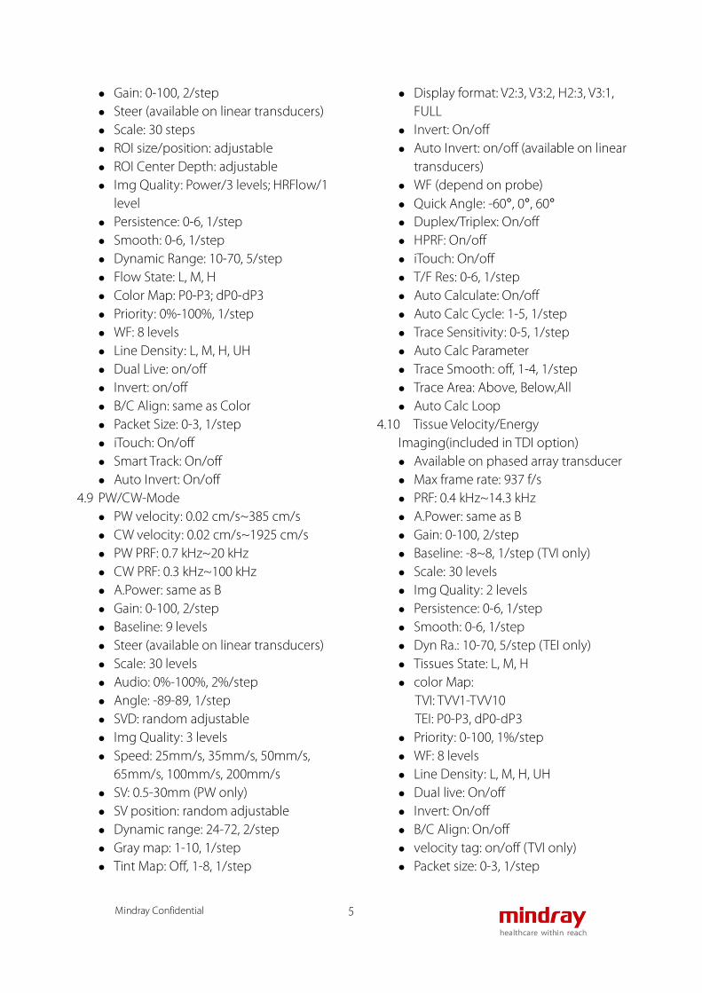
transducers) Steer (available on linear transducers) ROI size/position: adjustable ROI Center Depth: adjustable Img Quality: 3 levels Persistence: 0-6, 1/step Smooth : 0-6, 1/step Color Map: V0-V10; VV0-VV9 Flow State: L, M, H Priority: 0%-100%, 1%/step WF: 8 Levels Line Density: L, M,H,UH Dual Live: on/off Invert: on/off Auto Invert: on/off (available on linear
transducers) B/C Align: on/off velocity tag: on/off Packet Size: 0-3, 1/step iTouch: On/off Smart Track: On/off
4.8 Power Doppler Imaging PRF: 0.1 kHz ~ 14.3 kHz HR FlowTM: High Resolution Flow
provides better image quality and sensitivity
A.power: same as B
Mindray Confidential 5
Gain: 0-100, 2/step Steer (available on linear transducers) Scale: 30 steps ROI size/position: adjustable ROI Center Depth: adjustable Img Quality: Power/3 levels; HRFlow/1
level Persistence: 0-6, 1/step Smooth: 0-6, 1/step Dynamic Range: 10-70, 5/step Flow State: L, M, H Color Map: P0-P3; dP0-dP3 Priority: 0%-100%, 1/step WF: 8 levels Line Density: L, M, H, UH Dual Live: on/off Invert: on/off B/C Align: same as Color Packet Size: 0-3, 1/step iTouch: On/off Smart Track: On/off Auto Invert: On/off
4.9 PW/CW-Mode PW velocity: 0.02 cm/s~385 cm/s CW velocity: 0.02 cm/s~1925 cm/s PW PRF: 0.7 kHz~20 kHz CW PRF: 0.3 kHz~100 kHz A.Power: same as B Gain: 0-100, 2/step Baseline: 9 levels Steer (available on linear transducers) Scale: 30 levels Audio: 0%-100%, 2%/step Angle: -89-89, 1/step SVD: random adjustable Img Quality: 3 levels Speed: 25mm/s, 35mm/s, 50mm/s,
65mm/s, 100mm/s, 200mm/s SV: 0.5-30mm (PW only) SV position: random adjustable Dynamic range: 24-72, 2/step Gray map: 1-10, 1/step Tint Map: Off, 1-8, 1/step
Display format: V2:3, V3:2, H2:3, V3:1, FULL
Invert: On/off Auto Invert: on/off (available on linear
transducers) WF (depend on probe) Quick Angle: -60°, 0°, 60° Duplex/Triplex: On/off HPRF: On/off iTouch: On/off T/F Res: 0-6, 1/step Auto Calculate: On/off Auto Calc Cycle: 1-5, 1/step Trace Sensitivity: 0-5, 1/step Auto Calc Parameter Trace Smooth: off, 1-4, 1/step Trace Area: Above, Below,All Auto Calc Loop
4.10 Tissue Velocity/Energy Imaging(included in TDI option) Available on phased array transducer Max frame rate: 937 f/s PRF: 0.4 kHz~14.3 kHz A.Power: same as B Gain: 0-100, 2/step Baseline: -8~8, 1/step (TVI only) Scale: 30 levels Img Quality: 2 levels Persistence: 0-6, 1/step Smooth: 0-6, 1/step Dyn Ra.: 10-70, 5/step (TEI only) Tissues State: L, M, H color Map:
TVI: TVV1-TVV10 TEI: P0-P3, dP0-dP3
Priority: 0-100, 1%/step WF: 8 levels Line Density: L, M, H, UH Dual live: On/off Invert: On/off B/C Align: On/off velocity tag: on/off (TVI only) Packet size: 0-3, 1/step

Mindray Confidential 6
4.11 Tissue Velocity Doppler(included in TDI option) Available on phased array transducer Scale: 30 levels, 0.03 cm/s ~ 308 cm/s PRF: 0.7 kHz~20 kHz A.power: same as B Gain: 0-100,2/step Baseline: 9 levels Scale: 30 levels Audio: 0-100%, 2%/step Anlge: -89-89, 1/step SVD: random adjustable Img Quality: 2 levels Speed: 25mm/s, 35mm/s, 50mm/s,
65mm/s, 100mm/s, 200mm/s SV size: same as PW Dyn Ra.: 24-72, 2/step Gray Map: 1-10, 1/step Tint map: Off, 1-8, 1/step Display Format: V2:3, V3:2, H2:3, V3:1,
FULL Invert: On/off WF: 10 levels Quick Angle: -60°, 0, 60° Duplex/triplex: same as PW T/F Res: 0-6, 1/step iTouch: On/off
4.12 Tissue Velocity Motion (included in TDI option) A.power: same as B Smooth: 0-6, 1/step velocity tag: on/off Persistence: 0-6, 1/step Img Quality: 2 levels Flow State: L, M, H Speed: 25mm/s, 35mm/s, 50mm/s,
65mm/s, 100mm/s, 200mm/s Display format: V2:3, V3:2, H2:3, V3:1,
FULL Color Map: TVV1-TVV10 Packet Size: 0-3, 1/step Priority: 0%-100%,1%/step WF: 8 levels
4.13 iScapeTM View (option) Panoramic imaging Available on all transducers Acquisition method: B mode, Power
mode and Color mode Imaging length: 292.80 cm Tint map: off; 8 types Rotation: 0°-355°
4.14 Elastography (option) Available on linear transducers Support strain ratio measurement Unique shell analysis function Stress compensation technology
reduces deeper tissue artifacts, obtains more uniform stress throughout whole field
Stress indicator: supports frame by frame stress indication
Opacity: 0-5, 1/step Map: E1-E6 Smooth: 0-5, 1/step ROI: random adjustable ROI Center Depth: random adjustable Invert: on/off Depth: linear: 1.5~5cm Display Format: V1:1, H1:1, FULL Strain Scale: 0-5, 1/step Map Position: 0%-100%, 5%/step Dyn Ra.: 0~5, 1/step Strain Mode: 0-1, 1/step E Sensitivity: 0-5, 1/step Image Quality: three levels of
fundamental frequency, three levels of harmonic frequency
4.15 UWN+ Contrast ImagingTM*(option) Ultra Wideband Non-linear Plus
contrast imaging technology, which provides exceptional contrast agent detecting capability, not only extracts second harmonic, but also non-linear fundamental signals
Micro Flow Enhancement (MFE) available

Mindray Confidential 7
A.Power: same as B TGC: same as B Depth: same as B Gain: 0-100, 1/step Persistence: 0-7, 1/step Dyn Ra.: same as B Gray Map: 1-8, 1/step Tint Map: Off, 1-8, 1/step FOV: on/off FOV Size: random adjustable FOV Position: random adjustable ExFov: off, 1-2, 1/step iClear: Off, 1-7, 1/step Line Density: L, M, H, UH L/R Flip: on/off U/D Flip: on/off Rotation Counter-Clockwise: same as B Dual Live: on/off iTouch: On/off iTouch: -8-8, 2db/levels Image Quality: 3 levels Mix:
- Dual Live on: Contrast/C&T - Dual Live off: Contras/C&T/Tissue
Mix Map: 0-6, 1/step Timer1: on/off Timer2: on/off Destruct: on/off Destruct Time: 200-2000, 90/step Destruct Power: -30-0, 0.3/step MFE: on/off MFE Period: 0.1s, 0.2s, 0.4s, 0.6s, 0.8s,
1.0s, MAX Retro Capture: on/off Pro Capture: on/off Smooth: 0-6, 1/step CEUSPos: on/off
*The ME7 is designed for compatibility with commercially available ultrasound contrast agents. Because the availability of these agents is subject to government regulation and approval,
product features intended for use with these agents may not be commercially marketed nor made available before the contrast agent is cleared for use. Contrast related product features are enabled only on systems for delivery to an authorized country or region of use. Mindray medical systems makes no claims concerning the safety or effectiveness of contrast agents.
4.16 Stress Echo (option) Available on cardiac sector transducers 14 factory protocols User-defined protocols ECG triggered acquisition, display,
selection, comparison, evaluation and archiving of multiple cardiac loops during various stages of a stress echo examination
ASE16 (with score 4-7), ASE 17 (with score 4-7)
Customized stages: up to 7 views per stage, and up to 12 stages per study
View: standard views (PSLA, PSAX, A4C, A2C), and customized views
Image acquisition - R-wave trigger - Acquire mode: Manual ROI or full
screen - Ability to acquire frames or clips in B-
mode, LVO Image selection
- Attach the images with view annotation label (PSLA, PSAX, A4C,A2C, and customized views)
Review - Automatically adjust to the number
of images user defined Wall Motion Scoring
- ASE 16 (with score 4-7), or ASE 17(with score 4-7)
- Graphical display of scoring (Normal,

Mindray Confidential 8
Hyperkinetic, Severely Hyperkinetic, Akinetic, Dyskinetic)
LV volume measurement - Measurement of LV Volume in all
phases of cardiac cycle Report
- Reporting for both Wall Motion Scoring and LV volume measurement
4.17 LVO (option) Available on P4-2s Dedicated left ventricle contrast
imaging tool 4.18 iBeamTM
Spatial compound imaging 3 angles maximum Available on convex and linear
transducers 4.19 iTouchTM
Auto image optimization B-mode: gain, TGC Color: gain Power: gain PW: gain, scale, PRF, WF Contrast imaging: gain, TGC
4.20 Echo BoostTM Only for cardiac exams Improve the homogeneity of cardiac
images through the whole field of view Better contrast resolution of
myocardium tissue layers Better noise control in cardiac
chambers and muscles 4.21 B steer
Only for linear transducers 4.22 ExFov
Extended field of view Available for all convex and linear
transducers 4.23 Zoom
Zoom: Spot zoom (write zoom) up to 10x, Pan zoom (read zoom) 0.8x-10x
iZoom: convertible 3 steps ;normal image, zoom standard area, zoom only
image area 4.24 QSave
Quick save image parameter setting after image adjustment done
Support Save, Create, Restore 4.25 TT QA(option)
Tissue tracking quantitative analysis Mandatory ECG connection before TT
QA cine acquisition Six views for analysis: ALAX, A4C, A2C,
PSAXB, PSAXM, PSAXAP Reload: reload cine again for new study Edit: modify trace points Start tracking Accept & compute: start tracking
myocardium movement when user accept trace result
Display effect: 0/1; at 0, tracking in velocity vector arrow; at 1, tracking in dots
Trace method: 3 point or manual for ALAX, A4C, A2C; manual for PSAXB, PSAXM, PSAXAP
Bull’s Eye: trace result in bull’s eye model
Torsion: Torsion rate curve display LGC: available Valve’s open and close time index: MVC,
MVC’, AVC, AVO, MVO Data export: export data in CSV file Cycle: ECG triggered cardiac cycle
recognition for analysis Auto play: stop, X1/10, X1/5, X1/4, X1/3,
X1/2, X1, X2, X3 Thickness: 1-30mm, 1mm/step; adjust
trace thickness Track point: 20-40, 1/step Parameter: Volume, Speed, Displace., L
Strain, L Strain R, T Strain, T Strain R, Area, R Strain, R Strain R, C Strain, C Strain R, Rotation, Rot. R
Smooth: 0-4, 1/step 4.26 Smart 3DTM

Mindray Confidential 9
Acquisition Method: Rocked, Linear VR/MPR: set parameters for volume
rendered image or MPR plane Ref. Image: switch VR or A/B/C plane Display formats: Quad, Dual, Single,
MPR only, A4:1 VOI: on/off Reset: all, orientation, reset curve VR orientation: 0°, 90°, 180°, 270° for
quick rotation Inversion: inversion, gray Accept VOI: on/off Flip: flip VR Sync: synchronize VR with selected
plane Render modes: Surface, Min, Max, X-ray View direction: down/up, left/right,
front/back Threshold: 0%-100%, 1%/step (only on
VR) Opacity: 0%-100%, 5%/step (only on
VR) Smooth: 0-10, 1/ step Tint: off; 8 types Brightness: 0%-100%, 2%/step Contrast: 0%-100%, 2%/step Tool: Auto rotation
- Rotation control: play, single loop, loop
- Direction: left/right, up/down - Position: Set Start/Set end
Edit - Eraser: Soft eraser/ hard eraser,
Polygon, Contour, Rect, line - Eraser Diameter: 8-80, 1/step - Undo: Undo, Undo all
4.27 iNeedleTM(option) Needle visualization enhancement Best angle indicator Available on linear and curved
transducers 4.28 AutoEF (option)
Adjust Frame
Diastole FR Systole FR Volume curve: on/off Adjustment for the border of
endocardium 4.29 Smart track
Continuously track the flow and detect the best color box position and angle in real time scanning.
The Linear probes in carotid, IMT, Upper Ext A, Upper Ext V, Lower Ext A, Lower Ext V, EM Vascular exam modes support the Smart Track function.
4.30 RIMT (RF-Data based IMT) Available in single/dual B carotid exam
mode Side: left/right Calculation of 6 RIMT values, RIMT
average value, SD and ROI W Report operation:
- Data deleting - RIMT trend graphic viewing - Preview
4.31 eSpacial Navi (option) Needle guidance Display Favorite Only Auto Optimize Calibration Target Box In-Plane Position In-Plane Trajectory In-Plane Alignment In-Plane Target Box Out-Plane Position Out-Plane Trajectory Out-Plane Alignment Out-Plane
4.32 Smart VTI (option) Edit VTI LVOT Diam Save VTI Graph
4.33 Smart IVC (option) Edit Line: Angle

Mindray Confidential 10
Trend curve Change Resp Time: Resp Time 0.5-5.0 s Diagnosis comments Breath type: Spontaneous Breath,
Mechanical Ventilation 4.34 Smart B-line (option)
Auto Calc Scanning Areas: 6 Zones, 8 Zones, 12
Zones OverView: Image Map, Color Map, Prev
Item, Next Item Image and diagnosis comments
5 Cine Review and Post Processing 5.1 Cine review
Available in all modes Frame by frame manual cineloop
review or auto playback with variable speed
Independent cine review in 2D Dual and Quad mode one by one
Maximum cine memory is up to 25492 frames or 263.3 s (depend on the mode)
Retrospective storage (online setting available, 1-120 s, or 1-120 cycles, pre-settable) and prospective storage (1-480 s, or 1-390 cycles, pre-settable)
Frame compare: compare different frames for one cine in dual format
Cine compare: compare two or more than two cines in dual or quad format
Jump to first and jump to last: one keystroke review the first or last frame
Start point and end point: selectable 5.2 Raw data processing
B-mode: TGC Gain Dynamic range Gray map Tint map iClear
L/R Flip U/D Flip Rotation LGC Dual Live Auto Merge H Scale Echo Boost Smooth Zoom Value
M-mode: Gain Speed Dynamic Range Gray Map Tint Map Display format Edge Enhance
Color: Gain Baseline Smooth Color map Dual Live Invert Priority Velocity tag
PW: Gain Baseline Audio Angle Speed Dynamic range Gray map Tint Map Display format Invert WF Quick Angle T/F Res
6 Measurement/Analysis and

Mindray Confidential 11
Report* 6.1 Generic measurements
B-Mode - Distance - Ellipse - Trace - Spline - Cross - Angle(2L) - Angle(3P) - Double Dist - Trace Len - Trace Len(Spline) - Parallel - Distance P-L - IMT - B-Profile - B-Hist(Ellipse) - B-Hist(Trace) - B-Hist(Spline) - B-Hist(Rectangle) - Depth - Color Vel - Strain Hist - Elas. Hist - Color Vel Profile - Elas. - Strain - Smart Trace - ------------ - Volume - Volume(Ellipse) - Volume(E+Dist.) - Ratio(D) - ------------ - Volume - Volume - Volume(Ellipse) - Volume(E+Dist.) - Ratio(A) - Area1 - Area2
- Directional Ratio - D1 - D2 - RAC - Sag - XS - Volume Flow - Vas Area - TAMEAN - TAMAX - Elas. Ratio - A - B - Strain Ratio - A - B
M-Mode - HR - HR(R-R) - Slope - Distance - Time - Velocity
D-Mode
- PS/ED - Vel - HR - HR(R-R) - Time - Acceleration - D Trace - ------------ - Ratio(Vel) - Ratio(VTI) - ------------ - Volume Flow - Vas Area - TAMEAN - TAMAX
6.2 Clinical option measurement package Abdominal

Mindray Confidential 12
B-Mode - Aorta Bif - Aorta Aneurysm Status - Shunt Diam - Portal V Diam - M Portal V Diam - Splenic V Diam - PS Conflnc Diam - Renal V Diam - SMV Diam - IMV Diam - CHD - GB L - GB H - GB W - GB wall th - Cystic Duct - CBD - Panc duct - Panc head - Panc neck - Panc body - Panc tail - Appendix - Appendix Wall - Pylorus - Pylorus Wall - Renal L - Renal H - Renal W - Cortex - Adrenal L - Adrenal H - Adrenal W - Ureter - Cortex(Renal Transplant1) - Renal V Diam(Renal Transplant1) - Ureter Diam(Renal Transplant1) - Cortex(Renal Transplant2) - Renal V Diam(Renal Transplant2) - Ureter Diam(Renal Transplant2) - Pre-BL L - Pre-BL H
- Pre-BL W - Post-BL L - Post-BL H - Post-BL W - Spleen L - Spleen H - Spleen W - Spleen Area - Skin-L.Capsule Dist. - Hepatic Lesion1 Elas. - Hepatic Lesion2 Elas. - Hepatic Lesion3 Elas. - LSM - Free Fluid - ------------ - Renal Vol - Pre-BL Vol - Post-BL Vol - Mictur.Vol - ------------ - Aorta - Anterior-Posterior - Transverse - Outer Diameter - Inner Diameter - Outer Area - Inner Area - Celiac Axis - Anterior-Posterior - Transverse - SMA - Anterior-Posterior - Transverse - C Hepatic A - Anterior-Posterior - Transverse - Proper Hepatic A - Anterior-Posterior - Transverse - Hepatic A - Anterior-Posterior - Transverse - Splenic A

Mindray Confidential 13
- Anterior-Posterior - Transverse - GDA - Anterior-Posterior - Transverse - IMA - Anterior-Posterior - Transverse - Aorta Aneurysm - Long - Anterior-Posterior - Transverse - Celiac A Aneurysm - Long - Anterior-Posterior - Transverse - SMA Aneurysm - Long - Anterior-Posterior - Transverse - C Hepatic A Aneurysm - Long - Anterior-Posterior - Transverse - Proper Hepatic A Aneurysm - Long - Anterior-Posterior - Transverse - Hepatic A Aneurysm - Long - Anterior-Posterior - Transverse - Splenic A Aneurysm - Long - Anterior-Posterior - Transverse - GDA Aneurysm - Long - Anterior-Posterior - Transverse - IMA Aneurysm - Long - Anterior-Posterior
- Transverse - EVAR Residual Aneurysm Sac(2D) - Anterior-Posterior - Transverse - EVAR Inflow(2D) - Anterior-Posterior - Transverse - EVAR Graft Body(2D) - Anterior-Posterior - Transverse - EVAR Limb(2D) - Anterior-Posterior - Transverse - EVAR Outflow(2D) - Anterior-Posterior - Transverse - Aortic Bypass Graft Anast(2D) - Anterior-Posterior - Transverse - Aortic Bypass Graft Graft(2D) - Anterior-Posterior - Transverse - ABD Stenosis 1(2D) - Anterior-Posterior - Transverse - Outer Diameter - Inner Diameter - Outer Area - Inner Area - ABD Stenosis 2(2D) - Anterior-Posterior - Transverse - Outer Diameter - Inner Diameter - Outer Area - Inner Area - ABD Stenosis 3(2D) - Anterior-Posterior - Transverse - Outer Diameter - Inner Diameter - Outer Area - Inner Area

Mindray Confidential 14
- ABD Stenosis 4(2D) - Anterior-Posterior - Transverse - Outer Diameter - Inner Diameter - Outer Area - Inner Area - IVC - Anterior-Posterior - Transverse - Checklist - Hepatic V(2D) - Anterior-Posterior - Transverse - Lt Hepatic V(2D) - Anterior-Posterior - Transverse - M Hepatic V(2D) - Anterior-Posterior - Transverse - Rt Hepatic V(2D) - Anterior-Posterior - Transverse - Liver - L - H - W - R Liver Lobe - L - H - W - L Liver Lobe - L - H - W - Hepatic Lesion 1 - d1 - d2 - d3 - Hepatic Lesion 2 - d1 - d2 - d3
- Hepatic Lesion 3 - d1 - d2 - d3 - Hepatic Cyst 1 - d1 - d2 - d3 - Hepatic Cyst 2 - d1 - d2 - d3 - Hepatic Cyst 3 - d1 - d2 - d3 - GB - GB L - GB H - GB W - GB wall th - GB Finding 1 - d1 - d2 - d3 - GB Finding 2 - d1 - d2 - d3 - GB Finding 3 - d1 - d2 - d3 - GB Finding 4 - d1 - d2 - d3 - GB Finding 5 - d1 - d2 - d3 - Panc Finding 1 - d1

Mindray Confidential 15
- d2 - d3 - Panc Finding 2 - d1 - d2 - d3 - Panc Finding 3 - d1 - d2 - d3 - Panc Finding 4 - d1 - d2 - d3 - Panc Finding 5 - d1 - d2 - d3 - Kidney - Renal L - Renal H - Renal W - Cortex - Adrenal - Adrenal L - Adrenal H - Adrenal W - Renal Lesion 1 - d1 - d2 - d3 - Renal Lesion 2 - d1 - d2 - d3 - Renal Lesion 3 - d1 - d2 - d3 - Renal Cyst 1 - d1 - d2 - d3
- Renal Cyst 2 - d1 - d2 - d3 - Renal Cyst 3 - d1 - d2 - d3 - Kidney(Superior) - H - W - Kidney(Mid) - H - W - Kidney(Inferior) - H - W - Renal A - Long - Anterior-Posterior - Transverse - Renal A Aneurysm - Long - Anterior-Posterior - Transverse - Kidney(Renal Transplant1) - L - H - W - Adrenal(Renal Transplant1) - L - H - W - Finding 1(Renal Transplant1) - L - H - W - Finding 2(Renal Transplant1) - L - H - W - Finding 3(Renal Transplant1) - L

Mindray Confidential 16
- H - W - Finding 4(Renal Transplant1) - L - H - W - Finding 5(Renal Transplant1) - L - H - W - Finding 6(Renal Transplant1) - L - H - W - Renal Transplant 1(2D) - Cortex(Renal Transplant1) - Renal V Diam(Renal
Transplant1) - Ureter Diam(Renal Transplant1) - Kidney(Renal Transplant1) - L - H - W - Adrenal(Renal Transplant1) - L - H - W - Finding 1(Renal Transplant1) - L - H - W - Finding 2(Renal Transplant1) - L - H - W - Finding 3(Renal Transplant1) - L - H - W - Finding 4(Renal Transplant1) - L - H - W
- Finding 5(Renal Transplant1) - L - H - W - Finding 6(Renal Transplant1) - L - H - W - Kidney(Renal Transplant2) - L - H - W - Adrenal(Renal Transplant2) - L - H - W - Finding 1(Renal Transplant2) - L - H - W - Finding 2(Renal Transplant2) - L - H - W - Finding 3(Renal Transplant2) - L - H - W - Finding 4(Renal Transplant2) - L - H - W - Finding 5(Renal Transplant2) - L - H - W - Finding 6(Renal Transplant2) - L - H - W - Renal Transplant 2(2D) - Cortex(Renal Transplant2) - Renal V Diam(Renal

Mindray Confidential 17
Transplant2) - Ureter Diam(Renal Transplant2) - Kidney(Renal Transplant2) - L - H - W - Adrenal(Renal Transplant2) - L - H - W - Finding 1(Renal Transplant2) - L - H - W - Finding 2(Renal Transplant2) - L - H - W - Finding 3(Renal Transplant2) - L - H - W - Finding 4(Renal Transplant2) - L - H - W - Finding 5(Renal Transplant2) - L - H - W - Finding 6(Renal Transplant2) - L - H - W - Bladder - Pre-BL L - Pre-BL H - Pre-BL W - Post-BL L - Post-BL H - Post-BL W - Spleen - Spleen L
- Spleen H - Spleen W - Spleen Area - Hepatic Lesion1 ElasRatio - A - B - Hepatic Lesion2 ElasRatio - A - B - Hepatic Lesion3 ElasRatio - A - B D-Mode - Aorta - Celiac Axis - SMA - C Hepatic A - Proper Hepatic A - Hepatic A - Splenic A - GDA - IMA - Aorta(Post) - Celiac Axis(Post) - SMA(Post) - C Hepatic A(Post) - Proper Hepatic A(Post) - Hepatic A(Post) - Splenic Artery(Post) - GDA(Post) - IMA(Post) - EVAR Residual Aneurysm Sac - EVAR Inflow - EVAR Graft Body - EVAR Limb - EVAR Outflow - Aortic Bypass Graft Anast - Aortic Bypass Graft Graft - IVC Reflux - IVC - Hepatic V - Lt Hepatic V - M Hepatic V

Mindray Confidential 18
- Rt Hepatic V - Portal V - M Portal V - Splenic V - Renal V - SMV - IMV - Hepatic A Anast(Liver Transplant) - Hepatic V Anast(Liver Transplant) - Portal V Anast(Liver Transplant) - IVC(Liver Transplant) - Hep V Confl(Liver Transplant) - Donor IVC(Liver Transplant) - Renal A - Ren A Org - M Renal A - Renal A1 - Renal A2 - Hilum - Interlobar A - Arcuate A - Segment A - Artery Anast(Renal Transplant1) - Artery Anast 2(Renal Transplant1) - Vein Anast(Renal Transplant1) - Vein Anast 2(Renal Transplant1) - Renal A(Renal Transplant1) - Renal A1(Renal Transplant1) - Renal A2(Renal Transplant1) - Hilum(Renal Transplant1) - Interlobar A(Renal Transplant1) - Arcuate A(Renal Transplant1) - Segmental A(Renal Transplant1) - Renal Vein 1(Renal Transplant1) - Renal Vein 2(Renal Transplant1) - Artery Anast(Renal Transplant2) - Artery Anast 2(Renal Transplant2) - Vein Anast(Renal Transplant2) - Vein Anast 2(Renal Transplant2) - Renal A(Renal Transplant2) - Renal A1(Renal Transplant2) - Renal A2(Renal Transplant2) - Hilum(Renal Transplant2)
- Interlobar A(Renal Transplant2) - Arcuate A(Renal Transplant2) - Segmental A(Renal Transplant2) - Renal Vein 1(Renal Transplant2) - Renal Vein 2(Renal Transplant2) - TIPS - ------------ - SMA/Ao - CA/Ao - ------------ - ABD Stenosis 1 - Pre Sten - Sten - Post Sten - ABD Stenosis 2 - Pre Sten - Sten - Post Sten - ABD Stenosis 3 - Pre Sten - Sten - Post Sten - ABD Stenosis 4 - Pre Sten - Sten - Post Sten - Renal Transplant 1(Doppler) - Artery Anast(Renal Transplant1) - Artery Anast 2(Renal
Transplant1) - Vein Anast(Renal Transplant1) - Vein Anast 2(Renal Transplant1) - Renal A(Renal Transplant1) - Renal A1(Renal Transplant1) - Renal A2(Renal Transplant1) - Hilum(Renal Transplant1) - Interlobar A(Renal Transplant1) - Arcuate A(Renal Transplant1) - Segmental A(Renal
Transplant1) - Renal Vein 1(Renal Transplant1) - Renal Vein 2(Renal Transplant1) - Renal Transplant 2(Doppler)

Mindray Confidential 19
- Artery Anast(Renal Transplant2) - Artery Anast 2(Renal
Transplant2) - Vein Anast(Renal Transplant2) - Vein Anast 2(Renal Transplant2) - Renal A(Renal Transplant2) - Renal A1(Renal Transplant2) - Renal A2(Renal Transplant2) - Hilum(Renal Transplant2) - Interlobar A(Renal Transplant2) - Arcuate A(Renal Transplant2) - Segmental A(Renal
Transplant2) - Renal Vein 1(Renal Transplant2) - Renal Vein 2(Renal Transplant2)
Gynecology B-Mode
- UT L - UT H - UT W - Endo - Cervix L - Cervix H - Cervix W - Ovary L - Ovary H - Ovary W - Follicle1 L - Follicle1 W - Follicle1 H - Follicle2 L - Follicle2 W - Follicle2 H - Follicle3 L - Follicle3 W - Follicle3 H - Follicle4 L - Follicle4 W - Follicle4 H - Follicle5 L - Follicle5 W - Follicle5 H - Follicle6 L
- Follicle6 W - Follicle6 H - Follicle7 L - Follicle7 W - Follicle7 H - Follicle8 L - Follicle8 W - Follicle8 H - Follicle9 L - Follicle9 W - Follicle9 H - Follicle10 L - Follicle10 W - Follicle10 H - Follicle11 L - Follicle11 W - Follicle11 H - Follicle12 L - Follicle12 W - Follicle12 H - Follicle13 L - Follicle13 W - Follicle13 H - Follicle14 L - Follicle14 W - Follicle14 H - Follicle15 L - Follicle15 W - Follicle15 H - Follicle16 L - Follicle16 W - Follicle16 H - DWT - BSD(R) - BSD(Va) - RVA(R) - RVA(Va) - UTA(R) - UTA(Va) - URA - PVA(R) - PVA(Va) - PUA(R)

Mindray Confidential 20
- PUA(Va) - BPW-SP Dist.(R) - BPW-SP Dist.(Va) - Cx-SP Dist.(R) - Cx-SP Dist.(Va) - RA-SP Dist.(R) - RA-SP Dist.(Va) - Shuttle(R) - Shuttle(Va) - Rectocele Depth - Intus. Depth - ARA(R) - ARA(Va) - ARA(C) - LH AP Diam(R) - LH AP Diam(Va) - LH AP Diam(C) - LH Lateral Diam(R) - LH Lateral Diam(Va) - LH Lateral Diam(C) - LH Area(R) - LH Area(Va) - LH Area(C) - LA Angle(R) - LA Angle(Va) - LA Angle(C) - LA Thickness(R) - LA Thickness(Va) - LA Thickness(C) - LUG(R) - LUG(Va) - LUG(C) - GYN Lesion1 Strain - GYN Lesion2 Strain - GYN Lesion3 Strain - Lesion1 Elas. - Lesion2 Elas. - Lesion3 Elas. - Fibroid1 Strain - Fibroid2 Strain - Fibroid3 Strain - Fibroid1 Elas. - Fibroid2 Elas.
- Fibroid3 Elas. - ------------ - UT Vol - UT SUM - UT-L/CX-L - Ovary Vol - Follicle1 - Follicle2 - Follicle3 - Follicle4 - Follicle5 - Follicle6 - Follicle7 - Follicle8 - Follicle9 - Follicle10 - Follicle11 - Follicle12 - Follicle13 - Follicle14 - Follicle15 - Follicle16 - Mean DWT - BND - IAS Damage - EAS Damage - ------------ - Uterus - UT L - UT H - UT W - Endo - Uterine Cervix - Cervix L - Cervix H - Cervix W - Fibroid 1 - d1 - d2 - d3 - Fibroid 2 - d1 - d2

Mindray Confidential 21
- d3 - Fibroid 3 - d1 - d2 - d3 - Uterine Finding 1 - d1 - d2 - d3 - Uterine Finding 2 - d1 - d2 - d3 - Uterine Finding 3 - d1 - d2 - d3 - Uterine Finding 4 - d1 - d2 - d3 - Uterine Finding 5 - d1 - d2 - d3 - Uterine Finding 6 - d1 - d2 - d3 - Ovary - Ovary L - Ovary H - Ovary W - Ovarian Cyst 1 - d1 - d2 - d3 - Ovarian Cyst 2 - d1 - d2 - d3 - Ovarian Cyst 3 - d1
- d2 - d3 - Ovarian Finding 1 - d1 - d2 - d3 - Ovarian Finding 2 - d1 - d2 - d3 - Ovarian Finding 3 - d1 - d2 - d3 - Ovarian Finding 4 - d1 - d2 - d3 - Ovarian Finding 5 - d1 - d2 - d3 - Ovarian Finding 6 - d1 - d2 - d3 - Follicle1 - Follicle1 L - Follicle1 W - Follicle1 H - Follicle2 - Follicle2 L - Follicle2 W - Follicle2 H - Follicle3 - Follicle3 L - Follicle3 W - Follicle3 H - Follicle4 - Follicle4 L - Follicle4 W - Follicle4 H - Follicle5

Mindray Confidential 22
- Follicle5 L - Follicle5 W - Follicle5 H - Follicle6 - Follicle6 L - Follicle6 W - Follicle6 H - Follicle7 - Follicle7 L - Follicle7 W - Follicle7 H - Follicle8 - Follicle8 L - Follicle8 W - Follicle8 H - Follicle9 - Follicle9 L - Follicle9 W - Follicle9 H - Follicle10 - Follicle10 L - Follicle10 W - Follicle10 H - Follicle11 - Follicle11 L - Follicle11 W - Follicle11 H - Follicle12 - Follicle12 L - Follicle12 W - Follicle12 H - Follicle13 - Follicle13 L - Follicle13 W - Follicle13 H - Follicle14 - Follicle14 L - Follicle14 W - Follicle14 H - Follicle15 - Follicle15 L - Follicle15 W - Follicle15 H
- Follicle16 - Follicle16 L - Follicle16 W - Follicle16 H - GYN Lesion 1 - d1 - d2 - d3 - GYN Lesion 2 - d1 - d2 - d3 - GYN Lesion 3 - d1 - d2 - d3 - Residual Urine - BL Height - BL Depth - GYN Lesion1 Strain Ratio - A - B - GYN Lesion2 Strain Ratio - A - B - GYN Lesion3 Strain Ratio - A - B - Lesion1 Elas. Ratio - A - B - Lesion2 Elas. Ratio - A - B - Lesion3 Elas. Ratio - A - B - Fibroid1 Strain Ratio - A - B - Fibroid2 Strain Ratio - A - B

Mindray Confidential 23
- Fibroid3 Strain Ratio - A - B - Fibroid1 Elas. Ratio - A - B - Fibroid2 Elas. Ratio - A - B - Fibroid3 Elas. Ratio - A - B
Obstetrics B-Mode - GS - YS L - CRL - NT - BPD - OFD - HC - AC - FL - TAD - APAD - TCD - CM - IT - LVW - HW - OOD - IOD - HUM - Ulna - RAD - Tibia - FIB - CLAV - Vertebrae - MP - Foot - NBL - Ear
- APTD - TTD - FTA - THD - HrtC - TC - Umb VD - F-kidney L - Mat Kidney - Cervix L - AF - NF - Orbit - PL Thickness - Sac Diam1 - Sac Diam2 - Sac Diam3 - AF1 - AF2 - AF3 - AF4 - LVIDd - LVIDs - LV Diam - LA Diam - RVIDd - RVIDs - RV Diam - RA Diam - IVSd - IVSs - IVS - LV Area - LA Area - RV Area - RA Area - Ao Diam - MPA Diam - LVOT Diam - LVOT Area - RVOT Diam - Facial Angle - HrtA

Mindray Confidential 24
- MV Diam(Z-Score) - PV Diam(Z-Score) - Ao Asc Diam(Z-Score) - Ao Desc Diam(Z-Score) - Duct Art Diam(Z-Score) - TV Diam(Z-Score) - LPA Diam(Z-Score) - RPA Diam(Z-Score) - IVC Diam(Z-Score) - AV Diam(Z-Score) - MPA Diam(Z-Score) - RV Diam(Z-Score) - LV Diam(Z-Score) - RV Area(Z-Score) - LV Area(Z-Score) - RVIDd(Z-Score) - LVIDd(Z-Score) - UT L - UT H - UT W - Endo - AH - PH - 3th Ventricle - NT Above Cord - NT Below Cord - Mandible - Prenasal th - Heart AP - Heart T - LV Width - LV Length - RV Width - RV Length - LA Width - RA Width - LVWd - LVWs - RVWd - RVWs - AV Diam - AV Area - PV Area
- F-kidney H - F-kidney W - Lung - Stomach - YS H - YS W - Amniotic Sac L - Amniotic Sac H - Amniotic Sac W - Ovary Cyst L - Ovary Cyst H - Ovary Cyst W - UT AW - UT PW - CSP - FMF - MMF - Lung CCAM L - Lung CCAM H - Lung CCAM W - AD - Lliac Wing Angle - FAGL - FAG - Intestinum Crassum - Liver Length - Rib Length - Shoulder Blade - ------------ - MAD - Mean Sac Diam - AFI - EFW - EFW2 - HC/AC(Campbell) - FL/AC - FL/BPD - AXT - CI - FL/HC(Hadlock) - AC(c) - HC(c) - HrtC/TC

Mindray Confidential 25
- TCD/AC - LVW/HW - LVD/RVD - LAD/RAD - AoD/MPAD - LAD/AoD - UT Vol - UT SUM - UT-L/CX-L - ------------ - AFI - AF1 - AF2 - AF3 - AF4 - Uterus - UT L - UT H - UT W - Endo - M-Mode - FHR (M) - LVIDd - LVIDs - RVIDd - RVIDs - IVSd - IVSs - RVIDd(Z-Score) - LVIDd(Z-Score) - MVE - TVE - AVE - MAPSE - TAPSE - LV ICT - LV IRT - LV ET - RV ICT - RV IRT - RV ET -
D-Mode - Umb A - Duct Veno - Placenta A - MCA - Fetal Ao - Desc Aorta - Ut A - Ovarian A - FHR (Doppler) - Asc Aorta - RVOT - LVOT - MV E - MV A - TV E - TV A - MV E' - MV A' - MV S' - TV E' - TV A' - TV S' - AV PV - AV VTI - PV PV - PV VTI - Duct Art PV - Duct Art VTI - AV TPV - PV TPV - Duct Art TPV - Thoracic Aorta - Hepatic Vein - IVC - Umb V - Ovary - Endometrium - Cervical Cancer - Fibroid - Duct Art - ICA - Celiac A

Mindray Confidential 26
- ------------ - MV E/A - TV E/A - MV E/E' - TV E/E'
Cardiology B-Mode - RVAWd(2D) - RVAWs(2D) - RVDd(2D) - RVDs(2D) - IVSd(2D) - IVSs(2D) - LVIDd(2D) - LVIDs(2D) - LVPWd(2D) - LVPWs(2D) - Diastole(2D) - Systole(2D) - LVLd apical - LVLs apical - LVAd apical - LVAs apical - LVAd sax MV - LVAs sax MV - LVAd sax Endo - LVAd sax Epi - LV Major - LV Minor - LV Area(d) - LV Area(s) - HR(2D) - RA Major - RA Minor - RA Area - RA Vol(A4C) - RAP - RV Area(d) - RV Area(s) - RV Major - RV Minor - LA Diam(2D) - LA Major
- LA Minor - LA Area - LVOT Diam - Ao Diam(2D) - ACS(2D) - AV Diam - Ao Isthmus(2D) - Ao Sinus Diam(2D) - Ao st junct(2D) - AVA - Ao Arch Diam(2D) - Ao Asc Diam(2D) - Ao Desc Diam(2D) - Duct Art Diam - Post Ductal - Pre Ductal - MCS(2D) - MV Diam - MV EPSS(2D) - MVA - TV Diam - TVA - PV Diam - RVOT Diam - MPA Diam(2D) - RPA Diam(2D) - LPA Diam(2D) - IVC Diam(Expir) - IVC Diam(Insp) - SVC Diam(Expir) - SVC Diam(Insp) - LCA Diam - RCA Diam - PEd(2D) - PEs(2D) - VSD Diam - ASD Diam - PDA Diam - PFO Diam - AutoEF - ------------ - LA/Ao(2D) - ------------

Mindray Confidential 27
- LV(2D) - Diastole(2D) - Systole(2D) - IVSd(2D) - LVIDd(2D) - LVPWd(2D) - IVSs(2D) - LVIDs(2D) - LVPWs(2D) - HR(2D) - Simpson - A4Cd - A4Cs - A2Cd - A2Cs - HR(2D) - Mod.Simpson - LVLd apical - LVLs apical - LVAd sax MV - LVAs sax MV - LVAd sax PM - LVAs sax PM - HR(2D) - S-P Ellipse - LVLd apical - LVAd apical - LVLs apical - LVAs apical - HR(2D) - B-P Ellipse - LVIDd(2D) - LVAd sax MV - LVIDs(2D) - LVAs sax MV - LVAd apical - LVAs apical - HR(2D) - Bullet - LVLd apical - LVLs apical - LVAd sax MV - LVAs sax MV
- HR(2D) - LV Mass(Cube-2D) - IVSd(2D) - LVIDd(2D) - LVPWd(2D) - LV Mass(A-L) - LVLd apical - LVAd sax Epi - LVAd sax Endo - LV Mass(T-E) - LVAd sax Epi - LVAd sax Endo - a - d - LA Vol(Simp) - LA Vol(A2C) - LA Vol(A4C) - LA Vol(A-L) - LA apical - LAA(A2C) - LAA(A4C) - MVA(VTI) - LVOT Diam - LVOT VTI - MV VTI - AVA(VTI) - LVOT Diam - LVOT VTI - AV VTI - CO(LVOT) - LVOT Diam - LVOT VTI - AV HR - CO(RVOT) - RVOT Diam - RVOT VTI - PV HR - CO(MV) - MV Diam - MV VTI - MV HR - CO(TV) - TV Diam

Mindray Confidential 28
- TV VTI - TV HR - PISA MR - MR Rad - MR Als Vel - MR VTI - PISA AR - AR Rad - AR Als Vel - AR VTI - PISA TR - TR Rad - TR Als Vel - TR VTI - PISA PR - PR Rad - PR Als Vel - PR VTI - Qp/Qs - LVOT Diam - LVOT VTI - RVOT Diam - RVOT VTI - Z-Scores ( 3Y) (2D) - AV Diam - Ao Sinus Diam - Ao st junct - PV Diam - Ao Arch IA-LCA - Ao Arch LCA-LSA - Ao Arch after LSA - Ao Isthmus - Thoracic Ao Diam - IVC Diam - MV Diam - TV Diam - MPA Diam - RPA Diam - LPA Diam - Z-Scores (<18Y) (2D) - LV Area(d) A4C - LV Area(s) A4C - LVIDd A4C(2D)
- LVIDs A4C(2D) - LA AP Diam A4C - LA LL Diam A4C - LA Area A4C - RA AP Diam A4C - RA LL Diam A4C - RA Area A4C - RV Area(d) A4C - RV Area(s) A4C - RVd Major A4C - RVs Major A4C - RVd Minor (basal) A4C - RVd Minor (midcavity) A4C - LV Area(d) A2C - LV Area(s) A2C - LVIDd A2C(2D) - LVIDs A2C(2D) M-Mode - RVAWd(M) - RVAWs(M) - RVDd(M) - RVDs(M) - Ao Arch Diam(M) - Ao Asc Diam(M) - Ao Desc Diam(M) - Ao Diam(M) - Ao Isthmus(M) - Ao Sinus Diam(M) - Ao st junct(M) - ACS(M) - HR(M) - IVSd(M) - IVSs(M) - LA Diam(M) - LPA Diam(M) - Diastole(M) - Systole(M) - LVET(M) - LVIDd(M) - LVIDs(M) - LVOT Diam - LVPEP(M)

Mindray Confidential 29
- LVPWd(M) - LVPWs(M) - MCS(M) - MPA Diam(M) - MV A Amp - MV E Amp - MV D-E Slope - MV D-E Amp - MV E-F Slope - MV EPSS(M) - PEd(M) - PEs(M) - RPA Diam(M) - RVET(M) - RVOT Diam - RVPEP(M) - MAPSE - TAPSE - MV ALL - IVC Diam(Insp)(M) - IVC Diam(Expir)(M) - SVC Diam(Insp)(M) - SVC Diam(Expir)(M) - ------------ - LA/Ao(M) - ------------ - LV(M) - Diastole(M) - Systole(M) - IVSd(M) - LVIDd(M) - LVPWd(M) - IVSs(M) - LVIDs(M) - LVPWs(M) - HR(M) - LV Mass(Cube-M) - IVSd(M) - LVIDd(M) - LVPWd(M) - LV Tei Index(M) - MV C-O dur(M) - LVET(M)
- Z-Scores ( 3Y) (M) - IVSd(M) - LVPWd(M) - Z-Scores (<18Y) (M) - LVIDd(M) - LVIDs(M) D-Mode - MV Aa(lateral) - MV Aa(medial) - AAo Vmax - AV VTI - AV HR - AV Vmax - AR DecT - AR Time - AR PHT - AR Ved - AR Vmax - AR VTI - MV ARa(lateral) - MV ARa(medial) - ASD Vmax - AV AccT - AV DecT - Coarc Post-Duct - Coarc Pre-Duct - DAo Vmax - MV DRa(lateral) - MV DRa(medial) - MV Ea(lateral) - MV Ea(medial) - IVC Vel(Expir) - IVC Vel(Insp) - IVCT - LPA Vmax - LVET(Doppler) - LVOT AccT - LVOT VTI - LVOT Vmax - LVPEP(Doppler) - MPA Vmax - dP/dt

Mindray Confidential 30
- Tau(BAI) - MR VTI - MR Vmax - MS Vmax - MV A Dur - MV A Vel - MV A VTI - MV AccT - MV DecT - MV E Dur - MV E Vel - MV E VTI - IVRT - MV VTI - MV HR - MV Vmax - PVein A Dur - PVein A Vel - PVein D Vel - PVein D VTI - PVein DecT - PVein S Vel - PVein S VTI - PDA Vel(d) - PDA Vel(s) - PR PHT - PR VTI - PR Ved - PR Vmax - PR DecT - PV AccT - PV VTI - PV HR - PV Vmax - RAP - RPA Vmax - RVET(Doppler) - RVOT Vmax - RVOT VTI - RVPEP(Doppler) - MV Sa(lateral) - MV Sa(medial) - SVC Vel(Expir)
- SVC Vel(Insp) - TR VTI - TR Vmax - TV A Dur - TV A Vel - TV AccT - TV DecT - TV E Vel - TV VTI - TV HR - TV Vmax - VSD Vmax - Hepatic V S Vel - Hepatic V D Vel - ------------ - MV E/A - MVA(PHT) - TV E/A - TVA(PHT) - ------------ - LV Tei Index(Doppler) - MV C-O dur(Doppler) - LVET(Doppler) - RVSP - TR Vmax - RAP - PAEDP - PR Ved - RAP - MVA(VTI) - LVOT Diam - LVOT VTI - MV VTI - AVA(VTI) - LVOT Diam - LVOT VTI - AV VTI - CO(LVOT) - LVOT Diam - LVOT VTI - AV HR - CO(RVOT) - RVOT Diam

Mindray Confidential 31
- RVOT VTI - PV HR - CO(MV) - MV Diam - MV VTI - MV HR - CO(TV) - TV Diam - TV VTI - TV HR - RV Tei Index - TV C-O dur - RVET(Doppler) - PISA MR - MR Rad - MR Als Vel - MR VTI - PISA AR - AR Rad - AR Als Vel - AR VTI - PISA TR - TR Rad - TR Als Vel - TR VTI - PISA PR - PR Rad - PR Als Vel - PR VTI - Qp/Qs - LVOT Diam - LVOT VTI - RVOT Diam - RVOT VTI
Urology B-Mode - Renal L - Renal H - Renal W - Cortex - Adrenal L - Adrenal H - Adrenal W
- Ureter - Cortex(Renal Transplant1) - Renal V Diam(Renal Transplant1) - Ureter Diam(Renal Transplant1) - Cortex(Renal Transplant2) - Renal V Diam(Renal Transplant2) - Ureter Diam(Renal Transplant2) - Prostate L - Prostate H - Prostate W - Seminal L - Seminal H - Seminal W - Urethra - Pre-BL L - Pre-BL H - Pre-BL W - Post-BL L - Post-BL H - Post-BL W - Testicular L - Testicular H - Testicular W - Epididymis L - Epididymis H - Epididymis W - Scrotal Wall - Testis V(2D) - Testis V(Valsalva 2D) - Prostate Mass1 Strain - Prostate Mass2 Strain - Prostate Mass3 Strain - Prostate Mass1 Elas. - Prostate Mass2 Elas. - Prostate Mass3 Elas. - ------------ - Renal Vol - Prostate Vol - Pre-BL Vol - Post-BL Vol - Mictur.Vol - Testicular Vol - ------------

Mindray Confidential 32
- Kidney - Renal L - Renal H - Renal W - Cortex - Adrenal - Adrenal L - Adrenal H - Adrenal W - Renal Lesion 1 - d1 - d2 - d3 - Renal Lesion 2 - d1 - d2 - d3 - Renal Lesion 3 - d1 - d2 - d3 - Renal Cyst 1 - d1 - d2 - d3 - Renal Cyst 2 - d1 - d2 - d3 - Renal Cyst 3 - d1 - d2 - d3 - Kidney(Superior) - H - W - Kidney(Mid) - H - W - Kidney(Inferior) - H - W - Renal A
- Long - Anterior-Posterior - Transverse - Renal A Aneurysm - Long - Anterior-Posterior - Transverse - Kidney(Renal Transplant1) - L - H - W - Adrenal(Renal Transplant1) - L - H - W - Finding 1(Renal Transplant1) - L - H - W - Finding 2(Renal Transplant1) - L - H - W - Finding 3(Renal Transplant1) - L - H - W - Finding 4(Renal Transplant1) - L - H - W - Finding 5(Renal Transplant1) - L - H - W - Finding 6(Renal Transplant1) - L - H - W - Renal Transplant 1(2D) - Cortex(Renal Transplant1) - Renal V Diam(Renal
Transplant1)

Mindray Confidential 33
- Ureter Diam(Renal Transplant1) - Kidney(Renal Transplant1) - L - H - W - Adrenal(Renal Transplant1) - L - H - W - Finding 1(Renal Transplant1) - L - H - W - Finding 2(Renal Transplant1) - L - H - W - Finding 3(Renal Transplant1) - L - H - W - Finding 4(Renal Transplant1) - L - H - W - Finding 5(Renal Transplant1) - L - H - W - Finding 6(Renal Transplant1) - L - H - W - Kidney(Renal Transplant2) - L - H - W - Adrenal(Renal Transplant2) - L - H - W - Finding 1(Renal Transplant2) - L
- H - W - Finding 2(Renal Transplant2) - L - H - W - Finding 3(Renal Transplant2) - L - H - W - Finding 4(Renal Transplant2) - L - H - W - Finding 5(Renal Transplant2) - L - H - W - Finding 6(Renal Transplant2) - L - H - W - Renal Transplant 2(2D) - Cortex(Renal Transplant2) - Renal V Diam(Renal
Transplant2) - Ureter Diam(Renal Transplant2) - Kidney(Renal Transplant2) - L - H - W - Adrenal(Renal Transplant2) - L - H - W - Finding 1(Renal Transplant2) - L - H - W - Finding 2(Renal Transplant2) - L - H - W
Mindray Confidential 34
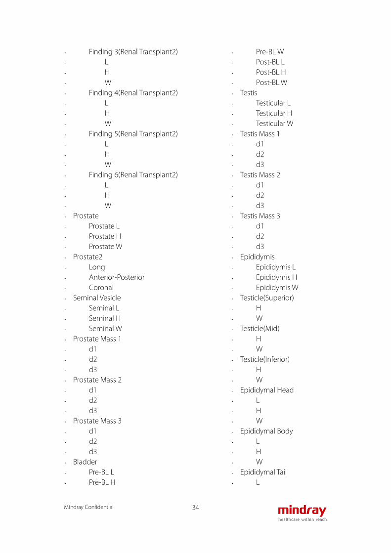
- Finding 3(Renal Transplant2) - L - H - W - Finding 4(Renal Transplant2) - L - H - W - Finding 5(Renal Transplant2) - L - H - W - Finding 6(Renal Transplant2) - L - H - W - Prostate - Prostate L - Prostate H - Prostate W - Prostate2 - Long - Anterior-Posterior - Coronal - Seminal Vesicle - Seminal L - Seminal H - Seminal W - Prostate Mass 1 - d1 - d2 - d3 - Prostate Mass 2 - d1 - d2 - d3 - Prostate Mass 3 - d1 - d2 - d3 - Bladder - Pre-BL L - Pre-BL H
- Pre-BL W - Post-BL L - Post-BL H - Post-BL W - Testis - Testicular L - Testicular H - Testicular W - Testis Mass 1 - d1 - d2 - d3 - Testis Mass 2 - d1 - d2 - d3 - Testis Mass 3 - d1 - d2 - d3 - Epididymis - Epididymis L - Epididymis H - Epididymis W - Testicle(Superior) - H - W - Testicle(Mid) - H - W - Testicle(Inferior) - H - W - Epididymal Head - L - H - W - Epididymal Body - L - H - W - Epididymal Tail - L

Mindray Confidential 35
- H - W - Prostate Mass1 Strain Ratio - A - B - Prostate Mass2 Strain Ratio - A - B - Prostate Mass3 Strain Ratio - A - B - Prostate Mass1 Elas. Ratio - A - B - Prostate Mass2 Elas. Ratio - A - B - Prostate Mass3 Elas. Ratio - A - B D-Mode - Renal A - Ren A Org - M Renal A - Renal A1 - Renal A2 - Hilum - Interlobar A - Arcuate A - Segment A - Artery Anast(Renal Transplant1) - Artery Anast 2(Renal Transplant1) - Vein Anast(Renal Transplant1) - Vein Anast 2(Renal Transplant1) - Renal A(Renal Transplant1) - Renal A1(Renal Transplant1) - Renal A2(Renal Transplant1) - Hilum(Renal Transplant1) - Interlobar A(Renal Transplant1) - Arcuate A(Renal Transplant1) - Segmental A(Renal Transplant1) - Renal Vein 1(Renal Transplant1)
- Renal Vein 2(Renal Transplant1) - Artery Anast(Renal Transplant2) - Artery Anast 2(Renal Transplant2) - Vein Anast(Renal Transplant2) - Vein Anast 2(Renal Transplant2) - Renal A(Renal Transplant2) - Renal A1(Renal Transplant2) - Renal A2(Renal Transplant2) - Hilum(Renal Transplant2) - Interlobar A(Renal Transplant2) - Arcuate A(Renal Transplant2) - Segmental A(Renal Transplant2) - Renal Vein 1(Renal Transplant2) - Renal Vein 2(Renal Transplant2) - Testis A - Testis V - Testis V(Valsalva) - Epididymis A - Epididymis V - ------------ - Renal Transplant 1(Doppler) - Artery Anast(Renal Transplant1) - Artery Anast 2(Renal
Transplant1) - Vein Anast(Renal Transplant1) - Vein Anast 2(Renal Transplant1) - Renal A(Renal Transplant1) - Renal A1(Renal Transplant1) - Renal A2(Renal Transplant1) - Hilum(Renal Transplant1) - Interlobar A(Renal Transplant1) - Arcuate A(Renal Transplant1) - Segmental A(Renal
Transplant1) - Renal Vein 1(Renal Transplant1) - Renal Vein 2(Renal Transplant1) - Renal Transplant 2(Doppler) - Artery Anast(Renal Transplant2) - Artery Anast 2(Renal
Transplant2) - Vein Anast(Renal Transplant2) - Vein Anast 2(Renal Transplant2) - Renal A(Renal Transplant2)

Mindray Confidential 36
- Renal A1(Renal Transplant2) - Renal A2(Renal Transplant2) - Hilum(Renal Transplant2) - Interlobar A(Renal Transplant2) - Arcuate A(Renal Transplant2) - Segmental A(Renal
Transplant2) - Renal Vein 1(Renal Transplant2) - Renal Vein 2(Renal Transplant2)
Vascular
B-Mode - CCA IMT - Bulb IMT - ICA IMT - ECA IMT - ------------ - IMT - CCA IMT - Bulb IMT - ICA IMT - ECA IMT - CCA - Anterior-Posterior - Transverse - Outer Diameter - Inner Diameter - Outer Area - Inner Area - Bulb - Anterior-Posterior - Transverse - Outer Diameter - Inner Diameter - Outer Area - Inner Area - Carotid Bifurcation - Anterior-Posterior - Transverse - Outer Diameter - Inner Diameter - Outer Area - Inner Area
- ICA - Anterior-Posterior - Transverse - Outer Diameter - Inner Diameter - Outer Area - Inner Area - ECA - Anterior-Posterior - Transverse - Outer Diameter - Inner Diameter - Outer Area - Inner Area - Vert A - Anterior-Posterior - Transverse - Outer Diameter - Inner Diameter - Outer Area - Inner Area - Subclav A - Anterior-Posterior - Transverse - Outer Diameter - Inner Diameter - Outer Area - Inner Area - Innom A - Anterior-Posterior - Transverse - Outer Diameter - Inner Diameter - Outer Area - Inner Area - Mammary A - Anterior-Posterior - Transverse - Outer Diameter - Inner Diameter - Outer Area - Inner Area - CCA Aneurysm

Mindray Confidential 37
- Long - Anterior-Posterior - Transverse - Bulb Aneurysm - Long - Anterior-Posterior - Transverse - Carotid Bifurcation Aneurysm - Long - Anterior-Posterior - Transverse - ICA Aneurysm - Long - Anterior-Posterior - Transverse - ECA Aneurysm - Long - Anterior-Posterior - Transverse - Vert A Aneurysm - Long - Anterior-Posterior - Transverse - Subclav A Aneurysm - Long - Anterior-Posterior - Transverse - Innom A Aneurysm - Long - Anterior-Posterior - Transverse - Mammary A Aneurysm - Long - Anterior-Posterior - Transverse - Carotid Graft 1 Anast - Long - Anterior-Posterior - Transverse - Carotid Graft 1 Graft - Long - Anterior-Posterior - Transverse
- Carotid Graft 2 Anast - Long - Anterior-Posterior - Transverse - Carotid Graft 2 Graft - Long - Anterior-Posterior - Transverse - Carotid Graft 3 Anast - Long - Anterior-Posterior - Transverse - Carotid Graft 3 Graft - Long - Anterior-Posterior - Transverse - Carotid Stent 1 - Long - Anterior-Posterior - Transverse - Carotid Stent 2 - Long - Anterior-Posterior - Transverse - Carotid Stent 3 - Long - Anterior-Posterior - Transverse - Carotid Stenosis 1 - Anterior-Posterior - Transverse - Outer Diameter - Inner Diameter - Outer Area - Inner Area - Carotid Stenosis 2 - Anterior-Posterior - Transverse - Outer Diameter - Inner Diameter - Outer Area - Inner Area - Carotid Stenosis 3

Mindray Confidential 38
- Anterior-Posterior - Transverse - Outer Diameter - Inner Diameter - Outer Area - Inner Area - Carotid Stenosis 4 - Anterior-Posterior - Transverse - Outer Diameter - Inner Diameter - Outer Area - Inner Area - Axill A - Anterior-Posterior - Transverse - Outer Diameter - Inner Diameter - Outer Area - Inner Area - Brachial A - Anterior-Posterior - Transverse - Outer Diameter - Inner Diameter - Outer Area - Inner Area - Radial A - Anterior-Posterior - Transverse - Outer Diameter - Inner Diameter - Outer Area - Inner Area - Ulnar A - Anterior-Posterior - Transverse - Outer Diameter - Inner Diameter - Outer Area - Inner Area - Axill A Aneurysm - Long
- Anterior-Posterior - Transverse - Brachial A Aneurysm - Long - Anterior-Posterior - Transverse - Radial A Aneurysm - Long - Anterior-Posterior - Transverse - Ulnar A Aneurysm - Long - Anterior-Posterior - Transverse - UE A Graft 1 Native Inflow - Anterior-Posterior - Transverse - UE A Graft 1 Anast - Anterior-Posterior - Transverse - UE A Graft 1 Graft - Anterior-Posterior - Transverse - UE A Graft 1 Native Outflow - Anterior-Posterior - Transverse - UE A Graft 2 Native Inflow - Anterior-Posterior - Transverse - UE A Graft 2 Anast - Anterior-Posterior - Transverse - UE A Graft 2 Graft - Anterior-Posterior - Transverse - UE A Graft 2 Native Outflow - Anterior-Posterior - Transverse - UE A Graft 3 Native Inflow - Anterior-Posterior - Transverse - UE A Graft 3 Anast - Anterior-Posterior

Mindray Confidential 39
- Transverse - UE A Graft 3 Graft - Anterior-Posterior - Transverse - UE A Graft 3 Native Outflow - Anterior-Posterior - Transverse - UE A Stent 1 - Long - Anterior-Posterior - Transverse - UE A Stent 2 - Long - Anterior-Posterior - Transverse - UE A Stent 3 - Long - Anterior-Posterior - Transverse - UE A Stenosis 1(2D) - Anterior-Posterior - Transverse - Outer Diameter - Inner Diameter - Outer Area - Inner Area - UE A Stenosis 2(2D) - Anterior-Posterior - Transverse - Outer Diameter - Inner Diameter - Outer Area - Inner Area - UE A Stenosis 3(2D) - Anterior-Posterior - Transverse - Outer Diameter - Inner Diameter - Outer Area - Inner Area - UE A Stenosis 4(2D) - Anterior-Posterior - Transverse
- Outer Diameter - Inner Diameter - Outer Area - Inner Area - C.Iliac A - Anterior-Posterior - Transverse - Outer Diameter - Inner Diameter - Outer Area - Inner Area - Ex.Iliac A - Anterior-Posterior - Transverse - Outer Diameter - Inner Diameter - Outer Area - Inner Area - IIA - Anterior-Posterior - Transverse - Outer Diameter - Inner Diameter - Outer Area - Inner Area - CFA - Anterior-Posterior - Transverse - Outer Diameter - Inner Diameter - Outer Area - Inner Area - DFA - Anterior-Posterior - Transverse - Outer Diameter - Inner Diameter - Outer Area - Inner Area - SFA - Anterior-Posterior - Transverse - Outer Diameter

Mindray Confidential 40
- Inner Diameter - Outer Area - Inner Area - Pop A - Anterior-Posterior - Transverse - Outer Diameter - Inner Diameter - Outer Area - Inner Area - TP Trunk A - Anterior-Posterior - Transverse - Outer Diameter - Inner Diameter - Outer Area - Inner Area - A.Tib. A - Anterior-Posterior - Transverse - Outer Diameter - Inner Diameter - Outer Area - Inner Area - Peroneal A - Anterior-Posterior - Transverse - Outer Diameter - Inner Diameter - Outer Area - Inner Area - P.Tib. A - Anterior-Posterior - Transverse - Outer Diameter - Inner Diameter - Outer Area - Inner Area - Dors.Ped. A - Anterior-Posterior - Transverse - Outer Diameter - Inner Diameter
- Outer Area - Inner Area - C.Iliac A Aneurysm - Long - Anterior-Posterior - Transverse - Ex.Iliac A Aneurysm - Long - Anterior-Posterior - Transverse - IIA Aneurysm - Long - Anterior-Posterior - Transverse - CFA Aneurysm - Long - Anterior-Posterior - Transverse - DFA Aneurysm - Long - Anterior-Posterior - Transverse - SFA Aneurysm - Long - Anterior-Posterior - Transverse - Pop A Aneurysm - Long - Anterior-Posterior - Transverse - TP Trunk A Aneurysm - Long - Anterior-Posterior - Transverse - A.Tib. A Aneurysm - Long - Anterior-Posterior - Transverse - Peroneal A Aneurysm - Long - Anterior-Posterior - Transverse - P.Tib. A Aneurysm

Mindray Confidential 41
- Long - Anterior-Posterior - Transverse - Dors.Ped. A Aneurysm - Long - Anterior-Posterior - Transverse - LE A Graft 1 Native Inflow - Anterior-Posterior - Transverse - LE A Graft 1 Anast - Anterior-Posterior - Transverse - LE A Graft 1 Graft - Anterior-Posterior - Transverse - LE A Graft 1 Native Outflow - Anterior-Posterior - Transverse - LE A Graft 2 Native Inflow - Anterior-Posterior - Transverse - LE A Graft 2 Anast - Anterior-Posterior - Transverse - LE A Graft 2 Graft - Anterior-Posterior - Transverse - LE A Graft 2 Native Outflow - Anterior-Posterior - Transverse - LE A Graft 3 Native Inflow - Anterior-Posterior - Transverse - LE A Graft 3 Anast - Anterior-Posterior - Transverse - LE A Graft 3 Graft - Anterior-Posterior - Transverse - LE A Graft 3 Native Outflow - Anterior-Posterior - Transverse
- LE A Stent 1 - Long - Anterior-Posterior - Transverse - LE A Stent 2 - Long - Anterior-Posterior - Transverse - LE A Stent 3 - Long - Anterior-Posterior - Transverse - LE A Stenosis 1(2D) - Anterior-Posterior - Transverse - Outer Diameter - Inner Diameter - Outer Area - Inner Area - LE A Stenosis 2(2D) - Anterior-Posterior - Transverse - Outer Diameter - Inner Diameter - Outer Area - Inner Area - LE A Stenosis 3(2D) - Anterior-Posterior - Transverse - Outer Diameter - Inner Diameter - Outer Area - Inner Area - LE A Stenosis 4(2D) - Anterior-Posterior - Transverse - Outer Diameter - Inner Diameter - Outer Area - Inner Area - LE A Finding 1 - Long - Anterior-Posterior

Mindray Confidential 42
- Transverse - LE A Finding 2 - Long - Anterior-Posterior - Transverse - LE A Finding 3 - Long - Anterior-Posterior - Transverse - LE A Finding 4 - Long - Anterior-Posterior - Transverse - LE A Finding 5 - Long - Anterior-Posterior - Transverse - LE A Finding 6 - Long - Anterior-Posterior - Transverse - Int Jug V - Anterior-Posterior - Transverse - Checklist - Innom V - Anterior-Posterior - Transverse - Checklist - Subclav V - Anterior-Posterior - Transverse - Checklist - Ax V - Anterior-Posterior - Transverse - Checklist - Brachial V - Anterior-Posterior - Transverse - Checklist - Radial V - Anterior-Posterior
- Transverse - Checklist - Ulnar V - Anterior-Posterior - Transverse - Checklist - Volar V - Anterior-Posterior - Transverse - Checklist - Cephalic V - Anterior-Posterior - Transverse - Checklist - Basilic V - Anterior-Posterior - Transverse - Checklist - CA Junction - Anterior-Posterior - Transverse - Checklist - Upper Arm Cephalic V - Anterior-Posterior - Transverse - Checklist - Cephalic-Antecubital V - Anterior-Posterior - Transverse - Checklist - Forearm Cephalic V - Anterior-Posterior - Transverse - Checklist - BA Junction - Anterior-Posterior - Transverse - Checklist - Upper Arm Basilic V - Anterior-Posterior - Transverse - Checklist - Basilic-Antecubital V

Mindray Confidential 43
- Anterior-Posterior - Transverse - Checklist - Forearm Basilic V - Anterior-Posterior - Transverse - Checklist - Digital V - Anterior-Posterior - Transverse - Checklist - Median Cubital V - Anterior-Posterior - Transverse - Checklist - AVF-Inflow Artery - Anterior-Posterior - Transverse - AVF-Anast - Anterior-Posterior - Transverse - AVF-Outflow Vein Level 1 - Anterior-Posterior - Transverse - AVF-Outflow Vein Level 2 - Anterior-Posterior - Transverse - AVF-Outflow Vein Level 3 - Anterior-Posterior - Transverse - AVF-Outflow Vein Level 4 - Anterior-Posterior - Transverse - AVF-Outflow Vein Level 5 - Anterior-Posterior - Transverse - AVF-Outflow Vein Level 6 - Anterior-Posterior - Transverse - AVF-Stenosis 1 - Anterior-Posterior - Transverse - AVF-Stenosis 2
- Anterior-Posterior - Transverse - AVF-Stenosis 3 - Anterior-Posterior - Transverse - AVF-Aneurysm 1 - Anterior-Posterior - Transverse - AVF-Aneurysm 2 - Anterior-Posterior - Transverse - AVF-Aneurysm 3 - Anterior-Posterior - Transverse - AV Graft-Inflow Artery - Anterior-Posterior - Transverse - AV Graft-Arterial Anast - Anterior-Posterior - Transverse - AV Graft-Graft - Anterior-Posterior - Transverse - AV Graft-Venous Anast - Anterior-Posterior - Transverse - AV Graft-Outflow Vein Level 1 - Anterior-Posterior - Transverse - AV Graft-Outflow Vein Level 2 - Anterior-Posterior - Transverse - AV Graft-Outflow Vein Level 3 - Anterior-Posterior - Transverse - AV Graft-Outflow Vein Level 4 - Anterior-Posterior - Transverse - AV Graft-Outflow Vein Level 5 - Anterior-Posterior - Transverse - AV Graft-Outflow Vein Level 6 - Anterior-Posterior

Mindray Confidential 44
- Transverse - C.Iliac V - Anterior-Posterior - Transverse - Checklist - Ex.Iliac V - Anterior-Posterior - Transverse - Checklist - IIV - Anterior-Posterior - Transverse - Checklist - CFV - Anterior-Posterior - Transverse - Checklist - Femoral V - Anterior-Posterior - Transverse - Checklist - DFV - Anterior-Posterior - Transverse - Checklist - Pop V - Anterior-Posterior - Transverse - Checklist - P.Tib. V - Anterior-Posterior - Transverse - Checklist - Peroneal V - Anterior-Posterior - Transverse - Checklist - Sural V - Anterior-Posterior - Transverse - Checklist - Soleal V - Anterior-Posterior
- Transverse - Checklist - A.Tib. V - Anterior-Posterior - Transverse - Checklist - TP Trunk V - Anterior-Posterior - Transverse - Checklist - Saph V - Anterior-Posterior - Transverse - Checklist - SSV - Anterior-Posterior - Transverse - Checklist - SF Junction - Anterior-Posterior - Transverse - Checklist - GSV Thigh - Anterior-Posterior - Transverse - Checklist - GSV Knee - Anterior-Posterior - Transverse - Checklist - GSV Calf - Anterior-Posterior - Transverse - Checklist - SP Junction - Anterior-Posterior - Transverse - Checklist - SSV Thigh Extension - Anterior-Posterior - Transverse - Checklist - AASV

Mindray Confidential 45
- Anterior-Posterior - Transverse - Checklist - PASV - Anterior-Posterior - Transverse - Checklist - Thigh Perf - Anterior-Posterior - Transverse - Checklist - Prox Calf Perf - Anterior-Posterior - Transverse - Checklist - Mid Calf Perf - Anterior-Posterior - Transverse - Checklist - Dist Calf Perf - Anterior-Posterior - Transverse - Checklist - Pseudoaneurysm - Long - Anterior-Posterior - Transverse - Neck - - M-Mode - - D-Mode - ACA - A1 ACA - MCA - M1 MCA - M2 MCA - AComA - Terminal ICA - PComA - PCA - P1 PCA - P2 PCA
- Opthalmic A - ICA Siphon - Terminal Vert A - BA - Ba V - CCA - ICA - ECA - Bulb - Carotid Bifurcation - Vert A - Subclav A - Innom A - Mammary A - Subclav V - CCA Aneurysm - ICA Aneurysm - ECA Aneurysm - Bulb Aneurysm - Carotid Bifurcation Aneurysm - Vert A Aneurysm - Subclav A Aneurysm - Innom A Aneurysm - Mammary A Aneurysm - Carotid Graft 1 Native Inflow - Carotid Graft 1 Anast Pre - Carotid Graft 1 Anast Max - Carotid Graft 1 Anast Post - Carotid Graft 1 Graft - Carotid Graft 1 Native Outflow - Carotid Graft 2 Native Inflow - Carotid Graft 2 Anast Pre - Carotid Graft 2 Anast Max - Carotid Graft 2 Anast Post - Carotid Graft 2 Graft - Carotid Graft 2 Native Outflow - Carotid Graft 3 Native Inflow - Carotid Graft 3 Anast Pre - Carotid Graft 3 Anast Max - Carotid Graft 3 Anast Post - Carotid Graft 3 Graft - Carotid Graft 3 Native Outflow - Carotid Stent 1

Mindray Confidential 46
- Carotid Stent 2 - Carotid Stent 3 - Axill A - Brachial A - Ulnar A - Radial A - UE A Graft 1 Native Inflow - UE A Graft 1 Anast - UE A Graft 1 Graft - UE A Graft 1 Native Outflow - UE A Graft 2 Native Inflow - UE A Graft 2 Anast - UE A Graft 2 Graft - UE A Graft 2 Native Outflow - UE A Graft 3 Native Inflow - UE A Graft 3 Anast - UE A Graft 3 Graft - UE A Graft 3 Native Outflow - UE A Stent 1 - UE A Stent 2 - UE A Stent 3 - C.Iliac A - Ex.Iliac A - IIA - CFA - DFA - SFA - Pop A - TP Trunk A - A.Tib A - Peroneal A - P.Tib A - Dors.Ped. A - LE A Graft 1 Native Inflow - LE A Graft 1 Anast Pre - LE A Graft 1 Anast Max - LE A Graft 1 Anast Post - LE A Graft 1 Graft - LE A Graft 1 Native Outflow - LE A Graft 2 Native Inflow - LE A Graft 2 Anast Pre - LE A Graft 2 Anast Max - LE A Graft 2 Anast Post
- LE A Graft 2 Graft - LE A Graft 2 Native Outflow - LE A Graft 3 Native Inflow - LE A Graft 3 Anast Pre - LE A Graft 3 Anast Max - LE A Graft 3 Anast Post - LE A Graft 3 Graft - LE A Graft 3 Native Outflow - LE A Stent 1 - LE A Stent 2 - LE A Stent 3 - Axill V - Brachial V - Radial V - Ulnar V - Cephalic V - Basilic V - AVF-Inflow Artery - AVF-Anast - AVF-Outflow Vein Level 1 - AVF-Outflow Vein Level 2 - AVF-Outflow Vein Level 3 - AVF-Outflow Vein Level 4 - AVF-Outflow Vein Level 5 - AVF-Outflow Vein Level 6 - AVF-Stenosis 1 - AVF-Stenosis 2 - AVF-Stenosis 3 - AV Graft-Inflow Artery - AV Graft-Arterial Anast - AV Graft-Graft - AV Graft-Venous Anast - AV Graft-Outflow Vein Level 1 - AV Graft-Outflow Vein Level 2 - AV Graft-Outflow Vein Level 3 - AV Graft-Outflow Vein Level 4 - AV Graft-Outflow Vein Level 5 - AV Graft-Outflow Vein Level 6 - ASP - BSP - ------------ - CCA(Sten) - Pre Sten

Mindray Confidential 47
- Sten - Post Sten - ICA(Sten) - Pre Sten - Sten - Post Sten - ECA(Sten) - Pre Sten - Sten - Post Sten - Bulb(Sten) - Pre Sten - Sten - Post Sten - Carotid Bifurcation(Sten) - Pre Sten - Sten - Post Sten - Vert A(Sten) - Pre Sten - Sten - Post Sten - Subclav A(Sten) - Pre Sten - Sten - Post Sten - Innom A(Sten) - Pre Sten - Sten - Post Sten - Mammary A(Sten) - Pre Sten - Sten - Post Sten - Carotid Stenosis 1 - Pre Sten - Sten - Post Sten - Carotid Stenosis 2 - Pre Sten - Sten - Post Sten - Carotid Stenosis 3
- Pre Sten - Sten - Post Sten - Carotid Stenosis 4 - Pre Sten - Sten - Post Sten - Axill A(Sten) - Pre Sten - Sten - Post Sten - Brachial A(Sten) - Pre Sten - Sten - Post Sten - Ulnar A(Sten) - Pre Sten - Sten - Post Sten - Radial A(Sten) - Pre Sten - Sten - Post Sten - UE A Stenosis 1 - Pre Sten - Sten - Post Sten - UE A Stenosis 2 - Pre Sten - Sten - Post Sten - UE A Stenosis 3 - Pre Sten - Sten - Post Sten - UE A Stenosis 4 - Pre Sten - Sten - Post Sten - C.Iliac A(Sten) - Pre Sten - Sten - Post Sten

Mindray Confidential 48
- Ex.Iliac A(Sten) - Pre Sten - Sten - Post Sten - IIA(Sten) - Pre Sten - Sten - Post Sten - CFA(Sten) - Pre Sten - Sten - Post Sten - DFA(Sten) - Pre Sten - Sten - Post Sten - SFA(Sten) - Pre Sten - Sten - Post Sten - Pop A(Sten) - Pre Sten - Sten - Post Sten - TP Trunk A(Sten) - Pre Sten - Sten - Post Sten - A.Tib. A(Sten) - Pre Sten - Sten - Post Sten - Peroneal A(Sten) - Pre Sten - Sten - Post Sten - P.Tib. A(Sten) - Pre Sten - Sten - Post Sten - Dors.Ped. A(Sten) - Pre Sten - Sten
- Post Sten - LE A Stenosis 1 - Pre Sten - Sten - Post Sten - LE A Stenosis 2 - Pre Sten - Sten - Post Sten - LE A Stenosis 3 - Pre Sten - Sten - Post Sten - LE A Stenosis 4 - Pre Sten - Sten - Post Sten - C.Iliac V - PV - Reflux - Checklist - Ex.Iliac V - PV - Reflux - Checklist - IIV - PV - Reflux - Checklist - CFV - PV - Reflux - Checklist - Femoral V - PV - Reflux - Checklist - DFV - PV - Reflux - Checklist - Pop V - PV

Mindray Confidential 49
- Reflux - Checklist - P.Tib. V - PV - Reflux - Checklist - Peroneal V - PV - Reflux - Checklist - Sural V - PV - Reflux - Checklist - Soleal V - PV - Reflux - Checklist - A.Tib. V - PV - Reflux - Checklist - TP Trunk V - PV - Reflux - Checklist - Saph V - PV - Reflux - Checklist - SSV - PV - Reflux - Checklist - SF Junction - PV - Reflux - Checklist - GSV Thigh - PV - Reflux - Checklist - GSV Knee
- PV - Reflux - Checklist - GSV Calf - PV - Reflux - Checklist - SP Junction - PV - Reflux - Checklist - SSV Thigh Extension - PV - Reflux - Checklist - AASV - PV - Reflux - Checklist - PASV - PV - Reflux - Checklist - Thigh Perf - PV - Reflux - Checklist - Prox Calf Perf - PV - Reflux - Checklist - Mid Calf Perf - PV - Reflux - Checklist - Dist Calf Perf - PV - Reflux - Checklist - ABI - ASP - BSP

Mindray Confidential 50
Small Parts B-Mode - Thyroid L - Thyroid H - Thyroid W - Isthmus H - THY Mass1 Strain - THY Mass2 Strain - THY Mass3 Strain - THY Mass1 Elas. - THY Mass2 Elas. - THY Mass3 Elas. - THY Nodule1 Strain - THY Nodule2 Strain - THY Nodule3 Strain - THY Nodule1 Elas. - THY Nodule2 Elas. - THY Nodule3 Elas. - Breast Mass1 Strain - Breast Mass1 Elas. - Breast Mass2 Strain - Breast Mass2 Elas. - Breast Mass3 Strain - Breast Mass3 Elas. - Breast Mass4 Strain - Breast Mass4 Elas. - Breast Mass5 Strain - Breast Mass5 Elas. - Breast Mass6 Strain - Breast Mass6 Elas. - Breast Mass7 Strain - Breast Mass7 Elas. - Breast Mass8 Strain - Breast Mass8 Elas. - Breast Mass9 Strain - Breast Mass9 Elas. - Breast Mass10 Strain - Breast Mass10 Elas. - Testicular L - Testicular H - Testicular W - Epididymis L - Epididymis H
- Epididymis W - Scrotal Wall - Testis V(2D) - Testis V(Valsalva 2D) - ------------ - Thyroid Vol - Testicular Vol - ------------ - Thyroid(Superior) - H - W - Thyroid(Mid) - H - W - Thyroid(Inferior) - H - W - Parathyroid 1 - L - H - W - Parathyroid 2 - L - H - W - Parotid - L - H - W - Lymph Node 1 - L - H - W - Lymph Node 2 - L - H - W - Lymph Node 3 - L - H - W - Lymph Node 4 - L

Mindray Confidential 51
- H - W - Lymph Node 5 - L - H - W - Lymph Node 6 - L - H - W - Thyroid - Thyroid L - Thyroid H - Thyroid W - Thyroid Mass 1 - d1 - d2 - d3 - Thyroid Mass 2 - d1 - d2 - d3 - Thyroid Mass 3 - d1 - d2 - d3 - Thyroid Nodule 1 - d1 - d2 - d3 - Thyroid Nodule 2 - d1 - d2 - d3 - Thyroid Nodule 3 - d1 - d2 - d3 - Thyroid Cyst 1 - d1 - d2 - d3 - Thyroid Cyst 2
- d1 - d2 - d3 - Thyroid Cyst 3 - d1 - d2 - d3 - Isthmus Finding 1 - d1 - d2 - d3 - Isthmus Finding 2 - d1 - d2 - d3 - Isthmus Finding 3 - d1 - d2 - d3 - THY Mass1 Strain Ratio - A - B - THY Mass2 Strain Ratio - A - B - THY Mass3 Strain Ratio - A - B - THY Mass1 Elas. Ratio - A - B - THY Mass2 Elas. Ratio - A - B - THY Mass3 Elas. Ratio - A - B - THY Nodule1 Strain Ratio - A - B - THY Nodule2 Strain Ratio - A - B

Mindray Confidential 52
- THY Nodule3 Strain Ratio - A - B - THY Nodule1 Elas. Ratio - A - B - THY Nodule2 Elas. Ratio - A - B - THY Nodule3 Elas. Ratio - A - B - Breast Mass 1 - L - H - W - Nip. Dist. - Skin Dist. - Breast Mass 2 - L - H - W - Nip. Dist. - Skin Dist. - Breast Mass 3 - L - H - W - Nip. Dist. - Skin Dist. - Breast Mass 4 - L - H - W - Nip. Dist. - Skin Dist. - Breast Mass 5 - L - H - W - Nip. Dist. - Skin Dist. - Breast Mass 6
- L - H - W - Nip. Dist. - Skin Dist. - Breast Mass 7 - L - H - W - Nip. Dist. - Skin Dist. - Breast Mass 8 - L - H - W - Nip. Dist. - Skin Dist. - Breast Mass 9 - L - H - W - Nip. Dist. - Skin Dist. - Breast Mass 10 - L - H - W - Nip. Dist. - Skin Dist. - Breast Mass1 Strain Ratio - A - B - Breast Mass1 Elas. Ratio - A - B - Breast Mass2 Strain Ratio - A - B - Breast Mass2 Elas. Ratio - A - B - Breast Mass3 Strain Ratio - A

Mindray Confidential 53
- B - Breast Mass3 Elas. Ratio - A - B - Breast Mass4 Strain Ratio - A - B - Breast Mass4 Elas. Ratio - A - B - Breast Mass5 Strain Ratio - A - B - Breast Mass5 Elas. Ratio - A - B - Breast Mass6 Strain Ratio - A - B - Breast Mass6 Elas. Ratio - A - B - Breast Mass7 Strain Ratio - A - B - Breast Mass7 Elas. Ratio - A - B - Breast Mass8 Strain Ratio - A - B - Breast Mass8 Elas. Ratio - A - B - Breast Mass9 Strain Ratio - A - B - Breast Mass9 Elas. Ratio - A - B - Breast Mass10 Strain Ratio - A - B
- Breast Mass10 Elas. Ratio - A - B - Testis - Testicular L - Testicular H - Testicular W - Testis Mass 1 - d1 - d2 - d3 - Testis Mass 2 - d1 - d2 - d3 - Testis Mass 3 - d1 - d2 - d3 - Epididymis - Epididymis L - Epididymis H - Epididymis W - Testicle(Superior) - H - W - Testicle(Mid) - H - W - Testicle(Inferior) - H - W - Epididymal Head - L - H - W - Epididymal Body - L - H - W - Epididymal Tail - L - H

Mindray Confidential 54
- W D-Mode - STA - ITA - Isthmus - Parathyroid 1 - Parathyroid 2 - Testis A - Testis V - Testis V(Valsalva) - Epididymis A - Epididymis V
Orthopedics - B-Mode - HIP - HIP-Graft - HIP( ) - HIP( ) - d/D
Emergency&Critical
- B-Mode - Renal L - Renal H - Renal W - CBD - Portal V Diam - CHD - GB wall th - Aorta Bif - Ureter - Pre-BL L - Pre-BL H - Pre-BL W - Post-BL L - Post-BL H - Post-BL W - GS - YS L - CRL - BPD - UT L - UT H
- UT W - Endo - Ovary L - Ovary H - Ovary W - ------------ - Renal Vol - Pre-BL Vol - Post-BL Vol - Mictur.Vol - Ovary Vol - UT Vol - UT SUM - ------------ - Uterus - UT L - UT H - UT W - Endo - Ovary - Ovary L - Ovary H - Ovary W - Kidney - Renal L - Renal H - Renal W - Cortex - Bladder - Pre-BL L - Pre-BL H - Pre-BL W - Post-BL L - Post-BL H - Post-BL W - M-Mode - FHR (M) - D-Mode - FHR (Doppler)
AutoCalc - PS

Mindray Confidential 55
- ED - MD - PPG - TAMAX - Vol Flow(TAMAX) - TAMEAN - Vol Flow(TAMEAN) - DT - MPG - MMPG - VTI - AT - S/D - D/S - PI - RI - PV - HR
6.3 Report Specific report template by application User-defined report template Editable value in report Images selectable Able to Export as PDF/RTF file
6.4 Smart OBTM Auto measurement for OB, a special
tool for easy OB scan, and greatly reduce time and increase productivity
Support BPD, HC, OFD, FL, AC Measurement result can be modified
by user 6.5 Smart NT
NT auto measurement Auto detection of NT inside ROI
6.6 iStorage Data transfer
* Not all measurements are listed in this part; For more detailed information please refer to User Manual
7 Exam Storage and Management 7.1 Exam storage
SSD: 128 GB, more than 45.6 GB internal
hard drive reserved for patient data storage
SSD: Capable of storage up to approximately 173242 single frames (FRM format)
Storage area - Pre-settable: image area, standard
area, full-screen - Image area: 1430*810 - Standard area:1920*920 - Full-screen: 1920*1080
7.2 Exam management iStationTM workstation dedicated for
patient exam management Patient exam query/retrieve Support review of current and past
exam New exam, Active exam, Continue
exam functions, End exam are available Support measurements and
calculations on archived exam and images
Export image as BMP/JPG/TIFF/DCM/FRM format (FRM: system format)
Export cine as DCM/AVI/CIN/MP4 format (CIN: system format)
Support backup/send to USB devices, DVD-RW media
7.3 iWorksTM (option) Auto workflow protocol Templates are user configurable Functions: pause, stop, replace, repeat,
skip, insert single step, return and continue, steps in thumbnail
iWorks setup mode: B/Dual/B+Color/B+PW/B+Color+PW/B+CW/B+Color+CW/ B+M
iWorks setup annotation: support up to 2 annotations, location and font size are configurable
iWorks setup bodymark: select existing library, and probe indicator is pre-

Mindray Confidential 56
settable iWorks setup measurement: select
existing measurement library Template import and export are
available
8 Connectivity 8.1 Ethernet Network Connection
Wireless connection: Internal WIFI (including EAP Network)
8.2 DICOM 3.0 DICOM Basic (option)
- Verify (SCU, SCP) - Print - Store - Storage Commitment - Media Exchange
DICOM Worklist (option) DICOM Query/Retrieve (option) DICOM Modality Performed Procedure
Step - MPPS (option) DICOM OB/GYN structure report
(option) DICOM Cardiac structure report
(option) DICOM Vascular structure report
(option) DICOM Breast structure report (option) DICOM Abdomen SR (option)
8.3 iStorage Direct network storage tool between
ultrasound system and personal computer
8.4 MedSight 8.5 MedTouch 8.6 Net Storage
Support sending images or exams to the shared directory of your PC server.
8.7 Security Anti-Virus: McAfee and Windows
Defender VPN
9 Probes
9.1 Curved array C5-1s
- Application: Abdomen, Gynecology, Obstetrics, Vascular, Nerve, Musculo-skeletal, Urology, Thoracic/Pleural, Small Parts
- Bandwidth: 1.2-6.0 MHz - Depth: 4.0-40.0 cm - Number of Elements: 128 - FOV (max): 61° - Extended FOV: 72° - Convex Radius: 60 mm - Physical Footprint: 76.5*28 mm - Footprint: 64.9*16.2 mm - B-mode Frequencies: 1.2~3.8, 1.7~5.2,
2.0~6.0 MHz - Harmonic Frequencies: 4.0, 5.0, 6.0
MHz - Doppler Frequencies: 2.0, 2.5, 3.0 MHz - Biopsy Guide: NGB-022, available,
multi angle, reusable; LPUBKG60, disposable
V11-3s - Application: Gynecology, Obstetrics,
Urology - Bandwidth: 3.0-11.0 MHz - Depth: 1.5-28.0 cm - Number of Elements: 128 - FOV (max): 140° - Extended FOV: 179° - Convex Radius: 11 mm - Physical Footprint: 24.85*21.8 mm - Footprint: 24*9 mm - B-mode Frequencies: 3.0~7.0, 4.0~9.0,
5.0~11.0 MHz - Harmonic Frequencies: 8.0, 9.0, 10.0
MHz - Doppler Frequencies: 4.4, 5.0, 5.7 MHz - Biopsy Guide: Biopsy Guide: NGB-004,
available, single angle, reusable; NGB-045, available, single angle, reusable
9.2 Linear array L11-3VNs

Mindray Confidential 57
- Application: Abdomen, Pediatric, Small Parts, Musculo-skeletal, Vascular, Nerve, Thoracic/Pleural
- Bandwidth: 3.0-12.8 MHz - Depth: 1.5-35.0 cm - Number of Elements: 192 - Field of View (max): 3.80 cm - Steered Angle: ±12°, ±6°, 0 (B steer); -
30°~30° (Color/PW steer) - Physical Footprint: 56.5*24 mm - Footprint: 43.5*10.5 mm - B-mode Frequencies: 3.0~8.3,
4.4~10.2, 5.6~12.8 MHz - Harmonic Frequencies: 8.0, 10.0, 12.0
MHz - Doppler Frequencies: 4.2, 5.0, 7.2 MHz - Biopsy Guide: not available
L12-3RCs - Application: Abdomen, Pediatric,
Small Parts, Musculo-skeletal, Vascular, Thoracic/Pleural
- Bandwidth: 3.0-12.8 MHz - Depth: 1.5-35.0 cm - Number of Elements: 192 - Field of View (max): 3.80 cm - Steered Angle: ±12°, ±6°, 0 (B steer); -
30°~30° (Color/PW steer) - Physical Footprint: 55.6*22 mm - Footprint: 43.5*8.2 mm - B-mode Frequencies: 3.0~8.3,
4.4~10.2, 5.6~12.8 MHz - Harmonic Frequencies: 8.0, 10.0, 12.0
MHz - Color Frequencies: 4.4, 5.0, 7.2 MHz - Doppler Frequencies: 4.2, 5.0, 7.2 MHz - Biopsy Guide: NGB-043 available,
multi-angle, reusable; NGB-044 available, multi-depth, reusable
L13-3s - Application: Abdomen, Pediatric,
Small Parts, Musculo-skeletal, Vascular, Nerve, Thoracic/Pleural
- Bandwidth: 3.2-12.3 MHz
- Depth: 1.5-35.0 cm - Number of Elements: 128 - Field of View (max): 3.79 cm - Steered Angle: ±12°, ±6°, 0 (B steer); -
30°~30° (Color/PW steer) - Physical Footprint: 61*24.4 mm - Footprint: 44.2*8.5 mm - B-mode Frequencies: 3.2~9.6,
5.4~11.0, 6.6~12.3 MHz - Harmonic Frequencies: 8.0, 9.4, 10.6
MHz - Doppler Frequencies: 4.0, 5.0, 6.2 MHz - Biopsy Guide: NGB-007, available,
multi-angle, reusable L13-3Ns
- Application: Abdomen, Pediatric, Small Parts, Musculo-skeletal, Vascular, Nerve, Thoracic/Pleural
- Bandwidth: 3.0-13.0 MHz - Depth: 1.5-35.0 cm - Number of Elements: 192 - Field of View (max): 3.82 cm - Steered Angle: ±12°, ±6°, 0 (B steer); -
30°~30° (Color/PW steer) - Physical Footprint: 56.8*21.2 mm - Footprint: 43.5*8.2 mm - B-mode Frequencies: 3.0~9.3,
5.4~11.2, 6.6~13.0 MHz - Harmonic Frequencies: 9.0, 10.0, 11.0
MHz - Doppler Frequencies: 4.2, 5.0, 6.2 MHz - Biopsy Guide: NGB-053, available,
multi-angle, reusable L14-6Ns
- Application: Abdomen, Pediatric, Small Parts, Musculo-skeletal, Vascular, Nerve, Ophthalmic, Thoracic/Pleural
- Bandwidth: 3.5-16.0 MHz - Depth: 1.5-35.0 cm - Number of Elements: 192 - Field of View (max): 3.80 cm - Steered Angle: ±12°, ±6°, 0 (B steer); -
30°~30° (Color/PW steer)

Mindray Confidential 58
- Physical Footprint: 45.7*10.9 mm - Footprint: 44.2*8.5 mm - B-mode Frequencies: 3.5~9.3,
5.4~11.2, 6.6~16.0 MHz - Harmonic Frequencies: 10.0, 11.0, 12.0
MHz - Doppler Frequencies: 5.0, 6.2, 7.3 MHz - Biopsy Guide: NGB-007, available,
multi-angle, reusable L20-5s
- Application: Abdomen, Small Parts, Musculo-skeletal, Vascular, Nerve, Ophthalmic
- Bandwidth: 6.0-23.0 MHz - Depth: 1.5-35.0 cm - Number of Elements: 192 - Field of View (max): 2.86 cm - Steered Angle: ±12°, ±6°, 0 (B steer); -
30°~30° (Color/PW steer) - Physical Footprint: 42.23* 22.10 mm - Footprint: 31.5*4.5 mm - B-mode Frequencies: 6.0~13.0,
9.0~16.6, 12.5~23.0 MHz - Harmonic Frequencies: 14.0, 16.0, 18.0
MHz - Color Frequencies: 8.9, 11.4, 13.3 MHz - Doppler Frequencies: 8.9, 10.0, 13.3
MHz - Biopsy Guide: not available
9.3 Phased array P4-2s
- Application: Abdomen, Gynecology, Obstetrics, Cardiac, Pediatric, Vascular, Thoracic/Pleural, Cephalic
- Bandwidth: 1.5-4.5 MHz - Depth: 2.0-38.0 cm - Number of Elements: 64 - Field of View (max): 90° - Physical Footprint: 25.2*20.6 mm - Footprint: 23.4*15.2 mm - B-mode Frequencies: 1.5~2.5, 2.5~3.5,
3.5~4.5 MHz - Harmonic Frequencies: 3.4, 3.8, 3.8,
4.2, 4.2 MHz - Doppler Frequencies: 2.0, 2.3, 2.5 MHz - CW Frequency: 2.0 MHz - Biopsy Guide: NGB-011, available,
multi angle, reusable
10 Peripheral Devices and
Accessories (Option) 10.1 Black/white digital video printer
MITSUBISHI P95DW-N 10.2 Black/white analog video printer
SONY UP-X898MD 10.3 Color digital video printer
SONY UP-D25MD 10.4 Footswitch
USB port: 971-SWNOM (2-pedal/3-pedal)
USB port: FS-81-SP (1-pedal) Support User-definable functions
(Freeze, Save, Print) 10.5 Built-in DVR
Built-in digital video recorder, save space and is a useful tool for education and memory
Max storage length each time: 60 min 10.6 Built-in Battery for Main Unit
Replaceable and rechargeable lithium battery
Empty battery recharged to full in 4h Continuous work time: about 1.5 hour
in B mode 10.7 Mobile Trolley
MT3 - Power supply module - Platform Height: 830-1150 mm;
adjustable - Probe holders (option) - Probe extend module (option) - Cover grounding cable (option) - Auxiliary output cable (option)
10.8 Barcode reader
Mindray Confidential 59
- 1-D barcode reader: SYMBOL LS2208 - 2-D barcode reader: SYMBOL DS4308 - JADAK HS-1M - JADAK HS-1R (supporting RFID)
11 System Inputs and Outputs 11.1 I/O Port
USB 3.0: 4 ports Ethernet: 1 port HDMI: 1 port
11.2 ECG module 11.3 Probe Extend module
Probe port: 2
12 Safety and Conformance 12.1 Quality standards
ISO 9001 ISO 13485
12.2 Design standards CSA C22.2 No. 601-1 EN 60601-1 and IEC 60601-1 EN 60601-1-2 and IEC 60601-1-2 EN 60601-1-6 and IEC 60601-1-6 EN 60601-2-37 and IEC60601-2-37 EN 62304 and IEC 62304 EN 62366 and IEC 62366 EN ISO 17664 and ISO 17664
12.3 CE declaration The system is fully in conformance with the Council Directive 93/42/EEC Concerning Medical Devices, as amended by 2007/47/EC. The number adjacent to the CE marking (0123) is the number of the EU-notified body that certified meeting the requirements of Annex II of the Directive.
NOTICE: Not all features or specifications described in this document may be available in all probes and/or modes. Mindray reserves the right to make changes in specifications and features shown herein, or discontinue the product at any time without notice or obligation. Contact Mindray
Representative for the most current information.

Copyright © 2022 FDOKUMEN