ULTRASOUND 2019
145
BMUS ULTRASOUND 2019 51ST ANNUAL SCIENTIFIC MEETING OF THE BRITISH MEDICAL ULTRASOUND SOCIETY Harrogate Convention Centre 10th - 12th December
-
Upload
khangminh22 -
Category
Documents
-
view
7 -
download
0
Transcript of ULTRASOUND 2019

WELCOMEBMUSULTRASOUND 201951ST ANNUAL SCIENTIFIC MEETING OF THE BRITISH MEDICAL ULTRASOUND SOCIETY
Harrogate Convention Centre
10th - 12th December

The programme is built around the conference theme ‘Diagnosis, Disease and Delivery’ with the aim of not only providing the established high quality ultrasound education, but also giving consideration to disease processes and the impact on the changing landscapes of ultrasound on the delivery of services.
The highlight of the conference is the Donald MacVicar Brown keynote lecture which this year will be delivered by the eminent Dr Trish Chudleigh, of The Rosie Hospital, Cambridge University Hospitals NHS Foundation Trust and University of Hertfordshire, whose name has become synonymous with obstetric ultrasound, respected both nationally and internationally.
This year sees the continuing appearance of the student stream, veterinary ultrasound and breast ultrasound. BMUS also is excited to see the introduction of bowel ultrasound to the main educational streams building on the success of the practical bowel sessions of previous years.
The technical exhibition is always a highlight of the BMUS ASM. The ultrasound manufacturers/companies will be there in support of the ultrasound community with technology, products and education creating the infamous welcoming and lively atmosphere of the BMUS ASM conference.
There are many ‘thank-you’s’ that should be given but firstly to the delegates for your continuing support and attendance which makes BMUS successful in its aims each year. To the stream leads and members of the Scientific and Education Committee whose dedication, enthusiasm and expertise ensures that BMUS continues year on year to deliver high quality ultrasound education. Finally to the Joy Whyte and the BMUS Office Team – their commitment to the BMUS education cause is unyielding and their event organisational skills ensure the meeting is a success.
Welcome to Harrogate and the 2019 BMUS ASM – Enjoy the meeting!
Catherine KirkpatrickChair, Annual Scientific Meeting Organising Committee 2019.
SCIENTIFIC PROGRAMME
We are delighted to welcome you to Harrogate for the 51st British Medical Ultrasound Annual Scientific Meeting (BMUS ASM) at the Harrogate Convention Centre. A Victorian Spa town steeped in history as a place to restore health for body and mind. The BMUS ASM brings cutting edge technology and experts in medical ultrasound together to provide progressive medical ultrasound education.
WELCOME
Mrs Jane Arezina, Leeds
Mr Gareth Bolton, Cumbria
Mrs Helen Brown, Birmingham
Dr Oliver Byass, Hull
Dr Peter Cantin, Plymouth
Dr Trish Chudleigh, Cambridge
Dr Nick Dudley, Lincoln
Mrs Hazel Edwards, Stevenage
Prof Rhodri Evans, Swansea
Mrs Kirstie Godson, Wakefield
Dr Nigel Grunshaw, Furness
Mrs Alison Hall, Stafford
Mrs Gill Harrison, London
Ms Therese Herlihy, Dublin
Mrs Terry Humphrey, Leeds
Mr Gerry Johnson, Manchester
Mrs Catherine Kirkpatrick, Lincoln
Prof Adrian Lim, London
Mrs Angie Lloyd-Jones, Runcorn
Mrs Andrea McCulloch, Oxford
Mrs Alison McGuinness, Wakefield
Dr Carmel Moran, Edinburgh
Dr Mary Moran, Dublin
Mrs Pamela Parker, Hull
Dr Keshthra Satchithananda, London
Ms Alison Smith, London
Dr Mike Smith, Cardiff
Prof Gail ter Haar, London
Mrs Lorelei Waring, Lancaster
Mrs Emma Waldegrave, London
Dr Barry Ward, Newcastle
Mrs Rachel Wilson, Hull
Ms Abbie Campbell (BMUS Events & Marketing Co-ordinator)
Mrs Tracey Clarke (BMUS Office Administrator)
Mrs Mandy Cove (BMUS Events & Marketing Co-ordinator)
Ms Emma Tucker (BMUS Development Manager)
Mrs Joy Whyte (BMUS Executive Officer)
BMUS would like to thank the following members of the BMUS Education Group, the 2019 Scientific Organising Committee and staff for their contribution and delivery of the 2019 education programmes.
3

5
CONTENTSInformationGeneral Information 6 - 7Ultrasound 2019 Conference App 8CPD and Feedback 8
Scientific ProgrammeDay 1 – Tuesday 10th December 2019 18 - 27Day 2 – Wednesday 11th December 2019 30 - 40Day 3 – Thursday 12th December 2019 42 - 50
2019 Keynote Lectures:• Donald MacVicar Brown – Tuesday 10th December 2019 13
2019 Young Investigator Session 15 - 16
Poster Exhibition 53 - 81Proceedings 101 - 143
Social EventsWelcome Reception – Tuesday 10th December 2019 26Annual Gala Dinner and Awards Ceremony – Wednesday 11th December 2019 41• Sonographer of the Year Award 2019 28
Technical ExhibitionList of Exhibitors 10Exhibition Plan 11Education on the Stand 12Exhibitor Profiles 85 - 99
Future EventsBMUS Study Days 2020 82Annual Scientific Meeting 2020 146 - 147
SCIENTIFIC PROGRAMME

SCIENTIFIC PROGRAMME
6
CONFERENCE TIMES
Tuesday 10th December09.15 President's Welcome09:30 - 17:00 Scientific Sessions
17:00 Welcome Reception, Exhibition Hall Studio One
Wednesday 11th December08.30 -16.30 Scientific Sessions
19.00 BMUS Winter Ball and Awards Ceremony, The Majestic Hotel, Ripon Rd, Harrogate HG1 2HU
Thursday 12th December09:00 -16:00 Scientific Sessions
DELEGATE BADGES
Attendees are required to wear their badges at all times to gain access to any part of the event.
Access to the practical training sessions is via the appropriate wristband which will be in your delegate pack.
NB. Should you have booked to attend one of the specific education sessions i.e Student Focussed Day, ThUNNDAR Day or the Veterinary Day, your badge will not allow you to attend any of the main programme general education sessions.
Please leave your badges at the registration desk at the end of your meeting attendance in order that these can be recycled.
CONTINUING PROFESSIONAL DEVELOPMENT (CPD)
The meeting has been awarded the following BMUS CPD credits and Category I RCR CPD creditsAll 3 days- 21 credits Day 1 - 7 credits Day 2 - 7 credits Day 3 - 7 credits
CPD certificates will be provided in an e-format by 31st January on completion of the online feedback form.
FEEDBACK
Feedback will be collected via completion of an electronic feedback form which will be sent to delegates at the end of their attendance at the meeting, the online feedback form will also be available on the 2019 Conference App to complete. BMUS would be grateful if delegates would take time to complete these forms, as the feedback forms helps the preparation of future meetings.
CATERING AND REFRESHMENTS
Lunches and refreshments are included in the registration fee. Catering and coffee points are located in the Exhibition Hall
CLOAKROOM
A manned cloakroom is located on the ground floor in the foyer area adjacent to the registration points. Please do not leave bags or personal items unattended whilst attending the conference. While every effort is made to keep your belongings secure, neither the Harrogate Convention Centre or BMUS can be held liable for any loss of damage.
WIFI
Free WiFi is available throughout the venue via the venue’s network free Wifi. This can be accessed by entering the following brief details into the login page.Username: BMUS2019
Password: bmus2019
SOCIAL MEDIA
We will be updating our social media throughout the conference. Our hashtag is #Ultrasound2019 - feel free to get tweeting and posting!Our Twitter handle is @BMUS_UltrasoundOur Facebook page is BMUS (British Medical Soc)
VIDEO FOOTAGE
The lectures in Plenary 1, 2 of the conference will be recorded. However please note that the footage will be available on the BMUS website to BMUS members only.
GENERAL INFORMATION
Harrogate Convention Centre, King’s Road, Harrogate, HG1 5LA

7
GENERAL INFORMATION
SCIENTIFIC PROGRAMME
PERSONAL RECORDING LECTURES
As the invited speakers have the option to refuse being included in the conference recording, personal recording of any lectures during the conference is strictly forbidden.
GENERAL DATA PROTECTION REGULATIONS
BMUS takes its responsibility in respect of your personal information seriously, and all information given at the time of registration is treated as confidential and will not be divulged to third parties without your permission.
However delegate badges do offer the facility for data capture, this information is limited to the delegate’s name, workplace and e-mail address. If you do not wish to share your personal information, you should NOT allow your delegate badge to be scanned.
It must be noted that by permitting your delegate badge to be scanned, you are giving permission for your personal information to be accessed.
PHOTOGRAPHY
Please be aware that during the event BMUS employs a photographer to take images for later publication in our BMUS newsletter, e-Newsletter Ultrapost and on the BMUS website.
Harrogate Convention Centre, King’s Road, Harrogate, HG1 5LA

SCIENTIFIC PROGRAMME
DAY ONE Tuesday 10th December
Plenary 1 - Members Suite
THU
RSDAY
TUESD
AYW
EDN
ESDAY
This year BMUS will be collecting delegate feedback through survey monkey. You can access this either through the BMUS conference app or via an email that will be sent to you during the conference. We are keen to collect feedback on all aspects of the Annual Scientific Meeting and have produced a comprehensive set of questions covering each session. Please take some time to complete this as it will be used to inform future events.
Once we have received your feedback survey your CPD certificate will be issued from the BMUS office. CPD certificates will only be issued to surveys that are fully complete. You should receive your CPD certificate by the end of January. The feedback survey will close midnight on 9th January 2020.
The feedback we receive will help us to; • Prepare future events
• Provide constructive feedback to our speakers
• Feedback to the venue
• Measure the success and value of the ASM
Thank you in advance for taking the time to complete the feedback questionnaire. If you have any problems please email [email protected].
The App is freely available and will help you get the most out of your time at Ultrasound 2019
The Ultrasound 2019 App includes• Interactive scientific programme
• Details on the exhibition and the companies in attendance
• Ability to create personalised agendas
• Ability to download electronic copies of the posters and abstracts
• An exhibition floorplan
• Access to BMUS Twitter and Facebook feeds
• Access to the feedback survey
This year a couple of sessions will be interactive. You will need the app to take part in these sessions. Through the app you can also propose questions for discussion at the Professional Issues Question Time Session. You can download the app from google play and apple app store. It will function on both apple and android devices.
If you are a member, your membership acts as your code to access to the app. If you are a non-member you will be given a code to access to the app. Both the code and membership number can be found above the QR code on your delegate badge
ASM DELEGATE FEEDBACK AND CPD POINTS
Download the Ultrasound 2019 App to enhance your conference experience
THE ULTRASOUND APP 2019
8

SCIENTIFIC PROGRAMME
Amity InternationalAP Insurance
APT VisionBowen Therapy
Bracco Imaging SpACanon Medical Systems Ltd
Casmed International LtdCJ Medical
Diagnostic HealthcareEsaote
Future MedicalGE Healthcare
GermitecGlobe Locums
BMUS would like to express its grateful thanks to the following companies for their support of Ultrasound 2019
Hitachi Medical Systems LtdIntelligent UltrasoundKnight ImagingMediScientificMermaid Medical UKMIS Healthcare NanosonicsPDS MedicalPhilips Physiological MeasurementsSiemens HealthineersThe College of RadiographersTristel Solutions LtdUKAS BMUS

SCIENTIFIC PROGRAMME
10
LIST OF EXHIBITORS 2019
Main Exhibition Hall
COMPANY
Esaote (Bronze Sponsor)
Nanosonics
Casmed
Future Medical
Mermaid Medical
Knight Imaging
Germitec
Tristel
Hitachi Medical Systems (Platinum Sponsor)
AP Insurance Brokers Ltd
The Society & College of Radiographers
Globe Locums
CJ Medical
Canon Medical Systems (Gold Sponsor)
UKAS
Medi-Scientific
Diagnostic Healthcare
Bracco
APT Vision Ltd
PDS Medical
Intelligent Ultrasound (Medaphor)
MIS Healthcare (Silver Sponsor)
GE Healthcare (Gold Sponsor)
Philips (Silver Sponsor)
Amity International
Siemens Healthineers (Bronze Sponsor)
Physiological Measurements Ltd
Bowen Therapy
BMUS
STAND NUMBER
1-4
5 - 6
7
8
10
12
13
14
15
16
17
18
19
20
21
22-23
26
27
28
29
30
33
34
35
36
37
39
Studio One Foyer

11
SCIENTIFIC PROGRAMME
TECHNICAL EXHIBITION FLOOR PLAN 2019
16
1630
27
21
26
10
18
12
19
3335
CATERING
15
37
14
137 8 39
POST
ER E
XHIB
ITIO
N
POSTERS
17
16
34
20
EXHIBITION ENTRANCETO LECTURES FROM REGISTRATION
3628
29
23
22&
1-4
5 & 6
POST
ER E
XHIB
ITION
51st Annual Scientific Meeting of the British Medical Ultrasound Society

SCIENTIFIC PROGRAMME
12
EDUCATION ON THE STAND
Tuesday 10th December
11:10 – 11.30
Tristel 3T A Digital Traceability System Designed to Train, Track and Trace
Efficiently, Securely and Accurately. Presentation by Esther Jansen
13:20 – 13.40Each piece of imaging equipment sold through Canon Medical UK helps to improve the lives of some families in Africa through carbon
offsetting. Presentation by Paul Chiplin, CO2Balance
13:40 – 14.00The use of ultrasound, everywhere! Breaking barriers with Philips
Lumify. Presentation by Claire Neil and Linda Arundale
15:40 – 16.00 Shear wave Elastography: A non-invasive assessment of liver fibrosis Presentation by Nadine Deschamps and Shivali Shah
Wednesday 11th December
10:10 – 10.30Head and Neck Ultrasound
A concise overview featuring Rhodri Evans. Presentation by Prof Rhodri Evans
12:10 – 12.30Break-through technology in vascular ultrasound: The XL14-3 XMatrix
Linear transducer. Presentation by Jim Jago
12:30 – 13.003D Gynaecology
Keeping it simple- Obtaining the Coronal View of the Uterus Presentation by Pieter Steensma and Karen Steer
Thursday 12th December
13:25 – 13.55Live Demonstration and an overview of Head and Neck Anatomy
and Ultrasound TechniquePresented by Prof Rhodri Evans
Main Exhibition Hall

13
SCIENTIFIC PROGRAMME
KEYNOTE LECTURE 2019
The Donald MacVicar Brown lecture has been a fixture of the Annual Scientific Meeting since 1996.This keynote lecture commemorates and celebrates the origins of medical ultrasound..
This plenary keynote lecture honours the 1958 publication of the Ian Donald, John MacVicar and Tom Brown paper in the British journal ‘The Lancet’. Their paper – ‘Investigation of Abdominal Masses by Pulsed Ultrasound’ is credited with transforming maternity care. The lecture is delivered by an invited speaker, recognised by BMUS for their inspirational work and contribution to medical ultrasound.
This year the lecture is delivered by Dr Trish Chudleigh, and is entitled ‘The Handmaid’s Tale’
Dr Trish Chudleigh graduated from Liverpool University in 1976 with a degree in zoology and began what was to become her ultrasound career in the medical physics department at the Royal Free Hospital, London. Here her teachers included Les Berger and Bill Smith, and their equipment the iconic Diasonograph. Trish then moved to King’s College Hospital to work with Stuart Campbell, beginning a twenty-year association with arguably one of the most exciting and influential obstetric ultrasound departments to emerge worldwide during the late 70s. With the commercialisation of real time during the 80s, the department was at the forefront of fetal abnormality diagnosis, the development of routine screening and the practical training of both. A multitude of obstetric ultrasound glitterati passed through the department over those years, with Trish having a significant hand in developing the scanning skills of most of them.
Trish has been involved in the academic and clinical teaching of ultrasound - through promotion of its best practice and contribution to national guidance - for almost as long as she has been scanning. She is perhaps best known as co-author of the standard obstetric ultrasound teaching text, Obstetric Ultrasound How, Why and When, first published in 1986 and currently in its 4th edition. She was a founder member of the multidisciplinary United Kingdom Association of Sonographers which published the UK’s first set of ultrasound guidelines in 1993. As the first UKAS Chairman in 1990, Trish’s stated aim was to see the profession of sonography established by 2000 – a wish that sadly remains unfulfilled almost 20 years later. Her long association with the Fetal Anomaly Screening Programme began in 2004 with her contribution to the establishment of its Down’s screening programme, then to its Fetal Anomaly Standards and then to its national NT and cardiac training programmes.
A member of BMUS since 1978, Trish served as a council member between 1994 and 1998 and, in the following 10 years, as a member, variously, of the Safety Group, the Fetal Measurements Working Party and the Scientific and Education committee. She was made an honorary member of the Society in 2016.
Trish is currently employed at the Rosie Hospital, Cambridge and the University of Hertfordshire, with additional activities involving ISUOG Basic Training, the National Institute for Health Research HTA First Trimester Anomalies research, the East of England Maternity Forum and the establishment of an ultrasound training school in Cambridge.
Dr Chudleigh will deliver her talk ‘The Handmaid’s Tale’ on Day 1, Tuesday 10th December at 16.00 in Plenary 1, Hall D. .
Dr Trish Chudleigh
Donald MacVicar Brown Lecture

YOUNGINVESTIGATORSESSION

15
Ultrasound assessment of Colonic IBD - a retrospective review, Mohammad Bilal Fazal, Piyush Singh, Albert Davies, Nigel Grunshaw, Furness General Hospital
My name is Bilal. I graduated from Hull York Medical School in 2018 and now I am working as a second year foundation doctor at Furness General Hospital in Cumbria. A consultant radiologist at my trust, who is a mentor and teacher, has a special interest in ultrasonography. Through working with him on a project related to ultrasound in inflammatory bowel disease, I have realised the scope of radiology and how fascinating it is. I hope to work as a radiologist in the future and will be pursuing this specialty as a career.
YOUNG INVESTIGATOR SESSION 2019
SCIENTIFIC PROGRAMME
The young investigator session is a showcase of the best abstracts submitted by authors 39 years old or younger. The best presentations from this session wins the BMUS Young Investigator of the year award and is given the opportunity to present their work on behalf of BMUS at the EUROSON 2020 Congress in Bergen, Norway.
Nicole Anstey
New design for a portable diagnostic power meter for use in hospitals, Nicole Anstey, Christopher R Fury, Piero Miloro, Srinath Rajagopal, Bajram Zeqiri, National Physical Laboratory
Nicole is a researcher in acoustics. She started at the National Physical Laboratory as a junior science apprentice in 2013, joining the Sound and Air group full time in 2015 and moving to the Ultrasound group at the end of 2016. She is responsible for the Power calibration service and the Tissue Mimicking Materials manufacture service. Her research interests include power measurements and phantom development.
Bilal Fazal

16
SCIENTIFIC PROGRAMME
Simone Ambrogio
Towards a standard test phantom for Magnetic Resonance guided High Intensity Focused Ultrasound (MRgHIFU), Simone Ambrogio1, Piero Miloro2, David Sinden2, Bajram Zeqiri2, Fiammetta Fedele1, Kumar V Ramnarine1, 1Medical Physics Guy's and St Thomas' NHS Foundation Trust, 2National Physical Laboratory
Simone was born in Reggio Calabria (Italy) in 1988. He received his BSc and MSc in Biomedical Engineering from the University of Pisa (Italy) in 2011 and 2015, respectively. He was trained in Quality Control, during his MSc dissertation, as member of Elettronica Bio Medicale Srl (now Althea Group), leader in Health Technology Management for the Italian National Health System. In September 2015 he joined, as an Early Stage Researcher within the VPH-CaSE (https://www.vph-case.eu/), the R&D team of Leeds Test Objects Ltd, a world leading company specialised in the design of medical imaging phantoms. While completing his PhD at the University of Sheffield, he was employed, as Senior Research Physicist within a NHS Knowledge Transfer Partnership, by Guy’s and St Thomas’ NHS Foundation Trust and the National Physical Laboratory in March 2019. His work focuses on the development of a standard test object for supporting QA, training and optimisation of MRgHIFU procedures..
Visualising sub-millimetre intrahepatic vascular structures in patients with fatty liver disease and hepatocellular carcinoma, Tim Hoogenboom, Adrian Lim, Simon Taylor-Robinson, Rohini Sharma, Elsa Angelini, Imperial College Healthcare NHS Trust
Tim Hoogenboom studied medical imaging and radiotherapy in the Netherlands and worked as a sonographer in Amsterdam for several years before moving to London to pursue a research degree. After completing an MRes in clinical research, he worked as a clinical research associate to support and monitor non-commercial drug trials for King’s Health Partners sponsored studies. Tim is currently employed as a research sonographer at Imperial College London while completing his PhD research on image analysis and machine learning of ultrasound images. He is interested in the use of data science to increase the amount of information obtained from imaging and improve the communication thereof.
Tim Hoogenboom

TUESD
AYW
EDN
ESDAY
THU
RSDAY
SCIENTIFICPROGRAMME2019TUESDAY 10TH DECEMBER WEDNESDAY 11TH DECEMBERTHURSDAY 12TH DECEMBER

18
AT A GLANCE DAY ONE Tuesday 10th December
SCIENTIFIC PROGRAMME
Session Start Times
LECTURES PRACTICAL SESSIONS
Hall DQueen's Suite King's Suite
Lower Ground Floor
Room 1 Room 2 Room 3
09.15 President’s Welcome
9.30 Obstetrics 1 Bowel 1 Physics 1Student
Focussed Session 1
11.00 REFRESHMENT BREAK11.10 - EDUCATION ON THE STAND : Tristel
11.30 Obstetrics 2 Bowel 2 Physics 2Student
Focussed Session 2
Top Tips to Renal Doppler
13.00
LUNCH EDUCATION ON THE STAND
13.20 - Canon Medical Systems 13.40 - Philips
14.00 Obstetrics 3 Cross Sectional Imaging Physics 3
Student Focussed Session 3
Bowel Workshop
15.30 REFRESHMENT BREAK15.40 - EDUCATION ON THE STAND : Philips
16.00Donald MacVicar Brown Lecture
Dr Trish ChudleighThe Handmaid’s Tale
17.00 END OF DAY 1
17.00 Welcome Reception – Exhibition Hall, Studio One

TUESD
AYW
EDN
ESDAY
THU
RSDAY
OPENING AND PRESIDENTIAL ADDRESS
09.15 Prof Rhodri Evans, BMUS President
OBSTETRICS SESSION 1 – EXPANDING OUR HORIZONS WITH PLACENTA ACCRETA AND VASA PRAEVIA
09.30 - 11.00 Chairs – Miss Alison Smith, Guy’s and St Thomas’ NHS Foundation Trust, Mrs Ellen Dyer, Addenbrookes Hospital, Cambridge University Hospitals NHS Foundation Trust
Diagnosis, detection and delivery - Towards the thoughtful and inspired practitioner
This year’s obstetrics and gynaecology streams have been brought together in recognition of the interplay between the two ultrasound specialisms, with early pregnancy bridging both streams.
The three obstetrics sessions have been designed to expand our knowledge and challenge our current thinking of ultrasound interpretation, through a combination of presentations from experts in the field and interactive multidisciplinary round table discussion - with the overall aim of inspiring and updating the delegates.
09.30 The clinician’s view, Mr Christoph Lees, Imperial College Healthcare NHS Trust
09.55 Putting theory into practice – The sonographer’s role, Dr Trish Chudleigh, The Rosie Hospital, Cambridge University Hospitals NHS Foundation Trust and University of Hertfordshire
10.05 Placenta Accreta Spectrum (Audit to assess detection and imaging pathways), Paul McTigue, Mid Yorkshire Hospitals NHS Trust (Proffered Paper)
10.15 Ultrasound training – What do pregnant women really think?, Stephanie Johnson, Kellen Gipson, Joanne Matthews, The Rosie Hospital, Cambridge University Hospitals NHS Foundation Trust (Proffered Paper)
10.25 A Case Study of Skeletal Dysplasia, Rebecca Rice, Fetal Assessment Unit, National Maternity Hospital, Dublin (Proffered Paper)
10.30 Looking through the keyhole at Megacystis, Maria Chaney Cahill, Health Sciences, University College Dublin (Proffered Paper)
10.35 The 18+0 to 20+6 week fetal anomaly ultrasound scan image review, Elizabeth Bullivant, Sheffield Teaching Hospitals NHS Foundation Trust (Proffered Paper)
10.40 Panel Discussion - The practicalities of taking this forward
OBSTETRICS SESSION 2 – IS THERE A CONSENSUS BETWEEN THE DECIDERS, THE PROVIDERS AND THE EXPERTS?
11.30 – 13.00 Chairs – Mrs Catherine Kirkpatrick, United Lincolnshire Hospital Trust, Dr Trish Chudleigh, The Rosie Hospital, Cambridge University Hospitals NHS Foundation Trust and University of Hertfordshire
11.30 What should be included in a 12 week ‘early anomaly’ scan?, Mrs Alison McGuinness, Mid Yorkshire Hospitals NHS Trust
19
DAY ONE Tuesday 10th December
Plenary 1 - Hall D
SCIENTIFIC PROGRAMMETU
ESDAY

20
DAY ONE Tuesday 10th December
Plenary 1 - Hall D
THU
RSDAY
TUESD
AYW
EDN
ESDAY
11.45 Echogenic bowel – is it or isn’t it, and what needs to be done about it?, Dr Trudy Sevens, Sheffield Hallam University
12.00 Will image review improve implementation of Saving Babies’ Lives Version 2?, Mrs Ellen Dyer, Addenbrookes Hospital, Cambridge University Hospitals NHS Foundation Trust
12.15 Perinatal Institute Survey Results Presentation, Jennifer Grosvenor, Perinatal Institute
12.20 Perinatal Institute Survey Results Discussion, Jennifer Grosvenor, Perinatal Institute
12.30 Panel Discussion - Do any of the above have medicolegal implications?
OBSTETRICS SESSION 3 – TOWARDS A BETTER OUTCOME DURING PREGNANCY AND AFTER DELIVERY
14.00 – 15.30 Chairs – Dr Trudy Sevens, Sheffield Hallam University, Mrs Alison McGuinness, Mid Yorkshire Hospitals NHS Trust
14.00 Key concepts of current fetal genetic testing, Dr Katrin Prescott, Chapel Allerton Hospital
14.30 Antenatal surgery for spina bifida, Mr Pranva Pandya, University College Hospital
15.00 A case of Tuberous Sclerosis in the 3rd Trimester, Irina Sochirca, Ultrasound, Royal Free Hospital (Proffered Paper)
15.05 Are we getting our wires crossed? Transposition of the great arteries, Katie Doohan, School of Medicine, University College Dublin (Proffered Paper)
15.10 NCARDS assessment of the routine screening progamme, Mr Nicholas Aldridge, (NCARDRS) National Congenital Anomaly and Rare Disease Registration Service
DONALD MACVICAR BROWN KEYNOTE LECTURE
16.00 – 17.00 Chairs – Prof Rhodri Evans, Hywel dda Health Board and Swansea University, Mrs Catherine Kirkpatrick, United Lincolnshire Hospitals NHS Trust
16.00 The Handmaid’s Tale, Dr Trish Chudleigh, The Rosie Hospital, Cambridge University Hospitals NHS Foundation Trust and University of Hertfordshire
SCIENTIFIC PROGRAMME

21
DAY ONE Tuesday 10th December
SCIENTIFIC PROGRAMME
Plenary 2 - Queen's Suite Seminar Room 1
TUESD
AYW
EDN
ESDAY
THU
RSDAY
SCIENTIFIC PROGRAMME
BOWEL ULTRASOUND – THE ESSENTIALS
09.30 – 11.00 Chairs – Mrs Catherine Kirkpatrick, United Lincolnshire Hospitals NHS Trust, Dr Peter Cantin, University Hospitals Plymouth NHS Trust, Derriford Hospital
There is a large black hole in the middle of the abdomen that is frequently ignored by ultrasound practitioners yet there is a huge amount of pathology that can be identified in the GI tract and mesentery.
This session will focus on understanding and identifying how disease processes affect the bowel and explore in depth common conditions that can be visualised with ultrasound including inflammatory bowel disease, tumours and conditions which affect the mesentery and fat.
The role of ultrasound and how it contributes to patient management will be discussed.
09.30 Bowel Ultrasound made easy (well sort of), Dr Nigel Grunshaw, Furness General Hospital, University Hospitals Morecambe Bay NHS Trust
10.00 Learn to love your fatty bits, Dr Tony Higginson, Queen Alexandra Hospital, Portsmouth Hospitals NHS Trust, Dr Nigel Grunshaw, Furness General Hospital, University Hospitals Morecambe Bay NHS Trust
10.20 Tumours and how not to miss them, Dr Richard Beable, Queen Alexandra Hospital, Portsmouth Hospitals NHS Trust
10.40 Panel Discussion
10.50 An unusual cause of a huge abdominal mass, Catherine Sharp, St James’s Hospital (Proffered Paper)
BOWEL ULTRASOUND – INFLAMMATORY BOWEL DISEASE
11.30 – 13.00 Chairs – Prof Rhodri Evans, Hywel dda Health Board and Swansea University, Prof Adrian Lim, Imperial College Healthcare NHS Trust
11.30 Inflammatory Bowel Disease the essentials, Dr Richard Beable, Queen Alexandra Hospital, Portsmouth Hospitals NHS Trust
12.00 Inflammatory Bowel Disease ultrasound on trial – Evidence from METRIC, Dr Tony Higginson, Queen Alexandra Hospital, Portsmouth Hospitals NHS Trust
12.15 Inflammatory Bowel Disease ultrasound in practice – The rapid access clinic, Dr Nigel Grunshaw, Furness General Hospital, University Hospitals Morecambe Bay NHS Trust
12.30 Skills shortage errors and training, Dr Tony Higginson, Queen Alexandra Hospital, Portsmouth Hospitals NHS Trust
12.45 Panel Discussion/Cases

22
DAY ONE Tuesday 10th December
THU
RSDAY
TUESD
AYW
EDN
ESDAY
Plenary 2 - Queen's Suite Seminar Room 1
FUNDAMENTALS OF CROSS-SECTIONAL IMAGING – OPTIMISING TECHNIQUE, INTERPRETATION AND REPORTING
14.00 – 15.30 Chairs – Mr Gerry Johnson, Tameside and Glossop Integrated Care NHS Foundation Trust, Mr Colin Griffin, Royal Liverpool and Broadgreen University Hospitals NHS Trust
Imaging techniques are becoming ever more integrated and the modern sonographer is now required to have a basic understanding of alternative imaging techniques in order to practise effectively.
This session is intended to provide a basic overview of imaging modalities other than ultrasound.
Delegates should have a deeper understanding of how other imaging modalities (MRI, CT, plain x-ray and nuclear medicine) are utilised after attending this session.
14.00 Principles of CT, Dr James Coates, The York Hospital
14.25 Principles of MRI, Dr Ben Layton, Manchester University NHS Trust
14.50 Principles of Nuclear Medicine and PET, Dr Brent Drake, University Hospitals Plymouth NHS Trust
15.10 Principles of extended ultrasound practice, Mrs Pamela Parker, Hull and East Yorkshire Hospitals NHS Trust
SCIENTIFIC PROGRAMME
Plenary 3 - Queen's Suite Seminar Room 2
PHYSICS 1
9.30 – 11.00 Chairs – Dr Nick Dudley, United Lincolnshire NHS Hospitals Trust, Dr James Jago, Philips Healthcare, Bothell, WA, USA
This session provides an overview of scanner knobology and quality assurance testing for sonographers, radiologists, physicists and technologists, with a special emphasis on some of the newer technological features available on modern ultrasound equipment
09.30 Squeezing the pips – How to get even better clinical images from your ultrasound scanner, Dr Barry Ward, Newcastle upon Tyne Hospitals NHS Foundation Trust
10.00 Image quality: Finding the evidence, Mr David Rowland, Leeds Teaching Hospitals NHS Trust
10.30 An ultrasound Quality Assurance programme for ISAS Accreditation, Dr Sian Curtis, University Hospitals Bristol NHS Foundation Trust

TUESD
AYW
EDN
ESDAY
THU
RSDAY
23
TUESD
AY
DAY ONE Tuesday 10th December
SCIENTIFIC PROGRAMME
PHYSICS 2
11.30 – 13.00 Chairs – Dr Barry Ward, Newcastle upon Tyne Hospitals NHS Foundation Trust, Dr Sian Curtis, University Hospitals Bristol NHS Foundation Trust
This session provides a whistle-stop tour of the various exciting new technological features that are available on many modern ultrasound scanners, and is designed to appeal to sonographers, radiologists, physicists and technologists.
11.30 Some say ultrasound is too user-dependent and subjective: All we know is that some recent innovations could change that!, Dr James Jago, Philips Healthcare, Bothell, WA, USA
12.00 Rising to the top: How bubbles are transforming ultrasound imaging and therapy, Dr James Choi, Imperial College London
12.30 Measurement of maximum flow velocity in clinical ultrasound scanners using a variety of transducers, Kumar V Ramnarine, Medical Physics Guy’s and St Thomas’ NHS Foundation Trust (Proffered Paper)
12.40 Investigating the relationship between in-air reverberations and temperature, Pedrum Kamali-Zonouzi, Royal Surrey County Hospital NHS Foundation Trust (Proffered Paper)
12.50 Question and Answer Session
PHYSICS 3
14.00 – 15.30 Chairs – Dr Barry Ward, Newcastle upon Tyne Hospitals NHS Foundation Trust, Mr David Rowland, Leeds Teaching Hospitals NHS Trust
This session provides a wide range of important, useful information for sonographers, radiologists, physicists and technologists, on the repair and replacement of ultrasound and the assessment of ultrasonic outpout
14.00 Quantifying the output of medical ultrasound devices: What? Why? How?, Dr Andrea Hurrell, Precision Acoustics Ltd, Dorchester
14.30 Ultrasound probe repairs – quality and safety, Mr Darren Wooley, Multi-Medix Ltd
15.00 Towards a standard test phantom for Magnetic Resonance guided High Intensity Focused Ultrasound (MRgHIFU), Simone Ambrogio, Guy’s and St Thomas’ NHS Foundation Trust (Proffered Paper)
15.10 New design for a portable diagnostic power meter for use in hospitals, Nicole Anstey, National Physical Laboratory, (Proffered Paper)
15.20 Question and Answer Session
Plenary 3 - Queen's Suite Seminar Room 2

THU
RSDAY
TUESD
AYW
EDN
ESDAY
SCIENTIFIC PROGRAMME
Plenary 4 - Queen's Suite Seminar Room 3
DAY ONE Tuesday 10th December
24
STUDENT FOCUSSED SESSION 1
10.00 - 11.00 Chairs – Ms Jane Arezina, University of Leeds, Mr Gareth Bolton, University of Cumbria
10.00 Welcome and Introduction, Ms Jane Arezina, University of Leeds, Mr Gareth Bolton, University of Cumbria
10.05 Imperforate Hymen: The role of ultrasound, Ruth Kelly, Midlands Regional Hospital Portlaoise, University College Dublin (Proffered Paper)
10.15 Round Ligament Varicosities: A rare presentation in pregnancy, Ruth Kelly, Midlands Regional Hospital Portlaoise, University College Dublin (Proffered Paper)
10.25 Is it me or is it the fetus? – Trusting your instincts as a new practitioner, Orna Murphy, Lister Hospital East and North Hertfordshire NHS Trust (Proffered Paper)
10.35 A retrospective clinical audit to determine sonographer compliance with the “twice on the couch” policy, Anthony Rawson, Northern Lincolnshire and Goole NHS Foundation Trust, University of Leeds (Proffered Paper)
10.45 Outcomes of ultrasound-guided transperineal prostate biopsy under local anaesthetic - A safe and tolerable technique?, Adam Morrell, Radiology Leeds Teaching Hospitals NHS Trust, (Proffered Paper)
10.55 Questions and Close
STUDENT FOCUSSED SESSION 2
11.30 – 13.00 Chairs – Ms Jane Arezina, University of Leeds, Mr Gareth Bolton, University of Cumbria
This session will cover Consultant Practice, safety and ultrasound Physics, and Medical Legal implications
11.30 Using ELfH as part of an ultrasound programme, Mrs Shelley Smart, University of Cumbria, Mrs Dorothy Keane, The Society and College of Radiographers, Ms Lyndsey Callion, Health Education England
11.50 Assessment of the efficacy of the British Thyroid Association’s 2014 guidelines for sonographic assessment of thyroid nodules, Henry Sheppard, Barts and The London School of Medicine (Proffered Paper)
12.00 Advanced scanner controls and safety, Dr Nick Dudley, United Lincolnshire NHS Hospitals Trust
12.20 Superficial Vein Thrombosis, not a deep problem?, Deirdre Walsh, Radiography and Diagnostic Imaging, School of Medicine University College Dublin (Proffered Paper)
12.30 Advanced Practitioner Sonographer, Northern Medical Ultrasound “No Win, No Fee” (Medico-Legal Case), Mrs Susan Foster, Northern Medical Ultrasound

TUESD
AYW
EDN
ESDAY
THU
RSDAY
SCIENTIFIC PROGRAMME
25
DAY ONE Tuesday 10th December
TUESD
AY
Plenary 4 - Queen's Suite Seminar Room 3
STUDENT FOCUSSED SESSION 3
14.00 – 15.30 Chairs – Mr Gareth Bolton, University of Cumbria, Ms Jane Arezina, University of Leeds
E-Learning for health, student research and case study presentations will be a feature of this session
14.00 Consultant Practice, Mrs Catherine Kirkpatrick, United Lincolnshire Hospitals NHS Trust
14.20 Master’s Degree (MSc) in Medical Ultrasound (Direct Graduate Entry Route) - A Therapeutic Radiographers Perspective, Laura Morton, University of Cumbria (Proffered Paper)
14.30 The diagnostic performance of Shear-Wave Elastography (SWE) as a predictor of malignancy in thyroid nodules, Jake Wheater, York Teaching Hospitals NHS Foundation Trust, (Proffered Paper)
14.40 The role of simulation in sonographer education – a PhD, Mrs Catriona Hynes, Sheffield Hallam University
15.10 Should the velocity pulse trace of the main portal vein be measured as well as Colour Doppler when screening cirrhotic livers for portal hypertension?, Katie Newstead, Ultrasound Derby University (Proffered Paper)
15.20 Splenic Artery Aneurysm, Eamon McNulty, HEM Clinical Ultrasound LTD (Proffered Paper)

SCIENTIFIC PROGRAMME
26
DAY ONE Tuesday 10th December
THU
RSDAY
TUESD
AYW
EDN
ESDAY
11.10 Tristel 3T - Stand 14
A Digital Traceability System Designed to Train, Track and Trace Efficiently, Securely and Accurately with Esther Jansen
13.20 Canon Medical Systems - Stand 20
Each piece of imaging equipment sold through Canon Medical UK helps to improve the lives of some families in Africa through carbon offsetting With Paul Chiplin, CO2Balance
Learn how our Carbon neutral initiative allows every purchase of imaging equipment to be translated into tonnes of carbon and offset to a high impact project in developing countries. By providing boreholes and stoves to people in Uganda and Kenya who previously had to burn firewood to sterilise water and to cook with – find out more on how you can help too.
13.40 Philips – Stand 35
The use of ultrasound, everywhere! Breaking barriers with Philips Lumify. Presentation by Claire Neil and Linda Arundale
15.40 Philips – Stand 35
Shear wave Elastography: A non-invasive assessment of liver fibrosis. Presentation by Nadine Deschamps and Shivali Shah
17.00 Welcome Drinks Reception in Exhibition Hall
Education On The Stand - Exhibition Hall (Studio 1)

27
DAY ONE Tuesday 10th December
SCIENTIFIC PROGRAMMETU
ESDAY
WED
NESD
AYTH
URSD
AY
Practical Workshop Sessions - King's Suite
TOP TIPS TO RENAL DOPPLER
11.30 – 13.00 Led by - Mrs Pamela Parker, Hull and East Yorkshire Hospitals NHS Trust, Dr Peter Cantin, University Hospitals Plymouth NHS Trust, Derriford Hospital
Doppler ultrasound of the renal tract is often perceived as ‘difficult’ by many ultrasound practitioners.
This workshop is intended to increase practitioners’ confidence in undertaking Doppler ultrasound of the urinary tract and build on existing knowledge and skills.
Expert faculty will guide you through the scanning process with helpful hints, tips and tricks working in small groups with hands-on experience with live scnning of our models.
Hits and tips to optimising doppler imaging, Dr Heather Venables, University of Derby
Doppler of the transplant kidney, Mrs Terry Humphrey, Leeds Teaching Hospitals NHS Trust
Renal Doppler in the obstructed kidney, Dr Oliver Byass, Hull University Teaching Hospitals NHS Trust
FACULTYMrs Terry Humphrey, Leeds Teaching Hospitals NHS TrustDr Oliver Byass, Hull University Teaching Hospitals NHS TrustMr Colin Griffin, Royal Liverpool and Broadgreen University Hospitals NHS TrustMr Gerry Johnson, Tameside and Glossop Integrated Care NHS Foundation TrustMrs Heather Venables, University of DerbyDr Roger Donnan, North Beverley Medical Centre
BOWEL ULTRASOUND WORKSHOP
14.00 – 15.30 Led by - Dr Nigel Grunshaw, Furness General Hospital, University Hospitals Morecambe Bay NHS Trust
Following on from the morning’s plenary session, this session will focus on giving practical hands on small group experience in GI ultrasound.
The workshop will start with a brief overview of the technique of scanning followed by hands on experience in small groups under the guidance of GI Ultrasound experts.
The workshop will end with a Q and A session with the faculty.
Introduction – Technique and anatomy, Dr Nigel Grunshaw, Furness General Hospital, University Hospitals Morecambe Bay NHS Trust
FACULTYDr Peter Cantin, University Hospitals Plymouth NHS Trust, Derriford HospitalDr Tony Higginson, Queen Alexandra Hospital, Portsmouth Hospitals NHS Trust Dr Richard Beable, Queen Alexandra Hospital, Portsmouth Hospitals NHS Trust

28
ABSTRACTS EXHIBITOR PROFILESonographer of the Year Award 2019
It was requested that the individuals nominated should:
• be champions of the profession
• promote excellence in the field of ultrasound
• provide leadership
• go the extra mile for patients and colleagues
• educate future generations with unyielding enthusiasm
• be a BMUS member
From the nominations received, BMUS has great pleasure introducing the FIVE successful shortlisted nominees.
Allison Harris City, University of LondonNominated by
Gill Harrison and Catherine McKenna
Terry Humphrey Leeds General Infirmary Nominated by Cathy Sharp
Jon MayesNottingham University Hospitals NHS Trust (NUH) and University
of Derby
Nominated by Gillian Coleman
M Haroon QarimLondon North West Hospitals
University Healthcare NHS Trust
Nominated bySujata Patel
Shaunna SmithUltrasound Department, Hull University Teaching Hospitals
NHS Trust
Nominated by Andrew Hunter
Earlier this year the profession was asked to nominate inspirational sonographers who they considered had gone above and beyond on a day to day basis, were an utmost credit to their profession and deserved to be recognised for making a difference.
The winner of the award for 2019 will be announced during the Winter Ball and Awards Ceremony at the Majestic Hotel, on Wednesday 11th December.
BMUS

29
Ultrasound Reimagined
We are very excited to be releasing 4 NEW systems at this years BMUS congress. Our continued passion driven by Samsung’s advanced technology and development continues to innovate and enhance the systems we offer.
Please come visit us and experience our full portfolio.
EMAIL: [email protected] TEL: 0208 205 9500 SALES WEB: www.mishealthcare.co.uk
BMUS STAND 33
NEW NEW
HERA i10 RS80 EVO RS85 V2
NEW NEW
Hera W9

30
Plenary 1 - Members Suite
AT A GLANCE DAY TWO Wednesday 11th December
SCIENTIFIC PROGRAMME
Session Start Times
LECTURES PRACTICAL SESSIONS
Hall DQueen's Suite King's Suite
Lower Ground FloorRoom 1 Room 2 Room 3 Room 4
08.30 Gynaecology 1 MSK 1 Professional Issues 1
Young Investigator
2019
Top Tips for Liver Imaging
10.00 REFRESHMENT BREAK10.10 - EDUCATION ON THE STAND: Phillips
10.30 Gynaecology 2 MSK 2 Head & Neck ThUNNDAR Liver
Elastography
12.00 LUNCH
12.10 - EDUCATION ON THE STAND : Philips
BMUS AGM12.20
12.30 - EDUCATION ON THE STAND : GE Healthcare
13.00 Gynaecology 3 MSK 3 Breast ThUNNDARProstate
Coffee and Chat
Obstetrics Practical
Session A: 13.00 Session B: 14.00
14.30 REFRESHMENT BREAK
15.00 Gynaecology 4BMUS
Question Time
MSK 4 ThUNNDAR
16.30 END OF DAY 2
19.00Winter Ball and Awards Ceremony
The Carriage Suite, Majestic Hotel, Ripon Road, Harrogate 19.00 till late

TUESD
AYW
EDN
ESDAY
THU
RSDAY
SCIENTIFIC PROGRAMME
31
DAY TWO Wednesday 11th December
Plenary 1 - Hall D
WOMEN’S HEALTH - EARLY PREGNANCY AND GYNAECOLOGY
08.30 - 10.00 Chairs - Dr Jackie Ross, King’s College Hospital, Dr Trish Chudleigh, The Rosie Hospital, Cambridge University Hospitals NHS Foundation Trust and University of Hertfordshire
The early pregnancy and three gynaecological sessions have been designed to challenge our current approach to ultrasound interpretation, through a combination of conventional expert lectures and interactive multidisciplinary round table discussion - with the overall aim of inspiring and updating the delegates.
08.30 What gets us into trouble in the early pregnancy unit (learning from complaints), Dr Jackie Ross, King’s College Hospital NHS Trust
09.00 What to say and how and when to say it – discussing early pregnancy scan findings, Ms Roxanne Sicklen, Royal Free London NHS Trust
09.30 Ask the experts – Interactive case studies discussion, Dr Jackie Ross, King’s College Hospital NHS Trust, Ms Roxanne Sicklen, Royal Free London NHS Trust, Mrs Ellen Dyer, Addenbrookes Hospital, Cambridge University Hospitals NHS Foundation Trust
POLYCYSTIC OVARIAN SYNDROME, SUBFERTILITY AND YOU
10.30 - 12.00 Chairs - Dr Jackie Ross, King’s College Hospital NHS Trust & Miss Alison Smith, Guy’s & St Thomas’ NHS Foundation Trust
10.30 The new European Society of Human Reproduction and Embryology (ESHRE) Guidelines for the assessment and management of polycystic ovarian syndrome (PCOS), Dr Nicola Tempest, Liverpool Women’s NHS Foundation Trust
11.00 Diagnosis, treatment options and the resulting pathways for subfertile couples, Dr Lukasz Polanski, Guy’s & St Thomas’ NHS Foundation Trust
11.30 Moving from a competent sonographer to a thoughtful practitioner - what happens in an IVF unit, Ms Tamara Labrunee, Guy’s & St Thomas’ NHS Foundation Trust
WOMEN’S HEALTH - TOWARDS RESEARCH
13.00 - 14.30 Chairs - Dr Lisa Story, Guy’s & St Thomas’ NHS Foundation, Dr Jonathan Gaughran, Guy’s & St Thomas’ NHS Foundation Trust
13.00 Translabial ultrasound for the localisation of midurethral sling mesh, Cathy Stewart, CS Partners Medical (Proffered Paper)
13.10 Grossly normal pelvic scan, Irina Sochirca, Royal Free Hospital (Proffered Paper)
13.15 A Case Study of Haematometra, Rebecca Rice, Fetal Assessment Unit, National Maternity Hospital, Dublin (Proffered Paper)
13.20 Caesarean section scars and cervical length, Dr Lisa Story, Guy’s & St Thomas’ NHS Foundation Trust
13.55 Premature ovarian failure, Dr Brianna Cloke, Guy’s & St Thomas’ NHS Foundation Trust
TUESD
AY

SCIENTIFIC PROGRAMME
32
DAY TWO Wednesday 11th Decemberr
Plenary 1 - Hall D
THU
RSDAY
TUESD
AYW
EDN
ESDAY
EVERYTHING WE NEED TO KNOW ABOUT THE ENDOMETRIUM
15.00 - 16.30 Chairs - Dr Trish Chudleigh, The Rosie Hospital, Cambridge University Hospitals NHS Foundation Trust and University of Hertfordshire, Mrs Ellen Dyer, Addenbrookes Hospital, Cambridge University Hospitals NHS Foundation Trust
15.00 The sonographer’s role, Miss Alison Smith, Guy’s & St Thomas’ NHS Foundation Trust
15.30 Gynaecologist’s tale, Dr Jonathan Gaughran, Guy’s & St Thomas’ NHS Foundation Trust
16.00 Roundtable discussion of cases

31
DAY TWO Wednesday 11th December
SCIENTIFIC PROGRAMME
Plenary 2 - Queen’s Suite Seminar Room 1
TUESD
AYW
EDN
ESDAY
THU
RSDAY
MSK 1 - ANATOMY AND SCAN TECHNIQUE FOR SHOULDER, HAND AND WRIST AND FOOT AND ANKLE
08.30 - 10.00 Chairs - Mrs Nicki Delves, Queen Victoria Hospital NHS Trust, Mr Mark Maybury, University Hospitals Birmingham NHS Trust
Live scanning demonstrations of ‘how to’ scan some of the more common MSK regions with anatomy talks.
08.30 How to scan the shoulder: A talk and live scanning demonstration, Mrs Kirstie Godson, University of Leeds, Mrs Rachel Levins, Mid Yorkshire Hospitals NHS Trust
09.00 How to scan the hand and wrist: A talk and live scanning demonstration, Miss Katie Simm, Whiston Hospital, Mr Richard Brindley, New Cross Hospital
09.30 How to scan the foot and ankle: A talk and live scanning demonstration, Mrs Sophie Cochran, United Lincolnshire Hospitals NHS Trust, Mrs Clare Drury, Hull University Teaching Hospitals NHS Trust
MSK 2 - CASE STUDY PRESENTATIONS
10.30 - 12.00 Chairs - Mrs Alison Hall, Cannock Chase Hospital and Keele University, Mr Mark Maybury, University Hospitals Birmingham NHS Trust
Interactive session presenting pathological cases with discussion on role of ultrasound and other imaging modalities and differential diagnoses.
Open forum: reporting MSK examinations.
10.30 The mechanisms of tendinopathy, Mr Michael Bryant, Blackpool Teaching Hospitals Trust
11.00 The plantar plate: What is it, where is it and why should I care?, Dr Syed Ali, Royal Preston Hospital
11.30 Reporting MSK examination: Open forum, Mrs Sara Riley, Leeds Teaching Hospitals NHS Trust
MSK 3 - ULTRASOUND IN RHEUMATOLOGY
13.00 - 14.30 Chairs - Mrs Nicki Delves, Queen Victoria Hospital NHS Trust, Mrs Kirstie Godson, University of Leeds
The role of ultrasound in rheumatology including seronegative rheumatology and ultrasound guided synovial biopsies in RA research - the tissue is the issue.
13.00 The use of ultrasound in Seronegative Inflammatory Arthritis, Mrs Alison Hall, Cannock Chase Hospital and Keele University
13.30 The role of ultrasound in the diagnosis and management of vasculitis, Prof Fred Joshua, Australasian Society for Ultrasound in Medicine, Prince of Wales Hospital, New South Wales
14.00 Ultrasound guided synovial biopsies in RA research: The tissue is the issue, Mr Mark Maybury, University Hospitals Birmingham NHS Trust

THU
RSDAY
TUESD
AYW
EDN
ESDAY
SCIENTIFIC PROGRAMME
34
DAY TWO Wednesday 11th December
Plenary 2 - Queen’s Suite Seminar Room 1
PROFESSIONAL ISSUES: SAFE PRACTICE AND PROMOTING EXCELLENCE IN ULTRASOUND
08.30 – 10.00 Chairs - Mrs Catherine Kirkpatrick, United Lincolnshire Hospitals NHS Trust, Mrs Pamela Parker, Hull and East Yorkshire Hospitals NHS Trust
How do we continue to promote safe practice in ultrasound? Do we become blasé in the quagmire of essential paperwork? Is it always about not missing the pathology? Can we embark on a journey of ‘Getting it Right First Time’? This session explores how professional standards, research and government bodies can influence and promote safe practice and excellence in ultrasound departments. This session is suitable for all ultrasound practitioners/sonographers/Radiologists/managers.
08.30 Medico-legal aspects of ultrasound practice: What do sonographers need to know?, Dr Tracey Elliot, University of Leicester
08.45 Protecting the patient – Who is responsible, Mr Nigel Thomson, Society and College of Radiographers
09.10 Getting it right first time (GIRFT) in ultrasound/Radiology, Dr Katherine Halliday, GIRFT Lead
09.40 An analysis of adverse incidents within ultrasound in 2018, Lynne Williams, Clinical Quality InHealth (Proffered Paper)
09.50 GIRFT – How collaborative working can improve patient gynaecological pathways, Nicola Davidson, Worcestershire Acute NHS Trust (Proffered Paper)
Plenary 3 - Queen’s Suite Seminar Room 2
PROFESSIONAL ISSUES: QUESTION TIME
15.00 Chair – Prof Rhodri Evans, Hywel dda Health Board and Swansea University
It’s back by popular demand! Tackling the important issues - the good, the bad and possibly the awkward.
Get your questions in early via the BMUS App, and the expert panel will try to answer or debate the issues and current affairs of the ultrasound world.
Panel members:Dr Peter Cantin, University Hospitals Plymouth NHS Trust, Derriford HospitalMrs Catherine Kirkpatrick, United Lincolnshire Hospitals NHS TrustMrs Pamela Parker, Hull and East Yorkshire Hospitals NHS Trust Prof Adrian Lim, Imperial College Healthcare NHS Trust Mrs Charlotte Beardmore, Society and College of RadiographersMs Jodie Long, Australasian Sonographers Association

35
DAY ONE Tuesday 4th December
SCIENTIFIC PROGRAMME
Plenary 1 - Members Suite
TUESD
AYW
EDN
ESDAY
THU
RSDAY
SCIENTIFIC PROGRAMME
DAY TWO Wednesday 11th December
Plenary 3 - Queen’s Suite Seminar Room 2
HEAD AND NECK SESSION
10.30 – 12.00 Chairs - Prof Rhodri Evans, Hywel dda Health Board and Swansea University, Mr Gerry Johnson, Tameside and Glossop Integrated Care NHS Foundation Trust
This head and neck session will focus on the multi-disciplinary team and where ultrasound is most useful in the patient pathway. It hopes to enable the ultrasound practitioner understand wider aspects of neck pathology and delivery of service whilst advancing knowledge and improving the way disease is diagnosed. Highlights will include Clinical Examination of the head and neck, The U3 Conundrum and infection in the head and neck.
10.30 Clinical examination of the head and neck: It’s all in the history, Miss Susannah Penney, Manchester University NHS Foundation Trust
10.55 The U3 Conundrum: Can we improve? Dr Steve Colley, Queen Elizabeth Hospital Birmingham
11.25 Advanced techniques in head and neck, Dr Andrew McQueen, Newcastle upon Tyne NHS Foundation Trust
11.50 Ultrasound of the eye in A/E, Osman Younus, Queen Elizabeth Hospital Birmingham (Proffered Paper)
BREAST SESSION
13.00 – 14.30 Chairs – Dr Keshthra Satchithananda, King’s College Hospital NHS Foundation Trust, Prof Adrian Lim, Imperial College Healthcare NHS Trust
In line with ‘Diagnosis, Disease and Delivery’ theme of this year’s ASM the breast session will aim to cover the expectations from a breast surgeon from the imaging in a symptomatic clinic, combining with presentations covering the most common pathologies that will illustrate that breast disease is a spectrum of changes. Education on the imager’s role within the symptomatic clinic and on the new exciting, evolving technologies that are relevant to this area.
13.00 What the surgeon wants from the symptomatic breast imager?, Mr Paul Thiruchelvam, Imperial College NHS Trust
13.20 The Radiologists view of imaging in a symptomatic breast clinic, Dr Caroline Rubin, The Royal College of Radiologists, University Hospital Southampton NHS Trust
13.40 Breast biopsy pathology more than binary outcome?, Dr Wen Ng, Guy’s and St Thomas’ NHS Foundation Trust
14.00 New advances in breast ultrasound imaging, Prof Adrian Lim, Imperial College Healthcare NHS Trust
14.20 Panel Discussion
PROFESSIONAL ISSUES: QUESTION TIME
15.00 Chair – Prof Rhodri Evans, Hywel dda Health Board and Swansea University
It’s back by popular demand! Tackling the important issues - the good, the bad and possibly the awkward.
Get your questions in early via the BMUS App, and the expert panel will try to answer or debate the issues and current affairs of the ultrasound world.
Panel members:Dr Peter Cantin, University Hospitals Plymouth NHS Trust, Derriford HospitalMrs Catherine Kirkpatrick, United Lincolnshire Hospitals NHS TrustMrs Pamela Parker, Hull and East Yorkshire Hospitals NHS Trust Prof Adrian Lim, Imperial College Healthcare NHS Trust Mrs Charlotte Beardmore, Society and College of RadiographersMs Jodie Long, Australasian Sonographers Association
12.20 BMUS Annual General Meeting - All Members Welcome
THU
RSDAY
TUESD
AYW
EDN
ESDAY
SCIENTIFIC PROGRAMME
36
YOUNG INVESTIGATOR SESSION 2019
09.00 - 10.00 Chairs – Prof Carmel Moran, University of Edinburgh, Mrs Terry Humphrey, Leeds Teaching Hospitals NHS Trust
The young investigator session is a showcase of the best abstracts submitted by authors 39 years old or younger. The best presentation from this session wins the BMUS Young Investigator of the year award and is given the opportunity to present their work on behalf of BMUS at the EUROSON 2020 Congress in Bergen, Norway.
09.00 Ultrasound assessment of Colonic IBD - a retrospective review, Mohammad Bilal Fazal, Furness General Hospital
09.15 Visualising sub-millimetre intrahepatic vascular structures in patients with fatty liver disease and hepatocellular carcinoma, Tim Hoogenboom, Imperial College London
09.30 New design for a portable diagnostic power meter for use in hospitals, Nicole Anstey, National Physical Laboratory
09.45 Towards a standard test phantom for Magnetic Resonance guided High Intensity Focused Ultrasound (MRgHIFU), Simone Ambrogio, Guy’s and St Thomas’ NHS Foundation Trust
MSK 4
15.00 – 16.30 Chairs - Mrs Alison Hall, Cannock Chase Hospital and Keele University, Mrs Kirstie Godson, University of Leeds
15.00 Ultrasound guided injections, Dr Sue Innes, University of Essex
15.30 Ultrasound in sports medicine, Dr Lorenzo Masci, Pure Sports Medicine
16.00 Audit; Sonographic report correlation against surgical findings during elective shoulder surgery, Vincent Gallagher, East Sussex Healthcare Trust (Proffered Paper)
16.10 Primary lung tumour invading the chest wall on ultrasound, Mark Charnock, Sheffield Teaching Hospitals NHS Trust (Proffered Paper)
16.20 Question and Answer Session
DAY TWO Wednesday 11th December
Plenary 3 - Queen’s Suite Seminar Room 2
Plenary 4 - Queen’s Suite Seminar Room 3
TUESD
AYW
EDN
ESDAY
THU
RSDAY
SCIENTIFIC PROGRAMME
37
DAY TWO Wednesday 11th December
Plenary 3 - Queen’s Suite Seminar Room 3
TUESD
AY
NEW TECHNOLOGIES FOR CLINICAL AND RECLINICAL RESEARCH INTO ULTRASOUND THERAPY AND IMAGING
10.30 - 16.00 Chairs - Prof Gail Ter Haar, The Institute of Cancer Research, Prof Carmel Moran, University of Edinburgh
10.30 Introduction
10.35 OptimUS: An open source general purpose ultrasound simulation platform, Pierre Gelat, Mechanical Engineering University College London
11.00 Acoustic and thermal characterisation of polyvinyl alcohol (PVA) hydrogels as tuneable tissue phantoms for HIFU treatment, Lisa Braunstein, Division of Radiotherapy and Imaging Institute of Cancer Research
11.25 Prediction of Pelvic Tumour Coverage by Magnetic Resonance Guided High-Intensity Focused Ultrasound (MRgHIFU) from referral ultrasound, Ngo Fung Daniel Lam, Joint Department of Physics The Institute of Cancer Research
13.00 A controlled study of proliferation and Prostaglandin E2 up-regulation in pre-osteoblasts stimulated by low intensity pulsed ultrasound, Jill Savva, Centre for Medical and Industrial Ultrasonics, University of Glasgow
13.25 Interleaving passive acoustic mapping with compounded diverging-wave imaging for HIFU treatment monitoring, Chunqi Li, School of Electronics and Electrical Engineering, University of Leeds
13.50 A Thermochromic Tissue Mimicking Material (Th-TMM) for High Intensity Focused Ultrasound and Hyperthermia Procedures, Simone Ambrogio, Medical Physics, Guy’s and St Thomas’ NHS Foundation Trust
14.15 Elucidation of biological mechanisms of clinically viable low frequency (20 kHz) ultrasound applicator for chronic wounds therapy, Olivia Ngo, School of Biomedical Engineering and Mechanical Engineering Drexel University and University of Glasgow
14.40 Refreshment Break
15.00 Demonstration of the ability to use microbubbles combined with low pressure focused ultrasound to induce cavitation in orthotopic pancreatic tumors, Petros Mouratidis, The Institute of Cancer Research
15.20 Discussion
Afternoon Session

THU
RSDAY
TUESD
AYW
EDN
ESDAY
SCIENTIFIC PROGRAMME
38
COFFEE & CHATJoin our experts for an informal ‘coffee and chat’ session discussing ultrasound of the Prostate.
13.00 Prostate Ultrasound, with Dr Oliver Byass, Hull University Teaching Hospitals NHS Trust and Mrs Pamela Parker, Hull and East Yorkshire Hospitals NHS Trust
DAY TWO Wednesday 11th December
TOP TIPS FOR LIVER IMAGING – SEGMENTS AND DOPPLER
08.30 – 10.00 Led by - Dr Peter Cantin, University Hospitals Plymouth NHS Trust, Derriford Hospital, Mr Gerry Johnson, Tameside and Glossop Integrated Care NHS Foundation Trust
This workshop is intended to offer delegates a strategy to understand segmental anatomy of the liver and to apply this in their general clinical practice and will comprise both formal lectures and practical experience using live models, under the tuition of expert faculty members.
At the end of the workshop, delegates will have an understanding of liver segmental anatomy and be able to isolate liver abnormalities to a particular liver segment.
08.30 Liver segments, Prof Adrian Lim, Imperial College Healthcare NHS Trust
08.45 30 minute scan
09.15 Liver doppler principles, Mr Colin Griffin, Royal Liverpool University Hospital
Education On The Stand - Exhibition Hall (Studio 1)
10.10 Philips – Stand 35
Head and Neck Ultrasound: A concise overview. Presentation by Prof Rhodri Evans
12.10 Philips – Stand 35
Break-through technology in vascular ultrasound: The XL14-3 XMatrix Linear transducer. Presentation by Jim Jago
13.00 GE Healthcare- Stand 34
3D Gynaecology- Keeping it simple- Obtaining the Coronal View of the Uterus. Presentation by Pieter Steensma, WH Clinical and Commercial Segment Lead and Karen Steer, Lead Clinical Product Specialist WHC
Queen’s Suite Seminar Room 4
Practical Workshop Sessions - King's Suite

TUESD
AYW
EDN
ESDAY
THU
RSDAY
39
SCIENTIFIC PROGRAMME
DAY TWO Wednesday 11th December
TUESD
AY
FACULTYMr Colin Griffin, Royal Liverpool University HospitalProf Adrian Lim, Imperial College Healthcare NHS TrustDr Tina Fang, King’s College Hospital NHS TrustDr Roger Donnan, North Beverley Medical Centre
LIVER ELASTOGRAPHY
10.30 – 12.00 Led by - Prof Adrian Lim, Imperial College Healthcare NHS Trust
This workshop will outline the different Elastography technologies available followed by a practical session on how to perform Shearwave Elastography using different scanners. Potential clinical applications will be discussed.
The workshop is aimed at sonographers and sonologists who would like to start utilising Elastography in their routine clinical practice.
FACULTYDr Tina Fang, King’s College Hospital NHS TRUSTDr James Burn, Imperial College Healthcare NHS Trust
OBSTETRICS PRACTICAL SESSION A
13.00 – 13.45 Led by Dr Trish Chudleigh, The Rosie Hospital, Cambridge University Hospitals NHS Foundation Trust and University of Hertfordshire
This obstetric practical session looks at the fetal anatomy that, first, we may be able to image at the routine 12 week scan. We then move on to what, as experienced and not so experienced sonographers delivering a high quality obstetric service, we realistically should be able to image routinely at the 12 week scan. Finally, we hope to reach a consensus from our participants as to what fetal anatomy we realistically can assess at the routine 12 week scan.
FACULTYDr Trudy Sevens, Sheffield Hallam UniversityMrs Ellen Dyer, Addenbrooke’s Hospital, Cambridge University Hospitals NHS Foundation Trust Mrs Alison McGuinness, Mid Yorkshire Hospitals NHS Trust
OBSTETRICS PRACTICAL SESSION B
14.00 – 14.45 Led by Dr Trish Chudleigh, The Rosie Hospital, Cambridge University Hospitals NHS Foundation Trust and University of Hertfordshire
This obstetric practical session looks at the fetal anatomy that, first, we may be able to image at the routine 12 week scan. We then move on to what, as experienced and not so experienced sonographers delivering a high quality obstetric service, we realistically should be able to image routinely at the 12 week scan. Finally, we hope to reach a consensus from our participants as to what fetal anatomy we realistically can assess at the routine 12 week scan.

SCIENTIFIC PROGRAMME
40
Plenary 1 - Members Suite
THU
RSDAY
TUESD
AYW
EDN
ESDAY
FACULTYDr Trudy Sevens, Sheffield Hallam UniversityMrs Ellen Dyer, Addenbrooke’s Hospital, Cambridge University Hospitals NHS Foundation Trust Mrs Alison McGuinness, Mid Yorkshire HospitalS NHS Trust
DAY TWO Tuesday 11th December

BMUS Winter Ball and Awards The Majestic Hotel, Harrogate
The evening festivities will include deluxe menu, entertainment and disco
An evening not to be missed, we have the ‘JukeBox Band’ Performing, a fun filled photobooth where you
can really let your hair down and a DJ to help you dance the night away.
The winners of this year’s prizes will be announced after dinner
Tickets are priced at £39.90
19.00 Pre-Dinner Drinks 19.45 Dinner and Awards Ceremony
Carriages as 12.30
A wonderful opportunity to begin your Christmas Festivities with old and new friends, come and join us for another fun packed BMUS event
Wednesday 11th December

SCIENTIFIC PROGRAMME
42
AT A GLANCE DAY THREE Thursday 12th December
Session Start Times
LECTURES PRACTICAL SESSIONS
Hall DQueen's Suite King's Suite
Lower Ground Floor
Room 1 Room 2 Room 3 Room 4
09.00 General Medical 1 Head & Neck Vascular 1 Veterinary 1
Academic Writing Coffee &
ChatMSK Workshop
10.50 REFRESHMENT BREAK
11.20 General Medical 2
Professional Issues 1 Vascular 2 Veterinary 2 MSK Workshop
(cont’d)
13.10 LUNCH13.25 : Education on the Stand – Hitachi Medical Systems
14.00 General Medical 3
Professional Issues 2 Vascular 3 Veterinary 3
16.00 CONFERENCE CLOSE

TUESD
AYW
EDN
ESDAY
THU
RSDAY
43
THE DILATED KIDNEY
9.00 - 10.50 Chairs – Mr Gerry Johnson, Tameside and Glossop Integrated Care NHS Foundation Trust, Mrs Pamela Parker, Hull and East Yorkshire Hospitals Trust
This session is intended to look at imaging and management of the dilated kidney throughout all stages of life.
It is intended that delegates will possess a deeper knowledge of the ultrasound and other imaging findings and subsequent management options after attending this session.
09.00 The dilated kidney in the neonate and child: Ultrasound findings, causes, alternative imaging, Dr Monique Shahid, Leeds Children’s Hospital
09.25 The dilated kidney in the adult: Ultrasound findings, causes, alternative imaging, Dr Oliver Byass, Hull University Teaching Hospitals NHS Trust
09.50 The role of imaging guided intervention in the treatment of the renal obstruction, Dr Atif Khan, Leeds Teaching Hospitals NHS Trust
10.15 CT or ultrasound in patients with macro and microscopic haematuria?, Dr Allen Ikwuagwu, Royal Blackburn Teaching Hospital
10.40 Abdominal distension in children, Clement Leung, Radiology Peninsula Radiology Academy, Plymouth (Proffered Paper)
10.45 Rare urachus abnormality: Inflammatory Myofibroblastic Tumour (IMT), Khalida Jan, Radiology South Tyneside and Sunderland NHS Foundation Trust (Proffered Paper)
THE JAUNDICED PATIENT
11.20 – 13.10 Chairs – Mrs Terry Humphrey, Leeds Teaching Hospitals NHS Trust, Dr Peter Cantin, University Hospitals Plymouth NHS Trust, Derriford Hospital
This session is intended to provide an overview of imaging and management of the jaundiced patient through all ages of life. As well as examining sonographic findings, this session will cover interpretation of LFTs, alternative imaging and clinical management of these patients.
11.20 Biochemical markers - what are LFT’s all about and who on earth is ELF?, Dr Lyndsay Corless, Hull Royal Infirmary
11.45 The jaundiced child: Ultrasound findings, causes, alternative imaging, Dr Helen Woodley, Leeds Children’s Hospital
12.10 The jaundiced adult: Ultrasound findings, causes, alternative imaging, Dr Raneem Albazaz, Leeds Teaching Hospitals NHS Trust
12.35 Jaundice after liver transplantation: What the surgeon needs to know, Mr Dhakshina Vijayanand, Leeds Teaching Hospitals NHS Trust
Plenary 1 - Hall D
DAY THREE Thursday 12th December
SCIENTIFIC PROGRAMMETU
ESDAY

THU
RSDAY
TUESD
AYW
EDN
ESDAY
SCIENTIFIC PROGRAMME
44
13.00 Ultrasound diagnosis of biliary obstruction: Are the recommended cut-off criteria of common bile duct diameter and liver function testing safe?, Matthew Hiles, Hull and East Yorkshire Hospitals NHS Trust (Proffered Paper)
CLINICAL CONUNDRUMS - WHAT TO IMAGING; HOW TO MANAGE
14.00 – 16.00 Chairs – Mrs Terry Humphrey, Leeds Teaching Hospital, Mrs Pamela Parker, Hull and East Yorkshire Hospitals Trust
This session is intended to examine current clinical issues in ultrasound which cause considerable debate and difficulty.
14.00 Developing world ultrasound 2019, Prof Richard Beese, Queen Elizabeth Hospital
14.25 Management of Incidental Findings, Dr Peter Cantin, University Hospitals Plymouth NHS Trust, Derriford Hospital
14.50 Incidentalomas: Point of Care Ultrasound and Haemangiomas, Stephen Wolstenhulme, Leeds Teaching Hospitals NHS Trust (Proffered Paper)
15.00 The utility of ultrasonography in the diagnosis of testicular torsion - A Systematic Review, Pearly Yuen, Radiology Royal Victoria Infirmary, Newcastle upon Tyne Hospitals NHS Foundation Trust (Proffered Paper)
15.10 Chest ultrasound - Is there more to see than fluid, Dr Adrian Wong, Royal Surrey County Hospital
15.35 Palliative care imaging - A role for hospice based ultrasound, Mrs Jo Eastman, The Royal Marsden NHS Trust
DAY THREE Thursday 12th December
Plenary 1 - Hall D
Plenary 2 - Queen's Suite Seminar Room 1
HEAD AND NECK SESSION 2
09.00 – 10.50 Chairs – Prof Rhodri Evans, Hywel dda Health Board and Swansea University, Mr Gerry Johnson, Tameside and Glossop Integrated Care NHS Foundation Trust
This head and neck session will focus on the multi-disciplinary team and where ultrasound is most useful in the patient pathway. It hopes to enable the ultrasound practitioner understand the wider aspects of ultrasound in head and neck pathology and delivery of service whilst advancing knowledge and improving the way disease is diagnosed.
Highlights will include: Endocrinology for the ultrasound practitioner, the rare and unusual world of head and neck lumps, Focussed ultrasound for brain therapy and alternative imaging in the head and neck.
09.00 Endocrinolgy for the ultrasound practitioner, Mrs Amy Barnes, University of Leicester NHS Trust

TUESD
AYW
EDN
ESDAY
THU
RSDAY
45
09.30 The rare and unusual world of head and neck lumps – Thinking outside of the box!, Prof Rhodri Evans, Hywel dda Health Board and Swansea University,
10.00 Alternative Imaging in the Head and Neck: Introduction to Cross-Sectional Imaging for ultrasound practitioner, Dr Damian Mullan, The Christie NHS Foundation Trust
10.30 Focussed ultrasound for brain therapy, Prof Wladyslaw Gedroyc, St Mary’s Hospital, London
PROFESSIONAL ISSUES - HORIZON SCANNING- PEERING INTO OUR FUTURE
11.20 – 13.10 Chairs – Mrs Catherine Kirkpatrick, United Lincolnshire Hospitals NHS Trust, Mr Colin Griffin, Royal Liverpool University Hospital
This session endeavours to explore the future of ultrasound and sonographers.
Is all the effort exploring changes to our profession worth it? Can we ensure the future of ultrasound is a bright one? It hopes to highlight the patient as an obvious pivotal stakeholder in the future direction of sonography as a profession.
11.20 Radiology and sonography – now and the future, Prof Rhodri Evans, Hywel dda Health Board and Swansea University
11.40 Ultrasound Education & CASE. Where are we now?, Mr Simon Richards, Teesside University
12.00 Why I love Sonographers!, Mr Paddy Hall, Patient Representative
12.20 Sonography as a profession. Where are we now? Mrs Pamela Parker, Hull and East Yorkshire Hospitals NHS Trust
12.40 Current ultrasound practice in Europe: A European Federation of Radiographer Societies (EFRS) survey of national radiographer societies, Gill Harrison, European Federation of Radiographer Societies, School of Health Sciences, City University of London (Proffered Paper)
12.50 Question and Answer Session
PROFESSIONAL ISSUES - STANDARD SETTING THE WHYS AND WHEREFORES
14.00 – 16.00 Chairs – Dr Nick Dudley, United Lincolnshire NHS Hospitals Trust, Mrs Hazel Edwards, East and North Herts NHS Trust
Do we have to have departmental protocols ?
Why should we benchmark against our peers? Is there any point in ISAS accreditation? This session hopes to explore the how, why and when of standard setting in ultrasound.
This session is suitable for all ultrasound practitioners/sonographers/Radiologists/managers
14.00 Why are standards and benchmarks important to ultrasound departments, Mrs Hazel Edwards, East and North Herts NHS Trust
SCIENTIFIC PROGRAMME
DAY THREE Thursday 12th December
Plenary 2 - Queen's Suite Seminar Room 1
TUESD
AY

SCIENTIFIC PROGRAMME
46
14.25 How does publishing, research and audit affect patient safety?, Prof Gill Harrison, City University of London
14.45 The quality challenge; using the imaging standard to ensure a safe, patient centred, quality service, Mrs Chris Woodgate, Society and College of Radiographers
15.15 Equipment QA - The forgotten standard? Why it matters, Dr Nick Dudley, United Lincolnshire NHS Hospitals Trust
15.50 Question and Answer Session
DAY THREE Thursday 12th December
Plenary 2 - Queen's Suite Seminar Room 1
THU
RSDAY
TUESD
AYW
EDN
ESDAY
VASCULAR SESSION 1
09.00 – 10.50 Chairs – Dr Colin Deane, King’s College Hospital NHS Trust, Mrs Emma Waldegrave, Lewisham and Greenwich NHS Trust
This session aims to educate the generalist or Vascular Scientist on up to date protocols and guidance for upper limb vein scanning; for DVT assessment and more advanced upper limb scanning for vascular access in patients undergoing haemodialysis.
09.00 Duplex ultrasound assessment of the upper limb veins: a how-to guide with practical advice and guidance on patient positioning, exam techniques, common pitfalls and diagnostic reporting of findings, Mrs Heather Anderson, NHS Lothian, The Society for Vascular Technology of Great Britain and Ireland Education Committee Chair
09.30 Duplex evaluation of the arteriovenous fistula for vascular access, where are we now? An up to date review of current imaging techniques and diagnostic criteria, Mr Matthew Bartlett, Royal Free London NHS Trust
10.00 Paediatric DVT - out ruling all potential causes, Paediatric DVT – out ruling all potential causes, Frances Glynn, University College Dublin and University Hospital Galway (Proffered paper)
10.10 Upper limb doppler ultrasound - A dual site study analysing clinical outcomes and indications, Nehan Khalid, Hull and East Yorkshire Hospitals NHS Trust (Proffered paper)
10.20 Use of ultrasound Guidance in Peripheral Venous Cannulation, Daniel El-Dalil, Emergency Department Chesterfield Royal Hospital (Proffered paper)
10.30 Is there value in repeating lower limb doppler ultrasound (sonovenograms) for suspected deep vein thrombosis, without clinical reassessment? - A pilot study, Claire Ryan, Leeds Teaching Hospitals NHS Trust (Proffered paper)
Plenary 3 - Queen's Suite Seminar Room 2

47
VASCULAR SESSION 2
11.20 – 13.10 Chairs – Mrs Heather Anderson, NHS Lothian, The Society for Vascular Technology of Great Britain and Ireland Education Committee Chair, Mrs Emma Waldegrave, Lewisham and Greenwich NHS Trust
The mid-morning vascular session aims to comprehensively cover Renovascular assessment of the kidney. Up to date protocols and grading criteria for the assessment of RAS will be reviewed, and case studies presented to enhance learning and audience interaction.
Following this lecture, a Consultant Nephrologist will review and update the audience on a range of renal pathologies both acute and chronic in nature.
The session concludes with proffered scientific vascular papers
11.20 Duplex evaluation of the Kidney including, assessment of renovascular disease, detection of hemodynamically significant renal artery stenoses and renal transplant surveillance, Dr Colin Deane, King's College Hospital NHS Trust
12.05 An overview of renal diseases; Symptoms, Diagnosis, and Treatment. Diagnostic ultrasounds role in the patient pathway, Dr Stephen Acton, Royal Wolverhampton NHS Trust
12.35 Evaluating the role of ultrasound in the diagnosis of Bilateral Persistent Sciatic Arteries, Sharon Bell, Shrewsbury and Telford Hospitals NHS Trust (Proffered Paper)
12.45 Analysis of carotid referral practice at a Hyperacute Stroke Unit (HASU), Amy Partridge, Northwick Park Hospital (Proffered Paper)
12.55 Questions and Discussion
VASCULAR SESSION 3
14.00 – 16.00 Chairs – Mrs Emma Waldegrave, Lewisham and Greenwich NHS Trust, Mr Matthew Bartlett, Royal Free London NHS Trust
This afternoon vascular session has some excellent speakers lined up to provide in-depth, up to date guidance on a range of topics including Temporal artery ultrasound for the diagnosis of GCA, Vascular assessment of the kidney and peripheral vasculitis. Time permitting this session will conclude with some interesting case studies provided from faculty.
14.00 Vascular assessment of the liver, Mr Colin Griffin, Royal Liverpool and Broadgreen University Hospitals NHS Trust
14.30 Peripheral Vasculitis, Prof Fred Joshua, Australasian Society for Ultrasound in Medicine and Prince of Wales Hospital, New South Wales
15.00 Clinical conundrums of giant cell arteritis, Dr Shaza Obaid, United Lincolnshire Hospitals NHS Trust
15.30 Using doppler in Giant Cell Arteritis, Prof Fred Joshua, Australasian Society for Ultrasound in Medicine and Prince of Wales Hospital, New South Wales
Plenary 3 - Queen's Suite Seminar Room 2
TUESD
AYW
EDN
ESDAY
THU
RSDAY
SCIENTIFIC PROGRAMME
DAY THREE Thursday 12th December

THU
RSDAY
TUESD
AYW
EDN
ESDAY
SCIENTIFIC PROGRAMME
48
VETERINARY SPECIAL SESSION 1
09.00 – 10.50 Chairs – Mrs Angie Lloyd-Jones, Northwest Veterinary Specialists, Miss Hannah Yeates, IMV Imaging
This session will:• Cover a range of interesting topics which promote the use of diagnostic ultrasound as
necessary adjunct to disease detection, diagnosis and delivery of appropriate patient management.
• Continue to promote high standards of clinical ultrasound practice and diagnostic confidence by inviting speakers from all levels of experience from the target audience group as well as Specialist Veterinary Clinicians with a wider knowledge, explaining how ultrasound supports their professional ultrasound practice.
• Offer a range of interesting case studies endorsing the importance of excellent ultrasound system skills and techniques which will support the Primary Referral Veterinary Clinician, Diagnostic Medical Sonographers and other Veterinary Clinicians wishing to embark on a career pathway which involves the regular use of ultrasound.
• Discuss the current ultrasound training routes for Veterinary Clinicians and Diagnostic Sonographers.
• Promote and debate the development of a CASE-accredited Ultrasound Training programme which offers stand-alone competency-based ultrasound training for Primary Referral Vets, Sonographers and Veterinary Nurses wishing to develop an interest in small animal ultrasound practice.
09.00 Plethora of Interesting Cases, Mrs Natasha Dickinson, Cave Veterinary Specialists
09.25 Ultrasound Pearls and Challenges: A Primary Veterinary Clinician’s Perspective, Ms Niamh Casey, Village Vets
09.50 CASE – Accreditation of ultrasound training; Is it applicable to Veterinary work?, Mr Simon Richards, Teesside University
10.20 Ultrasound of Joints; When is it complementary to radiography?, Dr Thomas Maddox, University of Liverpool
VETERINARY SPECIAL SESSION 2
11.20 – 13.10 Chairs – Mrs Angie Lloyd-Jones, Northwest Veterinary Specialists, Miss Hannah Yeates, IMV Imaging
11.20 Black box ultrasound – The rationale for a systematic approach in Veterinary medicine, Mr Will Humphreys, University of Liverpool
11.45 The use and abuse of ultrasound in small animal internal medicine, Dr Rebecca Littler, Northwest Veterinary Specialists
12.30 Left Atrial Rupture - Echocardiographic Diagnosis – An interesting case, Maria Mateos, Northwest Veterinary Specialists (Proffered Paper)
DAY THREE Thursday 12th December
Plenary 4 - Queen's Suite Seminar Room 3

TUESD
AYW
EDN
ESDAY
THU
RSDAY
49
DAY THREE Thursday 12th December
SCIENTIFIC PROGRAMME
Plenary 4 - Queen's Suite Seminar Room 3
TUESD
AY
12.40 Local anaesthetics are my magic bullet, Mr Miguel Martinez, Northwest Veterinary Specialists
13.10 LUNCHTIME System review and live demo of Abdominal Ultrasound Technique, with Aimee Bebbington and Siemens Healthineers, featuring ‘James’ the well-behaved rescue dog!
VETERINARY SPECIAL SESSION 3
14.00 – 16.00 Chairs – Mrs Angie Lloyd-Jones, Northwest Veterinary Specialists, Miss Hannah Yeates, IMV Imaging
14.00 Real-time remote-guided ultrasonography - Using technology to deliver continuous training and improved diagnosis, Ms Nuala Summerfield, Virtual Veterinary Specialists
14.45 Responsive Curriculum building – How it may help in defining practice standards, Mr Nigel Garrett, University of Central Lancashire
15.10 Debate:
Is it now time for formal stand-alone Veterinary ultrasound training programmes / modules?
Should CASE accredit these stand-alone ultrasound training programmes/modules in line with Diagnostic Medical ultrasound Training?
Queen's Suite Seminar Room 4
COFFEE & CHAT
09.00 Academic Writing, with Prof Gill Harrison, City University of London and Mrs Hazel Edwards, East and North Herts NHS Trust
Come and join our experts for an informal ‘coffee and chat’ session on Academic Writing and recieve top tips on how to get your research published.
Education On The Stand - Exhibition Hall (Studio 1)
13.25 Hitachi Medical Systems – Stand 15
Live Demonstration and an overview of H&N Anatomy and Ultrasound Technique. Presentation by Prof Rhodri Evans, the Head and Neck Ultrasound Specialist, from Hywel dda Health Board.
Prof Evans will be giving a live demonstration and an overview of H&N Anatomy and Ultrasound Technique using the latest CMUT Technology from Hitachi.

THU
RSDAY
TUESD
AYW
EDN
ESDAY
SCIENTIFIC PROGRAMME
50
MUSCULOSKELETAL PRACTICAL SESSION
09.00 – 10.50 Led by Mrs Alison Hall, Cannock Chase Hospital and Keele University
This half day practical workshop will build on information gained from the previous day’s lectures. Each station will be focussing on 3 areas - shoulder, hand/wrist and foot/ankle where experienced sonographers/Radiologists will be on hand to help delegates practice and build on patient positions and ultrasound techniques.
Upper limb scanning
FacultyMrs Nicki Delves, Queen Victoria Hospital NHS TrustMrs Sara Riley, Leeds Teaching Hospitals NHS TrustMiss Katie Simm, Whiston Hospital Dr Syed Ali, Royal Preston HospitalMr Mark Maybury, University Hospitals Birmingham NHS TrustMrs Kirstie Godson, University of LeedsMrs Clare Drury, Hull and East Yorkshire Hospitals NHS TrustMrs Sophie Cochran, United Lincolnshire Hospitals NHS Trust
MUSCULOSKELETAL PRACTICAL SESSION CONTINUED
11.20 – 13.10 Led by Mrs Alison Hall, Cannock Chase Hospital and Keele University
Lower Limb Scanning
FacultyMrs Nicki Delves, Royal Surrey County HospitalMrs Sara Riley, Leeds Teaching Hospitals NHS TrustMiss Katie Simm, Whiston Hospital Dr Syed Ali, Royal Preston HospitalMr Mark Maybury, University Hospitals Birmingham NHS TrustMrs Kirstie Godson, University of LeedsMrs Clare Drury, Hull and East Yorkshire Hospitals NHS TrustMrs Sophie Cochran, United Lincolnshire Hospitals NHS Trust
DAY THREE Thursday 12th December
Practical Workshop Session - King's Suite
24th April 2020Gynaecology Study Day
This course is approved for 6 CPD CreditsMember £110 | Non-Member £160
www.bmus.org
BMUS
Glasgow Caledonian University

24th April 2020Gynaecology Study Day
This course is approved for 6 CPD CreditsMember £110 | Non-Member £160
www.bmus.org
BMUS
Glasgow Caledonian University

POSTEREXHIBITION2019

53
POSTER EXHIBITION
POSTER EXHIBITION 2019
Bowel
1. Ascites: In neonates, children and adults, Clement Leung, Peter Cantin, Judith Foster, Catherine Gutteridge, Radiology University Hospitals Plymouth NHS Trust
Ascites is the abnormal accumulation of intraperitoneal fluid. This may be due to an intra-abdominal source or an important sign of an underlying systemic condition. In order to successfully treat ascites, it is important to accurately diagnose the cause.
Ultrasound is very sensitive in detecting small volumes of fluid in the recesses. It may also help differentiate the nature of the ascites as simple fluid is anechoic, whereas complex fluid with exudative, haemorrhagic, or neoplastic components may have particulate debris, layered appearance and septations.
There is a large differential diagnosis for the presence of ascites, which is different for neonates, children and adults. Through a series of cases, this presentation aims to demonstrate that ultrasound is a reliable imaging modality in the detection and characterisation of ascites and is central in making crucial clinical decisions with regard to guiding the need and timing of further imaging and definitive management.
2. Colonic Diverticulitis: Is transabdominal ultrasound comparable to Computed Tomography in diagnosis?, Miracle Lloani, Nottingham University Hospitals NHS Trust
Background:Colonic Diverticulitis (CD) occurs when mucosal protrusions (diverticula) from the colon wall become infected and inflamed. Its prevalence increases with age.
Uncomplicated cases can be treated conservatively but complications such as large abscesses, fistulas or perforation may require urgent interventional or surgical management.
The SCoR/BMUS guidelines (2019) list diverticulitis among the bowel conditions where ultrasound can and should contribute in diagnosis however in the author’s institution, patients with suspected diverticulitis are referred directly for only a Computed Tomography (CT) scan.
Objective:To determine if the value of trans-abdominal ultrasound is comparable with CT in diagnosing CD.
Methods:Several databases were searched using keywords derived from the research question. The results were limited to peer-reviewed articles in English and published between 2008 and 2018. The three most relevant articles which met the inclusion criteria were selected for critical appraisal.
Results:1. Min et al (2017), discovered no significant difference between the sensitivity of ultrasound (89.04%) results
alone and the net sensitivity (97.26%) of both ultrasound and CT (P= 0.101). The most common form of CD included in this study was uncomplicated.
2. Van Randen et al (2011), demonstrated a higher sensitivity for CT (81%), compared to ultrasound (61%) although this study did not differentiate between complicated and uncomplicated diverticulitis.
3. In Nielsen et al (2013), ultrasound misdiagnosed 79% of patients presenting with a complicated d diverticulitis while ultrasound and CT results were consistent in 83% of the patients with uncomplicated diverticulitis.

POSTER EXHIBITION
54
Conclusion:Ultrasound can effectively diagnose only uncomplicated diverticulitis. CT is more accurate in detecting complicated forms of diverticulitis. Misdiagnosing a complicated diverticulitis has serious implications on patients outcome and management.
This review supports local practice in the use of CT as the main imaging modality for suspected cases of CD. Ultrasound can be utilized where CT is contraindicated.
3. Inflammatory Bowel Disease in Dog and Human bowel, a comparison, Victoria Campbell, Ultrasound Royal Devon & Exeter NHS Trust
Introduction:Ultrasound of small intestine has become routine investigation in small animals, becoming more common with human due to improved imaging. Ultrasound can provide information on bowel wall thickness, layering of the wall, peristalsis and luminal contents. Canine intestinal ultrasound plays an important role in the recognition, diagnosis, and monitoring of many gastroenterological diseases and is becoming increasingly important in the management of Inflammatory Bowel Disease (IBD). jpg 1
jpg 1
jpg 2
IBD is an irritant disease that results in inflamed cells within the gastrointestinal wall. Suggestions for the cause are hypersensitivity to certain foods, bacteria, parasites or other foreign element. There is thickening of the bowel. Also two patterns of increased mucosal echogenicity have been reported, hyperechoic striations and hyperechoic speckles.
Dog bowel showing mucosal hyperechoic striations, lymphagiectasia. Thickened jejunal segments with hyperechoic striations within the mucosal layer represent dilated lacteals. jpg 2

55
POSTER EXHIBITION
Hyperechoic speckles within the mucosa are a sensitive parameter for determining the presence of inflammatory disease, may represent chronic changes that may require a longer period of time to resolve. jpg 3
Normal jejunum/ileum wall thickness is 3-3.8mm and 1.5mm for the large intestine.
jpg 3
Human Bowel:Bowel wall thickness is the most common indicator for the detection of inflammatory activity within the intestine. Wall thickness of the alimentary tract differs by region and depends on the degree of distension and contraction and probe compression. Common cut off values are 2 mm of wall thickness for the small intestine and 3-4 mm for the large intestine, although there is no standardization.
Loss of layering is indicator of active inflammation. jpg 4 jpg 5
Conclusion: Transferring skills between canine and human imaging show similarities of disease.
References:Royal Canin. Penninck d’Anjou, Small animal ultrasonography. BMJ.gut.org
jpg 4 jpg 5

POSTER EXHIBITION
56
4. Point of Care Ultrasound in the assessment of volume status: Glass half full or glass half empty? Daniel Beese, James Wallis, St George’s University Hospitals NHS Trust
Objective: Evaluate use of point-of-care (POC) Inferior Vena Cava (IVC) ultrasound (US) in assessment of volume status.
Methods: Review of literature regarding value of IVC Ultrasound in predicting response to fluid challenge.
Results: Estimation of intravascular volume status is vital in assessment of the acutely unwell patient. It has been shown that clinical judgement alone is a poor predictor of this, and thus POC Ultrasound of the IVC has been investigated as a surrogate marker. Several studies have shown ultrasound measurement of maximal IVC diameter and collapsibility index to predict hypovolemia and fluid responsiveness. It has been successfully taught to medical residents within four hours, and appears to have good inter-rater reliability.
The technique has become commonplace in emergency departments across the UK, yet the validity remains unclear. A number of studies have refuted the claim that IVC collapsibility can predict fluid responsiveness in both spontaneously breathing and ventilated patients9. Furthermore, IVC diameter has been shown to vary dependent on sampling location and anatomical differences. This leads to a significant rate of false negatives, whereby radiological hypervolaemic/euvolaemic patients actually remain fluid responsive.
A recent meta analysis suggested there was a moderate level of evidence that POCUS of the IVC can identify hypovolemic patients, but concluded further research was necessary before it could be used with confidence in the ED. Lee et al provide a useful algorithm of how POCUS can practically be used to guide fluid resuscitation.
Conclusions:We conclude that POCUS can be a useful tool to guide fluid resuscitation, but results should be interpreted with caution and used alongside clinical judgement. Further research in this area is required.
General Imaging

57
POSTER EXHIBITION
5. Combined Prospective and Retrospective assessment of haematoma formation post percutaneous ultrasound non targeted transthoracic liver biopsy: A single centre study, Wajiha Arshad, Pamela Parker, Oliver Byass, Nehan Khalid, Hull University Teaching Hospitals NHS Trust
Aims and objectives: • To assess the frequency of haematoma formation post ultrasound guided liver biopsy, and the sequelae. Can we change the current standard in our hospital to combine all cases to one single medical day unit?
Methods: • An audit tool was created for obtaining and collecting data regarding indications for a liver biopsy, how
many attempts were made, how many cores taken and if there were any immediate post procedural complications
• 4 months of data was used
• 2.5 months of data was used retrospectively and if there was any follow up in terms of scans or admissions following complications of the biopsy this was also noted
• 1.5 months of data was collected prospectively with a radiology registrar using a portable ultrasound scanner to assess for haematoma formation 2-4 hours post procedure
Results: • 31 patients were included in this audit, 10 were prospectively assessed and 21 were retrospectively
assessed using online patient records and the PACS system to note any follow up scans
• In terms of indications for the biopsy, the two largest indications were 32% patients had a biopsy for NAFLD & NASH and 26% had a biopsy for metastatic disease seen on previous scans
• The number of cores taken varied from 1-4 in a single sitting
• 10/31 male 32%
• 21/31 female 78%
• 100% patients did not have any immediate post procedural complications (pain, hypotension, vasovagal, perforation)
• 0% of scanned patients had a haematoma
• 1 patient had significantly deranged clotting and received 4 units of Fresh Frozen Plasma prior to the biopsy
• 11 patients underwent further imaging
• 5 tumour staging
• 2 routine? fibrosis ultrasounds
• 1? ascites (2 months post biopsy)
• 1 CTKUB
• 1 MRCP – GB calculus (?medication)
Conclusion:
• Excellent post biopsy haematoma rates
• Although a small sample, results are encouraging
• Scope for reshaping services and improving patient pathway which is currently being analysed

POSTER EXHIBITION
58
6. A Series of Unfortunate Events: Epididymo-orchitis Complicated by Testicular Abscess, Necrosis and Rupture, Emily Pratt1, Yvonne Green2, Therese Herlihy1, Kevin Cronin1, 1Diagnostic Imaging University College Dublin, 2Radiology Mater Misericordiae University Hospital, Dublin
Background: Acute epididymo-orchitis is the inflammation of both the epididymis and the testis and is a common cause of acute scrotum in male adults. Testicular infections are typically treated with antibiotics with little to no morbidity. However, in rare cases progression to complications such as abscess formation, testicular infarct and rupture may occur. The following case follows the care pathway of one such patient and examines the role of ultrasound in diagnosis and treatment planning.
Case Report: A 47-year-old male presented with severe right testicular pain and associated swelling and induration of the scrotum. The patient was being treated for epididymitis at the time of presentation. An ultrasound was requested to assess the testes to determine a cause for the pain.
Ultrasound Findings and Discussion: The ultrasound scan demonstrated epididymitis, a scrotal collection inferior to the right testicle as well as no intra-testicular flow concerning for testicular torsion or infarct due to infection. Exploratory surgery found no torsion and the testicle was deemed viable. A drain was inserted to treat the collection. A post drain removal ultrasound scan demonstrated a focal abscess, testicular necrosis and possible rupture of the inferior pole which was confirmed intraoperatively and histologically. A successful orchidectomy was performed.
Conclusion: Ultrasound enables rapid and accurate diagnosis of testicular disease such as epididymo-orchitis, as reflected in this case. Acute scrotum is a common presentation. However, awareness of rare complications such as abscess formation, testicular infarct and rupture is paramount for positive patient prognosis.
7. Ultrasound image optimisation: The differential diagnosis of testicular lesions, Andrew Walsh, Leeds and West Yorkshire Radiology Academy, Leeds Teaching Hospitals NHS Trust
Background:Testicular lesions encompass a broad spectrum, for which treatment strategies range from a ‘leave-alone’ approach, to aggressive, early surgical intervention. Since ultrasound is the preferred (and often only) modality of choice for imaging the testes, it is crucial to ensure that as unequivocal a diagnosis as possible is reported to the referrer. The practitioner dependent nature of ultrasound has an impact on diagnosis and treatment.
We present a selection of testicular lesions, seen in the ultrasound department of a busy tertiary centre, as a series of self-contained ‘miniature case presentations’.
Case reports:1. A 59 year old male with tuberculous epididymitis.
2. A 31 year old male with a testicular epidermoid tumour.
3. A 79 year old male with a hyper vascular scrotal wall mass.
4. A 49 year old male with multiple testicular abscesses.
A brief clinical background is given for each case, followed by annotated images.
For each case, a particular emphasis is put on the imaging optimisation used when interrogating the findings. This is to impact on our increased diagnostic confidence, conclusion and patient management recommendations.
Discussion:The improper use of ultrasound scanner presets and settings can have an effect upon the image quality. This will have a significant impact on the scrotal image interpretation.

59
POSTER EXHIBITION
We were able exploit the characteristics of each lesion by making proper use of image optimisation; employing techniques such as color and power Doppler; varying the focal zones position and making use of artefacts such as posterior acoustic enhancement and shadowing. This is to form clear and sensible diagnoses and recommendations, which if incorrect, would have had significant patient management implications.
8. Sonographer Led Contrast Enhanced Ultrasound Service: Is there more we can offer?, Andrew Hunter, Pamela Parker, Timothy Guest, Emma Helbren, Hull University Teaching Hospitals NHS Trust
Background:A Contrast Enhanced Ultrasound (CEUS) service has been delivered in our institution since 2004. The service started primarily assessing incidental focal liver lesions. Previous audit of the CEUS service in 2008 demonstrated concordance between CEUS and secondary imaging in 88% with a discrepancy rate of 12%. CEUS in lesions of >10 mm was recommended at that time.
The advent of increasing numbers of non-contrast renal CT, and the development of CT colonoscopy, has seen an increase in the need to characterise incidental lesions. The service has developed into a sonographer led service delivered by two experienced sonographers and has changed emphasis in recent years.
The CEUS service, however, is still largely dependent upon radiologists’ and clinicians’ experience of CEUS and not delivered as a planned pathway for incidental findings. This is despite the 2012 NICE guidance and the EFSUMB guidelines of 2017 & 2018 which all advocate the use of CEUS as a first line investigation.
Aim:The aim of this audit was to evaluate the current CEUS findings with any secondary imaging with a view to establishing a defined pathway which incorporates this technique as a primary investigation in the characterisation of incidental lesions.
Method:All CEUS examinations of incidental lesions performed in 2017 (207) were reviewed and compared with previous and subsequent imaging. CEUS findings and any secondary imaging tests were scored as benign, malignant or indeterminate.
Results:83% of CEUS examination were characterised as benign, 11% malignant and 6% were indeterminate
46% of cases had secondary imaging; there was agreement between primary and secondary imaging in 92%. There were discrepancies in 8% (5) of cases which are discussed in this poster.
Conclusion:CEUS is a useful and accurate imaging tool for the characterisation of incidental lesions and a defined imaging pathway has been developed accordingly.
9. Sonographer led Extracorporeal Shockwave Lithotripsy (ESWL), Nazia Hussain, Radiology Bradford Teaching Hospitals NHS Foundation Trust
Extracorporeal Shockwave Lithotripsy (ESWL) is used in the treatment of urinary tract calculi; it uses high energy shock waves to fragment the stone so the smaller fragments can easily pass. This negates the need for surgical intervention.
In recent years ESWL has become known to be a minimally invasive, low risk and highly successful treatment choice for managing renal and ureteric calculi.
With new advancements in lithotripsy technology Ultrasound is now being readily used to aid in targeting renal stones and maintaining the focus during treatment; the main aim being to increase the accuracy of stone treatment and the efficacy of ESWL.
In view of the new 2019 NICE guidance on treatment this method is set to take precedence in the treatment of acute urinary tract calculi.
This poster gives an insight into this advancing treatment method and our experience as sonographers delivering this service to the patients of Bradford and Airedale.

POSTER EXHIBITION
60
10. Combining PSAd and adapted TRUS Prostate Biopsy technique: Can the clinical significant cancer yield be improved? Shaunna Smith, Oliver Byass, Pamela Parker, Andrew Hunter, Hull University Teaching Hospitals NHS Trust
Background:Traditional extended core technique deployed for Transrectal Ultrasound Guided Prostate Biopsy (TRUS Bx) procedures results in a high yield of prostate cancer detection. However, there is a number of patients with a negative but in whom have a raised or rising PSA.
Using the traditional technique biopsies are usually taken from the peripheral rather than anterior region of the prostate. Consequently, tumours originating from the anterior / apical region may be under-sampled and it is known that up to 30% of cancer is situated in this region1,2,3. However, sampling the anterior zone can increase the level of pain the patient experiences during the procedure, frequently to an unacceptable level4.
It is noted that men with a prostate specific antigen density (PSAd) >0.19 have a significant risk of clinically significant cancers5 and suggested that combining PSAd with an abnormal DRE may lead to improved detection is the TRUS Bx technique is adapted6.
Aim:The aim of this study was to evaluate if an adapted TRUS Bx technique in men with a PSAd of > 0.15 improves clinically significant cancer yield at first TRUS Bx.
Method:TRUS Bx technique was adapted for all patients with a PSAd of >0.15 attending for TRUS from 01/06/18 to 28/02/19. The biopsy technique was recorded and compared with the subsequent histology results.
Results to date:150 men had adapted technique TRUS Bx. 58 (39%) were found to have clinically significant cancer. In 10 of these (17 %) clinically significant cancer was detected in the cores taken from the anterior or apex of the prostate only but in none of the other standard cores.
Conclusion:This initial study has demonstrated that using an adapted extended core TRUS Bx technique in men with a PSAd > 0.15 increases the yield of clinically significant cancer.
References:1. Sahu M, Wijesekera N, Donohue J. Anterior prostate cancer: Current perspectives and diagnostic dilemmas. Journal of Clinical Urology. 2016;10(1):49-55.
2. Werahera P, Crawford, E, La Rosa F. et al. Anterior tumors of the prostate: diagnosis and significance. Canadian Journal of Urology. 2013;20(5):6897–6906.
3. Sato S, Takahashi H, Kimura T, Egawa S, Furusato B, Ikegami M. Clinicopathological importance of anterior prostate cancer in Japanese Men. Pathology International. 2017;67(3):156-162.
4. Nazir B. Pain during Transrectal Ultrasound-Guided Prostate Biopsy and the Role of Periprostatic Nerve Block: What Radiologists Should Know. Korean Journal of Radiology. 2014;15(5):543.
5. Kotb A, Spaner S, Crump T, Hyndman M. The role of mpMRI and PSA density in patients with an initial negative prostatic biopsy. World Journal of Urology. 2018;36(12):2021-2025.
6. Kuppusamy S, Fatt Quek K. et al. Revisiting prostate specific antigen density (PSAD):a prospective analysis in predicting the histology of prostate biopsy. Int J Clin Exp Med. 2018;11(4):3873-3879.

61
POSTER EXHIBITION
11. Fusion guided trans rectal ultrasound prostate biopsy. Does targeted biopsy following multi parametric Magnetic Resonance Imaging (mpMRI) result in a more accurate detection of significant prostate cancer, Trevor Parker1, Pedro Morago2, Pamela Parker1, 1Hull University Teaching Hospitals NHS Trust, 2Health and Social Care Research University of Teeside
Aim: To ascertain whether the introduction of a new diagnostic pathway for prostate cancer using targeted fusion guided transrectal (fTRUS) biopsy following MRI has resulted in a more accurate detection of significant prostate cancer.
Methodology: A service evaluation, using retrospective secondary data of men on a new suspected prostate cancer pathway who had fTRUS and a subsequent prostatectomy, was undertaken comparing the histology from the fTRUS biopsy with the histology from the prostatectomy. The fTRUS target biopsy sample population comprised 40 men who were aged 70 or less and had a PSA of 10 or less. A comparator sample of 40 men of the same demographic who had undergone standard biopsy and prostatectomy prior to the new pathway were selected. Anonymised histology data was provided by the pathology department.
Analysis: The diagnostic outcomes for each man were established and the data analysed descriptively. Characteristics of the tests, including sensitivity and specificity, were calculated and compared with results from current literature.
Results: fTRUS target biopsy achieved a diagnostic accuracy rate of 92% compared to 77% with standard biopsy. There were also fewer false positives and false negatives in the fTRUS target group.
Conclusion: The results confirm that fTRUS target biopsy following MRI has increased the accuracy of detecting significant cancer.
12. Slowly but Surely: Contrast Enhanced Ultrasound (CEUS) for indeterminate renal lesions, Steven Kennish, Jamie Wild, Jonathan Smith, Catherine Clout, Clinical Radiology Sheffield Teaching Hospitals NHS Foundation Trust
Aim:A review of prospectively collected data was undertaken to assess the diagnostic utility of Contrast Enhanced Ultrasound (CEUS) performed for patients with renal lesions deemed indeterminate on onventional greyscale ultrasound, pre and post contrast CT and MRI.
Methods:A prospective record of patient identification number, demographics, previous imaging, CEUS diagnosis, complications, subsequent histopathology and follow-up was accrued since the introduction of CEUS as a problem-solving tool for assessing indeterminate renal lesions at our institution in November 2017. Evaluation to determine whether CEUS provided additional diagnostic value over conventional imaging using histopathology, discharge or imaging follow-up schedules as references was undertaken.

POSTER EXHIBITION
62
Results:20 patients underwent CEUS to evaluate 22 indeterminate renal lesions from 09/11/2017 – 23/05/2019. All patients were thought to have Bosniak 2F/3 cysts or possibly solid hypo-vascular lesions on prior imaging.
8 of 21 lesions were deemed to be solid hypo-enhancing tumours on CEUS and histopathology from biopsy and/or resection confirmed 4 Papillary Renal Cell Carcinomas (pRCC), 1 Oncocytic Papillary Renal tumour and 1 Oncocytoma. Confirmatory histopathology was not obtained in one patient with 2 solid lesions, one in a transplant kidney and the other in a native kidney because the patient died of unrelated factors.
4 lesions were found not to enhance on CEUS allowing for discharge.
12 of 21 lesions (57%) were categorised as either malignant or benign allowing for definitive management such as surgery or discharge. 9 (43%) lesions were categorised as either Bosniak 2F/3 cysts necessitating imaging follow-up.
Conclusions:CEUS has a valuable role as a problem-solving tool for renal lesions deemed indeterminate on conventional imaging. It guides whether to biopsy/resect, follow-up or discharge patients in all cases. Solid renal tumours diagnosed on CEUS tend to be hypo-vascular pRCC because characteristic microvascular enhancement (slowly but surely) has not been detected with conventional imaging.
13. Ultrasound imaging of the right-side upper abdomen: Revisiting lying compared to standing erect with gastric filling!, Stephen Wolstenhulme1, Helga Consiglio2, 1Radiology Leeds Teaching Hospitals NHS Trust, Leeds, UK, 2Obstetrics and Gynaecology Mater Dei Hospital, Malta
Aim:The purpose of the technical tip is to showcase the role of assessing the patient’s right side upper abdomen, with ultrasound, in the standing erect position, with gastric filling, compared to the supine position.
Method:Patients with suspected pathology were scanned fasting; first examined in the supine position without gastric filling. They then drank 200ml of water (gastric filling) The patients were reassessed in a standing erect position. Images of patients with/out pathology in the supine and erect positions will be showcased.
Results:The standing erect and gastric filling intervention demonstrated improved visualisation and diagnostic confidence for gallbladder, lower end CBD and pancreas pathology, compared to the supine without gastric filling.
14. Ultrasound in adult patients with difficult peripheral intravenous access: Education intervention for clinicians, Stephen Wolstenhulme1, Luke McMenamin1, Mohit Arora1, Stuart Nuttall1, Asoka Weerasinghe2, 1Leeds Teaching Hospitals NHS Trust, 2Emergency Medicine Mid-Yorkshire NHS Trust
Aim:The primary aim of this systematic review (SR) was to evaluate, in patients, over the age of 18 years, with difficult peripheral intravenous (PIV) access, the efficacy and efficiency of ultrasound guided PIV compared to the traditional ‘landmark technique’.
The secondary aim was to evaluate the ultrasound guided PIV access educational interventions used, to aid continuing professional development, for medical and non-medical clinical practitioners.
Method:A literature search was undertaken using seven databases. Search terms were selected by an initial literature search. A PRISMA chart, quality assurance, data extraction, data analysis and synthesis were done.
Results:Five randomized studies were included in the review. Heterogeneity in ultrasound equipment, probes, technique, and practitioner education and experience were found.
63
POSTER EXHIBITION
Two studies showed conflicting results for the time taken to gain PIV access using ultrasound. Two studies showed success rates of 88.6% and 96% using ultrasound. One of five studies (20%) stated the length of education and experience of the practitioner doing ultrasound guided PIV access. The clinicians who did the ultrasound procedures included anaesthetists and anaesthesiologists; emergency department physicians and technicians; and nurses.
Conclusion:Limited robust evidence to demonstrate, in patients with difficult access, the efficacy and efficiency of ultrasound guided PIV access was demonstrated.
Poor data on the type of the ultrasound education intervention was demonstrated.
Ultrasound guided PIV access guidelines need to be established.
The introduction of a new algorithm for the role of ultrasound guided PIV access requires in-depth clinical and academic education of medical and non-medical clinicians.
A large prospective multi-centre randomized controlled trial is required to confirm the effectiveness and efficiency of the educational intervention for ultrasound guided PIV access.
15. Ultrasonography and bladder volume accuracy: A comparative study using 2 portable bladder canners and a 3D Ultrasound system, Rosie Conlon, Brian Mc Glone, Oonagh Crean, Caroline Ahern, Joao Cardoso, National Rehabilitation Hospital, Dublin
Post Void Residual (PVR) is an important component in continence assessment and bladder management. There are reports showing poor reproducibility and accuracy with bladder scanners compared to real time ultrasound. A further disadvantage is that the bladder scanner does not allow visualisation of the bladder and surrounding anatomy directly and this can have diagnostic consequences.
The aim of this study is to compare the ultrasound bladder volume accuracy and level of agreement between two different portable bladder scanners and a stationary 3D ultrasound machine.
Methods:60 healthy volunteers were scanned after bladder filling.
Using Bladder Scanner One and Bladder Scanner Two and a 3D stationary ultrasound system, three examinations of each subject were performed by 3 operators pre and post micturition. The ultrasound bladder volumes were compared with the voided measurements.
Results:The median of the volume measured in each group are as follows - stationary ultrasound machine (226ml, IQR 151 to 316), Bladder Scanner One (234mls, IQR 154-411), Bladder Scanner Two (244mls, IQR 166-466).
Volumes measured were statistically different across all groups (p value less than 0.001). Measured volume by real micturition was statistically different from that measured by each scanner (p value less than 0.001. The correlation coefficients were for the stationary ultrasound machine 0.8, Bladder Scanner One 0.75 and for Scanner Two 0.63. The effects of BMI, gender and age were not significant.
Conclusion:All scanners underestimated the bladder volume.
Our results demonstrate that accuracy was greatest when using the 3D stationary ultrasound machine, although one specific hand held scanner performed similarly and measured the bladder volume more accurately and in a reproducible manner for a range of bladder volumes and is superior to the other hand held scanner. When uterine fibroids were present the bladder volumes measured by the hand held scanners were falsely affected.

POSTER EXHIBITION
64
Gynaecology
16. Cervical cancer: The role of ultrasound from diagnosis to treatment, Catriona Watts, Radiology Hull University Teaching Hospitals NHS Trust
Background:Despite a well-established screening programme which aims to prevent cervical cancer by treating pre-cancerous disease, data from 2014-2016 reveals around 3200 new cases of cervical cancer are detected each year, with peak incidence in females aged 24-29. With presenting symptoms including unusual vaginal bleeding and pain, symptoms that can be attributed to many different gynaecological conditions, ultrasound will likely be one of the initial diagnostic tests performed to investigate.
Case report:Referred by her GP for ultrasound, a 42 year patient presented with symptoms of abdominal pains, nausea, vaginal discharge and dysmenorrhoea. There was no relevant gynaecological history, however, the patient was not up to date with smear tests.
Discussion:A trans-vaginal scan revealed an ill-defined, hyper-vascular lesion in the anterior wall of the cervix with associated endometrial canal distension.
Urgent referral to the Gynaecology team was made and an adenosquamous cancer of the cervix was confirmed, following MRI assessment of the pelvis and a biopsy of the cervix with a predicted FIGO staging of 2b.
This patient has undergone combined treatment of chemotherapy, with external beam radiotherapy and intra-uterine brachytherapy.
3 Intra-uterine brachytherapy treatments have been given with trans-abdominal ultrasound guidance. This has been particularly important in this case for correct applicator placement given the risk of perforation due to the steeply anteverted position of the uterus on insertion. Whilst minimising the complications of uterine perforation, correct dosage to the treatment site is given, thus avoiding unnecessary higher doses to surrounding tissues.
This case study is an example of the role of ultrasound not only in the initial detection of a FIGO stage 2b cervical cancer, but how it can aid in the successful placement of intrauterine brachytherapy applicators to positively contribute to the overall treatment.
17. Has ultrasound the sensitivity and specificity to make an accurate diagnosis of Adnexal Torsion?, Sarah Kordula, College of Health and Social Care, Emergency Gynaecology and Early Pregnancy University of Derby, Nottingham University Hospitals NHS Trust
The Gynaecology triage unit at NUH NHS Trust operates 24/7, with the majority of women presenting with acute pelvic pain, that may not necessarily be associated with early pregnancy. Adnexal torsion (AT) is sometimes considered to be a possible cause of acute pelvic pain and has a prevalence of 2.7- 3%. Although low, its misdiagnosis has far reaching consequences beyond future fertility.
Sonographers seem to be of the opinion that ultrasound (US) cannot really be used to accurately diagnose AT, due to its perceived lack of sensitivity and specificity. The author has a strong desire to review the accuracy of these opinions.
AT is sometimes also described as ovarian torsion (OT) however the broader terminology is becoming the term of choice in more recent publications, as torsion can occur with or without tubal involvement. A literature search strategy was planned using PICO to identify key terms to search and then MeSH terms were employed for the time period between 2008 and 2018.
Results provided insight into why AT is challenging to identify, and awareness relating to stages of the process of torsion. Specific clinical signs and US imaging hallmarks could be used in conjunction, and as U/S may not be the first imaging undertaken in an emergency, results were compared with Computerised Tomography (CT) and Magnetic Resonance Imaging (MRI).

65
POSTER EXHIBITION
Key hallmarks combined with clinical information could lead to early accurate surgical intervention and safer patient outcomes, as well as prevent unnecessary surgery.
Use of this practical poster placed within the emergency gynaecology department and in-house dissemination for wider learning, could support both the sonography and emergency gynaecology teams to be better informed of ultrasound identifiers and clinical signs which may be indicative of adnexal torsion.
18. Hysterosalpingo-Contrast Sonography and the Subtle Secondary Signs of Tubal Patency, Mary O’Reilly, Deirdre Lynch, Joan McGee, Radiology Our Lady’s Hospital, Navan, Co. Meath, Ireland
1 in 6 couples are affected by Subfertility, classified as the failure to conceive after 12 months of regular, unprotected vaginal intercourse. Female problems are responsible for 70% of cases with Tubal factors contributing to 20% of these cases. Tubal patency testing is therefore an essential first step in assessing the subfertile patient. Hysterosalpingo-Contrast Sonography is an all-inclusive screening test, evaluating the myometrium, endometrium, ovaries, tubal structure and patency. A variety of pelvic conditions which may be the cause for infertility can be illustrated. It is a well-tolerated procedure and an excellent alternative to HSG and Laparoscopy. There is no ionizing radiation used. This is especially important in situations where fallopian tubes may momentarily spasm, thus, prolonging the examination time while waiting on the spasm to resolve. In addition, the risks of contrast reactions that may occur with HSG are eliminated. There is no general anaesthetic involved, an obvious advantage over Laparoscopy.
Our Ultrasound Department services a local population of 184,135. It is a 2-room department servicing Inpatients, Outpatients, Emergency Department, Medical Assessment Unit and GP patients. In 2011 a dedicated HyCoSy service was established for this region, offered two days per week to assess tubal patency. In cases where tubal patency is confirmed, we have noted subtle secondary sonograpic findings. These include:
• Free fluid containing echogenic bubbles indicating peritoneal spillage• Echogenic rim around the posterior uterus• Echogenic rim around the ovaries with subsequent loss of sonographic detail of ovarian tissueCurrent literature portrays ultrasound appearances of tubal patency however, the author found no publications about the more unusual secondary sonographic signs described. This pictorial review aims to share, demonstrate and increase awareness of these findings.
19. A Case of Uterine Arteriovenous Malformation: Ultrasound Appearance, Rebecca Chambers, Ultrasound Manchester Fertility
Background:Uterine arteriovenous malformation (UAVM) is a rare gynaecological condition which can be life threatening when presenting with severe vaginal bleeding. Arteriovenous malformations are abnormal communications between arteries and veins in a tissue without the presence of an intervening capillary network.
Case Report:A 29-year-old female presented to the clinic following recent termination of pregnancy at 15 weeks gestation for foetal abnormalities. The patient was experiencing painless prolonged vaginal bleeding eight weeks post surgical evacuation. The transvaginal ultrasound examination the scan revealed marked enlargement of the anterior myometrial wall with dilated vessels. The endometrium was visualised separately but was distorted posteriorly. UAVM was suspected and a referral was made to the gynaecological assessment unit at her local hospital. Unfortunately, the scan findings at the hospital were ambiguous and retained products of conception was misdiagnosed. The patient underwent hysteroscopy and dilation and curettage which resulted in a complication of excessive blood loss requiring blood transfusion. Over time, the patient’s symptoms subsided and conservative management was recommended. Within eight weeks of the initial diagnosis of UAVM, the ultrasound examination revealed full resolution.
Discussion:The diagnosis of UAVM is challenging not only given the rarity of the condition but because they may present similarly to, or in conjunction with, other pregnancy related pathologies, such as RPOC, postpartum endometritis, as well as gestational trophoblastic disease. Accurate differentiation from other uterine pathology is critical as procedures such as hysteroscopy or dilatation and curettage should be avoided in cases of UAVM as there is a risk of causing profuse bleeding and even death.

POSTER EXHIBITION
66
This poster aims to highlight the importance of consideration of UAVM and to demonstrate the characteristic ultrasound presentation.
20. Ultrasound Appearance of Uterine Vascular Lesions: Pictorial Review, Yien Sien Lee, Harvey James Teo Eu Leong, Department of Diagnostic Imaging and Intervention KK Women’s and Children’s Hospital, Singapore
Background:Uterine vascular lesions are rare but potentially life-threatening lesions. In patients with vaginal bleeding, pregnancy related event and prior instrumentation, diagnosis of vascular lesions such as arteriovenous malformation (AVM), pseudoaneurysm and direct arterial injury may be considered. Ultrasound (US) is the initial imaging modality for evaluation of abnormal bleeding. Angiography is performed in cases requiring therapeutic embolization. We report 3 cases where recognition of the US appearance of these lesions led to early diagnosis and appropriate treatment.
Case report 1:A 38 year old woman presents with bleeding after Caesarian section. US showed a mass composed of tubular structures and marked vascularity in the endometrium. CT angiography showed a vascular mass at the uterine fundus. Angiography confirmed left uterine artery pseudoaneurysm which was then embolized. Follow up US showed resolution of the vascular mass.
Case report 2:A 22 year old pregnant woman with no prior instrumentation presents with heavy bleeding. US showed a mass in the endometrium with cystic spaces, intense vascularity and elevated peak systolic velocity. Angiography showed AVM and patient underwent therapeutic embolization.
Case report 3:A 17 year old woman with prior termination of pregnancy presents with irregular bleeding. US demonstrates a mass with tubular cystic spaces with arterial and venous flow in the endometrium extending into the anterior myometrium. Angiography confirms right uterine artery pseudoaneurysm with AVM.
Discussion:Uterine vascular lesions often manifest as a mass containing cystic spaces or tubular structures in the endometrium and/or myometrium on gray scale US. Colour Doppler US plays a significant role in demonstrating vascularity of these lesions. Differential diagnoses include retained products of conception, gestational trophoblastic disease and subinvolution of placental bed. Angiography confirms the diagnosis and therapeutic embolization may be performed.

67
POSTER EXHIBITION
Head and Neck
21. The Abnormal Neck Node: A Sonographers Guide, Nicola Davidson, Imaging University Hospitals Birmingham NHS Foundation Trust
When performing head and neck ultrasound, abnormal lymph nodes are often identified as a cause for neck lumps. The presence of an abnormal lymph node will usually only form part of the clinical picture and once found will begin a process of further investigation.
With the nasopharynx and oropharynx unable to be appreciated with ultrasound and due to the complexities of the lymphatic drainage system the primary pathology may not be in the immediate vicinity. Also with the changes in disease process with the increase in HPV positive head and neck cancers it can often lead to multiple imaging investigations being requested to ensure that the all areas are thoroughly assessed.
This poster will be a sonographers’ guide to head and neck lymph nodes and how location can indicate the possible primary diagnosis. This reference guide will give a visual representation of the common locations of spread for different head and neck cancers to support sonographers in performing a comprehensive scan which will help with the planning of the patient pathway.
22. A pictorial case review of the sonographic findings of facial lumps in adults, Nadia Mahmood, Dominic Aldridge, Guven Kaya, Jane Topple, David Howlett, Radiology Eastbourne District General Hospital
Background:Facial lumps are an infrequently encountered, but important clinical presentation with a wide spectrum of differentials ranging from benign surveillable lesions to malignant pathologies. High resolution ultrasound is not only the first line, but often the only imaging modality used and accurate diagnosis based on sonographic findings is key to directing appropriate management. We present a pictorial review of histologically confirmed cases in adults presenting with a cheek mass, who are referred for ultrasound assessment.
Case report:The review covers a broad spectrum of both benign and malignant pathologies with description of salient sonographic features that allow accurate diagnosis. CT/MR correlation has been included where available. The benign diagnoses include abscesses, epidermoid cysts, mucocoeles, vascular malformations and benign tumours. More rare diseases such as IgG-4 disease and nodular fasciitis are also depicted. Examples of malignant conditions covered includes primary presentations of lymphoma, mucoepidermoid cancer and spindle cell carcinoma and metastatic disease to the face from a number of primary malignancies. Bony lesions and those involving the parotid gland have been excluded.
Discussion:Facial lumps tend to be superficial and are readily amenable to high resolution ultrasound assessment. The primary role of ultrasound is to provide anatomical detail, information regarding the vascularity of a lesion and accurate initial characterisation. It also guides the need for further cross-sectional imaging in larger/more extensive or potentially malignant lesions and is subsequently used to direct percutaneous needle biopsy to establish a tissue diagnosis. We present several histologically confirmed cases of facial lumps, detail the imaging findings and potential differential diagnoses.
23. The role of new ultrasound technology in head and neck management, Ruth Berry, Kathryn Rowles, Gerry Johnson, Radiology Tameside and Glossop Integrated Care NHS Trust
Background:This is a retrospective and prospective study performed in the one stop head and neck clinic to evaluate the potential of micro vascular imaging.
All scans have been or will be performed by one of the team of sonographers, consisting of one consultant radiographer and two advanced practitioners.
The ultrasound machine used will be the Canon 450.

POSTER EXHIBITION
68
Case report:This study will primarily assess the use of micro vascular imaging in lateral neck investigations. Investigation into its efficacy in determining squamous cell carcinoma and also infective lymphadenopathy.
Micro vascular imaging can detect established vascularity in lymph nodes in cases where colour doppler can only give a glimpse of vascular presence.
In our study we intend to explore whether, alongside the characteristic appearances on ultrasound, the presence/absence of established vascularity seen on micro vascular imaging can improve diagnosis.
The results are to be described.
Discussion:A working hypothesis is - can the use of micro vascular imaging alter the care pathway with regard to reducing the role/necessity of core biopsy or even excision biopsy?
24. Crucial sonographic features of Extrathyroidal Extension: A Pictorial Review, Chia-Yi Lin, Teo Si Min, Dorothy Hkawn Seng, Lim Kheng Choon, Division of Radiological Sciences Singapore General Hospital
Background:Extrathyroidal extension (ETE), a feature associated with papillary thyroid carcinoma (PTC), describes the extension of the primary tumour beyond the thyroid capsule and its invasion into adjacent structures.
Among the suggested risk factors for patients with PTC, ETE has been identified as one with significant prognostic implications. This is attributed to its association with greater morbidity and mortality due to higher risk of incomplete surgical excision. Sonography is the first-line imaging modality for assessing thyroid nodules, and allows preoperative evaluation of ETE, which in turn facilitates surgical planning of thyroid cancers.
Objectives:This didactic exhibit aims to improve the diagnostic confidence of ultrasound practitioners in recognizing ETE by describing the important sonographic features of ETE and reviewing possible pitfalls which may cause misinterpretations.
Methodology:The Radiology Information System database was searched and thyroid ultrasound examinations performed in Singapore General Hospital between January 2016 and June 2019 were retrospectively reviewed. Serial static images of ultrasound studies that demonstrate histology proven ETE, as well as studies with false positives and negatives of ETE were collected.
Results:Pictorial review with actual case illustrations on thyroid nodules is presented to describe the pertinent sonographic features of ETE. These include capsular abutment by the nodule, bulging of the normal thyroid contour, and loss of the echogenic capsule. Where available, cases demonstrating false positive and false negative of ETE in sonography were also presented to enhance ultrasound practitioners’ awareness of possible ultrasound pitfalls which may lead to wrong diagnoses.
Conclusion: It is imperative for ultrasound practitioners to recognize ETE, as this will facilitate timely diagnosis and management, which is in turn critical in ensuring good patient outcome.
69
POSTER EXHIBITION
Musculoskeletal
25. Abdominal wall lumps, not always a hernia! Mark Charnock, Sheffield Teaching Hospitals NHS Trust
Background: An abdominal wall lump is a bulge or swelling, which can be located anywhere within the abdomen. The most common causes are hernias and commonly these patients are referred from primary care for imaging to confirm the clinical diagnosis. Ultrasound is the first line imaging modality for abdominal wall masses.
Case report: This case study reports on a patient who presented to their GP with a 6 month history of a pelvic pain with a lump on the lateral aspect of a caesarean section. The patient was referred by the GP for an abdominal wall ultrasound scan to exclude a hernia. The abdominal wall scan detected an intramuscular mass in the rectus muscle. As the lump was painful and under the caesarean section scar, an endometrial deposit was suspected. The patient had an MRI scan, which was indeterminate and the patient was referred to the local Sarcoma MDT. An ultrasound guided biopsy confirming the diagnosis of an endometrial deposit and the patient had this subsequently excised.
Discussion: Abdominal wall endometriosis can occur following a caesarean section, with the classic presentation of a mass that increases in size and becomes painful during menses. Despite this, differential diagnoses include malignancies such as a sarcoma, metastatic disease or fibromatosis and benign abnormalities including a suture granuloma, hernia or abscess. As such, an iltrasound guided biopsy is required to provide a definitive diagnosis.
26. Review of bone pathology detected with ultrasound, Peter Abernethy, Mark Thurston, Dinuka Kuruppu, Kim Farmer, Tom Bean, Simon Thorogood, Radiology Peninsula Radiology Academy, Plymouth
We aim to review the imaging findings and clinical history of cases with bone pathology detected or diagnosed on ultrasound often unexpected or incidentally.
Retrospective review of imaging database and saved cases.
We will present ultrasound findings for pathology including osteochondromas, osteomyelitis, fractures and osteosarcoma.
A wide range of bone pathology can present and mimic soft tissue disease for which ultrasound is often used as the first line investigation. Familiarity with the potential pathology and its ultrasound appearances may help instigate timely further investigation and treatment.
27. Ultrasound guided percutaneous trigger finger release: A recent, one-year experience in our centre, Gabriel Constantinescu1, Dr Lisa Meacock1, Dr Shayan Ahmed2, 1Radiology Kings College Hospital NHS Foundation Trust, 2Radiology Barts and Royal London Hospitals NHS Foundation Trust
Aims:To highlight the benefits and limitations of this simple, outpatient technique in Musculoskeletal (MSK) Radiology practice.
Stenosing tendinosis/tenosynovitis (trigger finger /trigger thumb) is a relatively common cause of pain and disability presenting in General Practice, Rheumathology, and Orthopaedic Clinics. It presents with pain, popping, catching feeling and limited finger movement. Significant thickening of the A1 flexor tendon pulley thus restricting its motion causes it. It can be primary or associated with condition such as diabetes, gout, repetitive finger stress, etc.
Material and methods:41(n=41) patients with signs and symptoms of trigger finger were referred to our Service within one-year period (01.01.2018 - 31.12.2018). Ultrasound evidence of A1 pulley thickening was present in all cases.
31(n1=31) were females and 10(n2=10) males. The age range at presentation varied between 22 to 71 years (median age 46.5).

POSTER EXHIBITION
70
8(k1=8) patients presented with trigger thumb, 33(k2=33) with trigger finger.
17(p1=17) patients had predisposing conditions (diabetes, gout). 24(p2=24) had no predisposing conditions.
9 (p3=9) patients showing minimal A1 pulley thickening were not included in the study.
Radiological, ultrasound (US) guided percutaneous A1 tendon pulley release was performed in all 41 selected patients. No complications occurred in the interval.
Results:35(r1=35) patients responded very well following A1 pulley release, with no need for further intervention at 6 months follow up.
4(r2=4) patients returned for a second release procedure (all 4 diabetics).
2(r3=2) patients failed to respond and were referred for surgery.
Conclusion:Radiological, ultrasound guided percutaneous A1 pulley release is a very good alternative treatment in the vast majority of referred patients where conservative treatment fails. It is fast reliable, and devoid of complications in most cases. Diabetic patients tend to respond less well. A small number of patients with significant A1 pulley fibrosis will require surgery. We further aim to study the additional benefit of corticosteroid injection to prevent pulley scarring.
28. Nevi analysis by ultrasound, Rute Santos, Vanessa Fonseca, Medical Imaging and Radiotherapy Coimbra Health School, Polytechnic Institute of Coimbra
Ultrasonography, including B mode and elastography, is a noninvasive imaging technique, free of ionizing radiation and it represents an excellent tool in the diagnosis of different skin changes.
Purpose: To evaluate and characterize the nevi by ultrasound elastography.
Materials and methods: This study was composed by 66 participants aged between 18 and 70 years. Participants who had scars and/or tattoos in the study area were excluded. After the informed consent, all participants answered personal and sociodemographic questions, such as age, ethnicity, sun exposure, use of sunscreen, among others. Then, the selected nevi were photographed, and ultrasound was performed on different areas of the skin and the elastography on the nevi.
Results: According to rule ABCDE, the signs were normal and, through the elastography, it was possible to verify that there was predominance of green color, which is compatible with intermediate stiffness.
Conclusion: It was possible to verify that the assessed nevi are normal, being concordant with the predominance of green color in elastography and the ultrasound elastography showed to be a good method to evaluate nevi.

71
POSTER EXHIBITION
29. Primary bone tumours: Rare but not to be missed, Dr Catrin Evans1, Helen Thomas2, 1North Bristol Trust, 2Withybush Hospital, Hywel dda Trust
Background: We present two cases of primary bone tumours that have presented to Sonographer lists in the past 12 months. It is stated that a general radiologist will expect to see two or less cases of osteogenic sarcoma in a working lifetime; primary bone tumours are rare and account for 0.2% of all malignant tumours in adults. Whilst undoubtedly rare and unusual, a diagnosis of an osteogenic sarcoma or Ewings sarcoma may well present as a soft tissue mass on a general ultrasound list.
We present the features to look for on ultrasound plus the actions that should be instigated if a primary bone malignancy is suspected.
Cases:• 14 year old girl presented to A&E with h/o thigh pain and swelling. Referred for ultrasound with clinical
question? haematoma.• 18 year old male student, referred for ultrasound via GP with 3 month history of intermittent swelling left
forearm with clinical question? repetitive strain injury.
Learning points: 1. Always ensure that when imaging soft tissue masses that the underlying bone is assessed.
2. Always assess any soft tissue mass with colour flow assessment and ensure that this is recorded.
3. Look at soft tissue margins, sarcomas may often have well defined margins whereas osteomyelitis and haematomas will be ill-defined.
4. Look at underlying bone cortex – Signs to look out for are codmans triangle, spiculated cortex, soft tissue tumour arising from area of bone destruction with displacement of soft tissues and possible preservation of fat plane.
5. When malignancy is suspected ensure radiographs are taken immediately if none available.
6. Ensure that senior review and urgent discussion occurs with a Radiologist and MRI is organised.
Obstetrics
30. An MDT approach to fetal anomalies, Heidi McCarthy, Bradford Hospitals NHS Trust
This poster aims to inform delegates about how Bradford’s Multi-Disciplinary Team work closely together at a weekly meeting to improve the quality of care given to women found to have a fetal abnormality at scan.
Our team includes Obstetric Consultants with a special interest in fetal medicine, sonographers, Screening Co-ordinators, Neonatologists with a special interest in palliative care and the palliative care team at a local children’s hospice. We meet once a week to discuss fetal abnormalities found the previous week and update any outcomes from previously diagnosed pregnancies.
31. Point of Care Obstetric Ultrasound in the Developing World, Taran Bansal, Queen Elizabeth Hospital London
Point of Care Obstetric Ultrasound can be used in austere and developing world locations to triage pregnancy into high and low risk. Ultrasound can determine the number of fetuses, placental position, feral viability and feral presentation allowing high risk pregnancies to be selected for referral to tertiary centres. This would improve outcomes for mother and baby.
We present a pictorial review of the ultrasound findings and literature review of this exciting area of medicine.

POSTER EXHIBITION
72
32. Chorionic Bumps: A Potential Pitfall in Early Pregnancy Ultrasound, Claire Lamb, Stockport NHS Foundation Trust
Chorionic Bumps are described as being an irregular, convex bulge into the chorio-decidual surface of a gestation sac and most likely represent a haematoma. They are most commonly seen within the first trimester and can differ greatly in size and appearance, occasionally mimicking an embryo. Chorionic Bumps are thought to affect approximately 4-7 per 1000 pregnancies and are often found incidentally. Research is currently very limited and the significance of this finding in relation to pregnancy outcome is unclear. The aim of this poster is to demonstrate the varying appearances of Chorionic Bumps and highlight this as a potential pitfall in early pregnancy ultrasound.
33. The Fetal Anomaly Screening Programme: Data Collection and Improvement Methodology to raise standards: A Case Study, Chelsea Hinton, Peter Cantin, Ultrasound University Hospitals Plymouth NHS Trust
Background and purpose:National guidelines by the Fetal Anomaly Screening Programme (FASP) state that detection rates should be 75% and above for cleft lips during the anomaly scan performed at 18-20+6 weeks gestation. We undertake annual audits to assess our adherence to this target. In 2017, our detection rate fell below the standard to 50%. We wanted to improve our detection rate for and prevent further missed cleft lips and adhere to the FASP targets.
Methodology:This poster will describe the methodology used in identification of areas where practice could be improved and Plan-Do-Study-Act (PDSA) methodology in making improvement where necessary.
Robust collection of outcome data was central in identifying areas of practice requiring improvement together with a communication and education strategy for ultrasound staff in achieving improvements where necessary.
The results obtained (using numerical data if available):Yearly audit of fetal anomaly detection outcomes for 2018 demonstrated that all cleft clips were detected prenatally at the 18-20+6 weeks scan.
The conclusions drawn from the work:Robust collection of outcome data is central to ensuring quality of service in relation to screening for fetal abnormality. A clear strategy for improving areas of practice which fall below national standards is important. This use of PDSA methodology enabled our department to demonstrably improve diagnostic standards for pregnant women in our institution.
34. Four Unusual Case Studies of The Fetal Thorax, Patricia Smith, Royal United Hospital, Bath
Background:It is estimated that 2-3% of UK live births are born with a significant congenital anomaly (Ward-Platt, 2018). Early antenatal detection not only gives access to appropriate counselling for parents but also to initiate timely treatment pathways (PHE, 2018 and Letourneau et al, 2017). FASP aims to identify potential fetal anomalies out of an otherwise healthy population through a combination of biochemical and ultrasound markers. One of the components for the FASP anomaly scan is to standardise screening for cardio-thoracic anomalies. Whilst four of the more common cardiac anomalies are audited, there are many different cardio-thoracic anomalies detectable during the 20 week anomaly scan ( PHE, 2018 and Hunter et al, 2018).
Cases:This poster aims to share four rare cardio-thoracic anomalies detected during the 20 week anomaly scan at the Royal United Hospital Bath: aortic tunnelling, supraventricular fibrillation, a rhabomyoma and absent right lung.
Each case will be reported alongside FASP protocols with ultrasound findings and follow up investigation together with prognoses & literature studies on the anomalies. Ultrasound technique will be discussed alongside common pitfalls and some potential differential diagnoses.

73
POSTER EXHIBITION
Discussion:Antenatal diagnosis is important in the management of major cardio-thoracic anomalies. In some cases, prenatal treatment can offer the best prognosis.
For other cardio-thoracic anomalies, early detection will enable multi-disciplinary teams to schedule postnatal treatment. Furthermore, a timely diagnosis allows parents and families to adjust expectations, become involved in decisions and prepare for treatment. (ARC, 2018 and Lotto et al, 2018).
35. Looking through the keyhole at Megacystis, Maria Chaney Cahill1,2, Patricia Wogan2, Mary Moran1, 1Radiography and Diagnostic Imaging, School of Medicine, University College Dublin, Ireland, 2Maternity Department, St Luke’s General Hospital, Kilkenny
Background:Megacystis is a sonographic finding of a bladder that measures large for gestational age. The global incidence is approximately 0.38% with male fetuses predominantly affected. The fetal kidneys produce urine from around 10 weeks gestation, and from 10-14 weeks the normal sagittal diameter of the bladder is <7mm. A 37-year-old primigravida had a routine ultrasound at 12 weeks where a megacystis was identified.
Ultrasound Findings:
The ultrasound findings at 12 weeks demonstrated a fetal bladder measuring 22mm x 16mm x 15mm. A large cord cyst was also noted. However, there was no evidence of bladder exstrophy at cord insertion site, ruling out both a patent urachus and allantoic cyst. Fetal stomach and kidneys were identified and the fetal bowel appeared echogenic. Following referral to Fetal Medicine the CVS result showed normal chromosomes, with no other abnormalities detected. A scan at 16 weeks showed severe oligohydramnios, fetal growth restriction and a bladder diameter of >40mm, with a keyhole appearance.
Discussion:The continued pressure from a hyperextended bladder can cause irreparable damage to the kidneys. Megacystis if severe in the first trimester can often lead to renal failure, with a combination of renal failure, oligohydramnios and the effect of the large bladder leading to pulmonary hypoplasia. In the absence of chromosomal abnormalities and the appearance of the fetal bladder the findings in this case were suggestive of PUV with a poor prognosis expected.
Conclusion:The couple attended for a termination of pregnancy at 17 weeks gestation. However, prior to the procedure it was noted that the fetal heart had already stopped and there was a subsequent miscarriage.
36. Thinking outside the sac... adnexal masses in obstetric ultrasound, Rina Pindoria, London North West University Healthcare NHS Trust
Aims:1. To review adnexal masses seen on routine obstetric ultrasound cases to determine the value
of reporting ultrasound characteristics in assessing the risk of malignancy.
2. To examine the value of scanning and accurate reporting with follow up scans and adequate referrals to aid patient’s management and exclude the risk of malignancy.
Method:A pictorial review was conducted on interesting cases with adnexal masses on routine obstetric ultrasound and subsequent scans. By using the simple rules of IOTA criteria to determine the risk of malignancy in each case to evaluate subsequent management, including a conservative approach and post-partum planning/intervention.
Results:Three cases were reviewed and scored via simple IOTA rules to classify masses as benign /malignant.
Case 1: 8cm multiseptated cyst seen on NT scan with some loculations and no vascularity. This grew to 20cm at 36 weeks gestation. The size added an “M” characteristic, so MRI was performed. It was reported as large multiseptated cyst with no solid/papillary projections and features of a benign cyst. Histology confirmed a mucinous cystadenoma.

POSTER EXHIBITION
74
Case 2: Bilateral cysts with solid components measuring 8cm and 4cm seen on nuchal scan with differential of dermoid cysts. MRI confirmed bilateral dermoid with “B” features which were monitored monthly.
Case 3: 20cm collection in POD with septations at dating scan, followed up and reviewed with patient history - peritoneal thickening consistent with previous imaging which was result of TB several years earlier.
Conclusions:The management of adnexal masses discovered in pregnancy is contentious. Application of IOTA criteria is beneficial. Observation is reasonable if asymptomatic and the sonographic features indicate a benign etiology.
The majority of adnexal masses in pregnancy are incidentally discovered on routine prenatal ultrasound examination. Accurate application of IOTA criteria in serial scanning appears to be useful to determine the increased risk of malignancy.
37. Scanxiety: Can we reduce anxiety in primigravida patients attending for the fetal anomaly scan, Helen Ong, South Eastern Health and Social Care Trust, N Ireland
The Royal College of Obstetricians and Gynaecologists (RCOG) state that women must be fully aware of the objectives of the anomaly scan and this should be as explicit as possible so that they can make a decision with their partner whether to proceed or not. The lack of information surrounding the scan could be contributing to patient anxiety and thus adding extra stress for sonographers undertaking these examinations. Through performing a quality improvement programme using qualitative and quantitative data we aimed to reduce patient anxiety by 10% for primigravida patients attending for the anomaly scan. A patient video was produced which so far, we believe is the first of its kind in the UK. Our results to date have shown that for the majority of patients their anxiety is being reduced following watching the video prior to the scan. The patient video includes a comprehensive description of the scan detailing screening and its limitations, but at the same time ensuring that patients are aware regarding the positive experiences that an anomaly scan can provide. Full results of this study will be available by October 2019 as this study is not yet complete. We can see from results thus far that social media can be used to facilitate a programme to improve public knowledge and perspectives regarding the fetal anomaly scan. Our aim once the study is complete is for patients to access the video prior to attending for the scan, by uploading using a QR code available at the time of the first appointment.
38. Pregnant Portuguese women knowledge about obstetric ultrasound, Rute Santos, Raquel Fernandes, Medical Imaging and Radiotherapy, Coimbra Health School, Polythecnic Institute of Coimbra
Obstetric ultrasound is part of the prenatal care as a diagnostic test, allowing the identification of fetal abnormalities early, through the evaluation of pre-established parameters and when performed at the recommended gestation time.
The perception and knowledge of Portuguese pregnant women on obstetric ultrasound diagnosis is something not yet studied and essential for the effectiveness of diagnosis and follow-up of pregnancy, as well as it is essential that pregnant women have an active participation in the change of the National Health Plan in Portugal, revision and extension to 2020.
Purpose: To evaluate the knowledge and perception of pregnant women on diagnostic ultrasound.
Materials and methods:
The study was conducted using a questionnaire to 198 pregnant women whose pregnancy surveillance was performed in Portugal. The study was carried out from October to March 2019. Data were recorded and analyzed using Statistical Package for Social Science (SPSS), version 25.
Results: The mean age of pregnant women was 30.6 years ± 5.0 (range, 18 to 43 years). The level of knowledge of pregnant women on diagnostic obstetrical ultrasound was 1% Insufficient, 14.6%, Adequate, and 84.3%, with a good level of knowledge. However, there are specific questions that presented a high rate of incorrect answers.

75
POSTER EXHIBITION
Regarding the expectations regarding obstetric ultrasound diagnosis, 61.6% of the participants considered that they should perform more ultrasounds during pregnancy, especially during the 2nd and 3rd trimesters of pregnancy.
79.3% of participants indicated that they would like to have more information on obstetric ultrasound available mainly through health professionals and childbirth preparation classes.
Conclusion:Most pregnant women demonstrate knowledge regarding the purpose of diagnostic obstetrical ultrasound but lesser knowledge of the parameters it evaluates. It is an important time for pregnant women being most interested in having more information available on obstetric ultrasound diagnosis.
Paediatrics
39. Neonatal Cranial Ultrasound: A systematic approach, tips and common pathologies, Quynh Tran, Fariba Williams, Mariesa Taylor-Allkins, Tom Watson, Great Ormond Street Hospital for Children
Learning Objectives:An educational poster of ultrasound scanning techniques and image examples of both normal and abnormal neonatal cranial pathology encountered at a quaternary paediatric referral centre.
Background:Great Ormond Street NHS Hospital, London (GOSH) is a quaternary paediatric referral centre which sees a wide variety of patients and pathologies. Ultrasound is frequently used at GOSH to investigate the cranial contents and is particularly useful for identifying any abnormalities within the first few months of life and until the anterior fontanelle closes. It is an effective, non-invasive technique with high sensitivity and specificity. The pitfalls however, are that it is highly operator dependant and requires a good and established knowledge of technique and anatomy, and confidence in usage of the equipment.
Contents:• Our ultrasound scanning techniques and protocols used to enhance imaging and reduce inter-observer
variability.
• The variety of clinical presentations requiring specialist cranial imaging.
• Common abnormalities with the presentations, including vein of Galen, ventriculomegaly and its follow-up, sagittal sinus thrombus and corpus callosum cysts
• A pictorial review of the scanning technique with tips, and the variety of pathologies encountered.
Summary:Cranial pathology is a commonly encountered entity however can often be met with a degree of trepidation when ultrasound is used as an imaging modality. We have shown the techniques employed at a quaternary paediatric centre alongside both normal anatomy and examples of pathology.
40. Ultrasound of the Paediatric Transplant Kidney: Scanning technique and complications, Mariesa Taylor-Allkins, Riwa Meshaka, Fariba Williams, Tom Watson, Great Ormond Street Hospital for Children
Overview and Objective:End stage renal disease is uncommon in paediatrics, accounting for only 10 children per million each year. Despite these figures, Great Ormond Street is the largest paediatric renal transplantation centre in the UK, on average undertaking 26 transplants each year with the figures exponentially increasing. This leads to a demand of up to 62 ultrasound studies per month requiring the ultrasound team to have experience in providing high quality imaging studies. The technically complex interpretations for the immediate and long term follow up are essential and the multi-disciplinary team input relies on Ultrasound as the main non-invasive imaging modality to evaluate the transplant kidney, enabling assessment of the anatomy and vasculature with minimal distress to the already delicate patient. It is therefore important for the ultrasound practitioner to have a thorough understanding of the technique to provide consistency for these regular follow up investigations.

POSTER EXHIBITION
76
Contents:This educational poster will provide the target audience with information and imaging correlation ofthe normal renal graft anatomy and varying appearances
• the spectrum of post-surgical complications which includes an AV fistula, post-biopsy haematoma, renal artery stenosis, peri-nephric collection and recurrent native renal disease
• applying a systematic approach for an effective ultrasound technique
Summary:This aims to increase confidence for assessment of the paediatric transplant patient but can also be applied to the adult population.
41. Concordance of ultrasound and magnetic resonance imaging in the vascular workup of patients undergoing renal transplantation, Fariba Williams, Noor Jawad, Stephen Marks, Tom Watson, Radiology Great Ormond Street Hospital for Children
Objective:Patients with end-stage renal disease are at risk of vascular thrombosis, either due to their underlying condition or as a result of treatment and years of lines for haemodialysis. Pre-operative vascular assessment helps to identify anomalous or occluded vessels, which then influences the operative approach. The aim is to compare the consistency of ultrasound (US) and magnetic resonance (MR) imaging in the vascular assessment of paediatric patients undergoing renal transplantation.
Methods:A retrospective review of patients transplanted at our institution from 2013-2018, comparing results of patients who had ultrasound and MR within 4 months of each other. The imaging findings were compared to the gold standard of the intraoperative findings. Patients were retrospectively stratified into high and low clinical risk groups.
Results:137 transplants were performed, 85 of which had pre-operative workup with ultrasound only. 22 patients worked up with MR only. 30 patients had workup with ultrasound and MR. All went onto successful transplantation.
There was concordance between ultrasound and MR in 17 patients (55%), 11 of these had a normal ultrasound and MR and one patient showed an occluded external iliac vein which was confirmed on MR. The ultrasound was equivocal in 10 cases (32%), and of these 8 went on to have an abnormal MR and 2 a normal MR. 71% of the low risk group had normal ultrasound and MR. In the high-risk group, 46% were normal on ultrasound and MR. No low risk patients went on to have an abnormal MR.
Conclusion:Ultrasound is an excellent screening tool in paediatric patients undergoing workup before renal transplantation and is a reliable technique to identify abnormalities. High and low risk stratification is essential in identifying patients likely to have vascular complications. MR is beneficial for patients with clinical concerns and ambiguous findings.
42. Ultrasound of Urachal Anomalies in Children: A Pictorial Essay, Eu Leong Harvey James Teo, Lee Yien Sien, Department of Diagnostic Imaging and Intervention KK Women’s and Children’s Hospital, Singapore
Background:The urachus connects the dome of the bladder to the umbilical cord in foetal life. This connection usually involutes at birth. Urachal anomalies occur when the urachus persists beyond birth. These anomalies may cause severe morbidity and mortality due to complications such as infection and malignancy. These anomalies often present in childhood and ultrasound is often the first and only modality needed to diagnose these lesions. The purpose of this poster is to illustrate the sonographic features of these anomalies and their complications.
Case reports:Four types of urachal anomalies may occur and these are the patent urachus, where the entire communication between the umbilicus and the urachus has not involuted; the urachal cyst which is a fluid-filled dilation of the
77
POSTER EXHIBITION
mid-urachus; the umbilical-urachal cyst which is a blind focal dilatation of the umbilical end of the urachus; and the vesicourachal diverticulum which is a blind focal dilatation of the bladder end of the urachus. The sonographic features of these lesions and their complications will be shown.
Conclusion:After reviewing this poster, the reader will be familiar with the normal embryological development of the urachus and the sonographic characteristics of these anomalies and complications. The reader will be able to diagnose these anomalies early, helping to optimize an appropriate treatment plan.
43. Ultrasound Diagnosis of Burkitt Lymphoma presenting as ileo-colic intussusception: A case report, Eu Leong Harvey James Teo, Yien Sien Lee, Department of Diagnostic Imaging and Intervention KK Women’s and Children’s Hospital, Singapore
Background:Idiopathic ileo-colic intussusception is a frequent cause of abdominal pain in children less than 5-years of age. Ileo-colic intussusception occurring in adolescents should raise the suspicion of a pathological lead point which rarely may be due to Burkitt lymphoma. We present a case of a 15-year old boy where the diagnosis of Burkitt lymphoma was suspected on the initial ultrasound. The objective of this case report is to highlight the ultrasound findings that led to the early diagnosis of Burkitt lymphoma which positively impacted the management and prognosis of this patient.
Case Report:A 15-year old boy presented with increasing frequency of abdominal colic over a period of 4 months. Physical examination detected a mass in the epigastric region. AXR showed the presence of a soft tissue density over the epigastric region. Ultrasound revealed an ileo-colic intussusception containing a large soft tissue lead point with diffuse asymmetrical ileal wall thickening and surrounding lymph node enlargement. These findings led to a suspicion of Burkitt lymphoma and a decision was made not to proceed with an air enema reduction which would normally have been performed in idiopathic and uncomplicated cases of ileo-colic intussusception. A confirmatory CT scan and laparascopic examination was performed instead. Laparoscopy confirmed the presence of an irreducible ileo-colic intussusception and biopsies of the enlarged lymph nodes confirmed the diagnosis of Burkitt lymphoma. The patient underwent chemotherapy. Follow up CT showed the ileo-colic intussusception to be still present but the lead point and lymph nodes to be much smaller in size.
Discussion:This case highlights the importance of recognizing a lead point within an ileo-colic intussusception and the findings of Burkitt lymphoma within bowel. These findings in an adolescent should strongly raise the suspicion of Burkitt lymphoma.
44. Return to Sonography, Sujata Patel, London North West University Healthcare NHS Trust
Aims:There is paucity of information about the practical aspects of “Return to Sonography”. We considered retraining a sonographer who had not practised for 20 years and wished to return for obstetric scanning, to determine the feasibility, process required and length of time for retraining.
Methods:HCPC requirements for re-registration are for 60 days of training, a mixture of hands on, self-learning and lecture-based. We appreciated that the “hands on learning” aspect would be the most challenging, but would be enhanced by access to our simulator. Completion of FASP online courses was a prerequisite.
Following the initial practical sessions with dating and growth scans, anomaly scans were introduced with the objective of completing all four levels of City University competencies for obstetric scanning.
Professional Issues
POSTER EXHIBITION
78
Results:The availability of a simulator for practice and online modules proved invaluable combined with the interactions with the PGC/D students in our academy.
Conclusions:“Return to sonography” is a daunting process particularly after a lengthy break. We should not underestimate the demands of the practical aspects and reliance based on the goodwill, hard work and motivation of both the trainee and trainers. Furthermore, the stress on the trainee must be taken into account, with retraining ideally taking place in a dedicated environment.
45. Ultrasound Academy, The Way Forward in Ultrasound Training, Mohammad Haroon Qarib, The Ultrasound Academy, Central Middlesex Hospital, London North West University Healthcare NHS Trust
Background:The national shortage of sonographers and increasing demand for ultrasound services have inspired the London North West University Healthcare NHS Trust Radiology Management a new approach to sonographer training – the first of its kind, The Ultrasound Academy which opened at Central Middlesex Hospital in September 2017.
Objectives:The objective of this poster is to present a new, pioneering way of training sonographers in order to reduce the shortage of sonographers and produce highly competent sonographers. The students at the Academy have the opportunity to train in a hospital-based, state-of the art learning environment, where they are able to use the ultrasound simulator, student workshops, high quality ultrasound machines and dedicated student ultrasound rooms and extended scanning slots to reduce the pressure of training who undertake ultrasound examinations. The student sonographers benefit from an ongoing on-to-one support. The Academy has teamed up with City University and Samsung to enable the students to learn in best possible circumstances. The Ultrasound Academy provided training for 8 students at PGDip and PGC levels.
Conclusions:The Ultrasound Academy seems to be the new and effective way of ultrasound training to tackle the shortage of sonographers in the UK.
46. I Don’t Understand: Setting Communication Standards in Ultrasound, Lynne Williams, Clinical Quality InHealth
Background:Effective communication is essential in all aspects of healthcare. Ensuring that patients understand the process of their appointment, and what will occur, has a positive impact on the efficiency of the appointment; the quality of the scan; the experience of the patient and is critical to informed consent.
There are three basic forms of communication: Verbal, written and body language.
Purpose:This project was instigated because of an analysis of all ultrasound related complaints over a 12-month period. It was discovered that 58% of complaints were communication related. A project was begun to improve the communication skills of all clinical staff involved in the ultrasound service. This was accomplished with a series of practical workshops and reflective practice.
Summary of Content:This poster looks at the different ways that we all communicate with our patients, to raise awareness and perception of communication and how to improve those skills.
Also, to think about our responses to patients and to reflect on how patients perceive us.
Additionally, to assess how complaints may occur and also to think about informed consent.
Professional Issues

79
POSTER EXHIBITION
47. Diagnostic Thoracic Ultrasound imaging: An exploration of respiratory physiotherapists’ interest and use in clinical practice: A national survey, Simon Hayward1, Mike Smith2, Sue Innes3, 1Blackpool Teaching Hospital NHS Foundation Trust, 2Cardiff University, 3University of Essex
Aim: Thoracic ultrasound (TUS) is an imaging modality used to assess the lung and has enhanced accuracy compared to chest radiography. This ability to accurately assess pulmonary pathologies would be a useful skill for respiratory physiotherapists to possess. Physiotherapists do not routinely learn to perform TUS; however, they are now beginning to learn TUS as autonomous practitioners. The aim of this study was to explore the use of TUS by respiratory physiotherapists through a national UK-wide questionnaire.
Methods: A questionnaire comprising of open/closed questions was distributed at three national study days and via a newsletter containing a link to the questionnaire. The questionnaire was open for a six-week period between July and August 2018.
Results: A total of 133 questionnaires were completed and returned. Of the 133 respondents, 23% (n=31) reported that they used TUS imaging in clinical practice with 76% (n=101) reporting that they did not. The most common roles of TUS in practice were to: enhance the ability to differentially diagnose respiratory pathologies, aid respiratory assessment and support clinical reasoning. Of the 133 respondents, 44% (n=58) reported that they had undertaken training in TUS imaging and 56% (n=75) reported that they had not. The most common factors identified regarding TUS implementation were mentor availability, team support, ultrasound machine availability/cost, time pressures, evidence based support for its use and availability/cost of training. The majority of responses for all factors were predominantly negative with the exception of “team support” which was more evenly balanced between positive and negative.
Conclusion: This survey has provided an understanding of TUS practice amongst respiratory physiotherapists in the UK. The survey results demonstrated the barriers that inhibit current practice and highlighted the importance of mentors to support professional progression. There was a good understanding by all respondents regarding the clinical application of TUS.

POSTER EXHIBITION
80
48. Feasibility of using portal vein pulsatility index for risk stratification in patients with non-alcoholic fatty liver disease, Samantha McNeill1, Nicolas Ellerby2, Angela Liarios1, Nicholas Stern1, Jolanta Webb1, 1Aintree University Hospital, 2Royal Liverpool and Broadgreen Hospital
Aim: To establish feasibility of stratifying risk of progression of non-alcoholic fatty liver disease (NAFLD) to steatohepatitis (NASH)/cirrhosis by noninvasive means of calculating portal vein pulsatility index (PVPI).
Methods: We have retrospectively reviewed portal vein flow (PVF) and calculated portal vein pulsatility index (PVPI) in a cohort of 62 patients referred for Fibroscan with either clinically or biopsy confirmed diagnosis of NAFLD, and compared it to the Fibroscan grading of fibrosis and liver biopsy where available.
Result:Of the 62 patients, only 28 patients who underwent ultrasound had adequately documented colour and spectral Doppler PV assessment. There were technical shortcomings also of the documented PVF: lack of optimisation of: angle of PV insonation, wall filter, colour velocity, spectral velocity scale; inadequate length of trace; no caliper measurement of flow velocity; too large gate size with consequent sampling of hepatic arterial flow instead of PVF. Value of 0.48 +/- 0.31 was used as normal PVPI in accordance with literature; just one patient in our cohort had increased PVPI. No correlation with Fibroscan grading of fibrosis was present. Of the 7 patients who had liver biopsy, three had no adequate PVF assessment, in all remaining ones mild/moderate fibrosis on biopsy corresponded to a normal PVPI.
We have organised a teaching session for ultrasound staff on significance and correct technique of PVF assessment, and we have planned to prospectively measure PVPI in a new cohort of NAFLD patients, applying correct ultrasound technique.
Conclusions: PVPI has been reported to have a potential to become a useful prognostic index for patients with NAFLD. We found inadequate documentation of PVF preventing accurate PVPI measurement in our cohort of patients. We have planned a prospective PVPI comparison with Fibroscan after a teaching session on correct PVF technique.
49. A Single-Trust Service Evaluation to Identify if the Ankle Brachial Pressure Index Test alone can accurately diagnose Peripheral Arterial Disease without a Duplex Ultrasound in Primary Care Referred Patient, Suzzanna Leeming, Sheffield Teaching Hospitals NHS Trust
Objectives:Non-invasive lower limb assessment for patients with suspected peripheral arterial disease (PAD), including those with diabetes, is commonly performed using arterial duplex ultrasound (DUS) and ankle brachial pressure indices (ABPIs). To improve the pathway for these patients and reduce scan time, Trust A has proposed to remove the DUS from the examination if the ABPI is within normal range. The aim of this study was to investigate whether ankle brachial pressure indices (ABPIs) can provide reliable, diagnostic results for peripheral arterial disease unaccompanied by a lower limb arterial duplex ultrasound (DUS) in primary care referred patients. The secondary aim was to determine whether patient diabetic status had an effect on the reliability of ABPIs.
Methods:A retrospective service evaluation was performed. Consecutive patients were identified between 01 September 2017 and 28 February 2018 at one National Health Service (NHS) Trust. Cohen’s kappa, McNemar’s ?2, and diagnostic accuracy tests were performed on the study population and diabetic and non-diabetic sub-groups. Significance was set at p<0.05. Ethical approval was granted by Trust A and the University of Leeds.
Vascular

81
POSTER EXHIBITION
Results:136 cases were identified. Using DUS as the reference standard the results demonstrate “good” to “very good” agreement between ABPIs and DUS in the study population (K=0.768) and non-diabetic cases (K=0.813). For diabetic patients the agreement was “moderate” (K = 0.416) and the sensitivity of ABPIs was 40% indicating a reduced performance by ABPIs in this patient group.
Conclusions:DUS provides invaluable information for the diagnosis of PAD in GP referred patients both diabetic and non-diabetic. Therefore, DUS is important and required in the diagnosis of PAD and cannot be justifiably removed.

POSTER EXHIBITION
82
BMUS Comes to Cambridge: Saving Babies’ Lives, Version 2 Study Day29th January, Cambridge School of Clinical Medicine
BMUS and UCD Study Day29th February, University College Dublin
BMUS General Medical Study Day20th March, 1 Wimpole Street, London
BMUS Gynaecology Ultrasound Study Day 24th April, Glasgow Caledonian University
BMUS MSK Study Weekend2nd - 3rd May, Manchester
BMUS MSK Dissection Cadaveric Study Day5th September, Keele
BMUS Paediatric Study Day25th September, Leeds
BMUS Obstetrics Study Day3rd October, Bristol
BMUS Physics Study Day23rd October, Leeds
Study Days 2020
www.bmus.org/mediacentre/events/


84
EXHIBITORPROFILE2019

85
EXHIBITOR PROFILE
EXHIBITOR PROFILE
Stand 1 - 4
Stand 5 - 6
ESAOTE– BRONZE SPONSOR The Esaote Group is a leader in the biomedical equipment sector, in the areas of ultrasound, dedicated MRI, and software for managing the diagnostic process. Headquartered in Genoa, Italy, the Group has its production and research units in Italy (Genoa and Florence) as well as in the Netherlands (Maastricht) and it is active in around 80 countries worldwide. Esaote has always invested 8-10% of its total turnover in research and development, a sector in which about 20% of its employee’s work. Esaote, which proudly collaborates with clinical and scientific research centres and universities around the world, has received numerous accolades for its performance in terms of innovation, its investments in research, human capital and technology, as well as for extraordinary product design. Information on Esaote and its products is available at www.esaote.com
NANOSONICStrophon is the safe, versatile and simple way to high level disinfect your ultrasound probes and prevent patient cross contamination.
trophon was invented and manufactured by Nanosonics, a leader in the development of innovative technology for infection control. Established in 2001 and head-quartered in Sydney Australia, the Nanosonics group has offices in the USA and across Europe.
Nanosonics’ mission is to improve the safety of patients, clinics, their staff and the environment by transforming the way infection prevention practices are understood and conducted and introducing innovative technologies that deliver improved standards of care.
To find out more, www.nanosonics.co.uk

Stand
EXHIBITOR PROFILE
86
Stand 7
Stand 8
CASMEDFor over 20 years Casmed has supplied high quality devices for use in all aspects of ultrasound including needle guides and cyst aspiration needles. We offer one of the industry’s most complete selections of high quality, disposable probe covers designed to fit all makes of ultrasound probes together with balloons for endoscopy ultrasound in both latex and non-latex.
Our non-latex covers, made from polyisoprene, have excellent elasticity and combine positive aspects of latex without the negative aspect of allergy.
Probe covers prevent the risk of transmission of infection to patients and staff during procedures and eliminates costly and time-consuming disinfection. Added benefits include protection of equipment; savings in the number of probes required and reduced maintenance costs.
Covers are recommended for use in cardiology, gynaecology, urology, radiology, gastroenterology, intra-operative and surgical procedures. Our extensive range is available in both sterile and non-sterile covers and each cover is individually tested and packed.
www.casmed.co.uk
FUTURE MEDICALFuture Medical buy used imaging equipment including ultrasounds, probes, CT, MRI, X-Ray, mammo’s and more, and can also dispose if required. We take pride in being able to project manage any medical equipment de-installation, whilst offering the highest returns on your used items. When we purchase, we are also able to provide asset reports for any radiology equipment, helping you to build your business case to buy a replacement system. Future Medical have the expertise to streamline your removal project providing you with a flexible removal schedule from our in-house engineering team.
www.futuremedical.co.uk

Stand
87
EXHIBITOR PROFILE
Stand 10
Stand 12
MERMAID MEDICALMermaid Medical Group is a private and independently owned company founded in 2007 with our HQ in Copenhagen, Denmark. Within our areas of business, we strive to be the preferred partner for both producers of medical devices as well as end-users in the healthcare sector. Mermaid Medical Group is the exclusive distributor for several world-leading producers of medical devices. Our global sales force works out of 10 sales offices in Europe, USA and Asia. We supply medical devices to hospitals and other end-users.
• Breast Biopsy Needles
• Breast Markers and Localisation Wires
• Innovative Retractor Light for Breast Surgery
• How Needle Guidance can speed up your ultrasound procedures
Within our areas of business, we strive to be the preferred partner who sets the standard for the best.
We bring the best competences together in order to deliver quality products and services for the benefit of the healthcare sector and patients.
Respect, Joy of work, Commitment, Ambition, Honesty.
www.mermaidmedical.com
KNIGHT IMAGINGPacific Radiology is New Zealand’s leading radiology services provider. We offer the full range of imaging modalities with a Knight Imaging supplies an extensive range of ultrasound, Vascular and general medical couches, specialist patient chairs and X-ray / ultrasound imaging accessories.
Founded in 1981, the company is now part of the LabLogic group, which itself is a key supplier of scientific instruments to the Medical, Defence and Scientific sectors.
With an excellent record of customer service through its field engineers and continued investment in both staff training and product development - such as the very successful Mammography chair, the Cardiff - Knight Imaging is a company which fully understands its customers’ needs and can more than meet their expectations.
https://www.knightimaging.co.uk/

EXHIBITOR PROFILE
88
Stand 13
GERMITECGermitec has developed a UV-C based High-Level Disinfection (HLD) platform named ANTIGERMIX as an alternative to chemical soaking. Two products dedicated to ultrasound probes have been created: AS1 disinfects external, vaginal and rectal probes and AE1 disinfects TEE probes. In 2019, Germitec launched its latest product: “Hypernova Chronos”.
https://www.germitec.com
TRISTELTristel are the only company in the world to combine a proprietary chlorine dioxide formulation (ClO2) with innovative packaging to create high-performance disinfectants for medical instruments and surfaces.
The Tristel Trio Wipes System is compliant with the latest European legal requirements for a medical device disinfectant applied manually (BS EN 14885:2018). It is approved by more than 40 manufacturers of nasendoscopes, laryngoscopes, TOE probes, intracavity ultrasound probes, manometry catheters and ophthalmic devices. The Tristel Trio Wipes System has been tried and trusted in more than 45 countries around the world for 16 years.
https://www.tristel.com
Stand 14

89
EXHIBITOR PROFILE
Stand 16
Stand 15
HITACHI TECHNOLOGY INNOVATIONS AT BMUS 2019 Hitachi’s dedication to diagnostic ultrasound has created an extensive portfolio of ultrasound systems designed to meet the unique requirements of Radiologists and Sonographers. Over eighty years combined experience has enabled Hitachi to produce intuitive systems with exceptional functionality from the premium level ARIETTA 850 with CMUT technology through to high and mid-range ultrasound, including an expansive range of portable systems. Adaptive technology including Artificial Intelligence delivers a new level in image quality combined with user-friendly design to streamline your practice. Explore and discover more with our live scanning booth for hands-on evaluation with designer coffee, biscuits and a complementary mug!
Hitachi Medical Systems UK Ltd, Platinum sponsors at BMUS ASM 2019 | Harrogate
www.hitachi-medical-systems.co.uk/
AP INSURANCE AP Insurance Brokers are experts in providing independent insurance advice to the Medical Sector. We have considerable experience in this increasingly specialised area and are especially knowledgeable within the field of ultrasound. The world of Malpractice Insurance is an evolving one, especially for Sonographers, and our experts are here to talk you through the processes and the pitfalls.
We will take time to understand your business requirements before finding a bespoke solution for you – allowing you to make an informed choice about the types of insurance you must have, should have and could have.
www.apinsurance.co.uk
Platinum Sponsor

EXHIBITOR PROFILE
90
THE SOCIETY AND COLLEGE OF RADIOGRAPHERSThe Society and College of Radiographers is committed to developing and promoting the science and practice of all forms of medical imaging and radiation therapy, including ultrasound. We put the unified voice of sonographers in the UK to good effect in developing and promoting policy on current and emerging educational, professional and workforce issues related to sonography. We are a trade union as well as a professional body and are able to represent sonographers effectively in the workplace.
Do please visit our stand where there will be opportunity to discuss membership, win a valuable prize and review the wide range of e-LfH learning units for obstetric and non-obstetric ultrasound that are available. Members and non-members are all very welcome.
[email protected] www.sor.org
Stand 17
Stand 18
GLOBE LOCUMSSince our formation in 2011, Globe Locums has become one of the preeminent specialist Sonography recruitment firms in the UK. Our Global reach encompasses UK, Ireland, Australia, New Zealand & Asia Pacific, allowing Globe Locums to source both locum and permanent Sonographers of the highest calibre. Globe Locums is clinician owned and run allowing our ethical focus to grow with our company, making us unique in a very busy marketplace.
We are always available on [email protected] and 02072292620 for an informal chat.
91
EXHIBITOR PROFILE
Stand 19
CJ MEDICALWireless Point Of Care Ultrasound. Innovative wireless, Handheld, Portable, Affordable, Ipad & Iphone connectivity. CJ Medical are committed to facilitating and promoting innovative technologies supported by professional education. The wireless POCUS range comprises Linear, Convex, Double Headed, Dual Endo Cavity and a Bladder Scanner.
www.cjmedical.com
Stand 20
CANON MEDICAL SYSTEMS - GOLD SPONSORCanon Medical Systems Corporation develops and manufactures diagnostic imaging systems including CT, MRI, Ultrasound, X-ray systems and clinical laboratory systems and provides them to around 140 countries and regions around the world, offering technology that provides faster diagnosis and early treatment. Under the slogan "Made for Life" (Made for Partnerships, Made for Patients, Made for You.), Canon Medical Systems is making positive contributions toward hospital management, and provides a range of patient-friendly healthcare systems and services.
https://uk.medical.canon

EXHIBITOR PROFILE
92
Stand 22 & 23
UKASUKAS accreditation against the Quality Standard for Imaging (QSI) ensures patients consistently receive high quality services, delivered by competent staff working in safe environments. Supported by NHS England and recognised by the CQC, accreditation gives confidence to patients, commissioners and staff about safety, effectiveness and sustainability. UKAS assessment for accreditation is against the QSI Standard, a quality enhancing standard based on clinical and patient reported performance measures. The Royal College of Radiologists and the College of Radiographers have appointed UKAS, the national accreditation body, to provide imaging services accreditation for public and private organisations across the UK.
www.ukas.com
Stand 21
MEDISCIENTIFICMediScientific will be exhibiting at BMUS for the first time – showing the extensive range of ultrasound phantoms from manufacturer Kyoto Kagaku.
The Kyoto Kagaku anthropomorphic phantoms have a worldwide reputation for high quality and innovation. Their products are ideal for training & research as well as routine QA and a wide range of phantoms will be available on our stand for demonstration.
Special thanks to Canon Medical Systems Ltd who are providing an Xperia 200G ultrasound system to enable delegates to fully appreciate the quality of these phantoms.
https://www.mediscientific.co.uk/

93
EXHIBITOR PROFILE
DIAGNOSTIC HEALTHCAREOur passionate clinical leads strive to promote best practice and professional development with a personal commitment to both employees and clients alike.
With an ever expanding and evolving presence in the NHS we boast a flexible, stable and attractive approach to work including; contracted, temporary, locum arrangements, and attractive benefits with an option to join a reputable pension scheme. Diagnostic Healthcare (DHC) offers practitioners a supportive and diverse working environment with a variety of skill mix. All Sonographers are covered by DHC’s professional indemnity insurance, come and talk to us.
Training is a key philosophy engrained in our company providing opportunities for clinical placements and professional development.
Please visit www.dhc.uk.com
BRACCOBracco is a multinational group active in the healthcare sector with a total of 3,100 employees operating in more than 90 countries around the world. Over the years Bracco has intensified its commitment to innovative specialised research in imaging agents for diagnostic medicine, alongside the further development of medical devices and advanced injection systems for radiology and cardiology.
For more information and details on our products available please come and visit our stand.
More information can also be found on our website www.bracco.com
Stand 27
Stand 26

APT VISIONAPTVISION have created a unified Healthcare Information System embracing latest technologies to enable quicker and more effective outcomes for Patients, Clinicians, and Healthcare Providers. We have a unified total solution which includes end to end patient management workflow, but which can be modular and implemented as eleven individual packages or bundles to fulfil any brief and solve specific problems. These include
• Patient Web booking
• Patient Portal
• Clinician Web booking
• Clinician Portal
• Centralised network scheduling:
• Patient communication
• Core RIS –"Self-Service Package
EXHIBITOR PROFILE
94
Stand 28
Stand 29
• Shared and Home reporting
• Peer Review
• Business Intelligence and AI Ready
• MDT Management
https://www.aptvision.com/
PDS MEDICALPDS Medical are responsible for providing a number of community based diagnostic services on behalf of CCG throughout the UK. We deliver value for money, high quality services quickly and efficiently, contributing to the continuous effort to improve access and choice to patients and commissioners, as well as simplifying care pathways wherever possible. We pride ourselves on the use of the most innovative technologies and service developments. Our services follow nationally recommended best practice guidelines alongside locally developed protocols. Services are supported by experienced Clinicians and registered Consultant Specialists. We do not believe in a “one size fits all” approach.
www.pdsmedical.co.uk

95
EXHIBITOR PROFILE
StandStand 30
Stand 33
EXHIBITOR PROFILE
INTELLIGENT ULTRASOUND (MEDAPHOR)NEW ScanTrainer Compact is the portable system for medical professionals wishing to learn scanning and diagnostic skills in transvaginal and transabdominal ultrasound. From learning basic principles of ultrasound to scanning and diagnosing anomalies and pathology, ScanTrainer Compact is the most flexible and easy to use training solution available for your OBGYN programs.
NEW BodyWorks Eve is an ultra-realistic female patient simulator designed for interactive Point of Care Ultrasound (PoCUS) scenario training. Complete with 100 real patient scans and over 10,000 pathology variations across cardiac, lung, transabdominal and pelvic ultrasound, BodyWorks Eve is designed to meet the training needs of healthcare professionals who wish to practise PoCUS in Emergency Medicine and Critical Care.
www.intelligentultrasoundsimulation.com
MIS HEALTHCARE - SILVER SPONSORMIS Healthcare is a leading supplier of medical diagnostic equipment in the UK. Gold partners with Samsung we provide state of the art imaging systems. The Samsung range of Ultrasound equipment uses the latest in electronic design allowing faster processing and improved image quality with next generation beam formers. Our A.I technology continues to evolve using patented deep learning methods to further support and deliver next generation imaging. This year at BMUS we will be releasing 4 NEW systems into the UK to further enhance our portfolio to our customers. Please visit our stand to learn about our new exciting offerings.
www.mishealthcare.co.uk

EXHIBITOR PROFILE
96
Stand 34
Stand 35
GE HEALTHCARE - GOLD SPONSORGE Healthcare is a leading provider of medical imaging, monitoring, biomanufacturing, and cell and gene therapy technologies. GE Healthcare enables precision health in diagnostics, therapeutics and monitoring through intelligent devices, data analytics, applications and services. With over 100 years of experience and leadership in the healthcare industry and more than 50,000 employees globally, GE Healthcare helps healthcare providers, researchers and life sciences companies in their mission to improve outcomes for patients around the world. Follow us on Facebook, LinkedIn, Twitter and The Pulse for latest news, or visit our website www.gehealthcare.com for more information.
PHILIPS - SILVER SPONSORPhilips has been at the forefront of innovation in ultrasound for more than 30 years. Today, we continue to advance ultrasound through a unique combination of high quality, images and clinical information, helping clinicians achieve first time right diagnosis, every time, and in less time.
www.philips.co.uk/ultrasound/BMUS

97
EXHIBITOR PROFILE
Stand 36
Stand 37
AMITY INTERNATIONALAmity offers products for:• Ultrasound Probe Disinfection – A unique 3 wipe solution that Cleans, High-Level Disinfection (HLD) and Rinses, with
a full traceability system• Instrument (including flexible endoscopes) cleaning & High-Level Disinfection (HLD)• ScopePro – Replaces manual decontamination of flexible endoscopes• High Level and routine surface and environment cleaning and disinfection • Lubricants for medical devices• Whole Room Decontamination Systems – HPV & UVC• Air purification units – 40 times more efficient than HEPA-filtration• Automated dosing systems• Absorbents for body fluid spillages
email [email protected] to arrange a meeting.
www.aihc.co.uk
SIEMENS HEALTHINEERS - BRONZE SPONSOREnhance your diagnosis with improved imaging performance and adaptive technologies with ultrasound machines, solutions and services from Siemens Healthineers. Our portfolio of ACUSON ultrasound machines and advanced technologies offer complete versatility and functionality across your radiology department.
We are passionate about addressing industry challenges in ultrasound imaging from reducing sonographer work related musculoskeletal disorders, reducing repeat scans and referrals to other modalities while improving diagnostic confidence across patient populations and clinical settings.
Visit our stand for an opportunity to trial our latest innovation.
www.siemens.co.uk/healthcare

EXHIBITOR PROFILE
98
Stand 39
PHYSIOLOGICAL MEASUREMENTSPML are the largest independent community provider of NHS Community Ultrasound Services and have won numerous awards including The National Medilink Award for Partnership with the NHS.
Our community ultrasound services are aligned to our successful integrated patient pathway, providing patients with high quality investigations close to home. Utilising the latest innovative technology, we provide NOUS services with sonographers supported by consultant radiologists.
PML are looking to expand our team of Sonographers and offer a referral bonus. If you’re looking for your next challenge in either a full or part time position email [email protected]
BOWEN THERAPYBowen Therapy Professional Association (BTPA), is an independent non-profit making organisation. Practice standards are set by an elected committee of members. The group is part of the national Complementary and Natural Health Care (CNHC) council, which is a voluntary regulator for practitioners, set up in 2008 with government funding.
Bowen Therapy is a gentle technique using the pads of fingers and thumbs to roll over soft tissue and ligaments. Bowen is suitable to be used in all age groups and especially where pain is present. It does not attempt to force change; rather it asks the body to recognise and make changes necessary to bring it back to homoeostasis. Experience has shown that Bowen Therapy is very relaxing and in cases of emotional stress, where relaxation is a prime factor in easing pain and anxiety, it may help in improving the quality of life.
www.bowentherapy.org.uk
ACUSON Family
Expanding the vision of ultrasound
The
clin
ical
ove
rlay
is n
ot th
at o
f the
indi
vidu
al p
ictu
red.
It
was
mod
ified
for b
ette
r vis
ualiz
atio
n.
Visit stand 37 to find out more.siemens-healthineers.co.uk/ultrasound

99
EXHIBITOR PROFILE
BMUSCome and visit the BMUS Team outside the main exhibition hall, join in the conference quiz and find out about our latest plans for BMUS.
It is your chance to have your say on the future of the society and make suggestions for future workshops.
Visit www.bmus.org
BMUS
ACUSON Family
Expanding the vision of ultrasound
The
clin
ical
ove
rlay
is n
ot th
at o
f the
indi
vidu
al p
ictu
red.
It
was
mod
ified
for b
ette
r vis
ualiz
atio
n.
Visit stand 37 to find out more.siemens-healthineers.co.uk/ultrasound

StandPROCEEDINGS OF THE BRITISH MEDICAL ULTRASOUND SOCIETY 51ST ANNUAL SCIENTIFIC MEETING
10TH - 12TH DECEMBER 2019

D
EXHIBITOR PROFILE
Placenta Accreta Spectrum (Audit to assess detection and imaging pathways), Paul McTigue, Mid Yorkshire NHS Trust
A local audit performed to assess placenta accreta detection rates and imaging pathways.
Audit timeframe incorporates 2 years of obstetric scans from November 2016 – November 2018 across 3 hospital sites at Mid Yorkshire NHS Trust.
As per RCOG guidelines 2018 – Placenta praevia and Accreta (PAS) are auditable conditions and “There should be appropriate training for ultrasound staff in the antenatal diagnosis of placenta accreta spectrum”.
This study aimed to establish if current imaging pathway was sufficient at detecting PAS and if any amendments to local protocols were required. Cases were acquired from delivery and surgical notes, (Euroking) as well as reviewing all case referred for MRI. Imaging through each case obstetric history was evaluated retrospectively to ascertain if correct protocol was followed and if optimal techniques were utilised to exclude PAS.
Audit highlights:One case of placenta praevia was missed at 20 weeks scan.
Two cases that were high risk for accreta were not detected and resulted in emergency hysterectomy.
One case to uterine rupture may not have been avoided but accreta likely to have been a factor.
Three confirmed cases of placenta accreta spectrum were included of which only one was referred for MRI where PAS was diagnosed. This case was transferred to regional tertiary centre for delivery.
Learning points from audit – Importance of bladder filling to assess uterine/ bladder interface.
Atypical placenta appearances resultant from T21, chorangiosis and haematoma.
Proposed change in pathway – High risk patients to have dedicated scan to assess placentation at 24 weeks by experienced staff.
Ultrasound training – what do pregnant women really think?, Ellen Dyer, Stephanie Johnson, Kellen Gipson, Joanne Matthews, Trish Chudleigh, Rosie Hospital, Cambridge University Hospitals
Aim: To assess the experience of pregnant women attending for routine obstetric scans performed by student sonographers.
Background:With the demand for obstetric ultrasound scans ever increasing, and the widely recognised shortage of sonographers, we need to change our approach to ultrasound training. At the Rosie Hospital, Cambridge we are exploring new ways of increasing our training capacity for both sonographers and clinicians. As part of this work we wanted to ask women about their experience of participating in ultrasound training.
Method:In collaboration with the Trust’s Patient Experience Team, a questionnaire was developed for pregnant women attending for scan with student sonographers. Twenty-nine women were randomly selected and asked to complete questionnaires after attending for a scan with one of our three student sonographers. The data were then collated using Xcel and analysed using simple statistics. The qualitative data generated from the questionnaire were assessed for reoccurring themes.
101
EXHIBITOR PROFILE
Stand
ABSTRACTS
DAY ONE Tuesday 10th December
Obstetrics

ABSTRACTS
102
Results:Preliminary analysis suggests that 18/20 (90%) women, who responded to this question, were either impartial or preferred being scanned by student sonographers, while 16/28 (57%) women identified advantages in having scans with student sonographers, stating, for example, “more time to see baby” and “a more thorough scan”.
6/29 (21%) women scanned felt that they were not given an option to accept or decline participating in a training session. Two women surveyed said that they preferred having had a scan without a student sonographer.
Conclusion:The results of the questionnaire confirm anecdotal evidence that women do not mind being scanned by students. It did, however, identify an issue with patient consent for participation in training lists which needs to be addressed. Encouragingly, the results suggest that increasing the amount of training undertaking within our department would not impact negatively on women’s experience and they may even see it as advantageous.
A Case Study of Skeletal Dysplasia, Rebecca Rice1, Mary Moran2, 1National Maternity Hospital, Dublin, 2School of Medicine University College Dublin
Background: Routine fetal anatomy examinations are provided in many obstetric units. They mostly provide reassurance to parents and health care professionals by confirming normality. However, in 3% of cases, a fetal abnormality may be identified. Identification of skeletal anomalies is paramount to prenatal diagnoses of osteogenesis imperfecta and lethal cases of skeletal dysplasias.
Case Report and ultrasound Findings: The patient was a 43 year old woman. She previously had a first trimester miscarriage and a subsequent uncomplicated pregnancy with a term delivery. At 20+3 weeks gestation she attended for a routine anatomy scan. As this pregnancy had been accurately dated in the first trimester, the first point of concern was the small fetal measurements. All long-bones were short with fixed flexion and minimal movement of the limbs. There was abdominal ascites and wide-set eye orbits. A further examination by a fetal medicine specialist led to a prognosis of thanatophoric dysplasia based on the sonographic features. Thanatophoric Dysplasia (TD) is the most common lethal dominant skeletal dysplasia resulting from a fibroblast growth receptor FGFR3 gene mutation. Considering this, a poor prognosis was discussed with the parents.
Discussion: A thorough first trimester anatomy screen is not performed routinely in this author’s unit; however, if performed by an appropriately trained operator, up to 40% of lethal and severe malformations may be identified at this stage. Early examinations may identify increased nuchal translucency and/or fetal hydrops, which are common features of skeletal dysplasias in early pregnancy, with most skeletal anomalies identifiable by 14 weeks gestation.
Conclusion: Whilst usually anatomy scans are reassuring, sometimes anomalies will be identified. With advances in ultrasound technology and improvements in sonographer skills, more accuracy is being achieved, especially in cases like this where a lethal prognosis and diagnosis is confirmed.
Looking through the keyhole at Megacystis, Maria Chaney Cahill1,2, Patricia Wogan2, Mary Moran1, 1School of Medicine, University College Dublin, 2Maternity Department, St Luke’s General Hospital, Kilkenny, Ireland
Background:Megacystis is a sonographic finding of a bladder that measures large for gestational age. The global incidence is approximately 0.38% with male fetuses predominantly affected. The fetal kidneys produce urine from around 10 weeks gestation, and from 10-14 weeks the normal sagittal diameter of the bladder is <7mm. A 37-year-old primigravida had a routine ultrasound at 12 weeks where a megacystis was identified.
Ultrasound Findings:The ultrasound findings at 12 weeks demonstrated a fetal bladder measuring 22mm x 16mm x 15mm. A large cord cyst was also noted. However there was no evidence of bladder exstrophy at cord insertion site, ruling out both a patent urachus and allantoic cyst. Fetal stomach and kidneys were identified and the fetal bowel appeared echogenic. Following referral to Fetal Medicine the CVS result showed normal chromosomes, with no other abnormalities detected. A scan at 16 weeks showed severe oligohydramnios, fetal growth restriction and a bladder diameter of >40mm, with a keyhole appearance.

D
EXHIBITOR PROFILE
Discussion: The continued pressure from a hyperextended bladder can cause irreparable damage to the kidneys. Megacystis if severe in the first trimester can often lead to renal failure, with a combination of renal failure, oligohydramnios and the effect of the large bladder leading to pulmonary hypoplasia. In the absence of chromosomal abnormalities and the appearance of the fetal bladder the findings in this case were suggestive of PUV with a poor prognosis expected.
Conclusion:The couple attended for a termination of pregnancy at 17 weeks gestation. However, prior to the procedure it was noted that the fetal heart had already stopped and there was a subsequent miscarriage.
The 18+0 to 20+6 week fetal anomaly ultrasound scan image review, Elizabeth Bullivant, Frances Taylor, Sheffield Teaching Hospitals NHS Foundation Trust
Aim: Our aim as Screening Support Sonographer (SSS) and Deputy SSS was to implement a monthly review of anomaly scan still images obtained by sonographers as per the Fetal Anomaly Screening Programme (FASP) anomaly scan base menu. To develop an audit tool based on the BMUS recommended audit tool. To provide evidence that still images were to an acceptable standard and to identify any training needs and areas for improvement.
Method used: An audit tool was devised based on the static images required as per protocol. One study per sonographer per month was reviewed as per the audit tool and each case was assigned a ‘good’, ‘acceptable’ or ‘poor’ score. Results were kept in sonographer individual files for review and feedback given.
Results: The monthly image reviews did provide evidence of good clinical practice. This in turn provided valuable feedback for sonographers to demonstrate competency being met. The review was also a good learning tool and revision for sonographers. Where a ‘poor’ score was given an action plan was implemented. The review also provided evidence of how ultrasound machines were being used in terms of length of examinations, pre- sets used and image quality of different ultrasound machines. Gaps in protocol knowledge were also highlighted as were common training themes.
Conclusion: The audit provides valuable evidence to promote learning, changes in practice, equipment review and scheduling of the test. The frequency of the image review allows for highlighted issues to be resolved quickly. The weakness of the image review is that only images stored can be reviewed which does not include fetal heart views and it cannot assess other aspects of the scan. Time is also required for review and feedback.
Echogenic bowel - is it or isn’t it, and what needs to be done about it?’, Dr Trudy Sevens, Allied Health Professions Sheffield Hallam University
Fetal echogenic bowel detected by ultrasound in the second trimester of pregnancy is a non-specific finding that has many potential associations. It is therefore unsurprising that the significance of the finding prompts varying opinions from ‘providers’ of ultrasound screening services in relation to the subsequent obstetric care pathway.
Discrepancies also exist for the ‘deciders’ (sonographers) in ultrasound diagnosis of fetal echogenic bowel with both subjective and objective variations in practice. Local guidelines for diagnosing and reporting fetal echogenic bowel may also vary from National guidelines and policy. So how do sonographers decide which guidelines and policy to follow and when to use professional autonomy?
These decisions can come under the scrutiny of a sonographer expert witness, whose role is to examine the evidence and offer opinions for the courts with regards to whether or not a sonographer’s actions were justified. A key component is to decide if the sonographer acted in accordance with a responsible body of sonographers when diagnosing fetal echogenic bowel.
This session will discuss the consensus on how to reliably diagnose and report fetal echogenic bowel and the associated pitfalls.
A case of Tuberous Sclerosis in the 3rd Trimester, Irina Sochirca, Ultrasound Royal Free Hospital
Tuberous Sclerosis is a rare genetic disorder, that occurs 1 in 6,000 births, with an underlining mutation in TSC1 or
103
EXHIBITOR PROFILEABSTRACTS

ABSTRACTS
104
TSC2 genes in about 90% of cases. The tumours mostly affect the heart and brain, however also kidneys, eyes, lungs and skin. It is found in about 50% of cases of solitary rhabdomyoma after 20 weeks gestation and in 90% of cases of multiple rhabdomyomas. The prognosis ranges from mild symptoms of developmental delays to severe abnormalities, depending on presentation.
A 38 year old primigravida with a normal 20 week anomaly scan was referred to the main Ultrasound Department for a 28, 32, and 36 weeks serial growth scans in view of maternal age. During her 36 week growth scan, a solitary hyperechoic lesion was identified within the right ventricle and further referred to the local Fetal Medical Unit Department on suspicion of rhabdomyoma. The patient decided to seek a second opinion at University College London Hospital, where the finding was confirmed and subsequently also underwent a fetal brain MRI, where multiple brain lesions were also identified. After undergoing counselling, patient opted for fetocide at 38 weeks.
A retrospect review of the 28 and 32 week growth scans were reviewed, and it was suspected that the rabdomyoma was also present at 32 weeks and could have been detected earlier. Considering strong association of Tuberous Sclerosis with cardiac rhabdomyomas in the 3rd trimester, with a seemingly normal anomaly scan, a careful examination of the 4-chamber view of the fetal heart is recommended. Even though the final outcome were unlikely to be prevented, however the value of an earlier diagnosis cannot be underestimated.
Are we getting our wires crossed? Transposition of the great arteries, Katie Doohan1,2, Geraldine Gallagher2, Mary Moran1, 1School of Medicine, University College Dublin, Ireland, 2Letterkenny University Hospital, Ireland
Background:Incidences of Congenital Heart Disease range from 4-13/1000 livebirths, with 5-7% related to Transposition of the Great Arteries (TGA). TGA does not pose significant risk in utero however it is seen as a neonatal emergency once delivered. A para 20+ lady presented for the second trimester anomaly scan, which was it was incomplete due to the fetal lie and movements. A follow up was performed at 32+2/40.
Ultrasound Findings:The pre-set cardiac package, for the second and third trimester was selected. Normal situs was confirmed. Using colour Doppler blood flow appeared to be evenly dispersed through the four chambers of the heart, with an intact ventricular septum. On further assessment, an abnormality was suspected as there was no cross-over of the left and right outflow tracts, with the vessels parallel to each other. The three vessel view was also abnormal.
Discussion:In 98% of cases of TGA, there will be a normal four chamber view. The introduction of extended cardiac views in ultrasound has increased detection rates to 77%. During different stages of this pregnancy, we can see what appears to be a normal four-chamber view of the heart. Also, it is important to note the ventricular septum is intact. Approximately half of all cases of TGA involve defects of the ventricular septum. The outflow tracts appeared atypically, with no crossing over of the vessels apparent. Alongside this, there was also an abnormal three vessel view. Therefore, TGA was suspected. This diagnosis was confirmed by the Fetal Medicine Specialist, and referral to tertiary centre arranged.
Conclusion:Without the extended cardiac assessment, the patient and her care providers would have been oblivious to the complications that lay ahead. With this information, the woman delivered her baby in a tertiary facility and the baby was transferred to a specialist paediatric team for surgery.
The National Congenital Anomaly and Rare Disease Registration Service’s (NCARDRS) evaluation of the NHS Fetal Anomaly Screening Programme (FASP), Mr Nicholas Aldridge, Sarah Stevens, Nicola Miller, The National Congenital Anomaly and Rare Disease Registration Service, Public Health England
Background:Identifying false negative results poses a challenge to evaluating FASP performance, as diagnosis following screening may be delayed, postnatal, or within a different care setting. Having the necessary national data collection infrastructure, NCARDRS undertook an audit of FASP detection rates.
Methods:Data on pregnancies with 11 specified conditions with an expected delivery date of 01/04/2017 to 31/03/2018 were linked from NHS trusts, laboratories and other clinical services. Each anomaly was audited by NCARDRS and validated by the notifier.

105
For quality assurance, data were only included from providers notifying the minimum expected number of anomalies given local characteristics.
Condition-specific detection rates were calculated as the proportion of screened pregnancies and babies with confirmed fetal anomalies detected.
Results:5,946 diagnosed anomalies were included from 129 screening providers, booking population 658,730 (95.5% of the England booking population).
Ultrasound detection rates between 18+0 and 23+0 weeks gestation: anencephaly 100% (95%CI 78.5-100), bilateral renal agenesis 100% (90.8 - 100), congenital diaphragmatic hernia 62.7% (55.4-69.5), exomphalos 57.7% (38.9-74.5), cleft lip +/- palate 90.8% (88.0-93.1), lethal skeletal dysplasia 90.9% (80.4-96.1), serious cardiac 80.2% (77.3-82.8), spina bifida 95.1% (91.6-97.2), gastroschisis 85.7% (68.5-94.3), Patau’s syndrome 89.7% (76.4-95.9), Edwards’ syndrome 71.2% (60-80.3).
Conclusion:Detection rates between 18+0 and 23+0 weeks varied by condition. Most babies with anencephaly, gastroschisis, exomphalos, Patau’s syndrome, and Edwards’ syndrome were identified prior to the mid-pregnancy scan, and the inclusion of those detected using tests earlier in pregnancy markedly improved detection rates.
The audit enabled FASP to better understand how many women learned about their babies’ condition because they chose to have screening. This achievement demonstrates the value of a national register with the population coverage, case ascertainment and expertise to link information which would otherwise exist in isolation.
An Unusual Cause of a Huge Abdominal Mass, Catherine Sharp, St James’s Hospital, Leeds
Background:A 52 year old man presented in A/E with LUQ/chest tenderness, swelling and lethargy. The only relevant medical history to these symptoms was that he had been referred to cardiology the previous year. They reported no cardiac symptoms. He reported no weight loss and normal bowel habit.
Gastrointestinal stromal tumours (GIST) are rare but represent the most common mesenchymal tumours of the gastrointestinal tract accounting for approximately 90%. They account for 2-3% of all gastric malignancies and predominantly (50-60% of all cases) arise from the muscularis propria of the stomach. They can be intraluminal, extraluminal or mixed. Current thinking is that size (> 5cm) and mitotic count appear to be the most useful predictors of malignancy.
Findings:The LUQ swelling revealed a 22cm predominantly cystic, well defined mass occupying most of the left abdomen. There was a small peripheral solid component which showed increased perfusion. The mass was in close contact with the stomach. There was no evidence of invasion into the adjacent organs. No other abnormality was detected. The CT confirmed these findings and also detected a small 1.6cm aortocaval and small bowel mesentery nodule.
Histopathology:Endoscopic ultrasound (EUS) and guided fine needle biopsy (FNB) found blood and fragments of a moderately cellular spindle cell tumour. Appearances were highly suspicious of a GIST but immunohistochemistry was required to confirm this.
Conclusion:Ultrasound identified the potential cause of the mass as a GIST with a differential diagnosis being a renal/adrenal tumour or a mass relating to the tail of the pancreas. The CT scan confirmed the most probable cause of the left abdominal mass was a GIST. EUS and biopsy provided the definitive answer that this was an extraluminal gastrointestinal stromal tumour arising from the stomach.
Bowel
ABSTRACTS

ABSTRACTS
106
Fundamentals of Cross-Sectional Imaging
Squeezing the pips – how to get even better clinical images from your ultrasound scanner, Dr Barry Ward, Northern Medical Physics and Clinical Engineering Newcastle upon Tyne Hospitals NHS Foundation Trust
Over the last couple of years, major scanner manufacturers have introduced a range of exciting new features on their high-end equipment for general imaging applications. These include (a) radically new beamforming techniques, some of which eliminate manual focusing altogether, and (b) new, cutting-edge matrix transducer technologies, which allow 2D imaging with better slice thickness and contrast resolution. On top of this, real-time “biplane” and 3D imaging is also possible with one particular new transducer type.
In this work, we look at the B-mode and colour Doppler imaging performance of three current high-end devices – the GE Logiq E10, the Philips Epiq Elite and the Siemens Acuson Sequoia. In particular, we compare and contrast the various user controls that are available on the three scanners and their linear array transducers, and optimise image quality for one important clinical application, carotid artery scanning. This allows us to advise sonographers on how to get the most from their equipment, improving the quality of the diagnostic information that they’re able to provide to their clinical colleagues, while also practising the ALARA principle and minimising their patients’ acoustic exposures.
Image quality: Finding the evidence, Mr David Rowland, Medical Physics and Engineering Leeds Teaching Hospitals NHS Trust
Background:Established ultrasound Quality Assurance (QA) techniques can demonstrate faults and degradation associated with clinical ultrasound equipment. These can be implemented by both clinical and technical professionals.
There are also novel methods and tests that can be used to assess image quality which are usually implemented by a small number of specialist centres. There are also established clinical QA regimes that aim to ensure a consistent standard of service delivery.
Principles of CT, Dr James Coates, Radiology York Teaching Hospital Foundation Trust
Referring back to old imaging is a necessity and can be extremely helpful when performing and reporting all types of radiological studies, including ultrasound.
This presentation will aim to give sonographers a basic level of understanding of CT and so allowing them to refer back to prior studies and correlate current ultrasound findings.
A brief overview of CT physics will be covered, before going over key points of CT interpretation. Finally, a range of cases will be shown and discussed to demonstrate important points about CT interpretation and correlating with findings at ultrasound.
Principles of extended ultrasound practice, Mrs Pamela Parker, Ultrasound Hull University Teaching Hospitals NHS Trust
The aim of this presentation is to review what we know about existing functionality, recent imaging developments and emerging technologies that all ultrasound practitioners have access to on our ultrasound machine. The presentation will discuss the clinical impact of the ultrasound technology that is now standard on most ultrasound equipment, as well as evaluating what new developments bring to the diagnostic party. It is my belief that emerging ultrasound technologies can influence patient pathway innovation. As such, the next decade will see ultrasound at the forefront of delivering the very best in patient care. Join me on this discovery to hear how you can be the next ultrasound technology champion.
Physics

107
EXHIBITOR PROFILEABSTRACTS
Challenges of QA:One major aspect of ultrasound quality that is not well established or researched is the link between measurable acoustic parameters and real world clinical performance. Although many QA measures are very good at detecting equipment deterioration there is a lack of evidence that they achieve any absolute performance assessment that can reliably predict the clinical performance of equipment. Furthermore, they seem unable to reliably compare equipment of different makes, models or configurations or predict which makes and models are better suited to specific clinical applications.
The Proposal:An approach will be put forward to help physicists and other technical personnel assess equipment where clinical performance is suspected as being substandard but traditional QA has not detected a problem. The validity of different types of evidence will be discussed using case studies and observations from experience in delivering physics services to clinical ultrasound departments. Evidence gathering methods from other professions and industries will be considered with the aim of learning from their approach.
Summary:The aim is to build an understanding of how staff can effectively improve scientific and technical services to clinical Ultrasound Departments. This will help support sonographers and radiologists to deliver the best possible quality of service to their patients during routine day-to-day activities. The gathering of good evidence will help in equipment management and decision making.
An ultrasound QA programme for the Quality Standard for Imaging (formerly ISAS) Accreditation, Dr Sian Curtis, Rich Cowley, Tina Stoyles, Claire Giles, Victoria Davis, Jane Perry, Katy Phelps, Kate Wade, University Hospitals Bristol NHS Foundation Trust
QSI, the Quality Standard for Imaging (formerly the Imaging Services Accreditation Scheme) is a standard designed to address quality in delivery and quality improvement for diagnostic imaging and interventional radiology services.
UKAS Accreditation (United Kingdom Accreditation Service) is recommended by NHS England/Improvement and the Care Quality Commission and is recognition that the service provider has been assessed as competent in delivering high quality imaging services as set out in the QSI.
This presentation will discuss the development and challenges of setting up an ultrasound quality assurance programme for QSI accreditation in a Radiology department with 25 ultrasound machines and around 90 probes located across 5 different hospitals.
The team consisted of a Clinical Scientist (Medical Physics), a Clinical Technologist, Radiographers and Advanced Practitioners whose areas of expertise encompass the scientific, technical and clinical aspects of diagnostic ultrasound.
We needed to consider a number of factors in developing our QA programme, such as what tests to perform, how often, who would do the testing, the equipment required, the number and location of machines and probes, how and where the QA images and results would be stored, how we would communicate any faults or issues and most importantly, how to keep track of QA on 90 probes at different sites. It was essential to carry out the QA as efficiently as possible while minimising disruption to patient care and flow and ensuring correct infection control procedures were followed.
The QA programme was tested on a subset of machines at one site, we reflected on and learnt what worked and what didn’t, made changes, rolled it out across all the ultrasound machines within the Radiology department and re-assessed.
There were several more challenges to address but the result was an ultrasound QA programme which contributed to the Radiology department being awarded QSI accreditation.

ABSTRACTS
108
Some say ultrasound is too user-dependent and subjective: All we know is that some recent innovations could change that!, Dr James Jago, Ultrasound Philips Healthcare
STIG references aside, it has been accepted, almost since its origins, that ultrasound is more patient and operator dependent than some of the other imaging modalities, and that ultrasound is also inherently somewhat subjective and less quantitative. These limitations, along with the obvious strengths of accessibility, safety, and relatively low cost, have encouraged ultrasound to often be considered as a first-line assessment or even a screening tool for some diseases, with the definitive diagnosis and staging coming from CT, MR or PET. However, one of the great things about ultrasound is that it is always changing, and with the fairly recent emergence of sophisticated algorithms to enhance images and reduce artifacts, I believe we are beginning to tackle some of the user and patient dependency in image interpretation. In addition, ultrasound also has a growing pantheon of clinically relevant but quantifiable acoustic or physiological parameters, such as shearwave speed, acoustic attenuation, tissue strain, contrast wash-in/wash-out dynamics, volume flow quantification, and tissue or organ volume measurements, which provide ultrasound with an opportunity to expand its role in clinical management beyond screening/first assessment across many diseases. Floating over all of this is the potentially huge impact of Artificial Intelligence/Machine Learning, and especially Deep Learning, which may ultimately have an even more significant impact on ultrasound than any of the other main imaging modalities. Opportunities for AI exist in image enhancement, exam efficiency and workflow, quantification and Computer Aided Detection and Diagnosis.
Rising to the top: How bubbles are transforming ultrasound imaging and therapy, Dr James Choi, Bioengineering, Imperial College London
Microbubbles are small pockets of stabilised gas that can be administered into the bloodstream. On their own, they are benign and freely flow throughout our blood supply. But in the presence of ultrasound, the bubbles come alive, behaving in strange and brilliant ways. The purpose of this talk is to review how bubbles are being used to diagnose diseases and provide a glimpse into how they could be used to diagnose and treat diseases in the future. I will first explain what a bubble is made of and how they behave in an acoustic field. I will then describe how bubbles are being used as ultrasound imaging contrast agents to diagnose diseases. I will introduce two exciting new uses of microbubbles that are emerging: super-resolution and ultra-fast imaging. These technologies have broken the spatial and temporal resolution barriers that have so far limited ultrasound imaging. I will then discuss how bubbles can be used to treat diseases in a non-invasive and localised manner. I will introduce my research on delivering drugs to the brain and highlight some of the exciting technologies being developed across the globe.
Measurement of maximum flow velocity in clinical ultrasound scanners using a variety of transducers, Afnan Al-Gethami1, Simone Ambrogio2, Benedict Newman2, Grace Aneju2, Fiammetta Fedele2, Kumar V Ramnarine2, 1Medical Physics King’s College London, 2Medical Physics Guy’s and St Thomas’ NHS Foundation Trust
Background:The use of Doppler ultrasound for quantification of flow velocity is well established in a number of clinical areas. There are many sources of measurement error, hence it is important to assess accuracy of scanners in clinical use. Our hospital Doppler Quality Assurance (QA) checks highlighted errors in measurement of maximum velocity using certain probes which exceeded our tolerance of ±15% and which could not be adequately explained. The aim of this study was to systematically assess the errors in measurement of maximum velocity in a range of clinical ultrasound scanners and transducers.
Methods:A commercial Doppler QA flow phantom was used to produce a steady flow of a blood mimicking fluid through a 4mm diameter tube. A variety of probes (linear, curvilinear, phased array and matrix) on scanners from 2 manufacturers were assessed using pulsed Doppler modality. Maximum flow velocity was estimated from the sonogram for a range of flow speeds, beam vessels angles, Doppler gain, sample volume depths and sizes.
Results:The largest errors were found at large beam vessel angles and, for certain probes only, at small sample volume sizes. Large beam vessel angles (>80o), resulted in overestimation of maximum velocity by up to 170% due to geometrical spectral broadening, with up to 70% overestimation at angles <60o. Certain curvilinear and phased array probes from one manufacturer, showed increasing error with decreasing sample volume size (up to 70% overestimation, even with beam vessel angle <50o).

109
EXHIBITOR PROFILEABSTRACTS
Conclusion:This is the first study to report large errors in maximum velocity measurements at small sample volume sizes using certain curvilinear and phased array probes in clinical scanners; to our knowledge, a phenomenon only previously reported using a preclinical scanner with a 40 MHz linear array probe. Errors associated with measurement of maximum velocity using clinical scanners should be characterised and explicable, and requires further investigation.
Investigating the relationship between in-air reverberations and temperature, Pedrum Kamali-Zonouzi1, James Stevens1,2, Matthew Pryor1, 1Royal Surrey County Hospital NHS Foundation Trust, UK, 2University of Surrey, UK
Background: Although many papers have discussed the assessment of reverberation images, there has been limited investigation into the magnitude of the effect temperature has on in-air reverberation images. This work represents an initial investigation into the relationship between reverberations and temperature.
Methods:In-air ultrasound images were acquired across fourteen days using two L12-3 probes on a Philips HD11 ultrasound system with three images being captured during each imaging session. Probe surface temperature measurements were taken using a handheld Hanna Checktemp Q6 thermometer prior to capturing the images. Image acquisition parameters were kept consistent across both probes and throughout all imaging sessions. Image analysis was undertaken to identify the extent of variations between the reverberation images. Images were analysed using an ImageJ v1.47 macro which plotted a vertical line profile through each pixel column across every image. Reverberation profiles where then analysed to identify variations related to temperature changes.
Results: Initial results indicate a correlation (R2=0.68-0.92) between the areas under the DR curves and temperature. There was a 4.3°C range in probe surface temperatures throughout the fourteen days; with the greatest pixel value variation found across a row being 83 pixels (32% pixel value change). The correlation appears to be particularly strong around the multiple minimum and maximum pixel value points of the reverberation pattern (the regions that displayed the greatest pixel value variation).
Discussion: This work has examined the effect of temperature on the reverberation image which is currently not considered during QA sessions. This relationship demonstrates that it is important to consider the measurement protocol when setting up a QA programme to minimise the magnitude of the effect.
Conclusion: The results of this work indicate the existence of a relationship between temperature and the displayed in-air reverberation image. Further validation is required on different systems and other probe types.
Quantifying the output of medical ultrasound devices: What? Why? How?, Dr Andrew Hurrell, Precision Acoustics Ltd
It is commonly believed that medical ultrasound is unconditionally safe. The rise of high-street providers offering mothers-to-be vanity ultrasound scans the chance that “you can take home the results on a souvenir USB stick” should be a worry to all practitioners. It is symptomatic of “no-risk from ultrasound” misconception and provides no clinical benefit to mother or baby.
This talk begins by recapping the potential risk mechanisms associated with exposure to ultrasound, as well as some of the methods established within international standards to evaluate, and mitigate, potential harm. From this basis, the talk will then go on to present some of the tools that used in the characterisation of medical ultrasound fields alongside a brief overview of some of the key regulatory parameters. This presentation will also consider both diagnostic and therapeutic ultrasound fields, and the differences between the two. Finally, this presentation will go onto to show how ultrasonic output measurements find application in the research surrounding novel ultrasonic mediated therapies.

ABSTRACTS
110
Ultrasound Probe Repairs – Quality and Safety, Mr Darren Woolley, Multi-Medix Ltd
A growing number of repaired and refurbished diagnostic ultrasound probes are supplied by a small number of original equipment manufacturers (OEMs) and third-party vendors. OEMs ensure that their devices meet regulatory requirements; any repairs using non-OEM components carry the risk of non-compliance and affect quality and safety. Examples of probe repairs were assessed to illustrate their possible impact on function, quality and safety. Of 3,212 used probes assessed, 21 were found to have undergone repair involving functional components: 9 incorrectly wired, 11 non-OEM arrays and 1 re-lensed probe. For repairs not involving functional components, before and after electronic probe testing may be enough to determine correct performance. For repairs involving functional components, a more comprehensive suite of tests is necessary to determine that the materials, parts and final product match the performance of the OEM probe and meet regulatory requirements, ensuring quality and safety.
References:1 Dudley, N.J. and Woolley, DJ. Assessment of Repaired Diagnostic Ultrasound Probes, Ultrasound in Medicine and
Biology, Vol 45, No 10, pp.2844–2850, 2019.
Towards a standard test phantom for Magnetic Resonance guided High Intensity Focused Ultrasound (MRgHIFU), Simone Ambrogio1, Piero Miloro2, David Sinden2, Bajram Zeqiri2, Fiammetta Fedele1, Kumar V Ramnarine1, 1Medical Physics Guy’s and St Thomas’ NHS Foundation Trust,2National Physical Laboratory
Background:Magnetic Resonance guided High Intensity Focused Ultrasound (MRgHIFU) effectively ablates tissues by targeting the region of interest (ROI) and allows real-time monitoring of the temperature rise and assessment of the treatment effectiveness. Standards for characterisation of HIFU acoustic fields are well established, however the correlation between exposure parameters, tissue heating and therapeutic effectiveness is uncertain. The lack of universally accepted treatment planning and calibration protocols is limiting clinical acceptance. The aim of this study is to develop a standard test phantom which is suitable for performance assessment and Quality Assurance of HIFU systems.
Methods:A phantom consisting of 3D printed bone-equivalent disks, IEC agar based tissue mimic (TMM) and eight fine wire type-K thermocouples placed in strategic positions was manufactured. The phantom was sonicated with a Sonalleve MR-HIFU (Philips, Eindhoven, Netherlands) using different acoustic powers, cell sizes and ROIs (Figure 1). Sonications were performed on TMM and bone-equivalent regions both far from the thermocouples and on the thermocouples.
Results:MR-thermometry estimates were unreliable near the thermocouples due to imaging artefacts and temperature estimates from coronal, sagittal and transverse MRI-Thermometry planes differed by up to 40%. The maximum MRI-thermometry temperature estimate was within 5% of the corresponding thermocouple measurement. Treatment powers of 40W and 70W for 16s exposure resulted in peak temperature increase of 10.2?C and 22?C in TMM, respectively. An acoustic power of 40W in the bone-mimic resulted in peak temperature increase of 26?C, comparable to 24.7 ?C found in previous studies with ex-vivo lamb legs.
Conclusion:There was good agreement between maximum temperature estimates from MRI-thermometry and thermocouple measurements. Further tests are required to confirm the suitability of the test phantom for supporting calibration, QA, training, optimisation and validation of MRgHIFU procedures thereby ensuring safe and effective HIFU treatments.
New design for a portable diagnostic power meter for use in hospitals, Nicole Anstey, Christopher R Fury, Piero Miloro, Srinath Rajagopal, Bajram Zeqiri, National Physical Laboratory
Background:Output acoustic power is an important parameter in the assessment of diagnostic probes as it relates to the safety indices and can indicate transducer malfunctioning or degradation. Measurement of acoustic power is commonly made using radiation force balances. The high sensitivity to noise and vibration of these devices makes them difficult to use outside of laboratory environments. We have developed a device based on the pyroelectric effect, i.e. the ability of a material to generate a voltage when heated or cooled.

111
EXHIBITOR PROFILEABSTRACTS
Method:The sensor has dimensions of 80 mm x 65 mm and is made of a 28 µm thick PVDF element, covered by a 9 µm protective PVDF layer. It is backed with a highly acoustically absorbing material to maximize temperature elevation. The sensor is angled at 60° to avoid standing waves and is embedded in an enclosure covered by acoustically absorbing material. The response of the sensor and the electronics is tuned to show a sharp rise in pyroelectric voltage when the transducer is switched on (proportional to applied power) and a sharp decrease when the transducer is switched off.
Results:We present the results from four sensors, tested at four different frequencies from 1 MHz to 15 MHz and different transducer-sensor separations to evaluate effects of attenuation in water. The measured sensitivity values were consistent among the four sensors with values around 85 mV/W and sensitivity variation with frequency is within ±10% and is expected to be compensated for by calibration. Distance compensations must be applied for frequencies above 5 MHz due to attenuation in water.
Conclusion:The device shows promising results. A new version is under development that will embed reading electronics, providing a portable and self-standing tool for acoustic power measurements in hospitals, potentially down to a few milliwatts.
Student Focussed Session
Imperforate Hymen: The role of ultrasound, Ruth Kelly1,2, Bernice Dunne1,3, Mary Moran2, 1Midlands Regional Hospital Portlaoise, 2University College Dublin, 3Health Science Executive, Ireland
Background:An 11 year old female presented to the accident and emergency department of Hospital X. She complained of abdominal pain. The patient was known to be premenarchal. A urine sample was taken and a urinary tract infection was out ruled. The patient was then referred to the ultrasound department to assess for the presence of a pelvic pathology such as a cyst.
Ultrasound findings: Grayscale pelvic ultrasound showed both ovaries to be enlarged with multiple internal cysts. There was a centrally located large pelvic cystic lesion identified also. The lesion measured 11cm x 7cm x 6cm. The lesion had a mass effect on the uterus making it difficult to accurately assess the uterus. On colour Doppler investigation the lesion was avascular. The large, anechoic lesion had no internal septations. It appeared on initial ultrasound examination to originate from the uterus. Following these findings the patient was referred for an MRI examination. A 17cm x 7cm well defined cystic collection between the bladder and rectum was identified on the scan. The patient was diagnosed with an imperforate hymen and subsequently had surgical intervention with 300mls of menses drained. A follow up ultrasound of the pelvis demonstrated physiological cysts of the ovaries with a dilated right fallopian tube containing internal echoes. An additional MRI examination confirmed resolution of the previously noted preoperative hydrosalpinx. An adnexal cystic lesion was also noted with presumed endometrial tissue.

ABSTRACTS
112
Conclusion:Imperforate hymen is the most common obstructive disorder to occur in the female reproductive system. Typical presentation includes cyclical abdominal pain with amenorrhea. Patients may also note a pelvic mass or lower back pain. Whilst its incidence is reported, complications such as endometriosis and vaginal adenosis are rare. The current surgical intervention of choice for hymenal malformations such as imperforate hymen is hymenectomy.
Round Ligament Varicosities: A rare presentation in pregnancy, Ruth Kelly1,2, Bernice Dunne1,3, Mary Moran2, 1Midlands Regional Hospital Portlaoise, 2University College Dublin, 3Health Science Executive, Ireland
Background:A 31 year old female patient at 34 weeks gestation presents to the ultrasound department with pain in the left inguinal region accompanied by a swelling. The swelling was reducible upon compression with the ultrasound transducer and became more prominent when the patient stood upright. The patient’s symptoms began at 32 weeks gestation and she was referred for a pelvic ultrasound and a lower limb ultrasound. Clinicians were suspicious of an inguinal hernia or a left ileofemoral vein thrombosis. Her blood results showed an elevated D-dimer level.
Ultrasound findings:The patient underwent a pelvic and lower limb ultrasound followed by a targeted scan of the region of interest. Grayscale ultrasound displayed a mass in the left iliac fossa containing dilated, anechoic, tubular channels. Colour Doppler identified increased vascularity within these tubular channels that became more pronounced when the patient was scanned in the upright position. There was complete absence of bowel or lymph nodes within the inguinal mass ruling out the possibility of an inguinal hernia. This patient was diagnosed with round ligament varicosities (RLV), a rare presentation which is most commonly associated with pregnant patients. RLV are more commonly detected in pregnancy as there is smooth muscle relaxation causing dilation of the round ligament veins. There is also a raised cardiac output causing increased venous return and leading to engorgement of the vessels and pelvic venous impingement by the gravid uterus.
Conclusion:Ultrasound imaging in this patient’s care pathway accurately diagnosed round ligament varicosities and avoided unnecessary surgical exploration and its associated morbidities. The differential diagnosis of RLV should be considered in the presentation of an inguinal mass, especially in the prenatal patient. Ultrasound imaging provided a dynamic scan allowing the mass to be visualised as the patient was in the upright and supine positions.
Is it me or is it the fetus? – trusting your instincts as a new practitioner, Orna Murphy, East and North Hertfordshire NHS Trust
Fetal cardiac abnormalities are common with a prevalence of around 8 per 1000 live births (PHE, 2019) and are therefore encountered frequently during the 20 week anomaly scan. The impact of finding such an anomaly on both parents and staff should not be underestimated though and present a particularly challenging situation to a newly qualified sonographer.
This presentation describes a rare case of double inlet left ventricle as detected during routine screening and offers an insight into my experience as a novice practitioner. I will discuss my initial thoughts based on the images obtained during this difficult scan and will explain my rationale for making an onward referral to the Fetal Medicine Unit (FMU). The resulting findings from FMU and how they correlated with my findings will be revealed as well as the prognosis and actual outcome of the pregnancy.
Finally, aspects around preceptorship, clinical confidence and uncertainty will be covered. After all, new practitioners have the eternal worry of thinking ‘Is it me or is it the fetus?’ Lessons from the steep learning curve that comes from such situations will be shared.
Reference:1 Public Health England (PHE) (2019) NHS Fetal Anomaly Screening Programme - “Updated information on congenital
heart disease published for parents”. Available at: https://phescreening.blog.gov.uk/2019/01/30/updated-information-on-congenital-heart-disease-published-for-parents/ (accessed 26th July 2019).

113
EXHIBITOR PROFILEABSTRACTS
A retrospective clinical audit to determine sonographer compliance with the “twice on the couch” policy, Anthony Rawson, Northern Lincolnshire and Goole NHS Foundation Trust University of Leeds
Objectives: To determine whether two attempts to obtain the NT measurement were undertaken in patients who were referred for the quadruple test as the NT was not obtained in line with FASP guidelines. Secondly, identifying reasons for the failure to obtain the NT measurement.
Methods:A retrospective clinical audit was undertaken recording the documentation as evidence from obstetric reports or other work based portals to determine whether the “twice on the couch” policy had been followed.
Results: One hundred singleton pregnancies where retrospectively sampled with 100% (n=100) failing to report the “twice on the couch” policy. Deeper investigation of work-based portals found 75 patients adhered to the policy. The main reason for failing to obtain the NT measurement was fetal position (75%, n=88/117) followed by increased BMI (19%, n=22), fetal movements (4%, n=5) and other anatomical variants (2%, n=2). The majority of NT measurements took place using the obstetric departments newest ultrasound machine (Canon Aplio 600) (55%, n=96/175) compared to older machines (Canon Aplio MX, 500) (34%, n=28; 14% n=17). Of those patients whereby no evidence could be found, 68% (17/25) were scanned by a sonographer undertaking NT training. Using the FASP image guidance tool, 59% (n=59) of CRLs were scored poorly, with 29% (n=29) scored acceptable and 12% (n=12) scored as good. Those CRLs of whom no 2nd attempt could be demonstrated, 68% (17/25) where of a poor standard.
Conclusion: Accurate reporting is essential to demonstrate abidance of the guidelines and to support a patient’s future antenatal care. Clearer guidance about the “twice on the couch” policy may help clarify guidelines and local protocols to standardise sonographers practice in combined screening. The most appropriate ultrasound machine should be used for the best chance of obtaining the NT and a good CRL, which may reduce attempts and fail rate.
Outcomes of ultrasound-guided transperineal prostate biopsy under local anaesthetic - A safe and tolerable technique?, Adam Morrell1, Kirstie Godson2, Paul Baxter2, 1Radiology Leeds Teaching Hospitals NHS Trust, 2School of Medicine University of Leeds
Aim: Patients with a clinical suspicion of prostate cancer (PCa) will commonly undergo transrectal ultrasound guided prostate biopsy (TRUS-GB). The evolution of multi-drug resistant bowel flora has increased the rate of sepsis for TRUS-GB. Transperineal biopsy avoids the rectal wall but is overlooked as a primary biopsy method because it usually requires a theatre setting with a general anaesthetic. A transperineal ultrasound guided biopsy using a local anaesthetic (TPB-LA) may offer an alternative approach which can be carried out in an outpatient setting. This systematic review and meta-analysis will evaluate the safety and tolerability of TPB-LA.
Methods:A systematic literature search of the following databases was performed; AMED, EBM Reviews, The Cochrane library, MEDLINE, PubMed, CINAHL, Google Scholar and OpenGrey. The primary outcome is procedure pain assessed by visual analogue score (VAS). The secondary outcome is biopsy complications. The Prostate cancer detection rate is reported for completion. Data extraction and meta-analysis was performed for the eligible studies.
Results: A total of 295 articles were identified with 9 studies meeting the inclusion criteria. The pooled VAS pain scores for the stages of the procedure are as follows; Probe insertion = 2.65 (95% CI: 1.32 – 3.99), local anaesthetic administration = 3.12 (95% CI: 2.31 – 3.92), biopsy sampling 2.75 (95% CI: 1.90 – 3.60), overall procedure = 2.73 (95% CI: 1.90 – 3.55). Haematuria is the most common complication with rates ranging from 19.8-66.7%. No cases of sepsis were recorded. The overall PCa detection rate is 52.73%.
Conclusion: TPB-LA is safe with low rates of infectious complications and although other complications are common they are often self-limiting. TPB-LA is a well-tolerated technique with the majority of patients experiencing mild pain for each stage of the procedure. TPB-LA in an outpatient setting should be considered as a primary biopsy method for PCa detection.

ABSTRACTS
114
Personalised e-learning for MSc medical ultrasound students, Ms Lyndsey Callion, Health Education England, Mrs Shelly Smart, Department of Medical & Sports Science University of Cumbria, Mrs Dorothy Keane, Society of Radiographers
Purpose:The use of e-Learning is widespread in healthcare education1, however it can be controversial and have mixed results2,3. Lecturers at the University of Cumbria have personalised e-learning programmes from e-Learning for Healthcare and the Society of Radiographers and used these as a basis for their course material. Lecturers have incorporated the e-learning within their teaching. Students who will start the course in January 2019 will have an opportunity to feedback on their experience.
Background:MSc Medical Imaging (Ultrasound) is a new fulltime programme at the University of Cumbria. This is intensive an accelerated pathway to a career in sonography, designed to address the UK-wide shortage. The University are working in partnership with Health Education England, e-Learning for Healthcare (HEE e-LfH) to provide a personalised learning pathway to help students acquire the academic knowledge to work in the field.
e-Lfh is a vast resource containing over 200 programmes, including several specialist imaging projects - Image Interpretation, Radiology, eProton, Radiotherp-e and e-IRMER. Within the radiography programme, Image Interpretation, there are over 400 sessions, a fantastic free resource, but where to start?
Lecturers at the University of Cumbria have curated relevant content into a learning path, allowing material to be structured in a format that mirrors their university programme.
Summary:A joint project between the University of Cumbria, Society or Radiographers and e-Learning for Healthcare. Demonstrating the value of collective working to make the most of existing educational resources by personalising the approach.
1 George, P.P., Papachristou, N., and Belisario, J.M., et al, 2014. Online eLearning for undergraduates in health professions: A systematic review of the impact on knowledge, skills, attitudes and satisfaction. J Glob Health, 4(1):010406.
2 Lahti,M,. Hätönen, H., Välimäki, M., 2014. Impact of e-learning on nurses’ and student nurses knowledge, skills, and satisfaction: A systematic review and meta-analysis. Int J Nurs Stud, 51(1), pp.136-149.
3 Rasmussen, K., Belisario. J.M., and Wark. P.A., et al, 2014. Offline eLearning for undergraduates in health professions: A systematic review of the impact on knowledge, skills, attitudes and satisfaction. J Glob Health, 2014;4(1):010405.
Assessment of the efficacy of the British Thyroid Association’s 2014 guidelines for sonographic assessment of thyroid nodules, Henry Sheppard1, Pauline Rajan2, Praharaju Janaki2, Osama Omrani1, 1Barts and The London School of Medicine, 2Radiology Princess Alexandra Hospital
Objectives:We aimed to assess the efficacy of the 2014 British Thyroid Association’s (BTA) guidelines for the sonographic assessment of thyroid nodules as a predictor of neoplasia. We also aimed to investigate whether we performed less fine needle aspirations (FNA) with the new guidelines in place. Finally, we assessed the most common nodule characteristics seen amongst those lesions given an indeterminate grading.
Methods:Retrospective data was obtained from thyroid scan reports over a 12-month period. The data was categorised into two groups, six months prior to and six months following the introduction of BTA guidelines. The information analysed included number of nodules found, FNAs performed and neoplasms identified. In the post-guideline introduction group, U-grades and corresponding cytological Thy-grades were recorded. We performed chi square analysis to compare the number of nodules receiving FNA in each group, as well as how many neoplastic nodules were identified. Within the subgroup of nodules given an indeterminate U3 grade, we reviewed how many nodules displayed each feature set out by the guidelines.

115
EXHIBITOR PROFILEABSTRACTS
Results:In the pre-guideline group, 303 nodules were assessed and 83 (27%) went to FNAC. Of these, 23 (28%) were found to be neoplastic. In the post-guideline group, a total of 264 thyroid nodules were identified with 81 (31%) receiving an FNA and 19 (23%) demonstrating neoplasia. Chi square analysis showed no significant differences between the groups. 63/130 (48%) of nodules with a BTA grade were U3 (indeterminate). Only 6 U3 nodules were neoplastic. The most common feature seen in U3 nodules was central vascularity.
Conclusion:We did not perform less FNAs with the new guidelines in place. There was no significant difference in the number of neoplastic nodules identified. A large proportion of nodules were deemed U3 (indeterminate) and most of these were benign.
Advanced scanner controls and safety, Dr Nick Dudley, Lincoln County Hospital
Since safety guidelines were published there have been several technological developments, including harmonic imaging, compounding, advanced image processing, speed of sound correction and elastography. Some revision of basic safety considerations will be followed by an overview of these new technologies and their safety implications.
Vascular Abstract-Superficial Vein Thrombosis, not a deep problem?, Deirdre Walsh, University College Dublin
Background:A 57 year old male with a previous medical history of prostate cancer presented to the department for a left lower limb duplex Doppler ultrasound. He had a visibly hardened linear area on the medial aspect of his left thigh. The area had become swollen and inflamed in recent days, raising concern. He had no history of deep vein thrombosis (DVT) but due his medical history, one could not be ruled out without further investigation.
Ultrasound findings:On commencement of scan, the great saphenous vein (GSV) did not completely compress in the transverse plane but the common femoral vein (CFV) did. Following closer examination echogenic material was demonstrated within the GSV. The GSV was interrogated in the longitudinal plane and thrombus was clearly demonstrated. Colour Doppler was applied and there was no flow visualized within the GSV. The curvilinear probe was used to evaluate the extent of the thrombus. The clot was greater than 5cm in length and less than 3cm from the saphenofemoral junction, which required urgent treatment. Spectral Doppler with augmentation was used to confirm the patency of the CFV as it was feared that thrombus may be located in the venous system more superiorly. Good compression, augmentation and colour flow was noted throughout the CFV and remaining venous system.
Conclusion:The patient was prescribed anticoagulation therapy and was to be reviewed in 3-4 weeks. This was to ensure the clot did not increase in size and propagate into the deep system resulting in a DVT or pulmonary embolism (PE). If anticoagulation therapy is not suitable, the thrombus should be treated surgically. Identifying the size and location of the thrombus in patients with SVT is crucial. Patients with SVT are 4-6 times more likely to later develop a DVT or PE.
No Win No Fee, Mrs Sue Foster, Northern Medical Ultrasound
The aim of this presentation is to give an insight into dealing with a medical negligence claim. We will explore the value and purpose of professional indemnity insurance, how to react to a claim if you should ever need to, but more importantly how to avoid being the subject of a litigation claim.
The presentation will walk you through a personal experience of a claim and the range of emotions involved in dealing with such claims.
Medical negligence is a taboo subject but this presentation aims to make you aware of the best way to deal with such claims, so that should you ever be involved you can act in a professional manner that does not compromise your position.

ABSTRACTS
116
Masters Degree (MSc) in medical ultrasound (Direct Graduate Entry Route) – A Therapeutic Radiographer's Perspective, Laura Morton, University of Cumbria
Two-year pre-registration Masters level degrees have been available in the United Kingdom (UK), in nursing, occupational therapy and physiotherapy, since the 1990s and they were introduced to facilitate widening participation in a climate of shortages in the relevant workforces1.
Much of the literature suggests that these programmes attract older, motivated candidates with a wider academic base who work hard and perform well, ultimately producing high quality clinical practitioners2.
According to the Society & College of Radiographers (SCoR) in 2014, 18.1% of sonographer posts in the United Kingdom (UK) were unfilled1. In response to the growing sonographer shortages, and the negative impact this is having on ultrasound departments3,4 new educational solutions have been developed within the UK ultrasound community5 which has allowed many with a none traditional background to enter the profession.
From the perspective of a therapeutic radiographer, this presentation will be a personal account of my pathway into ultrasound, and how my background has helped me with the transition.
1 Milligan, J. (2014). Superhuman? Perceptions of Accelerated Students and Graduates Working in Healthcare. Nursing Education Today, 34, 749-753
2 Bowie, B.H. & Carr, K.C. (2013). From Coach to Colleague: Adjusting Pedagogical Approaches and Attitudes in Accelerated Nursing Programmes, Journal of Professional Nursing. 29 (6), 395-401
3 Waring, L., Miller, P.K., Sloane, G.C. & Bolton, G.C. (2018). Charting the practical dimensions of understaffing from a managerial perspective: The everyday shape of the UK’s sonographer shortage, Ultrasound, 26 (4), 206-213
4 Miller, P.K., Waring, L., Bolton, G.C. & Sloane, C. (2018). Personnel Flux and Workplace Anxiety: Personal and Interpersonal Consequences of Understaffing in UK Ultrasound Departments. Radiography available on-line https://doi.org/10.1016/j.radi.2018.07.005
5 Parker, P. & Harrison, G. (2015). Educating the Future Sonographic Workforce: Membership Survey Report from the British Medical Ultrasound Society, Ultrasound, 23 (4), 231-241
The diagnostic performance of shear-wave elastography (SWE) as a predictor of malignancy in thyroid nodules, Jake Wheater1, Kirstie Godson2, Jane Arezina2, 1Ultrasound York NHS Hospital, 2Medical Imaging University of Leeds
Objectives:The purpose of this systematic review and meta-analysis is to assess if shear-wave elastography (SWE) can be an accurate diagnostic predictor of malignancy in thyroid nodules.
Methods:Potential studies were searched for in; OVID Medical Database, PubMed, Cochrane Library, University of Leeds Library and ‘opengrey.eu’ online. There were 11 eligible studies found. The major study characteristics such as sensitivities, specificities, false positive rates and false negative rates were extracted. Study quality was assessed using the CASP checklist for diagnostic test accuracy. All 11 studies were suitable for both qualitative and quantitative review.
Results:There were 2301 patients involving 3039 thyroid nodules identified, of which 1291 were malignant. A significant level of heterogeneity between the studies was found with the acceptance of Higgins test (P>0.05) and a random effects model used thereafter for statistical calculations. The summary log diagnostic odds ratio (slDOR) = 2.68, the area under the curve (AUC) = 0.85 +/- 0.07.
Conclusion:SWE is an accurate diagnostic predictor of malignancy in thyroid nodules when used under the correct conditions and within its limitations, suggesting a useful role in everyday clinical practice, most likely as an imaging adjunct where malignancy is clinically suspected. Further research in larger international studies are advised, considering limited literature on this topic.
ABSTRACTS

D
EXHIBITOR PROFILEABSTRACTS
117
ABSTRACTS
Splenic Artery Aneurysm, Eamon McNulty, HEM Clinical Ultrasound Service Limited
Background:A splenic artery aneurysm (SAA) is defined as a focal dilatation of the splenic artery, measuring more than 1cm in diameter. They are typically saccular in nature and are the third most common intra-abdominal aneurysm, subsequent to aortic and iliac aneurysms (Martin, Teixeira-Farinha et al. 2018). The true prevalence remains controversial, with estimates varying widely from 0.2-10.4% in the general population (Kim, Chung et al. 2017). As with all aneurysms the main concern is rupture; which has a mortality rate of between 25 and 40% (Kassem and Gonzalez, 2019).
Case Report:A 70-year-old female presented with urine frequency, query incomplete bladder emptying. There was no other past medical history of note. The scan revealed a 14 mm anechoic, rounded structure adjacent to the splenic hilum. Colour Doppler demonstrated arterial flow towards the spleen and a rounded area of turbulence arising from the side of the vessel.
Discussion:SAAs are increasingly being detected incidentally during ultrasound examinations. Nevertheless, small or multiple aneurysms may be difficult to visualise; therefore, ultrasound practitioners must be vigilant when assessing the abdominal vasculature. SAAs typically appear on ultrasound as rounded vascular lesions at the splenic hilum. However, occasionally they are more proximal in location along the splenic artery, which are often more challenging to visualise. When SAA is suspected, ultrasound practitioners should consider the following questions to assess the likelihood of rupture:
Is the aneurysm greater than 2 cm? (Ouchi, Kato et al. 2018)
Is the patient pregnant? (Fang, Chen et al. 2018, Nasser, Kansoun et al. 2018)
Is the patient symptomatic? (Kassem and Gonzalez, 2018)
Is the patient a liver transplant recipient? (Bradley, Quenzer and Wittler, 2019)
If the answer is yes to any of these questions an urgent vascular referral should be considered.
References:1. Bradley, S., Quenzer, F. and Wittler, M. (2019) ‘Ruptured Visceral Artery Aneurysms: A Deadly Cause of Epigastric
Pain’, Clinical Practice and Cases in Emergency Medicine. Available at: https://doi.org/10.5811/cpcem.2019.1.41057 (Accessed: 27th July 2019).
2. Fang, G., Chen, B., Fu, W., Guo, D., Xu, X., Jiang, J., Luo, J. and Dong, Z. (2018) ‘Strategies for Endovascular Treatment of Complicated Splenic Artery Aneurysms’, Journal of Vascular Surgery, 68 (3), pp.787-794.
3. Kassem, M. and Gonzalez, L. (2018) Splenic Artery Aneurysm. Available at: https://www.ncbi.nlm.nih.gov/books/NBK430849/ (Accessed: 27th July 2019).
4. Kim, J., Chung, H., Kim, J., Park, S., Lee, S. and Do, B. (2017) ‘Splenic artery aneurysm with the double-rupture phenomenon’, Clinical and Experimental Emergency Medicine, 4 (2), p.113.
5. Martin, D., Teixeira-Farinha, H., Dattner, N., Rotman, S., Demartines, N. and Sauvain, M. (2018) ‘Spontaneous non-traumatic splenic artery aneurysm rupture: a case report and review of the literature’, European Review For Medical And Pharmacological Sciences, 22 (10), pp.3147–3150.
6. Nasser, H., Kansoun, A., Sleiman, Y., Mendes, V., Van Vyve, E., Kachi, A., Berjawi, T., Hamdan, W. and El Nakadi, I. (2018) ‘Different laparoscopic treatment modalities for splenic artery aneurysms: about 3 cases with review of the literature’, Acta Chirurgica Belgica, 118 (4), pp.212-218.
7. Ouchi, T., Kato, N., Nakajima, K., Higashigawa, T., Hashimoto, T., Chino, S. and Sakuma, H. (2018) ‘Splenic Artery Aneurysm Treated With Endovascular Stent Grafting: A Case Report and Review of Literature’, Vascular and Endovascular Surgery, 52 (8), pp.663-668.
8. Pratap, A., Pokala, B., Vargas, L., Oleynikov, D. and Kothari, V. (2018) ‘Laparoscopic endoscopic combined surgery for removal of migrated coil after embolization of ruptured splenic artery aneurysm’, Journal of Surgical Case Reports. Available at: https://doi.org/10.1093/jscr/rjx242 (Accessed: 27th July 2019).

EXHIBITOR PROFILEABSTRACTS
119118
Translabial ultrasound for the localisation of midurethral sling mesh, Cathy Stewart1, Miss Natalia Price2, 1Ultrasound CS Partners Medical Ltd, 2Oxford Gynaecology and Pelvic Floor Centre
Polypropylene midurethral slings have been a widely used surgical treatment for stress urinary incontinence. However, short and long-term complications have been reported, including visceral injury and chronic pain, as well as vaginal and lower urinary tract mesh erosion. Serious complications affect 10% of women within 5 years of the procedure and may be more common with trans-obturator slings (Kershaw 2019). Complete sling excision can be associated with resolution of pain (Goodall 2018). Around 3% of women who undergo these procedures will receive a sling excision within 10 years (Gurol-Urganci, 2018).
However, there is no validated imaging modality for localisation of the tape. Here we summarise our experience in identification of sling mesh position using 3D trans-labial ultrasound.
Methods: This was a prospective study of patients presenting for possible TVT surgical removal between July 2016 and February 2019. 53 women were examined with successful imaging on 100% of patients. TVT was visualised as an echogenic net-like structure between the Urethra and vagina. The integrity, shape, distance from urethral lumen and the relationship to vagina was noted.
Results:Of the 53 women in the inclusion period, TVT mesh was successfully identified in 100%.
In 43 the TVT was in a single piece, in 10 in 2 or more pieces. In 29 TVT was measured <4.1mm distal from urethral lumen. In 4 the mesh appeared to be within the urethral lumen subsequently confirmed by Cystoscopy. In 11 the TVT was irregularly shaped, in 3 TVT fragments were imaged and localised prior to surgery.
Conclusion:Translabial ultrasound is a valid, reproducible, first-line technique to visualise TVT mesh, significantly contributing to pre-surgery discussion and laparoscopic guidance, being very useful at identifying possible mesh remnants within the vagina after previous partial removal. However, scan findings need to be interpreted carefully based on symptoms and surgical history.
Grossly normal pelvic scan, Irina Sochirca, Royal Free Hospital
Cervical cancer is one of the most preventable type of cancers in developed countries with cervical screening program. This type of cancer is usually slow growing and exclusively caused by HPV virus.
A 47-year-old nulliparous woman attended A&E, presenting with menorrhagia and pelvic pain. She was found to be anaemic and required a blood transfusion. She reported attending A&E departments of several NHS hospitals for the past 2 years for the same symptoms. No record of recent smear tests were found. The patient was scanned in the acute gynaecology department, and the ultrasound showed marginally increased vascularity of the cervix, and negative sliding sign of the ovaries. The left ovary was normal in size, however appeared somewhat solid-looking. The rest of the study appeared grossly normal. Considering the presentation of non-mobile ovaries, endometriosis was suspected, and additional assessment of the pelvic floor with bladder and ureters were obtained. Several hypoechoic lesions that were suspected to be deep infiltrating endometriosis were found, with involvement of the bladder, uterovesicle pouch and the left ureter, as well as left renal hydronephrosis was noted. Study was extended to CT and MRI, which suggested bladder invasion and invasion to the lower uterine corpus and proximal upper third of the vagina. There was also involvement of the left ovary and right ureteric involvement and on the CT scan there is evidence of mediastinal lymphadenopathy but no lung lesions. The cervical biopsy has confirmed a moderately differentiated squamous cell carcinoma, with diagnosis of stage IV cervical cancer. She was offered palliative chemo therapy and left ureteric stenting.
The case study highlights the importance of developing advanced skills in pelvic floor assessment.
DAY TWO Wednesday 11th December
Gynaecology

119
ABSTRACTS
A Case Study of Haematometra, Rebecca Rice1, Mary Moran2, 1Fetal Assessment Unit National Maternity Hospital, 2School of Medicine University College Dublin
Background: Regular cervical screening has been paramount in the detection of early stage cervical disease. Programmes for follow up and treatment of same have been satisfactory. However, with any sort of invasive treatment or surgery, damage to the cervix may occur and effects may include cervical stenosis. Haematometra is a rare but potential comorbidity of cervical stenosis, where uterine contents collect within the cavity, unable to pass through the cervix causing amenorrhagia, pain and occasionally infection and damage to the uterus.
Case Report and Ultrasound Findings: A 37-year-old woman presented to her GP and the casualty department of a women’s hospital concerned with abdominal pain and bloating. Past gynaecological and obstetric history included, in chronological order, an early miscarriage, a spontaneous vaginal delivery, CONE biopsy for CIN3 (completely excised) and a low segment caesarean section for failure to dilate/cervical stenosis. She had amenorrhagia since the last delivery, despite stopping breastfeeding five months previously. A pelvic ultrasound was ordered and performed both transabdominally and transvaginally (with consent). The uterus was found to be enlarged and the uterine cavity filled from fundus to external cervical os with homogenous avascular echoes consisting of a “ground glass” appearance. There was no evidence of the same within the vagina. A diagnosis of haematometra was made. Treatment was surgical cervical dilatation and suction removal of the haemorrhagic materials.
Discussion: Whilst screening and treatment for cervical disease has been instrumental in women’s health, the comorbidities of treatment cannot be overlooked. Whilst it is not common, in cases of cervical stenosis it may be necessary to implement a thorough surveillance policy for antenatal and postnatal care.
Conclusion: Accurate history taking and thorough knowledge of pelvic anatomy when performing a pelvic ultrasound examination are integral for a timely and concise diagnosis.
Professional Issues
An analysis of adverse incidents within ultrasound in 2018, Lynne Williams, Clinical Quality InHealth
Background:It is inevitable in health care that adverse events and incidents will occur. There are many reasons why an adverse event can happen, including equipment failure, poor communication, unsafe procedures, etc. One of the primary causes, also, is human error.
We encourage all staff to report adverse incidents, in order that systems can be put in place to guard against a repeated event. Staff are encouraged to document all incidents and near misses on our on-line portal. Their line-managers and relevant members of the clinical quality team receive notification of new incidents. All new incidents are discussed at a dedicated weekly meeting so that relevant action can be taken and learning can be shared.
Purpose:This audit looks at those incidents reported within the ultrasound team in an 11-month period, from 1/1/18/ to 21/11/18. All incidents were analysed and classified in to one of 15 sub-groups. It was found that 65% of the incidents reported fell in to just four of the groups. These groups included booking errors, reporting queries, patient pathway delays and clinic cancellations.
Summary of Findings:In the time-period analysed, approximately 74338 ultrasound scans were performed, and there were 133 reported incidents, giving a 0.17% incident reporting rate. The current incident reporting target is 0.6% of all patients seen. Therefore, the ultrasound reporting rate, for the period audited was considerably lower. Further work is needed to engender a culture within ultrasound to encourage staff to report more frequently to create a culture of learning and of safety.
ABSTRACTS
120
Head and Neck
Advanced ultrasound technology - Lessons from the Head and Neck, Dr Andrew McQueen, Radiology Freeman Hospital
High resolution ultrasound has developed into a powerful diagnostic tool and represents the essential imaging investigation for neck lumps. Multiple anatomic features are analysed, enabling the malignant risk of thyroid and salivary lesions to be accurately stratified, allowing lymph node architecture to be characterised and guiding tissue sampling in a fast, safe and clinically focused setting. This talk will focus primarily on the key technical aspects of neck ultrasound and how these influence and alter the detection of valuable sonographic findings and determine image interpretation. Advanced technical features which help (and can hinder) the ultrasound practitioner are covered.
The concept of multiparametric neck ultrasound (assessing additional features beyond traditional ultrasound) will also be discussed with an overview of the current uses & evidence base in clinical practice. Finally, novel clinical and technical applications of ultrasound in the head & neck are continuing to emerge and become more widespread - the arrival of AI into the neck ultrasound room being an important example.
Ultrasound of the Eye in A/E, Osman Younus, Queen Elizabeth Hospital,
High frequency ultrasound is an extremely useful examination of the anatomy and pathology of the chambers and accessories of the eye. Ultrasound can be used as an extension of the clinical and ophthalmic examination of the eye. Ultrasound has the advantage in examining the eye when ophthalmic and clinical exam cannot be performed such as in cataracts and trauma. We demonstrate the use of ultrasound in a pictorial review of common eye conditions that present to A/E.
GIRFT – How collaborative working can improve patient gynaecological pathways, Nicola Davidson, Joanne Seymour, Carolyn Stabler, Radiology Worcestershire Acute NHS Trust
As the pressure on ultrasound services rises with the increasing demand for scans and adhering to national targets, this inevitably results in an increase in waiting times for patients. Within our Trust we were seeing a steady rise in requests for gynaecology scans which contributed to 45% of all ultrasound requests received from local GP’s.
In 2018 we embarked on a collaborative review of gynaecology services, with the local CCG’s and Gynaecology Team within the Trust, to review the patient pathways and ensure the there was an emphasis on ‘getting it right first time’.
During sessions with the GP’s it was quickly identified that they wanted guidance and often used the ultrasound scan as a way to be directed along the most appropriate pathway. A CCG task group supported by the Gynaecologists and Radiology were then able to develop guidance for the ‘common gynaecological conditions’. Following this many of the traditional reasons GP’s would have sent the patient for an ultrasound scan were removed in favour of Gynaecological referral for other examinations such as hysteroscopy or routes of treatment suggested.
To support this new guidance a training day was provided for GP’s and a robust vetting protocol developed by radiology to match the new guidance. This has ensured that all unsuitable requests are returned to the GP detailing the correct pathway which should be followed.
Although this journey has not been simple, there are many benefits to the patient pathway and experience including reduced waiting times and specialist referral early in the pathway if required.

121
EXHIBITOR PROFILEABSTRACTS
Musculoskeletal
Reporting MSK Examinations, Mrs Sara Riley, Leeds Radiology Academy
The report is often most contentious part of the musculoskeletal ultrasound examination and the practitioner should carefully consider the impact of the report on the management of the patient. The session will start with a short presentation outlining some of the pointers and pitfalls of good report writing in MSK ultrasound. This will be followed by an open forum giving the audience an opportunity to discuss some of their concerns.
Ultrasound guided synovial biopsies in RA research – the tissue is the issue, Mr Mark Maybury, Rheumatology Research Group, Inflammation Research Facility University of Birmingham Laboratories Queen Elizabeth Hospital Birmingham, Dr Andrew Filer, Inflammation and Ageing University of Birmingham
Synovial biopsy is not new to Rheumatology, since the 1930’s attempts have been made to retrieve synovial cells from joint linings with minimal disruption to the tissue and the patient alike. Over the following decades advances in surgical techniques, needle, and ultrasound technology culminated in the development of minimally invasive ultrasound guided synovial biopsies in the late 90’s. Such procedures are very well tolerated by patients, leading to the availability of tissue even at early stages of disease or during remission. This has led to a resurgence of interest in synovial tissue research, particularly regarding the pathogenesis of RA and the effects of therapies on the inflamed synovium. Areas to be covered in this talk include: the history behind ultrasound guided synovial biopsies, safety profile and patient tolerability, the techniques employed with respect to four regions (MCP, wrist, knee and ankle) and clinical utility and training.
Ultrasound Guided Musculoskeletal Injections – What do students think about the teaching? Dr Sue Innes, Ms Caroline Kerry, School of Sport Rehabilitation and Exercise Sciences University of Essex
Aim:Students who seek education in ultrasound guided injections intend to develop skills that reflect clinical competency and enable employability using this intervention. This research aimed to evaluate students’ reactions to educational methods used in a university based module focused on ultrasound-guided musculoskeletal injections.
Method:A focus-group discussion was followed by in-depth interviews of participants registered on a university module focusing on ultrasound-guided musculoskeletal injections. Participants were from a range of professional backgrounds, the 4 selected for interview were 2 sonographers and 2 physiotherapists. The discussion explored participants’ reactions to educational approaches used in the module and the impact of the teaching components on perceived competence and employability. The teaching strategies included injection of animal parts, blunt pressure indentation on peers, a shoulder injection simulator, case-based discussions with expert tutors and mentorship in the clinical environment. Data was transcribed verbatim and analysed thematically.
Results:Analysis of focus-group data revealed 4 emergent themes: ‘Practical skill acquisition’, ‘Responding to imaging findings’, ‘Patient focused care’ and ‘Clinical reasoning underpinning the intervention’. A topic guide was developed to enable these themes and others to be explored in the interviews. Analysis of the interview data added two more themes, ‘Impact of clinical mentorship’ and ‘Employability’. The contribution each of the teaching approaches made to themes varied but the value of expert tutors was repeatedly highlighted. Animal parts were valued as an introduction to technical elements of needle visualisation, blunt skin indentation provided exposure to human anatomy and this was further developed with other modalities.
Conclusion:Participants valued all of the approaches used to facilitate learning and acknowledged each contributed to their learning in different ways. Perceived clinical competence was facilitated by optimising links with clinical scenarios and clinical-reasoning. Some professional variation was observed, most notably with employability where sonographers reported noteworthy challenges.

ABSTRACTS
122
ABSTRACTS
POCUS in MSK medicine, Dr Lorenzo Masci, The Institute of Sport Exercise and Health
With the advent of more portable and cheaper diagnostic ultrasound, an increasing number of clinicians in MSK medicine are using diagnostic ultrasound to supplement their clinical assessment. Between 2000-2009 there was 717% increase in the number diagnostic ultrasound scans performed (Smith et al, 2009). The reasons for the increase in the use of ultrasound by clinicians are numerous and include reduced cost, greater portability, the ability to confirm or exclude a clinical diagnosis within the same consultation and improved accuracy and effectiveness of interventions guided with ultrasound.
The ‘one-stop shop’ aims at assessment, investigation and treatment initiation at one single clinic visit. These clinics have been shown to be cost-effective and to improve patient satisfaction in various specialties. Given the increase in the number of diagnostic ultrasounds performed in MSK medicine, the use of the ‘one-stop shop’ for soft tissue and degenerative musculoskeletal conditions using point of care ultrasound (POCUS) seems attractive. Previous studies have shown reduced onward referral rates (Sivan et al 2010), increased patient satisfaction (Middleton et al 2004) and improved cost-effectiveness (Saeggar et al, 2011).
This lecture will summarize the current consensus regarding the use of POCUS in assisting clinical management and improving accuracy and effectiveness of interventions such as injections and sonosurgery.
Audit: Sonographic report correlation against surgical findings during elective shoulder surgery, Vincent Gallagher, Jamie Buchanan, Justin Harris, East Sussex Healthcare Trust
Background:Diagnostic ultrasound examination is an expanding clinical tool used to help inform clinical practice. The popularity of this modality appears to be coming from non-radiological professions1. This diagnostic tool has proven to be as accurate as MRI in detecting rotator cuff pathology. Criticism around the accuracy of this modality is often attributed to operator dependence.
Study Aim:To offer assurance in sonographic shoulder report scanning, interpretation (description, report writing) and improve practice, if required.
Method:A sonographer performed an ultrasound scan on the morning of surgery. The sonographer was blinded to the patient’s history and previous investigations.
The sonographer’s scan results were reported using the British Elbow and Shoulder Society (BESS) criteria of report writing2.
The sonographer attended surgery to gain immediate visual feedback.
The sonographic report was scored by the orthopaedic surgeon, using criteria by Riley et al3.
Results:
Table 1: Description of the agreement scores (sonographic report/ surgical findings)3.
Grade 1 Agree completely with the sonographer’s report N=47
Grade 2 Minor discrepancy unlikely to alter patient care N=5
Grade 3 Potentially significant discrepancy N=2
Grade 4 Definite, significant discrepancy likely to have adverse consequences to the patient N=0
Criteria % Target, 2018-2019 % Achieved
The sonographic report will closely correlate with the surgical findings;
(Riley, grade 1 or 2 score) 3.90 % 96.3%

123
EXHIBITOR PROFILEABSTRACTSABSTRACTS
Criteria % Target, 2018-2019 % Achieved
The sonographic report will closely correlate with the surgical findings;
(Riley, grade 1 or 2 score) 3.90 % 96.3%
Table 2: Audit Standard.
Conclusion:The sonographer’s reports had a high degree of correlation with that found during surgery.Accuracy and confidence of sonographic reporting improved throughout the data collection. Ultrasound in the hands of this sonographer was found to be more accurate than MRI in reporting full thickness tears of the rotator cuff.Musculoskeletal ultrasound appears relatively cheap and accurate modality in the detection of rotator cuff pathology, which can aid surgical planning.
References:1. Sharpe RE, Nazarian LN, Parker L, Rao VM and Levin DC (2012). Dramatically Increased Ultrasound Utilisation from
2000-2009, Especially by Podiatrists in Private Offices. Journal American College of Radiology, Vol 9 (2): 141-146.
2. Point of Access Ultrasound by Shoulder Surgeons, http://www.bess.org.uk/media/final%20final%20draft%20BESS%20guidelines%20May%202011-1.pdf Accessed 11/05/2018.
3. Riley SJ, Groves CJ and Chandramohan M (2010). Musculoskeletal Ultrasound: Audit of sonographers reporting. Ultrasound, Vol 18: 36-40.
Primary lung tumour invading the chest wall on ultrasound, Mark Charnock, Radiology Department Sheffield Teaching Hospitals
Background: Soft tissue masses are commonly encountered in primary care, with the vast majority being benign abnormalities such as a lipoma or epidermoid cyst. Lung cancers account for around 20% of all cancer deaths and the chest wall is involved in around 5% of primary lung tumours. This care study reports on a primary lung tumour invading the chest wall, that was originally detected on ultrasound.
Case report: This case study reports on a male patient aged 67 who was a smoker. The patient presented to their GP with a 3 month history of an increasing lump on the left upper chest wall. The patient was referred by the GP for a soft tissue ultrasound scan and a lipoma was suspected. The ultrasound scan demonstrated a hypoechoic and hypervascular soft tissue mass, extending out of the chest into the subcutaneous tissues and starting to erode the overlying rib. The appearances were highly suspicious of a malignancy. The patient was sent for a chest X-ray, which confirmed an opacity in the left upper zone and referred to the Chest MDT. A staging CT scan confirmed the ultrasound appearances of a primary lung cancer with nodal metastases. The patient had an ultrasound guided biopsy confirming the diagnosis of a squamous cell carcinoma. The patient subsequently had a upper lobectomy with a combination or radiotherapy and chemotherapy.
Discussion: Tumours of the chest wall are varied and are divided into benign and malignant tumours, and those that arise from the rib cage. Primary lung tumours are uncommonly seen on ultrasound and this case highlights the ultrasound imaging features.

ABSTRACTS
D124
Young Investigator 2019
Ultrasound assessment of Colonic IBD - a retrospective review, Mohammad Bilal Fazal, Piyush Singh, Albert Davies, Nigel Grunshaw, Furness General Hospital
Objectives:Although there is increasing appreciation of the value of ultrasound in inflammatory bowel disease, most focus has been on small bowel and ileal Crohn’s disease. The role of ultrasound in colonic IBD is less well evaluated. The objective of this study was to review the accuracy of ultrasound in a mixed population referred for ultrasound assessment for possible inflammatory bowel disease.
Methods:Retrospective review of 3 years, 2016-2018, of patients referred for ultrasound evaluation of possible inflammatory bowel disease. Inclusion was based on patients who had undergone colonic ultrasound assessment performed as part of the routine scan and who underwent full optical colonoscopy within 3 months of the ultrasound procedure. All scans were performed by one operator. Correlation was made between the endoscopic findings, histology and the ultrasound report. Distal rectosigmoid disease was not included in the ultrasound assessment. Ultrasonic identification of colonic inflammatory disease was made on the basis of colonic wall thickening, particularly involving mucosa and submucosa, increase in vascularity, ulceration and change in the pericolonic fat.
Results:60 patients were identified. Male:Female ratio 28:32. Age range 16 - 86. There were 14 cases of Crohn’s Colitis, 5 of Ulcerative Colitis and 1 Colitis unclassified. There were 16 true positives, 39 true negatives, 1 false positive and 4 false negatives. Overall sensitivity was 80.00%, Sensitivity 97.50%, PPV 94.12%, NPV 90.7%. Analysis of false negative cases noted 1 had superficial endoscopic changes but equivocal histology, 1 had mild UC, 1 had undergone a recent steroid course and 1 in retrospect was abnormal on image review.
Conclusion:Ultrasound may be more useful than hitherto appreciated in colonic IBD. It appears to be particularly useful in Crohn’s colitis and may offer a useful adjunct to other imaging especially in patients who are unable to undergo full optical colonoscopy.
Visualising sub-millimetre intrahepatic vascular structures in patients with fatty liver disease and hepatocellular carcinoma, Tim Hoogenboom, Adrian Lim, Simon Taylor-Robinson, Rohini Sharma, Elsa Angelini, Imperial College Healthcare NHS Trust
Background: Intrahepatic vascular changes, through neoangiogenesis and arterialisation of existing vascular structures, are key in the progression of fatty liver disease (FLD) and hepatocarcinogenesis. These changes occur early in pathogenesis, are associated directly with the oxidative stress that drives FLD, and are present before signs of fibrosis.
A means to assess these changes shows potential as a biomarker, particularly in the context of early detection of hepatocellular carcinoma, by identifying those patients most at risk without relying on measures of fibrosis, and drug development, as vascular changes are likely to occur before amelioration of fibrotic processes. At present it is challenging to examine small intrahepatic vascular structures (IHVS) non-invasively.
Aims: To develop a reliable method for visualising sub millimetre IHVS in patients with FLD and/or HCC, of sufficient quality for image analysis.
Methods: 125 participants underwent abdominal ultrasound Doppler imaging of hepatic parenchyma before and after Sonovue injection, using an Aplio500 system (Canon Medical Systems Europe, Zoetermeer, The Netherlands). Liver stiffness measurements were obtained from all participants, histology data was available for 30 participants.
Rotational, rigid (affine) and non-rigid motion correction was applied to a subset of imaging and the most suitable method was reviewed based on improvement of visualisation of IHVS on maximum intensity projections.

125
EXHIBITOR PROFILEABSTRACTS
Results: While non-enhanced Doppler imaging could visualise small vascular structures in some cases, CEUS was required for the majority of cases and in particular those with significant attenuation due to FLD. Motion correction was not required for any cases, due to the ability to collect multiple videos within a single contrast volume, but rigid motion correction could improve image quality.
Conclusion: We were able to reliably assess intrahepatic vascular structures with sufficient quality for image analysis using a CEUS enhanced Doppler technique.
New design for a portable diagnostic power meter for use in hospitals, Nicole Anstey, Christopher R Fury, Piero Miloro, Srinath Rajagopal, Bajram Zeqiri, National Physical Laboratory
Background:Output acoustic power is an important parameter in the assessment of diagnostic probes as it relates to the safety indices and can indicate transducer malfunctioning or degradation. Measurement of acoustic power is commonly made using radiation force balances. The high sensitivity to noise and vibration of these devices makes them difficult to use outside of laboratory environments. We have developed a device based on the pyroelectric effect, i.e. the ability of a material to generate a voltage when heated or cooled.
Method:The sensor has dimensions of 80 mm x 65 mm and is made of a 28 µm thick PVDF element, covered by a 9 µm protective PVDF layer. It is backed with a highly acoustically absorbing material to maximize temperature elevation. The sensor is angled at 60° to avoid standing waves and is embedded in an enclosure covered by acoustically absorbing material. The response of the sensor and the electronics is tuned to show a sharp rise in pyroelectric voltage when the transducer is switched on (proportional to applied power) and a sharp decrease when the transducer is switched off.
Results:We present the results from four sensors, tested at four different frequencies from 1 MHz to 15 MHz and different transducer-sensor separations to evaluate effects of attenuation in water. The measured sensitivity values were consistent among the four sensors with values around 85 mV/W and sensitivity variation with frequency is within ±10% and is expected to be compensated for by calibration. Distance compensations must be applied for frequencies above 5 MHz due to attenuation in water.
Conclusion:The device shows promising results. A new version is under development that will embed reading electronics, providing a portable and self-standing tool for acoustic power measurements in hospitals, potentially down to a few milliwatts.
ABSTRACTS
126
Towards a standard test phantom for Magnetic Resonance guided High Intensity Focused Ultrasound (MRgHIFU), Simone Ambrogio1, Piero Miloro2, David Sinden2, Bajram Zeqiri2, Fiammetta Fedele1, Kumar V Ramnarine1, 1Medical Physics Guy’s and St Thomas’ NHS Foundation Trust, 2National Physical Laboratory
Background:Magnetic Resonance guided High Intensity Focused Ultrasound (MRgHIFU) effectively ablates tissues by targeting the region of interest (ROI) and allows real-time monitoring of the temperature rise and assessment of the treatment effectiveness. Standards for characterisation of HIFU acoustic fields are well established, however the correlation between exposure parameters, tissue heating and therapeutic effectiveness is uncertain. The lack of universally accepted treatment planning and calibration protocols is limiting clinical acceptance. The aim of this study is to develop a standard test phantom which is suitable for performance assessment and Quality Assurance of HIFU systems.
Methods:A phantom consisting of 3D printed bone-equivalent disks, IEC agar based tissue mimic (TMM) and eight fine wire type-K thermocouples placed in strategic positions was manufactured. The phantom was sonicated with a Sonalleve MR-HIFU (Philips, Eindhoven, Netherlands) using different acoustic powers, cell sizes and ROIs (Figure 1). Sonications were performed on TMM and bone-equivalent regions both far from the thermocouples and on the thermocouples.
Results:MR-thermometry estimates were unreliable near the thermocouples due to imaging artefacts and temperature estimates from coronal, sagittal and transverse MRI-Thermometry planes differed by up to 40%. The maximum MRI-thermometry temperature estimate was within 5% of the corresponding thermocouple measurement. Treatment powers of 40W and 70W for 16s exposure resulted in peak temperature increase of 10.2?C and 22?C in TMM, respectively. An acoustic power of 40W in the bone-mimic resulted in peak temperature increase of 26?C, comparable to 24.7?C found in previous studies with ex-vivo lamb legs.
Conclusion:There was good agreement between maximum temperature estimates from MRI-thermometry and thermocouple measurements. Further tests are required to confirm the suitability of the test phantom for supporting calibration, QA, training, optimisation and validation of MRgHIFU procedures thereby ensuring safe and effective HIFU treatments.
New Technologies For Clinical and Preclinical Research into Ultrasound Therapy and Imaging
OptimUS: An open source general purpose ultrasound simulation platform, Pierre Gelat1, Seyyed Reza Haqshenas1, Elwin van't Wout2, Samuel Groth3, Garth Wells3, Timo Betcke1, Nader Saffari1, 1University College London, 2Pontificia Universidad Católica de Chile, 3University of Cambridge
The clinical deployment of ultrasound therapies is hindered by challenges in treatment planning based on numerical models. For realistic clinical scenarios, simulation methods that employ volumetric meshes require several hours/days to run on a computer cluster. The wider clinical adoption and translation of therapeutic ultrasound will be greatly facilitated by the ability to produce fast and accurate patient specific simulations, with minimal computational overheads. For ultrasound scientists involved in preclinical research and for manufacturers of ultrasonic equipment, the ability to use open source software to rapidly visualise pressure fields in complex media bears significant advantages.
OptimUS is a general purpose open source ultrasound research platform which uses a multi-domain boundary element formulation to calculate and visualise ultrasonic fields in complex media. It features an easy-to-use Python front-end. OptimUS works by discretizing only the contours of the different tissue types thus considerably reducing computational overheads. Compounding this with efficient preconditioners and matrix compression techniques, the interactions of incident ultrasonic fields with multiple tissue domains may be accurately computed within a realistic clinical timeframe, and with no staircasing or numerical dispersion effects. Constrained optimisation techniques provide the possibility to focus through scatterers such as bone and to reduce scattering at boundaries where there is significant contrast in tissue properties. OptimUS also provides a simple framework for modelling complex ultrasonic sources such as array transducers, as well as planar and bowl transducers.
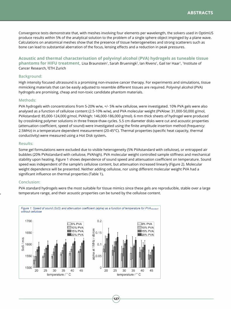
127
EXHIBITOR PROFILEABSTRACTS
Convergence tests demonstrate that, with meshes involving four elements per wavelength, the solvers used in OptimUS produce results within 5% of the analytical solution to the problem of a single sphere object impinged by a plane wave. Calculations on anatomical meshes show that the presence of tissue heterogeneities and strong scatterers such as bone can lead to substantial aberration of the focus, lensing effects and a reduction in peak pressures.
Acoustic and thermal characterisation of polyvinyl alcohol (PVA) hydrogels as tuneable tissue phantoms for HIFU treatment, Lisa Braunstein1, Sarah Brueningk2, Ian Rivens1, Gail ter Haar1, 1Institute of Cancer Research, 2ETH Zurich
Background: High intensity focused ultrasound is a promising non-invasive cancer therapy. For experiments and simulations, tissue mimicking materials that can be easily adjusted to resemble different tissues are required. Polyvinyl alcohol (PVA) hydrogels are promising, cheap and non-toxic candidate phantom materials.
Methods: PVA hydrogels with concentrations from 5-20% w/w, +/- 5% w/w cellulose, were investigated. 10% PVA gels were also analysed as a function of cellulose content (2.5-10% w/w), and PVA molecular weight (PVAlow: 31,000-50,000 g/mol, PVAstandard: 85,000-124,000 g/mol, PVAhigh: 146,000-186,000 g/mol). 6 mm thick sheets of hydrogel were produced by crosslinking polymer solutions in three freeze-thaw cycles. 5.5 cm diameter disks were cut and acoustic properties (attenuation coefficient, speed of sound) were investigated using the finite amplitude insertion method (frequency: 2.5MHz) in a temperature dependent measurement (20-45°C). Thermal properties (specific heat capacity, thermal conductivity) were measured using a Hot Disk system.
Results: Some gel formulations were excluded due to visible heterogeneity (5% PVAstandard with cellulose), or entrapped air bubbles (20% PVAstandard with cellulose, PVAhigh). PVA molecular weight controlled sample stiffness and mechanical stability upon heating. Figure 1 shows dependence of sound speed and attenuation coefficient on temperature. Sound speed was independent of the sample’s cellulose content, but attenuation increased linearly (Figure 2). Molecular weight dependence will be presented. Neither adding cellulose, nor using different molecular weight PVA had a significant influence on thermal properties (Table 1).
Conclusion: PVA standard hydrogels were the most suitable for tissue mimics since these gels are reproducible, stable over a large temperature range, and their acoustic properties can be tuned by the cellulose content.
ABSTRACTS

ABSTRACTS
128
Prediction of pelvic tumour coverage by Magnetic Resonance guided High-Intensity Focused Ultrasound (MRgHIFU) from referral imaging, Daniel Lam1, Ian Rivens1, Sharon Giles2,3, Emma Harris1, Nandita de Souza3, Gail ter Haar1, 1Therapeutic Ultrasound, Division of Radiotherapy and Imaging, The Institute of Cancer Research, London, United Kingdom, 2MRI Unit, The Royal Marsden NHS Foundation Trust, London, UK, 3Cancer Research UK Cancer Imaging Centre, Division of Imaging and Radiotherapy, The Institute of Cancer Research, London, UK
Background: Triaging is required to determine whether a patient’s pelvic tumour is suitable for magnetic resonance-guided high intensity focused ultrasound (MRgHIFU). Currently, clinicians assess patient suitability by examining MR planning images obtained with patients lying on the MRgHIFU bed in a clinician-determined “suitable” treatment position. Software automation of the triaging process using pre-existing (typically supine) referral imaging may improve the patient workflow. Software-based triaging requires accurate prediction of the treatable tumour volume. This requires segmentation of acoustic obstructions (e.g. bone), organs at risk (e.g. rectum), and the target tumour. Once the ideal treatment orientation has been determined, tumour accessibility (the proportion of tumour volume reachable with the focus) can be evaluated after accounting for the effect of body deformation in the new position. Tumour treatability can be evaluated via simulation of acoustic propagation, and the resultant thermal bioeffects. In a first step towards automated triaging, software designed to predict tumour accessibility has been developed and tested.
Methods:DIXON 3D MRI datasets were acquired for five volunteers and three patients (NCT02714621). Referral and treatment images of all subjects lying supine on the Achieva® MR bed and at an incline on a Sonalleve® V2 MRgHIFU treatment bed, respectively, were obtained. Referral datasets were registered to treatment datasets manually. Body outline was segmented automatically. Bones were segmented manually. Organs-at-risk and bowel gas were ignored for volunteers and manually segmented for patients. Tumours were segmented manually by a radiographer. The accessible tumour volume (patients) or soft tissue volume (volunteers) was quantified in both (registered) referral and treatment datasets using an exhaustive search method, after taking into account acoustic coupling limitations and the expected body deformation, and the results compared.
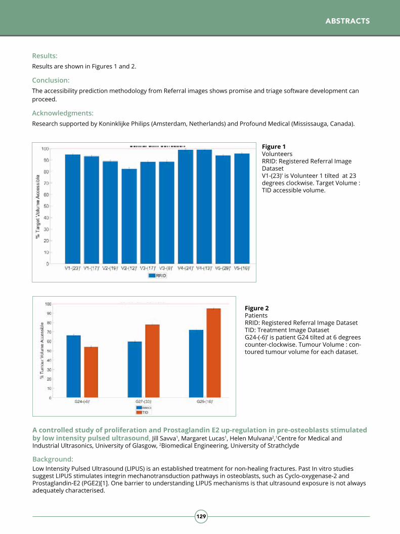
129
EXHIBITOR PROFILEABSTRACTS
Results:Results are shown in Figures 1 and 2.
Conclusion: The accessibility prediction methodology from Referral images shows promise and triage software development can proceed.
Acknowledgments:Research supported by Koninklijke Philips (Amsterdam, Netherlands) and Profound Medical (Mississauga, Canada).
Figure 2PatientsRRID: Registered Referral Image DatasetTID: Treatment Image DatasetG24-(-6)’ is patient G24 tilted at 6 degrees counter-clockwise. Tumour Volume : con-toured tumour volume for each dataset.
Figure 1VolunteersRRID: Registered Referral Image DatasetV1-(23)' is Volunteer 1 tilted at 23 degrees clockwise. Target Volume : TID accessible volume.
A controlled study of proliferation and Prostaglandin E2 up-regulation in pre-osteoblasts stimulated by low intensity pulsed ultrasound, Jill Savva1, Margaret Lucas1, Helen Mulvana2,1Centre for Medical and Industrial Ultrasonics, University of Glasgow, 2Biomedical Engineering, University of Strathclyde
Background:Low Intensity Pulsed Ultrasound (LIPUS) is an established treatment for non-healing fractures. Past In vitro studies suggest LIPUS stimulates integrin mechanotransduction pathways in osteoblasts, such as Cyclo-oxygenase-2 and Prostaglandin-E2 (PGE2)[1]. One barrier to understanding LIPUS mechanisms is that ultrasound exposure is not always adequately characterised.

ABSTRACTS
130
Figure 1: (a) Ratios of viable cells post-/pre-exposure vs. controls and (b) PGE2 concentration 20-hours post-exposure, at 45kHz and 1MHz and 0.0 (control) to 0.2MI.
Conclusion:After exposure to 1MHz LIPUS, cell proliferation and PGE2 concentration peaked at 0.1MI. 45kHz results were prone to variation but PGE2 and proliferation increased at 0.2MI, suggesting 45kHz LIPUS may be more effective at higher MI. We hypothesise that variation across frequencies is due to the shorter rise time of the 1MHz pulse, producing a step-change in radiation force: a mechanism of interest in mechanotransduction pathways. This, along with MI>0.2 at 45kHz, is the focus of future work.
1. Padilla, F., et al., Stimulation of Bone Repair with Ultrasound, in Therapeutic Ultrasound, J.-M. Escoffre and A. Bouakaz, Editors. 2016, Springer International Publishing: Cham. p.385-427.
Interleaving passive acoustic mapping with compounded diverging-wave imaging for HIFU treatment monitoring, Chunqi Li1, Harry Clegg1, Thomas Carpenter1, Luzhen Nie1, David M.J. Cowell1, Steven Freear1, James R. McLaughlan1,2, 1School of Electronics and Electrical Engineering, University of Leeds, LS2 9JT, 2Leeds Institute of Medical Research, University of Leeds, St. James’s University Hospital, LS9 7TF
Ultrasound-guided HIFU therapy could provide a low cost and convenient approach for real-time feedback. It, however, currently can only provide limited information on the formation of thermal lesions. Cavitation activity can play an important role during HIFU exposures that can lead to the heating enhancement and/or asymmetric lesion formation. Thus monitoring its progression during exposures, through interleaved passive acoustic mapping (PAM) and high frame rate imaging (HFR) could provide valuable temporal and spatial information of this phenomenon.
We designed an experimental setup allowing full control of the acoustic field. Pre-osteoblast cells were exposed to LIPUS at 45kHz and 1MHz, with Mechanical Index (MI) up to 0.2. Potential healing effects were assessed by cell proliferation and up-regulation of PGE2 compared to controls.
Methods:Ten custom-built, acoustically-transparent culture vessels (biocells) were seeded with MC3T3-E1 cells (25,000 cells/cm2). After 24-hours incubation, media was refreshed and each biocell placed in a tank of sterilised water at 34±1°C. Two transducers (45kHz; 1MHz) delivered LIPUS for 20 minutes with pulse width 200µs, repetition rate 1kHz and MI 0 (control), 0.05, 0.15 or 0.2. Field mapping was conducted before exposure using 0.5 mm or 2 mm needle hydrophone (Precision Acoustics).
PGE2 concentration in the media 20-hours post-exposure was measured by ELISA kit (Abcam). Proliferation was quantified by counting viable cells pre- and post-exposure using fluorescence microscopy. To maximise sample size, cell counts were undertaken in the centre, at maximum MI, and off-centre, at lower local MI.
Results:
(a) (b)

131
EXHIBITOR PROFILEABSTRACTS
As HFR imaging needs to be interleaved with the HIFU exposure, minimising the imaging time is necessary to ensure maximum heating efficiency. Compounded diverging-wave imaging by phased-array was confirmed as the optimal HFR modality and a suitable compromise between HIFU duty cycle (95%) and HFR acquisition time was demonstrated with minimal impact on thermal lesion formation (Figures 1a-c). During exposures PAM can be performed to complement HFR imaging. In this study, several time-domain PAM beamformers were assessed, these included time exposure acoustics (TEA), capon beamforming (CB), robust capon beamforming (RCB), eigenspace robust capon beamforming (E-RCB) and wiener E-RCB (WE-RCB) (Figures 2a-e). It was shown that WE-RCB provided improved signal-to-noise ratio (SNR) (2±0.2 dB higher than E-RCB). However, RCB without "DC components" outperforms WE-RCB in terms of point spread function (PSF) size, SNR, axial and lateral resolution, without the need to combine more computationally complex techniques such as Eigen-space and Wiener. Moreover, the results demonstrate the 'X-type' artefacts, common to PAM images, are not only originating from interactions between bubble clusters but also from DC components. In order to achieve real-time feedback, the PAM algorithms will be implanted on FPGAs. Finally, a systematic ultrasound-guided HIFU treatment system including HFR, PAM and US temperature mapping techniques could be developed to provide meaningful feedback on ablation progress for clinical guidance.
A Thermochromic Tissue Mimicking Material (Th-TMM) for high intensity focused ultrasound and hyperthermia procedures, Simone Ambrogio1, Raphaela M Baesso2, Bajram Zeqiri2, Fiammetta Fedele1, Kumar Ramnarine1, Piero Miloro2, 1Guy's and St Thomas' NHS Foundation Trust, 2National Physical Laboratory
Introduction:Measurement of temperature rise due to acoustic exposure provides important information on targeting accuracy, effectiveness and safety of high intensity therapeutic ultrasound (HITU) and hyperthermia procedures. A thermochromic tissue mimicking material (Th-TMM), both ultrasound and magnetic resonance compatible, is proposed in this study.
Methods:Th-TMM samples where manufactured by freezing/thawing 4 times a solution of PVA (10wt%), glycerol (8wt%), benzalkonium chloride (0.5wt%) and a thermochromic ink (5wt%) (LCR Hallcrest). The samples were divided in three groups. Group A was immersed in a temperature-controlled water bath to assess the sensitivity in the range of interest (40-65°C). Measurements of speed of sound and attenuation were carried out on Group B. Group C was sonicated in a water tank with a 2MHz HIFU transducer, at 20W (acoustic power), for exposure times ranging from 20s to 60s, in continuous wave.

ABSTRACTS
132
Elucidation of biological mechanisms of clinically viable low frequency (20 kHz) ultrasound applicator for chronic wounds therapy, Olivia Ngo1,2, Karissa Barbarevech1, Jacob Hyatt1, Ian Robinson1, RoseAnn DiMaria-Ghalili1, Michael Neidrauer1, Michael Weingarten1, Kara Spiller1, Peter A Lewin1, 1Drexel University, 2University of Glasgow
Introduction: The primary goal of this work was to investigate the biological mechanisms of action underlying the clinical pilot study success of a novel, wearable ultrasound applicator that we previously showed can reduce healing time of chronic wounds by 60% in a pilot study of 8 diabetic patients. Chronic wounds cost $25 billion annually in the US alone and a reduction of healing time directly reduces healthcare costs. Macrophages are critical in inflammation and have been shown to respond to external mechanical stimuli. Chronic inflammation has been implicated as a potential source of poor healing of chronic wounds.
Materials and Methods: In contrast to commercial ultrasonic debridement systems, a light-weight (<20 g) ultrasound applicator prototype operating at 20 kHz was developed. Its peak acoustic output pressure amplitude is limited to 55 kPa, corresponding to a spatial peak temporal peak (SPTP) intensity of 100 mW/cm2, which is below the inertial cavitation threshold; hence, in the verified absence of temperature elevation, the tissue-ultrasound interaction is dependent on radiation force alone. A custom-curated panel of 227 genes was analyzed 24 hours after exposing THP-1 macrophages in vitro with the clinical applicator (n=3). Student t-test with Benjamini-Hochberg correction was applied for statistical significance.
Results and Discussion: Preliminary gene analysis showed 8 genes (CD163, FOXQ1, CD80, CCL8, DEFB3, IL15, NIMA, CSF3, FPR1) were significantly (p<0.05) downregulated. These genes are responsible for functions related to recruitment, activation, proliferation and differentiation of immune cells indicating a decrease in inflammatory macrophage associated genes.
Results:The samples in Group A provided appreciably different shades of magenta colour when immersed in water with temperature ranging from 45°C to 65°C. The speed of sound and attenuation of the Th-TMM were 1560 ± 10 ms-1 and 0.13*f^1.5 ± 10% dBcm-1MHz-1.5, where f is frequency. The spatial distribution of the temperature increase and focus shape after the HIFU exposures are shown in Figure 1. The ROI (temperature greater than 45°C) is bigger for longer exposures. Th-TMM melting occurs for exposures longer than 30s.
Conclusion:A Th-TMM sensitive to a temperature range relevant to HIFU and hyperthermia procedures has been manufactured and tested. Further experiments are needed to optimise the acoustic properties, to quantify the temperature distribution within the ROI, to determine cavitation thresholds and to cross-reference the results with independent measurement methods (such as MRI-thermometry). Overall, early experiments showed the material has great potential and can be used in test objects for R&D of new procedures and QC of clinical devices.

133
POSTER EXHIBITION
Conclusions: Macrophage gene regulation appears to be a potential chronic wound healing mechanism activated by this therapeutic ultrasound applicator. A downregulation of these inflammation associated genes could indicate a less inflammatory macrophage after ultrasound treatment. Understanding the effects of therapeutic ultrasound on macrophage behaviour may lead to personalized wound care treatments procedures.
Demonstration of the ability to use microbubbles combined with low pressure focused ultrasound to induce cavitation in orthotopic pancreatic tumors, Petros Mouratidis1, Gail ter Haar1, 1The Institute of Cancer Research
High intensity focused ultrasound (HIFU) can be used for the treatment of solid tumours by creating cavitation which results in mechanical disruption of their tumour microenvironment. Pancreatic tumours which are characterised by a dense stroma should be particularly prone to these acoustic cavitation effects.
Our pilot preclinical studies have shown that treatment of pancreatic tumours with pulsed HIFU (pHIFU) can result in biological and immunological anticancer effects. The application of these pHIFU exposure parameters in a clinical setting is, in parts, hindered by the high acoustic pressures (>10MPa) required, which are considered to be neither practical nor safe for the patient.
The aim of this study was to investigate the feasibility of combining pHIFU with cavitation enhancing agents (microbubbles) to induce acoustic cavitation in pancreatic tumours at clinically-relevant low rarefactional pressures. Syngeneic orthotopic pancreatic KPC tumours (KrasLSL.G12D/+; p53R172H/+; PdxCre tg/+) were grown in immune-competent murine C57BL/6 subjects. Subjects were injected intravenously with 100?l of microbubbles, and tumours were exposed to pHIFU (peak negative pressures = -17.6 to -2.7 MPa, duty cycle = 1%, pulse repetition frequency = 1 Hz, 25 repeats, f = 1.5 MHz) using the small animal Alpinion VIFU 2000 Therapeutic ultrasound platform. Acoustic cavitation was monitored during pHIFU bursts using a weakly focused polyvinylidine fluoride, broadband (0.1 to 20 MHz) passive cavitation detector. Data recording was controlled and processed using custom-written MatLab software. Tumours were imaged using 2D high frequency (14 MHz) B-mode ultrasound (E-Cube 9, Alpinion, USA).
Results showed that broadband and half harmonic emissions were detected at peak negative pressures as low as -3 MPa when microbubble contrast agent was injected immediately before the pHIFU treatment. These results suggest that the use of microbubbles could reduce the pressure threshold needed for the induction of acoustic cavitation during pHIFU treatment of pancreatic tumours.
ABSTRACTS

ABSTRACTS
134
Abdominal distension in children, Clement Leung1, Jane Ferguson2, 1Radiology Peninsula Radiology Academy, 2Radiology Royal Devon and Exeter NHS Foundation Trust
Background:Peritoneal mesothelioma is strongly associated with asbestos and metal fibres exposure, an occupational hazard, and it predominantly affects middle aged to older males (50 to 70 years). Rarely, 1-2 cases per million, does it arise as primary neoplasm from peritoneum. Patients may be asymptomatic or present with abdominal distension, nausea and vomiting, abdominal pain, weight loss and/or malaise.
Case Report:A 13 year old girl presents with six weeks of rapid abdominal distension with no change in bowel habit. Her appetite was fine and she felt well and active. She has had chicken pox and was post-menarche. There was no family history of any medical conditions. On examination, her abdomen was tense but non-tender. There was no palpable mass or peripheral lymphadenopathy, and no signs of anaemia or jaundice. Trans-abdominal ultrasound revealed extensive ascites with low level echoes throughout. Around the liver, there was multiple subcapsular moderately echogenic lesions indenting the liver surface. No intrahepatic lesion and normal gallbladder was noted. There was a large lobulated solid pelvic mass with some internal heterogeneity and a more discrete left sided cystic structure containing multiple peripheral solid nodules. Uterus and ovaries were not identified. Further cross-sectional imaging with MRI was acquired. Peritoneal biopsy confirmed the diagnosis of malignant epithelial mesothelioma. Immunohistochemical marker and genomic profiling were also performed. Treatment options include cytoreductive surgery, intraperitoneal chemotherapy and systematic chemotherapy.
Discussion:Malignant deposits follow flow of peritoneal fluid and gravity into dependent pelvic spaces and up into right subphrenic spaces. Psychosocial support for the patient, sibling and family is vital in the overall management of paediatric oncology patients.
Rare Urachus abnormality: Inflammatory Myofibroblastic Tumour (IMT), Khalida Jan, Radiology South Tynesdide and Sunderland NHS Foundation Trust
Background:Inflammatory Myofibroblastic tumour (IMT) is an infrequent, locally aggressive, benign soft tissue tumour with uncertain malignant potential, originally reported in the lungs by Brunn (1939) with subsequent occurrences in the digestive system. It is rare in the urinary system, first discovered in the bladder by Roth (1980). A Urachal IMT is extremely rare and challenging to diagnose. Urachal remnants represent rare congenital anomalies resulting in a failure of the obliteration of the allantois at birth which connects the dome of the bladder to the umbilicus. Awareness and recognition of urachal anomalies and knowledge of IMT is important to distinguish whether the tumour originates from the bladder or the Urachus, because the surgical treatment options are completely different.
Case Report:A 8 year old male presented for abdominal ultrasound with a 3/52 history of frank haematuria, dysuria, pyrexia, ‘flu like’ symptoms and constipation but no history of UTI, trauma or surgery. On Physical examination he was pale and thin. Laboratory results indicated raised inflammatory markers i.e. CRP and ESR. Ultrasound assessment revealed a large well defined, mixed echo, vascular mass at the umbilicus extending into the dome of the urinary bladder raising the suspicion of a urachal malignant neoplasm.
Discussion: The normal obliteration of the Urachus after birth terminates in the midline umbilical ligament. Incomplete regression results in several anomalies. Recognition is required because of increased potential for infection and neoplastic differentiation with ultrasound being the primary investigative tool in classifying and characterisation of the type of anomaly. Awareness and understanding of the urachal anatomy, embryology and the entity of the Urachal IMTs is a prerequisite for timely and accurate diagnosis.
DAY THREE Thursday 12th December
General Imaging and Paediatrics

135
ABSTRACTS
StandUltrasound diagnosis of biliary obstruction: Are the recommended cut-off criteria of common bile duct diameter and liver function testing safe?, Matthew Hiles1, Stephen Wolstenhulme2, Shaunna Smith1, 1Radiology Hull and East Yorkshire Hospitals Trust, 2Leeds Teaching Hospitals Trust
Background: Transabdominal Ultrasound (TAUS) including measurement of the Common Bile Duct (CBD), and Liver Function Tests (LFTs), are the recommended initial investigations into suspected biliary pathology but estimates of test specificity are average. Recent guidelines propose increasing the normal limit of CBD diameter and combining elevated LFTs including elevated bilirubin as alternative diagnostic criteria.
Objectives: To determine and compare diagnostic accuracies of current and alternate criteria for diagnosis of biliary pathology using Magnetic Resonance Cholangiopancreatography (MRCP) as the reference standard.
Methods: A retrospective service evaluation was conducted on 508 eligible subjects between September 2013 and August 2018. All subjects received TAUS and laboratory blood analyses followed by MRCP for investigation into suspected biliary pathology. Ultrasound, MRCP and laboratory findings were collected by retrospectively applying the current and alternate diagnostic criteria to the study cohort. Sensitivity and specificity with confidence intervals were calculated for both criteria by populating 2 X 2 contingency tables.
Results: Of the 508 subjects, reference standard MRCP identified 328 with complex biliary pathology, 79 with simple gallstones, and 101 with no abnormality. Application of the current diagnostic criteria correctly identified the target condition in 256 subjects with 72 incorrectly identified as negative. Application of the alternate diagnostic criteria correctly identified 227 subjects as positive with 101 incorrectly identified as negative. Sensitivity and specificity were 78% (CI 73% - 82%) and 44% (CI37% - 52%) respectively for the current diagnostic criteria, and 69% (CI 64% - 74%) and 67% (CI 59% - 74%) respectively for the alternate diagnostic criteria.
Conclusion: The alternate diagnostic criteria yielded greater specificity but inferior sensitivity when compared to the current criteria used in daily practice. Increased false negative diagnoses are unacceptable due the risk of infectious morbidity associated with undiagnosed biliary obstruction. The alternate diagnostic criteria should be considered unsafe for clinical use.
Incidentalomas: Point of Care ultrasound and haemangiomas, Helga Consiglio1, Stephen Wolstenhulme2, 1Obstetrics and Gynaecology Mater Dei Hospital, Msida, Malta, 2Radiology Leeds Teaching Hospitals NHS Trust, Leeds, UK
Background and Aim:Internationally, Point of Care ultrasound (POCUS) is a developing modality. POCUS examinations are carried out by medical and non-medical clinicians, from many disciplines. They have varying degrees of ultrasound (US) experience and education. This focussed examination may detect incidentalomas.
The aim of this case report is to demonstrate the role of POCUS in the management of a patient with focal liver lesion (FLL) incidentalomas.
Case Report:A 24 year old woman was referred to an Urologist and a “same-day kidney POCUS” clinic due to a gynaecologist’s suspicion of urinary tract endometriosis. She had symptoms of urgency, dysuria during the menstrual cycle, altered bowel habit and weight loss.
A POCUS examination was done by a trainee sonographer (obstetrician) and supervised by an expert non-medical sonographer/ lecturer. The findings were: normal kidneys and bladder. Four solid FLLs were identified. The largest, in segment II/III measured 3.5cm and had mixed echotexture. The other three lesions measured between 1 and 2.9cm. The conclusion was atypical haemangiomas; needs further characterization.
An US specialist (Consultant Radiologist) confirmed the POCUS findings. Magnetic Resonance Imaging was requested. This agreed with the POCUS and the specialist’s findings of haemangiomas. The patient was reassured and discharged.

ABSTRACTS
136
Discussion:POCUS equipment is relatively cheap and compact. It provides same-day access to imaging with the potential of reducing: anxiety, repeat visits and costs. POCUS is a rule-in examination and not a definitive diagnostic examination. FLL incidentalomas may be identified. These include haemangiomas, focal nodular hyperplasia, focal steatosis, adenomas and malignancy. POCUS clinicians should refer the patient with unexpected pathology to specialist (radiologists and non-medical sonographers) in medical imaging departments. POCUS and specialist US services should complement each other for the benefit of the patient.
The utility of ultrasonography in the diagnosis of testicular torsion - A systematic review, Pearly Yuen, Royal Victoria Infirmary, Newcastle upon Tyne Hospitals NHS Foundation Trust
Aims and Objectives:Although ultrasound is a valuable tool in establishing a diagnosis for acute scrotum patients, its role in detection of testicular torsion remains controversial and has historically been reliant on Colour Doppler ultrasound alone. This review aims to assess the value of combined B-mode and Colour Doppler ultrasound features in detection of testicular torsion in acute scrotum patients.
Methodology:A thorough literature search through several electronic databases, grey literature and hand searching was conducted to identify relevant literature which met the inclusion criteria of this review. Relevant literatures were systematically reviewed and data from included studies were extracted, tabulated and analysed.
Results:The included studies demonstrated strong agreement on the value of ultrasound as a triage tool in acute scrotum patients. Sole reliance on Colour Doppler ultrasound features showed potential for false-negative results. Whereas, several B-mode appearances were found to be good indicators for testicular torsion and showed high sensitivity and specificity values. Where B-mode and Colour Doppler ultrasound features were combined, sensitivity towards testicular torsion detection increased, with 3 studies achieving 100%. However, all studies recommended correlation with clinical examination findings and that any investigations should be conducted within appropriate time to avoid delays to treatment. Several limitations in methodology of the included studies such as small sample populations, lack of information on the ultrasound operators and selective tabulation of data were noted.
Conclusion:Ultrasound, utilising the combination of B-mode and Colour Doppler, is a useful and reliable adjunct to clinical examination in the detection of testicular torsion in acute scrotum patients, especially in indeterminate and low suspicion cases. However, a larger scale primary study is required to further validate the results of this review due to limitations in methodology of included studies.
Palliative care imaging – a role for hospice based ultrasound, Mrs Jo Eastman, Saint Francis Hospice
The aim of this talk is to present a review of the experience of establishing the provision of an ultrasound scanning service in an adult hospice setting.
Palliative care is more than just end of life care. It is an interdisciplinary approach to specialised nursing and medical care for people with life-threatening illness. It focuses on providing relief from the symptoms, pain, physical and mental stress at any stage of that illness.
Ultrasound imaging is a valuable resource that has been widely used in hospitals for many years. Its usage has been to slow to develop in hospices despite improvements to the cost of machines and their accessibility. It can help clinicians make what are often difficult management decisions at a crucial point in a patient’s life.
Over the course of six years, 429 scans were carried out on 327 patients by the author (an experienced sonographer) with an age range of 23 to 96. Indications for scans included suspected urinary retention, DVT, and assessment of abdominal ascites or pleural fluid. A wide range of pathologies were found, from disease progression to gallstones accounting for pain. The author will share some of her experiences and observations of scanning in these challenging but rewarding circumstances.
Conclusion:The author, supported by the clinical team, has been able to demonstrate that a hospice based ultrasound service is achievable, effective and safe. Clinicians value the greater certainty available with the use of sonography when signs and symptoms are subtle or complex, while patients appreciate not having to travel for imaging.

137
Sonography as a Profession. Where are we now?, Pamela Parker, Ultrasound Hull University Teaching Hospitals NHS Trust
As we go to press we are in the middle of an election campaign in the UK. What will this hold of for the NHS, our patients and our profession? The journey of developing the career framework for the professional sonographer has been on-going for some years, and continues but where have we been on this journey? More importantly, where are we heading? In the advent of a new government, following today’s election, it is difficult to predict the direction our journey but this presentation will discuss options open to the profession as a whole.
The presentation will give an update of the journey to date and will discuss the challenges that have been overcome. New challenges will have to be faced, in no small part does this include the strong desire to ensure the profession of sonographer is recognised as an independent entity and receives the statutory regulation recognition that it deserves. The audience is encouraged to actively participate in the question and answer session that follows; your opinion matters.
Current ultrasound practice in Europe: A European federation of radiographer societies (EFRS) survey of national radiographer societies, Gill Harrison1,2, Malene Roland V. Pedersen1,3, Barbara Kraus1,4, Sandra Noij-Rijkes1,5, Rute Andreia Martins dos Santos1,6, 1European Federation of Radiographer Societies, 2School of Health Sciences, City, 3Lillebaelt Hospital, Denmark and University of Southern Demark, The Netherlands, 4Radiology Technician Teaching and Research University of Applied Sciences, Vienna, 5Inholland University of Applied Science, The Netherlands, 6Coimbra Health School, Polytechnic Institut
Aims:A survey of the European Federation of Radiographer Societies’ (EFRS) national radiographer societies was carried out to collate information and opinions about ultrasound practice across the wider European countries.
Methods:A SurveyMonkey questionnaire was developed and sent by e-mail to 54 European, Baltic and Asian countries’ radiographer societies. A range of questions were used including free text options, to expand on specific topic areas.
Results:Responses were received from 27 countries, with 40 individual responses from radiographer societies. Radiologists perform ultrasound examinations in 39 (97.5%) countries, followed by ‘specialised medical doctors’ in 32 (80%) countries. Radiographers perform ultrasound examinations in 13 (48%) countries and had limited involvement in 3 (11%) countries, leaving 41% of countries with no radiographer involvement in ultrasound. Common reasons for radiographers not undertaking ultrasound examinations were professional resistance or legislation preventing it and limited educational provision specifically for ultrasound. Where education was available, focused courses predominated (39%) and post graduate ultrasound courses (35%). The UK was the only country to have a dedicated BSc (Hons) programme specifically for ultrasound education.
Fully independent interpretative reporting by radiographers was uncommon (4%). The majority provide descriptive or checklist reports, which are finalised or written by a radiologist. Indemnity insurance was provided by 25% of radiographer societies for radiographers performing ultrasound examinations, one highlighted that it was secondary insurance to support employers’ primary insurance.
Conclusion:The findings were varied, with some countries fully engaged with radiographers performing ultrasound examinations, a few were trying to develop the role and others had a range of barriers to expanding radiographers’ involvement into ultrasound. Barriers were explored in the survey and the findings can be used to assist countries who want to develop the sonographer role for radiographers, to overcome some of the challenges and provide safe, high quality patient care.
Professional Issues
ABSTRACTS

ABSTRACTS
138
Why are standards and benchmarks important to ultrasound departments?, Mrs Hazel Edwards, Radiology East and North Herts NHS Trust
Standards and benchmarks help with the delivery of quality healthcare - our priority. This talk will define what is meant by a standard and benchmark and will explain where they come from and who sets them. Research drives standards from which guidance and recommendations can be made. These in turn allow local protocols to be developed for the benefit of both patients and staff. These benefits will be highlighted along with some of the problems ultrasound departments may face in the absence of robust protocols. Finally, reasons why departments and individuals may sometimes fail to adhere to their own protocols will be given.
Publishing research and audit: How does it affect patient safety?, Gill Harrison, School of Health Sciences City, University of London & Society and College of Radiographers
Audit and research are essential components of safe, effective practice in healthcare. There is an expectation that audit will be undertaken to assess the quality of healthcare provision. Additionally. one of the pillars of advanced practice is research and audit. The difference between research and audit will be considered during the presentation.
When considering audits of practice, it’s worth thinking about the following questions:
• How many audits are undertaken in the UK?
• How many do you see published?
• Is there a discrepancy? If so why?
Within this presentation, the publication for audit and research findings will be discussed in relation to patient care and patient safety. Consideration will be given to potential issues with publishing work and the impact that might have on patient safety.
Equipment QA - The forgotten standard? Why it matters, Dr Nick Dudley, Lincoln County Hospital
An oft stated reason for the lack of ultrasound QA is that there is no legislative requirement. Those of us working with or alongside x-rays have been distracted by the various ionising radiations regulations that explicitly require QA. However, there is other legislation that requires QA, especially The Health and Social Care Act 2008 (Regulated Activities) Regulations 2014 and Care Quality Commission (Registration) Regulations 2009 which require risks to be assessed and mitigated and equipment to show no more than minor deterioration. These requirements cannot be met without QA. The drivers for QA and the consequences of ignoring the need will be explored and minimum evidence based standards will be presented.
Vascular
Paediatric DVT: Out-ruling all potential causes, Frances Glynn1,2, Rona O’Riordan2, Therese Herlihy1, Kevin Cronin1, 1University College Dublin, 2University Hospital Galway
Background:Deep vein thrombosis (DVT) events are rare in children in comparison to in adults. However, DVT is becoming an increasingly common complication, particularly among hospitalised children. This case presents a 6-year-old male with a background of spina bifida and limited mobility admitted with left lower limb swelling, increased leg temperature and skin discolouration. The paediatric team ordered a lower limb Doppler ultrasound (US) as they queried the presence of a DVT.
Case report:B-mode, colour and spectral Doppler US assessment of the left leg veins was performed. The external iliac vein, common femoral vein, proximal to mid femoral vein were all patent, compressible and showed augmented flow. The mid femoral vein to popliteal vein were non-compressible with absent flow, consistent with DVT. The patient received anti-thrombotic treatment but with progressive symptoms, a serial follow-up US scan was performed 3 weeks after the initial diagnosis showing resolution of the DVT.

139
EXHIBITOR PROFILEABSTRACTS
Discussion:The US findings of a resolved DVT led to a query over the initial cause of the DVT and of the progressing symptoms. Femur radiographs revealed a longstanding distal femur fracture with extensive callous bone formation and soft tissue swelling. The combination of patient immobility and bone trauma meant that this patient was at a particularly high risk of developing a DVT, thus highlighting the importance of out-ruling all potential causes of DVT when diagnosed in children.
US plays an important role in both the diagnosis of paediatric DVT and follow-up of its resolution. However, consistency in scanning approach and reduced interscan variability is crucial to improve confidence in US findings. Paediatricians should have a very high index of suspicion for DVT, especially in hospitalised children. It also is important to out-rule all potential causes of DVT in children when an US diagnosis is made.
Upper limb doppler ultrasound: A dual site study analysing clinical outcomes and indications, Wajiha Arshad, Nehan Khalid, Siva Muthukumarasamy, Hull and East Yorkshire Hospitals Trust
Aims and Objectives:• Assessing the accuracy and appropriateness of information provided by clinicians on DVT requests to guide
justification
• Analyse the outcome data in positive scans for dual sites
• Monitor follow up imaging relating to the symptoms associated with the initial presentation
Methods:• Data was collected between 01/01/2018 and 31/12/2018 including both inpatient and outpatient scans during this
period, of patients aged 16 and above
• Each patient record was assessed for indication of scan, past medical history, ultrasound result, relevant follow up imaging and patient demographics
• These were input into an anonymised database for analysis
Results:• 92 patients had ultrasound doppler studies during 01/01/2018 – 31/12/2018
• 54% female, 46% male
• Mean age was 61 years, however the range was 16-92 years
• Indication of scan – 4 patients had clinical documentation of cellulitis, 88 patients had swelling and pain in the affected limb
• 6 patients did not have a documented d-dimer (7%)
• 29 had positive findings
• 1 had thrombophlebitis
• The remaining 28 (30%) had deep vein thrombosis (stenotic v occlusive)
• Of these 28, the most prevalent location was in the subclavian vein 10 patients (36%)
• 4 of these extended into the external jugular vein
• 2 were occlusive
• 1 extended into the axillary vein
• Of note 4 patients had brachial DVTs (14%), 2 of which were secondary to PICC line insertions
• 4 patients had follow up, two were CT venograms to analyse the venous occlusions
Conclusion:Upper limb ultrasound is a useful tool in analysing venous occlusive disease in the acute setting. Our study highlights its effectiveness with only 4 patients continuing to have follow up imaging. The most prevalent documented area of thrombus burden in our study was the subclavian vein.

ABSTRACTS
140
Use of ultrasound guidance in Peripheral Venous Cannulation, Daniel El-Dalil, Emergency Department Chesterfield Royal Hospital
Aim:To evaluate the use ultrasound guidance in Peripheral Venous Cannulation (PVC) in the Emergency Department.
Method :Review of the literature surrounding the use of ultrasound guidance in PVC.
Results:PVC is a routine procedure in emergency medicine allowing vital fluids and medications to be given to patients. Cannulation can be difficult to achieve in certain groups of patients, such as intravenous drug users, patients with raised Body Mass Index (BMI) and patients with circulatory shock1,2. With the rising prevalence of obesity3, the number of patients with difficult venous access is likely to rise.
Ultrasound is widely used to aid PVC in patients with difficult venous access4. Two meta analyses demonstrated with statistical significance that ultrasound guided PVC was more successful than traditional cannulation techniques5,6. Research has shown potential advantages of using ultrasound include reduced number of cannulation attempts and reduced time to complete the procedure7, however some studies have shown that there were no significant difference in these parameters6.
Central venous catheters can be used in patients with difficult venous access; these however are a resource intensive intervention and not without risk8. Studies show that ultrasound guided PVC has been associated with reduced number of central venous catheter insertions [8] and can be easily taught to junior doctors and emergency nurse practitioners2,9.
Conclusion:Ultrasound guidance can improve success rates of PVC in those with difficult venous access2,5,6,8,9 and is an alternative technique with a lower risk profile to central venous cannulation9.
As a consequence of this review, the induction programme for the new cohort of junior doctors at Chesterfield Royal Hospital Emergency Department will include training in the use of ultrasound guided venous access.
References:1. Beecham, G. B., & Tackling, G. (2019). Peripheral Line Placement. In StatPearls [Internet]. StatPearls Publishing.
2. Khan, M. S., Sabnis, V. B., Phansalkar, D. S., Prasad, S. P., & Karnam, A. H. F. (2019). Use of ultrasound in peripheral venous catheterization in adult emergency and critical care units. Anaesthesia, Pain & Intensive Care, 303-310.
3. Wang, Y. C., McPherson, K., Marsh, T., Gortmaker, S. L., & Brown, M. (2011). Health and economic burden of the projected obesity trends in the USA and the UK. The Lancet, 378(9793), 815-825.
4. Schmidt, G. A., Blaivas, M., Conrad, S. A., Corradi, F., Koenig, S., Lamperti, M., ... & Slama, M. (2019). Ultrasound-guided vascular access in critical illness. Intensive care medicine, 45(4), 434-446.
5. Van Loon, F. H. J., Buise, M. P., Claassen, J. J. F., Dierick-van Daele, A. T. M., & Bouwman, A. R. A. (2018). Comparison of ultrasound guidance with palpation and direct visualisation for peripheral vein cannulation in adult patients: a systematic review and meta-analysis. British journal of anaesthesia, 121(2), 358-366.
6. Egan, G., Healy, D., O’Neill, H., Clarke-Moloney, M., Grace, P. A., & Walsh, S. R. (2013). Ultrasound guidance for difficult peripheral venous access: systematic review and meta-analysis. Emerg Med J, 30(7), 521-526.
7. Costantino, T. G., Parikh, A. K., Satz, W. A., & Fojtik, J. P. (2005). Ultrasonography-guided peripheral intravenous access versus traditional approaches in patients with difficult intravenous access. Annals of emergency medicine, 46(5), 456-461.
8. Galen, B. T., & Southern, W. N. (2018). Ultrasound-guided peripheral intravenous catheters to reduce central venous catheter use on the inpatient medical ward. Quality Management in Healthcare, 27(1), 30-32.
9. Brannam, L., Blaivas, M., Lyon, M., & Flake, M. (2004). Emergency nurses’ utilization of ultrasound guidance for placement of peripheral intravenous lines in difficult access patients. Academic Emergency Medicine, 11(12), 1361-1363.

141
EXHIBITOR PROFILEABSTRACTS
Is there value in repeating lower limb doppler ultrasound (sonovenograms) for suspected deep vein thrombosis, without clinical reassessment? - A pilot study, Claire Ryan, Oliver Hulson, Stephen Wolstenhulme, Adam Morrell, Gurpreet Ross, Catherine Sharp, Reshma Koshy, Shishir Karthik, Leeds Teaching Hospitals NHS Trust
Introduction:Lower limb deep venous thrombosis (DVT) causes significant morbidity[1] and is difficult to identify clinically[2]. At our centre, we assess for DVTs using sonovenograms. The National Institute for Health and Care Excellence (NICE) recommends repeat sonovenogram 6-8 days following an initial negative scan, if the Well’s Score ?2 and D-dimer >230ng/ml[3]. Anecdotal accounts from sonographers and radiologists indicate some returning patients in whom symptoms have improved. We thereby question justification for rescanning without clinical reassessment.
Aims:To ascertain:
1. The frequency of positive repeat scans.
2. Risk factors making positive repeat imaging more likely.
3. Should we introduce repeat clinical assessment and D-dimer before rescanning?
Methods:This is a pilot study, covering one month (October 2018), with the view to extend to a year (January ’18 - January ’19). Patients with two scans within six weeks (negative for DVT on initial scan) were selected. We are retrospectively analysing data from the Trust’s ultrasound database, the Radiology Information System (CRIS) and the Trust’s electronic patient records system (PPM+).
Results:Of 83 patients (29 M, 54 F; age range 18-94), one had a positive repeat sonovenogram (1.2%). The request conveyed clinical reassessment had occurred, which was unusual, and D-dimer increased (232-241) - the sole case where D-dimer was repeated (1.2%). There were no risk factors identified; although, a Bakers’ Cyst was diagnosed on the first scan.
Conclusion:Preliminary data suggests positive yield following an initial negative sonovenogram is low (1.2%) and repeating the test is of questionable value. Perhaps a more targeted reassessment would be preferable - particularly as, anecdotally, the positive study involved both clinical and D-dimer reassessment. However, a larger study is required to ascertain the role of clinical reassessment, before proposing its introduction into standard clinical practice.
References:1. White RH. The epidemiology of venous thromboembolism. Circulation 2003;107(23 Suppl 1):I4-8.
2. Goodacre S, et al. Meta-analysis: The value of clinical assessment in the diagnosis of deep venous thrombosis. Ann Intern Med 2005; 143(2): 129-139
3. NICE Clinical Guideline [CG144] Venous thromboembolic diseases: diagnosis, management and thrombophilia testing; Recommendations 2012 (updated 2015): 4.
Evaluating the role of ultrasound in the diagnosis of bilateral persistent sciatic arteries, Sharon Bell, Radiology Shrewsbury and Telford Hospital NHS Trust
Background:Peripheral vascular disease (PVD), affects 13% of the population aged over 50 in the western world and can be divided into two main groups: organic and functional. Other less common causes to include in the differential diagnosis in younger patients are Buerger’s disease and anatomical variants such as duplication of the superficial femoral artery and persistent sciatic arteries (PSA).
PVD is a major cause of morbidity and mortality globally. Symptoms vary from cold extremities, intermittent claudication, to acute and critical ischaemia. All of these have a significant financial burden on critical healthcare resources.
This case report evaluates the role of ultrasound in the diagnosis of bilateral persistent sciatic arteries.

ABSTRACTS
142
Case report:A 16 year old female was referred by her GP to the vascular clinic presenting with bilateral intermittent claudication. On examination missing dorsals pedis pulses were noted bilaterally. No previous imaging was available and the patient was referred for a lower limb Doppler ultrasound.
Initial ultrasound scanning demonstrated no evidence of diseased vessels causing the patients pain, but unusual arterial anatomy. Both superficial femoral arteries tapered to the distal thigh where collaterals were demonstrated. Following the scan a diagnosis of persistent bilateral sciatic arteries was made.
Discussion:Ultrasound due to its many advantages is usually the first line imaging investigation to assess the peripheral arteries. In this case ultrasound was able to demonstrate that there was no evidence of any organically diseased vessels, but an anatomical variant causing the patient’s symptoms. However, it was unable to demonstrate the overall view of the lower limb vasculature, the origin of the PSA, or provide all information to classify the sub type in view of any potential treatment or surgery.
Although ultrasound is useful in the diagnosis of PSA , it lacks the ability to assess the whole of the peripheral circulation.
Analysis of carotid referral practice at a hyperacute stroke unit (HASU), Aaron Goh, Amy Partridge, Kristen Swinamer, Sophie Renton, Kamran Modaresi, Northwick Park Hospital
Background:From the National Vascular Registry data, rates of carotid endarterectomies (CEA) performed nationally are decreasing yearly. Our unit represents one of eight HASUs in London and we had experienced a similar reduction in CEA rates. We examine our referral process to identify the number of carotid duplexes performed in 2018, ascertain the pick-up rate for CEA and determine if all appropriate patients were referred for vascular input.
Methods:A retrospective analysis was performed of all duplexes performed at our centre in 2018. This was cross-referenced with electronic patient records and physical notes.
Results:A total of 1209 carotid duplex scans were performed during the study interval, with 114 patients (9.4%) identified with significant (>50%) ICA stenosis. Of these, 34 patients (29.8%) underwent CEA. The outcomes of the patients are listed in Table 1. All the 10 patients that were not referred had ICA stenoses of 50-59% as measured using the NASCET criteria. The 2 patients with no electronic documentation were referred to the vascular outpatient clinic.
Significant (>50%) ICA stenosis Total (n=114)
CEA performed 34
Offered CEA, but declined 4
Procedure cancelled due to technical/medical reasons 4
Complete ICA occlusions of the symptomatic side 10
Assess by vascular and decision made for BMT 7
Symptoms not in keeping with TIA / stroke 23
Significant stenoses (>50%) on contralateral side not in keeping with symptoms 20
Not referred as inpatient Not referredNo documentation
12102
BMT: Best medical therapy; ICA: internal carotid artery; TIA: transient ischaemic attack
Conclusion:We have identified that 12 patients with symptomatic stenoses were not referred to vascular surgery for inpatient assessment, which would represent up to a 35% increase in CEA activity. We recommend closer collaboration between stroke and vascular teams to identify patients who would benefit from CEA.

143
EXHIBITOR PROFILEABSTRACTS
Veterinary
Left Atrial Rupture - Echocardiographic Diagnosis: An Interesting Case, Maria Mateos, Cardiology Northwest Veterinary Specialists
A left atrial rupture was diagnosed in a dog with advanced myxomatous mitral valve disease that experienced a sudden onset collapse episode after intense exercise. Two-dimensional echocardiography showed the presence of pericardial fluid, myxomatous mitral valve disease, severe mitral regurgitation and presence of organized echogenic material originating from the wall of the left atrium, extending adjacent to the left ventricular free wall within the pericardial space, suggestive of a thrombus. Supportive treatment, with oxygen, furosemide and pimobendan, was started. The dog’s state of health improved on treatment and was discharged after one day. The patient is alive at the time of writing.

ABSTRACTS EXHIBITOR PROFILEJoin BMUS NOW And Support Your Profession!Did you know that it costs less than £1.85* a week to be a member of BMUS and that you can pay quarterly for your subscription?
• Membership of a truly multi-disciplinary society fostering the exchange of information and ideas between its members.
• The quarterly 'Ultrasound' journal - the official journal of the British Medical Ultrasound Society - Ultrasound covers all aspects of medical ultrasound, from the theoretical through to clinical applications and their impact on both workforce and patients. The journal fulfils both an educational and a scientific research role and publishes a mixture of invited review articles, current research and descriptions of new techniques and equipment.
• Free online access to the full back catalogue of the 'Ultrasound' journal and 'BMUS Bulletin'.
• Ultrapost, the society’s weekly e-newsletter.
• A quarterly printed newsletter giving information about the Society; its meetings, committees, activities and diary dates.
• Opportunity to gain ‘continuing professional development’ opportunities via our online CPD portal.
• Access to professional guidance documents via the members only section of the BMUS website.
• Access to the diagnosis for 'Interesting Cases’ published on the BMUS webpages.
• Reduced registration fees at the BMUS Annual Scientific Meeting and the special topic meetings run by the society.
• As a member of BMUS you will also be a member of EFSUMB and WFUMB, and can enjoy the benefits of both societies including reduced registration fees at meetings organised by WFUMB and EFSUMB.
• Discounts on BMUS publications and Ultrasound in Medicine and Biology, the official journal of WFUMB.
• Eligibility to apply to become a member of BMUS Council or a member of one of the Society's Committees.
• Eligibility to apply for the society's scholarships and to influence the Society's affairs by voting at the AGM.
Benefits of being a BMUS member include:
Join Online now at www.bmus.org

The Philips EPIQ Elite for general imaging ultrasound system brings an exceptional level of performance, workflow and advanced intelligence for today’s most demanding practices. Expect fast, consistent, easier exams across all clinical segments.
EPIQ Elite enhances diagnostic confidence, helping you deliver a new class of care to your patients. There is always a way to make life better.
Next-generation ultrasound.Elevated performance.
See the EPIQ Elite video at www.philips.com/epiq
Ultrasound

BMUS 2020