ACUSON P500™ 2.0 Ultrasound System
18
ACUSON P500™ 2.0 Ultrasound System Datasheet
-
Upload
khangminh22 -
Category
Documents
-
view
4 -
download
0
Transcript of ACUSON P500™ 2.0 Ultrasound System

ACUSON P500™ 2.0 Ultrasound System
Datasheet
Kam
MDD_Stamp
ACUSON P500 Ultrasound System
2 siemens.com/ultrasound
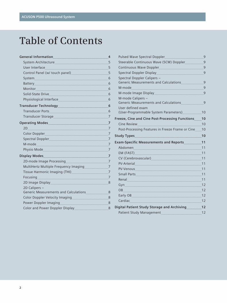
Table of ContentsGeneral Information 4
System Architecture 5
User Interface 5
Control Panel (w/ touch panel) 5
System 6
Battery 6
Monitor 6
Solid-State Drive 6
Physiological Interface 6
Transducer Technology 6
Transducer Ports 6
Transducer Storage 7
Operating Modes 7
2D 7
Color Doppler 7
Spectral Doppler 7
M-mode 7
Physio Mode 7
Display Modes 7
2D-mode Image Processing 7
MultiHertz Multiple Frequency Imaging 7
Tissue Harmonic Imaging (THI) 7
Focusing 7
2D Image Display 8
2D Calipers – Generic Measurements and Calculations 8
Color Doppler Velocity Imaging 8
Power Doppler Imaging 8
Color and Power Doppler Display 8
Pulsed Wave Spectral Doppler 9
Steerable Continuous Wave (SCW) Doppler 9
Continuous Wave Doppler 9
Spectral Doppler Display 9
Spectral Doppler Calipers – Generic Measurements and Calculations 9
M-mode 9
M-mode Image Display 9
M-mode Calipers – Generic Measurements and Calculations 9
User defined exam (User-Programmable System Parameters) 10
Freeze, Cine and Cine Post-Processing Functions 10
Cine Review 10
Post-Processing Features in Freeze Frame or Cine 10
Study Types 10
Exam-Specific Measurements and Reports 11
Abdomen 11
EM (FAST) 11
CV (Cerebrovascular) 11
PV-Arterial 11
PV-Venous 11
Small Parts 11
Renal 11
Gyn 12
OB 12
Early OB 12
Cardiac 12
Digital Patient Study Storage and Archiving 12
Patient Study Management 12
Kam
New Stamp

ACUSON P500 Ultrasound System
siemens.com/ultrasound 3
Advanced Technologies and Features 13
Dynamic Persistence 13
Auto Flash Color Artifact Suppression 13
Dynamic TCE Technology 13
Advanced SieClear Spatial Compounding 13
TEQ Technology 13
Advanced Physio Module (Option) 13
Continuous Wave Doppler 13
Spectral and Color Doppler Tissue Imaging (DTI) 14
Clarify Vascular Enhancement (VE) Technology 14
SieScape Panoramic Imaging (Option) 14
CartoSound Communication (Option) 14
syngo Arterial Health Package (AHP) – Option 14
Intracardiac Echocardiography (ICE) Imaging Options 15
eSieScan Workflow Protocols (Option) 15
eSie Measure Workflow Acceleration Package (Option) 15
Enhanced Needle Visulaization (Option) 15
Barcode Reader (Option) 15
Dual USB Foot Switch (Option) 16
DICOM 3.0 Connectivity (Option) 16
DICOM Structured Reporting (Option) 16
Wireless Data Transfer 16
Ultrasound System Security – Virus Protection 16
Documentation Devices 16
Optional On-Board Video Devices 16
System Output 17
Video Standard 17
Video/Audio Input 17
Universal Video Converter 17
System Interface Connections 17
Acoustic Output Management 17
Heat Out 17
Electrical/Environmental Specifications 17
Integrating the Healthcare Enterprise (IHE) 17
Standards Compliance 18
Quality Standards 18
Design Standards 18
Acoustic Output Standards 18
CE Declaration 18
EU Authorized Representative 18
Kam
New Stamp

ACUSON P500 Ultrasound System
4 siemens.com/ultrasound
The ultrasound imaging system is intended for diagnostic ultrasound imaging and fluid flow analysis for the following applications: abdominal, obstetrics, gynecology, vascular, musculoskeletal, small parts, pediatric, cardiac and intracardiac applications.
The system also provides the ability to measure anatomical structures (abdomen, fetal, gynecology, cardiac, small parts, peripheral vascular, cerebrovascular and musculoskeletal) and calculation packages that provide information to the clinician that may be used adjunctively with other medical data obtained by a physi-cian for clinical diagnosis purposes.
• Full M&A Calculation Package with Real Time Auto Doppler Calculations
• Vascular Calcs
• Cardiac Calcs
• OB Calcs and Tables
• Fetal Trending
• Multi Gestational Calcs
• Gynecological Calcs
• Renal Calcs
General Information
• ACUSON P500 2.0 will support primary applications in abdominal, obstetrics, gynecology, pelvis, renal, emergency medicine, vascular and pediatrics, small parts, musculoskeletal, intra cardiac, adult and pedi-atric echo. Secondary applications will be supported for FAST and Vascular Access and other interventional procedures.
• ACUSON P500 2.0 is expected to sell into the hos-pital and private office midrange market segments of Emergency Medicine, Critical Care, Anthesiology, Vascular Surgen, Musculoskeletal, Internal medicine, Endocrinology, Breast, Gynecological, Rehabilitation/Sports Medicine, and Radiology where hand carried ultrasound is required.
• Unique selling points (key differentiators)
– Robust investment protection for our customers including a rugged and durable industrial design, extended factory warranty, and asset tracking tools.
– Flexible and seamless connectivity allowing effi-cient workflow in remote locations and in portable bedside operations (WLAN).
– Intuitive user interface and workflow by incorpo-rating touch technology.
– Latest imaging technologies for better plunkability and image quality for each application
The ACUSON P500 delivers sophisticated technologies from across our portfolio to ensure unprecedented image quality, plunkability, and smart workflow all in a compact format. This new platform based on Siemens’ pioneering technologies will give your organization flexibility to grow into the future.
Kam
New Stamp
ACUSON P500 Ultrasound System
siemens.com/ultrasound 5
User Interface
On-screen text, conventional control panel overlay, touch panel overlay, and operating instructions are available in the following languages:
– English
– French
– Italian
– German
– Spanish
• Operating instructions are available in 24 additional languages.
• Thumbnail menu provides on-screen thumbnail images and dynamic clips during exams
• Touch panel supports thumbnail menu on right and a control menu on left
• Soft key at the bottom and left panel of the touch screen and user defined keys give more UI flexibility
Control Panel (w/ touch panel)
• Dual input devices: conventional control panel and touch screen
• Backlit supporting QWERTY and hard keys
• Variable brightness or a difference color indicating an active status
• QWERTY key board for text entry
• Alternative system operation by touching the image display
• Multi-level task light: Disable (Off), Available (Low light), On (Highlight)
• Customizable soft keys for easy and immediate viewing of on-screen menus
• Full-size QWERTY keyboard supports text entry and function keys
• User definable keys
• The system shall have clear protective control panel cover to support infection control
The primary target users for ACUSON P500 2.0 are anesthesiologists, physicians, radiologists, cardiologists, sonographers, and others highly trained in ultrasound imaging and using the system for a clinical diagnosis. Traditional users will exercise many imaging modes and perform significant image optimization, measurement, and analysis tasks. Another primary target user is emergency medicine (EM) physicians. This non-traditional user tends to be located in established markets, such as the U.S.A. and Europe and has less ultrasound training. EM physi-cians use ultrasound for a variety of diagnostic exams and guided procedures; they will use a variety of imaging modes, but require extremely plunkable presets to reduce the need for manual image optimization. As a secondary target user, anesthesiologists use HCU to guide peripheral nerve block procedures, and rehab/sports medicine physicians use HCU to assess muscle, tendon, and joint health, as well as, guide aspirations and injections. These users will only use a limited number of imaging modes (B and C) and basic image optimization controls (depth, gain).
System Architecture
• All-digital beam former technology
• Parallel Quad beam processing
• 2D-mode line density: Up to 512 lines
• Processing channels: Up to 86,016 channels
• Total system dynamic range: > 205 dB
• Tissue Harmonic Imaging
• Advanced SieClear™ multi-view spatial compounding
• Dynamic TCE™ tissue contrast enhancement technology
• Application specific imaging presets
• TEQ™ Multiparametric Optimization technology
• Enhanced measurement and report functionality
• Knowledge-based workflow tools including user Interface
• Quick boot (ready to scan in less than 30 seconds)
• Operate for approximately one hour on battery power under normal use conditions
• Streamline connectivity with solutions such as DICOM Store, DICOM Modality Worklist, and DICOM structured reporting for Vascular, OB/GYN and Cardiac exams.
• Integrated Image Review Workplace provides an integrated application that provides digital acquisition, storage, review, and transfer of ultrasound studies. Studies shall be reviewable and quantifiable on board the system, stored on the hard drive and/or trans-ferred to USB flash drive for cost effective archival.
Kam
New Stamp

ACUSON P500 Ultrasound System
6 siemens.com/ultrasound
System
• Dimensions
– Closed: 40.2 cm (L) × 37.4 cm (H) × 7.15 cm (D)
– In working position: 40.2 cm (L) × 37.4 cm (H) × 36 cm (D)
• Weight
– 7.2 kg (w battery pack)
Cart
• Smart Cart – Maximum Physical Dimensions 59 cm (W) × 89.2 – 109.2 cm (H) × 79 (D) cm
– Weight: 63.8 kg with multiple transducer module
– Wheels Castor size: 125 mm Front castor (2 ea): Bi-brake system (direction lock and total lock) Rear castor (2 ea): Total lock
• Simple Cart
– Maximum Physical Dimensions 60.5 cm (W) × 65.9 – 92.1 cm (H) × 53.5 (D) cm Weight: 18.5 kg
– Wheels Castor size: 125 mm Front castor (2 ea): Bi-brake system (direction lock and total lock) Rear castor (2 ea): Total lock
Battery
• One removable Li-ion batteries
• Operating time: Approximately 1 hour
• Recharging time: 100% = 3 hours
• After 300 cycles remains 80% of maximum charge
• Nominal operating voltage is 14.8 V
• Capacity: 6.45 A
• Power: 96 W
Monitor
• Infrared Panel Display (LCD), 15.4-inch color, high resolution, and progressive scan (non-interlaced) with Multi-domain Vertical Alignment (MVA) technology
• Resolution: 1280 × 800 pixels
• Recordable image area clips: 800 × 600 pixels
• Monitor view angle: 176 degrees
Solid-State Drive
• Internal 180 GB SSD
• Allows storage of patient studies that include images, clips, reports and measurements
• Image storage capacity up to 30,000 frames of static image with compression, approximately 2,000 clips (8 seconds in length)
Physiological Interface
• Standard 3-lead ECG interface
• Auxiliary Input for ECG and other physiology signals from third party devices
• Continuous display in all real-time modes
• R-Wave single and dual trigger function
• Respiratory trace
• Heart rate display
• Adjustable gain and trace position on-screen
Transducer Technology
Ultra-sensitive, wideband transducers, matched with user-selectable MultiHertz™ multiple frequency imaging, improve resolution and penetration. Up to seven 2D and THI frequencies and up to six color Doppler and spectral Doppler frequencies expand the clinical versatility of a single transducer, thereby maximizing transducer investment.
• microCase™ transducer miniaturization technology and SuppleFlex cables
• SuppleFlex™ transducer cables and integrated cable management provide protection during exams and transport
• Independent 2D and color frequencies for optimal resolution and penetration
• Frequency range: 1.3 – 16 MHz
• Hanafy lens acoustic technology
• Universal, stainless steel and disposable biopsy guides for specified linear and curved array transducers
Note: Please refer to the dedicated transducer flyer on our website at siemens.com/ultrasound for more information
Transducer Ports
• Multiple transducer adaptors for 3 active transducer ports supported
• (3) active transducer ports that support high density phased array, curved array and linear array transducers.
• Electronic transducer selection (instantaneous switching between transducers).
Kam
New Stamp
ACUSON P500 Ultrasound System
siemens.com/ultrasound 7
Transducer Storage
• (4) Configurable transducer holders support all transducer designs
• Transducer holders can be removed for cleaning
Operating Modes
2D
• Fundamental 2D
• Phased THI
• Alternative THI
• Filtered THII
Color Doppler
• Velocity-based color Doppler
• Power Doppler
Spectral Doppler
• Pulsed wave
• Continuous wave
• Steerable continuous wave (SCW)
• Duplex and Triplex modes
M-mode
Physio Mode
• ECG trace in all modes
• Aux ECG
• Respiratory
Display Modes
• Selectable dual screen display formats in 2D, 2D/color, M-mode and/or spectral Doppler
• Mode: top-bottom or side-by-side in real-time and digital cine replay
– Virtual Format
– Dual mode with one freeze and live
– Zoom
2D-mode Image Processing
• All-digital parallel signal processing with frame rates up to 1260 fps (transducer dependent)
• MultiHertz imaging: Up to 10 user-selectable transmit frequencies
• Res/Speed selection: 6 levels
• Persistence: 5 levels
• Edge enhancement: 4 levels
• Dynamic range selection: 10 – 90 dB in 5 dB increments
• Gain: -30 – +30dB in 1 dB increments
• TEQ gain setting: 11 levels
• Dynamic TCE technology: 3 levels
• Depth/Gain Compensation: 6 controls
• User-selectable grayscale maps: 7 maps
• User-selectable 2D colorization: 16 maps
• Maximum Display Depth: 30 cm
• Minimum Display Depth: 2 cm
MultiHertz Multiple Frequency Imaging
Siemens’ unique MultiHertz multiple frequency imaging is designed to combine the resolution and penetration of several transducers into one. At the push of a button, the user can independently change frequencies for 2D, THI, color and spectral Doppler to select the optimal combina-tion for image resolution, penetration and sensitivity.
• Transmit frequencies: Up to 13 user-selectable frequencies
• 2D and M-mode: Up to 3 frequencies
• THI: Up to 4 frequencies
Tissue Harmonic Imaging (THI)
Selectable harmonic frequencies increase success with difficult-to-image patients, improving diagnostic confi-dence, and dramatically improving contrast and spatial resolution by reducing noise and clutter in the image.
• MultiHertz multiple frequency imaging capability in THI
• Available on the CH5-2, P4-2, VF10-5, VF13-5, EC9-4, VF16-5, L10-5v and P8-4 transducers
Focusing
• Transmit focal zones: Up to 4 zones
• Digital dynamic receive focusing with dynamic apodization
• Multi-position, user-selectable position
• Can use multiple focal zones simultaneously
Kam
New Stamp

ACUSON P500 Ultrasound System
8 siemens.com/ultrasound
2D Image Display
• Full screen, and Dual Select screen formats as well as Dual, Dual seamless, Dual select and Dual from Freeze
• Curved Vector format
• L/R flip and U/D flip for all formats in real-time and digital cine replay
• Image depth: 2 – 30 cm in 1.0 cm increments (transducer dependent)
• Virtual Format Imaging (transducer dependent)
– Left/right steer
– Trapezoid Imaging
• Digital read Zoom with image pan
– Available on live and cine replay images
– Up to 10x zoom (transducer dependent)
2D Calipers – Generic Measurements and Calculations
• Multiple cursor sets on frozen, live, dual screen and cine playback images
• Distance measurements: Up to 8 measurements per screen
– Distance measurement
– Depth measurement from skin line
– Angle measurement
– Area and circumference: ellipse, trace
• Compound Measurements
– Volume: user-selectable preset by 1 distance, 2 distance, 3 distance, or 1 ellipse and 1 distance
– Flow volume: 1 velocity and 1 distance, or 1 velocity and 1 ellipse
– Stenosis: user-selectable preset calculated by 2 ellipse, or 2 distance measurements
Color Doppler Velocity Imaging
• Available on all imaging array transducers
• Multi-beam formation technology provides quad signal processing for color Doppler frame rates up to 170 fps (transducer dependent)
• Transmit frequencies: Up to 3 user-selectable frequencies per transducer
• Left/right steer on all linear transducers
• Color Doppler invert
• Advanced processing in color Doppler mode resulting in excellent spatial resolution and superior Flash suppression
• AutoColor flow state optimization with high, medium and low flow settings
• Color Doppler velocity maps: Up to 9 user-selectable maps (9 velocity)
• Velocity scale range: ± 0.6 – ± 244.4 cm/sec
• PRF scale range: 200 – 14,000 Hz (transducer dependent)
• Gain: -20 – 20 dB in 1 dB increments
• Color Doppler line density: 3 selections
• Wall filter: 4 selections
• Color smoothing: 4 levels
• Tissue/color priority: 5 selections
• Color Doppler persistence: 5 levels
Power Doppler Imaging
• Available on all imaging array transducers
• Multi-beam formation technology provides quad signal processing for power Doppler frame rates up to 170 fps (transducer dependent)
• Left/right steer on all linear array transducers
• Transmit frequencies: Up to 2 user-selectable frequencies per transducer
• Power Doppler maps: Up to 8 maps (8 non-directional)
• PRF scale range: 200 – 14,000 Hz (transducer dependent)
• Gain: -20 – 20 dB in 1 dB increments
• Power Doppler line density: 3 selections
• Wall filter: 4 selections
• Power Doppler smoothing: 4 levels
• Tissue/power Doppler priority: 5 selections
• Color persistence: 5 levels
Color and Power Doppler Display
• 2D/C mode
• Dual 2D/C mode
• 2D/C/D mode (simultaneous triplex), 2D/C/D mode (update)
Kam
New Stamp
ACUSON P500 Ultrasound System
siemens.com/ultrasound 9
Pulsed Wave Spectral Doppler
• Available on all imaging array transducers
• Transmit frequencies: Up to 3 user-selectable frequencies per transducer
• Sweep speed: 10 selections
• Post-processing gray maps: 7 maps
• Doppler colorization maps: 12 user-selectable maps
• Gain: -30 – 30 dB in 1 dB increments
• PRF range: 200 – 14,000 Hz
• Velocity scale range: ± 1.5 – ± 950 cm/sec with 0° angle correction
• Angle correction: 0 – 89° in 1° increments
• Gate size: 1.0 – 20 mm
• Wall filter: 7 selections (transducer dependent)
• Baseline shift: 13 levels
• Spectral invert
• Autotrace function
Steerable Continuous Wave (SCW) Doppler
• Available on P4-2, P8-4, AcuNav when CW option is purchased
• Transmit frequency: 1 frequency
• Sweep speed: 10 selections
• Post-processing gray maps: 7 maps
• Doppler colorization: 12 maps
• Gain: -30 – 30 dB in 1 dB increments
• PRF range: 1.56 – 19.5 kHz sample rate
• Velocity scale range: ± 30 – ± 600 cm/sec with 0° angle correction
• Wall filter: 7 selections (transducer dependent)
• Baseline shift: 13 levels
• Spectral invert
• Autotrace function is not supported in SCW mode
Continuous Wave Doppler
• Supported on the CW2 and CW5 transducers
Spectral Doppler Display
• Full screen Doppler trace, 2D/Doppler mode, triplex or update 2D/C/Doppler
• Imaging display: 4 formats
– Top-bottom: 1/3-2/3, 1/2-1/2, 2/3-1/3
– Side-by-side: 40-60
Spectral Doppler Calipers – Generic Measurements and Calculations
• Multiple cursor sets on frozen and cine playback images
• Velocity/Frequency/Pressure Gradient
• Heart rate/Heart cycle/Time
• Autotrace measurements in real-time and freeze including calculations for PS, ED, TAMx, TAMn, PI, RI and S/D
• Resistive Index (RI)
• Pulsatility Index (PI), including Peak-to-Peak method
• Time Average Velocity max (TAV)
• Systolic/diastolic ratio (S/D)
• Velocity Time Integral (VTI)
• Acceleration
• Flow volume using combined velocity and distance, or velocity and ellipse measurements
• Doppler angle correction after measurement
M-mode
• Available on all imaging array transducers
• Frequencies: Up to 3 user-selectable frequencies, including fundamental and harmonics
• Edge enhancement: 4 selections
• Display dynamic range: 10 – 90 dB in 5 dB increments
• Gain: -30 – 30 dB in 1 dB increments
• Gray maps: 7 maps
• M-mode colorization maps: 16 maps
• Sweep speed: 10 selections
M-mode Image Display
• Full screen M-mode, 2D/M-mode
• Imaging display: 4 formats
– Top-bottom: 1/3-2/3, 1/2-1/2, 2/3-1/3
– Side-by-side: 40-60
M-mode Calipers – Generic Measurements and Calculations
• Multiple cursor sets on frozen and cine playback images
– Distance
– Time
– Slope
– Heart rate
Kam
New Stamp
ACUSON P500 Ultrasound System
10 siemens.com/ultrasound
User defined exam (User-Programmable System Parameters)
All imaging modes and parameters are customizable and programmable using User-defined exam (User-programm-able system parameters).
• Up to 32 User-defined exam parameters supported
• User-defined exam combines all preferred imaging mode parameters, annotation, text and measurements into a single user preset
Freeze, Cine and Cine Post-Processing Functions
Cine Review
The cine feature is standard and offers the ability to review real-time acquired data. All real-time, post-acquisi-tion optimization functions are available in cine review.
• Frame-by-frame cineloop review and continuous cine motion review, including control of playback rate
• Independent cine review in mixed modes (2D/M, 2D/Doppler, 2D/C/Doppler)
• Independent cine review in 2D Dual Select mode with image align playback feature
• Cine memory: 1,357 MB
• Cine store capacity: Up to 1,260 frames
• Acoustic clip capture from cine review
Post-Processing Features in Freeze Frame or Cine
• 2D-mode
– Zoom/pan
– Gray map
– Colorization map
– Measurements/reports/annotations/pictograms
– DTCE
– Left/Right Flip
– Up/Down Flip
– Dyn R
• Color Doppler
– Zoom/pan
– Color map
– Color invert
– Measurements/reports/annotations/pictograms
– Priority
• Spectral Doppler
– Gray map
– Doppler colorization map
– Angle correct
– Measurements/reports/annotations/pictograms
– Gain
– VolumeSweep
• M-mode
– Gray map
– M-mode colorization map
– Measurements/reports/annotations/pictograms
– Sweep
– DR
– Anatomical M-mode
• Anatomical M-mode – live, cineloop and DIMAQ image review
Study Types
The ACUSON P500 is designed to support many imag-ing applications. Factory-supplied exam and transducer dependent imaging resets have been carefully optimized for each application to provide consistency, reliability and increased productivity. All applications include body markers, text and annotation labels. Selected applications support customized worksheets and reports. The follow-ing are supported:
• Abdomen
• Emergency Medicine (FAST)
• CV (Cerebrovascular)
• PV-Arterial
• PV-Venous
• Small Parts
• Renal
• GYN
• OB (w/o fetal echocardiography)
• Early OB
• Pelvis
• Breast
• MSK
• Cardiac
• Intracardiac
• Pediatric Echo
• Pediatric Abdomen
• Neo Head
• Super MSK
Kam
New Stamp
ACUSON P500 Ultrasound System
siemens.com/ultrasound 11
Exam-Specific Measurements and Reports
All measurement and report packages are available for use with all exam types.
All exam specific measurement and reports support:
• All general measurements and calculations
• Comprehensive, customizable, patient reports and worksheets
• Customizable Anatomy Descriptions
• Physician summary utility – supports on-system report generation including customizable letterhead, patient data, results, graphs, images, comments, recommendations and a customizable signature line
The following Measurements and Reports packages are available on the ACUSON P500:
Abdomen
• Depth from skin line
• Distance (straight or irregular)
• Circumference/Area (ellipse and irregular)
• Angle
• Volume (1 – 3 distances, ellipse, or distance + ellipse)
• Abdominal Doppler (RI, PI, acceleration, flow volume, velocity and frequency)
EM (FAST)
• Support FAST(Report template) for Emergency
• Support measurement labels for Emergency
• FHR, Gall Bladder Wall Thickness, CBD, Aorta, EDV, ESV
• TeichHolz (M Mode: IVSd, LVIDd, LVPWd, IVSs, LVIDs, LVPWs)
• GS, CRL, BPD, Bladder Transverse and Sagittal Dimensions
CV (Cerebrovascular)
• Support a cerebrovascular worksheet and report.
• Support labels for the cerebrovascular exam includ-ing PS, ED, PI, RI, S/D ratio, area percent stenosis and diameter percent stenosis measurements for left and right ICA prox, ICA mid, ICA dist, CCA prox, CCA mid, CCA dist, ECA, and Vertebral Artery.
• Report includes PS, ED, PI, RI, S/D ratio, area percent stenosis, and diameter percent stenosis measure-ments for left and right ICA prox, ICA mid, ICA dist, CCA prox, CCA mid, CCA dist, ECA, and Vertebral Artery.
PV-Arterial
• Support the ability to create a DICOM Vascular structured report.Support labels for peripheral vas-cular exam including PS, ED, PI, RI, S/D ratio, area percent stenosis, and diameter percent stenosis mea-surements for right and left CIA, EIA, CFA, PFA, SFA prox, SFA mid, SFA dist, POP A, TPT, ATA, PTA, PER A, and DPA.
• Right and left extremity measurements
PV-Venous
• Include a venous report for manual entry of measurements and comments.
Small Parts
• Support a Small Parts worksheet and report.
• Support labels for a Small Parts (Thyroid) exam
• Total 15 nodules measurementt
Renal
• Support a renal worksheet and report.
• Support labels for a Renal exam
• Right and Left Kidney dimensions (TRV, AP, SAG) and volume, Bladder volumes (PreVoid, PostVoid), Aorta (Prox, Mid, Dist), Right and Left Renal Artery (Prox, Mid, Dist)
• Support customizable Right and Left Renal Artery/Aorta ratio calculation
Kam
New Stamp
ACUSON P500 Ultrasound System
12 siemens.com/ultrasound
Gyn
• Support a gynecological worksheet and report
• Support labels for a Gyn exam
• Uterus dimensions, Endometrium, Cervix, Right and Left Ovary, Right and Left Follicles (15 follicles per side), Right and Left Uterine Artery, Right and Left Ovarian Arteries
OB
• Support multiple fetus reporting capabilities
• Support calculating the ultrasound menstrual age and gestational age
• Mean Sac Diameter (MSD), Crown Rump Length (CRL), Biparietal Diameter (BPD), Head Circumference (HC), Abdominal Circumference (AC), Femur Length (FL)
• Available estimated date of confinement calculation
Early OB
• Early OB patient worksheet and report
• Support labels for an early OB exam
• Mean Sac Diameter (MSD), Gestational Sac (GS), Crown Rump Length (CRL), Biparietal Diameter (BPD), Head Circumference (HC), Abdominal Circumference (AC), Femur Length (FL), Yolk Sac, Nuchal Trans-lucency (NT), Facial Angle, Fetal Heart Rate
The ACUSON P500 supports customizable labeled measurements (20 B-mode, 10 Doppler and 10 M-mode) for the following exam types: Abdomen, Peripheral Venous, Renal, MSK, Breast, Pelvis and EM. Additionally, all reporting packages support customizable user-defined labels and descriptors (up to 40).
Cardiac
• Adult, Pediatric, Intracardiac standard measurements
• Volume formulas for Left Ventricular function assessment in 2D-mode and M-mode
• 2D-mode, M-mode and Doppler calculations
• M-mode Slope, Heart Rate, Time and Distance measurements
• Spectral Doppler Acceleration, Trace, Heart Rate, Time and Velocity measurements
• Cardiac patient report and worksheet for 2D-mode, M-mode and spectral Doppler
Digital Patient Study Storage and Archiving
The INTEGRATED IMAGE REVIEW WORKPLACE allows for digital acquisition, storage and review of complete ultra-sound studies, including static images, dynamic clips, measurements, calculations and reports.
Studies can be reviewed and quantified on-board, stored on the system SSD and transferred to the USB Flash drive for cost-effective archival.
Patient Study Management
Playback of digitally stored images in a selectable 1-up, 4-up, 9-up, or 16-up screen format. The patient study screen allows search, selection and deleting of studies or export to USB drive.
• 90 GB of the 180 GB internal SSD reserved for patient data managementt
– Patient database sorting by Name, ID and Study Date
• Hard drive capacity:
– Approximately 30,000 B/W and color images
• Storage and retrieval of static images
• Storage and retrieval of cine clips
– Retrospective clip capture during real-time imaging with a selectable duration of 1, 2, 3 or 4 seconds or a selectable duration of 1, 2, 3 or 4 beat capture; ECG triggerable
– Prospective clip capture during real-time imaging with a selectable duration of 1 ,2,3,4,8, & 60 seconds or a selectable duration of 1 ,2,3,4,8, & 60 beat capture; ECG triggerable
• Export of patient studies from hard drive
• Storage and retrieval of reports
• Supports measurements and calculations on archived study and on saved images
• Acoustic clip capture from cine review
• M-mode still frame scroll and store
• PW spectral Doppler still frame scroll and store
• Export of patient studies from hard drive to USB Flash Drive. Studies can be individually selected or batched copied
• Images are exported in PC compatible JPEG format, TIFF format, AVI format or DICOM format
• Supports export to USB Flash Drive
Kam
New Stamp
ACUSON P500 Ultrasound System
siemens.com/ultrasound 13
Advanced Technologies and Features
Dynamic Persistence
Dynamic Persistence is associated with B-mode and Color. It prevents ghosting when probe or patient motion is detected, and enhances color sensitivity and reduces B-mode noise when no motion is detected.
Auto Flash Color Artifact Suppression
Siemens proprietary and ground-breaking technology detects and prevents motion artifacts associated with probe and patient movement, and enhances color imaging sensitivity when no motion detected.
Dynamic TCE Technology
• Dynamic TCE technology is a proprietary, advanced post-processing method for speckle reduction
• Compatible with other advanced imaging modes including Advanced SieClear compounding, THI, and TEQ technology
• Supports all primary and secondary exam types
• Three levels available: Low, Medium and High
Advanced SieClear Spatial Compounding
This feature combines two distinct technologies to create exceptional image quality: Advanced SieClear spatial compounding and SieClear compounding. This combina-tion of technologies provides exceptional improvements in border definition.
• Up to 7 steering angles available on linear transducer, 7 available on curved array transducers
• Supports all primary and secondary exam types
TEQ Technology
TEQ technology offers a sophisticated solution for 2D with a push of a button. TEQ technology significantly reduces time spent optimizing imaging performance, while improv-ing the consistency and quality of diagnostic exams.
• Pre-processing to RF echo data before image is formed
• Optimizes gain and brightness in two dimensions for 2D-mode imaging
• User-configurable updates for:
– Default image brightness and gain
– Auto-refresh on mode transitions (2D/THI), freeze/unfreeze
• Auto-refresh on mode transitions (2D/THI)
• Preset option for auto-refresh upon unfreeze events
• Compatible with other advanced imaging options including THI, Dynamic TCE technology, and Advanced SieClear compounding
• Available on all transducers
Advanced Physio Module (Option)
• Provides the ability to configure ECG capabilities for specialty applications that do not require Continuous Wave Doppler capabilities
• AUX ECG and Respiratory functions are supported in addition to conventional Physio Module
Continuous Wave Doppler
Contains the prerequisites to perform cardiac and certain vascular exams:
• Physiological interface
– Standard 3-lead ECG interface
– Aux ECG interface
– R-Wave single and dual trigger function
– Heart rate display
– Adjustable gain and trace position on screen
– Selection for external ECG input
– Respiratory function
• Steerable Continuous Wave Doppler module
Kam
New Stamp
ACUSON P500 Ultrasound System
14 siemens.com/ultrasound
Spectral and Color Doppler Tissue Imaging (DTI)
• Supports both color DTI and spectral DTI functionality and includes a quantification package
• Enables assessment of LV diastolic function on the ACUSON P500
– System Spectral Doppler DTI capability utilizes real-time Doppler shift information from moving tissue to better visualize and quantify myocardial diastolic function
– The spectral Doppler DTI calculation package provides guided velocity and acceleration measure-ments and includes a measurement report package
– Color DTI capability can be used for qualitative evaluation of wall motion and displays the relative change of velocities
– Color DTI M-mode
– DTE (Doppler Tissue Energy)
Clarify Vascular Enhancement (VE) Technology
Clarify™ VE technology is a real-time, adaptive, pixel-by-pixel analysis implemented through a simple, time-saving user interface that provides multiple levels of clarification to optimize tissue contrast resolution and definition of both tissue and vessel walls according to user preference. Clarify VE technology is available on all curved and linear transducers.
SieScape Panoramic Imaging (Option)
Grayscale panoramic imaging allows acquisition and display of images up to 60 cm in length to a maximum curvature of 360°.
• Available on all curved and linear transducers
• Can be displayed in full field acquisition or zoomed for detail viewing
• Measurements available
CartoSound Communication (Option)
This license enables Ethernet communication between the ACUSON P500 2.0 system and the Biosense Webster CartoSound® Module.
Stress Echo Imaging (Option)
The stress echo package provides tools for ECG-triggered acquisition, display, selection comparison, evaluation and archiving of multiple cardiac loops during various stages of a stress echo examination.
• Standard acquisition protocols for treadmill, ergometric and pharmacological stress including:
– Multiple factory default stress echo protocols
– Customizable stress echo protocols
– Flexible combination of imaging modes while in stress echo package
– Ability for customized studies through Protocol Editor, with up to 12 stages, 6 views per stage, 20 loops per view or 120 sec prospective clip capture
– Full screen or ROI (region of interest) acquisition
• Complete R-R capture with clip editing
• Easy workflow throughout the exam protocol
• Stage Timer
• Prospective continuous capture (up to 120 seconds) or Retrospective labeled capture
• Reference image display during acquisition
• Immediate review of acquired loops
• Flexibility to skip views or stages
• Flexibility to re-acquire and overwrite already acquired images
• Indication of current view, acquired views and skipped views in the workflow diagram
• Wall Motion Scoring, 16/17 segment model with graphical display and report printing
• LV Volume Measurements with report printing
syngo Arterial Health Package (AHP) – Option
syngo® Arterial Health Package provides the clinician with the capability to measure Carotid Intima-Media Thickness (CIMT) and the option to reference normative tables that have been validated and published in peer-reviewed studies.1 The information is intended to provide a straightforward tool for communicating with patients the relative state of their cardiovascular system.
1 This feature should be utilized according to the ASE Consensus Statement, “Use of Carotid Ultrasound to Identify Subclinical Vas-cular Disease and Evaluate Cardiovascular Disease Risk: A Consensus Statement from the American Association of Echocardiography; Carotid Intima-Media Thickness Task Force, Endorsed by the Society for Vascular Medicine.”
Kam
New Stamp

ACUSON P500 Ultrasound System
siemens.com/ultrasound 15
Intracardiac Echocardiography (ICE) Imaging Options
Adult and pediatric intracardiac echocardiography (ICE) option using the ACUSON AcuNav™ ultrasound catheter family.
• Disposable ACUSON AcuNav 8F ultrasound catheter
– 8 French catheter (cross-sectional diameter 3.3 mm), 90 cm insertable length
• Disposable ACUSON AcuNav 10F ultrasound catheter
– 10 French catheter (cross-sectional diameter 2.7 mm), 90 cm insertable length
• Ultrasound catheters must be purchased separately
• Reusable SwiftLink™ catheter connector (additional SwiftLink connectors are optional)
• 10 sterile covers
• Complete echocardiography exam capabilities during intracardiac minimally invasive procedures, such as atrial fibrillation ablation, transcatheter septal closure device placement, pacemaker lead placement, trans septal catheterization and balloon valvuloplasty or septostomy
• Visualization of cardiac anatomy and regional myocardial tissue motion, great vessels and vascular anatomy, blood flow direction, blood flow velocity and other devices located within the heart
• Sterile, steerable, single-use catheter
• High resolution, high frame rate imaging in multiple modes, including: 2D, M-mode, PW
eSieScan Workflow Protocols (Option)
eSieScan™ workflow protocols allow the operator to focus on patient care, rather than system interaction. eSieScan protocols anticipate and execute your exam based on customizable programs. eSieScan workflow protocols dramatically decrease keystrokes, enabling shorter exams times, better throughput and reduced intraoperator varia bility. eSieScan protocols are available for Cardiac exam.
eSie Measure Workflow Acceleration Package (Option)
The eSie Measure™ workflow acceleration package is the first innovative application that provides semi-automated measurements for routine echo exams, improving effi-ciency and consistency for end users. Based on a knowl-edge base of over a thousand expert-traced datasets, the eSie Measure package improves accuracy and reproduc-ibility. Manual measurement accounts for a large portion of an echo exam time and requires repetitive key strokes which can lead to long term stress injury. With a push of a button, the eSie Measure package semi-automatically generates reliable measurement data for 2D, M-mode and spectral Doppler, increasing consistency, reproducibility and accuracy of each exam, while reducing key strokes.
Enhanced Needle Visulaization (Option)
Enhanced needle visualization allows for advanced image formation to improve the display of the needle. Utilizes unique Pixel former architecture for multiple needle inter-rogation angles without reducing frame rate. Proprietary blending algorithm and speckle filters deliver optimal needle visualization while maintaining image quality.
• Multiple angle needle enhancement for in-plane imaging: angle needle up to 40°
• Non-angle needle enhancement for out-of-plane imaging
• Scan frequency optimized for needle enhancement
• Simple on-off system control that can also be c ontrolled from transducer
• Needle visualization enhancement setting can be stored with user-defined Exam Type for each customization
• Available on all linear transducers
Barcode Reader (Option)
• Allows fast and accurate patient information data input
• Easy attachment to USB port
• Supports 2D and 1D patient barcode
• Can scan up to 3 individual barcodes: patient, physician and sonographer
• Inputs the following patient identifying data:
– Patient Name (First and Last)
– Patient ID
– Physician ID
– Sonographer
Kam
New Stamp
ACUSON P500 Ultrasound System
16 siemens.com/ultrasound
Dual USB Foot Switch (Option)
• Programmable dual USB connected Foot Switch
DICOM 3.0 Connectivity (Option)
DICOM option contains DICOM Connectivity and DICOM Worklist. DICOM 3.0 Connectivity enables digital data trans-fer via a DICOM network for both printing and storage. The ACUSON P500 acts as a DICOM Storage Class User. DICOM Worklist enables query and direct download of the patient worklist schedule from the Hospital/Radiology Infor mation System (HIS/RIS) to the ACUSON P500, auto-matically populating the Patient Registration” screen with patient demographic information.
Functionality supported:
• Connectivity to PACS system for storage of all digital images and dynamic clips with patient demographic data
• In-Progress Store during the exam
• DICOM Storage Commitment
• DICOM Exchange Media export to USB Flash drive
• DICOM Region Calibration
• DICOM interchange media viewer software SHOWCASE®
• Interchange media database that identifies the USB drive to which the patient study has been saved
DICOM Structured Reporting (Option)
DICOM Structured Reporting (SR) provides a standardized report architecture to allow for easy transfer of measure-ments to offline PCs, workstations and archiving systems. DICOM Structured Reporting will automatically populate measurements to their respective fields in an external software package. (To send the SR data over the network, the DICOM 3.0 connectivity option is required.) This option is available for the following applications:
• Vascular
• OB/GYN
• Cardiac
Wireless Data Transfer
Utilizes internal Wi-Fi module to enable wireless connec-tivity between the ultrasound system and the facility’s LAN to provide functionality equivalent to a wired network.
The Wireless Option supports connectivity with:
• DICOM services - Modality worklist, storage commit-ment and store
• Siemens Remote Service – Remote update handling for storage distribution and NetViewer for remote application support and remote trouble-shooting
• Telexy Qview (Sold separately by Telexy Healthcare and requires a wireless or LAN connection.)
Technical Specification
• Standards : IEEE 802.11n, 802.11g, 802.11b, 802.11a
• Security features: WEP, WPA, WPA2 personal, WPA and WPA2 Enterprise
Ultrasound System Security – Virus Protection
Embedded virus protection solution that protects the system against advanced persistent threats, viruses, malware and other executing software by detecting and preventing any unwanted change to improve IT compli-ance and security.
Documentation Devices
Optional On-Board Video Devices
• Up to 2 documentation devices (B/W printer and color printer/DVD recorder) can be integrated into the system cart and controlled from the system control panel
• Supported devices:
– Mitsubishi USB P95DW B/W Printer
– Sony UP-D898 B/W Printer
– Mitsubishi USB CP30DW Color Printer
– Sony UP-D25MD Color Printer
• TEAC DVD UR-50BD-SK Recorder (NTSC/PAL switchable, 115 V/230 V)
Kam
New Stamp

ACUSON P500 Ultrasound System
siemens.com/ultrasound 17
System Output
Video Standard
• HDMI 1.4
Video/Audio Input
• (1) HDMI 1.4 out
Universal Video Converter
• The external input/output (I/O) video converter box converts:
– Digital video input signals to analog video output signals.
– Video signals from the ultrasound system for display on an external display device.
• The I/O converter box supports one digital input video format (DVI) and three digital output video formats (DVI, D-sub, and S-Video).
System Interface Connections
• Network
• (1) Ethernet connector, type RJ45 (10/100/1000 BaseT)
• Peripherals
– (4) USB 2.0 ports on System, (4) USB 2.0 ports on Smart Cart
– (3) AC Main Outlet on Smart Cart
Acoustic Output Management
• On-screen acoustic power indicator (AIUM/NEMA output display standard)
Heat Out
• Maximum heat output : 2150 BTU/hr
Electrical/Environmental Specifications
The ACUSON P500 is available in one mainframe configuration, suitable for use in 100/115 V and 230 V environments.
• Power connections: 100 – 240 VAC, 1.8 A, 50/60 Hz
• Power consumption: maximum 500 VA with OEM’s
• Atmospheric pressure range (without OEM’s): 700 – 1060 hPa or up to 10,000 ft during operation
• Ambient temperature range (without OEM’s): 10 – 40° C (50 – 104° F), during operation
• Ambient humidity range (without OEM’s): 30 – 80% during operation
Integrating the Healthcare Enterprise (IHE)
Having all relevant information at one’s fingertips is a prerequisite for optimal and efficient patient care. Seam- less integration of the hospital’s IT and Imaging Systems and their capabilities to exchange information without restriction are key success factors for facilitating daily work. This is why Siemens has been instrumental in launching and advancing the IHE (Integrating the Healthcare Enter-prise) Initiative. Our commitment and dedication enable us to provide clinicians with the ACUSON P500 one of many innovative products embedded with the building blocks necessary in supporting clinicians’ need for seam-less health information exchange.
For more information on the ACUSON P500 and the Siemens commitment to the IHE initiative, please visit www.siemens.com/IHE
Kam
New Stamp
ACUSON P500 Ultrasound System
18 siemens.com/ultrasound
Standards Compliance
The ACUSON P500 is in compliance with the following standards, including all applicable amendments at the time of product release.
Quality Standards
• FDA QSR 21 CFR Part 820
• ISO 9001
• ISO 13485
Design Standards
• ANSI/AAMI ES 60601-1
• CSA C22.2 No. 601-1
• EN 60601-1 and IEC 60601-1
• EN 60601-1-1 and IEC 60601-1-1
• EN 60601-1-2 and IEC 60601-1-2 (Class B)
Note: The system is a Class A device when the barcode reader, DVR and AcuNav are in use.
• EN 60601-2-18 and IEC 60601-2-18
• EN 60601-2-37 and IEC60601-2-37
• EN60601-1-6 and IEC 60601-1-6
• ISO 14971
• EN 62304 and IEC 62304
• EN62366 and IEC 62366
Acoustic Output Standards
• IEC 62359 (Test Methods for the Determination of TI and MI)
• AIUM/NEMA UD-2, Acoustic Output Measurement Standard for Diagnostic Ultrasound
• AIUM/NEMA UD-3, Standard for Real-time Display of Thermal and Mechanical Acoustic output Indices on Diagnostic Ultrasound Equipment
CE Declaration
This product is provided with a CE marking in accordance with the regulations stated in Council Directive 93/42/EEC of June 14, 1993 concerning Medical Devices. The CE marking only applies to medical devices that have been put on the market according to the above referenced Council Directive. Unauthorized changes to this prod-uct are not covered by the CE marking and the related Declaration of Conformity.
EU Authorized Representative
Siemens AG Healthcare GmbH Henkestr. 127 91052 Erlangen Germany Phone: +49 9131 84 0
Siemens Healthcare reserves the right to modify the design and specifications contained herein without prior notice. Please contact your local Siemens Healthcare Sales Representative for the most current information.
Kam
New Stamp
Kam
New Stamp
Kam
New Stamp