ACUSON X300 Ultrasound System, Premium Edition
22
ACUSON X300 Ultrasound System, Premium Edition Release 5.0 www.siemens.com/healthcare
-
Upload
khangminh22 -
Category
Documents
-
view
1 -
download
0
Transcript of ACUSON X300 Ultrasound System, Premium Edition
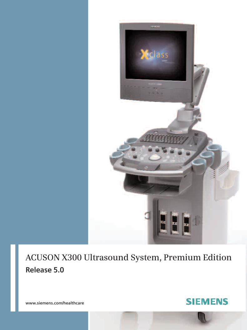
ACUSON X300 Ultrasound System, Premium EditionRelease 5.0
www.siemens.com/healthcare

2
• Streamline connectivity with solutions such as
DICOM Print/Store, DICOM Worklist, DICOM
MPPS and DICOM structured reporting for
OB/GYN, Vascular and Cardiac exams.
• Increase functionality with fourSight™ 4D
transducer technology, Advanced fourSight™
technology, integrated stress echo, fourSight™
TEE View, syngo® Mitral Valve Assessment
(MVA), Axius™ edge assisted Ejection Fraction,
syngo® Auto Left Heart (Auto LH), syngo®
Velocity Vector Imaging™ (VVI) technology,
syngo® Arterial Health Package (AHP), syngo®
Auto OB measurements and ACUSON AcuNav™
ultrasound catheters.
User Interface
• Intuitive Windows-based operating principles
• User-centric control panel with home-base
layout and control customization
• On/Off task light and back-lit illumination of
control panel
• Variable brightness indicates active state of
function keys
• Customizable Soft Key selections are displayed
on-screen to provide easy and immediate
access to imaging controls and activation of
specific functions
ACUSON X300 Ultrasound SystemPremium Edition
GENERAL INFORMATION
The ACUSON X300™ ultrasound system, premium
edition (PE) is a portable, compact, shared serv-
ice imaging solution. The ACUSON X300 PE
delivers exceptional clinical performance across a
wide variety of applications and streamlines exam
workflowwith an easy-to-use ErgoDynamic™
imaging system design.
The ACUSON X300 PE combines best-in-class
image quality and a robust set of features to
meet daily clinical needs. It enhances diagnostic
confidence with high quality 2D, 3D and 4D
imaging, color and power Doppler, and steerable
continuous wave and pulsed wave Doppler
capabilities. It also provides a pathway to
seamlessly integrate future technology
advancements.
SYSTEM ARCHITECTURE
All-digital signal processing and multibeam formation
technology provides best-in-class image quality in 2D
and 3D imaging, Native™ tissue harmonic imaging,
DTO™ Dynamic Tissue Optimization technology and
Doppler modes and Dynamic TCE™ tissue contrast
enhancement technology for greater diagnostic con-
fidence. The DIMAQ-IP integrated workstation pro-
vides digital acquisition, storage, review and transfer
of ultrasound studies. Studies can be reviewed and
quantified on-board, stored on the system hard drive
and transferred to the built-in DVD multi-drive (DVD-
R/RW & CD-R/RW) or USB Flash drive for cost-effective
archival.
The all-digital system architecture enables seamless
integration of optional features to:
• Enhance productivity through TGO™ tissue
grayscale optimization technology, SieScape™
panoramic imaging, and Clarify™ vascular
enhancement (VE) technology.

3
• Monitor tilt of 10 degrees up, 65 degrees down
and swivel of -80 to +80 degrees
• Digital on-screen display of brightness and
contrast controls
• Energy saving display power management
• 4 levels of illumination intensity: Off, 1, 2, 3
Audio Speakers
• High performance audio speakers are integrated
in the monitor
Physiological Interface
• Standard 3-lead ECG interface
• Auxillary Input for ECG and other physiology
signals from 3rd party devices
• Continuous display in all real-time modes
• R-Wave single and dual trigger function
• Respiratory trace
• Heart rate display
• Adjustable gain and trace position on screen
• Recovery Time: Less than 2 seconds
Hard Drive
• Internal 160 GB hard drive
• Allows storage of patient studies that include
images, reports and measurements
• Image storage capacity up to 150,000 images
with compression: color or B/W DVD Multi-Drive
(DVD-R/RW & CD-R/RW)
• Removable 650 MB, 700 MB and 790 MB CD-R
and 650 MB or 700 MB CD-RW
• Removable 4.7 GB single layer DVD and 8.5 GB
single side double layer DVD
• Allows storage and archiving of complete
patient studies including images, dynamic clips,
reports and measurements
• Storage capacity dependent upon patient
study size
• Export of images in TIFF and clips in AVI file
or DICOM format
• DICOM viewer for export of DICOM format to
CD/DVD (Vista compatible)
• Easily accessible, full size, QWERTY keyboard
for text entry, function keys and system
programming
• Thumbnail menu provides on-screen thumbnail
images and dynamic clips during exams
• Wrist support to help reduce operator repetitive
stress injuries
• Height adjustment of control panel – 100 mm
up/down with lock lever
• Multi-directional articulating monitor arm to
help improve ergonomics
• Arm rotation: -90 to +90 degrees
• FPD rotation: -80 to +80 degrees
• Tilt: -10 to +65 degrees
• Up: 125 mm
• Pull: 250 mm
• Wheel-lock mechanism
• Front castor (2 ea): Bi-brake system (direction
lock and total lock)
• Rear castor (2 ea): total lock
• Up to 32 QuickSet™ user-programmable system
parameters allows users to customize system
for individual transducer/application settings.
QuickSet parameters combine all preferred
imaging mode parameters, annotation, text and
measurements into one user preset
Language Support
• On-screen text, control panel overlay and
operating instructions are all available in
English, French, German, Spanish, Italian,
and Chinese
Monitor
• Flat Panel Display (FPD), 15-inch color, high
resolution, and progressive scan (non-interlaced)
with In Plane Switching (IPS) technology
• Resolution: 1024 x 768 pixels
• Total screen area: 1024 x 768 pixels
• Recordable image area clips: 800 x 600 pixels
• Total screen capture: 1024 x 768 pixels

- Auxiliary pencil transducer
- Steerable CW on selected phased array
transducers
• ECG trace in all modes
• Duplex mode
• Triplex mode
• Flexible combination of imaging modes in
side-by-side Dual and Dual Select in real-time,
and digital cine replay
• Selectable split screen display formats in 2D or
2D/color with M-mode and/or spectral Doppler
mode: top-bottom or side-by-side in real-time
and digital cine replay
• 4B mode allowing simultaneous display of 4
static B-mode images
• Virtual Format
• Dual from freeze
• Split/Zoom
Tissue Harmonic Imaging
Selectable harmonic frequencies increases success
with difficult-to-image patients, improving diagnos-
tic confidence, and dramatically improving contrast
and spatial resolution by reducing noise and clutter
in the image.
• MultiHertz™ multiple frequency imaging
capability in THI
• Available on the CH5-2, C6-2, C7F2, C8-5, P8-4,
P4-2, P5-1, EC9-4, EV9-4, EV9F4, P9-4, VF8-3,
VF10-5, VF13-5SP, VF13-5 and V5M
transducers
MultiHertz Multiple Frequency Imaging
Siemens’ unique MultiHertz multiple frequency
imaging is designed to combine the resolution
and penetration of several transducers in one. At
the push of a button, the user can independently
change frequencies for 2D, THI, color and spec-
tral Doppler to select the optimal combination
for image resolution, penetration and sensitivity.
• Depending on the transducer, up to seven
user selectable transmit frequencies are
available
Transducer Ports
• Three active universal transducer ports that
support high density phased array, curved
array and linear array transducers
• Electronic transducer selection (instantaneous
switching between transducers)
• Left-most transducer port supports TEE using
MP adapter
• Industrial design provides easy access to the
transducer ports
Transducer Storage
• Six configurable transducer holders support all
transducer designs and provide gel bottle
storage
• SuppleFlex™ transducer cables and integrated
cable management for protection during
exams and transport
• Special transducer holder provides secure
storage and easy access to endocavity transducer
• Transducer holders can be removed for cleaning
• Optional side pocket storage for one (1)
transducer connector and one (1) standard
gel bottle
Acoustic Output Management
• On-screen acoustic power indicator (AIUM/NEMA
output display standard)
OPERATING/DISPLAY MODES
• 2D imaging in fundamental and harmonic
modes (both phase inversion and filtered)
• DTI™ Doppler tissue imaging capability
• Color M-mode
• M-mode
• Anatomical M-mode live, cineloop and DIMAQ
image review
• Color Doppler velocity mode
• Power Doppler mode
• Directional power Doppler
• Pulsed Wave spectral Doppler mode (PW)
• Continuous Wave spectral Doppler mode (CW)
4

5
• 16 user-selectable 2D colorization maps
• Maximum Depth: 30 cm with CH5-2, P4-2
and P5-1
• Minimum Depth: 2 cm with VF13-5SP
2D Image Display
• Full screen, Curved Vector, Split, Quad and
Dual Select screen formats
• L/R flip and U/D flip for all formats in real-time
and digital cine replay
• Split/Zoom
• Dual image display from freeze
• Image depth from 2 cm to 30 cm in 1.0 cm
increments – transducer dependent
• Virtual Format Imaging (transducer dependent)
- Left/right steer
- Trapezoid Imaging
• Digital read/write Zoom with image pan
- Available on live and cine replay images
- At least 2.5x and up to 10x zoom – transducer
dependent
• 4B mode
2D Calipers – Generic Measurements and
Calculations
• Multiple cursor sets on frozen, live, dual screen
and cine playback images
- Up to four 2D and M-mode frequencies
- Up to five THI frequencies
- Up to two frequencies in color, power or
pulsed wave Doppler modes
- One frequency in SCW Doppler mode
Beamforming in 2D-mode
• New generation all-digital beamformer
technology enables parallel Quad beam processing
of the RF signal data in the time and amplitude
domains
• 2D-mode line density up to 512 lines
• Up to 4,416 processing channels
• Total system dynamic range > 205 dB
Focusing
• Up to 4 transmit focal zones
• Digital dynamic receive focusing with dynamic
apodization
SynAps Synthetic Aperture Technology
• SynAps™ synthetic aperature technology is
available on the CH5-2, C8-5, VF8-3 and VF10-5
transducers for higher image resolution at depth
• User can turn SynAps technology On and Off
2D-mode Image Processing
• All-digital parallel signal processing with frame
rates up to 1172 FPS – transducer dependent
• MultiHertz multiple frequency imaging with up
to seven user selectable transmit frequencies
• Six levels Res/Speed selection: 0 – 5
• Five persistence levels: 0 – 4
• Four edge enhancement levels: 0 – 3
• Display dynamic range: 30 to 70 dB in five
decibel increments
• Adjustable gain from 0 to 60 dB in one decibel
increments
• DTO technology – 3 levels
• Dynamic TCE technology - 3 levels
• Eight DGC controls for Depth/Gain Compensation
• Nine gain balanced user-selectable gray maps

• Eight sweep speed selections: 1, 2, 3, 4, 5, 6, 7, 8
• Eight selectable post processing gray maps
• 12 user-selectable Doppler colorization maps:
0 – 11
• Adjustable gain from 0 dB to 90 dB in
one decibel increments
• PRF range: 1.56 kHz to 34.7 kHz sample rate
• Velocity scale range is ±30 ~ ±650 cm/sec with 0
degree angle correction
• Eight wall filter selections – transducer
dependent
• 17 levels of baseline shift
• Spectral invert
• Autotrace function is not supported in
SCW Doppler mode
Spectral Doppler Display
• Full screen Doppler trace, 2D/Doppler mode,
triplex or update 2D/C/Doppler
• Four imaging display formats – top-bottom:
1/3-2/3, 1/2-1/2, 2/3-1/3; side-by-side: 40-60
Spectral Doppler Calipers – Generic
Measurements and Calculations
• Multiple cursor sets on frozen and cine
playback images
• Velocity/Frequency/Pressure Gradient
• Heart rate/Heart cycle/Time
• Autotrace measurements and calculations
including PS, ED, TAMx, TAMn, PI, RI and S/D
• Resistive Index (RI)
• Pulsatility Index (PI), including Peak-to-Peak
method
• Time Average Velocity max (TAV)
• Systolic/diastolic ratio (S/D)
• Velocity Time Integral (VTI)
• Acceleration/Deceleration
• Flow volume using combined velocity and
distance, or velocity and ellipse measurements
• Doppler angle correction after measurement
• Up to eight distance measurements per screen
- Distance measurement
- Depth measurement from skin line
- Angle measurement
- Area and circumference: ellipse, trace
• Compound Measurements:
- Volume: user-selectable preset by 1 distance,
2 distance, 3 distance; 1 ellipse and 1
distance
- Flow volume: 1 velocity and 1 distance, or 1
velocity and 1 ellipse
- Stenosis: user-selectable preset calculated by
2 ellipse, or 2 distance measurements
Pulsed Wave Spectral Doppler
• Available on all imaging array transducers
• Up to two user-selectable transmit frequencies
per transducer
• DTI Doppler tissue imaging available on select
transducers
• Eight sweep speed selections: 1, 2, 3, 4, 5, 6, 7, 8
• Eight selectable post-processing gray maps
• 12 user-selectable Doppler colorization maps:
0 – 11
• Adjustable gain from 0 to 90 dB in one decibel
increments
• PRF range: 100 to 19,500 Hz
• Velocity scale range is ± 1.5 ~ ± 350 cm/sec
with 0 degree angle correction
• Angle correction 0 - 89 degree in one degree
increments
• Gate size: from 1.0 mm up to 20 mm
• Eight wall filter selections – transducer dependent
• 17 levels of baseline shift
• Spectral invert
• Autotrace function
Steerable Continuous Wave (SCW) Doppler
• Available on all phased array transducers when
Advanced Cardiac-vascular option is purchased
• One transmit frequency
6
7
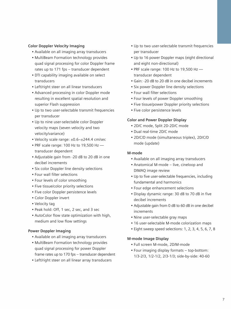
• Up to two user-selectable transmit frequencies
per transducer
• Up to 16 power Doppler maps (eight directional
and eight non-directional)
• PRF scale range: 100 Hz to 19,500 Hz —
transducer dependent
• Gain: -20 dB to 20 dB in one decibel increments
• Six power Doppler line density selections
• Four wall filter selections
• Four levels of power Doppler smoothing
• Five tissue/power Doppler priority selections
• Five color persistence levels
Color and Power Doppler Display
• 2D/C mode, Split 2D-2D/C mode
• Dual real-time 2D/C mode
• 2D/C/D mode (simultaneous triplex), 2D/C/D
mode (update)
M-mode
• Available on all imaging array transducers
• Anatomical M-mode – live, cineloop and
DIMAQ image review
• Up to five user-selectable frequencies, including
fundamental and harmonics
• Four edge enhancement selections
• Display dynamic range: 30 dB to 70 dB in five
decibel increments
• Adjustable gain from 0 dB to 60 dB in one decibel
increments
• Nine user-selectable gray maps
• 16 user-selectable M-mode colorization maps
• Eight sweep speed selections: 1, 2, 3, 4, 5, 6, 7, 8
M-mode Image Display
• Full screen M-mode, 2D/M-mode
• Four imaging display formats – top-bottom:
1/3-2/3, 1/2-1/2, 2/3-1/3; side-by-side: 40-60
Color Doppler Velocity Imaging
• Available on all imaging array transducers
• MultiBeam Formation technology provides
quad signal processing for color Doppler frame
rates up to 171 fps – transducer dependent
• DTI capability imaging available on select
transducers
• Left/right steer on all linear transducers
• Advanced processing in color Doppler mode
resulting in excellent spatial resolution and
superior Flash suppression
• Up to two user-selectable transmit frequencies
per transducer
• Up to nine user-selectable color Doppler
velocity maps (seven velocity and two
velocity/variance)
• Velocity scale range: ±0.6~±244.4 cm/sec
• PRF scale range: 100 Hz to 19,500 Hz —
transducer dependent
• Adjustable gain from -20 dB to 20 dB in one
decibel increments
• Six color Doppler line density selections
• Four wall filter selections
• Four levels of color smoothing
• Five tissue/color priority selections
• Five color Doppler persistence levels
• Color Doppler invert
• Velocity tag
• Peak hold: Off, 1 sec, 2 sec, and 3 sec
• AutoColor flow state optimization with high,
medium and low flow settings
Power Doppler Imaging
• Available on all imaging array transducers
• MultiBeam Formation technology provides
quad signal processing for power Doppler
frame rates up to 170 fps – transducer dependent
• Left/right steer on all linear array transducers

- 2D-mode colorization map
- Measurements/reports/annotations/pictograms
• Color Doppler
- Zoom/pan
- Color map
- Color invert
- Measurements/reports/annotations/pictograms
• Spectral Doppler
- Gray map
- Doppler colorization map
- Angle correct
- Measurements/reports/annotations/pictograms
• M-mode
- Gray map
- M-mode colorization map
- Measurements/reports/annotations/pictograms
TRANSDUCER TECHNOLOGY
Ultra-sensitive, wideband transducers, matched with
user-selectable MultiHertz imaging, improve
resolution and penetration. Depending on the
transducer, the user can select from up to seven 2D
and THI frequencies and up to two color Doppler and
spectral Doppler frequencies, expanding the clini-
cal versatility of a single transducer, and thereby
M-mode Calipers – Generic Measurements and
Calculations
• Multiple cursor sets on frozen and cine play
back images
- Distance
- Time
- Slope
- Heart rate
FREEZE, CINE AND CINEPOST-PROCESSING FUNCTIONS
Cine Review
Cine feature is standard and offers post-acquisition
optimization of all real-time post-processing
functions.
• Frame-by-frame cineloop review and continuous
cine motion review, including control of playback
rate
• Independent cine review in mixed modes
(2D/M, 2D/Doppler, 2D/C/Doppler)
• Independent cine review in 2D Dual Select
mode with image align playback feature
• Maximum standard cine memory is up to
2729 frames
• Acoustic clip capture from cine review
• Retrospective clip capture during real-time
imaging with a selectable duration of 1, 2, 3,
or 4 seconds or a selectable duration 1, 2, 3
or 4 beat capture; ECG triggerable
• Prospective clip capture during real-time
imaging with a selectable duration of 1 to
120 seconds a selectable duration 1 to 120
beat capture; ECG triggerable
• Anatomical M-mode – live, cineloop and
DIMAQ image review
Post Processing Features in Freeze Frame or Cine
• D-mode
- Zoom/pan
- Gray map
8

9
• Gynecology
• Early Obstetrics
• Adult Cardiac (Transthoracic)
• Pediatric Cardiac (Transthoracic)
• Neonatal Echo
• TEE Adult
• Intacardiac Echo (ICE)
• Vascular (C-Vas, P-Vas, Venous)
• Small Parts (Breast, Testicle, Thyroid)
• Orthopedics
• Musculoskeletal
• Urology (Prostate)
• Cranial (TCI)
• Emergency Medicine (EM)
• Penile
EXAM-SPECIFIC MEASUREMENTSAND REPORTS
The measurement function is arranged by exam
type and is available for use with all exam types.
The ACUSON X300 PE and has measurement and
report packages for the following exam types:
Abdomen
• All general measurements and calculations
• Physician summary utility – supports on-system
report generation including customizable
letterhead, patient data, results, graphs,
images, comments, recommendations, and
customizable signature line
Obstetrics
• All general measurements and calculations
• Early Obstetrics Menstrual Age (MA)
measurements are MSD, CRL, and Yolk Sac
• Menstrual Age parameter labels are MSD, CRL,
BPD, OFD, HC, AC, ATD, ASD, FL, HL, UL, TL, FT,
FTA, and BN
• Five user-defined labels are available in 2D-mode
• Calculations include: EFW from the selected
maximizing transducer investment.
• Wideband MultiHertz imaging allows user
selection of independent 2D and color
frequencies for optimal resolution and
penetration
• Universal, stainless steel and disposable biopsy
guides for specified linear and curved array
transducers
• Innovative ultra low-loss lens materials and
microelectronic technologies for efficient
performance and increased signal bandwidth
• Frequency range: 1.2 MHz to 13 MHz
• Hanafy Lens acoustic technology
• Single crystal piezoelectric design
• microCase™ transducer miniaturization
technology and SuppleFlex cables
Intracardiac Echocardiography Transducers
• ACUSON AcuNav 10F ultrasound catheter –
Adult intracardiac echocardiography
• ACUSON AcuNav 8F ultrasound catheter –
Adult intracardiac echocardiography
• CartoSound™ Communication- Adult
intracardiac echocardiography
Note: See dedicated transducer flyer for more information.
STUDY TYPES
The ACUSON X300 PE is designed to support
most multi-specialty imaging applications.
Factory supplied exam and transducer dependent
imaging presets have been carefully optimized
for each application to provide consistency, relia-
bility, and increased productivity. Selected appli-
cations include body markers, text and annota-
tion labels, worksheets and reports.
• Abdominal
• Renal
• Obstetrics
• Cardiac patient report and worksheet for
2D-mode, M-mode, and Spectral Doppler
• Physician summary utility – supports on-system
report generation including customizable
letterhead, patient data, results, graphs,
images, comments, recommendations, and
customizable signature line
Cerebrovascular
• All general measurements and calculations
• ICA prox, ICA mid, ICA dist, ECA and VA
measurements
• Area Percent Stenosis and Diameter Percent
Stenosis measurements
• Cerebrovascular patient report
• Physician summary utility – supports on-system
report generation including customizable
letterhead, patient data, results, graphs,
images, comments, recommendations, and
customizable signature line
Peripheral Vascular
• All general measurements and calculations
• CIA, EIA, CFA, PFA, SFA prox, SFA mid, SFA dist,
POP A, TRUNK ATA, PTA, PER A and DPA
measurements
• Right and left extremity measurements
• Peripheral vascular patient report
• Physician summary utility – supports on-system
report generation including customizable
letterhead, patient data, results, graphs,
images, comments, recommendations, and
customizable signature line
Venous
• All general measurements and calculations
• Right and left extremity measurements
• Venous patient report
• Physician summary utility – supports on-system
report generation including customizable
letterhead, patient data, results, graphs,
images, comments, recommendations, and
customizable signature line
reference, HC/AC, TCD/AC, LVW/HW, CorBPD,
FL/AC, FL/BPD, CI, AFI, AXT, and Fetal Heart
• Face Angle
• Customizable Anatomy Descriptions
• Calculations for both Gestational Age (GA),
ultrasound menstrual age, and Estimated Date
of Confinement (EDC)
• Early OB and Standard OB patient reports
include worksheets for viewing the progress of
the report and editing during the exam process
• Multiple fetus reporting capabilities
• Growth Analysis Graphs with exam file linking
• OB patient report and worksheet including
Fetal Heart report page
• Physician summary utility – supports on-system
report generation including customizable
letterhead, patient data, results, graphs,
images, comments, recommendations, and
customizable signature line
Gynecology
• All general measurements and calculations
• Micturated and residual volume calculation
• Uterus and right and left follicle ovary
measurements
• Gynecology patient report
• Physician summary utility – supports on-system
report generation including customizable
letterhead, patient data, results, graphs,
images, comments, recommendations, and
customizable signature line
Cardiac
• Adult and pediatric standard measurements
• Volume formulas for Left Ventricular function
assessment in 2D-mode and M-mode
• 2D-mode, M-mode, and Doppler calculations
• M-mode Slope, Heart Rate, Time, and Distance
measurements
• Spectral Doppler Acceleration, Trace, Heart
Rate, Time and Velocity measurements
10

11
Thyroid
• All general measurements and calculations
• Thyroid volume
• Physician summary utility – supports on-system
report generation including customizable
letterhead, patient data, results, graphs,
images, comments, recommendations, and
customizable signature line
Urology
• All general measurements and calculations
• Residual volume calculations
• Prostate and urology patient report
• Physician summary utility – supports on-system
report generation including customizable
letterhead, patient data, results, graphs,
images, comments, recommendations, and
customizable signature line
Testicle
• All general measurements and calculations
• Physician summary utility – supports on-system
report generation including customizable
letterhead, patient data, results, graphs,
images, comments, recommendations, and
customizable signature line
Musculoskeletal
• All general measurements and calculations
• Right and left hip angle measurement
• Classification and Graf Sonometer
• Hip angle patient report
• Physician summary utility – supports on-system
report generation including customizable
letterhead, patient data, results, graphs,
images, comments, recommendations, and
customizable signature line
TCI
• All general measurements and calculations
• MCA, ICA-Siphon, ACA-A1, ACA-A2, ACoA,
PCA-P1, PCA-P2, PCoA, PCA, Basilar A and Vert
A measurement
• TCI patient report
• Physician summary utility – supports on-system
report generation including customizable
letterhead, patient data, results, graphs,
images, comments, recommendations, and
customizable signature line
Penile
• All general measurements and calculations
• Corp Cav, Corp Spong, Cav A, Pre-Inj Cav A,
Post-Inj Cav A and Urethra B mode measurement
• Iliac A, Dorsal A, Urethral A, Bulbar A, Brach A,
Cav A, Pre-Inj Cav A, Post-Inj Cav A, Sup Dorsal
V and Dp Penile V D mode measurement
• Penile patient report
• Physician summary utility – supports on-system
report generation including customizable
letterhead, patient data, results, graphs,
images, comments, recommendations, and
customizable signature line
Emergency Medicine Calculations
• FAST – Focused Acute Sonography in Trauma
reporting elements to support emergency
medicine utility
• Aorta – essential aorta measurements and
reporting elements to support emergency
medicine clinical utility

• OB – subset of essential OB measurements and
reporting elements to support emergency
medicine clinical utility
• Physician summary utility – supports on-system
report generation including customizable
letterhead, patient data, results, graphs,
images, comments, recommendations, and
customizable signature line
• The ACUSON X300 PE supports customizable
labeled measurements (20 B-mode, 5 Doppler
& 5 M-mode) for the following exam types:
Abdomen, Muskuloskeletal, Breast, Thyroid,
Testicle,Venous, Renal, Pedatric Abdomen,
Neonatal Head, Superficial Muskuloskeletal,
Digital, Small Parts and Aorta.
DIGITAL PATIENT STUDY STORAGEAND ARCHIVING
The DIMAQ-IP integrated workstation allows for
digital acquisition, storage and review of com-
plete ultrasound studies, including static images
and dynamic clips, measurements, calculations
and reports.
Patient Study Management
Playback of digitally stored images in a selectable
1-up, 4-up, 9-up, 16-up or 25-up screen format.
The patient study screen allows searching, selecting
and deleting studies, and exporting studies to
DVD multi-drive (DVD-R/RW and CD-R/RW)
• 100 GB internal hard drive reserved for
patient data management
• Compatible with removable 650 MB, 700 MB
and 790 MB CD-R and 650 MB or 700 MB CD-RW
• Removable 4.7 GB single layer DVD and
8.5 GB single side double layer DVD
• Hard drive capacity:
- Approximately 150,000 B/W and color
images
• Storage and retrieval of frozen static images
• Storage and retrieval of reports
• Instant dial-in and replay of static images
in 1-up screen format
• Supports measurements and calculations on
archived study and on saved and retrieved
images
• Acoustic clip capture from cine review
• Retrospective clip capture during real-time
imaging with a selectable duration of 1, 2, 3
or 4 seconds or a selectable duration of 1, 2,
3 or 4 beat capture; ECG triggerable
• Prospective clip capture during real-time
imaging with a selectable duration of 1 to
120 seconds or a selectable duration of 1 to
120 beat capture; ECG triggerable
• Export of patient studies from hard drive to
DVD-R/RW and CD-R/RW drive. Studies can be
individually selected or batched copied
• Images are exported in PC compatible TIFF
format, AVI format, or DICOM format
• M-mode still frame scroll and store
• PW spectral Doppler still frame scroll and store
• Patient database sorting by Name, ID, and
study date
• USB flash drive
OPTIONS
PLUS (Option)
The Plus Option is a beamforming hardware option
that improves image quality, penetration and sensi-
tivity for high density transducers on the ACUSON
X300 PE. The following transducers deliver
enhanced performance with installation of the Plus
Option: C7F2, EV9F4, C6-2, CH5-2, VF8-3, VF10-5,
C8-5.
Dynamic TCE Technology (Option)
Dynamic TCE technology is an advanced post pro-
cessing method for speckle reduction and edge
enhancement. Three levels available: low, medium
and high.
12

13
worklist schedule from the Hospital/Radiology
Information System (HIS/RIS) to the ACUSON
X300 PE, automatically populating the "New
Patient" screen with patient demographic
information. (Requires DICOM 3.0 Connectivity
option.)
DICOM MPPS–Modality Performed Procedure
Step (Option)
Enables automatic exchange of Modality
Performed Procedure Step information with the
Hospital/Radiology Information System (HIS/RIS).
(Requires DICOM 3.0 Connectivity option and
DICOM Modality Worklist option.)
DICOM OB/GYN Structured Reporting (Option)
DICOM Structured Reporting (SR) provides a
standardized report architecture to allow for easy
transfer of OB and GYN measurements to offline
PCs, workstations and archiving systems. DICOM
OB/GYN Structured Reporting will automatically
populate OB/GYN measurements to their respective
fields in an external software package. (To send
the OB/GYN SR data over the network, the DICOM
3.0 connectivity option is required.)
DICOM Vascular Structured Reporting (Option)
DICOM Structured Reporting (SR) provides a
standardized report architecture to allow for easy
transfer of Vascular measurements to offline PCs,
workstations and archiving systems. DICOM
Vascular Structured Reporting will automatically
populate Vascular measurements to their respective
fields in an external software package. (To send
the Vascular SR data over the network, the DICOM
3.0 connectivity option is required.)
DICOM Cardiac Structured Reporting (Option)
DICOM Structured Reporting (SR) provides a
standardized report architecture to allow for easy
Integrated Gel Warmer (Option)
• Enables gel warming
• Temperature control precision: ±1°
- Low: 31° C
- Medium: 34° C
- High: 37° C
• Easy-to-use Power on/off control switch
• LED color for status indicator:
- Standby mode: Off
- Operating mode: Orange
• Saftey protection for electrical overload
• Weight: approximately 385g
• Size: 87 x 170 x 85 (mm)
DICOM 3.0 Connectivity (Option)
Enables digital data transfer via a DICOM network
for both printing and storage. The ACUSON X300
PE acts as a DICOM Storage Class User and DICOM
Print Class User.
Functionality supported:
• Connectivity to PACS system for storage of all
digital images and dynamic clips with patient
demographic data
• In-Progress Store during the exam
• Image printing to DICOM color and
grayscale printers
• DICOM Storage Commitment
• DICOM Exchange Media export to DVD-R/RW
and CD-R/RW
• DICOM Region Calibration
• DICOM interchange media viewer software
SHOWCASE®
• Interchange media database that identifies the
CD to which the patient study has been burned
DICOM Modality Worklist (Option)
Enables query and direct download of the patient
14
• Curved VOI
• 4D cine
• MPR Measurements
• 3-Scape™ real-time 3D imaging is supported
on the following transducers: CH5-2, C6-2,
EV9-4
Advanced fourSight technology (Option)
Advanced fourSight technology offers broad
3D/4D acquisition, data rendering and post-
processing functionality.
• MultiSlice format allows the user to select
range, slice spacing and format for viewing
each slice. The MultiSlice format supports up
to 36 slices at once.
• Thick Slice Imaging (TSI) enables definition of
a view plane and creates a thick slice around
the region of interest. TSI delivers improved
contrast resolution and provides more
information in a single image.
• Curved MPR enables real-time multiplanar
reformatting of images into any linear or
curved plane. This permits the user to set
points along a curved object in order to bring
all objects along this line into the same plane
for viewing, such as the fetal spine.
Advanced Cardiac-vascular (Option)
Contains the prerequisites to perform cardiac and
certain vascular exams:
• Physiological interface
- Standard 3-lead ECG interface
- Aux ECG interface
- R-Wave single and dual trigger function
- Heart rate display
- Adjustable gain and trace position on screen
- Selection for external ECG input
- Respiratory function
• Steerable Continuous Wave Doppler module
• Auxiliary Continuous Wave Doppler module
transfer of Cardiac measurements to offline PCs,
workstations and archiving systems. DICOM
Cardiac Structured Reporting will automatically
populate Cardiac measurements to their respective
fields in an external software package. (To send
the Cardiac SR data over the network, the DICOM
3.0 connectivity option is required.)
QuikStart (Option)
QuikStart reduces time required for power-up and
power-down events by allowing the system to
enter a specialized suspend mode.
• Standby mode power-down in 3 sec; complete
boot-up in less than 12 sec
• Standby time: more than 40 min
• Full recharge in less than 180 min
Tissue Grayscale Optimization (TGO) (Option)
TGO tissue grayscale optimization technology
provides one-button image optimization. It
automatically adjusts image brightness to the
tissue type being imaged and equalizes the overall
image gain. The user-definable threshold accom-
modates different user preferences for gain
settings and various room lighting conditions. TGO
technology improves the consistency and quality
of the ultrasound images to enhance productivity
by removing time-consuming and operator
dependent manual adjustments. TGO technology
can be used with every transducer, for every exam
type and at every imaging frequency, including
THI.
fourSight 4D transducer technology (Option)
fourSight 4D technology provides 2D and 3D
imaging utilizing mechanical acquisition, as well as
real-time 4D imaging, up to 30 volumes per
second, with the C7F2 and the EV9F4 4D transducers.
The fourSight 4D imaging option offers an easy-
to-use interface for rapid acquisition and volume
rendering and includes the following:

15
and vessel walls according to user preference.
Clarify VE technology is available on all curved
and linear transducers.
SieScape Imaging (Option)
B/W panoramic imaging allows acquisition and
display of images up to 60 cm in length to a
maximum curvature of 360 degrees.
• Available on all curved and linear transducers
• Can be displayed in full length or zoomed for
detailed viewing
• Measurements available
Advanced Physio Module (Option)
The Physio Module provides the ability to configure
ECG capabilities for specialty applications that do
not require continuous wave Doppler capabilities.
Aux ECG and Respiratory function are supported
in addition to conventional Physio Module.
OPTIONAL APPLICATIONS
CartoSound Communication (Option)
This license enables Ethernet communication
between the ACUSON X300 ultrasound system,
premium edition and the Biosense Webster
CARTOSOUND™ system.
syngo Auto OB Measurements (Option)
syngo Auto OB measurements is an innovative
algorithm which provides the ability to auto-meas-
ure major fetal structures required for biometric
measurements: BPD, HC, OFD, AC, FL and HL. This
technology reduces user variance when performing
these measurements. The measurement results are
saved to the report.
Stress Echo Package (Option)
The stress echo package provides tools for ECG
triggered acquisition, display, selection, compari-
son, evaluation and archiving of multiple cardiac
loops during various stages of a stress echo
examination.
Spectral and Color Doppler Tissue Imaging
(DTI) (Option)
• Provides color DTI and spectral DTI exams
with quantification package
• Spectral DTI with quantification package
• Enables assessment of LV diastolic function
on the ACUSON X300 PE
- Spectral Doppler DTI capability utilizes
real-time Doppler shift information from
moving tissue to better visualize and
quantify myocardial diastolic function
- The spectral Doppler DTI calculation pack-
age provides guided velocity and acceleration
measurements and includes a measurement
report package
- Color DTI capability can be used for
qualitative evaluation of wall motion and
displays the relative change of velocities
- Color DTI M-mode
- DTE(Doppler Tissue Engery)
SieClear multi-view spatial compounding
(Option)
The SieClear compounding option increases con-
trast resolution and improves tissue differentia-
tion of low contrast lesions by reducing image
speckle. Tissue boundaries and interfaces appear
sharper and more continuous. SieClear com-
pounding is accessible in THI and is compatible
with other advanced imaging options including
SieScape panoramic imaging, TGO ultrasound
technology and Clarify VE technology.
Clarify vascular enhancement (VE) technology
(Option)
Clarify VE technology is a real-time, adaptive,
pixel-by-pixel analysis implemented through a
simple, time-saving user interface that provides
multiple levels of clarification to optimize tissue
contrast resolution and definition of both tissue

• D’Art is an intuituve navigation tool with
adjustable indicator for line of sight direction
• Utilizes two cut planes positioned
perpendicular to the selected line of site
• Dynamic MultiPlanar reconstruction
• Viewing presentation enhanced by zoom,
pan and rotate functions
• Easy to use, rapid acquisition, enhanced
workflow
syngo Mitral Valve Assessment (MVA) (Option)
An integrated method which, in conjunction with
syngo fourSight TEE view, enables a detailed
assessment of mitral valve morphology from 3D
images for a more accurate and complete diagnosis
of pathology location and size. Performs measure-
ments for:
• Angle
• Size
• Distance
• Area
syngo Arterial Health Package
syngo® Arterial Health Package software (AHP)
provides the clinician with the capability to measure
Intima Media Thickness and the option to reference
normative tables that have been validated and
published in peer-reviewed studies. The information
is intended to provide a straightforward tool for
communicating with patients the relative state of their
cardiovascular system. This feature should be utilized
according to the ASE Consensus Statement, “Use of
Carotid Ultrasound to Identify Subclinical Vascular
Disease and Evaluate Cardiovascular Disease Risk: A
Consensus Statement from the American Association
of Echocardiography; Carotid Intima-Media Thickness
Task Force, Endorsed by the Society for Vascular
Medicine”.
• Standard acquisition protocols for treadmill,
ergometric, and pharmacological stress
including:
- Multiple factory default stress echo protocols
- Customizable stress echo protocols
- Flexible combination of imaging modes
while in the stress echo package
- Ability for customized studies through
Protocol Editor, with up to 12 stages, 6
views per stage, 20 loops per view or 120
second prospective clip capture
• Full screen or ROI (region of interest) acquisition
• Complete R-R capture with clip editing
• Easy workflow throughout the exam protocol
• Stage Timer
• Prospective continuous capture (up to 120
seconds) or Retrospective labeled capture
• Reference image display during acquisition
• Immediate review of acquired loops
• Flexibility to skip views or stages
• Flexibility to re-acquire and overwrite
already acquired images
• Indication of current view, acquired views
and skipped views in the workflow diagram
• Wall Motion Scoring, 16/17segment model
with graphical display and report printing
• LV Volume Measurements with report
printing
syngo fourSight TEE View (Option)
An integrated method which provides acquisition,
review, manipulation and dynamic display
capabilities of gated 3D datasets using the V5M
transesophageal transducer.
• 3D surface (volume) rendering for detailed
anatomical display
16
1ARIC - G Howard, et al: "Carotid artery intimal-media thickness distribution in general populations as
evaluated by B-mode ultrasound, ARIC Investigators": Stroke 1993;24;1297-1304
17
- Parametric color M-mode display of a selected
component of Tissue Velocity along the
dynamic tissue border trace over time
- Parametric color M-mode display of Tissue
Strain along the dynamic tissue border trace
over time
- Up to 20 different points can be selected on
the 2D image along the trace for graphic
displays of Velocity, Strain and Strain Rate.
Combined display or full-screen magnification
• 3D representations of parametric color M-mode
display along the trace over time for:
- Selected components of Tissue Velocity
- Tissue Strain
- Tissue Strain Rate
- Pan, Zoom, Rotate of the 3D parametric display
• Time curves of global and segmental
(6-segment model) LV volumes automatically
calculated by Simpson method
• Parametric color display of automatically
calculated global and segmental Ejection
Fraction
• Time curves and measurements of Dmin
(transverse diameter) and Dmax (longitudinal
diameter)
• Simultaneous display of volume time curves
and measurements for current and previous
cases
• Synchrony analysis
- 6-segment chamber model
- Time curve display and measurements for
segmental tissue motion parameters: Velocity
(tangential or radial), Strain (tangential or
radial), Strain Rate (tangential or radial) and
Displacement (tangential or radial)
- Automatic time-to-peak and phase analysis of
all motion parameter curves
- Parametric color 6-segment model display of
Time-to-Peak and phase information for
Velocity (tangential or radial), Strain
(tangential or radial), Strain Rate (tangential
or radial), Displacement (tangential or
radial) and Rotation
syngo Velocity Vector Imaging (VVI) Technology
(Option)
A dynamic 2D method to visualize, measure, and
display global and regional myocardial motion and
mechanics. Available both on and off the system.
• Uses grayscale images and a sophisticated
tracking algorithm to determine the velocity
and direction of myocardical tissue motion and
displays it in a dynamic vector presentation
overlapping the 2D clip
• Base of vectors tract tissue motion
• Length of vector indicates how fast the tissue
at the base of the vector is moving
• Direction of vectors point in the direction of
tissue motion
• Algorithm allows for processing ultrasound
clips obtained in all views of the heart, as well
as for generic moving tissue (e.g., vessel wall)
• Not limited by color Doppler dependencies of
frame rate, angle or mean velocities
• Compatible with all transducers
• The tracking algorithm for syngo VVI
incorporates multiple sources of information,
including speckle tracking:
- Manual tracing of the myocardial border on
any single frame of a clip
- Mitral plane motion tracking
- Tracking of the inward and outward motion
of the tissue border
- Tissue motion along the border trace using
sophisticated speckle tracking
- Periodic motion of the heart
- Spatial coherence of tissue motion
• Parameters and parametric displays supported
by syngo VVI include:
- Visual assessment of wall tracking
- Visual assessment of vector dynamics
- Display of time curves of the selected velocity
vector components
- ECG display
- Individual heartbeat, or average of multiple
heart beats analysis

Axius Edge Assisted Ejection Fraction (Option)
Axius ejection fraction calculates left ventricle volumes
and ejection fractions quickly and reliably.
• Enhances workflow by allowing the user to
indicate three boundary points for measurement
instead of a manual trace of the ventricle
• Utilizes the Simpson method for LV volume
calculations based on LV endocardial border
trace
Intracardiac Echocardiography Imaging Options
(ICE)
Adult and pediatric intracardiac echocardiography
(ICE) using the ACUSON AcuNav ultrasound
catheter family.
• Disposable ACUSON AcuNav 8F ultrasound
catheter
- 8 french catheter (cross-sectional diameter
3.3 mm), 110 cm insertable length
• Disposable ACUSON AcuNav 10F ultrasound
catheter
- 10 french catheter (cross-sectional diameter
2.7 mm), 90 cm insertable length
• Ultrasound catheters must be purchased
separately
• Reuseable SwiftLink™ catheter connector (DL-
260 type) (additional SwiftLink connectors
are optional)
• 10 sterile covers
• Complete echocardiography exam capabilities
during intracardiac minimally invasive
procedures, such as atrial fibrillation ablation,
transcatheter septal closure device placement,
pacemaker lead placement, transseptal
catheterization, and balloon valvuloplasty
or septostomy
• Visualization of cardiac anatomy and regional
myocardial tissue motion, great vessels and
vascular anatomy, blood flow direction and
velocity, other devices located within the heart
• Sterile, steerable, single-use catheter
• An arbitrary, multi-segment M-line can be
selected on a 2D clip display to obtain virtual
M-mode information derived from a 2D clip.
Virtual M-mode can be used as a background
for time curves providing reference on cardiac
cycle phase
• Compatible with standard acquisition frame
rate clips, and acoustic clip capture (e.g., iso-
volumetric contraction and relaxation events)
syngo Auto Left Heart (Auto LH) Technology
(Option)
• Calculated Confidence
- Uses pattern recognition
technology based on a comprehensive
database representing typical adult
transthoracic exams
- Provides robust performance and expert-like
consistency in every exam
- Excels in technically difficult studies as
learning is largely independent of
image quality
• Automated Workflow
- Bypasses the cumbersome “mark-and-trace”
method to calculate EF, end diastole volume
and end systole volume
- Automatically identifies ED and ES frames
and tracks endocardial borders frame to frame
- Promotes smooth, efficient workflow with
manual edit options
- Reduces observer variability to yield more
consistent, standardized measurements
• Consistent, Reliable and Convenient
- Extends calculation accuracy to broaden
clinical confidence
- Presents EF measurement in complete
perspective — with image frames for ED
and ES with numerical and graphical data
- Available off the system with syngo®
US Workplace
- Operates on DICOM clips from select Siemens
and non-Siemens ultrasound systems
18

19
DOCUMENTATION DEVICES
Optional On-Board Video Devices
• Up to two (B/W printer and color printer/DVD
Recorder) documentation devices can be
integrated into the system cart and controlled
from the system control panel
• Supported devices:
- Mitsubishi USB P93DW/P95 B/W Printer
- Mitsubishi USB CP30DW Color Printer
- Sony USB UP-23MD Color Printer
- JVC DVD BD-X201MS Recorder
(NTSC/PAL switchable, 115V/230V)
- Mitsubishi S-VHS VCR (NTSC and PAL versions)
SYSTEM INPUT/OUTPUT
Video Standard
• PAL/CCIR: 625 lines, 50 Hz
• NTSC/EIA: 525 lines, 60 Hz
Video/Audio Input
• (1) Composite color Video in, BNC-type
• (1) Y/C Video in, S-terminal (SVHS)
• (1) 2-Channel Audio in (Right/Left), RCA --
jack type
Video/Audio Output
• (1) Composite B/W Video out, BNC-type
• (1) Composite color Video out, BNC-type
• (1) RGB and Composite Sync out, mini D-SUB
(15 pin)
• (1) Y/C Video out, S-terminal (SVHS)
• (1) 2-Channel Audio (Right/Left), RCA jack type
• (1) VGA out, mini D-SUB (15pin)
Other Input/Output
• (1) Foot switch connector, phone jack-type
• (1) Remote control connector, mini-jack
(stereo)
• High resolution, high frame rate imaging in
multiple modes, including: 2D, M-mode, PW
spectral Doppler, CW spectral Doppler, color
Doppler velocity and Doppler tissue imaging
(optional)
• Digital phased array technology –
not mechanical scanning
• Imaging penetration up to 15 cm allows for
visualization of left-sided cardiac anatomy from
within the right atrium or right ventricle
• Two planes of bi-directional steering of the
catheter tip (160 degrees in each direction:
anterior-posterior/left right) for maneuverability,
rapid anatomic orientation and micro-positioning
• Longitudinal side-fire imaging provides
standard echocardiographic views, similar
to TEE, for easier orientation
• QuikSet user-programmable system parameters
provide instant image optimization
• Tension control knob for holding the desired
catheter curvature
• 64-element digital phased array with
simultaneous processing for the highest
imaging resolution in all modes
• Access to advanced imaging modes provides
full echocardiography capabilities, including:
- 2D imaging
- M-mode
- Pulsed Wave (PW) spectral Doppler
- Continuous Wave (CW) spectral Doppler
- Color Doppler velocity capability
- DTI Doppler Tissue Imaging capability
(optional)
- Comprehensive measurements and calculation,
including volume, function and hemodynamics
• MultiHertz imaging
• Zoom capabilities
INTEGRATING THE HEALTHCAREENTERPRISE (IHE)
Having all relevant information at one's fingertips
is a prerequisite for optimal and efficient patient
care. Seamless integration of the hospital’s IT and
Imaging Systems and their capabilities to exchange
information without restriction are key success fac-
tors for facilitating daily work. This is why Siemens
has been instrumental in launching and advancing
the IHE (Integrating the Healthcare Enterprise)
Initiative. Our commitment and dedication enable
us to provide clinicians with the ACUSON X300 PE
one of many innovative products embedded with
the building blocks necessary in supporting clini-
cians' need for seamless health information
exchange.
For more information on the ACUSON X300 PE
and the Siemens commitment to the IHE initiative,
please visit www.siemens.com/IHE.
STANDARDS COMPLIANCE
The ACUSON X300 PE meets the requirements
of the Medical Device Directive and carries the
CE Mark.
Quality Standards
ISO 9001:
ISO 13485:2003 / EN ISO 13485:
Design Standards
UL 60601-1
CSA C22.2 No. 601-1
EN 60601-1 and IEC 60601-1
EN 60601-1-1 and IEC 60601-1-1
EN 60601-1-2 and IEC 60601-1-2
EN 60601-2-37 and IEC60601-2-37
System Interface Connections
• Network
- (1) Ethernet connector, type RJ45
(10/100 BaseT)
• Peripherals
- (2) Serial port RS-232C connector, D-SUB (9-pin)
- (2) USB 2.0 ports
- (2) AC Main Outlet
SYSTEM DIMENSIONS
• Height: 137.9 – 166 cm (54.43 – 65.3 in)
• Width: 51.8 cm (20.4 in)
• Depth: 87.9 cm (34.6 in)
• Weight: 102 kg (225 lbs)/98 kg without OEM’s
ELECTRICAL/ENVIRONMENTALSPECIFICATIONS
The ACUSON X300 PE is available in one main-
frame configuration, suitable for use in 100/115V
and 230V environments.
• Power connections: 100-120/200-240 VAC,
50/60Hz
• Built-in AC isolation transformer
• Power consumption: maximum 600VA
with OEM’s
• Atmospheric pressure range: 700 hPa to
1060 hPa (525 to 795 mm Hg) or up to
3050 m (10,000 ft)
• Ambient temperature range (without OEM’s):
10°C to +40°C (50° to 104°F)
• Humidity: 30 – 80%, non-condensing,
during operation
• Maximum heat output: 2150 BTU/hr
20

21
Acoustic Output Standards
• IEC 61157 (Declaration of Acoustic Power)
• AIUM/NEMA UD-2, 1998 Acoustic
OutputMeasurement Standard for Diagnostic
Ultrasound
• AIUM/NEMA UD-3, 1998 Standard for Real-
TimeDisplay of Thermal and Mechanical Acoustic
Output Indices on Diagnostic Ultrasound
Equipment
CE Declaration
The 230V/115 V version of
the ACUSON X300 ultra-
sound system, PE is provid-
ed with a CE marking in accordance with the regu-
lations stated in Council Directive 93/42/EEC of
June 14, 1993 concerning Medical Devices.
Siemens Medical Solutions USA, Inc., is certified by
notified body 0123 to Annex 11.3 – Full Quality
System.
Authorized EC Representative:
Siemens AG
Healthcare Sector
Henkestrasse 127
91052 Erlangen
Germany
phone: +49 9131 84 0
Siemens reserves the right to modify the design
and specifications contained herein without pri-
ornotice.
Please contact your local Siemens Healthcare Sales
Representative for the most current information.

3-Scape, ACUSON, AcuNav, Axius, Clarify,
DTI, DTO, Dynamic TCE, ErgoDynamic,
fourSight, microCase, MultiHertz, Native,
QuickSet, SieClear, SieScape, SuppleFlex,
SwiftLink, SynAps, TGO, Velocity Vector
Imaging and X300 are trademarks of
Siemens Medical Solutions USA, Inc., and
syngo is a trademark of Siemens AG.
CartoSound is a trademark of BioSense
Webster, Inc.
SHOWCASE is a registered trademark of
Trillium Technology, Inc.
Microsoft and Windows are registered
trademarks of the Microsoft Corporation
in the United States and other countries.
WS 0309 I © 03.2009, Siemens Medical Solutions USA, Inc.
Global Siemens HeadquartersSiemens AGWittelsbacherplatz 280333 MunichGermany
Legal ManufacturerSiemens Medical Solutions USA, Inc.Ultrasound1230 Shorebird WayMountain View, CA 94043 USATelephone: +1-888-826-9702www.siemens.com/healthcare
Local Contact InformationSiemens Medical Solutions USA, Inc.51 Valley Stream ParkwayMalvern, PA 19355-1406 USATelephone: +1-888-826-9702www.usa.siemens.com/healthcare
Europe: + 49 9131 84-0Asia Pacific: + 65 6490 6000
Global Business Unit AddressSiemens Medical Solutions USA, Inc.Ultrasound1230 Shorebird WayMountain View, CA 94043 USATelephone: +1-888-826-9702www.siemens.com/healthcare
Global SiemensHealthcare HeadquartersGlobal Siemens AGHealthcare SectorHenkestrasse 12791052 ErlangenGermanyTelephone: + 49 9131 84-0www.siemens.com/healthcare