Sympathetic innervation of the spleen in male Brown Norway rats: A longitudinal aging study
25
Sympathetic Innervation of the Spleen in Male Brown Norway Rats: A Longitudinal Aging Study Sam D. Perez 1 , Dorian Silva 2 , Ashley Brooke Millar 2 , Christine A. Molinaro 2 , Jeff Carter 2 , Katie Bassett 2 , Dianne Lorton 4 , Paola Garcia 2 , Laren Tan 2 , Jonathon Gross 2 , Cheri Lubahn 4 , Srinivasan ThyagaRajan 2 , and Denise L. Bellinger 2 1 Department of Physiology and Pharmacology, Loma Linda University School of Medicine, 11021 Campus Street, Loma Linda, California 92350 2 Department of Pathology and Human Anatomy, Loma Linda University School of Medicine, 11021 Campus Street, Loma Linda, California 92350 4 Hoover Arthritis Research Center, Sun Health Research Institute, Sun City, AZ 85351 Abstract Aging leads to reduced cellular immunity with consequent increased rates of infectious disease, cancer and autoimmunity in the elderly. The sympathetic nervous system (SNS) modulates innate and adaptive immunity via innervation of lymphoid organs. In aged Fischer 344 (F344) rats, noradrenergic (NA) nerve density in secondary lymphoid organs declines, which may contribute to immunosenescence with aging. These studies suggest there is SNS involvement in age-induced immune dysregulation. Objectives—The purpose of this study was to longitudinally characterize age-related change in sympathetic innervation of the spleen and sympathetic activity/tone in male Brown Norway (BN) rats, which live longer and have a strikingly different immune profile than the F344 rat, the traditional animal model for aging research. Methods—Splenic sympathetic neurotransmission was evaluated between 8 and 32 months of age by (1) NA nerve fiber density; (2) splenic norepinephrine (NE) concentration; and (3) circulating catecholamines levels after decapitation. Results—We report a decline in noradrenergic nerve density in splenic white pulp (45%) at 18 months of age compared with 8 month-old (M) rats, which is followed by a much slower rate of decline between 18 and 32 months. Lower splenic NE concentrations were consistent with morphometric findings. Circulating catecholamines levels generally dropped with increasing age. Conclusion—These findings suggest there is a sympathetic-to-immune system dysregulation beginning at middle age. Given the unique T-helper-2 bias in Brown Norway rats, altered sympathetic-immune communication may be important for understanding the age-related rise in asthma and autoimmunity. © 2009 Elsevier B.V. All rights reserved. Corresponding Author: Denise L. Bellinger, Ph.D., Departments of Pathology and Human Anatomy, Loma Linda University School of Medicine, 11021 Campus Street, AH 325, Loma Linda, CA 92350, Tel.: 909-558-7069, Fax: 909-558-0432, [email protected]. Publisher's Disclaimer: This is a PDF file of an unedited manuscript that has been accepted for publication. As a service to our customers we are providing this early version of the manuscript. The manuscript will undergo copyediting, typesetting, and review of the resulting proof before it is published in its final citable form. Please note that during the production process errors may be discovered which could affect the content, and all legal disclaimers that apply to the journal pertain. NIH Public Access Author Manuscript Brain Res. Author manuscript; available in PMC 2013 April 10. Published in final edited form as: Brain Res. 2009 December 11; 1302: 106–117. doi:10.1016/j.brainres.2009.09.012. NIH-PA Author Manuscript NIH-PA Author Manuscript NIH-PA Author Manuscript
-
Upload
independent -
Category
Documents
-
view
0 -
download
0
Transcript of Sympathetic innervation of the spleen in male Brown Norway rats: A longitudinal aging study
Sympathetic Innervation of the Spleen in Male Brown NorwayRats: A Longitudinal Aging Study
Sam D. Perez1, Dorian Silva2, Ashley Brooke Millar2, Christine A. Molinaro2, Jeff Carter2,Katie Bassett2, Dianne Lorton4, Paola Garcia2, Laren Tan2, Jonathon Gross2, CheriLubahn4, Srinivasan ThyagaRajan2, and Denise L. Bellinger2
1Department of Physiology and Pharmacology, Loma Linda University School of Medicine, 11021Campus Street, Loma Linda, California 923502Department of Pathology and Human Anatomy, Loma Linda University School of Medicine,11021 Campus Street, Loma Linda, California 923504Hoover Arthritis Research Center, Sun Health Research Institute, Sun City, AZ 85351
AbstractAging leads to reduced cellular immunity with consequent increased rates of infectious disease,cancer and autoimmunity in the elderly. The sympathetic nervous system (SNS) modulates innateand adaptive immunity via innervation of lymphoid organs. In aged Fischer 344 (F344) rats,noradrenergic (NA) nerve density in secondary lymphoid organs declines, which may contributeto immunosenescence with aging. These studies suggest there is SNS involvement in age-inducedimmune dysregulation.
Objectives—The purpose of this study was to longitudinally characterize age-related change insympathetic innervation of the spleen and sympathetic activity/tone in male Brown Norway (BN)rats, which live longer and have a strikingly different immune profile than the F344 rat, thetraditional animal model for aging research.
Methods—Splenic sympathetic neurotransmission was evaluated between 8 and 32 months ofage by (1) NA nerve fiber density; (2) splenic norepinephrine (NE) concentration; and (3)circulating catecholamines levels after decapitation.
Results—We report a decline in noradrenergic nerve density in splenic white pulp (45%) at 18months of age compared with 8 month-old (M) rats, which is followed by a much slower rate ofdecline between 18 and 32 months. Lower splenic NE concentrations were consistent withmorphometric findings. Circulating catecholamines levels generally dropped with increasing age.
Conclusion—These findings suggest there is a sympathetic-to-immune system dysregulationbeginning at middle age. Given the unique T-helper-2 bias in Brown Norway rats, alteredsympathetic-immune communication may be important for understanding the age-related rise inasthma and autoimmunity.
© 2009 Elsevier B.V. All rights reserved.
Corresponding Author: Denise L. Bellinger, Ph.D., Departments of Pathology and Human Anatomy, Loma Linda University Schoolof Medicine, 11021 Campus Street, AH 325, Loma Linda, CA 92350, Tel.: 909-558-7069, Fax: 909-558-0432, [email protected].
Publisher's Disclaimer: This is a PDF file of an unedited manuscript that has been accepted for publication. As a service to ourcustomers we are providing this early version of the manuscript. The manuscript will undergo copyediting, typesetting, and review ofthe resulting proof before it is published in its final citable form. Please note that during the production process errors may bediscovered which could affect the content, and all legal disclaimers that apply to the journal pertain.
NIH Public AccessAuthor ManuscriptBrain Res. Author manuscript; available in PMC 2013 April 10.
Published in final edited form as:Brain Res. 2009 December 11; 1302: 106–117. doi:10.1016/j.brainres.2009.09.012.
NIH
-PA Author Manuscript
NIH
-PA Author Manuscript
NIH
-PA Author Manuscript
Keywordsnoradrenergic nerves; secondary lymphoid organ; stress-induced plasma catecholamines;fluorescence histochemistry; cardiovascular measures; aged
1. INTRODUCTIONThe central nervous system (CNS) regulates immune function, at least in part, vianoradrenergic (NA) sympathetic nerves that innervate primary and secondary immuneorgans (reviewed in Bellinger et al., 2008b). Norepinephrine (NE) released by sympatheticnerves interacts with adrenergic receptors (AR) expressed on the surface of cells of theimmune system (reviewed in Kin and Sanders, 2006; Bellinger et al., 2008b). Thesympathetic nervous system (SNS) modulates many aspects of immune function. Based onin vitro and in vivo studies in young adult rodents, the roles ascribed to the SNS include: (1)limiting the magnitude of both acute and chronic inflammatory responses by shifting thecytokine balance from a pro-inflammatory towards an anti-inflammatory cytokine profile(Vizi and Elenkov, 2002); (2) promoting T-helper-2 (Th2)-driven antibody responses via β2-AR signaling of B cells by up-regulating B cell accessory molecule expression andincreasing B cell responsiveness to interleukin (IL)-4 (reviewed in Kin and Sanders, 2006);(3) enhancing cell-mediated responses, such as a delayed type hypersensitivity (DTH)reaction to a contact sensitizing agent through direct interaction with both T cells andantigen-presenting cells (Madden et al., 1989; Li et al., 2004); and (4) influencing Th1-driven antibody responses through β2-AR stimulation of Th1 cells (Madden et al., 1995;Kruszewska et al., 1995; reviewed in Kin and Sanders, 2006; Bellinger et al., 2005). Ingeneral, agents that activate the SNS tend to reduce T cell responses (Madden et al., 1989;Sanders et al., 1997; reviewed in Kin and Sanders, 2006), anti-viral immune reactivity(Dobbs et al., 1993), and natural killer (NK) cell activity (Irwin et al., 1988). Collectively,studies performed in young adult rodents demonstrate that genetic background, gender, siteof immunization, type of immune cells involved in the immune response, and timing ofexposure to catecholamines during the immune response all contribute to the complexity ofSNS interactions with the immune system and affect the outcome of SNS modulation of theimmune response (Madden et al., 1995; Kin and Sanders, 2006; Bellinger et al., 2008b).Furthermore, these complexities likely reflect an important role of the SNS in fine-tuning animmune response with the goal of effectively eliminating the threat to the host and restoringimmune system homeostasis.
NA innervation of, and NE content in, secondary lymphoid organs, such as the spleen, canbe affected by physiological changes (i.e., immune challenge or immunosuppression) (Yanget al., 1998; Lorton et al., 2008), immunodeficiency virus infection (Kelley et al., 2003;Sloan et al., 2008), psychosocial stress (Sloan et al., 2007), hypertension (Purcell andGattone, 1992), pregnancy and parturition (Bellinger et al., 2001) and with advancing age(Bellinger et al., 1987, 2001, 2002). Age-related changes in sympathetic innervation of thespleen are species- and/or strain-specific. For example, sympathetic NA innervation ofspleens from aged C57Bl/6J and BALB/cJ mice is preserved (Madden et al., 1997; Bellingeret al., 2001), but declines with advancing age in male C3H, MRL-lpr/lpr (Breneman et al.,1993) and New Zealand mice (NZB, NZW, and NZBW) (Bellinger et al., 1989). In murinestrains that develop autoimmune diseases (MRL lpr/lpr, NZB and NZBW) the onset ofsympathetic nerve loss in the spleen occurs with, or slightly precedes the onset of theautoimmune disease (Breneman et al., 1993; Bellinger et al., 1989).
In a previous study from our laboratory (Bellinger et al., 2002) we compared sympatheticinnervation of the spleen in four strains of young (3-month-old (M)) and old (21M) rats.
Perez et al. Page 2
Brain Res. Author manuscript; available in PMC 2013 April 10.
NIH
-PA Author Manuscript
NIH
-PA Author Manuscript
NIH
-PA Author Manuscript

These strains of rats were chosen for study because they are commonly used as models forhuman aging and/or used to study neural-immune interactions. We reported that NAinnervation of spleens from male Fischer 344 (F344) and Lewis rats decline in normal aging,whereas NA innervation was preserved in age-matched Brown Norway (BN) and BN XF344 (F1; BNF1) rats. The reason for this strain difference is unclear, but maybe a result ofthe BN and BNF1 rats having a longer life span (median age 32M) than F344 and Lewis rats(median age 24M) (Nadon, 2004). Unlike F344 rats, the most commonly used rat model foraging, in BN rats there is low morbidity from pituitary adenomas or glomerulonephrosiswith increasing age (Lipman et al., 1996, 1999; Nadon, 2004). BN and F344 rats also differin their behavior (Rex et al., 1996; Ramos et al., 1997), learning and memory (van der Staayet al., 1996), immune function (Sado et al., 1986; Koch 1976; Stankus and Leslie 1976;Festing, 1998; Lipman, 1996, 1999), and stress responses (Segar et al., 2008; Duclos et al.,2005; Sarrieau et al., 1998; Gómez et al., 1998), which may affect NA nerve integrity in thespleen with advancing age.
The purpose of the present study was to longitudinally examine the effect of age onsympathetic NA innervation of spleens from BN rats. Since age-related changes in SNS andsympatho-adrenal medullary system (SAM) reactivity may contribute to age-inducedchanges in NA nerve integrity and splenic NE content via increased local catecholamineconcentrations influencing uptake into the nerve terminals from the circulation, localsympathetic nerve activity, and/or affecting leukocyte migration (Bellinger et al., 2008b;Elenkov et al,. 2000; Madden et al., 1995), SNS and SAM reactivity to decapitation stresswas also assessed by measuring circulating catecholamine levels (NE and epinephrine (EPI),respectively). We report here an age-related decline in NA nerve density in the splenic whitepulp, evident by morphometric analysis at 18 months of age, and reduced splenic NEconcentration between 18 and 32 months of age. Decapitation stress-induced plasmacatecholamine levels were diminished in middle aged and aged rats. Given the welldocumented role of the SNS in immune modulation, altered sympathetic innervation likelyaffects immune competence in aging BN rats.
RESULTS3.1. Fluorescence Histochemistry for Catecholamines in the Spleen
In young adult rats (8M), dense plexuses of NA fibers entered the spleen with the splenicartery and its branches. The greatest density of NA nerves was found in the white pulpassociated with the central arteriole and its branches (Fig. 1A). Fluorescent NA nerve fibersextend from these vascular plexuses into the periarteriolar lymphatic sheath (PALS) amonglymphocytes and macrophages, as either linear or punctate profiles. NA nerve fibers alsocourse as less dense plexuses along the venous sinuses and trabeculae in the red pulp, but arerarely found in splenic follicles, where B cells predominantly reside.
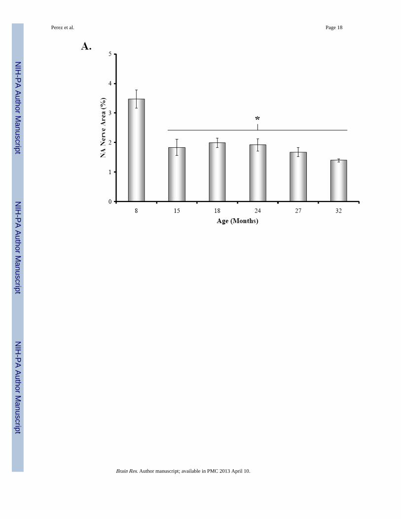
At 15 months of age, a decline in NA nerve density was observed in all compartments of thespleen, but most strikingly in the white pulp along the central arteriole (Fig. 1B). Between18 and 32 months of age, the density of NA plexuses in the splenic white pulp, along thecentral arteriole (Fig. 1C-1F) and other regions of the spleen appeared comparable to thatseen at 15 months of age. Morphometric analysis of fluorescent nerve profiles in the splenicwhite pulp across age (Fig. 2A) confirmed our qualitative assessment of sympatheticinnervation (F(5,30) = 15.14, p < 0.0001). There was a significant reduction in the meanpercentage of NA nerve area in spleens from 15M to 32M rats compared with 8M rats (p <0.001). A scatter plot showing the percent area of NA nerve fibers in splenic white pulpsacross age is shown in Fig. 2B. Linear regression analysis revealed a significant negativecorrelation (p < 0.0001) between sympathetic nerve area and increasing age.
Perez et al. Page 3
Brain Res. Author manuscript; available in PMC 2013 April 10.
NIH
-PA Author Manuscript
NIH
-PA Author Manuscript
NIH
-PA Author Manuscript
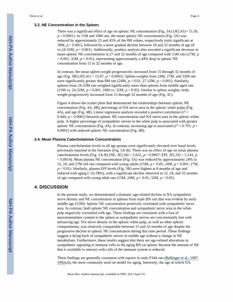
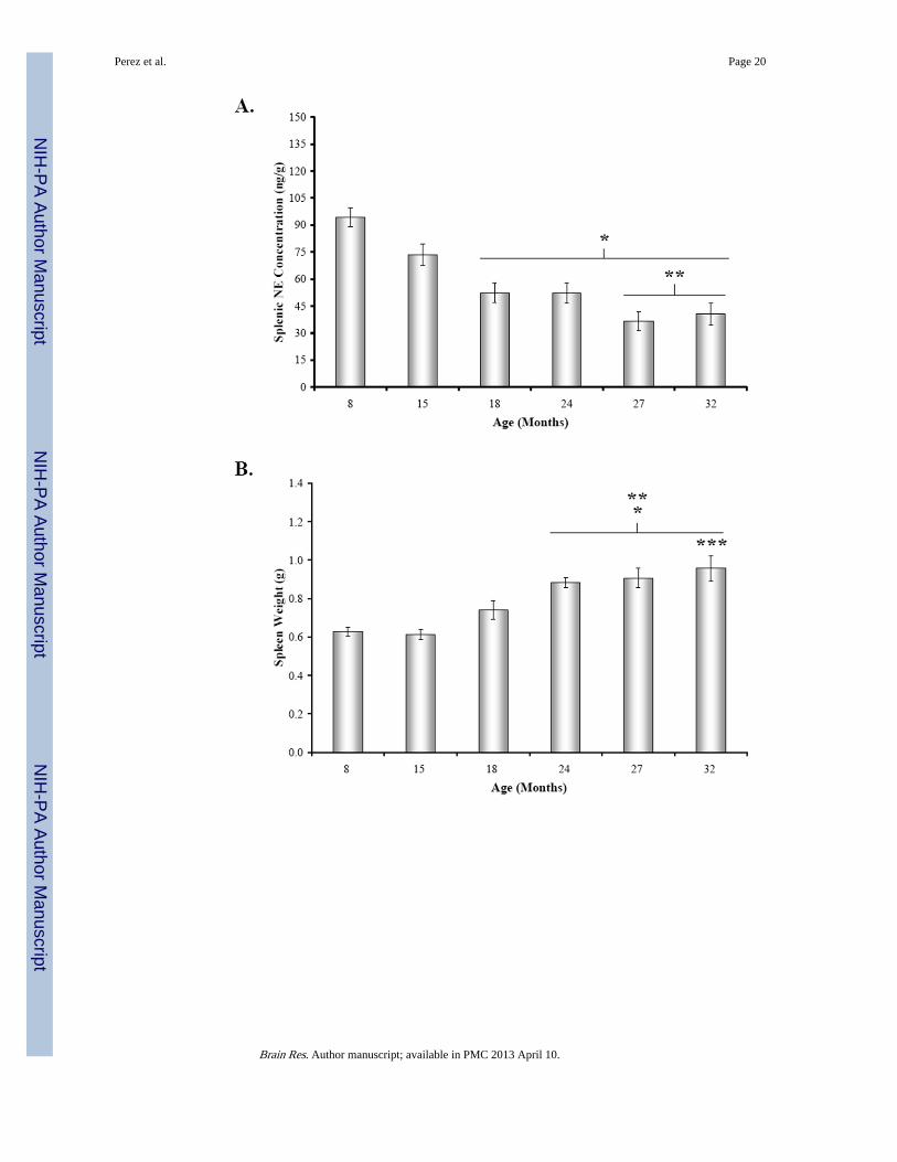
3.2. NE Concentration in the SpleenThere was a significant effect of age on splenic NE concentration (Fig. 3A) (F(5,41)= 15.39,p < 0.0001). In 15M and 18M rats, the mean splenic NE concentration (Fig. 3A) wasreduced by approximately 25 and 45% of the 8M values, respectively (only significant at18M, p < 0.001), followed by a more gradual decline between 18 and 32 months of age (8vs.24-32M, p < 0.001). Additionally, posthoc analysis also revealed a significant decrease inmean splenic NE concentration at 27 and 32 months of age compared with 15M rats (27M, p< 0.001; 32M, p < 0.01), representing approximately a 44% drop in splenic NEconcentration from 15 to 32 months of age.
In contrast, the mean spleen weight progressively increased from 15 through 32 months ofage (Fig. 3B) (F(5,41) = 12.67, p < 0.0001). Spleen weights from 24M, 27M, and 32M ratswere significantly greater than 8M rats (24M, p < 0.01; 27-32M, p < 0.001). Similarly,spleens from 24-32M rats weighed significantly more than spleens from middle aged rats(15M vs. 24-32M, p < 0.001; 18M vs. 32M, p < 0.05). Similar to spleen weights, bodyweight progressively increased from 15 through 32 months of age (Fig. 3C).
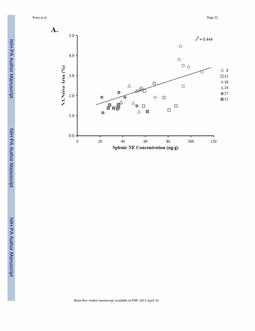
Figure 4 shows the scatter plots that demonstrate the relationships between splenic NEconcentration (Fig. 4A, 4B), percentage of NA nerve area in the splenic white pulps (Fig.4A), and age (Fig. 4B). Linear regression analysis revealed a positive correlation (r2 =0.444; p < 0.0001) between splenic NE concentration and NA nerve area in the splenic whitepulp. A higher percentage of sympathetic nerves in the white pulp is associated with greatersplenic NE concentration (Fig. 4A). In contrast, increasing age is associated (r2 = 0.791; p <0.0001) with reduced splenic NE concentration (Fig. 4B).
3.4. Mean Plasma Catecholamine ConcentrationPlasma catecholamine levels in all age groups were significantly elevated over basal levelspreviously reported in the literature (Fig. 5A-B). There was an effect of age on mean plasmacatecholamine levels (Fig. 5A-B) (NE, F(5,34) = 5.652, p < 0.0007; EPI, F(5,33) = 5.143, p< 0.0014). Mean plasma NE concentration (Fig. 5A) was reduced by approximately 24% in15, 24, and 27M old rats compared with young adults (15M, p < 0.05; 24M, p < 0.001; 27M,p < 0.01). Similarly, plasma EPI levels (Fig. 5B) were highest at 8 months of age andreduced with aging (~32-58%), with a significant decline observed at 15, 24, and 32 monthsof age compared with young adult rats (15M, 24M, p < 0.01; 32M, p < 0.05).
4. DISCUSSIONIn the present study, we demonstrated a dramatic age-related decline in NA sympatheticnerve density and NE concentration in spleens from male BN rats that was evident by earlymiddle age (15M). Splenic NE concentration positively correlated with sympathetic nervearea. In contrast, both splenic NE concentration and sympathetic nerve area in the whitepulp negatively correlated with age. These findings are consistent with a loss ofneurotransmitter content in the spleen as sympathetic nerves are concomitantly lost withadvancing age. NA nerve density in the splenic white pulp, as well as other spleniccompartments, was relatively comparable between 15 and 32 months of age despite theprogressive decline in splenic NE concentration during this time period. These findingssuggest a dying back of sympathetic nerves in middle age without a change in NEmetabolism. Furthermore, these results suggest that there are age-related alterations insympathetic signaling to immune cells in the aging BN rat spleen, because the amount of NEthat is available to interact with cells of the immune system is reduced.
These findings are generally consistent with reports in male F344 rats (Bellinger et al., 1987,1992a,b), the most commonly used rat model for aging. Interestly, the age at which NA
Perez et al. Page 4
Brain Res. Author manuscript; available in PMC 2013 April 10.
NIH
-PA Author Manuscript
NIH
-PA Author Manuscript
NIH
-PA Author Manuscript
nerve loss becomes evident is comparable in both strains of rats (18 months of age) despitethe fact that male BN rats live 20% longer than male F344 rats (75% mortality by 34 and 26months of age, respectively (Nadon, 2004)). However, there are some noteworthydifferences between these two strains. First, the decline in splenic nerve density in the whitepulp is less extensive in BN compared with F344 rats (~56% vs. 75% at around the time of75% mortality). This finding suggests that while the mechanisms that initiate the age-relateddecline in sympathetic innervation of the spleen may be similar in these two strains, themicroenvironment of spleen may differ such that greater resilience is afforded tosympathetic nerve fibers in spleens from BN rats. Whether this is a reflection of the strikingdifferences in the immune profiles of these two strains awaits further study. Similarly, howdifferential preservation of sympathetic innervation of secondary lymphoid tissues, and itsconsequences on immune regulation contributes to greater longevity, requires furtherinvestigation.
Another difference between these two strains is that the decline in nerve density in old BNrats is comparable to the loss of splenic NE concentration (approximately 56-57% declinefor both parameters at 32 months of age). In contrast, the extent of NA nerve lost at 27months of age in F344 rats is much greater (75%) than the loss in splenic NE concentration(~50%) (Bellinger et al., 1987, 1992b). These data, along with functional data in F344 ratsindicating increased signaling of splenocytes via NE binding with β-AR (Bellinger et al.,2008a) suggest compensatory mechanisms in NE metabolism in response to NA nerve lossin F344 rats, which do not occur with nerve loss in BN rats. NE turnover studies are neededto directly address this hypothesis; however preliminary findings from our laboratory(unpublished observations) showing that NE signaling of splenocytes via β-AR stimulationis significantly impaired in old BN rats indirectly supports this hypothesis.
In a previous study from our laboratory (Bellinger et al., 2002) no significant differences insplenic NE concentration between 8M and 21M male BN rats were reported. The reason forthe greater effect of aging on NA nerve integrity observed in the present study is not known,but may be due to differences in the vendor source. Male BN rats in the present study wereobtained through National Institute on Aging (NIA) from Harlan, whereas in the earlierstudy animals were purchases through NIA from Charles River Laboratories. Thesediscrepancies are not unique as other studies have reported differences in physiologicalparameters depending on the vendor source (Perrotti et al., 2001; Turnbull and Rivier, 1999;Pare and Kluczynski, 1997). For example, neuroendocrine and immune responses toinflammatory stimuli differed in Sprague-Dawley rats depending on whether they wereobtained through Harlan or Charles Rivers Laboratories (Turnbull and Rivier, 1999).
The loss of NA innervation of the spleen in male BN rats may be explained by (a) changesin neurotrophic support, neurotrophic receptor expression, and/or neurotrophic signaltransduction in splenic sympathetic NA nerve fibers, (b) cumulative effects of oxidativestress on the NA nerve fibers, and/or (c) an increase in systemic inflammation that occurswith aging. Neurotrophic growth factors such as neurotrophin-3 (NT-3) and nerve growthfactor (NGF) are important for maintaining and supporting NA nerve integrity (Levi-Montalcini, 2004). NGF, NT-3 and brain-derived neurotrophic factor are abundantly presentin the spleen (Yamamoto et al., 1996). While it is not known whether there is an age-relateddecrease in NGF activity in F344 and BN rats, there are reports of an age- and disease-related decline in the content of NGF in several organs. Nishizuka and coworkers reported adecline in NGF in specific brain regions with age (Nishizuka et al., 1991; Nitta et al., 1993).There is also a decline in NGF and NT3 in the spleens of Lewis rats with adjuvant-inducedarthritis (Bellinger et al., 2001). NGF, an essential neurotrophin factor for sympatheticneuron survival, is extensively distributed in the parenchyma of spleen. A decline in itsactivity may explain the age-related disappearance of NA nerve fibers in splenic white pulp.
Perez et al. Page 5
Brain Res. Author manuscript; available in PMC 2013 April 10.
NIH
-PA Author Manuscript
NIH
-PA Author Manuscript
NIH
-PA Author Manuscript

In support for a role for oxidative stress in NA nerve loss, administration of deprenyl, amonoamine oxidase inhibitor, reverses the 6-hydroxydopamine-induced and age-related lossof splenic NA innervation in young and old F344 rats respectively (reviewed inThyagaRajan and Felten, 2002). The reversal in NA neuronal loss was accompanied byenhanced cell-mediated immunity especially NK cell activity and IL-2 and IFN-γproduction (reviewed in ThyagaRajan and Felten, 2002). These studies suggest growthfactors and cytokines are important in maintaining sympathetic nerve fibers in the splenicwhite pulp across life span, and altered trophic support can have functional consequences.
Alternatively, individual variability in aging of sympathetic innervation of the spleen, orsurvival selectivity may contribute to the differences seen; however, the individualvariability we found in our study does not lend support to the former hypothesis. Otherfactors that may contribute include differences in animal transport to the study location, timeof year, and housing conditions. Collectively, these findings suggest that environmentalfactors may affect NA nerve integrity in the aging rat spleen. In support of this hypothesis,Sloan et al (2008) have shown that simian immunodeficiency virus infection decreasessympathetic innervation of primate lymph nodes, possibly due to reduced neurotrophicsupport.
Plasma NE and EPI levels reported here reflect a stress response to handling anddecapitation, as they are ~5-10 and 15-80 fold greater, respectively, than measurementstaken via indwelling catheters from awake, undisturbed rats (Popper et al., 1977; Mabry et al1995a,b,c; Paulose and Dakshinamurti, 1987; Carruba et al., 1981; Kvetnansky et al., 1978).Basal catecholamines were not measured in the present study, however, other studies in ratscommonly report no effect of age on circulating NE and EPI (McCarty, 1981, 1985; Korte etal., 1992; Mabry et al., 1995a,b,c), and some studies have found elevated levels(Michalikova et al., 1990). Plasma NE and EPI concentrations from 8M rats in our study arecomparable to levels previously reported in young BN rats (Gilad and Jimerson, 1981) andother rat strains under the conditions used to obtain blood in this study (Ben-Jonathan &Porter, 1976; Roizen et al., 1975; Popper et al., 1977). One study (Gilad and Jimerman,1981) has compared sympathetic reactivity to decapitation stress alone or withimmobilization in young BN and Wistar-Kyoto (WK) rats, of which the latter strain is morereactive to stress. They found that plasma catecholamine levels immediately afterdecapitation and 0 or 10 min after immobilization stress were significantly higher in WKthan in BN rats. No studies that we are aware of have examined the effect of decapitationstress in the long-lived BN rat across age. However, in another study from our laboratory, noage-related change in decapitation stress-induced plasma NE concentrations and reducedplasma EPI levels at 24 months were found in male F344 rats (Bellinger et al., 2008a), astrain with a little less than 1 year shorter median life span (Nadon, 2004) and greaterbehavioral responses to stress generally attributed to differences in hypothalamic-adrenocortical functioning (Marissal-Arvy et al., 1999; Sarrieau et al., 1998; Gómez et al.,1996, 1998; Kusnecov et al., 1995). The age-related decline in decapitation-stress inducedrise in plasma NE and EPI concentrations found in this study suggests diminished capacityof the SNS and SAM to effectively respond to an acute stressor in aging male BN rats.Relevant to the present study, Kusnecov et al. (1995) demonstrated significant differences infootshock stress during early diurnal and nocturnal periods of the day on T cell mitogen-induced lymphocyte proliferation and IL-6 response in male BN rats compared with threeother strains of rats, including F344 rats.
Stress studies by other investigators (Mabry et al., 1995a,b,c; Cizza et al., 1995) havedemonstrated variable age-related differences in SNS and SAM reactivity to other acute andchronic stressors, depending on the type, duration, and intensity of the stressor. For example,in contrast to our findings in F344 rats, Mabry and colleagues (1995a) reported greater
Perez et al. Page 6
Brain Res. Author manuscript; available in PMC 2013 April 10.
NIH
-PA Author Manuscript
NIH
-PA Author Manuscript
NIH
-PA Author Manuscript
plasma catecholamine responses in aged F344 rats (22M) and slower return to baseline aftertermination of the stressor than those of young adult rats (3M) after cold (20 and 25 °C)swim stress, but no aging difference when the water temperature was at 30 or 35 °C. Inanother aging study using Wistar rats (Michalíková et al., 1990), basal plasmacatecholamines were elevated in 11 and 28M rats compared with young rats, andimmobilization stress markedly increased plasma NE in 11M, but plasma NE and EPI wasmildly elevated in 28M animals. Although poorly characterized in rats, stress-inducedeffects on sympathetic reactivity in humans are not attributable to differences inthermoregulatory mechanisms or kinetic factors, such as neuronal uptake or plasmaclearance rates of catecholamines (Linares and Halter, 1987; Morrow et al., 1987; Poehlmanet al., 1990; Stromberg et al., 1991). Since visceral organs contribute very little to plasmaNE levels (reviewed in Bellinger et al., 1998), it seems unlikely that reduced SNS activity inthe spleen contributes to the lower plasma catecholamine levels. Collectively, these studiesindicate an age-related impairment in the ability of animals, including humans, to adapt toan ever-changing environment because of defects in hypothalamic regulation of SNS andSAM activity in aged animals. Thus, whereas basal levels of circulating hormones, like NEand EPI, are often not affected by aging, defects in neuroendocrine and autonomicregulation become unmasked when aged animals are exposed to acute stressors.
Our plasma catecholamine findings are relevant to sympathetic regulation of immunefunction in at least two ways. First, physical and psychosocial stressors can affect animmune function by elevating circulating stress hormones [Szelenyi and Vizi, 2007; Starkieet al., 2005; Moncek et al., 2003; Giovambattista et al., 2000; Condé et al., 1999; Hasko etal., 1995; Mujika et al., 2004; Brenner et al., 1998; Pederson et al., 1997; Pyne, 1994;Hinrichsen et al., 1992]. Second, exposure to environmental antigens is in and of itself astressor, affecting the reactivity of the SNS and SAM (Sakata et al., 1994; Moncek et al.,2003; Giovambattista et al., 2000).
The functional significance of altered sympathetic reactivity to stress and sympatheticinnervation of the aging rat spleen awaits further investigation. It is clear, at least in youngadults, the SNS plays an important role in regulating immune function and thatdysregulation of the SNS can affect immune-mediated diseases (reviewed in Kin andSanders, 2006; Bellinger et al., 2008b; Elenkov et al., 2000). It is also well documented thatas cell-mediated immunity declines with increasing age (reviewed in Chakravarti andAbraham, 1999; Shearer, 1997), there is a shift toward humoral-mediated immunity (Carusoet al., 1996; Castle et al., 1997). Given these data, it is tempting to speculate that altered NAneural signaling of the immune system may contribute to immune senescence. Whether thisis true or not, SNS dysregulation in aging is likely to affect the host’s ability to optimallydefend against infectious diseases, prevent autoimmunity, detect/eliminate cancerous cells,and influence circulating proinflammatory cytokine levels, which progressively rise withage. As the immune system shifts to a Th2 response with aging, tolerance mechanisms havebeen postulated to fail leading to the production of clinically significant autoreactiveantibodies (Stacy et al., 2002; Stephan et al., 1997).
BN rats are unique, because they have a vigorous Th2 immune responses, producingcytokines IL-4, IL-6, and IL-10, along with antibody isotypes IgG1 and IgE on antigenicstimulation (Fournié et al., 2001). They also share many immunological and physicalresponses seen in human asthma, such as high production of IgE antibody, contraction ofairway smooth muscle, airway hyperresponsiveness, involvement of leukotrienes in lungreactions, and infiltration of eosinophils and lymphocytes into the airway (Ohtsuka et al.,2005). BN rats are widely used to study several chemically-induced autoimmune syndromessuch as polyarthritis, vasculitis, lupus-like syndromes, and other types of T helper celldependent autoimmune diseases. With their striking Th2 bias, aged BN rats may provide a
Perez et al. Page 7
Brain Res. Author manuscript; available in PMC 2013 April 10.
NIH
-PA Author Manuscript
NIH
-PA Author Manuscript
NIH
-PA Author Manuscript
good model for discovering the mechanisms that predispose the elderly to increased risk forTh2-mediated autoimmune diseases and asthma. Careful analysis of species- and strain-related differences in how the SNS and immune system changes with advancing age andtheir relationship with frequencies of morbidity and mortality to certain types of disease mayreveal risk factors and/or aging phenotypes that strongly predict susceptibility to certaintypes of diseases associated with aging.
EXPERIMENTAL PROCEDURE2.1. Animals
Male, inbred, specific-pathogen-free BN/Bi (BN) rats at 8, 15, 18, 24, 27, or 32 months ofage (n of 8 per age group) were purchased under a National Institute of Aging (NIA)contract from Harlan Sprague-Dawley (Indianapolis, IN). Animals were housed two percage in the vivarium at Loma Linda University, an Accreditation of Laboratory Animal Care(ALAC)-accredited facility. The room temperature (22-25 °C) and humidity (30-40%) werecontrolled and maintained on a 12:12-h light-dark cycle. Rats had access to rodent chow andwater ad libitum. Animals were closely observed for changes in physical condition and/orpresence of age-related illness. All animal experiments were conducted in accordance withthe principles and procedures outlined in National Institutes of Health (NIH) Guide for theCare and Use of Laboratory Animals and were approved by the Animal Care and UseCommittee at Loma Linda University. At the time of sacrifice, all visceral organs wereautopsied for evidence of gross pathologies and tissues were dissected for study. Rats withany visible lesions, tumors, and evident pathology were removed from this study and theirtissues excluded from the analysis. Three additional animals per group at older ages in ourstudy were purchased to compensate for loss of animals from the study due to pathology andto maintain an n of 8 per treatment group.
2.2. Study DesignRats were housed in the vivarium at Loma Linda University for 1 week prior to studyinitiation to acclimate to vivarium conditions. After acclimation to vivarium conditions, ratswere sacrificed by decapitation, and spleens and trunk blood were immediately harvested.Spleens were cut cross-sectionally into 5 equally-sized pieces. The middle piece of thespleen was weighed, immediately frozen on dry ice, and stored at -80 °C until samples wereprepared for neurochemical measurement of NE by high-performance liquidchromatography with electrochemical detection (HPLC). The adjacent spleen pieces wereused for fluorescence histochemical staining to localize NA nerves. Trunk blood (8-10 mlper sample) was collected in 12×75 mm tubes containing 10 mmol/L disodiumethylenediaminetetraacetic acid (EDTA) kept on ice. All blood samples were collectedwithin 1 min of decapitation. After centrifugation (1200 rpm), the plasma was collected intomicrofuge tubes and stored at -80 °C until the determination of catecholamine levels.
2.3. Fluorescence Histochemistry for CatecholaminesThe glyoxylic acid method of histofluorescence for catecholamines was used to visualizeNA sympathetic nerves in spleens from BN rats. Spleen blocks from each rat were sectionedat 16 μm on a cryostat at -20 °C. The sections were thaw-mounted onto slides and stainedusing a modification of the glyoxylic acid condensation method (SPG method), aspreviously described by de la Torre (1980). Briefly, 3 sections were mounted on each slide,dipped into a solution containing 1% glyoxylic acid, 0.2 M potassium phosphate, and 0.2 Msucrose (pH 7.4), and then slides were air dried under a direct stream of cool air for 15minutes. Spleen sections were covered with several drops of mineral oil, placed on a copperplate in an oven at 95 °C for 2.5 minutes, then coverslipped. Catecholamine-containing
Perez et al. Page 8
Brain Res. Author manuscript; available in PMC 2013 April 10.
NIH
-PA Author Manuscript
NIH
-PA Author Manuscript
NIH
-PA Author Manuscript
nerve terminals were visualized using an Olympus BH-2 fluorescence microscope equippedwith epi-illumination accessories.
2.4 High-Performance Liquid Chromatography (HPLC) with Coulometric DetectionSpleen samples were transferred into labeled centrifuge tubes containing 10X volume pertissue wet weight of cold 0.1 M perchloric acid containing 0.25 μM 3, 4-dihydroxybenzylamine (DHBA) as an internal standard, sonicated using a Branson Sonifier250, and centrifuged at 10,000 rpm for 5 min. Supernatants were transferred tomicrofilterfuge tubes, centrifuged at 14,000 rpm for 20 min and stored at -80 °C untilassayed for NE content. Plasma samples (200 μl per sample) were pipetted into a 12×75 mmglass tube, followed by the addition of 1.0 ml of phosphate buffer (pH 7.0), 1.0 ml of 1.5 MTris buffer (pH 8.6), 50 μl of the internal standard, DHBA, and 50 mg of acid washedalumina. Plasma samples were vortexed and placed on a shaker for 5 min at 175 rpm andthen the alumina was allowed to settle. Next, the samples were aspirated, washed 3X withdouble distilled H2O, and centrifuged for 2 min at 14,000 rpm. The alumina was placed intoa new microfilterfuge tube and vortexed in 200 μl of 0.1 M HClO4. After the samples werecentrifuged again for 2 min at 9000 rpm, 50 μl of supernatant from each sample weretransferred to HPLC vials and loaded into an ESA Model 542 autosampler to quantify NEconcentrations ([NE]) by HPLC using a CouleChem HPLC System (ESA, Chelmsford,MA). The mobile phase was delivered at a flow rate of 1.0 ml/min by an ESA Model 582solvent delivery module through a reverse phase C18 5 μm, 8×100 mm Radial-Pakanalytical column. The potential through the guard cell and the two detector cells in the ESACouleChem III coulometric system were set at 400 mV, 350 mV, and -350 mV,respectively. Peak heights and area under the curves were analyzed using EZChrom EliteSoftware (Scientific Software Inc. Pleasanton, CA). Unknown sample catecholamineconcentrations were determined by comparing peak area (peak height) with those fromknown standards.
2.6. Data AnalysisMorphometric analysis of splenic NA nerves in the white pulp was carried out withoutknowledge of the treatment groups (i.e., blinded) using the Image Pro® Plus software(version 5.0; Media Cybernetics, Bethesda, MD), as previously described (Lorton et al.,2005; Bellinger et al., 1987, 2002). The white pulp was selected for analysis, because themajority of sympathetic nerve fibers innervate this splenic compartment. One randomlyselected splenic white pulp in the hilar region (the point of NA nerve entry into the spleen)of 4 spleen sections per rat from 6 animals per age group was used for analysis. The criteriafor selection of white pulps for analysis were that (1) there was only one cross-sectionthrough the central arteriole in the white pulp; (2) the size of the central arteriole wascomparable across all samples (80-100 μm across the largest diameter of the vessel) and (3)the arteriole was cut in true cross section. Splenic white pulps were digitally photographed at200X and the number of pixels containing NA nerve profiles in each image, based on sizeand color, were determined. At this magnification, all pixels of each image were within thewhite pulp. The number of positive pixels (i.e., those containing nerve fibers) was used todetermine the percentage of the total area positive for sympathetic nerves in each image. Theaverage percentage area positive for sympathetic nerves from the 4 white pulps that weresampled from each animal was calculated, and then the means from each animal per agegroup were averaged to determine the within group mean ± standard error of the mean(SEM).
Catecholamine concentrations, and spleen and body weights were expressed as a mean ±SEM. NE concentrations in the spleen, and plasma NE and EPI concentrations, weredetermined from known standards and concentrations corrected based on the recovery rate
Perez et al. Page 9
Brain Res. Author manuscript; available in PMC 2013 April 10.
NIH
-PA Author Manuscript
NIH
-PA Author Manuscript
NIH
-PA Author Manuscript

of the internal standard, DHBA. Plasma catecholamineconcentrations were expressed in ng/ml. Splenic NE concentration was expressed in ng per g tissue wet weight. A one-wayanalysis of variance (ANOVA) was performed on all data to determine between groupdifferences using GraphPad Prism 4.0®. Factors reaching significance levels of at least p <0.05 by ANOVA were subjected to Bonferroni post-hoc analysis to determine which groupscontributed to the significant ANOVA. Scatter plots and least-squares linear regressionanalysis were performed using GraphPad Prism 4.0® to determine correlations betweensplenic NE content and noradrenergic nerve density and age, and splenic NE concentrationand age.Lines of best fit with a 95% confidence interval were generated. Significance levelswere determined by calculating the correlation coefficients (r2 values) and degrees offreedom (n-2); p < 0.05 was considered significant.
AcknowledgmentsThis study was supported by NIH Grant NS44302.
REFERENCES CITEDBellinger DL, Silva D, Millar AB, Molinaro C, Ghamsary M, Carter J, Perez S, Lorton D, Lubahn C,
Arauja G, Thyagarajan S. Sympathetic nervous system and lymphocyte proliferation in the Fischer344 rat spleen: a longitudinal study. Neuroimmunomodulation. 2008a; 15:260–271.
Bellinger DL, Millar BA, Perez S, Carter J, Wood C, Thyagarajan S, Molinaro C, Lubahn C, Lorton D.Sympathetic modulation of immunity: relevance to disease. Cell Immunol. 2008b; 252:27–56.
Bellinger DL, Stevens SY, ThyagaRajan S, Lorton D, Madden KS. Aging and sympathetic modulationof immune function in Fischer 344 rats: effects of chemical sympathectomy on primary antibodyresponse. J Neuroimmunol. 2005; 165:21–32. [PubMed: 16005733]
Bellinger D, Tran L, Kang J, Lubahn C, Felten D, Lorton D. Age-related changes in noradrenergicsympathetic innervation of the rat spleen are strain dependent. Brain Behav Immun. 2002; 16:247–261. [PubMed: 12009685]
Bellinger, D.; Lorton, D.; Lubahn, C.; Felten, D. Innervation of lymphoid organs: association of nerveswith cells of the immune system and their implications in disease. In: Ader, R.; Felten, DL.; Cohen,N., editors. Psychoneuroimmunology. Academic Press; San Diego: 2001. p. 55-111.
Bellinger, DL.; Felten, DL. Sympathetic nervous system and aging. In: Mobbs, CV.; Hov, PF., editors.Interdiscipl Topics Gerontol, Functional Endocrinology of Aging. Vol. 29. Karger; Basel: 1998. p.166-203.
Bellinger DL, Lorton D, Felten SY, Felten DL. Innervation of lymphoid organs and implications indevelopment, aging, and autoimmunity. Int J Immunopharmacol. 1992a; 14:329–344. [PubMed:1319962]
Bellinger DL, Ackerman KD, Felten SY, Felten DL. A longitudinal study of age-related loss ofnoradrenergic nerves and lymphoid cells in the rat spleen. Exp Neurol. 1992b; 116:295–311.
Bellinger DL, Felten SY, Collier TJ, Felten DL. Noradrenergic sympathetic innervation of the spleen:IV. Morphometric analysis in adult and aged F344 rats. J Neurosci Res. 1987; 18:55–63. 126–129.[PubMed: 3682028]
Ben-Jonathan N, Porter JC. A sensitive radioenzymatic assay for dopamine, norepinephrine, andepinephrine in plasma and tissue. Endocrinology. 1976; 98:1497–1507. [PubMed: 1278114]
Bishopric NH, Cohen HJ, Lefkowitz RJ. Beta adrenergic receptors in lymphocyte subpopulations. JAllergy Clin Immunol. 1980; 65:29–33. [PubMed: 6243144]
Breneman SM, Moynihan JA, Grota LJ, Felten DL, Felten SY. Splenic norepinephrine is decreased inMRL-lpr/lpr mice. Brain Behav Immun. 1993; 7:135–143. [PubMed: 8347895]
Brenner I, Shek PN, Zamecnik J, Shephard RJ. Stress hormones and the immunological responses toheat and exercise. Int J Sports Med. 1998; 19:130–143. [PubMed: 9562223]
Bunag RD, Teravainen TL. Tail-cuff detection of systolic hypertension in different strains of ageingrats. Mech Ageing Develop. 1991; 59:197–213.
Perez et al. Page 10
Brain Res. Author manuscript; available in PMC 2013 April 10.
NIH
-PA Author Manuscript
NIH
-PA Author Manuscript
NIH
-PA Author Manuscript

Carruba MO, Picotti GB, Miodini P, Lotz W, Da Prada M. Blood sampling by chronic cannulationtechnique for reliable measurements of catecholamines and other hormones in plasma of consciousrats. J Pharmacol Methods. 1981; 5:293–303. [PubMed: 7311568]
Caruso C, Candore G, Cigna D, DiLorenzo G, Sireci G, Dieli F, Salerno A. Cytokine productionpathway in the elderly. Immunol Res. 1996; 15:84–90. [PubMed: 8739567]
Castle S, Uyemura K, Wong W, Modlin R, Effros R. Evidence of enhanced type 2 immune responseand impaired upregulation of a type 1 response in frail elderly nursing home residents. MechAgeing Dev. 1997; 94:7–16. [PubMed: 9147356]
Chakravarti B, Abraham GN. Aging and T-cell-mediated immunity. Mech Ageing Dev. 1999;108:183–206. [PubMed: 10405980]
Cizza G, Pacak K, Kvetnansky R, Palkovits M, Goldstein DS, Brady LS, Fukuhara K, Bergamini E,Kopin IJ, Blackman MR, et al. Decreased stress responsivity of central and peripheralcatecholaminergic systems in aged 344/N Fischer rats. J Clin Invest. 1995; 95:1217–1224.
Condé GL, Renshaw D, Zubelewicz B, Lightman SL, Harbuz MS. Central LPS-induced c-fosexpression in the PVN and the A1/A2 brainstem noradrenergic cell groups is altered byadrenalectomy. Neuroendocrinology. 1999; 70:175–185. [PubMed: 10516480]
de la Torre J. Standardization of the sucrose-potassium phosphate-glyoxylic acid histofluorescencemethod for tissue monoamines. Neurosci Lett. 1980; 17:339–340. [PubMed: 6133245]
Dobbs CM, Vasquez M, Glaser R, Sheridan JF. Mechanisms of stress-induced modulation of viralpathogenesis and immunity. J Neuroimmunol. 1993; 48:151–160. [PubMed: 8227313]
Duclos M, Bouchet M, Vettier A, Richard D. Genetic differences in hypothalamic-pituitary-adrenalaxis activity and food restriction-induced hyperactivity in three inbred strains of rats. JNeuroendocrinol. 2005; 17:740–752. [PubMed: 16219003]
Elenkov IJ, Wilder RL, Chrousos GP, Vizi ES. The sympathetic nerve—an integrative interfacebetween two supersystems: the brain and the immune system. Pharmacol Rev. 2000; 52:595–638.[PubMed: 11121511]
Festing, MFW. Inbred strains of rats and their characteristics. Mouse Genome Informatics, TheJackson Laboratory. 1998. (http://www.informatics.jax.org/external/festing/rat/docs/BN.shtml)
Fournié GJ, Cautain B, Xystrakis E, Damoiseaux J, Mas M, Lagrange D, Bernard I, Subra JF, PelletierL, Druet P, Saoudi A. Cellular and genetic factors involved in the difference between Brown-Norway and Lewis rats to develop respectively type-2 and type-1 immune-mediated diseases.Immunol Rev. 2001; 184:145–160. [PubMed: 12086309]
Gilad GM, Jimerson DC. Modes of adaptation of peripheral neuroendocrine mechanisms of thesympatho-adrenal system to short-term stress as studied in two inbred rat strains. Brain Res. 1981;206:83–93. [PubMed: 7470895]
Gilman SC, Woda BA, Feldman JD. T lymphocytes of young and aged rats. I. Distribution, density,and capping of T antigens. J Immunol. 1981; 127:149–153. [PubMed: 6972406]
Giovambattista A, Chisari AN, Gaillard RC, Spinedi E. Modulatory role of the epinergic system in theneuroendocrine-immune system function. Neuroimmunomodulation. 2000; 8:98–106. [PubMed:10965235]
Gómez F, De Kloet ER, Armario A. Glucocorticoid negative feedback on the HPA axis in five inbredrat strains. Am J Physiol. 1998; 274:R420–R427. [PubMed: 9486300]
Gómez F, Lahmame A, de Kloet ER, Armario A. Hypothalamic-pituitary-adrenal response to chronicstress in five inbred rat strains: differential responses are mainly located at the adrenocortical level.Neuroendocrinology. 1996; 63:327–337.
Goonewardene IM, Murasko DM. Age associated changes in mitogen induced proliferation andcytokine production by lymphocytes of the long-lived brown Norway rat. Mech Ageing Dev.1993; 71:199–212. [PubMed: 7510811]
Gruver AL, Hudson LL, Sempowski GD. Immunosenescence of aging. J Pathol. 2007; 211:144–156.[PubMed: 17200946]
Haskó G, Elenkov IJ, Kvetan V, Vizi ES. Differential effect of selective block of alpha 2-adrenoreceptors on plasma levels of tumour necrosis factor-alpha, interleukin-6 and corticosteroneinduced by bacterial lipopolysaccharide in mice. J Endocrinol. 1995; 144:457–462. [PubMed:7738470]
Perez et al. Page 11
Brain Res. Author manuscript; available in PMC 2013 April 10.
NIH
-PA Author Manuscript
NIH
-PA Author Manuscript
NIH
-PA Author Manuscript
Hinrichsen H, Fölsch U, Kirch W. Modulation of the immune response to stress in patients withsystemic lupus erythematosus: review of recent studies. Eur J Clin Invest. 1992; 22(Suppl 1):21–25. [PubMed: 1459182]
Holmes DJ. Experimental Rodent Strains. Brown Norway Rat. Sci Aging Knowl Environ. 2004;2004:as3.
Irwin M, Hauger RL, Brown M, Britton KT. CRF activates autonomic nervous system and reducesnatural killer cell cytotoxicity. Am J Physiol. 1988; 255:R744–R747. [PubMed: 2847561]
Kin NW, Sanders VM. It takes nerve to tell T and B cells what to do. J Leukoc Biol. 2006; 79:1093–104. [PubMed: 16531560]
Koch C. Genetic control of antibody responses to PHA in inbred rats. Scand. J Immunol. 1976;5:1149–1153.
Kohm AP, Sanders VM. Suppression of antigen-specific Th2 cell-dependent IgM and IgG1 productionfollowing norepinephrine depletion in vivo. J Immunol. 1999; 162:5299–5308. [PubMed:10228005]
Korte SM, Buwalda B, Bouws GA, Koolhaas JM, Maes FW, Bohus B. Conditioned neuroendocrineand cardiovascular stress responsiveness accompanying behavioral passivity and activity in agedand in young rats. Physiol Behav. 1992; 51:815–822. [PubMed: 1594680]
Kruszewska B, Felten SY, Moynihan JA. Alterations in cytokine and antibody production followingchemical sympathectomy in two strains of mice. J Immunol. 1995; 155:4613–4620. [PubMed:7594460]
Kusnecov AW, Shurin MR, Armfield A, Litz J, Wood P, Zhou D, Rabin BS. Suppression oflymphocyte mitogenesis in different rat strains exposed to footshock during early diurnal andnocturnal time periods. Psychoneuroendocrinology. 1995; 20:821–835. [PubMed: 8834090]
Kvetnansky R, Sun CL, Lake CR, Thoa N, Torda T, Kopin IJ. Effect of handling and forcedimmobilization on rat plasma levels of epinephrine, norepinephrine, and dopamine-beta-hydroxylase. Endocrinology. 1978; 103:1868–1874. [PubMed: 748021]
Laurant P, Adrian M, Berthelot A. Effect of age on mechanical properties of rat mesenteric smallarteries. Can J Physiol Pharmacol. 2004; 82:269–275. [PubMed: 15181465]
Levi-Montalcini R. The nerve growth factor and the neuroscience chess board. Prog Brain Res. 2004;146:525–527. [PubMed: 14699984]
Li X, Taylor S, Zegarelli B, Shen S, O’Rourke J, Cone RE. The induction of splenic suppressor T cellsthrough an immune-privileged site requires an intact sympathetic nervous system. JNeuroimmunol. 2004; 153:40–49. [PubMed: 15265662]
Linares OA, Halter JB. Sympathochromaffin system activity in the elderly. J Am Geriatr Soc. 1987;35:448–453. [PubMed: 3553283]
Lipman RD, Dallal GE, Bronson RT. Effects of genotype and diet on age-related lesions in ad libitumfed and calorie-restricted F344, BN, and BNF3F1 rats. J Gerontol A Biol Sci Med Sci. 1999;54:B478–B491. [PubMed: 10619311]
Lipman RD, Chrisp CE, Hazzard DG, Bronson RT. Pathologic characterization of brown Norway,brown Norway × Fischer 344, and Fischer 344 × brown Norway rats with relation to age. JGerontol A Biol Sci Med Sci. 1996; 51:B54–B59. [PubMed: 8548501]
Lorton D, Lubahn C, Sweeney S, Major A, Lindquist CA, Schaller J, Washington C, Bellinger DL.Differences in the injury/sprouting response of splenic noradrenergic nerves in Lewis rats withadjuvant-induced arthritis compared to rats treated with 6-hydroxydopamine. Brain Behav Immun.2009; 23:276–285. [PubMed: 18984038]
Lorton D, Lubahn C, Lindquist CA, Schaller J, Washington C, Bellinger DL. Changes in the densityand distribution of sympathetic nerves in spleens from Lewis rats with adjuvant-induced arthritissuggest that an injury and sprouting response occurs. J Comp Neurol. 2005; 489:260–273.[PubMed: 15984001]
Mabry TR, Gold PE, McCarty R. Age-related changes in plasma catecholamine and glucose responsesof F-344 rats to a single footshock as used in inhibitory avoidance training. Neurobiol Learn Mem.1995c; 64:146–155. [PubMed: 7582823]
Mabry TR, Gold PE, McCarty R. Age-related changes in plasma catecholamine responses to chronicintermittent stress. Physiol Behav. 1995b; 58:49–56.
Perez et al. Page 12
Brain Res. Author manuscript; available in PMC 2013 April 10.
NIH
-PA Author Manuscript
NIH
-PA Author Manuscript
NIH
-PA Author Manuscript
Mabry TR, Gold PE, McCarty R. Age-related changes in plasma catecholamine responses to acuteswim stress. Neurobiol Learn Mem. 1995a; 63:260–268.
Madden KS, Bellinger DL, Felten SY, Snyder E, Maida ME, Felten DL. Alterations in sympatheticinnervation of thymus and spleen in aged mice. Mech Ageing Dev. 1997; 94:165–175. [PubMed:9147368]
Madden KS, Sanders VM, Felten DL. Catecholamine influences and sympathetic neural modulation ofimmune responsiveness. Annu Rev Pharmacol Toxicol. 1995; 35:417–448.
Madden KS, Felten SY, Felten DL, Sundaresan PR, Livnat S. Sympathetic neural modulation of theimmune system. I. Depression of T cell immunity in vivo and in vitro following chemicalsympathectomy. Brain Behav Immun. 1989; 3:72–89.
Marissal-Arvy N, Mormède P, Sarrieau A. Strain differences in corticosteroid receptor efficiencies andregulation in Brown Norway and Fischer 344 rats. J Neuroendocrinol. 1999; 11:267–273.[PubMed: 10223280]
Marque V, Kieffer P, Atkinson J, Lartaud-Idjouadiene I. Elastic properties and composition of theaortic wall in old spontaneously hypertensive rats. Hypertension. 1999; 34:415–422. [PubMed:10489387]
McCarty R. Aged rats: diminished sympathetic-adrenal medullary responses to acute stress. BehavNeural Biol. 1981; 33:204–212. [PubMed: 7305814]
McCarty R. Sympathetic-adrenal medullary and cardiovascular responses to acute cold stress in adultand aged rats. J Auton Nerv Syst. 1985; 12:15–22. [PubMed: 3980919]
Michalíková S, Balázová H, Jezová D, Kvetnanský R. Changes in circulating catecholamine levels inold rats under basal conditions and during stress. Bratisl Lek Listy. 1990; 91:689–693.
Moncek F, Aguilera G, Jezova D. Insufficient activation of adrenocortical but not adrenomedullaryhormones during stress in rats subjected to repeated immune challenge. J Neuroimmunol. 2003;142:86–92. [PubMed: 14512167]
Morrow LA, Linares OA, Hill TJ, Sanfield JA, Supiano MA, Rosen SG, Halter JB. Age differences inthe plasma clearance mechanisms for epinephrine and norepinephrine in humans. J ClinEndocrinol Metab. 1987; 65:508–511.
Mujika I, Padilla S, Pyne D, Busso T. Physiological changes associated with the pre-event taper inathletes. Sports Med. 2004; 34:891–927. [PubMed: 15487904]
Nadon, NL. Gerontology and age-associated lesions. In: Suckow, MA.; Weisbroth, SH.; Franklin, CL.,editors. The Laboratory Rat. Academic Press; New York: 2006. p. 761-772.
Nishizuka M, Katoh-Semba R, Eto K, Arai Y, Iizuka R, Kato K. Age- and sex-related differences innerve growth factor distribution in the rat brain. Brain Res Bull. 1991; 27:685–688. [PubMed:1756386]
Nitta A, Hasegawa T, Nabeshima T. Oral administration of idebenone, a stimulator of NGF synthesis,recovers NGF content in aged rat brain. Neurosci Lett. 1993; 163:219–222. [PubMed: 8309637]
Ohtsuka R, Shutoh Y, Fujie H, Yamaguchi S, Takeda M, Harada T, Doi K. Changes in histology andexpression of cytokines and chemokines in the rat lung following exposure to ovalbumin. ExpToxicol Pathol. 2005; 56:361–368. [PubMed: 15945275]
Pare WP, Kluczynski J. Differences in the stress response of Wistar-Kyoto (WKY) rats from differentvendors. Physiol Behav. 1997; 62:643–648. [PubMed: 9272677]
Paulose CS, Dakshinamurti K. Chronic catheterization using vascular-access-port in rats: bloodsampling with minimal stress for plasma catecholamine determination. J Neurosci Methods. 1987;22:141–146. [PubMed: 3437776]
Pedersen BK, Bruunsgaard H, Klokker M, Kappel M, MacLean DA, Nielsen HB, Rohde T, Ullum H,Zacho M. Exercise-induced immunomodulation—possible roles of neuroendocrine and metabolicfactors. Int J Sports Med. 1997; 18(Suppl 1):S2–7. [PubMed: 9129258]
Perrotti LI, Russo SJ, Lagos F, Quinones-Jenab V. Vendor differences in cocaine-induced behavioralactivity and hormonal interactions in ovariectomized Fischer rats. Brain Res Bull. 2001; 54:1–5.[PubMed: 11226708]
Poehlman ET, McAuliffe T, Danforth E Jr. Effects of age and level of physical activity on plasmanorepinephrine kinetics. Am J Physiol. 1990; 258:E256–262. [PubMed: 2305875]
Perez et al. Page 13
Brain Res. Author manuscript; available in PMC 2013 April 10.
NIH
-PA Author Manuscript
NIH
-PA Author Manuscript
NIH
-PA Author Manuscript
Popper CW, Chiueh CC, Kopin IJ. Plasma catecholamine concentrations in unanesthetized rats duringsleep, wakefulness, immobilization and after decapitation. J Pharmacol Exp Ther. 1977; 202:144–148. [PubMed: 874810]
Pyne DB. Regulation of neutrophil function during exercise. Sports Med. 1994; 17:245–258.[PubMed: 8009138]
Purcell ES, Gattone VH 2nd. Immune system of the spontaneously hypertensive rat. I. Sympatheticinnervation. Exp Neurol. 1992; 117:44–50. [PubMed: 1618286]
Ramos A, Berton O, Mormède P, Chaouloff F. A multiple-test study of anxiety-related behaviours insix inbred rat strains. Behav Brain Res. 1997; 85:57–69. [PubMed: 9095342]
Rex A, Sondern U, Voigt JP, Franck S, Fink H. Strain differences in fear-motivated behavior of rats.Pharmacol Biochem Behav. 1996; 54:107–111. [PubMed: 8728546]
Roizen MF, Moss J, Henry DP, Weise V, Kopin IJ. Effect of general anesthetics on handling- anddecapitation-induced increases in sympathoadrenal discharge. J Pharmacol Exp Ther. 1978;204:11–18. [PubMed: 563442]
Sado Y, Naito I, Akita M, Okigaki T. Strain specific responses of inbred rats on the severity ofexperimental autoimmune glomerulonephritis. J Clin Lab Immunol. 1986; 19:193–199. [PubMed:3522918]
Sakata Y, Morimoto A, Murakami N. Changes in plasma catecholamines during fever induced bybacterial endotoxin and interleukin-1 beta. Jpn J Physiol. 1994; 44:693–703. [PubMed: 7760523]
Sanders VM, Munson AE. Beta adrenoceptor mediation of the enhancing effect of norepinephrine onthe murine primary antibody response in vitro. J Pharmacol Exp Ther. 1984; 230:183–192.[PubMed: 6086876]
Sanders VM, Baker RA, Ramer-Quinn DS, Kasprowicz DJ, Fuchs BA, Street NE. Differentialexpression of the β2-adrenergic receptor by Th1 and Th2 clones. J Immunol. 1997; 158:4200–4210. [PubMed: 9126981]
Sarrieau A, Chaouloff F, Lemaire V, Mormède P. Comparison of the neuroendocrine responses tostress in outbred, inbred and F1 hybrid rats. Life Sci. 1998; 63:87–96. [PubMed: 9674942]
Segar TM, Kasckow JW, Welge JA, Herman JP. Heterogeneity of neuroendocrine stress responses inaging rat strains. Physiol Behav. 2009; 96:6–11. [PubMed: 18722394]
Shearer GM. Th1/Th2 changes in aging. Mech Aging Dev. 1997; 94:1–6. [PubMed: 9147355]
Sloan EK, Capitanio JP, Tarara RP, Mendoza SP, Mason WA, Cole SW. Social stress enhancessympathetic innervation of primate lymph nodes: mechanisms and implications for viralpathogenesis. J Neurosci. 2007; 27:8857–8865. [PubMed: 17699667]
Sloan EK, Nguygen CT, Cox BF, Tarara RP, Capitanio JP, Cole SW. SIV infection decreasessympathetic innervation of primate lymph nodes: the role of neurotrophins. Brain Behav Immun.2008; 22:185–194. [PubMed: 17870298]
Stacy S, Krolick KA, Infante AJ, Kraig E. Immunological memory and late onset autoimmunity. MechAgeing Dev. 2002; 123:975–985. [PubMed: 12044946]
Stephan RP, Lill-Elghanian DA, Witte PL. Development of B cells in aged mice: decline in the abilityof pro-B cells to respond to IL-7 but not to other growth factors. J Immunol. 1997; 158:1598–1609. [PubMed: 9029095]
Stankus RP, Leslie GA. Rat interstrain antibody response and cross idiotypic specificity.Immunogenet. 1976; 3:65–73.
Starkie RL, Hargreaves M, Rolland J, Febbraio MA. Heat stress, cytokines, and the immune responseto exercise. Brain Behav Immun. 2005; 19:404–412. [PubMed: 16061150]
Stromberg JS, Linares OA, Supiano MA, Smith MJ, Foster AH, Halter JB. Effect of desipramine onnorepinephrine metabolism in humans: interaction with aging. Am J Physiol. 1991; 261:R1484–R1490. [PubMed: 1750571]
Szelényi J, Vizi ES. The catecholamine cytokine balance: interaction between the brain and theimmune system. Ann N Y Acad Sci. 2007; 1113:311–324. [PubMed: 17584982]
Thomas J, Bertrand H, Stacy C, Herlihy JT. Long-term caloric restriction improves baroreflexsensitivity in aging Fischer 344 rats. J Gerontol. 1993; 48:B151–B155. [PubMed: 8315219]
Perez et al. Page 14
Brain Res. Author manuscript; available in PMC 2013 April 10.
NIH
-PA Author Manuscript
NIH
-PA Author Manuscript
NIH
-PA Author Manuscript
ThyagaRajan S, Felten DL. Modulation of neuroendocrine–immune signaling by L-deprenyl and L-desmethyldeprenyl in aging and mammary cancer. Mech Ageing Dev. 2002; 123:1065–1079.[PubMed: 12044956]
Turnbull AV, Rivier CL. Sprague-Dawley rats obtained from different vendors exhibit distinctadrenocorticotropin responses to inflammatory stimuli. Neuroendocrinology. 1999; 70:186–195.[PubMed: 10516481]
van der Staay FJ, Blokland A. Behavioral differences between outbred Wistar, inbred Fischer 344,Brown Norway, and hybrid Fischer 344 × Brown Norway rats. Physiol Behav. 1996; 60:97–109.[PubMed: 8804648]
Vizi ES, Elenkov IJ. Nonsynaptic noradrenaline release in neuro-immune responses. Acta Biol Hung.2002; 53:229–244. [PubMed: 12064774]
Yamamoto M, Sobue G, Yamamoto K, Terao S, Mitsuma T. Expression of mRNAs for neurotrophicfactors (NGF, BDNF, NT-3, and GDNF) and their receptors (p75NGFR, trkA, trkB, and trkC) inthe adult human peripheral nervous system and nonneural tissues. Neurochem Res. 1996;21:929–938. [PubMed: 8895847]
Yang H, Wang L, Huang C. Plasticity of GAP-43 innervation of the spleen during immune response inthe mouse. Evidence for axonal sprouting and redistribution of the nerve fibers.Neuroimmunomodulation. 1998; 5:53–60. [PubMed: 9698258]
Abbreviations
DHBA 4-dihydroxybenzylamine
AR adrenergic receptors
BN Brown Norway
BNF1 BN X F344 (F1)
CNS central nervous system
DTH delayed type hypersensitivity
DP diastolic pressure
EPI epinephrine
EDTA ethylenediaminetetraacetic acid
F344 Fischer 344
SPG method glyoxylic acid condensation method
HPLC high-performance liquid chromatography with electrochemicaldetection
IL interleukin
M month-old
NK natural killer
NGF nerve growth factor
NT-3 neurotrophin-3
NZB, NZW, andNZBW
New Zealand black, white, and black and white mice,respectively
NA noradrenergic
NE norepinephrine
PALS periarteriolar lymphatic sheath
Perez et al. Page 15
Brain Res. Author manuscript; available in PMC 2013 April 10.
NIH
-PA Author Manuscript
NIH
-PA Author Manuscript
NIH
-PA Author Manuscript

HClO4 perchloric acid
SAM sympathetic-adrenal medullary system
SNS sympathetic nervous system
Th1 T-helper-1
Th2 T-helper-2
WK Wistar-Kyoto
Perez et al. Page 16
Brain Res. Author manuscript; available in PMC 2013 April 10.
NIH
-PA Author Manuscript
NIH
-PA Author Manuscript
NIH
-PA Author Manuscript
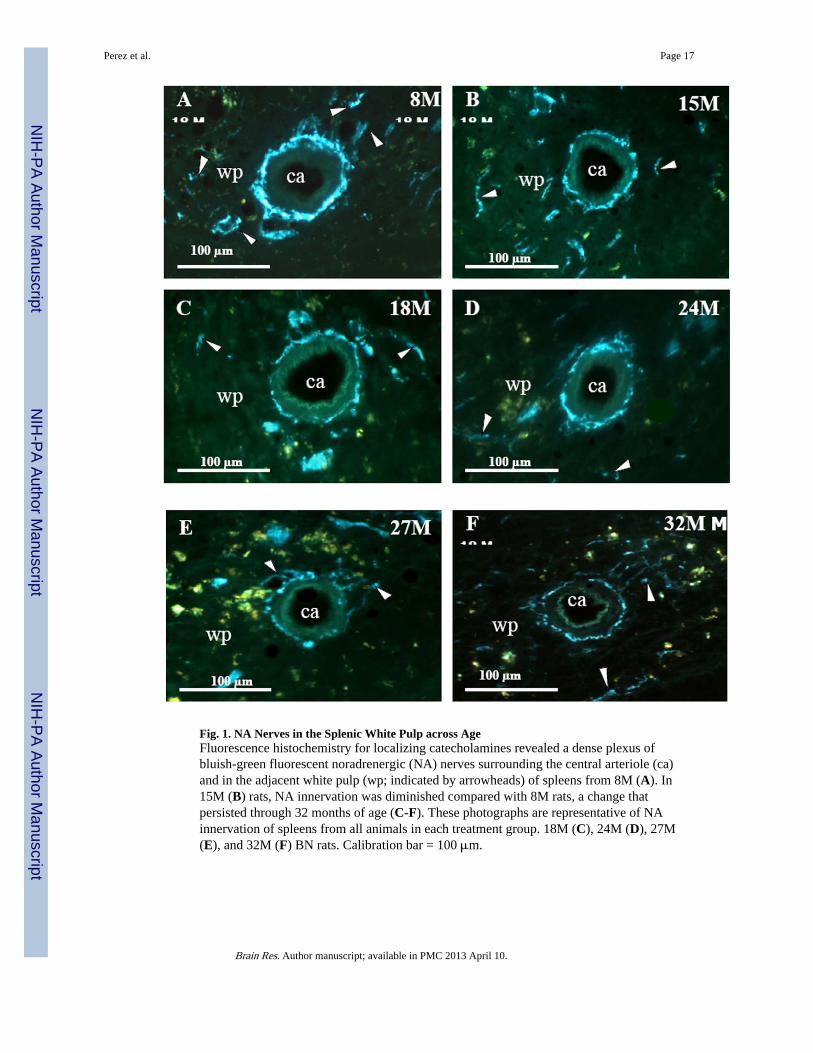
Fig. 1. NA Nerves in the Splenic White Pulp across AgeFluorescence histochemistry for localizing catecholamines revealed a dense plexus ofbluish-green fluorescent noradrenergic (NA) nerves surrounding the central arteriole (ca)and in the adjacent white pulp (wp; indicated by arrowheads) of spleens from 8M (A). In15M (B) rats, NA innervation was diminished compared with 8M rats, a change thatpersisted through 32 months of age (C-F). These photographs are representative of NAinnervation of spleens from all animals in each treatment group. 18M (C), 24M (D), 27M(E), and 32M (F) BN rats. Calibration bar = 100 μm.
Perez et al. Page 17
Brain Res. Author manuscript; available in PMC 2013 April 10.
NIH
-PA Author Manuscript
NIH
-PA Author Manuscript
NIH
-PA Author Manuscript
Perez et al. Page 18
Brain Res. Author manuscript; available in PMC 2013 April 10.
NIH
-PA Author Manuscript
NIH
-PA Author Manuscript
NIH
-PA Author Manuscript
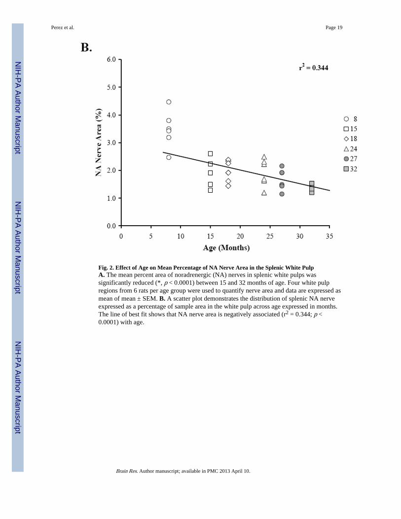
Fig. 2. Effect of Age on Mean Percentage of NA Nerve Area in the Splenic White PulpA. The mean percent area of noradrenergic (NA) nerves in splenic white pulps wassignificantly reduced (*, p < 0.0001) between 15 and 32 months of age. Four white pulpregions from 6 rats per age group were used to quantify nerve area and data are expressed asmean of mean ± SEM. B. A scatter plot demonstrates the distribution of splenic NA nerveexpressed as a percentage of sample area in the white pulp across age expressed in months.The line of best fit shows that NA nerve area is negatively associated (r2 = 0.344; p <0.0001) with age.
Perez et al. Page 19
Brain Res. Author manuscript; available in PMC 2013 April 10.
NIH
-PA Author Manuscript
NIH
-PA Author Manuscript
NIH
-PA Author Manuscript
Perez et al. Page 20
Brain Res. Author manuscript; available in PMC 2013 April 10.
NIH
-PA Author Manuscript
NIH
-PA Author Manuscript
NIH
-PA Author Manuscript
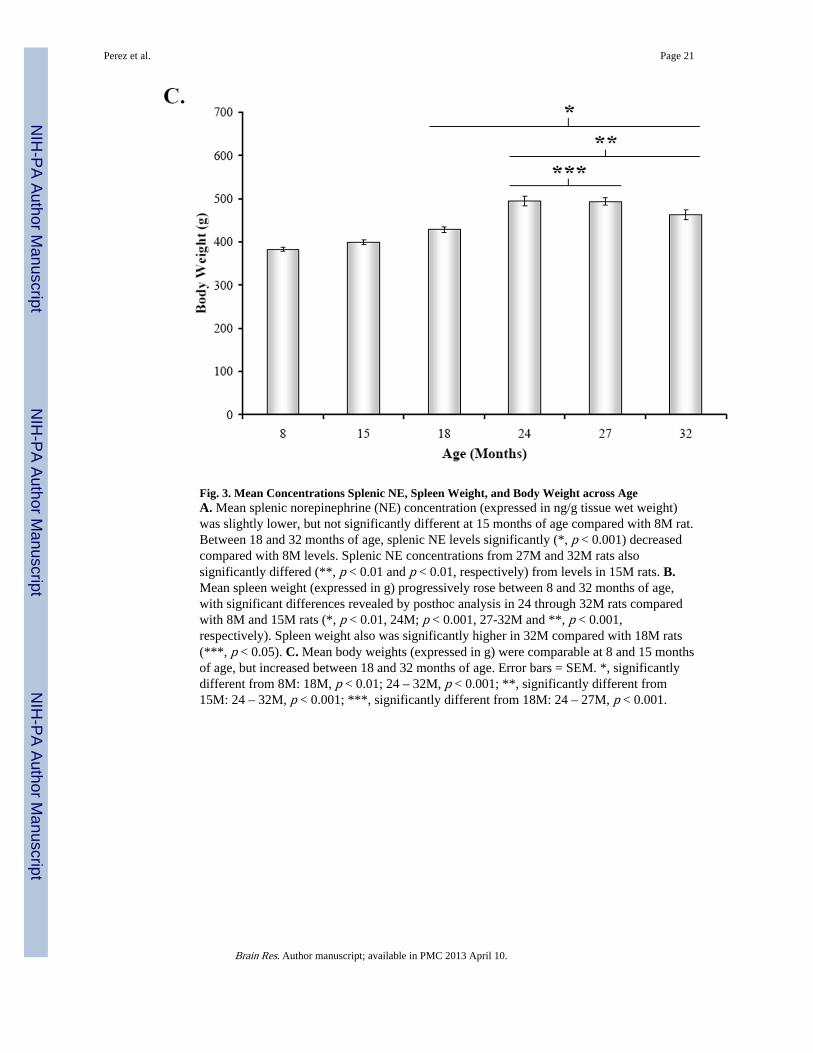
Fig. 3. Mean Concentrations Splenic NE, Spleen Weight, and Body Weight across AgeA. Mean splenic norepinephrine (NE) concentration (expressed in ng/g tissue wet weight)was slightly lower, but not significantly different at 15 months of age compared with 8M rat.Between 18 and 32 months of age, splenic NE levels significantly (*, p < 0.001) decreasedcompared with 8M levels. Splenic NE concentrations from 27M and 32M rats alsosignificantly differed (**, p < 0.01 and p < 0.01, respectively) from levels in 15M rats. B.Mean spleen weight (expressed in g) progressively rose between 8 and 32 months of age,with significant differences revealed by posthoc analysis in 24 through 32M rats comparedwith 8M and 15M rats (*, p < 0.01, 24M; p < 0.001, 27-32M and **, p < 0.001,respectively). Spleen weight also was significantly higher in 32M compared with 18M rats(***, p < 0.05). C. Mean body weights (expressed in g) were comparable at 8 and 15 monthsof age, but increased between 18 and 32 months of age. Error bars = SEM. *, significantlydifferent from 8M: 18M, p < 0.01; 24 – 32M, p < 0.001; **, significantly different from15M: 24 – 32M, p < 0.001; ***, significantly different from 18M: 24 – 27M, p < 0.001.
Perez et al. Page 21
Brain Res. Author manuscript; available in PMC 2013 April 10.
NIH
-PA Author Manuscript
NIH
-PA Author Manuscript
NIH
-PA Author Manuscript
Perez et al. Page 22
Brain Res. Author manuscript; available in PMC 2013 April 10.
NIH
-PA Author Manuscript
NIH
-PA Author Manuscript
NIH
-PA Author Manuscript
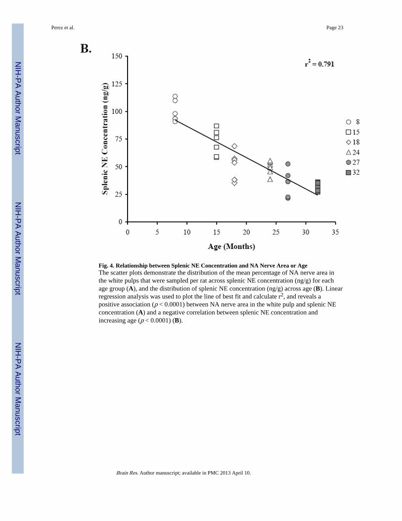
Fig. 4. Relationship between Splenic NE Concentration and NA Nerve Area or AgeThe scatter plots demonstrate the distribution of the mean percentage of NA nerve area inthe white pulps that were sampled per rat across splenic NE concentration (ng/g) for eachage group (A), and the distribution of splenic NE concentration (ng/g) across age (B). Linearregression analysis was used to plot the line of best fit and calculate r2, and reveals apositive association (p < 0.0001) between NA nerve area in the white pulp and splenic NEconcentration (A) and a negative correlation between splenic NE concentration andincreasing age (p < 0.0001) (B).
Perez et al. Page 23
Brain Res. Author manuscript; available in PMC 2013 April 10.
NIH
-PA Author Manuscript
NIH
-PA Author Manuscript
NIH
-PA Author Manuscript
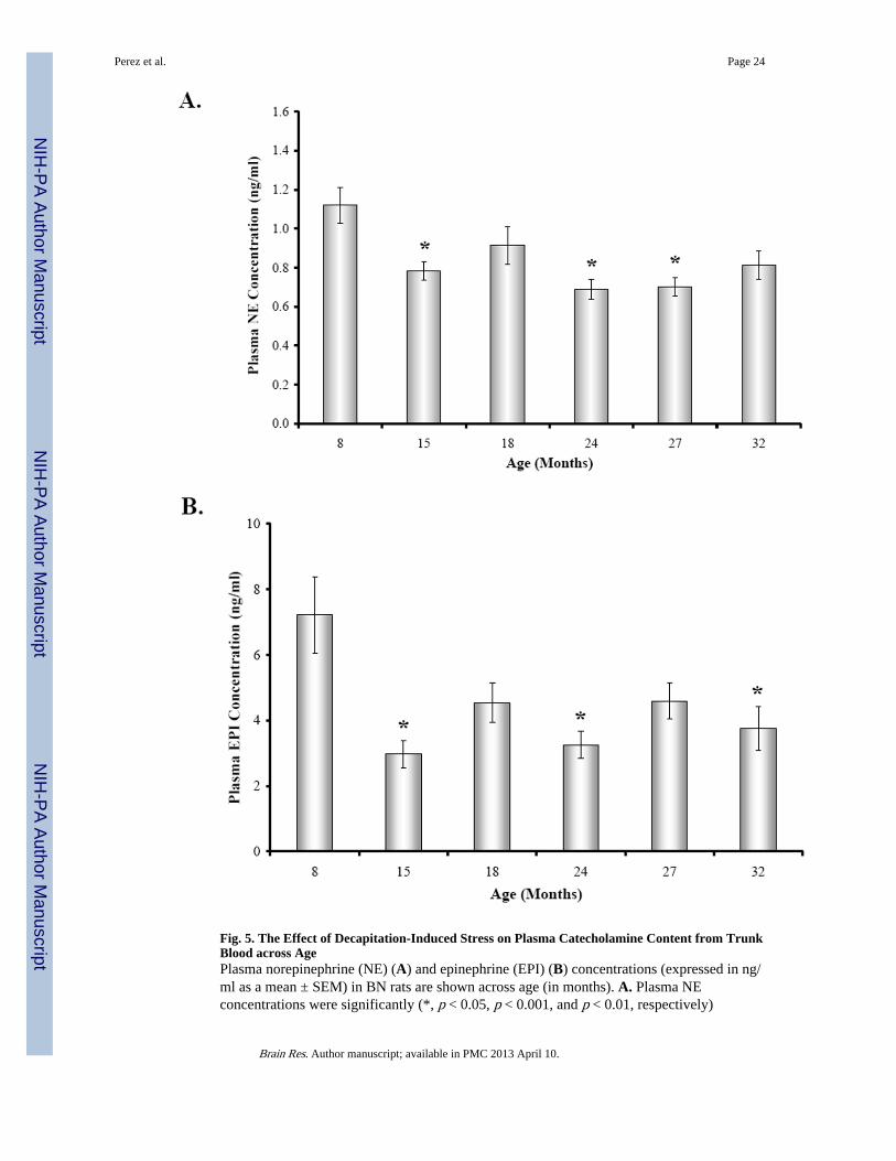
Fig. 5. The Effect of Decapitation-Induced Stress on Plasma Catecholamine Content from TrunkBlood across AgePlasma norepinephrine (NE) (A) and epinephrine (EPI) (B) concentrations (expressed in ng/ml as a mean ± SEM) in BN rats are shown across age (in months). A. Plasma NEconcentrations were significantly (*, p < 0.05, p < 0.001, and p < 0.01, respectively)
Perez et al. Page 24
Brain Res. Author manuscript; available in PMC 2013 April 10.
NIH
-PA Author Manuscript
NIH
-PA Author Manuscript
NIH
-PA Author Manuscript
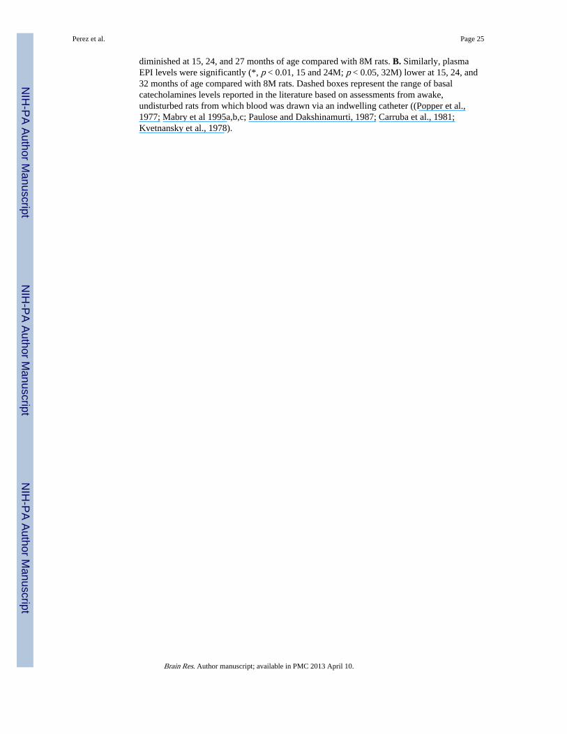
diminished at 15, 24, and 27 months of age compared with 8M rats. B. Similarly, plasmaEPI levels were significantly (*, p < 0.01, 15 and 24M; p < 0.05, 32M) lower at 15, 24, and32 months of age compared with 8M rats. Dashed boxes represent the range of basalcatecholamines levels reported in the literature based on assessments from awake,undisturbed rats from which blood was drawn via an indwelling catheter ((Popper et al.,1977; Mabry et al 1995a,b,c; Paulose and Dakshinamurti, 1987; Carruba et al., 1981;Kvetnansky et al., 1978).
Perez et al. Page 25
Brain Res. Author manuscript; available in PMC 2013 April 10.
NIH
-PA Author Manuscript
NIH
-PA Author Manuscript
NIH
-PA Author Manuscript