La sottoscritta PERRONE SERAFINA (detta Sara) nato a San ...
Upload
khangminh22Category
view
5download
0
University of Dundee
DOCTOR OF PHILOSOPHY
Cutaneous innervation of the hand
Sulaiman, Sara
Award date:2014
Link to publication
General rightsCopyright and moral rights for the publications made accessible in the public portal are retained by the authors and/or other copyright ownersand it is a condition of accessing publications that users recognise and abide by the legal requirements associated with these rights.
• Users may download and print one copy of any publication from the public portal for the purpose of private study or research. • You may not further distribute the material or use it for any profit-making activity or commercial gain • You may freely distribute the URL identifying the publication in the public portal
Take down policyIf you believe that this document breaches copyright please contact us providing details, and we will remove access to the work immediatelyand investigate your claim.
Download date: 10. Jul. 2022

DOCTOR OF PHILOSOPHY
Cutaneous innervation of the hand
Sara Sulaiman
2014
University of Dundee
Conditions for Use and DuplicationCopyright of this work belongs to the author unless otherwise identified in the body of the thesis. It is permittedto use and duplicate this work only for personal and non-commercial research, study or criticism/review. Youmust obtain prior written consent from the author for any other use. Any quotation from this thesis must beacknowledged using the normal academic conventions. It is not permitted to supply the whole or part of thisthesis to any other person or to post the same on any website or other online location without the prior writtenconsent of the author. Contact the Discovery team ([email protected]) with any queries about the useor acknowledgement of this work.

College of Art, Science and Engineering
Centre for Anatomy and Human Identification
CUTANEOUS INNERVATION OF THE HAND
By:
MS. SARA SULAIMAN
Supervised by:
PROFESSOR ROGER SOAMES
DR CLARE LAMB
A thesis Submitted in fulfillment of the requirement of the degree
DOCTOR OF PHILOSOPHY IN SCIENCE IN HUMAN ANATOMY
March 2014

II
Table of Contents
Content Page number
I. Table of Content II
II. List of figures V
III. List of tables XX
IV. List of abbreviations XXIII
V. Acknowledgments XXIV
VI. Declaration XXVI
VII. Summary XXVII
1. Introduction 1
1.1. The development of the sensory innervation pattern 5
1.2. Peripheral nerves internal anatomy and injury 7
1.3. Hand landmarks 18
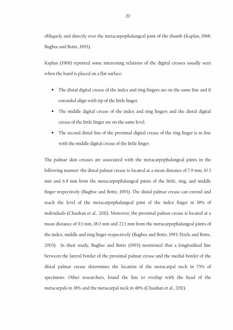
1.3.1. Flexion creases 18
1.3.2. Bony landmarks 22
1.3.3. Relationship of superficial landmarks to deeper structures of the hand
25
1.4. Palmar surface of the hand 27
1.4.1. Median nerve 31
1.4.1.1. Palmar cutaneous branch of the median nerve
31
1.4.1.2. Digital nerves 46
1.4.2. Ulnar nerve 49
1.4.3. Palmar communication branch between the median and the ulnar nerve
53
1.5. Dorsum of the hand 67
1.5.1. Superficial branch of the radial nerve 71
1.5.2. Dorsal branch of the ulnar nerve 88
1.5.3. Dorsal communication branch between the ulnar and radial nerve
93
1.6. Thesis aims and objectives 96

III
Content Page number
2. Methods 98
2.1. Sample 98
2.2. Anatomical landmarks 98
2.3. Measurements 102
2.4. Dissection process 103
2.5. Photographs and visual illustrations 126
2.6. Statistics 127
3. Results 128
3.1. Intra-observer results 128
3.2. Palmar cutaneous branch of the median nerve 129
3.3. Common digital nerve 144
3.4. Ulnar nerve 149
3.5. Palmar communicating branch between the median and ulnar nerve
159
3.6. Superficial branch of the radial nerve 165
3.7. Dorsal branch of the ulnar nerve 188
3.8. Sensory distribution in the dorsum of the hand 189
3.9. Dorsal communicating branch between the radial and ulnar nerve
194
4. Discussion 198
4.1. Palmar surface of the palm 198
4.1.1. Palmar cutaneous nerve of the median nerve 198
4.1.2. Common digital nerves 206
4.1.3. Ulnar nerve 209
4.1.4. Communicating branch between the median and the ulnar nerve
212
4.2. Dorsal Surface of the hand 224
4.2.1. Superficial branch of the radial nerve 224
4.2.2. Dorsal branch of the ulnar nerve 237
4.2.3. Sensory distribution in the dorsum of the hand
240

IV
Content
Page number
4.2.4. Dorsal communicating branch between the superficial branch of the radial nerve and the dorsal branch of the ulnar nerve
245
5. Conclusion 249
6. References 257
7. Appendix I 287
8. Appendix II 313
V
List of Figures
Figure Page number
Figure 1.1: An illustration of a cross section of the hand at the carpal
tunnel level showing the different compartments, spaces
and fascia in the hand (Agur et al., 2008).
1
Figure 1.2: The internal organization of the peripheral nerve (Moore
et al., 2011).
8
Figure 1.3: Illustrations of fascicular organization in the peripheral nerves. (A) The cable structure; (B) The plexiform structure (Stewart, 2003).
11
Figure 1.4: Diagrammatic illustration of the five degrees of nerve injuries as described by Sunderland. 1, first degree of injury; 2, second degree of injury; 3, third degree of injury; 4, fourth degree of injury; 5, fifth degree of injury (Campbell, 2008).
13
Figure 1.5: An illustration shows the process of Wallerian degeneration. (A) Injury occurs to the axon but the endoneurial tube is still intact; (B) Degeneration of axonal and myelin components of the distal segment. Elipsoids are formed by Schwann cells to facilitate phagocytosis by macrophages; (C) Degeneration and clearing of the distal segment are achieved. Regeneration of the axon begins guided by bands of Büngner (columns of Schwann cells); (D) regeneration continues in the endoneurial tube distal to the site of injury; (E) Full regrowth of axonal and associated myelin components (Nowak and Handford, 2003).
15
Figure 1.6: Hand flexion creases. 19
Figure 1.7: Important bony landmarks in the palmar surface of the hand.
22
Figure 1.8: Important landmarks in the dorsum of the hand. LT, Lister’s tubercle; APL, abductor pollicis longus; RSP, Radial styloid process; ASB, anatomical snuff box; EPB, extensor pollicis brevis; EPL, extensor pollicis longus; ECRL, extensor carpi radialis longus; ECRB, extensor carpi radialis brevis; ECU, extensor carpi ulnaris; USP, ulnar styloid process; LF, lunate fossa (Doyle and Botte, 2003).
23
VI
Figure Page number
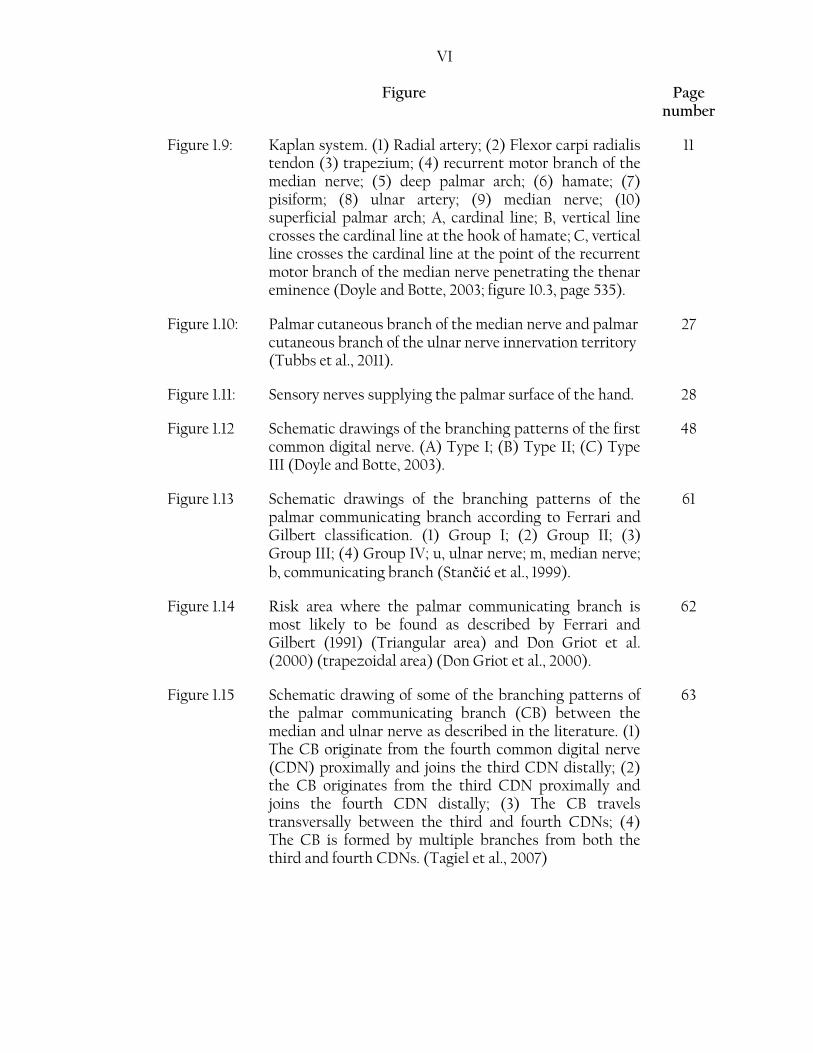
Figure 1.9: Kaplan system. (1) Radial artery; (2) Flexor carpi radialis tendon (3) trapezium; (4) recurrent motor branch of the median nerve; (5) deep palmar arch; (6) hamate; (7) pisiform; (8) ulnar artery; (9) median nerve; (10) superficial palmar arch; A, cardinal line; B, vertical line crosses the cardinal line at the hook of hamate; C, vertical line crosses the cardinal line at the point of the recurrent motor branch of the median nerve penetrating the thenar eminence (Doyle and Botte, 2003; figure 10.3, page 535).
11
Figure 1.10: Palmar cutaneous branch of the median nerve and palmar cutaneous branch of the ulnar nerve innervation territory (Tubbs et al., 2011).
27
Figure 1.11: Sensory nerves supplying the palmar surface of the hand. 28
Figure 1.12 Schematic drawings of the branching patterns of the first common digital nerve. (A) Type I; (B) Type II; (C) Type III (Doyle and Botte, 2003).
48
Figure 1.13 Schematic drawings of the branching patterns of the palmar communicating branch according to Ferrari and Gilbert classification. (1) Group I; (2) Group II; (3) Group III; (4) Group IV; u, ulnar nerve; m, median nerve; b, communicating branch (Stančić et al., 1999).
61
Figure 1.14 Risk area where the palmar communicating branch is most likely to be found as described by Ferrari and Gilbert (1991) (Triangular area) and Don Griot et al. (2000) (trapezoidal area) (Don Griot et al., 2000).
62
Figure 1.15 Schematic drawing of some of the branching patterns of the palmar communicating branch (CB) between the median and ulnar nerve as described in the literature. (1) The CB originate from the fourth common digital nerve (CDN) proximally and joins the third CDN distally; (2) the CB originates from the third CDN proximally and joins the fourth CDN distally; (3) The CB travels transversally between the third and fourth CDNs; (4) The CB is formed by multiple branches from both the third and fourth CDNs. (Tagiel et al., 2007)
63

VII
Figure Page number
Figure 1.16 Risk area where the palmar communicating branch (CB) is most likely to be found as described by Loukas et al. (2007). Light grey area indicates the range where all palmar CBs were found; dark grey area indicates the area where the majority (83%) of the palmar CBs were found (Loukas et al., 2007).
65
Figure 1.17 Schematic drawing showing the innervation territory classically described in the literature.
68
Figure 1.18 Risk area where the dorsal CB is most likely to be found as described by Loukas et al. (2008). Light grey area indicates the range where all dorsal CB were found; Dark grey area indicates where the majority (85%) of the dorsal CB were found; mean percentage is indicated by the dotted line at 41% (Loukas et al., 2008).
95
Figure 1.19 The suggested safe area where no major nerve passes as described by Mok et al. (2006). Dark lines outline the course of the SBRN supplying the lateral two and half digits and the DBUN supplying the medial two and half digits; CMCJ, metacarpophalangeal joint (Mok et al., 2006).
96
Figure 2.1: Dorsal landmarks used in this study. (1) Radial styloid process; (2) Ulnar styloid process; (3) Lister’s tubercle; (4) The third metacarpophalangeal joint; Red dot indicates the middle of the bistyloid line.
100
Figure 2.2: Palmar landmarks used in this study. (1) Transverse carpal ligament; (2) Scaphoid tubercle; (3) Bistyloid line; (4) Distal wrist crease; (5) Pisiform; (6) Third metacarpophalangeal joint; red dot indicates the middle of the bistyloid line.
101
Figure 2.3: Skin incisions outlined in the distal forearm and hand. (A) Lateral hand view; (B) Posterior forearm and dorsal hand view; (C) Anterior forearm and palmar hand view.
105
Figure 2.4: Skin removed in the distal forearm and hand. (A) Lateral hand view; (B) Posterior forearm and dorsal hand view; (C) Anterior forearm and palmar hand view.
106
VIII
Figure Page number
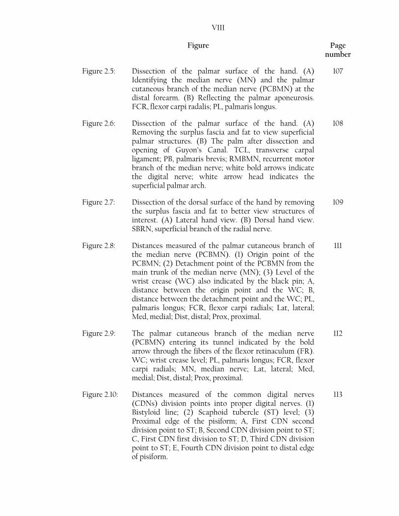
Figure 2.5: Dissection of the palmar surface of the hand. (A) Identifying the median nerve (MN) and the palmar cutaneous branch of the median nerve (PCBMN) at the distal forearm. (B) Reflecting the palmar aponeurosis. FCR, flexor carpi radalis; PL, palmaris longus.
107
Figure 2.6: Dissection of the palmar surface of the hand. (A) Removing the surplus fascia and fat to view superficial palmar structures. (B) The palm after dissection and opening of Guyon’s Canal. TCL, transverse carpal ligament; PB, palmaris brevis; RMBMN, recurrent motor branch of the median nerve; white bold arrows indicate the digital nerve; white arrow head indicates the superficial palmar arch.
108
Figure 2.7: Dissection of the dorsal surface of the hand by removing the surplus fascia and fat to better view structures of interest. (A) Lateral hand view. (B) Dorsal hand view. SBRN, superficial branch of the radial nerve.
109
Figure 2.8: Distances measured of the palmar cutaneous branch of the median nerve (PCBMN). (1) Origin point of the PCBMN; (2) Detachment point of the PCBMN from the main trunk of the median nerve (MN); (3) Level of the wrist crease (WC) also indicated by the black pin; A, distance between the origin point and the WC; B, distance between the detachment point and the WC; PL, palmaris longus; FCR, flexor carpi radials; Lat, lateral; Med, medial; Dist, distal; Prox, proximal.
111
Figure 2.9: The palmar cutaneous branch of the median nerve (PCBMN) entering its tunnel indicated by the bold arrow through the fibers of the flexor retinaculum (FR). WC; wrist crease level; PL, palmaris longus; FCR, flexor carpi radials; MN, median nerve; Lat, lateral; Med, medial; Dist, distal; Prox, proximal.
112
Figure 2.10: Distances measured of the common digital nerves (CDNs) division points into proper digital nerves. (1) Bistyloid line; (2) Scaphoid tubercle (ST) level; (3) Proximal edge of the pisiform; A, First CDN second division point to ST; B, Second CDN division point to ST; C, First CDN first division to ST; D, Third CDN division point to ST; E, Fourth CDN division point to distal edge of pisiform.
113

IX
Figure Page number
Figure 2.11: Distances measured of the common digital nerves (CDNs) division points into proper digital nerves. (1) Bistyloid line (BSL); (2) Third metacarpophalangeal joint; A, First CDN second division point to BSL; B, First CDN first division to BSL; C, Second CDN division point to BSL; D, Third CDN division point to BSL; E, Fourth CDN division point to BSL.
114
Figure 2.12: Distances measured of the ulnar nerve (UN) division points to the proximal edge of the pisiform. (1) The proximal edge of the pisiform; (2) The division point of the UN into deep and superficial branches; (3) The division point of the superficial branch into a proper digital nerve (PDN) to the medial side of the little finger and the fourth common digital nerve (CDN); A, The distance between the division point of the UN into deep and superficial divisions and the proximal edge of the pisiform; B, The distance between the division point of the superficial branch of the UN into a PDN to the medial side of the little finger and the fourth CDN to the proximal edge of the pisiform; MN, Median nerve; UN, Ulnar nerve.
115
Figure 2.13: Distances measured of the palmar communicating branch (CB) with reference to the transverse carpal ligament (TCL). (1) Distal attachment point of the palmar CB; (2) Proximal attachment point of the palmar CB; (3) The distal margin of the TCL; A, distance between the distal attachment point and the TCL; B, distance between the proximal attachment point of the palmar CB and the TCL; MN, median nerve; UN, ulnar nerve.
116
Figure 2.14: Distances measured of the palmar communicating branch (CB) with reference to the wrist crease (WC). (1) Third metacarpophalangeal joint; (2) Distal attachment point of the palmar CB; (3) Proximal attachment point of the palmar CB; (4) The WC; A, distance between the third MCP joint and the WC; B, distance between the distal attachment point and the WC; C, distance between the proximal attachment point of the palmar CB and the WC; MN, median nerve; UN, ulnar nerve.
117
X
Figure Page number
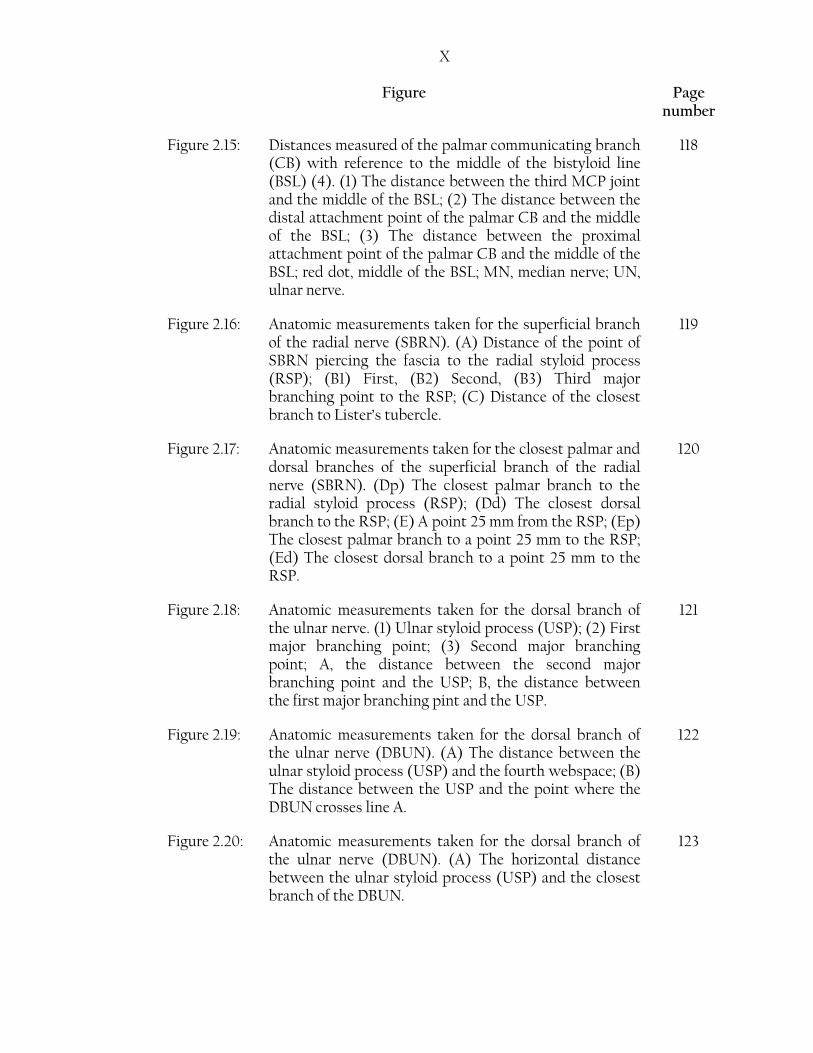
Figure 2.15: Distances measured of the palmar communicating branch (CB) with reference to the middle of the bistyloid line (BSL) (4). (1) The distance between the third MCP joint and the middle of the BSL; (2) The distance between the distal attachment point of the palmar CB and the middle of the BSL; (3) The distance between the proximal attachment point of the palmar CB and the middle of the BSL; red dot, middle of the BSL; MN, median nerve; UN, ulnar nerve.
118
Figure 2.16: Anatomic measurements taken for the superficial branch of the radial nerve (SBRN). (A) Distance of the point of SBRN piercing the fascia to the radial styloid process (RSP); (B1) First, (B2) Second, (B3) Third major branching point to the RSP; (C) Distance of the closest branch to Lister’s tubercle.
119
Figure 2.17: Anatomic measurements taken for the closest palmar and dorsal branches of the superficial branch of the radial nerve (SBRN). (Dp) The closest palmar branch to the radial styloid process (RSP); (Dd) The closest dorsal branch to the RSP; (E) A point 25 mm from the RSP; (Ep) The closest palmar branch to a point 25 mm to the RSP; (Ed) The closest dorsal branch to a point 25 mm to the RSP.
120
Figure 2.18: Anatomic measurements taken for the dorsal branch of the ulnar nerve. (1) Ulnar styloid process (USP); (2) First major branching point; (3) Second major branching point; A, the distance between the second major branching point and the USP; B, the distance between the first major branching pint and the USP.
121
Figure 2.19: Anatomic measurements taken for the dorsal branch of the ulnar nerve (DBUN). (A) The distance between the ulnar styloid process (USP) and the fourth webspace; (B) The distance between the USP and the point where the DBUN crosses line A.
122
Figure 2.20: Anatomic measurements taken for the dorsal branch of the ulnar nerve (DBUN). (A) The horizontal distance between the ulnar styloid process (USP) and the closest branch of the DBUN.
123

XI
Figure Page number
Figure 2.21: Distances measured of the dorsal communicating branch (CB) with reference to the bistyloid line (BSL). (1) Distal attachment point of the dorsal CB; (2) Proximal attachment point of the dorsal CB; (4) The BSL; (X) shows the location of the third metacarpophalangeal (MCP) joint. A, the distance between the third MCP joint and the BSL; B, distance between the distal attachment point and the BSL; C, distance between the proximal attachment point of the dorsal CB and the BSL.
124
Figure 3.1: Different origin direction of the palmar cutaneous branch of the median nerve (PCBMN) from the main trunk of the median nerve (MN). (A) Lateral origin, (B) Posterolateral origin, (C) Medial origin, (D) Anterior origin. FCR, flexor carpi radialis; PL, palmaris longus; SBRN, superficial branch of the radial nerve; Lat, lateral; Med, medial; Prox, proximal; Dist, distal.
131
Figure 3.2: Palmar cutaneous branch of the median nerve (PCBMN) crossing between the superficial and deep fibers of the flexor retinaculum to create a tunnel before entering the palm. Lat, lateral; Med, medial; Dist, distal; Prox, proximal.
133
Figure 3.3: Branching patterns of the palmar cutaneous branch of the median nerve (PCBMN). (A) Type I, (B) Variation to Type I, (C) Type II and (D) Type III. PL, palmaris longus; FCR, flexor carpi radialis; bold arrow indicates the PCBMN; MN, median nerve, Med, medial; Lat, lateral; Dist, distal; Prox, proximal.
136
Figure 3.4: Two palmar cutaneous branches of the median nerve (PCBMN). (A) Shows the different course that each branch takes. (B) After full dissection of the flexor retinaculum to view the deeper branch, the rectangular area outlined is enlarged in (C). PL, palmaris longus; FCR, flexor carpi radialis; bold arrows indicate the PCBMN branches; Lat, lateral; Med, medial; Prox, proximal; Dist, distal.
139

XII
Figure Page number
Figure 3.5: The palmar cutaneous branch of the median nerve (PCBMN) communicating with the lateral antebrachial cutaneous nerve (LABCN). (A) Full hand view showing the location of the two nerves, rectangular area outlined is enlarged in (B). (C) Shows the superficial branch of the radial nerve communicating with the LABCN prior to its communication with the PCBMN. PL, palmaris longus; FCR, flexor carpi radialis; Lat, lateral; Med, medial; Prox, proximal; Dist, distal; Palmar, palmar surface; Dorsal, dorsal surface.
140
Figure 3.6: The palmar cutaneous branch of the median nerve (PCBMN) communicating with the recurrent motor branch of the median nerve (RMBMN). The rectangular area outlined is magnified at the corner. Lat, lateral; Med, medial; Prox, proximal; Dist, distal.
141
Figure 3.7: Palmar cutaneous branch of the median nerve (PCBMN) communicating with the palmar cutaneous branch of the ulnar nerve (PCBUN) at the rectangular area outlined. Arrow head, PCBUN; PL, palmaris longus; Lat, lateral; Med, medial; Prox, proximal; Dist, distal
141
Figure 3.8: The palmar cutaneous branch of the median nerve (PCBMN) communicating with the first common digital nerve to the medial side of the thumb. (A) A full hand view. (B) Enlarged image of the communicating branch. Lat, lateral; Med, medial; Prox, proximal; Dist, distal.
142
Figure 3.9: An artery crossing the median nerve (MN) and separating the two branches of the palmar cutaneous branch of the median nerve (PCBMN) from the MN trunk. PL, palmaris longus; FCR, flexor carpi radialis; Lat, lateral; Med, medial; Prox, proximal; Dist, distal.
143
Figure 3.10: Common digital nerves (CDNs) and the superficial palmar arch. (A) Full hand view. (B) A neural loop is created by a branch from the 3rd CDN passing superficially to the superficial palmar arch. Arrow head indicates the superficial palmar arch; black arrows indicate the CDNs; Lat, lateral; Med, medial; Prox, proximal; Dist, distal.
145
XIII
Figure Page number
Figure 3.11: First common digital nerve (CDN) branching pattern. (A) Type I, (B) Type II, (C) Type III. The bold arrow indicates the first CDN; RMBMN, recurrent motor branch of the median nerve; MN, median nerve; UN, ulnar nerve; Lat, lateral; Med, medial; Prox, proximal; Dist, distal.
148
Figure 3.12: Patterns of division of the ulnar nerve (UN) in Guyon’s canal. (A) Type I, (B) Type II. PDN, proper digital nerve; CDN, common digital nerve; Lat, lateral; Med, medial; Prox, proximal; Dist, distal.
150
Figure 3.13: A special case where the ulnar nerve (UN) divided into four branches in Guyon’s canal. PDN, proper digital nerve; CDN, common digital nerve; CB, communicating branch; Lat, lateral; Med, medial; Prox, proximal; Dist, distal.
151
Figure 3.14: Kaplan anastomosis. (A) Dorsal branch of the ulnar nerve (DBUN) communicates with the superficial branch of the ulnar nerve. (B) DBUN communicates with the proper digital nerve to the little finger (PDN). CDN, common digital nerve; Lat, lateral; Med, medial; Prox, proximal; Dist, distal.
154
Figure 3.15: Fourth common digital nerve (CDN) receiving a branch from the third CDN which originate from the median nerve (MN). Bold arrow indicates the communicating branch. Lat, lateral; Med, medial; Prox, proximal; Dist, distal.
155
Figure 3.16: Fourth common digital nerve (CDN) communicating with the proper digital nerve (PDN) to the little finger. Bold arrow indicates the communicating branch. Lat, lateral; Med, medial; Prox, proximal; Dist, distal.
156
Figure 3.17: Deep motor branch of the ulnar nerve communicating with the proper digital nerve (PDN) to the little finger. CDN, common digital nerve; bold arrow indicates the communicating branch. Lat, lateral; Med, medial; Prox, proximal; Dist, distal.
156

XIV
Figure Page number
Figure 3.18: A special case of the ulnar nerve (UN) in Guyon’s canal. (A) Distal forearm view. (B) Nerves after passing through Guyon’s canal. FCU, flexor carpi ulnaris; UA, ulnar artery; UNs, superficial branch of the ulnar nerve; DBUN, dorsal branch of the ulnar nerve; CB, palmar communicating branch; arrow head indicates the proper digital nerve to the little finger; bold arrow indicates the deep branch of the ulnar nerve; tortuous arrow indicates the fourth common digital branch. Lat, lateral; Med, medial; Prox, proximal; Dist, distal.
158
Figure 3.19: Branching pattern of the palmar communicating branch (CB) Type I. (A) Type I A, (B) Type I B. CDN, common digital nerve; MN, median nerve; UN, ulnar nerve, bold arrow indicates the palmar CB. Lat, lateral; Med, medial; Prox, proximal; Dist, distal.
160
Figure 3.20: Branching pattern of the palmar communicating branch (CB) Type II. (A) Type II A, (B) Type II B. CDN, common digital nerve; MN, median nerve; UN, ulnar nerve; bold arrow indicates the palmar CB. Lat, lateral; Med, medial; Prox, proximal; Dist, distal.
161
Figure 3.21: Branching pattern of the palmar communicating branch (CB) Type III. (A) Type III A, (B) Type III B. CDN, common digital nerve; MN, median nerve; UN, ulnar nerve; bold arrow indicates the palmar CB. Lat, lateral; Med, medial; Prox, proximal; Dist, distal.
162
Figure 3.22: Branching pattern of the palmar communicating branch (CB) Type IV (A) and Type V (B). CDN, common digital nerve; MN, median nerve; UN, ulnar nerve; bold arrow indicates the palmar CB. Lat, lateral; Med, medial; Prox, proximal; Dist, distal.
163
Figure 3.23: First division of the superficial branch of the radial nerve (SBRN) after it becomes cutaneous. (A) Palmar and dorsal divisions. (B) Three divisions, one dorsal and two palmar (indicated by the arrow heads); pins indicate the location of the radial styloid process. Prox, proximal; Dist, distal; palmar, palmar surface; Dorsal, dorsal surface.
167

XV
Figure Page number
Figure 3.24: A. Special case of high division of the superficial branch of the radial nerve into palmar and dorsal divisions in the elbow region. Full hand view, arrow head indicates the dorsal branch; bold arrow indicates the palmar branch; black pin indicates the location of the radial styloid process. Prox, proximal; Dist, distal; Palmar, palmar surface; Dorsal, dorsal surface.
168
B. Special case of high division of the superficial branch of the radial nerve into palmar and dorsal divisions in the elbow region. The palmar branch crossing through brachioradialis indicated by the arrow head; bold arrow indicates the dorsal branch coursing between brachioradialis and extensor carpi radialis longus (ECRL) tendons. Prox, proximal; Dist, distal; Palmar, palmar surface; Dorsal, dorsal surface.
168
C. Special case of high division of the superficial branch of the radial nerve into palmar and dorsal divisions in the elbow region. The two different points where the palmar (bold arrow) and dorsal (arrow head) branches pierce the antebrachial fascia. BR, brachioradialis; ECRL, extensor carpi radialis longus; LABCN, lateral antebrachial cutaneous nerve. Prox, proximal; Dist, distal; Palmar, palmar surface; Dorsal, dorsal surface.
169
D. Special case of high division of the superficial branch of the radial nerve into palmar and dorsal divisions in the elbow region. The palmar branch indicated by the bold arrow communicates with the lateral antebrachial cutaneous nerve (LABCN), communication point indicated by the tortuous arrow. Arrow head indicates the dorsal branch; rectangular area shows the palmar and dorsal branch communicating; black pin indicates the location of the radial styloid process. Prox, proximal; Dist, distal; Palmar, palmar surface; Dorsal, dorsal surface.
170
Figure 3.25: Branching pattern of the superficial branch of the radial nerve (SBRN) Type I. (A) Type IA. (B) Type IB. (C) Type IC. Arrow head indicates the trifurcation of the SBRN, pins indicate the location of the radial styloid process. Prox, proximal; Dist, distal; Palmar, palmar surface; Dorsal, dorsal surface.
176

XVI
Figure Page number
Figure 3.26: Branching pattern of the superficial branch of the radial nerve (SBRN) Type II. (A) Type II A. (B) Type II B (C) Type II C. (D) Type II D. (E) Type II E. CT, common trunk; LABCN, lateral antebrachial cutaneous nerve; pins indicate the location of the radial styloid process. Prox, proximal; Dist, distal; Palmar, palmar surface; Dorsal, dorsal surface.
179
Figure 3.27: Branching pattern of the superficial branch of the radial nerve (SBRN) Type III. (A) Type III A. (B) Type III B (C) Type III C. (D) Type III D. CT, common trunk; pins indicate the location of the radial styloid process. Prox, proximal; Dist, distal; Palmar, palmar surface; Dorsal, dorsal surface.
182
Figure 3.28: Branching pattern of the superficial branch of the radial nerve (SBRN) Type IV. Prox, proximal; Dist, distal; Lat, lateral; Med, medial.
183
Figure 3.29: Branching pattern of the superficial branch of the radial nerve (SBRN) Type V. Prox, proximal; Dist, distal; Lat, lateral; Med, medial.
183
Figure 3.30: Branching pattern of the superficial branch of the radial nerve (SBRN) Type VI A. (A) Lateral view of Type VI A. (B) Dorsal view of Type VI A. Prox, proximal; Dist, distal; Med, medial; Lat, lateral; Palmar, palmar surface; Dorsal, dorsal surface.
184
Figure 3.31: Branching pattern of the superficial branch of the radial nerve (SBRN) Type VI B. (A) Lateral view of Type VI B. (B) Dorsal view of type VI B. CT, common trunk; pins indicate the radial styloid process. Prox, proximal; Dist, distal; Lat, lateral; Med, medial; Palmar, palmar surface; Dorsal, dorsal surface.
185
Figure 3.32: Branching pattern of the superficial branch of the radial nerve (SBRN) Type VI C. (A) Lateral view of Type VI C. (B) Dorsal view of type VI C. Prox, proximal; Dist, distal; Lat, lateral; Med, medial; Palmar, palmar surface; Dorsal, dorsal surface.
186

XVII
Figure Page number
Figure 3.33: Branching pattern of the superficial branch of the radial nerve (SBRN) Type VI D. This type was found in cadaver number 896 right and left hands, pictures were lost and the sketch was prepared based on the sketches taken from the two hands the type was found.
187
Figure 3.34: Superficial branch of the radial nerve (SBRN) communicating with the lateral antebrachial cutaneous nerve (LABCN) (A). The rectangular area outlined in (A) is enlarged and shown in (B). Prox, proximal; Dist, distal; Palmar, palmar surface; Dorsal, dorsal surface.
191
Figure 3.35: Lateral view of a special case of the lateral antebrachial cutaneous nerve (LABCN) entirely supplying the lateral side of the thumb. The medial side of the thumb is supplied by a branch of the superficial branch of the radial nerve (SBRN) after communicating with the LABCN (A). The rectangular area outlined in (A) is enlarged and shown in (B). Bold arrow indicates the communicating branch between the LABCN and the SBRN; Palmar, palmar surface; Dorsal, dorsal surface; Prox, proximal; Dist, distal.
192
Figure 3.36: Dorsal communicating branch (CB) between the superficial branch of the radial nerve (SBRN) and the dorsal branch of the ulnar nerve (DBUN) Type I. (A) Type I A. (B) Type I B. (C) Type I C. The arrow head indicates the CB. Prox, proximal; Dist, distal; Lat, lateral; Med, medial.
195
Figure 3.37: Dorsal communicating branch (CB) between the superficial branch of the radial nerve (SBRN) and the dorsal branch of the ulnar nerve (DBUN) (A) Type II. (B) Type III. The arrow head indicates the CB; Dist, distal; Prox, proximal; Lat, lateral; Med, medial.
196
XVIII
Figure Page number
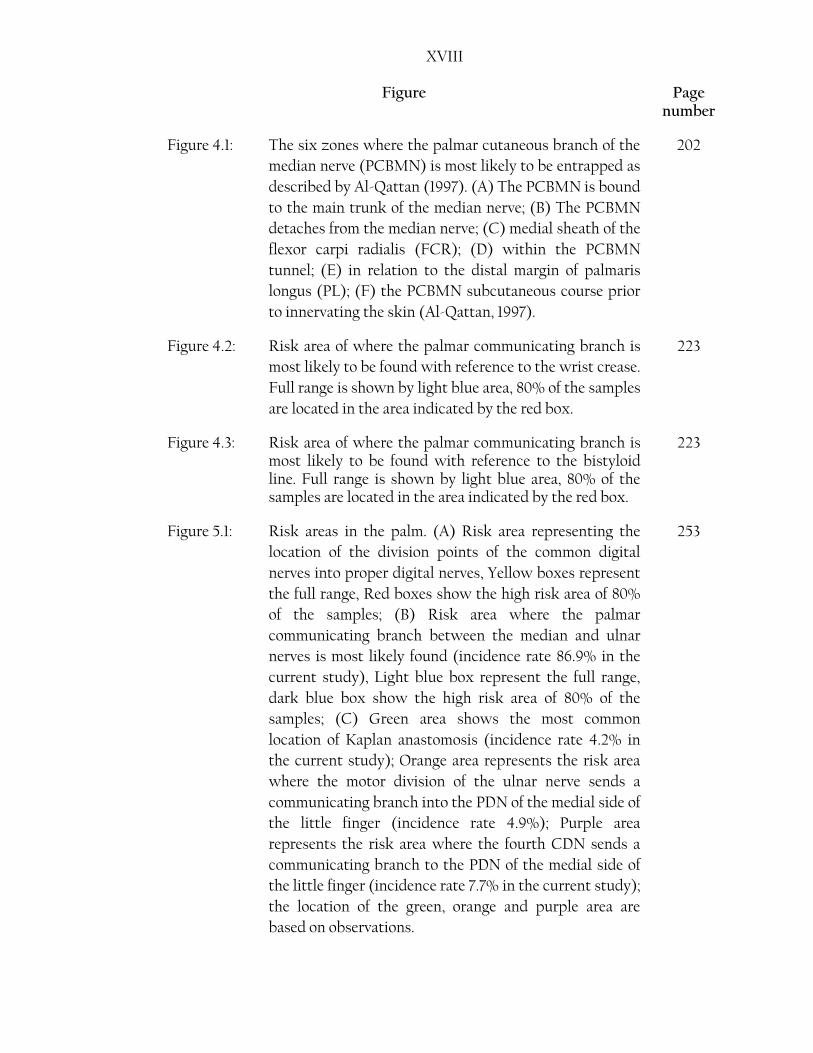
Figure 4.1: The six zones where the palmar cutaneous branch of the
median nerve (PCBMN) is most likely to be entrapped as
described by Al-Qattan (1997). (A) The PCBMN is bound
to the main trunk of the median nerve; (B) The PCBMN
detaches from the median nerve; (C) medial sheath of the
flexor carpi radialis (FCR); (D) within the PCBMN
tunnel; (E) in relation to the distal margin of palmaris
longus (PL); (F) the PCBMN subcutaneous course prior
to innervating the skin (Al-Qattan, 1997).
202
Figure 4.2: Risk area of where the palmar communicating branch is
most likely to be found with reference to the wrist crease.
Full range is shown by light blue area, 80% of the samples
are located in the area indicated by the red box.
223
Figure 4.3: Risk area of where the palmar communicating branch is most likely to be found with reference to the bistyloid line. Full range is shown by light blue area, 80% of the samples are located in the area indicated by the red box.
223
Figure 5.1: Risk areas in the palm. (A) Risk area representing the
location of the division points of the common digital
nerves into proper digital nerves, Yellow boxes represent
the full range, Red boxes show the high risk area of 80%
of the samples; (B) Risk area where the palmar
communicating branch between the median and ulnar
nerves is most likely found (incidence rate 86.9% in the
current study), Light blue box represent the full range,
dark blue box show the high risk area of 80% of the
samples; (C) Green area shows the most common
location of Kaplan anastomosis (incidence rate 4.2% in
the current study); Orange area represents the risk area
where the motor division of the ulnar nerve sends a
communicating branch into the PDN of the medial side of
the little finger (incidence rate 4.9%); Purple area
represents the risk area where the fourth CDN sends a
communicating branch to the PDN of the medial side of
the little finger (incidence rate 7.7% in the current study);
the location of the green, orange and purple area are
based on observations.
253

XIX
Figure Page number
Figure 5.2: The most common sensory innervation pattern and risk
areas in the dorsum of the hand as found in the current
study. Yellow box represent full range of the most
common location of the dorsal communicating branch
between the superficial branch of the radial nerve
(SBRN) and the dorsal branch of the ulnar nerve (DBUN)
(Type III) relative to the distance between the third
metacarpophalangeal joint and the middle of the
bistyloid line, 80% of the samples are found in the area
indicated by the red box; Light green area represent the
full range of the area where the DBUN crosses a line
extending between the ulnar styloid process and the
fourth webspace, 80% of the samples are found in the
area indicated by the dark green box; Pink area represent
a risk area where the lateral antebrachial cutaneous nerve
communicate with the SBRN (incidence rate 30.0%, this
area is based on observations).
255

XX
List of Tables
Table Page number
Table 1.1: Anatomical description of the palmar cutaneous branch of the median nerve as mentioned in the literature
33
Table 1.2: Examples of different incision suggestions for the treatment of carpal tunnel syndrome as reported in the literature
43
Table 1.3: Zones of Guyon’s canal and their boundaries 51
Table 1.4: The major anatomical studies investigating the incidence and pattern of the palmar communicating branch between the median and the ulnar nerve
55
Table 1.5: Patterns of distribution of the superficial branch of the radial nerve in the literature
73
Table 1.6: Anatomic measurements of the superficial branch of the radial nerve (SBRN) to bony landmarks that are discussed in the literature
84
Table 1.7: Anatomic measurements and description for the dorsal branch of the ulnar nerve as described in the literature
88
Table 2.1: Number of hands dissected during the study 98
Table 2.2: Summary of the anatomical landmarks used in this study 99
Table 2.3: List of the anatomical measurements for each of the nerves investigated in the study
125
Table 3.1: Number of samples investigated for each nerve 128
Table 3.2: Chi-Square results for repeated measures 129
Table 3.3: Anatomical measurements recorded for the palmar cutaneous branch of the median nerve (PCBMN) (mm)
137
Table 3.4: Anatomical measurements recorded in cases where two palmar cutaneous branches of the median nerve were found (mm)
138
XXI
Table Page number
Table 3.5: Absolute measurements for the branching points of the common digital nerves (CDNs) into proper digital nerves in the palm (mm)
146
Table 3.6: Branching points of the common digital nerves (CDNs) into the proper digital nerves to the distance between the third metacarpophalangeal joint and the bistyloid line (%)
147
Table 3.7: The first and second division points of Type I and the point of trifurcation of Type II to the proximal edge of the pisiform (mm)
152
Table 3.8: Distances measured for the proximal and distal attachments of the palmar communicating branch (CB) to different anatomical landmarks (mm)
165
Table 3.9: Measurements of the closest branches of the superficial branch of the radial nerve (SBRN) to various anatomical points (mm)
171
Table 3.10: The major division points of the superficial branch of the radial nerve to the radial styloid process (mm)
172
Table 3.11: Incidence rate for superficial branch of the radial nerve (SBRN) Type I branching pattern
175
Table 3.12: Incidence rate for superficial branch of the radial nerve (SBRN) Type II branching pattern
177
Table3.13: Incidence rate for superficial branch of the radial nerve (SBRN) Type III branching pattern
180
Table 3.14: Incidence rates for superficial branch of the radial nerve
(SBRN) Type IV, Type V and Type VI branching patterns.
182
Table 3.15: Anatomical measurements recorded for the dorsal branch of the ulnar nerve (mm)
188
Table 3.16: Distances of the first and second major branching point of the dorsal branch of the ulnar nerve to the ulnar styloid process (mm).
189
Table 3.17: Sensory innervation in the dorsum of the hand with reference to digits
189
XXII
Table Page number
Table 3.18: Distribution of the different branching patterns in the
dorsum of the hand
193
Table 3.19: Anatomical measurements for the dorsal communicating branch between the superficial branch of the radial nerve and the dorsal branch of the ulnar nerve (mm)
197
Table 4.1: Incidence rates of different patterns of distributions of the palmar cutaneous nerve of the median nerve in the literature compared with the types found in the current study
205
Table 4.2: Different incidence rates of the palmar communicating branch patterns as reported in the literature in comparison to the current study findings
217
Table 4.3: Anatomical measurements for the superficial branch of the radial nerve (SBRN) as reported in the literature in comparison to the current study (mm)
226
Table 4.4: Incidence rates of the branching patterns of the superficial branch of the radial nerve found in the current study
237

XXIII
List of Abbreviations
BSL Bistyloid line
BR Brachioradialis
CAHID Centre for Anatomy and Human Identification
CB Communicating branch
CDN Common digital nerve
CT Common trunk
DBUN Dorsal branch of ulnar nerve
ECRL Extensor carpi radialis longus
FCR Flexor carpi radialis
FCU Flexor carpi ulnaris
LABCN Lateral antebrachial cutaneous nerve
LT Lister’s tubercle
MCP Metacarpophalangeal joint
MN Median nerve
n Number
PCBMN Palmar cutaneous branch of the median nerve
PCBUN Palmar cutaneous branch of the ulnar nerve
PCL Palmar carpal ligament
PDN Proper digital nerve
PL Palmaris longus
RMBMN Recurrent motor branch of the median nerve
RN Radial nerve
RSP Radial styloid process
SBRN Superficial branch of radial nerve
ST Scaphoid tubercle
TCL Transverse carpal ligament
UA Ulnar artery
UD Undetermined
UN Ulnar nerve
USP Ulnar styloid process
WC Wrist Crease

XXIV
Acknowledgments
This work will not have seen the light without the blessings of God to whom I give my
ultimate thanks and gratitude. Moreover, I am grateful and indebted to many people who
filled me with their kindness, support and guidance throughout the period of my project.
First of all, I would like to thank my principal supervisor Professor Roger Soames for his
patience, guidance and the beautiful kind fatherly sprit he embraced upon me during my
time being his student. Moreover, no amount of thanks can express my gratitude to my
secondary supervisor, Dr. Clare lamb, who through her kindness, patience and immense
knowledge provided continuous help, direction and support through various aspects of my
project. I was lucky to have two supervisors who are approachable, encouraging and
always concerned about my wellbeing in addition to how my research is developing. Who
tolerated my crazy ideas, kept me focused and allowed me to create and thrive in my own
pace.
Special thanks and appreciation to my Thesis Committee members, Professor Caroline
Wilkinson and Professor Timothy Newman, who continuously gave invaluable suggestions
and comments expressing a great spirit of adventure in regard to research.
A special note of thanks to all the teaching and support staff at the Center for Anatomy
and Human Identification (CAHID) for providing a professional and stimulating
environment to work. I was privileged to work among such a team with commitment,
excitement and devotion to high standards of education. Special thanks to Dr. Catherine
Carr, Dr. Paul Felts, Ms. Netta Gallazzi, Ms. Vivienne McGuire, and Ms. Gillian Malone.
My sincere gratitude goes to Dr. Stephen Hubbard, for all his help in the statistical aspect
of this project. Despite his busy schedule, he still accommodated my questions and gave
valuable suggestions. Special thanks go to Dr. Luca Albergante for his valuable suggestions
and comments, his kindness and patience in explaining statistical tests for me. A note of
thanks goes to Ms. Victoria Mcculloch for providing some of the visual illustrations in this
project.
No amounts of thanks can describe my gratitude to my parents. Their unconditional love,
continuous support and kindness made me the person I am today. While many doubted me

XXV and my decision to go through this journey, their persistent encouragement and love
allowed me to dream, be ambitious and dare to believe that this is achievable. To them, I
am eternally grateful.
For a special, generous, forgiving, and supportive friend; Nasreen, thank you for being my
safe haven where I was able to hide during my low and blue days.

XXVI
Declaration
I hereby declare that this dissertation is a presentation of my original work conducted
under the supervisor of Professor Roger Soames, Cox Professor of Anatomy, and Dr. Clare
Lamb, Senior Lecturer in Anatomy at the Centre for Anatomy and Human Identification,
University of Dundee. Contributions of others involved are clearly indicated. All references
cited have been consulted by me. I certify that this work has never been accepted for the
award of any other higher degree.
Sarah Sulaiman
XXVII
Summary
With the increase of hand pathologies in the last decade, the need to better understand the
anatomy of the hand is becoming more vital. The cutaneous innervation of the hand is
classically described to be supplied by palmar cutaneous branch of the median nerve
(PCBMN), common digital nerves (CDNs), ulnar nerve (UN), palmar cutaneous branch of
the ulnar nerve, dorsal branch of the ulnar nerve (DBUN), superficial branch of the radial
nerve (SBRN) and occasionally the lateral antebrachial cutaneous nerve (LABCN).
Although the sensory distribution of the hand has been described in the literature, reports
have often shown contradicting views and occasionally different or incomplete
descriptions. Furthermore, clinical procedures in the hand and wrist can result in painful
and/or disabling postoperative complications. This thesis outlines, categorizes and
describes the distribution and branching patterns of cutaneous branches supplying the
palmar and dorsal surface of the hand and their relationship to the distal area of the
forearm and wrist. It also investigates the palmar and dorsal communicating branches,
their patterns and common locations. Moreover, the project discusses the impact of the
distribution and branching patterns of the cutaneous nerves on surgical and diagnostic
procedures performed in the hand, wrist and distal forearm. 160 cadaveric hands were
dissected in the Centre for Anatomy and Human Identification (CAHID), University of
Dundee. All cadavers were musculoskeletally mature adults with mean age of 82.5±9.4
(range: 53-101) years. Skin was removed from the distal half of the forearm to the
metacarpophalangeal joints. Nerves under investigation were identified, dissected, and
traced. Sketches, photographs, and measurements to predefined landmarks including the
wrist crease (WC), bistyloid line (BSL) and the third metacarpophalangeal (MCP) joint
were taken and results expressed as means, standard deviations and ranges. Patterns are
classified and expressed with frequencies. The PCBMN was found to originate from the
main trunk of the median nerve (MN) 54.1±15.7 mm proximal to the WC and course
distally between flexor carpi radialis and palmaris longus (if present) to innervate the
proximal palmar surface of the hand by branching into one of three types identified.
Furthermore, two PCBMN were found in 8.9% of cases. The second, third, fourth CDNs
were found to divide into proper digital nerves at a point located distal to the 70% of the
distance between the third MCP joint and the BSL in 88% of cases. The cutaneous
innervation of the palm was found to be relatively constant with the lateral 3½ digits being

XXVIII supplied by the MN and the medial 1½ being supplied by the UN. A palmar CB was found
between the third CDN-MN and fourth CDN-UN in 86.9% of the cases coursing in
different patterns and changing the palmar sensory innervation of that previously
described. The sensory innervation of the dorsum of the hand was variable. The most
common pattern was being supplied by the SBRN innervating the lateral dorsal skin and
the skin covering the lateral 2½ digits and the DBUN innervating the medial dorsal skin
and the skin covering the medial 1½ digits found in 37.3%. All radial supply to the dorsum
of the hand with the absence of the DBUN was found in 6.7%. The SBRN connected with
the LABCN in 30.7% and with the DBUN in 26.4% complicating the sensory innervation in
the dorsum of the hand. Understanding the cutaneous innervation of the hand,
appreciation of the possible variations and presence of communicating branches will result
in a better evaluation of signs and symptoms, establishing a proper therapeutic plan,
avoiding iatrogenic injuries during surgical interventions, and properly diagnose
postoperative complications leading to an increased quality of medical service and patient
satisfaction.

1
Figure 1.1: An illustration of a cross section of the hand at the carpal tunnel level
showing the different compartments, spaces and fascia in the hand
(Agur et al., 2008; figure 64, page 562).
1. Introduction
The human hand is a unique and complex structure. Hand injuries, trauma and
pathology account for a considerable amount of health care problems. It is only natural
that the hand has received a lot of attention in the literature as its relatively small size
and high density of structures create delicate and close anatomical relationships
(Figure 1.1). Understanding hand disease processes and treatment planning requires a
detailed knowledge of hand anatomy and a high appreciation of the anatomical
relationships between structures in the hand.
2
Understanding the anatomy of the hand starts from the skin. Its dorsal and palmar
superficial landmarks provide the basis for surgical planning and hand pathology
assessment. In 1968, Kaplan introduced a unique system exploring the hand’s
superficial landmarks and their relationships to deeper structures. Kaplan’s system is
still used today and is considered a useful surgical map during surgical incision
planning. Other bony landmarks, such as the ulnar and radial styloid processes, are
used in clinical and research settings as they are easily palpable and relatively constant
among different individuals. However, with so many landmarks in the hand and wrist
area, how each affects anatomical morphometry is of interest.
The cutaneous innervation is an important aspect to consider during the planning of
surgical approaches and anaesthesia. The literature describes the innervation of the
hand to be by the median nerve (MN), ulnar nerve (UN), radial nerve (RN) and
occasionally the lateral antebrachial cutaneous nerve (LABCN). The palmar cutaneous
branch of the median nerve (PCBMN) innervates the proximal two-fifths of the
midpalmar surface. It has received a lot of attention in the modern literature consistent
with the evolution of surgical carpal tunnel release techniques. With relatively limited
sample size anatomical studies, the course and branching patterns of the PCBMN have
been outlined, with many recommendations for ideal incision sites to minimize
postoperative complications. With many reports and different recommendations, a
better understanding of the cutaneous innervation of the palm may increase patient’s
postoperative satisfaction and lead to fewer complications.
The digital nerves innervate the distal three-fifths of the palm and the palmar surface of
the digits. The first, second and third common digital nerves (CDN) arise from the MN
after it passes through the carpal tunnel, whereas the fourth CDN and the proper
digital nerve (PDN) to the medial side of the little finger arise from the UN after it

3
passes through Guyon’s canal (ulnar canal) extending from the proximal edge of the
palmar carpal ligament to the fibrous arch of the hypothenar muscles in the medial side
of the palm (Gross and Gelberman, 1985). The point of division of the CDN into PDNs
has not been well investigated. Furthermore, the communicating branches (CB)
between the MN and the UN can cause sensory alterations affecting the typical signs
and symptoms of hand pathology; and when injured can complicate surgical procedures
such as carpal tunnel and flexor tendon release. The literature has differing reports of
the incidence rate of the CB and the most common branching patterns. Knowledge of
the most common location and branching patterns of the CB will help to better plan
therapeutic and diagnostic procedures to minimize iatrogenic injuries in the area.
The dorsum of the hand is supplied by the dorsal branch of the ulnar nerve (DBUN),
the superficial branch of the radial nerve (SBRN) and occasionally the LABCN. The
DBUN is classically described as innervating the medial dorsal skin of the hand and the
skin covering the medial 1½ digits. The course of the DBUN puts it in risk during wrist
arthroscopy and any procedure that requires a direct approach to the ulna such as open
reduction and internal fixation, ulnar lengthening and shortening procedures, delayed
union or non-union of ulnar fractures. It is also important to understand the course of
the nerve to properly prepare and harvest neurocutaneous flaps.
The second nerve innervating the dorsum of the hand is the SBRN which supplies the
laterodorsal surface of the hand and the skin covering the lateral 3½-2½ digits: it has
many configurations. Due to its course in the distal forearm and wrist; and the
variability of its position and branching patterns, it is vulnerable to injuries due to
trauma or iatrogenesis. Many therapeutic and diagnostic procedures are conducted on
the distal forearm and wrist area including fixation and reduction of distal radial

4
fractures, wrist arthroscopic procedures, de Quervain’s syndrome release, radial artery
harvest and cephalic vein cannulation. Understanding the anatomical course of the
SBRN and its relationship to other structures in the distal forearm is not only
important to ensure safe clinical practice; but also to create opportunities for the
development of new medical techniques and applications.
Although described in the literature, the innervation territory and the pattern of
distribution in the dorsum of the hand differ greatly among authors. Anatomical
variations, communicating branches and most common patterns of distribution are not
well described nor have the associations between the nerves or other anatomical
structures in the dorsum of the hand been fully investigated. Furthermore, there are
contradicting reports about the incidence of communicating branches between the
nerves that supply the dorsum of the hand and their common locations. Description of
the most common patterns of innervation and variations to these patterns in respect to
well-known anatomical landmarks can greatly help in clinical settings. It will also aid
in better evaluating electrophysiological studies, to properly diagnose and plan
treatment, and safely intervene, if required.
The current study investigates the cutaneous innervation of the hand focusing on the
PCBMN, CDNs, UN, SBRN, and DBUN. It describes their anatomical course, details
their common patterns of distribution and discusses their significance in clinical
settings. The study aims to fill the gap in the literature to better understand the
cutaneous nerve supply to the hand and appreciate the relations between the nerves
and other anatomical structures in the distal forearm and wrist.

5
1.1. The development of the sensory innervation pattern
The sensory innervation is organized segmentally where a region of skin (dermatome)
is supplied by axons derived from a dorsal root ganglion and carried through one or
more cutaneous nerves (Scott, 1992). The cutaneous fibres from each dorsal root
ganglion grow precisely and accurately along marked pathways to their target region
by receiving guidance from two sources: general and specific cues. The general cues
channel general populations (different cells) down a common pathway; whereas, the
specific cures are directed toward a particular population of cells directing them
towards the appropriate target region (Tosney and Oakley, 1990; Scott, 1992). The
following is an overview of the general and specific cues that direct the growth of
dermatomes; however, as far as can be ascertained, the exact influences and processes
of the development of the cutaneous innervation of the hand in humans (the nerves
investigated in this study) were not fully investigated in the literature.
Tosney and Oakley (1990) reported that nerves avoid some regions and are attracted to
others suggesting that the regions favourable for axonal growth are adjacent to
relatively inhibitory regions. In the chick embryo, growing axons avoid perinotochordal
mesenchyme and are attracted to the dorsal-anterior sclerotome establishing the
dorsal-ventral position; they avoid the posterior sclerotome and are attracted to the
anterior sclerotome establishing the anterior-posterior position of the spinal nerves;
they avoid the growing pelvic girdle and are attracted to the plexus mesenchyme
establishing the nerve trunk position (Tosney and Oakley, 1990; Scott, 1992). The
inhibitory and/or permissive characteristic of a region is controlled by complex cellular
interactions, phagocytosis, cell death with a molecular basis providing a relative
balance of substances guiding the sensory neuron (Tosney and Landmesser, 1985;
Goodman and Shatz, 1993; Kitsukawa et al., 1997).

6
Specific cues guiding the sensory peripheral fibres include the interactions between
sensory axons and other sensory and motor axons. However; studies have also shown
that cutaneous nerves were still able to grow in their approximate normal locations in
chick embryos with motorneurons precursor removed (Scott, 1988). These results
suggest that although motorneurons can provide cues to direct sensory axons, they are
not essential (Scott, 1992).
Skin movement is also thought to affect the orientation and growth of sensory axons.
Some studies suggest that axons from each dorsal root ganglion innervate skin
embryologically derived from the same ganglion root. Scott (1982) suggested that
sensory axons are specifically matched with the corresponding skin region creating
dermatomes with specific boundaries and little overlap. However; the literature
presents different dermatomal maps which can be attributed to the different methods
used in different studies to define dermatomes being behaviour response or electrical
neurophysiological response studies. Another reason for the inconsistency in
dermatomes boundaries is the overlap of skin areas supplied by nerve fibres of adjacent
dermatomes (Werner and Whitsel, 1967; Lee et al., 2008). Furthermore, as the skin and
limb grow, nerve endings enlarge and grow, which can affect the orientation of the
dermatome.
Attraction of axons to targeted epithelium is another mechanism where embryonic skin
is thought to secrete neurotropic agents, other than nerve growth factor, that attracts
cutaneous axons and thus acts like specific cues guiding cutaneous axons to their
targeted region early in development (Lumsden and Davies, 1983). Skin epidermis has
also shown inhibitory influences on axonal growth according to studies conducted on
chick embryos (Scott, 1982; Martin et al., 1989).

7
Another specific cue is thought to be administered by the association between sensory
axons and Merkel cells (Scott, 1992). Nerve growth factor is required for normal axonal
growth and development: one source of nerve growth factor are Merkel cells. Merkel
cells are thought to be a regulator of the density and distribution of nerve endings (Vos
et al., 1991).
Although dermatomes are areas of skin innervated by one dorsal root ganglion, fibres
can be carried through different nerve trunks. As the nerves enter the skin through
different points, they can compete for skin via different mechanisms, and thus create
different innervation patterns between individuals or even between the different sides
of the same individual (Diamond, 1981; Scott, 1982; Scott, 1992).
Furthermore, the full growth and form of a human being is a result of different
interactions between the developing individual, which is directed by genes, and its
environment. In the early phase of development, the embryo’s development and growth
is sensitive to environmental cues that can modify, interfere or even disrupt its
development (Gluckman et al., 2005). The effect of the environment is clearly evident in
the different fingerprints between identical twins (siblings with exactly the same
genotype), between the two hands of the same individual and even between the
different fingers of the same hand. Such differences are obtained during the
differentiation process of the skin and are caused by the different flow of the amniotic
fluids around the fetus and its position in the uterus during differentiation of skin.
However, fingerprints are expressed from the same gene therefore the resulting pattern
will not be totally random but will retain some similarities (Jain et al., 2002). It is
possible that environmental cues can influence the orientation and the distribution of
nerves creating different patterns among individuals or even between the different sides
of the same individual.

8
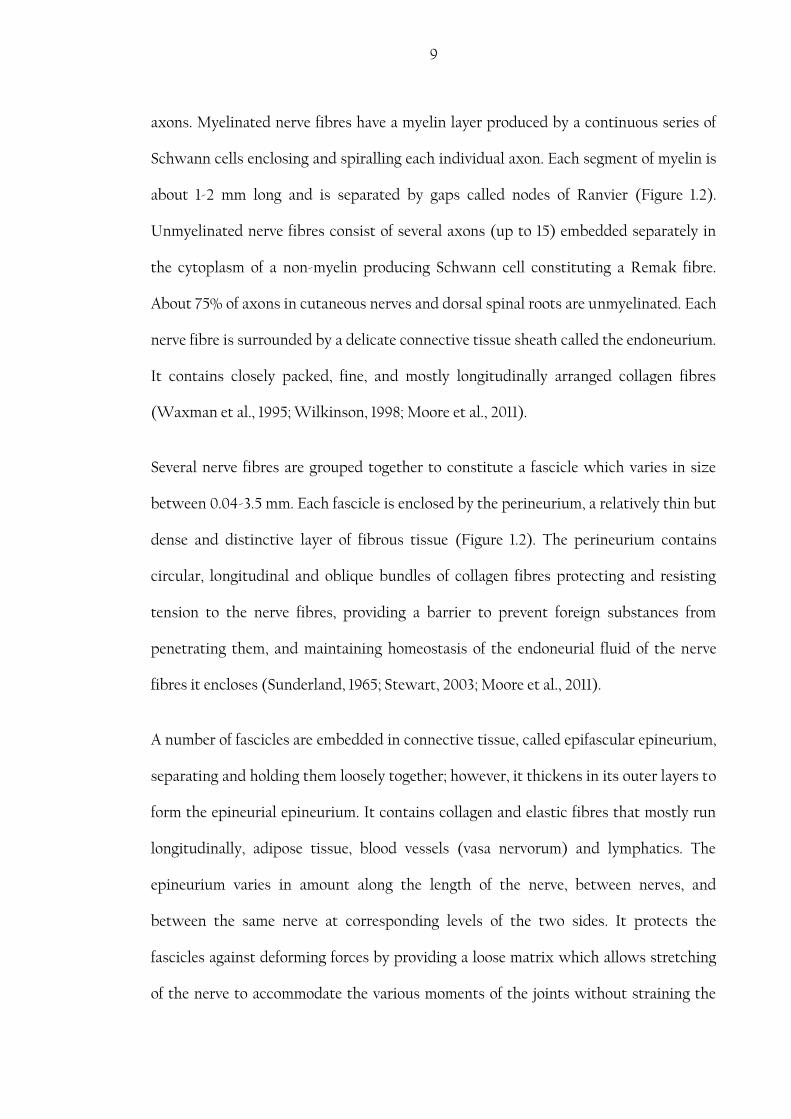
Figure 1.2: The internal organization of the peripheral nerve (Moore et al., 2011;
figure 1.21, page 33).
1.2. Peripheral nerves internal anatomy and injury
Peripheral nerves connect the peripheral structures of the body to the brain and spinal
cord (central nervous system) allowing for continuous reactions to the changes in the
external and internal environments. With a variable mix of fibres being myelinated or
unmyelinated, somatic or autonomic, the peripheral nerves are organized into bundles
enclosed in three sheaths of connective tissue (Moore et al., 2011).
The nerve fibre consists of an axon, neurolemma and an endoneurium. An axon is a
single process that carries impulse away from the neuron’s cell body: it is surrounded
by the cell membrane of Schwann cells, called neurolemma, separating it from other
9
axons. Myelinated nerve fibres have a myelin layer produced by a continuous series of
Schwann cells enclosing and spiralling each individual axon. Each segment of myelin is
about 1-2 mm long and is separated by gaps called nodes of Ranvier (Figure 1.2).
Unmyelinated nerve fibres consist of several axons (up to 15) embedded separately in
the cytoplasm of a non-myelin producing Schwann cell constituting a Remak fibre.
About 75% of axons in cutaneous nerves and dorsal spinal roots are unmyelinated. Each
nerve fibre is surrounded by a delicate connective tissue sheath called the endoneurium.
It contains closely packed, fine, and mostly longitudinally arranged collagen fibres
(Waxman et al., 1995; Wilkinson, 1998; Moore et al., 2011).
Several nerve fibres are grouped together to constitute a fascicle which varies in size
between 0.04-3.5 mm. Each fascicle is enclosed by the perineurium, a relatively thin but
dense and distinctive layer of fibrous tissue (Figure 1.2). The perineurium contains
circular, longitudinal and oblique bundles of collagen fibres protecting and resisting
tension to the nerve fibres, providing a barrier to prevent foreign substances from
penetrating them, and maintaining homeostasis of the endoneurial fluid of the nerve
fibres it encloses (Sunderland, 1965; Stewart, 2003; Moore et al., 2011).
A number of fascicles are embedded in connective tissue, called epifascular epineurium,
separating and holding them loosely together; however, it thickens in its outer layers to
form the epineurial epineurium. It contains collagen and elastic fibres that mostly run
longitudinally, adipose tissue, blood vessels (vasa nervorum) and lymphatics. The
epineurium varies in amount along the length of the nerve, between nerves, and
between the same nerve at corresponding levels of the two sides. It protects the
fascicles against deforming forces by providing a loose matrix which allows stretching
of the nerve to accommodate the various moments of the joints without straining the

10
fascicle (Sunderland, 1965; Moore et al., 2011; Kiernan and Rajakumar, 2014) (Figure
1.2).
The blood supply to the nerve is arranged as a vascular plexus that penetrates all of the
layers of the epineurium and perineurium. The vessels have a coiled configuration and
approach the nerve trunk segmentally to ensure continuity of the vascular supply
during the gliding of the nerves. The vessels cross into the endoneurium obliquely
creating a possible valve mechanism (Rempel et al., 1999).
The longitudinal organization of the fascicles and their contents in the peripheral nerve
is important in understanding the clinical signs and symptoms of different
neuropathologies (Stewart, 2003). Historical studies have highlighted two theories: the
first being that the fascicles are arranged like cables, where nerve fibres remain in the
same discrete fascicle throughout the length of the nerve; while the second describes a
plexiform internal pattern where the fascicles branch, split, join and intermingle
throughout the length of the nerve (Figure 1.3) (Langley and Hashimoto, 1917; Jabaley et
al., 1980; Stewart, 2003). The current view is that the fascicles are arranged like cables
with each fascicle containing motor or sensory fibres specific to a certain region in the
more distal regions of the nerve, such as the distal forearm or wrist. In proximal regions,
the fascicles branch and intermingle in a plexiform pattern. The degree of intermingling
differs between nerves; however, sensory or motor fibres to specific areas tend to
remain grouped together as a fascicle or within fascicles throughout the nerve (Stewart,
2003). This view can explain the different and variable manifestations of neuropathies.
Stewart (1987) studied the clinical manifestations of ulnar nerve neuropathies at the
level of the elbow in 24 patients and reported that branches originating distal to the
lesion can be involved or spared from any affect, indicating specific fascicle
involvement. Muscles of the distal forearm were spared from any affect, while intrinsic

11
Figure 1.3: Illustrations of fascicular organization in the peripheral nerves. (A) The
cable structure; (B) The plexiform structure (Stewart, 2003).
muscles of the hand were affected (Stewart, 1987). Such manifestations can easily
confuse physicians into identifying the lesion to be at the wrist or hand rather than the
elbow. Radial nerve lesions can also be manifested differently in Saturday-night palsy
(radial nerve compression resulting from pressure against a firm object as seen after a
deep sleep on the arm) (Trojaborg, 1970). One possible explanation is the differential
involvement of fascicles within the radial nerve (Trojaborg, 1970; Stewart, 2003).
Selective fascicles could be injured due to their close proximity to bone where the
potential for compression is greatest. During bone fractures, fascicles closer to the bone
fragments could be injured. Moreover, fascicles which have less protective coverings are
more vulnerable to injury. In addition, the vasa nervorum could be affected by changes
in intraneural pressure leading to ischaemia. Based on the findings, Stewart (1987)
suggested that the nerve fibres affected are those originating most distally from the

12
nerve as longer fibres are the more affected by the compromised axoplasmic flow. The
longer fibres could be at more risk of damage, as seen in carpal tunnel syndrome where
sensory disturbances are often reported in the most distal parts of the finger
(Sunderland, 1965; Trojaborg, 1970; Stewart, 1987). It is important to appreciate the
effect of the internal neural topography in hand neuropathologies.
Following injury to a peripheral nerve, complex but regulated events occur to restore
function. Understanding the process is important to appreciate the signs and
symptoms of different neuropathologies. Depending on the severity of injury, Seddon
(1943) identified three types of nerve injuries and classified them into: neurapraxia,
axonotmesis, and neurotmesis. Neurapraxia is the mildest form of nerve injury where
nerve continuity is maintained, but the ion-induced conduction is blocked at the injury
site leading to transient function loss as seen in compression injuries. Focal
demylination and/or ischaemia are thought to be the causes of conduction block
(Robinson, 2000). Axonotmesis occurs when the axons and the surrounding myelin
sheaths are damaged, while the perineurium and the epineurium are still intact as seen
in crush injuries, stretch injuries or percussion injuries. Axon and myelin degeneration
distal to the site of injury occurs leading to complete denervation. Recovery is possible
as the uninjured fibres sprout to reinnervate the target organ. Neurotmesis occurs after
the nerve is severed where recovery is usually incomplete and can only be achieved with
surgical intervention. It can be seen in sharp injuries, injection of noxious drugs,
traction injuries or percussion injuries (Seddon, 1943; Wilkinson, 1998; Robinson, 2000;
Burnett and Zager, 2004).
Sunderland (1951) further modified Seddon’s classification of peripheral nerve injury,
suggesting five categories based on the severity of the injury. A first degree injury
corresponds to neurapraxia and a second degree to axonotmesis. Third degree injury
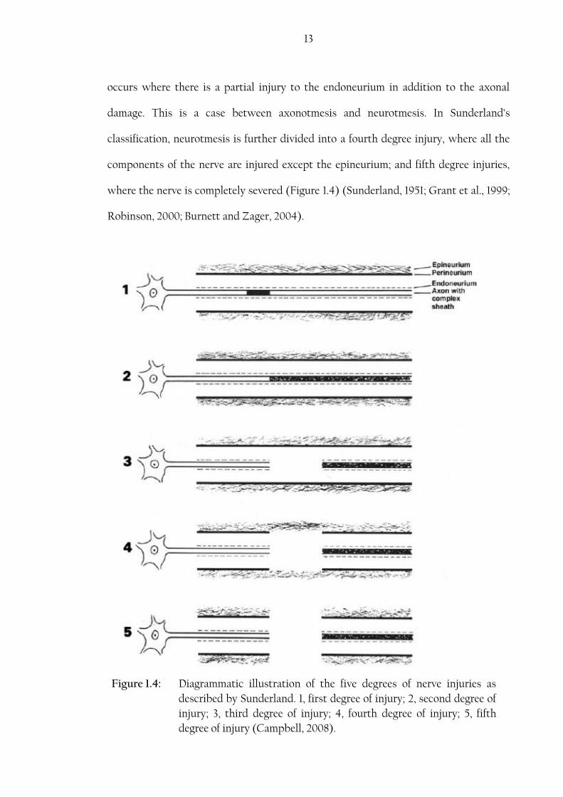
13
Figure 1.4: Diagrammatic illustration of the five degrees of nerve injuries as
described by Sunderland. 1, first degree of injury; 2, second degree of
injury; 3, third degree of injury; 4, fourth degree of injury; 5, fifth
degree of injury (Campbell, 2008).
occurs where there is a partial injury to the endoneurium in addition to the axonal
damage. This is a case between axonotmesis and neurotmesis. In Sunderland’s
classification, neurotmesis is further divided into a fourth degree injury, where all the
components of the nerve are injured except the epineurium; and fifth degree injuries,
where the nerve is completely severed (Figure 1.4) (Sunderland, 1951; Grant et al., 1999;
Robinson, 2000; Burnett and Zager, 2004).

14
Neurapraxia (first degree injuries), no structural changes are noticed and thus no true
degeneration or regeneration occurs. In axonotmesis (second degree injuries), a
calcium-mediated process known as Wallerian degeneration occurs distal to the site of
injury resulting in dissolution of the distal axon, remodelling of the nerve fibre
components and regeneration by regrowth of new axons. Figure 1.5 shows the steps of
Wallerian degeneration. Within hours of injury physical fragmentation of both axons
and myelin occurs, neurotubules and neurofilaments becomes disorganized and
irregular due to varicose swelling (Burnett and Zager, 2004). By 48 to 96 hours after
injury, axonal continuity is lost and impulse conduction is not possible. Schwann cells
become active within 24 hours of injury and divide rapidly to help in the degeneration
and regeneration processes. Endoneural mast cells secret histamine and serotonin
enhancing the permeability of the capillaries and facilitating the migration of
macrophages to the site of injury. Schwann cells pass the degenerated axonal and
myelin debris to macrophages that migrated to the site of injury through the permeable
capillaries in the region. The process of cleaning and phagocytosis at the site of injury
can extend from one week to several months (Campbell, 2008).

15
Figure 1.5: An illustration shows the process of Wallerian degeneration. (A)
Injury occurs to the axon but the endoneurial tube is still intact;
(B) Degeneration of axonal and myelin components of the distal
segment. Elipsoids are formed by Schwann cells to facilitate
phagocytosis by macrophages; (C) Degeneration and clearing of
the distal segment are achieved. Regeneration of the axon begins
guided by bands of Büngner (columns of Schwann cells); (D)
regeneration continues in the endoneurial tube distal to the site of
injury; (E) Full regrowth of axonal and associated myelin
components (Nowak and Handford, 2003; figure 21.6, page 600).
16
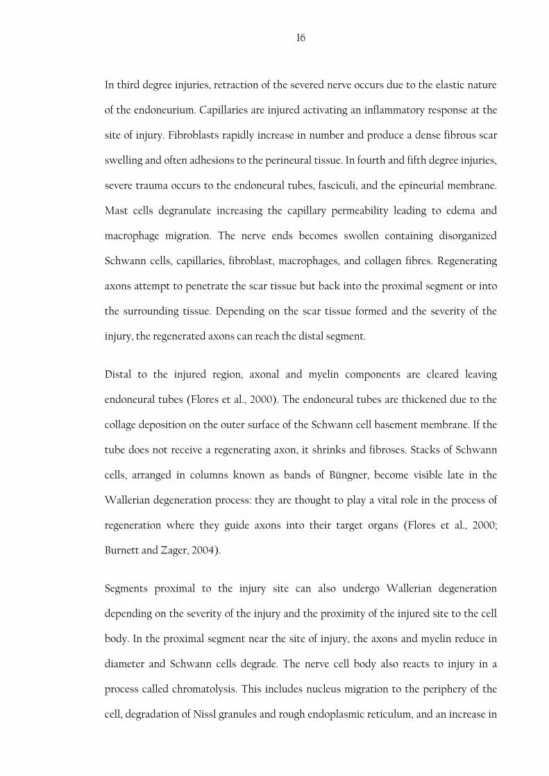
In third degree injuries, retraction of the severed nerve occurs due to the elastic nature
of the endoneurium. Capillaries are injured activating an inflammatory response at the
site of injury. Fibroblasts rapidly increase in number and produce a dense fibrous scar
swelling and often adhesions to the perineural tissue. In fourth and fifth degree injuries,
severe trauma occurs to the endoneural tubes, fasciculi, and the epineurial membrane.
Mast cells degranulate increasing the capillary permeability leading to edema and
macrophage migration. The nerve ends becomes swollen containing disorganized
Schwann cells, capillaries, fibroblast, macrophages, and collagen fibres. Regenerating
axons attempt to penetrate the scar tissue but back into the proximal segment or into
the surrounding tissue. Depending on the scar tissue formed and the severity of the
injury, the regenerated axons can reach the distal segment.
Distal to the injured region, axonal and myelin components are cleared leaving
endoneural tubes (Flores et al., 2000). The endoneural tubes are thickened due to the
collage deposition on the outer surface of the Schwann cell basement membrane. If the
tube does not receive a regenerating axon, it shrinks and fibroses. Stacks of Schwann
cells, arranged in columns known as bands of Büngner, become visible late in the
Wallerian degeneration process: they are thought to play a vital role in the process of
regeneration where they guide axons into their target organs (Flores et al., 2000;
Burnett and Zager, 2004).
Segments proximal to the injury site can also undergo Wallerian degeneration
depending on the severity of the injury and the proximity of the injured site to the cell
body. In the proximal segment near the site of injury, the axons and myelin reduce in
diameter and Schwann cells degrade. The nerve cell body also reacts to injury in a
process called chromatolysis. This includes nucleus migration to the periphery of the
cell, degradation of Nissl granules and rough endoplasmic reticulum, and an increase in

17
the synthesis of RNA, protein components, lipids, and hydrolytic enzymes (Flores et al.,
2000; Burnett and Zager, 2004). This process marks the shift of all function from
impulse transmission to cellular repair, producing important materials for axonal
regrowth during the regeneration phase (Waxman et al., 1995).
The regeneration process starts at the cell body with the reversal of chromatolysis.
Materials are transported from the cell body to the site of axonal regeneration, and the
regenerating axonal tip sprouts out. In severe injuries, scar tissue fills the gap between
the severed nerve endings and resists the advancement of the regenerating axons.
Furthermore, axons can grow and be misdirected into a functionally inappropriate
endoneural tubes or even fail to re-enter the endoneural tube. The resistance that the
regenerating axons receive at the site of injury leads to the creation of multiple smaller
axon sprouts which do not all reach their target organ (Burnett and Zager, 2004;
Campbell, 2008).
Axons entering the correct endoneural tube distal to the site of injury grow to reach the
target organ. Sometimes, several sprouts may enter the endoneural tube, as a result the
regenerated endoneural tube many contain more axons than the original. The
specialized growth cone at the tip of the axon sprout contains multiple filopodia
(cytoplasmic projections) that adhere to the basal lamina of Schwann cells and use it as
a guide. If the axon tip is delayed in getting into the distal segment, the endoneural tube
would have decreases in diameter and thus axonal regrowth is slowed (Fawett and
Keynes, 1990; Campbell, 2008).

18
1.3. Hand Landmarks:
Understanding the surface anatomy of the hand is important in planning surgical
procedures and the assessment of normal and abnormal function in the hand and wrist.
The most important landmarks on the palmar surface of the hand are flexion creases,
the pisiform, scaphoid tubercle, and hook of the hamate. On the dorsal surface of the
hand are Lister’s tubercle, the anatomic snuff box, the lunate fossa, and the radial and
ulnar styloid processes.
1.3.1. Flexion creases:
The skin of the palm is firmly attached to the deep fascia by several relatively constant
palmar creases. The palm creases are significant palmar landmarks because of their
relationship to underlying structures. They also ensure stability of the skin during
gripping and enable movement of the digits without impingement. Most creases do not
correspond to their respective joints, however there are two prominent creases at the
proximal interphalangeal joint, the proximal of which is usually used to determine the
location of the underlying joint (Doyle and Botte, 2003). Appreciation of the
relationship between the creases and the underlying osseous anatomy may be
extremely useful in clinical and surgical settings such as tendon repairs, webbed fingers
and Dupuytren’s disease corrective procedures, trigger finger surgery and even carpal
tunnel release. Creases can be grouped into four categories: the digital, palmar, thenar,
and wrist skin creases (Figure 1.6).

19
There are three horizontal digital creases: proximal, middle and distal digital creases in
all fingers, except the thumb, which lie close to the metacarpophalangeal joint and the
proximal and distal interphalangeal joints respectively. Bugbee and Botte (1993)
reported that the distal digital skin creases, consisting of one or two closely placed
lines, are always proximal to their associated distal interphalangeal joint by 7-7.8 mm.
The middle digital creases are two separated lines, more oriented to their
corresponding joint positions: they lie 1.6-2.6 mm proximal to the proximal
interphalangeal joint. The proximal digital skin creases lie between 14.4-19.6 mm distal
to their respective metacarpophalangeal joints and are usually two parallel lines
separated by 3-4 mm. The metacarpophalangeal crease of the thumb is vertical rather
than horizontal. The interphalangeal joint crease of the thumb is located 2.2 mm
proximal to the interphalangeal joint whereas the proximal joint crease crosses
Figure 1.6: Hand flexion creases (Source: Author).
20
obliquely and directly over the metacarpophalangeal joint of the thumb (Kaplan, 1968;
Bugbee and Botte, 1993).
Kaplan (1968) reported some interesting relations of the digital creases usually seen
when the hand is placed on a flat surface:
The distal digital crease of the index and ring fingers are on the same line and if
extended align with tip of the little finger.
The middle digital crease of the index and ring fingers and the distal digital
crease of the little finger are on the same level.
The second distal line of the proximal digital crease of the ring finger is in line
with the middle digital crease of the little finger.
The palmar skin creases are associated with the metacarpophalangeal joints in the
following manner: the distal palmar crease is located at a mean distance of 7.9 mm, 10.3
mm and 6.9 mm from the metacarpophalangeal joints of the little, ring, and middle
finger respectively (Bugbee and Botte, 1993). The distal palmar crease can extend and
reach the level of the metacarpophalangeal joint of the index finger in 18% of
individuals (Chauhan et al., 2011). Moreover, the proximal palmar crease is located at a
mean distance of 9.1 mm, 18.0 mm and 22.1 mm from the metacarpophalangeal joints of
the index, middle and ring finger respectively (Bugbee and Botte, 1993; Doyle and Botte,
2003). In their study, Bugbee and Botte (1993) mentioned that a longitudinal line
between the lateral border of the proximal palmar crease and the medial border of the
distal palmar crease determines the location of the metacarpal neck in 73% of
specimens. Other researchers, found the line to overlap with the head of the
metacarpals in 38% and the metacarpal neck in 48% (Chauhan et al., 2011).

21
The thenar crease usually intersects the lateral side of the proximal palmar crease at its
distal border and the distal wrist crease near the wrist centre at its proximal border.
Bugbee and Botte (1993) reported that in 54% of specimens, the thenar crease lies
directly over the metacarpal of the middle finger, being in line with the second web
space in 38% and located within the third web space in 8%. In the proximal palm, the
thenar crease crosses the capitate in approximately 50% of specimens. The thenar
crease passes 22.6 mm to the centre of the trapeziometacarpal joint and 18.7 mm distal
to the hook of the hamate on the medial side of the carpus (Bugbee and Botte, 1993). In
another study investigating the palmar creases in an Indian population, Chauhan et al.
(2011) reported that the thenar crease was located in the 2nd intermetacarpal space in
76% and crossed the scaphoid in 26% of individuals. Moreover, the thenar crease
coursed 7.2 mm and 15.6 mm distal to the trapeziometacarpal joint and the hook of the
hamate respectively. The study suggests that the difference in findings could be
attributed to the racial differences due to North Americans possessing wider carpal
regions than Indians.
There are three wrist creases, however only the distal wrist crease can be used as a
reliable landmark. The distal wrist crease is located over the proximal carpal row. The
centre of the lunate is located 9.2 mm proximal to the crease (Bugbee and Botte, 1993).
In their study, Bugbee and Botte (1993) reported that the distal wrist crease crosses the
scaphoid in 98% of individuals with the centre of the scaphoid waist laying 1 mm distal
to it. Moreover, the study also reported that the midpoint of the trapeziometacarpal
joint is located 19.4 mm distal to the crease. On the medial side, the pisiform lies
directly on or slightly distal to the distal wrist crease, whereas the hook of the hamate
is 12.6 mm distal to the crease. The crease is also located 11.7 mm distal to the base of
the ulnar styloid process (Bugbee and Botte, 1993).

22
1.3.2. Bony landmarks:
There are many important bony landmarks in the hand. The pisiform, hook of the
hamate, and the scaphoid tubercle are located in the palmar surface of the hand,
whereas Lister’s tubercle, the anatomic snuff-box, lunate fossa, and the radial and ulnar
styloid processes are found in the dorsal aspect of the hand (Friedman, 2008).
The pisiform bone is a palpable landmark which lies on the medial palmar surface of the
hand, on the distal wrist crease or slightly distal to it (Bugbee and Botte, 1993). It is
used to identify the tendon of flexor carpi ulnaris, the neurovascular bundle and the
Figure 1.7: Important bony landmarks in the palmar surface of the hand.

23
hook of the hamate (Doyle and Botte, 2003). The hook of the hamate is located on the
medial palmar surface of the hand, some 10 mm lateral and distal to the pisiform: it may
be difficult to palpate in some individuals because of its deep position. It is an
important landmark to locate the ulnar artery and nerve and the medial border of the
transverse carpal ligament (TCL) as it lies between the ulnar canal and the carpal
tunnel. On the lateral side of the palm, the scaphoid tubercle projects to the distal
lateral palm and almost always crosses the distal wrist crease (Figure 1.7).
Figure 1.8: Important landmarks in the dorsum of the hand. LT, Lister’s tubercle;
APL, abductor pollicis longus; RSP, Radial styloid process; ASB,
anatomical snuff box; EPB, extensor pollicis brevis; EPL, extensor
pollicis longus; ECRL, extensor carpi radialis longus; ECRB, extensor
carpi radialis brevis; ECU, extensor carpi ulnaris; USP, ulnar styloid
process; LF, lunate fossa (Doyle and Botte, 2003; Figure 9.1, page 487).
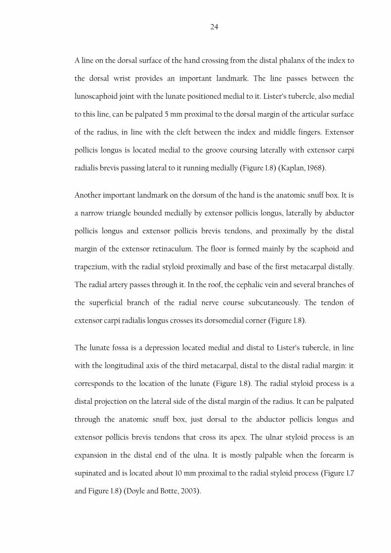
24
A line on the dorsal surface of the hand crossing from the distal phalanx of the index to
the dorsal wrist provides an important landmark. The line passes between the
lunoscaphoid joint with the lunate positioned medial to it. Lister’s tubercle, also medial
to this line, can be palpated 5 mm proximal to the dorsal margin of the articular surface
of the radius, in line with the cleft between the index and middle fingers. Extensor
pollicis longus is located medial to the groove coursing laterally with extensor carpi
radialis brevis passing lateral to it running medially (Figure 1.8) (Kaplan, 1968).
Another important landmark on the dorsum of the hand is the anatomic snuff box. It is
a narrow triangle bounded medially by extensor pollicis longus, laterally by abductor
pollicis longus and extensor pollicis brevis tendons, and proximally by the distal
margin of the extensor retinaculum. The floor is formed mainly by the scaphoid and
trapezium, with the radial styloid proximally and base of the first metacarpal distally.
The radial artery passes through it. In the roof, the cephalic vein and several branches of
the superficial branch of the radial nerve course subcutaneously. The tendon of
extensor carpi radialis longus crosses its dorsomedial corner (Figure 1.8).
The lunate fossa is a depression located medial and distal to Lister’s tubercle, in line
with the longitudinal axis of the third metacarpal, distal to the distal radial margin: it
corresponds to the location of the lunate (Figure 1.8). The radial styloid process is a
distal projection on the lateral side of the distal margin of the radius. It can be palpated
through the anatomic snuff box, just dorsal to the abductor pollicis longus and
extensor pollicis brevis tendons that cross its apex. The ulnar styloid process is an
expansion in the distal end of the ulna. It is mostly palpable when the forearm is
supinated and is located about 10 mm proximal to the radial styloid process (Figure 1.7
and Figure 1.8) (Doyle and Botte, 2003).
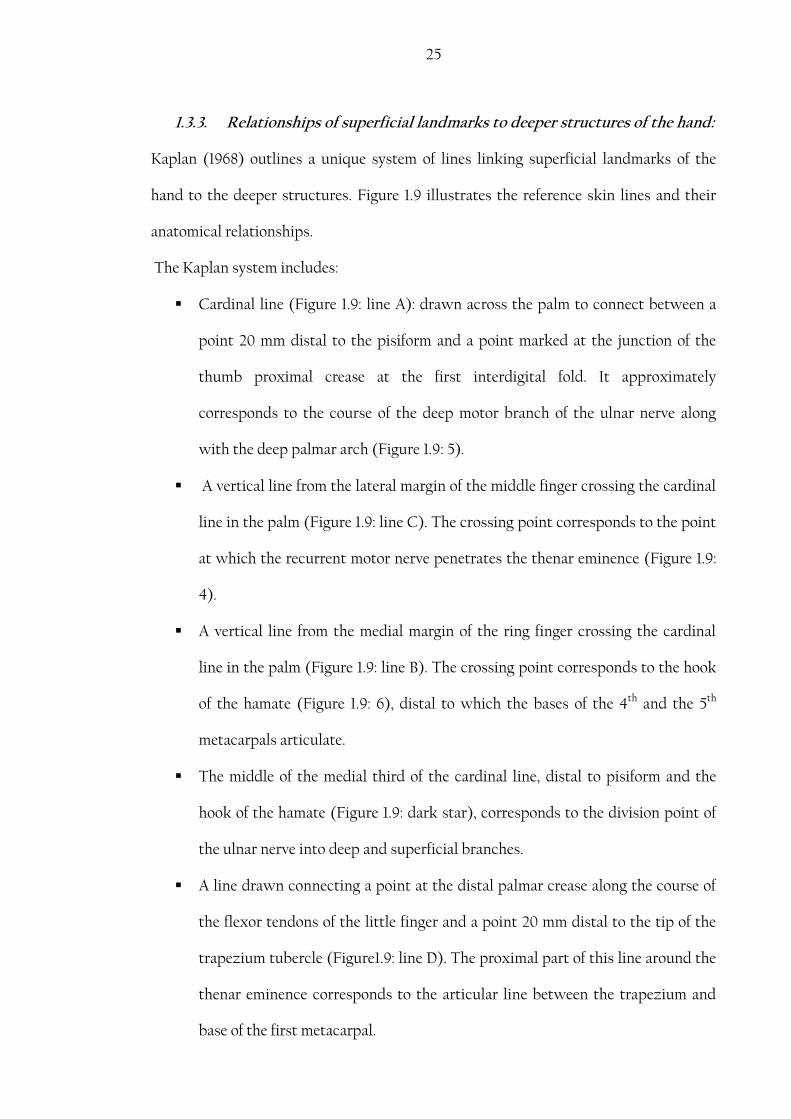
25
1.3.3. Relationships of superficial landmarks to deeper structures of the hand:
Kaplan (1968) outlines a unique system of lines linking superficial landmarks of the
hand to the deeper structures. Figure 1.9 illustrates the reference skin lines and their
anatomical relationships.
The Kaplan system includes:
Cardinal line (Figure 1.9: line A): drawn across the palm to connect between a
point 20 mm distal to the pisiform and a point marked at the junction of the
thumb proximal crease at the first interdigital fold. It approximately
corresponds to the course of the deep motor branch of the ulnar nerve along
with the deep palmar arch (Figure 1.9: 5).
A vertical line from the lateral margin of the middle finger crossing the cardinal
line in the palm (Figure 1.9: line C). The crossing point corresponds to the point
at which the recurrent motor nerve penetrates the thenar eminence (Figure 1.9:
4).
A vertical line from the medial margin of the ring finger crossing the cardinal
line in the palm (Figure 1.9: line B). The crossing point corresponds to the hook
of the hamate (Figure 1.9: 6), distal to which the bases of the 4th and the 5th
metacarpals articulate.
The middle of the medial third of the cardinal line, distal to pisiform and the
hook of the hamate (Figure 1.9: dark star), corresponds to the division point of
the ulnar nerve into deep and superficial branches.
A line drawn connecting a point at the distal palmar crease along the course of
the flexor tendons of the little finger and a point 20 mm distal to the tip of the
trapezium tubercle (Figure1.9: line D). The proximal part of this line around the
thenar eminence corresponds to the articular line between the trapezium and
base of the first metacarpal.

26
Figure 1.9: Kaplan system. (1) Radial artery; (2) Flexor carpi radialis tendon (3)
trapezium; (4) recurrent motor branch of the median nerve; (5) deep
palmar arch; (6) hamate; (7) pisiform; (8) ulnar artery; (9) median
nerve; (10) superficial palmar arch; dark star, division point of the
ulnar nerve into superficial and deep divisions; A, cardinal line; B,
vertical line crosses the cardinal line at the hook of hamate; C,
vertical line crosses the cardinal line at the point of the recurrent
motor branch of the median nerve penetrating the thenar eminence;
D, oblique line corresponds to the articular line between trapezium
and the base of the first metacarpal at its proximal part at the base
of the thenar eminence (Doyle and Botte, 2003; figure 10.3, page
535).
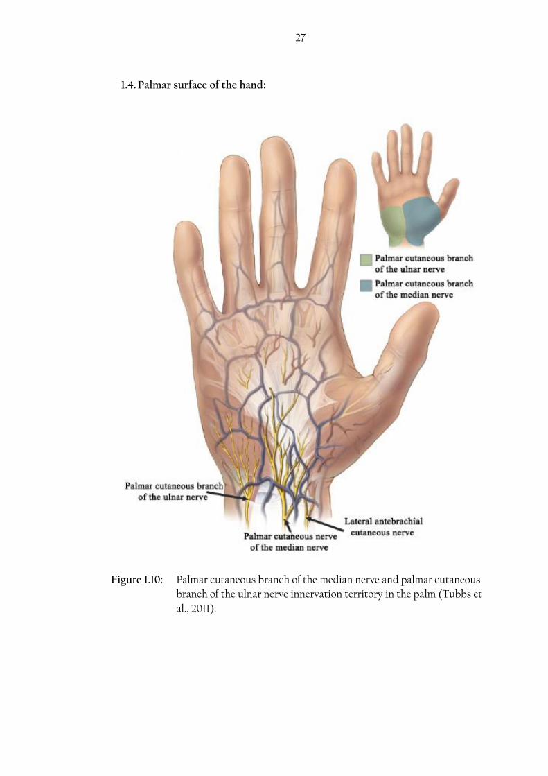
27
1.4. Palmar surface of the hand:
Figure 1.10: Palmar cutaneous branch of the median nerve and palmar cutaneous
branch of the ulnar nerve innervation territory in the palm (Tubbs et
al., 2011).
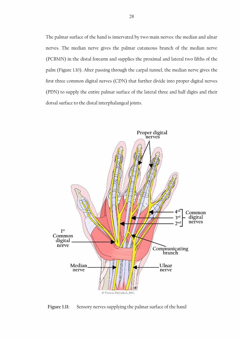
28
Figure 1.11: Sensory nerves supplying the palmar surface of the hand
The palmar surface of the hand is innervated by two main nerves: the median and ulnar
nerves. The median nerve gives the palmar cutaneous branch of the median nerve
(PCBMN) in the distal forearm and supplies the proximal and lateral two fifths of the
palm (Figure 1.10). After passing through the carpal tunnel, the median nerve gives the
first three common digital nerves (CDN) that further divide into proper digital nerves
(PDN) to supply the entire palmar surface of the lateral three and half digits and their
dorsal surface to the distal interphalangeal joints.

29
Originating at various levels in the forearm, the palmar cutaneous branch of the ulnar
nerve and the nerve of Henle supply the medial side of the palm (Figure 1.10). The ulnar
nerve passes through the ulnar canal (Guyon’s canal) and divides into a deep branch
supplying motor innervation to most of the intrinsic muscles of the hand and a
superficial branch that supplies sensory innervation to the medial one and half digits.
Deep to the superficial palmar arch, a communicating branch may be present between
the median and ulnar nerves. This communicating branch allows exchange of sensory
fibres between the ulnar and median nerves (Ferrari and Gilbert, 1991; Don Griot et al.,
2000; Griot et al., 2002; Loukas et al., 2007) (Figure 1.11).
The sensory distribution in the palm is fairly constant compared to that of the dorsal
surface (Stappaerts et al., 1996; Laroy et al., 1998). Nevertheless, there are variations in
reported boundaries of the innervation territory for each nerve. The median nerve is
usually described as innervating the palmar surface of the lateral 3½ digits in
anatomical charts and textbooks; however, according to Hoppenfeld (1976), as cited in
Stappaerts et al. (1996), the sensory innervation to the ring finger as being entirely
supplied by the ulnar nerve. Although less acknowledged by anatomists when
describing the palmar sensory distribution, the medial side of the ring finger can be
supplied by the ulnar nerve through fibres traveling in the palmar CB between the ulnar
and median nerve. Investigating sensory disturbances in gunshot victims from World
War I, Stopford (1918) reported that the ulnar nerve innervated the entire palmar
surface of the ring and little fingers in 18% and extended to supply the medial side of
the middle finger in 3% of the cases investigated. Investigating the median nerve supply
to the palmar surface, the study reported that sensory disturbances were not noted on
the radial side of the ring finger in 20%, nor was it found in the medial side of the
middle finger in 4%. However, the median nerve supplied the entire ring finger in 4%.
30
The study also mentions that the sensory loss was not absolute in the lateral side of the
ring finger suggesting a dual innervation of this area by the ulnar and median nerves
(Stopford, 1918). Linell (1921) also reported some variation to the classical description
of the nerve supply to the palmar surface from investigating 20 cases. In this study, the
ulnar nerve innervated the palmar surface of the entire medial two digits in one case
(5%) and innervated the medial side of the middle finger in three cases (15%) (Linell,
1921).
In velocity conduction studies the sensory distribution is remarkably constant. Laroy et
al. (1998) investigated the palmar sensory distribution in 64 hands and reported that
median nerve stimulation resulted in recordings obtained from the thumb, index,
middle and ring fingers; whereas stimulation of the ulnar nerve resulted in recordings
obtained from the little and ring fingers in all cases. The study also mentions that the
ring finger receives equal contributions from the ulnar and median nerves (Laroy et al.,
1998). Stappaerts et al. (1996) reported that median nerve stimulation resulted in
recordings from the lateral 4 fingers in all cases except two where an additional
recording was also obtained from the little finger. Furthermore, stimulation of the ulnar
nerve resulted in recordings from the little and ring fingers in all cases except three
where an additional recording was also obtained from the middle finger (Stappaerts et
al., 1996). Such variation from the typical description can be attributed to the presence
of a palmar CB allowing transfer of fibres between the two nerves. The innervation of
the palmar surface can differ between the hands of the same individual. Although still
high, symmetry was found in only 87% (Stappaerts et al., 1996).

31
1.4.1. Median nerve
In the axilla, anterior to the third portion of the axillary artery, the lateral and medial
roots of the lateral and medial cords of the brachial plexus join to form the median
nerve. Carrying fibres from the anterior rami of C5, C6, C7, C8 and T1, it supplies the
majority of the flexor compartment of the forearm, some hand muscles and contributes
to the cutaneous innervation of the hand. In the hand, the median nerve supplies the
middle and lateral third of the palmar surface, the palmar surface covering the lateral
3½ digits and the dorsal skin covering the distal phalanges of the lateral 3½ digits by
the palmar cutaneous branch of the median nerve and the common digital nerves.
1.4.1.1. Palmar Cutaneous Branch of the Median nerve
In the forearm, 50 to 70 mm proximal to the wrist crease, the median nerve gives off the
palmar cutaneous branch of the median nerve (PCBMN). The nerve originates
anterolaterally and continues with the main trunk epineurium for about 16-25 mm
before it detaches coursing distally between palmaris longus and flexor carpi radialis.
At the proximal edge of the TCL, the nerve takes a superficial course in line with the
axis of the ring finger, entering its own fibrous tunnel in the ligament. However, the
tunnel can be formed within the antebrachial fascia, the palmar carpal ligament (PCL),
or the flexor retinaculum (Al-Qattan, 1997). The anterior compartment of the forearm is
surrounded by the antebrachial fascia that thickens distally forming the PCL. Distal to
the PCL and at deeper levels, the antebrachial fascia continues distally as the flexor
retinaculum or the TCL (Moore et al., 2006). The nerve enters the tunnel 3 mm medial
to the thenar crease and pierces the palmar aponeurosis 15 mm distal to the scaphoid
tubercle (Naff et al., 1993; Matloub et al., 1998). It usually divides into a larger lateral
and one or more medial branches that supply the lateral two-fifths of the palm and skin

32
over the thenar eminence (Taleisnik, 1973). The nerve continues to branch and
penetrates the fascia and palmaris longus fibres blending with the palmar fascia. Table
1.1 summarizes the anatomical descriptions of the PCBMN in the literature. Das and
Brown (1976) described the most common course of the PCBMN, after investigating
120 cases of carpal tunnel release, was to originate 30-60 mm proximal to the wrist,
course deep to flexor digitorum superficialis, continue distally superficial to the TCL
(flexor retinaculum) and distribute various branches in the mid-palm. However, the
authors noted different patterns of the course of the nerve into the wrist where the
PCBMN can course through or deep to the TCL rather than superficial to it before it
became cutaneous (Das and Brown, 1976). In their study, Matloub et al. (1998) reported
that the most common course of the PCBMN was through the TCL (flexor
retinaculum) (37/40), where the nerve enters its tunnel between the superficial and the
deep layers. However, in a minority of cases, the nerve coursed superficial to the TCL
(flexor retinaculum) and no tunnel was found (3/40) (Matloub et al., 1998).

33
Tab
le 1
.1:
A
nat
omic
al d
escr
ipti
on o
f th
e p
alm
ar c
uta
neo
us
bra
nch
of
the
med
ian
ner
ve a
s m
enti
oned
in t
he
lite
ratu
re
Ref
eren
ce
No
. Cad
aver
s A
nat
om
ical
des
crip
tio
ns
Tal
eisn
ik (
1973
)
12 c
adav
eric
han
ds
Det
aile
d d
escr
ipti
on o
f th
e co
urs
e an
d r
elat
ion
ship
to
stru
ctu
res
in t
he
dis
tal f
orea
rm.
o
Ori
gin
ated
from
th
e an
tero
late
ral s
ide
of t
he
med
ian
ner
ve.
o
Pal
mar
cu
tan
eou
s b
ran
ch o
f th
e m
edia
n n
erve
(P
CB
MN
) re
mai
ns
bou
nd
to
the
mai
n
tru
nk
of t
he
med
ian
ner
ve fo
r 16
-25
mm
aft
er it
s or
igin
.
o
Th
e P
CB
MN
en
ters
a 9
-16
mm
lon
g tu
nn
el b
etw
een
th
e su
per
fici
al a
nd
dee
p la
yers
of t
he
tran
sver
se c
arp
al li
gam
ent.
o
D
ivid
es in
to o
ne
larg
er la
tera
l bra
nch
su
pp
lyin
g th
e th
enar
asp
ect
and
sm
alle
r u
lnar
b
ran
ches
th
at p
erfo
rate
th
e tr
ansv
erse
car
pal
liga
men
t.
Var
iati
ons
wer
e fo
un
d in
3/1
2.
Bez
erra
et
al.
(19
86)
50 a
du
lt fo
rmal
in
fix
ed u
pp
er
extr
emit
ies
Ori
gin
ated
from
th
e la
tera
l sid
e of
th
e m
ain
tru
nk
of t
he
med
ian
ner
ve, 4
5.6±
9.7
mm
(ra
nge
24
.6-
66.8
mm
) p
rox
imal
to
the
wri
st c
reas
e.
It p
erfo
rate
d t
he
ante
bra
chia
l fas
cia
7.9
±3.2
mm
(ra
nge
3.6
-18.
6 m
m)
pro
xim
al t
o th
e w
rist
cr
ease
.
It e
mer
ged
in t
he
pal
m 7
.6±3
.2 m
m (
ran
ge 1
.7-1
8.6
mm
) d
ista
l to
the
wri
st c
reas
e.
Th
e w
idth
of
the
ner
ve a
t th
e p
oin
t of
ori
gin
was
1.2
±0.3
mm
(ra
nge
0.7
-2.1
mm
).
Th
e le
ngt
h o
f th
e n
erve
from
its
poi
nt
of o
rigi
n t
o th
e p
oin
t it
giv
es it
s fi
rst
bra
nch
was
52.
4±1
6.2
(ran
ge 2
0.5
-89
.3 m
m).
Th
ree
typ
es w
ere
rep
orte
d b
ased
on
th
e ar
ea in
ner
vate
d in
th
e p
alm
:
o
Typ
e I:
29
/50
(58
%)
PC
BM
N g
ave
off
one
late
ral b
ran
ch in
ner
vati
ng
the
then
ar r
egio
n
and
an
oth
er in
term
edia
te s
up
ply
ing
the
inte
rmed
iate
reg
ion
of
the
han
d.
o
Typ
e II
: 17/
50 (
34%
) P
CB
MN
div
ided
into
th
ree
bra
nch
es, l
ater
al, i
nte
rmed
iate
an
d
med
ial i
nn
erva
tin
g th
e th
enar
, in
term
edia
te p
orti
on o
f th
e h
and
an
d t
he
hyp
oth
enar
re
gion
res
pec
tive
ly.
o
Typ
e II
I: 4
/50
(8%
) P
CB
MN
gav
e of
f on
e in
term
edia
te b
ran
ch in
ner
vati
ng
the
inte
rmed
iate
por
tion
of t
he
han
d, a
nd
an
oth
er m
edia
l bra
nch
inn
erva
tin
g th
e th
enar
re
gion
.

34
Ref
eren
ce
No
. Cad
aver
s A
nat
om
ical
des
crip
tio
ns
Var
iati
ons
to t
he
thre
e ty
pes
wer
e n
oted
: o
6/
50 (
12%
) p
rese
nte
d a
dee
p b
ran
ch t
hat
con
nec
ted
wit
h t
he
dig
ital
ner
ve o
f th
e m
edia
n n
erve
in
2 c
ases
. o
2/
50 (
4%
) p
alm
ar c
uta
neo
us
ner
ve o
f th
e m
edia
n n
erve
con
nec
ted
wit
h t
he
sup
erfi
cial
bra
nch
of
th
e ra
dia
l ner
ve.
H
obb
s et
al.
(19
90
)
25 fr
esh
cad
aver
ic
up
per
ex
trem
itie
s
from
25
cad
aver
s
Ori
gin
ated
from
th
e la
tera
l sid
e of
th
e m
ain
tru
nk
of t
he
med
ian
ner
ve in
all
cas
es.
Th
e n
erve
has
an
ave
rage
len
gth
of
129
mm
(ra
nge
70
-265
mm
).
Ori
gin
ated
84
mm
(ra
nge
30
-215
mm
) p
rox
imal
to
the
wri
st c
reas
e an
d t
rave
lled
45
mm
(ra
nge
30
-60
mm
) d
ista
l to
the
wri
st c
reas
e.
Bra
nch
es o
rigi
nat
ed p
rox
imal
to
the
wri
st c
reas
e av
erag
ed 0
.6 (
ran
ge 0
-2)
and
th
ose
orig
inat
ed
dis
tal t
o th
e w
rist
cre
ase
aver
aged
6 (
ran
ge 3
-12)
.
Fou
r b
ran
ches
(ra
nge
1-9
) w
ith
len
gth
of 1
6 m
m (
ran
ge 9
-40
mm
) em
erge
d f
rom
th
e la
tera
l sid
e of
th
e P
CB
MN
dis
tal t
o th
e w
rist
cre
ase.
Tw
o b
ran
ches
(ra
nge
0-5
) w
ith
len
gth
of 4
mm
(ra
nge
0-1
0 m
m)
emer
ged
fro
m t
he
med
ial s
ide
of t
he
PC
BM
N d
ista
l to
the
wri
st c
reas
e.
Com
mu
nic
ated
wit
h t
he
sup
erfi
cial
bra
nch
of t
he
rad
ial n
erve
in 2
cas
es.
Tw
o P
CB
MN
s or
igin
ated
from
th
e m
edia
n n
erve
at
dif
fere
nt
leve
ls w
ere
fou
nd
in 4
cas
es a
ll o
f w
hic
h a
re f
rom
mal
e h
and
s.
N
aff e
t al
. (19
93)
21 u
pp
er e
xtr
emit
ies
from
12
fres
h
cad
aver
s
Ori
gin
ated
from
th
e ra
dia
l sid
e of
th
e m
edia
n n
erve
mai
n t
run
k a
nd
cou
rsed
med
iall
y to
th
e fl
exor
car
pi r
adia
lis
(FC
R).
It e
nte
red
th
e fa
scia
l sh
eath
of
the
FC
R 1
5 m
m p
rox
imal
to
the
scap
hoi
d t
ub
ercl
e.
It e
nte
rs it
s tu
nn
el a
t th
e le
vel o
f th
e sc
aph
oid
tu
ber
cle.
It p
ierc
ed t
he
pal
mar
ap
oneu
rosi
s 15
mm
dis
tal t
o th
e sc
aph
oid
tu
ber
cle.
Ori
gin
ated
57
mm
pro
xim
al t
o th
e ra
dia
l sty
loid
pro
cess
(gr
eate
st d
ista
nce
110
mm
).

35
Ref
eren
ce
No
. Cad
aver
s A
nat
om
ical
des
crip
tio
ns
Dow
dy
et a
l. (1
99
4)
52 f
orm
alin
fix
ed
up
per
ex
trem
itie
s (4
9 w
ith
PL
)
Ori
gin
ated
41
mm
(ra
nge
20
-70
mm
) p
rox
imal
to
the
dis
tal w
rist
cre
ase.
Tra
vell
ed t
hro
ugh
th
e te
nd
on o
f pal
mar
is lo
ngu
s in
2/4
9 s
pec
imen
s.
o
In t
he
firs
t ca
se t
he
ner
ve o
rigi
nat
ed 3
0 m
m p
rox
imal
to
the
wri
st c
reas
e an
d p
ierc
ed t
he
ten
don
10
mm
pro
xim
al t
o it
s in
sert
ion
. o
In
th
e se
con
d c
ase
the
ner
ve o
rigi
nat
ed 4
5 m
m p
rox
imal
to
the
wri
st c
reas
e an
d p
ierc
ed
the
ten
don
15
mm
pro
xim
al t
o it
s in
sert
ion
.
DaS
ilva
et
al.
(19
96)
12 f
resh
up
per
ex
trem
itie
s fr
om 9
ca
dav
ers
Pen
etra
ted
th
e an
teb
rach
ial f
asci
a 19
mm
(ra
nge
14
-26
mm
) p
rox
imal
to
the
wri
st c
reas
e
Pen
etra
ted
th
e p
alm
ar a
pon
euro
sis
15 m
m d
ista
l to
the
wri
st c
reas
e.
Gav
e b
ran
ches
pro
xim
al t
o th
e w
rist
cre
ase
in 3
/12
spec
imen
s.
Tw
o ty
pes
wer
e re
por
ted
: o
T
ype
A: t
he
mai
n t
run
k b
ran
ches
into
3 m
ain
bra
nch
es; l
ater
al, i
nte
rmed
iate
an
d u
lnar
(7
/12)
. o
T
ype
B: T
he
mai
n t
run
k b
ran
ches
into
man
y sm
all n
on-d
isti
nct
bra
nch
es (
5/12
).
Du
pli
cate
PC
BM
N t
hat
cre
ated
a s
enso
ry lo
op a
nd
join
ed t
he
pal
mar
cu
tan
eou
s b
ran
ch o
f th
e m
edia
n n
erve
by
pen
etra
tin
g th
e la
tera
l sid
e of
th
e tr
ansv
erse
car
pal
liga
men
t w
as n
oted
in 1
/12
spec
imen
s.
Tw
o P
CB
MN
s w
ith
dif
fere
nt
orig
in p
oin
ts w
ere
not
ed in
1/1
2 sp
ecim
ens.
Imm
un
ohis
tolo
gica
l stu
die
s sh
owed
un
mye
lin
ated
fib
res
end
ing
in t
he
sup
erfi
cial
laye
r of
th
e T
CL
.
Mar
tin
et
al.
(19
96)
25
fres
h f
roze
n
cad
aver
ic h
and
s
Ori
gin
ated
from
th
e ra
dia
l sid
e of
th
e m
ain
tru
nk
of t
he
med
ian
ner
ve, 5
9 m
m (
ran
ge 4
1-78
mm
) p
rox
imal
to
the
wri
st c
reas
e.
Tw
o P
CB
MN
s w
ere
iden
tifi
ed in
tw
o sp
ecim
ens.

36
Ref
eren
ce
No
. Cad
aver
s A
nat
om
ical
des
crip
tio
ns
Wat
chm
aker
et
al. (
199
6)
25 fr
esh
up
per
ex
trem
itie
s
Tw
o n
erve
s w
ere
iden
tifi
ed in
tw
o sp
ecim
ens.
Ori
gin
ated
41
mm
pro
xim
al t
o th
e w
rist
cre
ase
(ran
ge 2
7-63
mm
).
Cou
rsed
2 m
m la
tera
l to
the
then
ar c
reas
e at
th
e le
vel o
f th
e w
rist
cre
ase
wit
h a
ran
ge o
f 6 m
m
late
ral a
nd
med
ial o
f th
e th
enar
cre
ase.
It c
ours
ed 4
-5.5
mm
late
ral t
o th
e in
ter-
then
ar d
epre
ssio
n.
Cou
rsed
9 m
m (
ran
ge 1
-16
mm
) la
tera
l to
the
axis
of t
he
rin
g fi
nge
r in
ex
ten
sion
; an
d c
ours
ed
med
ial t
o th
e ax
is d
uri
ng
flex
ion
.
Th
e n
erve
ove
rlap
ped
wit
h t
he
med
ial a
nte
bra
chia
l cu
tan
eou
s n
erve
in o
ne
case
(1/
25).
It c
ould
be
trac
ed 3
5-4
0 m
m d
ista
l to
the
wri
st c
reas
e b
y lo
up
e m
agn
ific
atio
n.
No
bra
nch
ing
pat
tern
an
d n
o co
mm
un
icat
ion
s w
ith
oth
er c
uta
neo
us
ner
ves
wer
e n
oted
. M
atlo
ub
et
al.
(19
98)
40
fres
h-f
roze
n
up
per
ex
trem
itie
s fr
om 3
0 c
adav
ers
Ori
gin
ated
from
th
e la
tera
l sid
e of
th
e m
ain
tru
nk
of t
he
med
ian
ner
ve, 4
4 m
m p
rox
imal
to
the
wri
st c
reas
e.
Tw
o P
CB
MN
s w
ere
fou
nd
in 6
/40
sp
ecim
ens.
Ave
rage
len
gth
of t
he
tun
nel
is 8
mm
(ra
nge
2-1
5 m
m).
PC
BM
N t
rave
lled
su
per
fici
al t
o th
e T
CL
(fl
exor
ret
inac
ulu
m)
in 3
/40
sp
ecim
ens.
PC
BM
N w
as p
arti
ally
cov
ered
by
fib
res
of p
alm
aris
lon
gus
at it
s in
sert
ion
poi
nt
into
th
e p
alm
ar
apon
euro
sis
in 3
/40
sp
ecim
ens.
Th
ree
dis
trib
uti
on p
atte
rns
wer
e n
oted
: o
T
ype
A: T
he
ner
ve d
ivid
ed in
to o
ne
late
ral a
nd
an
oth
er m
edia
l bra
nch
es t
hat
furt
her
d
ivid
ed in
to 1
-5 b
ran
ches
to
sup
ply
th
e sk
in (
20/4
0).
o
T
ype
B: S
ever
al la
tera
l (th
ree)
an
d m
edia
l (tw
o) b
ran
ches
com
e of
f th
e p
alm
ar c
uta
neo
us
bra
nch
of
the
med
ian
ner
ve b
efor
e it
div
ides
into
med
ial a
nd
late
ral b
ran
ches
(14
/40
).
o
Typ
e C
: Tw
o p
alm
ar c
uta
neo
us
bra
nch
es o
f th
e m
edia
n n
erve
ori
gin
ated
from
th
e m
ain
m
edia
n n
erve
tru
nk
an
d b
oth
gav
e sm
alle
r b
ran
ches
into
th
e p
alm
(6/
40
).
Th
e le
ngt
h o
f th
e P
CB
MN
from
th
e w
rist
cre
ase
to t
he
mos
t d
ista
l bif
urc
atio
n p
oin
t w
as 1
9 m
m
(ran
ge 1
0-2
5 m
m).

37
Ref
eren
ce
No
. Cad
aver
s A
nat
om
ical
des
crip
tio
ns
Th
e P
CB
MN
pas
sed
3 m
m (
ran
ge 0
-7 m
m)
med
ial t
o th
e th
enar
cre
ase.
Th
e an
gle
bet
wee
n t
he
term
inal
late
ral a
nd
med
ial s
ub
-bra
nch
es w
as 6
0.
Th
e m
ost
med
ial t
erm
inal
bra
nch
was
loca
ted
12
mm
(ra
nge
4-2
1 m
m)
to t
he
then
ar c
reas
e an
d
the
mos
t la
tera
l ter
min
al b
ran
ch w
as lo
cate
d 5
mm
(ra
nge
2-9
mm
) to
th
e th
enar
cre
ase.
Ch
ayn
es e
t al
. (2
00
4)
35 h
and
s fr
om 2
5 ca
dav
ers
Ori
gin
ated
from
th
e ra
dia
l sid
e of
th
e m
ain
tru
nk
of t
he
med
ian
ner
ve in
all
cas
es, 4
4.3
mm
(r
ange
24
.8-7
0.8
mm
) p
rox
imal
to
the
bis
tylo
id li
ne.
Mea
n w
idth
0.9
±0.3
mm
(ra
nge
0.4
-1.9
mm
).
Tw
o P
CB
MN
s w
ere
fou
nd
in o
ne
case
.
Bec
ame
sub
cuta
neo
us
5.7
mm
pro
xim
al t
o th
e b
isty
loid
lin
e (1
5.3
mm
pro
xim
al t
o 9
.6 m
m d
ista
l to
th
e b
isty
loid
lin
e).
Ave
rage
len
gth
54
.6 m
m (
ran
ge 9
.2-2
1.7
mm
).
On
e to
th
ree
med
ial b
ran
ches
ave
ragi
ng
3.4
mm
lon
g an
d 1
-7 la
tera
l bra
nch
es a
vera
gin
g 14
.6 m
m
lon
g w
ere
not
ed.
Com
mu
nic
ated
wit
h t
he
sup
erfi
cial
bra
nch
of t
he
rad
ial n
erve
in 5
cas
es.
Tw
o d
istr
ibu
tion
pat
tern
s w
ere
rep
orte
d:
o
Typ
e A
: Th
e n
erve
div
ided
into
tw
o b
ran
ches
(21
/35)
. o
T
ype
B: T
he
ner
ve d
ivid
ed in
to t
hre
e b
ran
ches
(14
/35)
.
Ch
eun
g et
al.
(20
04
)
120
up
per
ex
trem
itie
s fr
om 6
0
adu
lt C
hin
ese
cad
aver
s
Ori
gin
ated
from
th
e la
tera
l sid
e of
th
e m
edia
n n
erve
in 8
8.3%
, dis
tan
ce b
etw
een
pal
mar
is lo
ngu
s an
d P
CB
MN
at
the
leve
l of d
ista
l wri
st c
reas
e w
as 1
4 m
m (
ran
ge 6
-37
mm
).
Ori
gin
ated
from
th
e m
edia
l sid
e of
th
e m
edia
n n
erve
in 1
1.7%
, dis
tan
ce b
etw
een
th
e p
alm
aris
lo
ngu
s an
d P
CB
MN
at
the
leve
l of t
he
dis
tal w
rist
cre
ase
was
3 m
m (
ran
ge 0
-8 m
m).
Ori
gin
ated
32
mm
(ra
nge
12
-65
mm
) p
rox
imal
to
the
dis
tal w
rist
cre
ase.
Ozc
anli
et
al.
(20
10)
16 fr
esh
fro
zen
an
d 1
4
form
alin
fix
ed
cad
aver
ic u
pp
er
extr
emit
ies
Ori
gin
ated
from
th
e la
tera
l sid
e of
th
e m
edia
n n
erve
in 2
8/30
.
Ori
gin
ated
39
.2±1
8.6
mm
pro
xim
al t
o th
e d
ista
l wri
st c
reas
e, a
nd
con
tin
ued
in t
he
pal
m fo
r 19
.8±1
1.5
mm
dis
tal t
o th
e d
ista
l wri
st c
reas
e.

38
One of the early descriptions of the pattern of distribution of the PCBMN was
described by Taleisnik (1973). In this study of twelve hands the PCBMN usually
divided into one large lateral branch supplying the thenar region and another smaller
medial branch perforating the TCL and then continuing along the thenar crease
supplying the midpalm (Taleisnik, 1973). After dissecting 50 adult hands, Bezerra et al.
(1986) described three patterns of division: the most common type, found in 58% of
specimens, included two branches one lateral innervating the thenar region and
another intermediate coursing under the thenar crease and innervating the
intermediate portion of the hand. In 34% of specimens, the PCBMN divided into lateral,
intermediate and medial branches innervating the thenar, intermediate, and hypothenar
regions of the hand respectively. The third type was reported in 8% of specimens where
the PCBMN divided into intermediate and medial branches innervating the
intermediate and thenar regions of the hand respectively (Bezerra et al., 1986). In their
study, DaSilva et al. (1996) reported two patterns of division of the PCBMN. Pattern A
(58.3%) with the main trunk of the nerve dividing into three terminal branches; lateral,
medial and a longer intermediate one. Pattern B (41.7%) was identified with many non-
distinct small branches from the main trunk penetrating the palmar aponeurosis
(DaSilva et al., 1996). In another study, Chaynes et al. (2004) reported two types of
pattern of the PCBMN: the nerve divided into two main branches in 21/35 (60%) cases
and three branches in 14/35 (40%).
Many variations of the PCBMN have been reported in the literature. Two separate
branches of the PCBMN originating at the same level or at different levels have been
identified. It can have a more distal origin from the main trunk of the median nerve and
originate at the level of the proximal edge of the TCL. The PCBMN can originate from
the medial side of the main trunk of the median nerve rather than the commonly known

39
lateral origin; originate more proximally than usual or as high as the junction between
the upper third and the middle third of the forearm (Das and Brown, 1976; Hobbs et al.,
1990; Matloub et al., 1998). During localized anaesthesia to the region of the wrist or the
thenar muscles, care must be taken to block the PCBMN along with the median nerve
unless the block to the median nerve was made proximal to the origin of the PCBMN.
The PCBMN can also be absent, in which case its innervation area is taken over by the
musculocutaneous nerve, the superficial branch of the radial nerve, the palmar
cutaneous branch of the ulnar nerve or any combination of these nerves (Das and
Brown, 1976; Born and Mahoney, 1995; Doyle and Botte, 2003). The PCBMN may
communicate with the superficial branch of the radial nerve (SBRN). The connections
between the two nerves were reported in 8% and could explain the sensory
disturbances in the dorsum of the hand in patients who underwent wrist surgery
(Hobbs et al., 1990). In a more recent study, Chaynes et al. (2004) reported a higher
incidence rate of the connections (14.29%). According to Bezerra et al. (1986), both
Kuhlmann and Meyer-Otetea (1976) and Kuhlmann et al. (1978) reported higher
connection incidences between the PCBMN and the SBRN at 25% and 40%
respectively. These connections may be at risk during surgical procedures to the lateral
side of the wrist such as palmar wrist ganglion or procedures to the distal radius.
The PCBMN can also course deep to or through the fibres of the palmaris longus
tendon. Dowdy et al. (1994) investigated 52 cadavers and reported 2 cases where the
PCBMN passed through the fibres of palmaris longus tendon in its distal 20 mm
endangering the nerve during palmaris longus tendon harvest. Matloub et al. (1998)
also reported the PCBMN coursing deep to the terminal fibres of palmaris longus at its
distal insertion into the palmar aponeurosis in 3/40 specimens. Therefore, during the
harvest of palmaris longus tendon, it is recommended that the tendon be cut proximal

40
to the wrist crease with careful dissection of the lateral side where the PCBMN is most
likely to course (Matloub et al., 1998).
Due to its course in the forearm, the PCBMN is at risk of injury during any surgical
procedure along or adjacent to the ulnar side of flexor carpi radialis. A variation in the
course of the nerve in relation to flexor carpi radialis has been reported. Nagle and
Santiago (2008) noted that the PCBMN coursed superficial to the distal aspect of the
flexor carpi radialis to terminate over the thenar eminence. Tendinitis in the region of
the distal forearm, injury or inflammation that leads to soft tissue swelling, and scar
formation after elective carpal tunnel release can lead to symptoms of PCBMN
entrapment (Pardal-Fernández et al., 2011; Sierakowski et al., 2012). Patients would
suffer from loss of sensibility in the proximal two-fifths of the palm and discomfort, but
more importantly direct injury to the nerve can result in painful neuromas. The nerve
can be also entrapped as it passes through its own fibrous tunnel. Naff et al. (1993)
suggested that in cases where neuroma formation due to direct injury to the PCBMN
cannot be validated; symptoms can be relieved by decompression of the nerve rather
than completely sectioning it. Cases where PCBMN entrapment were identified were
due to continuous pressure at the base of the wrist, an abnormal palmaris longus that
causes pressure upon the nerve, the presence of cysts or ganglia (Buckmiller and
Rickard, 1987; De Smet, 1998), or entrapment within the antebrachial fascia (Semer et
al., 1996). Patients would suffer from symptoms including localized pain and numbness
at the base of the thenar eminence (Nakamichi and Tachibana, 2000). Al-Qattan (1997)
described six zones where the PCBMN is in danger of entrapment. It can be
compressed at its origin from the main median nerve trunk by masses or anomalous
tendons; by the flexor carpi superficialis fascia at the site it emerges at its lateral
margin; at its course close to the medial margin of flexor carpi radialis tendon by
41
tendinitis or ganglia arising from the flexor carpi radialis sheath; at its tunnel by ganglia
or an atypical palmaris longus; at the insertion of palmaris longus into the palmar fascia
in cases where the nerve pierces the distal part of the muscle tendon and at the
subcutaneous layer by fibrous and scar tissue formed after carpal tunnel release.
PCBMN entrapment can occur concomitant with compression of the main median
nerve trunk in carpal tunnel syndrome. Due to the overlap between the sensory
innervation of the PCBMN and the median nerve, it can be difficult to distinguish the
abnormalities caused by PCBMN entrapment from that caused by median nerve
compression in carpal tunnel syndrome (Wada et al., 2002; Imai et al., 2004). Imai et al.
(2004) suggested that the PCBMN entrapment can be assessed objectively by analysing
sensory nerve action potentials regardless of the presence of carpal tunnel syndrome. It
is unclear however, if PCBMN entrapment in these cases is independent or linked to
the median nerve neuropathy. Nonetheless, it is worthwhile evaluating for PCBMN
entrapment in all cases of carpal tunnel syndrome.
The large nerve branches can be easily identified and retracted, but smaller branches
will be difficult to preserve in open carpal tunnel release. In cases of a large branch
injury, neuromas formed will present with easily recognizable symptoms. However, the
smaller nerve branches can also be injured as they cross the TCL. Neuromas in this case
will not be formed but the severed fibres could be trapped in the scarred tissue and
cause unpleasant temporary or prolonged dysesthesia (Taleisnik, 1973). According to
Ahčan et al. (2003) there are at least 2700 axons in the palmar triangle surrounding the
incision used for carpal tunnel release, once injured can multiply by 10 folds creating
30,000 nerve sprouts. These nerve sprouts may be trapped within the scar tissue and
cause scar discomfort and pain (Ahčan et al., 2003). After anatomic, histological and
immunofluorescence studies, DaSilva et al. (1996) reported that the TCL is composed of
42
two layers. The superficial layer consists of loose connective tissue while the deep layer
is composed of thick fibroblasts and collagen. The study demonstrated that only the
superficial layer had unmyelinated nerve fibre endings whereas no fibres were detected
in the deeper thick layers (DaSilva et al., 1996). Such studies suggest that small nerve
endings will be injured during carpal tunnel release and will lead to inescapable
postoperative pillar pain and scar discomfort (DaSilva et al., 1996; Martin et al., 1996;
Ahčan et al., 2003). Moreover, in endoscopic release the main trunk of the PCBMN is in
jeopardy at the proximal incision, smaller branches cannot be preserved as they pass
through or superficial to the ligament and repeated passage of the endoscopic knife will
further damage the small PCBMN branches. Postoperative pain may be explained by
direct injury of the nerve fibres due to incisional trauma or indirectly by coagulation,
suturing, or channel preparations during endoscopic procedures. Scar tenderness can
be explained by nerve ending entrapment in scar tissue. The injured nerve endings
develop sensitivity towards mechanical, thermal and chemical stimulation (Ahčan et
al., 2002; Ahčan et al., 2003).
Since Phalen et al. (1950) described the diagnosis and the suggested sectioning of the
TCL as a treatment; the surgical treatment of carpal tunnel syndrome has rapidly
evolved to include many recommendations, techniques and approaches for placing the
incision. Transverse incisions at the TCL can injure the main nerve trunk at the
proximal wrist. Longitudinal incisions are advocated in many reports; however, there
is debate where the incision should be placed (Table 1.2.). Gerritsen et al. (2001)
reviewed fourteen studies to compare various surgical techniques in carpal tunnel
release. The study reported that the standard open technique seems to offer better post-
operative prognosis than endoscopic technique or open technique with a new incision.

43
Endoscopic carpal tunnel release had more neural complications but shorter recovery
time (Gerritsen et al., 2001).
Table 1.2: Examples of different incision suggestions for the treatment of carpal tunnel syndrome as reported in the literature.
No Reference Method
1. Phalen et al. (1950)
The first to suggest the surgical release of the transverse carpal ligament.
2. Taleisnik (1973) A longitudinal incision on the medial side of the axis of the ray of the ring finger.
3. Ariyan and Watson (1977)
A longitudinal incision in the palm until the wrist crease.
4. Engber and Gmeiner (1980)
A longitudinal incision in line with the ring finger ray axis.
5. Chow (1989) Endoscopic release of carpal tunnel syndrome.
6. Hobbs et al. (1990) A longitudinal incision 10 mm medial to the axis of the 3rd metacarpal (in line of the ring finger axis).
7. Tsai et al. (1995) One portal endoscopic technique with the aid of a custom-made glass tube and a meniscus knife.
8. DaSilva et al. (1996) A longitudinal incision along the axis of the ring finger.
9. Watchmaker et al. (1996)
A longitudinal incision 5 mm ulnar to the interthenar depression.
10. Serra et al. (1997)
Short incision 20 mm long in the midpalm in line with the third ray proximal to Kaplan’s cardinal line.
11. Matloub et al. (1998) A longitudinal incision 10-15 mm medial and parallel to the thenar crease, stopping 15 mm distal to the wrist crease.
Two small incisions:
The first: 20 mm from the centre of the palm extending proximally and perpendicular to the wrist crease.
The second: an incision on the medial side of palmaris longus tendon proximal to the wrist crease.

44
No Reference Method
12. Avci and Sayli (2000) Release by Knifelight inserted through a short 20 mm longitudinal incision in line with the ring finger and proximal to a transverse line crossing from the medial border of an abducted thumb.
13. Cheung et al. (2004) A longitudinal incision 10 mm medial to palmaris longus if present, if absent an incision as described by Taleisnik (1973) is used.
14. Tubbs et al. (2005)
Dorsal incision between the proximal fourth and fifth metacarpals.
15. Tubbs et al. (2011) A longitudinal incision along a line through the middle of the ring finger.
16. De la Fuente et al. (2013) Sectioning the TCL by a “U”-shaped surgical probe and “V”-shaped scalpel with the aid of ultrasound. The probes are inserted through a 15 mm transverse incision starting at the palmaris longus tendon and coursing medially at the level of the distal wrist crease.
Many open techniques have been described to approach the carpal tunnel with minimal
damage to the PCBMN. Taleisnik (1973) recommends the incision to be on the ulnar
side of the axis of the ring finger at the level of the heel of the hand. However, such an
approach will put the palmar branch of the ulnar nerve in danger of injury (Tubbs et al.,
2011). Moreover, the study did not specify how the axis is determined. Watchmaker et
al. (1996) were the first to show the variability of surface landmarks such as the ring
finger axis ray and the palmar creases, in different anatomic positions of the hand. Their
results confirmed that the axis of the ring finger differs significantly during flexion and
extension. The PCBMN coursed 9 mm lateral to the axis in extension, whereas it
coursed medial to the axis during flexion. Moreover, the axis of the ring finger varies
depending on whether the fingers were adducted, abducted or held in-between
(Watchmaker et al., 1996). Engber and Gmeiner (1980) investigated the palmar
cutaneous branch of the ulnar nerve in 21 cadaveric hands and suggested that an

45
incision in line with the ring finger axis will limit injury to both the palmar cutaneous
branch of the ulnar nerve and the PCBMN. Agreeing with Engber and Gmeiner (1980),
Hobbs et al. (1990) also suggested that incisions are made 10 mm ulnar to the axis of the
middle finger, which corresponds to an incision in line with the ring finger axis.
Furthermore, Siegel et al. (1993) recommended the carpal tunnel release incision is in
line with the axis of the ring finger avoiding injury to both PCBMN and the palmar
cutaneous branch of the ulnar nerve. However, the authors emphasised that such an
approach will preserve major neurovascular structures, but will not ensure complete
avoidance of injury as there is no substitute for a wide meticulous exposure (Siegel et
al., 1993). Martin et al. (1996) studied the cutaneous nerves in the palm of 25 fresh
cadavers and reported that an incision in the line of the ring finger axis will injure the
PCBMN in 3/25 cases and will injure cutaneous branches from the ulnar nerve in 16/25
cases. The tenderness often reported post-surgically by patients treated for carpal
tunnel syndrome is most probably due to injury to small cutaneous nerves originating
from the ulnar nerve rather than from PCBMN (Martin et al., 1996). However, Ahčan et
al. (2003) investigated the subcutaneous nerve fibres crossing an incision line with the
lateral border of the ring finger in 15 specimens and reported that fibres from the
PCBMN are endangered in 10/15 specimens but did not mention the subcutaneous
branches from the ulnar nerve.
DaSilva et al. (1996) studied the PCBMN in 12 fresh upper limbs and reported that in 7
cases the nerve divided into three branches as it passed into the proximal palm. The
intermediate branch was found directly on the axis of the ring finger. Interestingly, the
study also reported that the larger branches of the PCBMN are found superficial to the
palmar aponeurosis and terminate in the skin and only the smaller deeper branches
were found to terminate in the superficial layer of the carpal ligament (DaSilva et al.,
46
1996). Other studies suggested using the inter-thenar depression as a safe landmark for
carpal tunnel release (Watchmaker et al., 1996). However, this approach will endanger
the palmar cutaneous branch of the ulnar nerve. As the PCBMN enters its tunnel 3 mm
medial to the thenar crease, a thenar incision would most likely injure the nerve or its
branches (Ahčan et al., 2003). In an interesting Chinese study, Cheung et al. (2004)
suggested using palmaris longus as a landmark for carpal tunnel release incision. The
study recommends an incision 10 mm medial to the palmaris longus if present and
making an incision on the ulnar side of the ring finger axis as described by Taleisnik
(1973) if absent. Ozcanli et al. (2010) recommended the area between the superficial
palmar arch and the distal region of the PCBMN that extends 39.2±18.6 mm distal to
the distal WC as a safe zone for carpal tunnel release incision.
1.4.1.2. Digital nerves
The median nerve becomes superficial 50 mm proximal to the wrist. It courses deep to
palmaris longus (if present), medial to flexor carpi radialis and flexor pollicis longus. It
then enters the carpal tunnel superficial to flexor digitorm superficialis and deep to the
TCL.
The carpal tunnel is a passageway between the forearm and wrist. Its roof is the TCL,
the lateral wall contains the scaphoid and trapezium, the medial wall contains the hook
of the hamate and pisiform, and its floor is formed by the palmar radiocarpal ligaments.
The median nerve passes through the tunnel superficial to all the finger and thumb
flexor tendons as one trunk immediately deep to the TCL.
Usually the recurrent motor branch of the median nerve leaves from the anterolateral
surface at the distal margin of the TCL to supply the thenar muscles. After passing
through the carpal tunnel, the median nerve gives three common digital nerves (CDN)
47
at the junction between the middle and distal third of the metacarpal shafts (Doyle and
Botte, 2003).
The common digital nerves pass deep to the superficial palmar arch and the common
digital arteries. Dividing into proper digital nerves (PDN) at the level of the metacarpal
necks and continue superficial to the digital arteries. Bas and Kleinert (1999)
investigated the branching point of the dorsal proper digital nerves and found them to
be proximal to the A1 pulley zone (a zone spanning 20 mm proximal to the proximal
digital crease to 10 mm distal to the proximal digital crease) in 62% of cases, which is
more proximal than previously reported. The first common digital branch divides into
three proper digital nerves after giving a motor branch to the first lumbrical. The first
common digital nerve supplies the palmar surface of the thumb and lateral side of the
index finger. The second common palmar digital nerve gives a motor branch to the
second lumbrical before it courses distally and further divides to supply the medial side
of the index finger and lateral side of the middle finger. The third common digital nerve
can give a motor branch to the third lumbrical and continues distally to divide into
proper digital nerves to the medial side of the middle finger and lateral side of the ring
finger. It is not uncommon for the third common digital nerve to communicate with the
fourth common digital nerve of the ulnar nerve (Figure 1.11).
Jolley et al. (1997) investigated the first common digital nerve in 79 adult cadaver hands
and observed three patterns (Figure 1.12):
Type I: A PDN supplying the lateral side of the thumb and a CDN that further
divides to supply the medial side of the thumb and lateral side of the
index finger (Figure 1.12: A).

48
Type II: A trifurcation to produce three PDNs to supply the thumb and lateral
side of the index finger (Figure 1.12: B).
Type III: A CDN that further divided to supply the medial and lateral sides of the
thumb and a PDN that continued distally to supply the index finger
(Figure 1.12: C).
These three types were found in 69%, 25%, and 6% of hands respectively (Jolley et al.,
1997).
The median nerve can divide proximal to the distal edge of the carpal tunnel usually
into a larger lateral and a smaller medial component in the forearm or at the level of the
wrist (Eiken et al., 1971). Jeon et al. (2002) reported a case of high division of the third
CDN at the level of the wrist crease discovered during endoscopic release of the carpal
tunnel. Eiken et al. (1971) reported three cases of high division of the median nerve, as
high as the level of the middle third of the forearm, and emphasized that care should be
Figure 1.12: Schematic drawings of the branching patterns of the first common digital nerve. (A) Type I; (B) Type II; (C) Type III (Doyle and Botte, 2003; figure 10.3, page 571).
A B C
49
taken when removing peritendinous tissue as it may mask the 2nd division of the
median nerve. High bifurcation of the median nerve is usually accompanied by a median
artery coursing between the two divisions. However, in their study, Berry et al. (2003)
reported a high bifurcation of the median nerve with each division travelling in a
separate compartment of the carpal tunnel. Such high division of the median nerve is
considered a rare variation. Stančić et al. (1995) found it only once after exploring the
median nerve in 100 cases. In a Korean study, Ahn et al. (2000) reported one case
(0.3%) of high division of the median nerve after investigating 354 cases of elective
carpal tunnel release. In another study conducted on 60 Iranian cadaveric hands,
Alizadeh et al. (2006) reported high bifurcation of the median nerve in 5 cases (8.3%).
Such variations can put the median nerve at risk of injury during carpal tunnel release,
flexor tendosynovectomy or tendon repair in the carpal tunnel.
Common digital nerves can also divide into more than two proper digital nerves. Kuvat
and Tagil (2009) reported a case of duplication of the proper digital nerves in the index
finger. The patient had two medial and two lateral proper digital nerves on each side of
the index finger (Kuvat and Tagil, 2009).
1.2.2. Ulnar nerve
The ulnar nerve originates from the medial cord of the brachial plexus carrying fibres
from the anterior rami of C7, C8 and T1. It courses in the axilla medial to the axillary
artery and continues distally in the arm to pierce the medial intramuscular septum 80
mm proximal to the medial epicondyle and passes to the posterior compartment of the
arm. It enters the anterior compartment of the forearm by passing through the cubital
tunnel where it usually gives three main branches: a motor branch to the medial part of

50
flexor digitorum profundus to the little and ring fingers, a palmar cutaneous branch and
the dorsal branch of the ulnar nerve (Doyle and Botte, 2003).
At the level of the wrist, the ulnar nerve and artery pass through Guyon’s canal, first
described by Guyon in 1861. The canal is bounded between the pisiform and the hook
of the hamate, proximally covered by the TCL and distally by palmaris brevis. The
superficial branch of the ulnar nerve supplies the skin of the little finger and medial side
of the ring finger. The artery usually passes lateral to the nerve and contributes to the
formation of the superficial palmar arch. The ulnar nerve and artery pass lateral to the
pisiform, superficial to the flexor retinaculum, and deep to the superficial carpal
ligament. The canal is divided into three zones: Zone I of the ulnar tunnel (Guyon’s
canal) extends for about 30 mm from the transverse proximal edge of the TCL to the
bifurcation of the nerve into deep and superficial branches about 10 mm distal to the
pisiform, at about the level of the hamate. Zone II includes the motor division of the UN
and Zone III contains the superficial branch of the UN distal to the UN bifurcation.
Table 1.3 shows the boundaries and the content of each zone of Guyon’s canal.
The superficial branch of the ulnar nerve courses distally over abductor and flexor digiti
minimi, gives a motor branch to palmaris brevis, divides into a PDN to the medial side
of the little finger and the fourth CDN that further divides at the level of the metacarpal
shafts to supply the lateral side of the little finger and the medial side of the ring finger
(Doyle and Botte, 2003). The superficial branch of the ulnar nerve was reported to
supply the little finger and both sides of the ring finger in some cases (Bonnel and Vila,
1985). The superficial branch lies deep to the superficial palmar arch but superficial to
the flexor tendons.
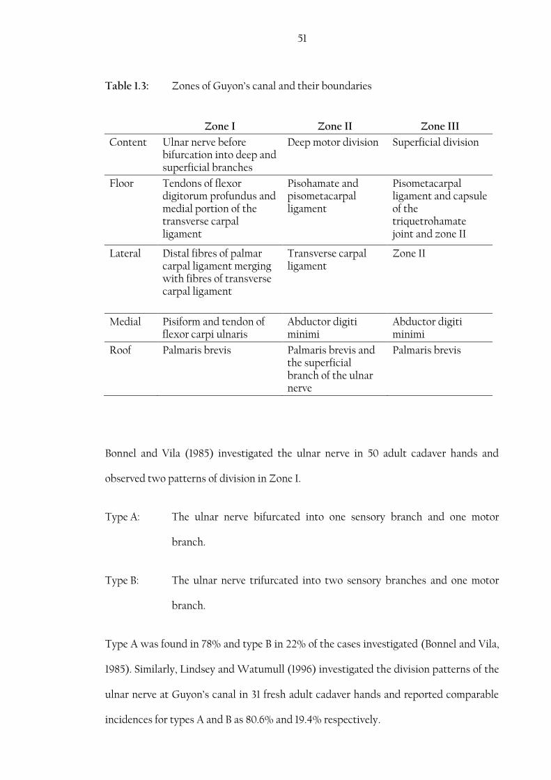
51
Table 1.3: Zones of Guyon’s canal and their boundaries
Zone I Zone II Zone III
Content Ulnar nerve before bifurcation into deep and superficial branches
Deep motor division Superficial division
Floor Tendons of flexor digitorum profundus and medial portion of the transverse carpal ligament
Pisohamate and pisometacarpal ligament
Pisometacarpal ligament and capsule of the triquetrohamate joint and zone II
Lateral Distal fibres of palmar carpal ligament merging with fibres of transverse carpal ligament
Transverse carpal ligament
Zone II
Medial Pisiform and tendon of flexor carpi ulnaris
Abductor digiti minimi
Abductor digiti minimi
Roof Palmaris brevis Palmaris brevis and the superficial branch of the ulnar nerve
Palmaris brevis
Bonnel and Vila (1985) investigated the ulnar nerve in 50 adult cadaver hands and
observed two patterns of division in Zone I.
Type A: The ulnar nerve bifurcated into one sensory branch and one motor
branch.
Type B: The ulnar nerve trifurcated into two sensory branches and one motor
branch.
Type A was found in 78% and type B in 22% of the cases investigated (Bonnel and Vila,
1985). Similarly, Lindsey and Watumull (1996) investigated the division patterns of the
ulnar nerve at Guyon’s canal in 31 fresh adult cadaver hands and reported comparable
incidences for types A and B as 80.6% and 19.4% respectively.

52
Compression of the ulnar nerve in Guyon’s canal can occur due to trauma, fractures,
ulnar artery aneurysm, lipomas, ganglion formations and muscle variations. Injury to
the ulnar nerve and artery can also occur during endoscopic decompression of the
carpal tunnel.
Olave et al. (1997) described a variation of the ulnar nerve dividing into medial and
lateral branches high in the forearm. The medial branch ran on the medial side of the
artery and gave the deep division of the ulnar nerve and proper palmar digital nerve to
the medial side of the little finger. The lateral branch rejoined the medial branch 35.0
mm distal to the wrist crease and gave off the palmar digital branch to the 4th
interosseous space (Olave et al., 1997). Such a variation puts the lateral branch at risk
during endoscopic carpal tunnel release.
Bozkurt et al. (2004) described another variation in the course of the ulnar nerve in the
palm where the PDN to the medial side of the little finger was formed by two branches
from the superficial division of the UN and a third branch from the deep motor branch
piercing the abductor digiti minimi. Such a variation could lead to unexplained sensory
disturbances to the medial side of the ring finger if the deep motor branch was
compressed or injured (Bozkurt et al., 2004).
Kaplan (1963) was the first to describe a connection between the dorsal branch of the
ulnar nerve (DBUN) and the superficial branch of the ulnar nerve: a similar case was
reported by Bonnel and Vila (1985). Hoogbergen and Kauer (1992) reported a case of
Kaplan anastomosis where the DBUN joined the deep branch of the ulnar nerve,
whereas Paraskevas et al. (2008) reported another case where the DBUN connected to
the main trunk of the ulnar nerve before its bifurcation.

53
McCarthy and Nalebuff (1980) reported a case where a branch from the DBUN
innervated the little finger without joining the superficial branch of the ulnar nerve as
described by Kaplan. Bozkurt et al. (2002) described a similar variation where the
medial side of the little finger was innervated by the dorsal branch of the ulnar nerve,
while the superficial branch of the ulnar nerve continued as a CDN to supply the fourth
web-space. Similarly, Konig et al. (1994) described two cases of the 23 specimens
investigated where the little finger was innervated by a branch from the DBUN without
passing through Guyon’s canal.
The Kaplan anastomosis and the DBUN branch to innervate the little finger are both at
risk during surgical procedures on the ulnar side of the wrist, as in Guyon’s canal
exploration and flexor carpi ulnaris surgery, or in cases of pisiform fractures. Injury to
the Kaplan anastomosis can lead to motor and sensory disturbances depending on the
merger point of the nerve (PDN to the medial side of the little finger, superficial branch
of the UN, motor division of the UN, main trunk of the UN). The presence of other
innervation patterns can provide misleading physiological results and complicate
surgical procedures.
1.4.2. Palmar communication between the median and the ulnar nerve
First described by Berrettini in 1741, the communicating branch between the ulnar and
median nerves usually connects between the third and fourth CDNs and passes deep to
the superficial palmar arch, distal to the TCL. It is also referred to as the ramus
communicans, or ramus anastomoticus, or nerve of Berrettini. Reports differ in the
incidence rate of communicating branches (CB) ranging between 4% and 100%.
However, with incidence rates as high as 100% Bonnel and Vila (1985), 95% Ferrari and

54
Gilbert (1991), 94% Don Griot et al. (2000), and 80% Meals and Shaner (1983), the CB
should be considered a normal anatomical finding rather than an anomaly.
Dogan et al. (2010) investigated the CB in the hands of 200 spontaneously aborted
fetuses and found the CB in 29.5%. The authors explained the low incidence rate to be
the result of racial differences and the gestational age as more than 80% of the CB were
found in fetuses more than 20 weeks old (Dogan et al., 2010). Table 1.4 summarizes the
major studies investigating the palmar CB between the MN and UN reported in the
literature.

55
Tab
le 1
.4:
Th
e m
ajor
an
atom
ical
stu
die
s in
vest
igat
ing
the
inci
den
ce a
nd
pat
tern
of
the
pal
mar
com
mu
nic
atio
n b
ran
ch b
etw
een
th
e m
edia
n
ner
ve a
nd
th
e u
lnar
ner
ve
NO
. R
efer
ence
N
o. C
adav
ers
Inci
den
ce
Cla
ssif
icat
ion
P
atte
rn
1.
M
eals
an
d
Shan
er,
1983
50 a
du
lt
cad
aver
pal
ms
(35
froz
en a
nd
15
form
alin
fi
xed
ca
dav
ers)
40
(80
%)
Mea
ls a
nd
Sh
aner
38 (
95%
) T
he
com
mu
nic
atio
n
bra
nch
(C
B)
bra
nch
ed
from
th
e 4
th
com
mon
d
igit
al n
erve
(C
DN
) (u
lnar
) to
th
e 3rd
CD
N (
med
ian
)
23
/38
fib
res
ente
red
th
e la
tera
l sid
e of
th
e ri
ng
fin
ger
10
/38
fib
res
ente
red
th
e la
tera
l si
de
of t
he
rin
g fi
nge
r an
d m
edia
l si
de
of t
he
mid
dle
fin
ger
5/
38 fi
nal
des
tin
atio
n o
f th
e fi
bre
s co
uld
not
be
det
erm
ined
1 (2
.5%
) T
he
CB
bra
nch
ed fr
om t
he
3rd
CD
N (
med
ian
) to
th
e 4
th C
DN
(u
lnar
)
1 (2
.5%
) B
oth
3rd
an
d 4
th C
DN
gav
e a
CB
th
at m
erge
d a
nd
con
tin
ued
in
th
e m
idp
alm
2.
B
onn
el a
nd
V
ila,
19
85
50
form
alin
fi
xed
ad
ult
ca
dav
er h
and
s
50 (
100
%)
Bon
nel
an
d
Vil
a 4
6 (9
2%
) T
he
CB
bra
nch
ed fr
om t
he
4th
CD
N (
uln
ar)
to t
he
3rd
CD
N (
med
ian
)
4 (
8%)
Bot
h 3
rd a
nd
4th
CD
Ns
gave
a C
B t
hat
mer
ged
in
to a
pro
per
dig
ital
n
erve
(P
DN
) to
th
e la
tera
l sid
e of
th
e ri
ng
fin
ger
3.
F
erra
ri a
nd
G
ilb
ert,
19
91
50 a
du
lt
cad
aver
han
ds
45
(90
%)
Fer
rari
an
d
Gil
ber
t 22
(4
8.9
%)
Th
e C
B c
ours
ed o
bli
qu
ely
from
th
e 4
th C
DN
(u
lnar
) to
th
e 3r
d C
DN
(m
edia
n),
th
e d
ista
nce
bet
wee
n t
he
orig
in o
f th
e C
B a
nd
th
e d
ista
l m
argi
n o
f th
e tr
ansv
erse
car
pal
lig
amen
t w
as m
ore
than
4 m
m a
nd
th
e
orig
in a
ngl
e is
less
th
an 5
4.
11 (
24.4
%)
Th
e C
B c
ours
es f
rom
th
e 4
th C
DN
(u
lnar
) to
th
e 3r
d C
DN
(m
edia
n)
par
alle
l to
th
e d
ista
l m
argi
n o
f tr
ansv
erse
car
pal
lig
amen
t, t
he
dis
tan
ce
bet
wee
n t
he
orig
in o
f th
e C
B a
nd
th
e d
ista
l mar
gin
of
tran
sver
se c
arp
al
liga
men
t w
as le
ss t
han
4 m
m w
ith
a r
igh
t an
gle
to t
he
uln
ar n
erve
.

56
NO
. R
efer
ence
N
o. C
adav
ers
Inci
den
ce
Cla
ssif
icat
ion
P
atte
rn
10
(22
.2%
) T
he
CB
cou
rsed
ob
liq
uel
y fr
om t
he
4th
CD
N (
uln
ar)
to t
he
3rd
CD
N
(med
ian
), o
rigi
nat
ed b
elow
th
e d
ista
l m
argi
n o
f th
e tr
ansv
erse
car
pal
li
gam
ent,
th
e or
igin
an
gle
is v
ery
acu
te.
2 (4
.4%
) T
he
CB
bra
nch
ed fr
om t
he
3rd
CD
N (
med
ian
) to
th
e 4
th C
DN
(u
lnar
)
4.
B
as a
nd
K
lein
ert,
19
99
30 f
resh
ad
ult
ca
dav
er p
alm
s 20
(67
%)
Mod
ifie
d
Mea
ls a
nd
Sh
aner
11 (
55%
) T
he
CB
bra
nch
ed fr
om t
he
4th
CD
N (
uln
ar)
to t
he
3rd
CD
N (
med
ian
)
4 (
20%
) T
he
CB
bra
nch
ed fr
om t
he
3rd
CD
N (
med
ian
) to
th
e 4
th C
DN
(u
lnar
)
5 (2
5%)
Mu
ltip
le C
Bs
cou
rsin
g in
bot
h d
irec
tion
s (d
iffu
se in
terc
onn
ecti
ons)
5.
St
ančić
et
al.,
199
9
100
fres
h
adu
lt c
adav
er
han
ds
81 (
81%
) F
erra
ri a
nd
G
ilb
ert
12 (
14.8
%)
Th
e C
B c
ours
ed o
bli
qu
ely
from
th
e 4
th C
DN
(u
lnar
) to
th
e 3r
d C
DN
(m
edia
n),
th
e d
ista
nce
bet
wee
n t
he
orig
in o
f th
e C
B a
nd
th
e d
ista
l m
argi
n o
f th
e tr
ansv
erse
car
pal
lig
amen
t w
as m
ore
than
4m
m a
nd
th
e
orig
in a
ngl
e is
less
th
an 5
4.
16 (
19.8
%)
Th
e C
B c
ours
es f
rom
th
e 4
th C
DN
(u
lnar
) to
th
e 3r
d C
DN
(m
edia
n)
par
alle
l to
th
e d
ista
l m
argi
n o
f th
e tr
ansv
erse
car
pal
lig
amen
t, t
he
dis
tan
ce b
etw
een
th
e or
igin
of
the
CB
an
d t
he
dis
tal
mar
gin
of
the
tran
sver
se c
arp
al l
igam
ent
was
les
s th
an 4
mm
wit
h a
rig
ht
angl
e to
th
e u
lnar
ner
ve.
53
(65.
4%
) T
he
CB
cou
rsed
ob
liq
uel
y fr
om t
he
4th
CD
N (
uln
ar)
to t
he
3rd
CD
N
(med
ian
), o
rigi
nat
ed b
elow
th
e d
ista
l m
argi
n o
f th
e tr
ansv
erse
car
pal
li
gam
ent,
th
e or
igin
an
gle
is v
ery
acu
te.
0 (
0)
Th
e C
B b
ran
ched
fro
m t
he
3rd
CD
N (
med
ian
) to
th
e 4
th C
DN
(u
lnar
)
6.
O
lave
et
al.
200
1 56
form
alin
fi
xed
ad
ult
B
razi
lian
ca
dav
eric
h
and
s
54
(96.
4%
) N
o cl
assi
fica
tion
u
sed
, on
ly
obse
rvat
ion
s w
ere
rep
orte
d
45
(83.
3%)
Ori
gin
ated
fro
m t
he
4th
CD
N (
uln
ar)
and
in
sert
ed i
nto
th
e 3rd
CD
N
(med
ian
) d
ista
lly
8 (1
4.8
%)
Ori
gin
ated
fro
m t
he
3rd C
DN
(m
edia
n)
and
in
sert
ed i
nto
th
e 4
th C
DN
(u
lnar
) d
ista
lly

57
NO
. R
efer
ence
N
o. C
adav
ers
Inci
den
ce
Cla
ssif
icat
ion
P
atte
rn
1
(1.9
%)
Tw
o b
ran
ches
on
e or
igin
ated
fro
m t
he
4th
CD
N (
uln
ar)
and
in
sert
ed
into
th
e 3rd
CD
N (
med
ian
) d
ista
lly
and
th
e ot
her
ori
gin
ated
fro
m t
he
3rd C
DN
(m
edia
n)
and
in
sert
ed i
nto
th
e 4
th C
DN
(u
lnar
) d
ista
lly
cros
sin
g ea
ch o
ther
.
7.
D
on G
riot
et
al.
200
0
53 f
orm
alin
fi
xed
from
ad
ult
cad
aver
h
and
s
50 (
94
%)
Mod
ifie
d
Mea
ls a
nd
Sh
aner
44
(88
%)
Th
e C
B b
ran
ched
from
th
e 4
th C
DN
(u
lnar
) to
th
e 3r
d C
DN
(m
edia
n)
4 (
8%)
Th
e C
B c
ours
es p
erp
end
icu
larl
y b
etw
een
th
e 3r
d a
nd
4th
CD
Ns
2 (4
%)
Th
e C
B b
ran
ched
from
th
e 3r
d C
DN
(m
edia
n)
to t
he
4th
CD
N (
uln
ar)
8.
G
riot
et
al.
200
2 26
form
alin
fi
xed
from
ad
ult
cad
aver
p
alm
s p
urp
osel
y se
lect
ed w
ith
C
B
- M
odif
ied
M
eals
an
d
Shan
er
21 (
80.8
%)
Th
e C
B b
ran
ched
from
th
e 4
th C
DN
(u
lnar
) to
th
e 3r
d C
DN
(m
edia
n)
11
/21
fib
res
join
ed t
he
late
ral s
ide
of t
he
rin
g fi
nge
r
7/
21 f
ibre
s en
tere
d t
he
late
ral
sid
e of
th
e ri
ng
fin
ger
and
med
ial
sid
e of
th
e m
idd
le fi
nge
r
3/
21 f
inal
des
tin
atio
n o
f th
e fi
bre
s co
uld
not
be
det
erm
ined
2 (7
.7%
) T
he
CB
bra
nch
ed fr
om t
he
3rd
CD
N (
med
ian
) to
th
e 4
th C
DN
(u
lnar
)
1/
2 fi
bre
s jo
ined
th
e m
edia
l sid
e of
th
e ri
ng
fin
ger
1/
2 fi
bre
s jo
ined
th
e m
edia
l sid
e of
th
e ri
ng
fin
ger
and
late
ral s
ide
of t
he
litt
le f
inge
r
3 (1
1.5%
) T
he
CB
cou
rses
per
pen
dic
ula
rly
bet
wee
n 3
rd a
nd
4th
CD
Ns
2/
3 b
oth
3rd
an
d 4
th C
DN
gav
e a
CB
th
at m
erge
d a
nd
con
tin
ued
in t
he
mid
pal
m
1/
2 m
ult
iple
CB
s co
urs
ing
in b
oth
dir
ecti
ons
(dif
fuse
inte
rcon
nec
tion
s)

58
NO
. R
efer
ence
N
o. C
adav
ers
Inci
den
ce
Cla
ssif
icat
ion
P
atte
rn
9.
K
awas
him
a et
al,
200
4
169
emb
alm
ed
cad
aver
ic
han
ds
10
9
(74
.5%
) K
awas
him
a et
al
.
24 (
14.2
%)
Typ
e A
: C
B b
ran
ched
fro
m t
he
3rd
CD
N (
med
ian
) to
th
e 4
th C
DN
(u
lnar
)
5 (3
%)
Typ
e B
: m
ult
iple
C
Bs
cou
rsin
g in
b
oth
d
irec
tion
s (d
iffu
se
inte
rcon
nec
tion
s)
60
(35.
5%)
Typ
e C
: No
CB
1 (0
.6%
) T
ype
D:
CB
bra
nch
ed f
rom
4th
CD
N (
uln
ar)
to t
he
late
ral
sid
e of
th
e ri
ng
fin
ger
79 (
46.
7%)
Typ
e E
: T
he
CB
bra
nch
ed f
rom
th
e 4
th C
DN
(u
lnar
) to
th
e 3r
d C
DN
(m
edia
n)
10.
Bia
fora
&
Gon
zale
z,
(20
07)
50 fo
rmal
in
fix
ed a
du
lt
cad
aver
ic
han
ds
37 (
74%
) M
eals
an
d
Shan
er
34 (
91.
9%
) T
he
CB
bra
nch
es f
rom
th
e u
lnar
ner
ve a
nd
in
sert
s in
to t
he
med
ian
n
erve
dis
tall
y
2/34
ad
dit
ion
al b
ran
ch j
oin
ing
the
pro
per
dig
ital
ner
ve o
f th
e la
tera
l si
de
of t
he
rin
g fi
nge
r
1/34
a b
ran
ch a
rose
fro
m t
he
CB
to
join
th
e p
rop
er d
igit
al b
ran
ch t
o th
e u
lnar
sid
e of
th
e m
idd
le f
inge
r
3 (8
.1%
) T
he
CB
bra
nch
es f
rom
th
e m
edia
n n
erve
an
d a
ttac
hes
to
the
uln
ar
ner
ve d
ista
lly
11.
Lou
kas
et
al.,
200
7 20
0 fo
rmal
in
fix
ed a
du
lt
cad
aver
ic
han
ds
170
(85
%)
Mod
ifie
d
Mea
ls a
nd
Sh
aner
143
(84
.1%
) T
he
CB
bra
nch
ed fr
om t
he
4th
CD
N (
uln
ar)
to t
he
3rd
CD
N (
med
ian
)
12 (
7.1%
) T
he
CB
bra
nch
ed fr
om t
he
3rd
CD
N (
med
ian
) to
th
e 4
th C
DN
(u
lnar
)
6 (3
.5%
) T
he
CB
cou
rses
per
pen
dic
ula
rly
bet
wee
n 3
rd a
nd
4th
CD
Ns
9 (
5.3%
) M
ult
iple
CB
s co
urs
ing
in b
oth
dir
ecti
ons
(dif
fuse
inte
rcon
nec
tion
s)

59
NO
. R
efer
ence
N
o. C
adav
ers
Inci
den
ce
Cla
ssif
icat
ion
P
atte
rn
12.
Tag
il e
t al
., 20
07
30
em
bal
med
ad
ult
cad
aver
s p
alm
s
18 (
60%
) M
odif
ied
M
eals
an
d
Shan
er
12 (
66.7
%)
Th
e C
B b
ran
ched
from
th
e 4
th C
DN
(u
lnar
) to
th
e 3r
d C
DN
(m
edia
n)
1 (5
.5%
) T
he
CB
bra
nch
ed fr
om t
he
3rd
CD
N (
med
ian
) to
th
e 4
th C
DN
(u
lnar
)
2 (1
1.1%
) T
he
CB
cou
rses
per
pen
dic
ula
rly
bet
wee
n 3
rd a
nd
4th
CD
Ns
3 (1
6.7%
) M
ult
iple
CB
s co
urs
ing
in b
oth
dir
ecti
ons
(dif
fuse
inte
rcon
nec
tion
s)
13.
Dog
an e
t al
., 20
10
200
from
sp
onta
neo
usl
y ab
orte
d
fetu
ses
59
(29
.5%
) M
odif
ied
K
awas
him
a et
al
. 20
04
, an
ad
dit
ion
al t
ype
was
ad
ded
(t
ype
F)
10 (
5%)
Typ
e A
: C
B b
ran
ched
fro
m t
he
3rd
CD
N (
med
ian
) to
th
e 4
th C
DN
(u
lnar
)
3 (1
.5%
) T
ype
B:
mu
ltip
le
CB
s co
urs
ing
in
bot
h
dir
ecti
ons
(dif
fuse
in
terc
onn
ecti
ons)
141
(70
.5%
) T
ype
C: N
o C
B
2 (1
%)
Typ
e D
: C
B b
ran
ched
fro
m 4
th C
DN
(u
lnar
) to
th
e la
tera
l si
de
of t
he
rin
g fi
nge
r
42
(21%
) T
ype
E:
CB
bra
nch
ed f
rom
th
e 4
th C
DN
(u
lnar
) to
th
e 3r
d C
DN
(m
edia
n)
2 (1
%)
Typ
e F
: Bo
th 3
rd a
nd
4th
CD
N g
ave
a C
B t
hat
mer
ged
an
d c
onti
nu
ed
in t
he
mid
pal
m s
up
ply
ing
the
late
ral r
ing
fin
ger

60
Many classification systems have been suggested in the literature. Meals and Shaner
(1983) were the first to classify the communicating branch according to its origin and
insertion point into three types:
Type I: The CB passed from the 4th CDN (ulnar) to the 3rd CDN (median).
Type II: The CB passed from the 3rd CDN (median) to the 4th CDN (ulnar).
Type III: Both the 3rd and 4th CDNs gave a branch that merged and continued in
the midpalm.
They further subdivided Type I into three subtypes according to the final destination of
the CB fibres (Meals and Shaner, 1983). However, they have not provided any
morphometric data to localize the CB and this limits the surgical use of their results.
Bonnel and Vila (1985) found the CB in all 50 cases investigated: they reported two
types of CB previously described by Meals and Shaner (1983) (Table 1.4); however, they
found that in the 4 cases of Type III, the fibres formed a PDN that supplied the lateral
side of the ring finger. Bonnel and Vila (1985) were also the first to use morphometric
data to localize the CB; however, their data was limited to the origin of the CB. Ferrari
and Gilbert (1991) suggested another classification system based on the relationship of
the CB to the TCL and angle of origin (Figure 1.13). Their four groups are:
Group I: The CB coursed obliquely from the 4th CDN (ulnar) to the 3rd CDN
(median), the distance between the origin of the CB and the distal
margin of the TCL was more than 4 mm and the origin angle less than
54 (Figure 1.13: 1).

61
Group II: The CB courses from the 4th CDN (ulnar) to the 3rd CDN (median)
parallel to the distal margin of the TCL, the distance between the origin
of the CB and the distal margin of the TCL was less than 4 mm with a
right angle to the ulnar nerve (Figure 1.13: 2).
Group III: The CB coursed obliquely from the 4th CDN (ulnar) to the 3rd CDN
(median), originated below the distal margin of the TCL, the origin angle
is very acute (Figure 1.13: 3).
Group IV: The CB branched from the 3rd CDN (median) to the 4th CDN (ulnar)
(Figure 1.13: 4).
Figure 1.13: Schematic drawings of the branching patterns of the palmar
communicating branch according to Ferrari and Gilbert
classification. (1) Group I; (2) Group II; (3) Group III; (4) Group IV;
u, ulnar nerve; m, median nerve; b, communicating branch (Stančić
et al., 1999).

62
Ferrari and Gilbert (1991) also described a triangular zone in the palm where the CB is
always found. This zone is bounded distally by the proximal transverse crease, laterally
by the longitudinal crease between the thenar and hypothenar eminence and extends
medially to the middle half of the hypothenar eminence (Ferrari and Gilbert, 1991). This
area however, is not based on morphometric measurements, but rather on observation
(Figure 1.14).
Figure 1.14: Risk area where the palmar communicating branch is most likely to
be found as described by Ferrari and Gilbert (1991) (Triangular area)
and Don Griot et al. (2000) (trapezoidal area) (Don Griot et al.,
2000).

63
Figure 1.15: Schematic drawing of some of the branching patterns of the palmar
communicating branch (CB) between the median and ulnar nerve as
described in the literature. (1) The CB originate from the fourth
common digital nerve (CDN) proximally and joins the third CDN
distally; (2) the CB originates from the third CDN proximally and
joins the fourth CDN distally; (3) The CB travels transversally
between the third and fourth CDNs; (4) The CB is formed by multiple
branches from both the third and fourth CDNs (Tagiel et al., 2007).
Although some might consider the Ferrari and Gilbert (1991) classification system to be
arbitrary and confusing, Stančić et al. (1999) followed it in their investigation of 100
adult cadaver hands. Interestingly, they reported different distribution and incidence
rates than did Ferrari and Gilbert (1991). (Table 1.4).
Bas and Kleinert (1999) published a detailed study of the nerve’s distribution in the
hand and digits. They modified the Meals and Shaner (1983) classification system by
adding a new type where the palm has multiple CBs coursing in both directions
between the MN and UN (diffuse interconnection) (Figure 1.15: 4). In 2000, Don Griot
et al. described a new type where the CB travels perpendicularly between the 3rd and
4th CDNs (Figure 1.15: 3). Then in another study, they further investigated 26 of the
palms with a CB and modified the Meals and Shaner (1983) classification system by
adding several subtypes based on the final destination of the fibres in the CBs (Griot et
al., 2002) (Table 1.4).

64
Don Griot et al. (2000) used morphometric measurements to localize the CB and
described a rectangular risk zone where the CB would be in danger of injury during
surgical procedures. Their risk zone corresponds to the morphometric parameters of
the proximal origin and distal insertion of the CBs. The proximal origin of the CB was
localized 22% to 69% of the distance between the BSL and the 3rd MCP joint, whereas
the distal insertion was found 32% to 81% of the same distance (Don Griot et al., 2000).
The previously indicated danger zone extends more distally and less medially and
laterally compared to that described by Ferrari and Gilbert (1991) (Figure 1.14).
In another study conducted on 56 Brazilian cadaveric hands, Olave et al. (2001) mapped
the location of the neurovascular structures in the palm in relation to the wrist crease
and the TCL. The study found the palmar CB in 54 (96.4%) cases, in the majority of
which (45/54, 83.3%) the CB originated from the 4th CDN (ulnar nerve) at a mean
distance of 33.9±5.5 mm and 30.2±8.2 mm to the distal WC in right and left male hands
respectively; while it emerged at 28.5±6.2 mm and 27.1.33 mm to the distal WC in right
and left female hands respectively. The CB inserted into the 3rd CDN (median nerve)
distally at 43.6±6.9 mm and 40.2±6.2 mm to the distal WC in right and left male hands
respectively; while it joined at 40.7±7.8 mm and 34.4±1.6 mm to the distal WC in right
and left female hands respectively. The study also reported that the CB was found
6.2±3.7 mm and 5.3±3.7 mm distal to the TCL at the axial line of the 4th metacarpal bone
in male and female right hands respectively; while it was found 5.1±2.8 mm and 4.0±1.9
mm distal to the TCL at the axial line of the 4th metacarpal bone in male and female left
hands respectively (Olave et al., 2001). Although the study presents significant results,
the data does not account for the relative position of the CB but rather to the exact
distances between structures.

65
Figure 1.16: Risk area where the palmar communicating branch (CB) is most
likely to be found as described by Loukas et al. (2007). Light grey
area indicates the range where all palmar CBs were found; dark grey
area indicates the area where the majority (83%) of the palmar CBs
were found (Loukas et al., 2007).
Loukas et al. (2007) studied the palmar CB between the MN and UN in 200 adult
cadavers and by using a modified Meals and Shaner (1983) classification system, were
able to group the CB into four types and then further subdivide them into 15 subtypes.
The authors described a risk area similar to the one suggested by Don Griot et al.
(2000) (Figure 1.16). By using the midpoint of the CB as a reference point, they
indicated that the danger zone extends between 35% and 62% of the distance between
the BSL and the 3rd MCP joint, but also mention that the CB was found outside this
zone in 27% of cases (Loukas et al., 2007).
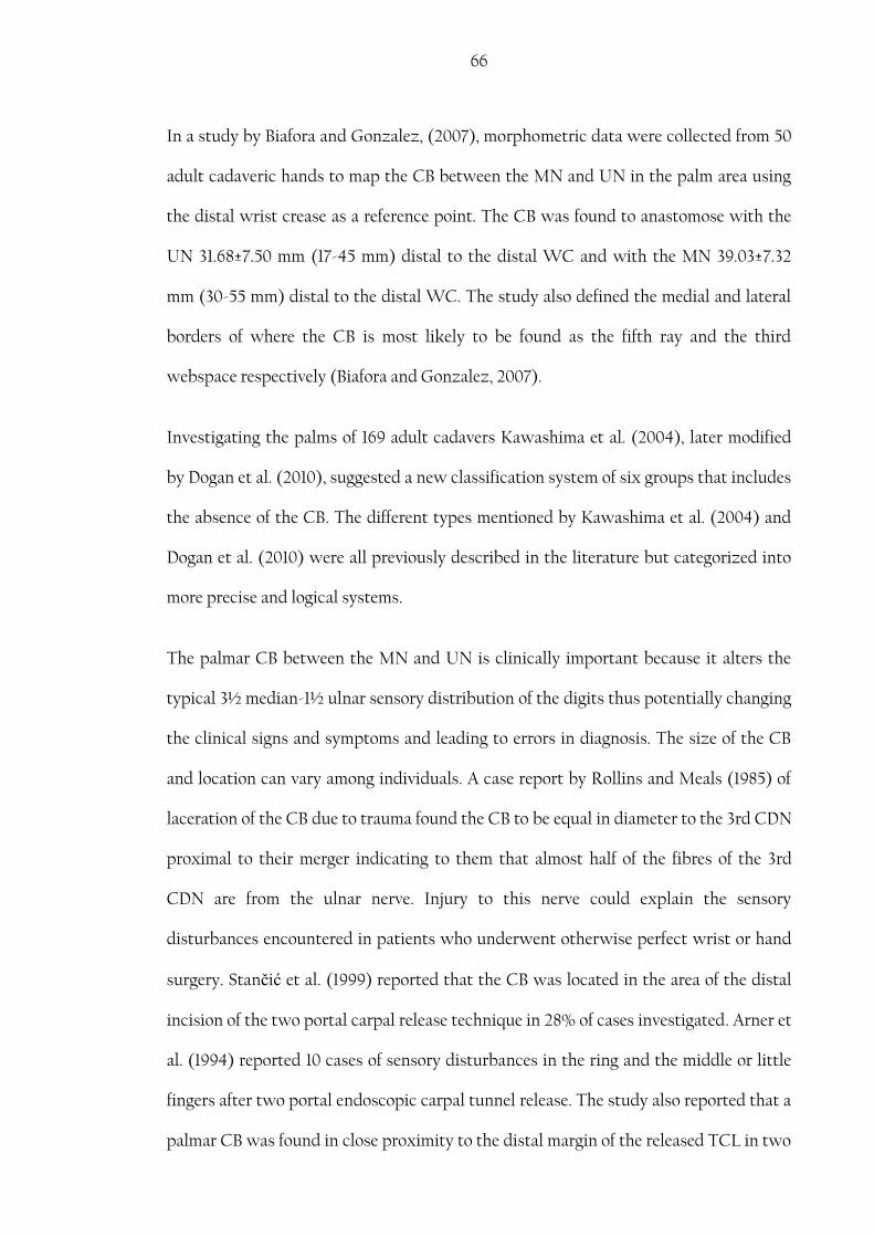
66
In a study by Biafora and Gonzalez, (2007), morphometric data were collected from 50
adult cadaveric hands to map the CB between the MN and UN in the palm area using
the distal wrist crease as a reference point. The CB was found to anastomose with the
UN 31.68±7.50 mm (17-45 mm) distal to the distal WC and with the MN 39.03±7.32
mm (30-55 mm) distal to the distal WC. The study also defined the medial and lateral
borders of where the CB is most likely to be found as the fifth ray and the third
webspace respectively (Biafora and Gonzalez, 2007).
Investigating the palms of 169 adult cadavers Kawashima et al. (2004), later modified
by Dogan et al. (2010), suggested a new classification system of six groups that includes
the absence of the CB. The different types mentioned by Kawashima et al. (2004) and
Dogan et al. (2010) were all previously described in the literature but categorized into
more precise and logical systems.
The palmar CB between the MN and UN is clinically important because it alters the
typical 3½ median-1½ ulnar sensory distribution of the digits thus potentially changing
the clinical signs and symptoms and leading to errors in diagnosis. The size of the CB
and location can vary among individuals. A case report by Rollins and Meals (1985) of
laceration of the CB due to trauma found the CB to be equal in diameter to the 3rd CDN
proximal to their merger indicating to them that almost half of the fibres of the 3rd
CDN are from the ulnar nerve. Injury to this nerve could explain the sensory
disturbances encountered in patients who underwent otherwise perfect wrist or hand
surgery. Stančić et al. (1999) reported that the CB was located in the area of the distal
incision of the two portal carpal release technique in 28% of cases investigated. Arner et
al. (1994) reported 10 cases of sensory disturbances in the ring and the middle or little
fingers after two portal endoscopic carpal tunnel release. The study also reported that a
palmar CB was found in close proximity to the distal margin of the released TCL in two

67
patients and a neuroma was found in the CB in one patient (Arner et al., 1994). In
another case report by May Jr and Rosen (1981) a large neuroma was found on the CB
following a carpal tunnel release of a 34 year old patient. The neuroma caused sensory
disturbances on the lateral side of the ring finger: symptoms were resolved once the
neuroma was resected (May Jr and Rosen, 1981). Due to its close proximity to the distal
margin of the TCL, the CB is at risk during surgical procedures to the hand and wrist in
carpal tunnel release, ring finger flexor tendon surgery, Dupuytren’s fasciectomy and
during exploration of the ulnar side of the TCL and Guyon’s canal (Rollins and Meals,
1985; Olave et al., 2001). Injury to the nerve causes tingling sensation or hyperesthesia in
the digits depending on the topography of the injured branch in the affected patient
(Loukas et al., 2007). Knowledge of the location and range of possible CBs will aid
clinicians to better assess and diagnose cases pre-operatively, recognize and preserve
CBs intra-operatively, and accurately evaluate complications post-operatively.
1.3. Dorsum of the hand:
The dorsum of the hand is supplied by the dorsal branch of the ulnar nerve (DBUN),
the superficial branch of the radial nerve (SBRN) and occasionally the lateral
antebrachial cutaneous nerve (LABCN). The DBUN originates about 80 mm proximal
to the wrist crease and courses distally and dorsally to innervate the skin of the medial
dorsal hand and the dorsal surface of the medial one and half digits (Figure 1.17) (Botte
et al., 1990). The SBRN originates at the level of the lateral epicondyle and courses
distally to become cutaneous by emerging between the tendons of brachioradialis and
extensor carpi radialis longus. It supplies the dorsal skin of the hand and the dorsal
skin of the lateral three and half digits as far as the distal interphalangeal joint. The
DBUN and SBRN may communicate at the dorsum of the hand. The communicating
branch allows exchange of sensory fibres between the two nerves.
68
Knowledge of the sensory innervation of the dorsum of the hand is based generally on
historical large studies examining the nerves of the dorsum of the hand. However, most
studies agree that variations in the sensory innervation to the dorsum of the hand are
not uncommon making the two nerves vulnerable to injury during arthroscopic and
open surgeries performed in the dorsum of the hand (Cirpar et al., 2012). Stopford
(1918) clinically examined more than 1000 gunshot patients from World War I and
reported anaesthesia patterns following peripheral nerve injuries. The study reported
that the DBUN was responsible for innervating the skin of the medial dorsal surface of
the hand and the medial one and half digits in 79% (Stopford, 1918). Dissecting 16 cases,
Linell (1921) found the DBUN to supply the dorsal surface of the medial one and half
digits in only 2 cases (12.5%), the dorsal surface of the medial two digits in 2 cases
(12.5%), the dorsal surface of the medial two and half digits in 11 cases (68.8%), and the
dorsal surface of the medial three and half digits in one case (6.25%). The study
SBRN usually
innervates the
lateral 3½ digits
DBUN usually
innervates the
medial 1½ digits
Figure 1.17: Schematic drawing showing the innervation territory classically
described in the literature (Source: Author).
69
suggested that the seventh cervical nerve fibres, responsible for supplying the middle
finger, travel in the DBUN from high connections at the level of the brachial plexus.
Auerbach et al. (1994) investigated the SBRN in 20 cadaveric upper limbs and reported
that the most common innervation pattern was the SBRN innervating the skin for over
the lateral two and half digits noted in 45% of the cases, whereas it innervated the
lateral three and half in 30%. The study also reported that the SBRN exceeded its
innervation territory supplying the lateral four digits and the lateral four and half digits
in one case each (Auerbach et al., 1994). In more recent studies, Mok et al (2006)
investigated the dorsum of the hand in 30 cadaveric hands and reported that the most
common pattern of distribution of the DBUN was innervation of the skin over the
medial two and half digits, noted in 24/30 cases. The classical description of the medial
one and half digits innervation was only present in 4/30 cases. The study also described
the most common pattern of distribution for the SBRN, present in 16/30 cases, being
innervation of the lateral two and half digits, whereas the classical description of the
lateral three and half digits was only found in 9/30 cases. Moreover, dual innervation
was noted in 10 cases where the two nerves innervated the medial side of the middle
finger and/or the lateral side of the ring finger; however, no mention of communicating
branches was made (Mok et al., 2006). In velocity conduction studies, a reduced or
absent action potential response may result due to variations in the innervation
territory. Authors have advocated that in cases of abnormal DBUN response, the SBRN
should be stimulated to confirm pathology and exclude atypical sensory patterns
(Dutra De Oliveira et al., 2000; Garibaldi and Nucci, 2000).
Communicating branches between the SBRN and the DBUN can alter the sensory
innervation in the dorsum of the hand. The incidence rate of these communicating
branches was reported as 4.2% by Botte et al. (1990), 15% by Auerbach et al. (1994), and
70
60% by Loukas et al. (2008). Such communication can explain the low, absent or/and
atypical response in conduction velocity studies1 (Garibaldi and Nucci, 2000).
An all radial supply to the dorsum of the hand has been mentioned in the literature
with various incidence rates. In dissection based studies it was found in 4.2% as
reported by Botte et al. (1990), 3.3% by Mok et al. (2006), 5.5% by Tiznado et al. (2012)
and 8% by Robson et al. (2008). In velocity conduction studies it was reported in 19%
by Pollak et al. (2013) and 12.9% by Stappaerts et al. (1996). In conduction velocity
studies, small nerves cannot produce enough response to be detected due to their small
diameter and/or the few number of axons present in that nerve. Examiners, studying
the dorsum of the hand, would therefore interpret an absent or low amplitude as the
nerve being absent and thus assume the dorsal surface of the hand to be solely supplied
by the SBRN. This can explain the higher rates reported in conduction velocity studies
compared to that reported in dissection based studies. Failure to recognise variations in
the sensory innervation of the dorsum of the hand could lead to misdiagnosis of ulnar
neuropathy in the forearm or at the elbow especially when there are more than two
variations coexisting (Leis et al., 2010).
Interestingly, the pattern of the innervation in the dorsum of the hand is not only
variable in general, but also variable between the two hands of the same individual. In a
study conducted on cadavers, Huanmanop et al. (2007) reported asymmetry in the
SBRN pattern in 17/79 cases (21.5%). In their study, Stappaerts et al. (1996)
investigated the cutaneous nerve’s action potential response from 31 subjects and
reported that symmetry between the right and left hands was found in only 9/31
subjects (29%). Also Dutra de Oliveira et al. (2000) reported significant differences
between right and left hand sides in 21% of their cases. In conduction velocity studies
1 The communicating branch between the SBRN and DBUN is further explained in section 1.3.3.

71
where the healthy hand is used as a reference to compare the affected hand, it is
important for clinicians interpreting the conduction velocity test results to be aware of
possible variations and asymmetry. The other healthy hand should not be taken as an
absolute reference.
Knowledge of the course and anatomical relations of the DBUN and the SBRN to other
structures in the distal forearm and wrist is critical in clinical settings where diagnostic
and therapeutic procedures can put them in danger of injury.
1.3.1. Superficial branch of the radial nerve
At the level of the elbow, the radial nerve divides into superficial and deep branches.
The radial nerve can divide 20-50 mm proximal or distal to the radiocapitellar
(humeroradial) joint. The superficial branch of the radial nerve (SBRN) courses
superior to supinator and continues deep to brachioradialis. In the distal half of the
forearm, the SBRN pierces the antebrachial fascia between the tendons of
brachioradialis and extensor carpi radialis longus to become subcutaneous about 90
mm proximal to the wrist. In 10% of specimens, the nerve may become subcutaneous by
piercing the tendon of brachioradialis (Doyle and Botte, 2003): a lower incidence was
reported in a Thai cadaver study conducted by Huanmanop et al. (2007). The SBRN
was noted to emerge between two parts of the brachioradialis tendon in 3.8%
(Huanmanop et al., 2007). Such a variation might cause nerve entrapment.
The nerve continues distally and divides into two (91.67%) (one palmar and one dorsal)
or three branches (8.33%) 51 mm proximal to the RSP (Ikiz and Üçerler, 2004). It is
estimated that the nerve becomes subcutaneous and divides for the first time after it
becomes subcutaneous in the distal 20-36% of the distance between the lateral humeral
epicondyle and the RSP. All branches of the SBRN pass lateral to Lister’s tubercle (LT),

72
with the closest branch to the first dorsal compartment passing within a mean distance
of 4 mm. With various patterns and configurations, the palmar branch continues
distally to supply the dorsolateral side of the thumb. It may divide into smaller
branches that spread to supply the palmolateral side of the thenar eminence. It is not
uncommon for the palmar branch to communicate with distal branches from the
LABCN. The dorsal branch continues distally and further divides at the level of the RSP
into two or three branches supplying the dorsomedial side of the thumb and the
dorsolateral side of the index finger; the dorsomedial side of the index and the
dorsolateral side of the middle finger; and the dorsomedial side of the middle finger and
the dorsolateral side of the ring finger. The dorsomedial branch to the thumb and the
dorsolateral branch to the index finger originated from the same main branch in 65% of
the specimens (Doyle and Botte, 2003). The SBRN branches are numbered from dorsal
to palmar and are called superficial radial 1 (SR1), SR2 and SR3 supplying the dorsal
surface of the middle and lateral side of the ring finger, the index and medial side of the
thumb, and the lateral side of the thumb respectively (Ikiz and Üçerler, 2004).
Knowledge of the branching patterns to the thumb and the digits may help prevent
iatrogenic injuries of the nerve and its branches during various clinical procedures.
Furthermore, understanding the patterns and the anatomical course can provide
opportunities to modify or create new clinical applications for reconstructive hand
surgeries. Various reverse neurofasciocutaneous flaps can be obtained from the SBRN
branches with acceptable donor site morbidity (Tellioǧlu et al., 2000). The pattern of
distribution of the SBRN exhibited many configurations. Obtaining a classification
system from those configurations is considered a challenge to anatomists. Table 1.5
describes the classification used in the literature and the incidence rate of each pattern.
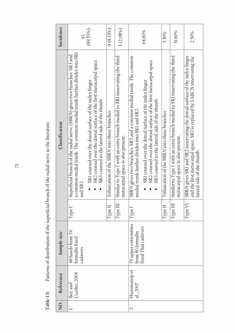
73
Tab
le 1
.5:
Pat
tern
s of
dis
trib
uti
on o
f th
e su
per
fici
al b
ran
ch o
f th
e ra
dia
l ner
ve in
th
e li
tera
ture
NO
R
efer
ence
S
amp
le s
ize
Cla
ssif
icat
ion
In
cid
ence
1
Ikiz
an
d
Üçe
rler
, 20
04
48
han
ds
from
24
fo
rmal
in f
ixed
ca
dav
ers
Typ
e I
Sup
erfi
cial
bra
nch
of
the
rad
ial n
erve
(SB
RN
) gi
ves
two
bra
nch
es: S
R3
and
a
com
mon
med
ial t
run
k. T
he
com
mon
med
ial t
run
k fu
rth
er d
ivid
es in
to S
R1
and
SR
2.
SR1
cou
rsed
ove
r th
e d
orsa
l su
rfac
e of
th
e in
dex
fin
ger
SR2
cou
rsed
ove
r th
e d
orsa
l su
rfac
e of
th
e fi
rst
met
acar
pal
sp
ace
SR3
cou
rsed
to
the
late
ral s
ide
of t
he
thu
mb
43
(89
.53%
)
Typ
e II
T
rifu
rcat
ion
of
the
SBR
N in
to t
hre
e b
ran
ches
. 4
(8.
33%
)
Typ
e II
I Si
mil
ar t
o T
ype
I w
ith
an
ex
tra
bra
nch
med
ial t
o S
R1
inn
erva
tin
g th
e th
ird
m
etac
arp
al s
pac
e is
als
o p
rese
nt.
1
(2.0
8%)
2
Hu
anm
anop
et
al.,
200
7
79 u
pp
er e
xtr
emit
ies
from
40
form
alin
fi
xed
Th
ai c
adav
ers
Typ
e I
SBR
N g
ives
tw
o b
ran
ches
: SR
3 an
d a
com
mon
med
ial t
run
k. T
he
com
mon
m
edia
l tru
nk
furt
her
div
ides
into
SR
1 an
d S
R2.
SR1
cou
rsed
ove
r th
e d
orsa
l su
rfac
e of
th
e in
dex
fin
ger
SR2
cou
rsed
ove
r th
e d
orsa
l su
rfac
e of
th
e fi
rst
met
acar
pal
sp
ace
SR3
cou
rsed
to
the
late
ral s
ide
of t
he
thu
mb
64.6
0%
Typ
e II
T
rifu
rcat
ion
of
the
SBR
N in
to t
hre
e b
ran
ches
1.
30%
Typ
e II
I Si
mil
ar t
o T
ype
I w
ith
an
ex
tra
bra
nch
med
ial t
o S
R1
inn
erva
tin
g th
e th
ird
m
etac
arp
al s
pac
e is
als
o p
rese
nt.
31
.60
%
Typ
e V
I SB
RN
giv
es S
R1
and
SR
2 in
ner
vati
ng
the
dor
sal s
urf
ace
of t
he
ind
ex fi
nge
r an
d t
he
firs
t m
etac
arp
al s
pac
e. S
R3
is r
epla
ced
by
LA
BC
N in
ner
vati
ng
the
late
ral s
ide
of t
he
thu
mb
. 2.
50%

74
NO
R
efer
ence
S
amp
le s
ize
Cla
ssif
icat
ion
In
cid
ence
3
Kor
cek
an
d
Won
gwor
awat
, 20
11
40
up
per
ex
trem
itie
s fr
om 2
4 fo
rmal
in
fix
ed a
du
lt c
adav
ers
Typ
e I
SBR
N d
ivid
es in
to S
R3
and
a c
omm
on m
edia
l tru
nk
furt
her
div
idin
g in
to
SR1
and
SR
2.
34 (
85.0
%)
Typ
e II
SB
RN
div
ides
into
SR
1 an
d a
com
mon
late
ral t
run
k t
hat
furt
her
div
ides
into
of
SR
2 an
d S
R3.
4
(10
.0%
)
Typ
e II
I SB
RN
div
ides
into
SR
1 an
d S
R2
. SR
3 is
mis
sin
g an
d r
epla
ced
by
LA
BC
N.
2 (5
.0%
)
4
Gu
pta
et
al.,
2012
60 u
pp
er e
xtr
emit
ies
from
30
sp
onta
neo
usl
y ab
orte
d fe
tuse
s
Typ
e I:
T
he
SBR
N in
ner
vate
d t
he
late
ral t
wo
and
hal
f d
igit
s 66
.70
%
Ia
: th
e m
ain
tru
nk
bif
urc
ated
into
SR
3 an
d a
med
ial c
omm
on t
run
k t
hat
fu
rth
er d
ivid
es in
to S
R2
and
SR
1:
SR 1
su
pp
lies
th
e 2n
d m
etac
arp
al s
pac
e
SR2
sup
pli
es t
he
2nd
met
acar
pal
sp
ace
SR3
sup
pli
es t
he
late
ral s
ide
of t
he
thu
mb
60%
Ib: t
rifu
rcat
ion
of
SBR
N
6.70
%
Typ
e II
: SB
RN
inn
erva
ted
th
e la
tera
l 3 d
igit
s: S
BR
N d
ivid
es in
to a
med
ial c
omm
on
tru
nk
su
pp
lyin
g th
e m
idd
le, i
nd
ex fi
nge
r an
d o
ccas
ion
ally
th
e m
edia
l sid
e of
th
e th
um
b; a
nd
SR
3 b
ran
ches
su
pp
lyin
g th
e th
um
b.
23.3
0%
IIa:
Th
e SB
RN
bif
urc
ated
into
SR
3 an
d a
med
ial c
omm
on t
run
k t
hat
furt
her
d
ivid
es in
to S
R2
and
SR
1. S
R3
sup
pli
es t
he
late
ral s
ide
of t
he
thu
mb
. 10
%
IIb
: Tri
furc
atio
n o
f th
e S
BR
N in
to 3
bra
nch
es. S
R3
inn
erva
tes
the
enti
re
thu
mb
10
%
IIc:
Th
e SB
RN
div
ides
into
4 b
ran
ches
. SR
3 in
ner
vate
s th
e la
tera
l asp
ect
of
the
thu
mb
. 3.
30%

75
NO
R
efer
ence
S
amp
le s
ize
Cla
ssif
icat
ion
In
cid
ence
T
ype
III:
SB
RN
inn
erva
ted
th
e la
tera
l 3 a
nd
hal
f d
igit
s. T
he
mai
n t
run
k d
ivid
ed in
to a
m
edia
l com
mon
tru
nk
an
d S
R3.
10
%
IIIa
: Th
e m
edia
l com
mon
bra
nch
div
ides
into
4 b
ran
ches
inn
erva
tin
g th
e la
tera
l sid
e of
th
e ri
ng,
th
e en
tire
mid
dle
an
d r
ing
fin
gers
. SR
3 in
ner
vate
s th
e en
tire
th
um
b.
6.67
%
IIIb
: Th
e m
edia
l com
mon
bra
nch
div
ides
into
5 b
ran
ches
inn
erva
tin
g th
e la
tera
l sid
e of
th
e ri
ng
fin
ger,
th
e en
tire
mid
dle
an
d in
dex
fin
gers
, an
d t
he
med
ial s
ide
of t
he
thu
mb
. SR
3 in
ner
vate
s th
e la
tera
l sid
e of
th
e th
um
b.
3.30
%

76
Due to its location and close anatomic relations to other structures in the wrist and
hand, the SBRN is at risk of direct or iatrogenic injury during distal radial fractures,
fixation and reduction of the distal radial fractures, arthroscopic procedures to the
wrist, cephalic vein cannulation, and first dorsal compartment release.
The SBRN is at risk of direct injury during distal radial fractures and indirectly during
the treatment of those fractures. Compressive casts or fracture bracelets may cause
nerve irritation and further complicate the injury (Ikiz and Üçerler, 2004). External
fixator pins are used to stabilize distal radial fractures and have proven to give good
clinical results. Anderson et al. (2004) investigated complications associated with
using external fixation techniques in 24 patients and reported that 66.7% of patients
developed different complications of which 21% were neuropathies involving the
median nerve or the SBRN. The surgeon’s technique in placing the pin influences the
incidence of SBRN injury. An open technique can provide better visualisation and
perseveration of the nerve. In Anderson et al. (2004) series 23 of the 24 patients had
open technique. Moreover, Kirschner wire fixation is used to reduce and stabilize distal
radial fractures. It is done through inserting the pins through the anatomic snuff box
and through the RSP putting the branches of the SBRN that are passing close or over
the region at risk of injury. Ikiz and Üçerler (2004) found a branch of the SBRN passing
in the anatomic snuff box in 16.67% of specimens. Klitscher et al. (2007) reported that
the closest branch to the RSP passed as close as 1 mm to the RSP. Steinberg et al.,
(1995) reported injury to the SBRN in 10% (2/20) and to the LABCN in 15% (3/20) of
the specimens during percutaneous Kirschner wire fixation through the anatomic snuff
box. The study also identified a safe zone of 68 mm2 in the anatomic snuff box where
injury to neurovascular structures could be avoided (Steinberg et al., 1995). However,
individual anatomical variations in the anatomic snuff box eliminate the existence of a

77
true safe zone (Whipple and Poehling, 1996; Korcek and Wongworawat, 2011). Singh et
al. (2005) investigated the incidence of SBRN injury due to radial Kirschner wire
insertion through the styloid process and reported that damage was found in 20%
(8/40) of cases investigated. The SBRN is not only at risk of injury during the insertion
of the Kirschner wires, but also during removal and by the continuous friction with the
bent or buried end of the wire (Singh et al., 2005; Sharma et al., 2006). Hochwald et al.
(1997) compared the risk of inserting a Kirschner wire by an open technique and a
limited open technique on the SBRN and noted that the SBRN was damaged in 7/22
(32%) in percutaneous insertion through the styloid process, while it was only injured
in one case when the limited open technique was used. Many reports advocate placing
the Kirschner wires into the distal radius by limited open incision with blunt
dissection to the bone and using a soft tissue protector as it provides better
visualization and allows identification and preservation of neurovascular structures
(Hochwald et al., 1997; Singh et al., 2005).
Elastic intramedullary nailing is frequently used for displaced and unstable paediatric
fractures in the forearm. It is a preferred technique as it limits soft tissue stripping,
results in limited surgical scars, is minimally invasive and allows early postoperative
movement (Klitscher et al., 2007; Cumming et al., 2008). Radial nailing is achieved
about 20 mm proximal to the epiphysis (Garg et al., 2008). Although relatively
infrequent, SBRN injury is one of the complications of this procedure (Cumming et al.,
2008; Fernandez et al., 2010; Kang et al., 2011). Fernandez et al. (2010) reported that
lesions of the SBRN may occur due to the primary fracture, insertion of the nails or at
the time of material removal. Careful dissection and identification of the SBRN
branches during the insertion of the nails can decrease the injury; however scar tissue

78
formation may hinder identification of the nerve and thus limit the possibility of
preservation (Fernandez et al., 2010).
The SBRN and the cephalic vein have a close anatomical relationship making the nerve
at risk during intravenous cannulation at the wrist (Boeson et al., 2000). Sawaizumi et
al. (2003) reported 11 cases of injury to the SBRN due to intravenous injection into the
cephalic vein at the wrist joint. Complete recovery was achieved within three months
in 36.4% of cases; however complications persisted in the remaining 63.6% (Sawaizumi
et al., 2003). The SBRN intersects the cephalic vein 54.3 mm proximal to the RSP;
however the intersection point could be variable (Klitscher et al., 2007). Gupta et al.
(2012) investigated the SBRN in 60 hands of spontaneously aborted fetuses and
reported that the cephalic vein intersected the nerve at the level of the wrist or distal to
it in all specimens. They also reported that the vein intersected the nerve once in 40%
and twice in 80% of specimens (Gupta et al., 2012). Robson et al. (2008) indicated that
the cephalic vein and the SBRN had a close association (2 mm) in 80% of cases
investigated. Boeson et al. (2000) recommends immediate withdrawal of the catheter if
the patient complained of paresthesias or numbness, whereas Sawaizumi et al. (2003)
believe that wrist venepuncture should only be performed when no other appropriate
alternative is available. Klitscher et al. (2007) and Robson et al., (2008) recommend
venepuncture to be performed prior to the nerve piercing the antebrachial fascia to
avoid any crossing points of the SBRN with the cephalic vein.
Radial forearm fasciocutaneous free flaps are frequently used for the reconstruction of
defects in the oral cavity, oropharyngeal cavity, upper limb, lower limb and genitalia
(Emerick and Deschler, 2007). Damage to the SBRN is a common complication of the
procedure. Radial nerve morbidity as a complication of radial fasciocutaneous free flap

79
harvest was reported as high as 54% (Lutz et al., 1999). Grinsell and Theile (2005)
investigated radial nerve morbidity associated with radial artery free flap harvest in two
techniques that depend on harvesting the venae commitantes or the cephalic vein with
the flap. The study concluded that the incidence of damage to SBRN branches was 9%
when harvesting the venae commitantes and doubled (18%) when the cephalic vein was
involved (Grinsell and Theile, 2005). The doubling of nerve morbidity due to including
the cephalic vein in the flap can be explained by the close anatomical association
between the cephalic vein and the SBRN that puts the nerve in danger of injury during
clinical procedures performed close to the cephalic vein (Robson et al., 2008).
The SBRN is an important structure to consider during clinical procedures to the radial
artery. Robson et al., (2008) reported that the radial artery was closely associated with
the SBRN at the level of the RSP in 12 (48%) of 25 specimens and to the LABCN in 6
(24%). The radial artery is harvested for coronary artery bypass surgery and is
frequently used for arterial blood gas analysis. Percutaneous needle puncture is
considerably painful and may provoke anxiety and discomfort in patients that require
multiple arterial blood gas analysis (Tran et al., 2002). Several studies have concluded
that the use of topical anesthetic gels did not contribute to the reduction of pain during
radial artery puncture in comparison to local infiltrative anesthesia (Tran et al., 2002;
Aaron et al., 2003; Robson et al., 2008). The pain might be induced as a result of
irritation or injury to the SBRN branches or the LABCN which are closely associated to
the radial artery. Robson et al. (2008) recommended approaching the artery from the
ulnar side as they found no nerve branches passing on the ulnar aspect of the artery.
The radial artery is harvested for coronary artery bypass surgery: complications at the
site of harvest have been reported in the literature. Greene and Malias (2001) reported
sensory disturbances related to the SBRN and the LABCN in 36 arms (10.7%). A higher

80
incidence rate was reported by Siminelakis et al. (2004) after investigating 54 patients
who underwent radial artery harvesting for coronary bypass surgery. Sensory
disturbances were reported in 34.09% of which 31.28% were dorsal disturbances and
2.27% were palmar (Siminelakis et al., 2004). In another study investigating the
complication of radial artery harvest in 560 patients, Denton et al. (2001) reported that
dorsal sensation abnormalities were found in 13.8%, palmar abnormities were reported
in 12.7% and decreased thumb function in 5.5% indicating the involvement of the
median nerve and the SBRN. The study however, did not explore the possibility of
involvement of LABCN injury and explains the neurological complications by direct
injury to the SBRN and indirect median nerve ischemia due to loss of collateral radial
blood supply (Denton et al., 2001). It is clear that the dorsal sensory abnormalities are
caused by SBRN injury only; moreover the palmar abnormalities may be the result of
injury to LABCN, SBRN branches, or both. Although endoscopic techniques use a small
skin incision which minimizes wound infection and scarring leading to less
postoperative recovery time and better cosmetic results, sensory disturbances have
been reported in conventional and endoscopic radial artery harvest. The distribution of
sensory disturbances indicates the involvement of both the SBRN and the LABCN in
the conventional open method. Endoscopic harvest showed paresthesia in smaller areas
confined to the SBRN sensory territory which suggests that only the SBRN is involved
(Shapira et al., 2006). In another study neurological complications including a lesion in
the radial artery and paraesthesia developed in 22 and 24 patients who underwent
radial artery harvest by conventional open and endoscopic methods respectively
(Bleiziffer et al., 2008).
Arthroscopy to the lateral area of the wrist or the dorsum of the hand can put the SBRN
at risk of injury. Arthroscopy is a diagnostic and therapeutic tool used in many clinical
81
conditions including triangular fibrocartilage complex tears, carpal interosseous
ligament injuries, reduction and fixation of distal radial fractures and resection of
dorsal ganglia. The SBRN passes superficially to the arthroscopic portals. Kiliç et al.
(2009) investigated the relation of wrist arthroscopy portals to the SBRN in 11
cadaveric hands. Due to the small area where arthroscopic portals can be positioned
and the great number of individual anatomic variations, the authors stated that it was
not possible to create guidelines to prevent SBRN injury and recommended making
skin-only incisions and proceed by blunt dissection to establish portals and minimize
the risk of injury to the SBRN (Kiliç et al., 2009). Ropars et al. (2010) investigated the
portals used in carpometacarpal and metacarpophalangeal joints in thumb arthroscopy
in 30 fresh cadaveric wrists and reported that SR2 and SR3 are at high risk of injury as
they pass close to the portals.
Arthroplasty to the distal scaphoid joint is used to eliminate pain and restore strength
and function in the wrist. The procedure includes a longitudinal 40 mm incision
between the scaphoid and trapezium ending just distal to the RSP. Careful dissection
and gentle retraction of the SBRN is essential to avoid injury to the nerves passing over
the region (Wessels, 2004). Wessels (2004) investigated arthroplasty to the distal
scaphoid joint in 63 patients and reported two cases of SBRN injuries. An interesting
case was reported by Kuruvilla et al. (2002) of a patient complaining of sensory
disturbances in the dorsomedial side of the hand after undergoing wrist arthroplasty
even though the surgical incision did not pass over the DBUN. The SBRN innervated
the entire dorsal surface of patient’s hand, thus a lesion on the medial branches of the
SBRN caused by the surgical incision resulted in sensory loss (Kuruvilla et al., 2002).
Extensive knowledge of the innervation of the wrist joint is essential in Wilhem’s
technique for wrist denervation. Careless dissection of the lateral side of the wrist over

82
the styloid process and the first dorsal compartment can injure sensory branches of the
SBRN and/or cause neuromas (Ekerot et al., 1983). Paresthesia in the lateral region has
been reported following the procedure indicating possible injury of the main nerve
(Ferreres et al., 2002).
De Quervain’s disease is an inflammatory disease of the first dorsal compartment. The
surgical treatment of this condition includes the release of the first dorsal compartment
to allow the inflammation to subside. Treatment varies from immobilization of the
wrist to surgery according to the severity of the condition. Branches of the SBRN pass
over the first compartment in 16.67% of cases (Ikiz and Üçerler, 2004). Investigating 40
Thai cadavers, Huanmanop et al. (2007) reported that the incidence of SBRN branches
passing over the first dorsal compartment is double that mentioned by Ikiz and Üçerler
(2004), being 38%. Klitscher et al. (2007) stated that the SBRN branches may pass as
close as 2 mm to the middle of the first dorsal compartment. Many surgical techniques
have been encouraged in the literature arguing their cosmetic outcome or reduced
complications. According to Robson et al. (2008), the transverse incision or the z
incision would have consistently crossed the SBRN branches in all cases investigated.
The authors advocate the use of a longitudinal incision 25 mm proximal to the RSP as it
avoided damaging any SBRN branches in 68%. The authors also stress the importance
of careful dissection to help identify and preserve the nerve branches in the remaining
32% (Robson et al., 2008). Surgeons should be aware of the presence and close
proximity of the nerve and should try to identify and preserve any nerve tissue to avoid
postoperative complications.
Procedures done on the wrist or the dorsum of the hand such as nerve blocks through
the anatomic snuff box or bone grafts taken from the distal radius between the first and
second dorsal compartments can injure the nerve branches passing over the region. It is
83
reported that the SBRN passes over the anatomic snuff box in 16.67% of specimens
(Ikiz and Üçerler, 2004); however Huanmanop et al. (2007) reported that SBRN
branches passed over the anatomical snuff box in 64.6% in Thai cadavers.
It is important to anesthetize the main trunk prior to first branching to achieve total
sensory blockade of the SBRN. Robson et al. (2008) recommend local anaesthetic
infiltration to be administered when the SBRN becomes subcutaneous prior to giving
its first branch between the distal 33% and 20% of the total length of the arm.
It is not uncommon for the SBRN to connect with the LABCN. Usually the LABCN
connects with the palmar branch to supply the lateral side of the thenar eminence.
Huanmanop et al. (2007) found the connections between the two nerves in 34 Thai
cadaveric arms (43%), while Ikiz and Üçerler (2004) reported it in 20.83%. LABCN
was found to extend over the anatomic snuff box in 52.5% and supply the same area as
the SBRN in 10%. The LABCN was also reported to take over the innervation territory
of SR3 (over the lateral and palmolateral side of the thenar eminence) in 5% (Korcek
and Wongworawat, 2011). Mackinnon and Dellon (1985) reported a partial or complete
overlap between the two nerves in 75% of cases and that injuries to SBRN will be
associated with LABCN injuries. Due to the connections between the two nerves,
sectioning SBRN alone to treat neuroma-related pain at the dorsolateral side of the
wrist might not have effective outcomes (Mackinnon and Dellon, 1985; Robson et al.,
2008). Knowledge of these connections is important in cases of denervation, wrist and
thenar region local anaesthesia or nerve block. Table 1.6 outlines anatomic
measurements of the SBRN described in the literature.

84
Tab
le 1
.6:
An
atom
ic m
easu
rem
ents
of t
he
sup
erfi
cial
bra
nch
of t
he
rad
ial n
erve
(SB
RN
) to
bon
y la
nd
mar
ks
that
are
dis
cuss
ed in
th
e li
tera
ture
NO
R
efer
ence
N
o o
f C
adav
ers
1st
bra
nch
Clo
sest
bra
nch
to
Oth
er f
ind
ings
L
iste
r’s
tub
ercl
e
the
cen
tre
of
the
1st
do
rsal
co
mp
artm
ent
1.
A
bra
ms
et a
l., 1
99
2
20 f
resh
ca
dav
ers
up
per
ex
trem
itie
s
51±1
8 m
m
pro
xim
al t
o th
e R
SP
16.0
±0.5
m
m
4.0
±4.0
mm
Bec
omes
su
bcu
tan
eou
s 9
0±1
4
mm
p
rox
imal
to
th
e ra
dia
l sty
loid
pro
cess
(R
SP).
Su
per
fici
al b
ran
ch o
f th
e ra
dia
l n
erve
(SB
RN
) ar
ose
at
the
leve
l of t
he
late
ral h
um
eral
ep
icon
dyl
e in
8/2
0
Wit
hin
21
mm
of t
he
late
ral e
pic
ond
yle
in t
he
12/2
0
2/20
th
e SB
RN
pie
rced
th
e te
nd
on o
f b
rach
iora
dia
lis.
Dia
met
er w
hen
it p
ierc
ed t
he
fasc
ia w
as 3
±0.5
mm
.
Its
firs
t d
ivis
ion
incl
ud
ed 3
bra
nch
es in
3/2
0.
SBR
N b
ecam
e su
bcu
tan
eou
s an
d f
irst
bra
nch
ed a
t th
e d
ista
l 36
%
and
20
%
of
the
dis
tan
ce
from
la
tera
l ep
icon
dyl
e to
th
e R
SP r
esp
ecti
vely
.
At
the
leve
l of
th
e ex
ten
sor
reti
nac
ulu
m,
the
pal
mar
an
d d
orsa
l b
ran
ch d
iam
eter
s w
ere
2.0
±0.4
an
d 2
.0±0
.2
resp
ecti
vely
.
Bra
nch
of
SB
RN
ly
ing
over
th
e ce
ntr
e of
th
e fi
rst
dor
sal c
omp
artm
ent
in 3
5%.
Con
nec
ted
w
ith
th
e la
tera
l an
teb
rach
ial
cuta
neo
us
ner
ve (
LC
AB
N)
in 7
/20
. 2.
Stei
nb
erg
et a
l.,
199
5 20
fre
sh
cad
aver
u
pp
er
extr
emit
ies
42
mm
p
rox
imal
to
Lis
ter’
s tu
ber
cle
- -
LA
BC
N c
ours
ed t
hro
ugh
th
e an
atom
ic s
nu
ff b
ox i
n
9/2
0 (
45%
).
Th
e d
orsa
l b
ran
ch 2
nd m
ajor
div
isio
n o
ccu
rred
4 m
m
pro
xim
al t
o L
iste
r’s
tub
ercl
e.
S3 b
ran
ch w
as o
n t
he
bor
der
of
the
anat
omic
sn
uff
box
in
all
cas
es, 7
.9±4
.9 m
m fr
om t
he
RSP
.
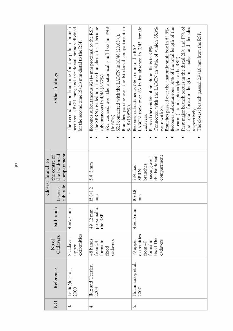
85
NO
R
efer
ence
N
o o
f C
adav
ers
1st
bra
nch
Clo
sest
bra
nch
to
Oth
er f
ind
ings
L
iste
r’s
tub
ercl
e
the
cen
tre
of
the
1st
do
rsal
co
mp
artm
ent
3.
Tel
lioǧ
lu e
t al
., 20
00
8
cad
aver
u
pp
er
extr
emit
ies
46±
5.7
mm
-
-
Th
e se
con
d m
ajor
bra
nch
ing
for
the
pal
mar
bra
nch
oc
curr
ed 4
.8±2
.1 m
m;
and
th
e d
orsa
l b
ran
ch d
ivid
ed
for
the
seco
nd
tim
e 16
±2.3
mm
dis
tal t
o th
e R
SP
.
4.
Ik
iz a
nd
Üçe
rler
, 20
04
4
8 h
and
s fr
om 2
4
form
alin
fi
xed
ca
dav
ers
49
±12
mm
p
rox
imal
to
the
RSP
15.8
±1.2
m
m
5.4
±3 m
m
Bec
omes
su
bcu
tan
eou
s 9
2±14
mm
pro
xim
al t
o th
e R
SP
Th
e SB
RN
div
ided
in
to t
hre
e b
ran
ches
aft
er i
t b
ecam
e su
bcu
tan
eou
s in
4/4
8 (8
.33%
).
SR2
cou
rsed
ove
r th
e an
atom
ical
sn
uff
box
in
8/4
8 (1
6.67
%).
SR3
con
nec
ted
wit
h t
he
LA
BC
N in
10
/48
(20
.83%
).
Bra
nch
es p
assi
ng
over
th
e 1s
t d
orsa
l co
mp
artm
ent
in
8/4
8 (1
6.67
%).
5.
Hu
anm
anop
et
al.,
200
7 79
up
per
ex
trem
itie
s fr
om 4
0
form
alin
fi
xed
Th
ai
cad
aver
s
46±
1.5
mm
10
±3.8
m
m
38%
has
SB
RN
b
ran
ches
p
assi
ng
over
th
e 1s
t d
orsa
l co
mp
artm
ent
Bec
omes
su
bcu
tan
eou
s 73
±1.5
mm
to
the
RS
P
LA
BC
N t
ook
ove
r S3
in
its
ab
sen
ce i
n 2
/43
fem
ale
cad
aver
s.
Pie
rced
th
e te
nd
on o
f b
rach
iora
dia
lis
in 3
.8%
.
Con
nec
ted
wit
h t
he
LA
BC
N i
n 4
3%,
of w
hic
h 8
5.3%
w
ere
wit
h S
3.
Bra
nch
es p
asse
d o
ver
the
anat
omic
sn
uff
box
in 6
4.6
%.
Bec
omes
su
bcu
tan
eou
s 30
% o
f th
e to
tal
len
gth
of
the
fore
arm
(la
tera
l ep
icon
dyl
e to
th
e R
SP).
Fir
st m
ajor
bra
nch
occ
urs
in
th
e d
ista
l 21
% a
nd
17%
of
the
tota
l fo
rear
m
len
gth
in
m
ales
an
d
fem
ales
re
spec
tive
ly.
Th
e cl
oses
t b
ran
ch p
asse
d 2
.9±1
.8 m
m fr
om t
he
RSP
.

86
NO
R
efer
ence
N
o o
f C
adav
ers
1st
bra
nch
Clo
sest
bra
nch
to
Oth
er f
ind
ings
L
iste
r’s
tub
ercl
e
the
cen
tre
of
the
1st
do
rsal
co
mp
artm
ent
6.
Kli
tsch
er e
t al
., 20
07
10 u
pp
er
extr
emit
ies
from
5
emb
alm
ed
cad
aver
s
54.7
±12.
2 m
m
16.4
±3.7
m
m
pal
mar
4
.4±1
.3 m
m;
dor
sal
15.2
±2.8
mm
Th
e se
con
d d
ivis
ion
of
the
SBR
N o
ccu
rred
at
the
leve
l of
th
e R
SP
(0±5
.7
mm
) w
ith
a
ran
ge
of
10
mm
p
rox
imal
to
the
RSP
to
10 m
m d
ista
l to
the
RSP
.
Th
e ce
ph
alic
vei
n a
nd
th
e SB
RN
cro
ssed
at
54.3
±7.6
m
m p
rox
imal
to
the
RSP
(ra
nge
:42
-68
mm
).
Clo
sest
pal
mar
an
d d
orsa
l b
ran
ches
at
the
leve
l of
th
e R
SP is
9.8
±4.5
mm
an
d 3
.5±2
.7 m
m r
esp
ecti
vely
.
7.
Rob
son
et
al.,
200
8 25
up
per
ex
trem
itie
s fr
om
form
alin
fi
xed
ca
dav
ers
49
.2±1
4.4
m
m
14.9
±5.3
m
m
dor
sal 6
.4±4
.1
mm
; pal
mar
4
.9±3
.0 m
m
Bec
omes
su
bcu
tan
eou
s 83
.1±1
1.4
mm
pro
xim
al t
o th
e R
SP.
Th
e SB
RN
bec
ame
sub
stan
ces
in t
he
dis
tal
32.8
±4.1
%
of t
he
tota
l len
gth
of t
he
fore
arm
.
25 m
m i
nci
sion
fro
m t
he
RSP
wil
l av
oid
th
e n
erve
s in
17
/25
case
s (6
8%)
and
in
8/
25
(32
%)
the
inci
sion
w
ould
at
leas
t p
arti
ally
cro
ss t
he
SBR
N.
Rad
ial
arte
ry w
as c
lose
ly a
ssoc
iate
d w
ith
th
e SB
RN
in
12
/25
(48%
).
SBR
N w
as c
lose
ly a
ssoc
iate
d w
ith
th
e ce
ph
alic
vei
n i
n
80%
. A
ssoc
iati
on w
ith
th
e ce
ph
alic
vei
n w
as g
rou
ped
in
to p
rox
imal
an
d d
ista
l as
soci
atio
n i
n 1
6/25
(64
%);
4
/25(
16%
) p
rox
imal
to
the
RSP
; 3/
25 (
12%
) d
ista
l to
R
SP; 2
/25
(8%
) n
o cl
ose
asso
ciat
ion
.
An
an
omal
ous
ner
ve d
istr
ibu
tion
was
not
ed i
n t
wo
han
ds.
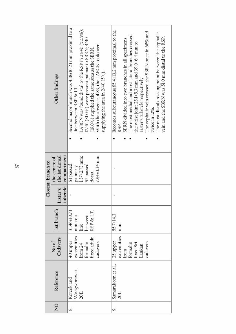
87
NO
R
efer
ence
N
o o
f C
adav
ers
1st
bra
nch
Clo
sest
bra
nch
to
Oth
er f
ind
ings
L
iste
r’s
tub
ercl
e
the
cen
tre
of
the
1st
do
rsal
co
mp
artm
ent
8.
Kor
cek
an
d
Won
gwor
awat
, 20
11
40
up
per
ex
trem
itie
s fr
om 2
4
form
alin
fi
xed
ad
ult
ca
dav
ers
31.4
6±10
.73
mm
to
a li
ne
bet
wee
n
RSP
& L
T.
- S3
pas
sed
p
alm
arly
1.
37±2
.73
mm
; S2
pas
sed
d
orsa
l 7.
94
±3.3
4 m
m
Seco
nd
maj
or b
ran
ch w
as 4
.18±
10.2
3 m
m p
rox
imal
to
a li
ne
bet
wee
n R
SP
& L
T.
LA
BC
N w
as fo
un
d d
ista
l to
the
RSP
in 2
1/4
0 (
52.5
%);
17
/40
(81
.0%
) w
ere
pre
sen
t p
alm
ar t
o SB
RN
; 4/4
0
(19
.0%
) su
pp
lied
th
e sa
me
area
as
the
SBR
N.
Wit
h t
he
abse
nce
of S
3, t
he
LA
BC
N t
ook
ove
r su
pp
lyin
g th
e ar
ea in
2/4
0 (
5%).
9.
Sa
mar
akoo
n e
t al
., 20
11
25 u
pp
er
extr
emit
ies
from
fo
rmal
in
fix
ed S
ri
Lan
kan
ca
dav
ers
55.7
±14
.3
mm
-
-
Bec
omes
su
bcu
tan
eou
s 85
.4±1
3.2
mm
pro
xim
al t
o th
e R
SP.
SBR
N d
ivid
ed in
to t
wo
bra
nch
es in
all
sp
ecim
ens.
Th
e m
ost
med
ial a
nd
mos
t la
tera
l bra
nch
es c
ross
ed
the
wri
st jo
int
25.1
±5.3
mm
an
d 3
9.0
±6.4
mm
to
Lis
ter’
s tu
ber
cle
resp
ecti
vely
.
Th
e ce
ph
alic
vei
n c
ross
ed t
he
SBR
N o
nce
in 6
8% a
nd
tw
ice
in 1
2%.
Th
e m
ost
dis
tal c
ross
ing
poi
nt
bet
wee
n t
he
cep
hal
ic
vein
an
d t
he
SBR
N w
as 5
1.0
mm
dis
tal t
o th
e R
SP.
88
1.3.2. Dorsal branch of the ulnar nerve
Arising from the medial aspect of the main trunk of the ulnar nerve, the dorsal branch
of the ulnar nerve (DBUN) supplies the dorsal surface of the medial 1½ digits. It
originates 83 mm from the proximal border of the pisiform, courses distally and
medially deep to flexor carpi ulnaris to become subcutaneous 50 mm proximal to the
pisiform. Proximal to the wrist it gives two or three branches, one of which supplies the
ulnocarpal joint capsule. The nerve can also divide before becoming subcutaneous
(Mok et al., 2006). The course of the nerve differs slightly according to the anatomical
position of the arm. The nerve passes close to the medial border of the widest diameter
of the ulnar head when the arm is supinated; whereas it passes on the palmoulnar
border of the ulna head when the arm is pronated (Botte et al., 1990). The nerve gives
one or two branches in the hand to supply the little finger and medial side of the ring
finger. The dorsal branches usually extend to the proximal interphalangeal joint. Table
1.7 shows the anatomic measurements for the point of origin and point of piercing the
fascia for the DBUN as reported in the literature.
Table 1.7: Anatomic measurements and description for the dorsal branch of the ulnar nerve as described in the literature
No. Reference No. of cadavers Anatomical descriptions
1. Botte et al., 1990
24 fresh upper extremities from adult cadavers
Originated 64±23 mm from the distal aspect of the head of the ulna and 83±24 mm from the proximal border of the pisiform. The origin point is located at the distal 26% of the total ulna length.
At origin, the nerve had a slightly oval or round cross-section, 2.4 mm (1.6-3.5) in diameter.
Pierces the fascia 50±18 mm proximal to the pisiform.
The nerve gave 3-9 branches along its course with a diameter of 0.7-2.2 mm.

89
No. Reference No. of cadavers Anatomical descriptions
The nerve was absent in one case and communicated with the superficial branch of the radial nerve (SBRN) in another
2. Bertelli and Pagliei, 1998
35 upper extremities
Originated 64.8 mm (50-160 mm) proximal to the pisiform.
Measured 2.34 mm (1.2-3 mm) in diameter.
3. Grossman et al., 1998
10 fresh upper extremities from adult cadavers and reported 6 cases of DBUN entrapment
Originates 55 mm proximal to the head of ulna.
The dorsal branch of the ulnar nerve (DBUN) reaches the dorsal aspect of the hand by crossing volar to the ulnar head.
No communications were found between the DBUN and the SBRN.
4. Casoli et al., 2004
22 fresh upper extremities from adult cadavers.
Originated 80 mm (60-105 mm) proximal to the pisiform.
Formed an angle of 30-60 with the ulnar nerve coursing to the dorsum of the hand.
The nerve divided into two terminal branches.
5. Mok et al., 2006
30 formalin fixed upper extremities from adult cadavers
Became subcutaneous 26 mm (6-41 mm) proximal to the ulna styloid process (USP).
Divides for the first time 20 mm (-44-20 mm) distal to the USP.
The second major division occurs 26 mm (-3-43 mm) distal to the USP.
6. Tindall et al., 2006
10 left and 10 right upper extremities from 20 formalin fixed cadavers.
The DBUN crossed a line between the USP and the fourth web space at an average of 24mm distal to the USP corresponding to the proximal 23% of the previously described distance.
The DBUN coursed 14mm (10-19 mm) medially to the USP.
7. Goto et al., 2010
30 formalin fixed upper extremities from adult cadavers
Originated from the main trunk of the UN at 34±13 mm (7-61 mm) proximal to the USP.
Two types were noted: o Proximal type: 21/30 o Distal type: 9/30
90
No. Reference No. of cadavers Anatomical descriptions
8. Puna and Poon, 2010
32 upper extremities from 19 soft embalmed cadavers
Originated 51±14 mm (25-80 mm) proximal to the USP and 19±6 mm (10-30 mm) palmar and lateral to the subcutaneous border of the ulna.
Crossed the subcutaneous border of the ulna from palmar to dorsal 2±11 mm (-25-25 mm) proximal to USP.
9. Cavusoglu et al., 2011
14 formalin fixed upper extremities from 7 cadavers and 22 formalin-preserved upper extremities from 22 cadavers.
Originates 180±2.9 mm distal to medial epicondyle and 83.6±2.4 mm proximal to the pisiform
Pierces the fascia 54.7±1.5 mm to the pisiform.
The DBUN bifurcated into two terminal branches.
The DBUN diameter prior to its branching from the UN was 1.65±0.03 mm and at the dorsum of the hand was 1.6±0.03 mm.
Goto et al. (2010) classified the course of the DBUN based on its division pattern with
respect to the ulnar styloid process into two groups:
Proximal type: The DBUN courses around the ulno-dorsal aspect of the hand
proximal to the USP.
Distal type: The DBUN courses around the ulno-dorsal aspect of the hand
distal to the USP.
The proximal type was found in 21/30 (70%), while the distal type was found in 9/30
specimens (30%) (Goto et al., 2010).
Variations in the anatomical course of the DBUN have been reported in the literature.
The nerve can arise more proximally from the main trunk of the ulnar nerve even at the
level of the elbow (Grossman et al., 1998). In a case report by Lama et al. (2009), the

91
DBUN originated from the main trunk of the UN at the level of the elbow near the
cubital fossa and terminated by dividing into two lateral branches that supplied the
dorsal medial half of the hand and the medial three digits, and a medial branch that
connected with a branch from the superficial division of the ulnar nerve to supply the
medial side of the little finger (Kaplan anastomosis). The nerve was found to be absent
in some cases, during which the SBRN, the musculocutaneous nerve or the posterior
cutaneous nerve of the forearm takes over supplying the region otherwise supplied by
the DBUN.
Understanding the course and the relationships of the DBUN to the distal area of the
forearm and the dorsal wrist and hand is important to avoid iatrogenic injuries to the
nerve during diagnostic or therapeutic procedures such as wrist arthroscopy,
electrodiagnostic studies, nerve block, carpal tunnel release, and harvesting ulnar nerve
flaps. It can also be at risk during surgical procedures that require a direct approach to
the subcutaneous border of the ulna as open reduction and internal fixation, ulnar
lengthening and shortening procedures, delayed union or non-union of ulnar fracture
treatments, ulna osteotomy and chronic osteomyelitis treatment (Puna and Poon,
2010). DBUN injury may present with numbness, dysesthesia, and pain due to neuroma
formation. It is also important to appreciate its possible involvement in dorsal hand
injuries or forearm pathologies as metacarpal bone fractures, extensor tendon injury,
tenosynovitis of extensor carpi ulnaris, and puncture wounds (Grossman et al., 1998).
DBUN injury is one of the complications of wrist arthroscopy. Rodeo et al. (1993)
mentioned that the low complications of wrist arthroscopy are due to the low
frequency of the procedure and the knowledgeable and experienced surgeons
performing it (Rodeo et al., 1993). For over 15 years, wrist arthroscopy is being used
more frequently to treat various wrist and hand conditions including evaluation and

92
treatment of the triangular fibrocartilage complex, dorsal ganglion cysts, distal radial
articular fractures, carpal fractures, carpal instability and inflammatory arthritis of the
radiocarpal joint (Beredjiklian et al., 2004). As the nerve passes close to the 6th lateral
portal used in wrist arthroscopy, it is in danger of injury during dissection or retraction;
or strangulation between sutures. Beredjiklian et al. (2004) reported that
complications occurred in 5.2% of patients who underwent wrist arthroscopy and that
1.9% of these cases were related to the DBUN or UN neurapraxia. Ahsan and Yao
(2012), conducted a literature review on wrist arthroscopy complications and stated
that they could be underestimated. Tsu-Hsin Chen et al. (2006) reported DBUN injury,
whereas Nguyen et al. (2011) described cases where the ulnar nerve was sectioned
during wrist arthroscopy to repair a triangular fibrocartilage complex. McAdams and
Hentz (2002) investigated triangular fibrocartilage tear repair by using the inside-out
technique in wrist arthroscopy and the effect on the DBUN. They reported that the
nerve can pass as close as 0.4 mm to the surgical sutures (average, 1.9 mm) and was
sandwiched between two sutures in 50% of specimens (McAdams and Hentz, 2002).
Tindall et al. (2006) suggested the proximal fifth (19%) of the distance between the
USP and the fourth web space as a safe zone where the 6R portal can be placed with no
damage to the DBUN. Incising the skin and identifying the nerve before establishing
arthroscopic portals or suture passage can prevent accidental sectioning or nerve
strangulation (McAdams and Hentz, 2002; Tsu-Hsin Chen et al., 2006).
The DBUN is also used in preparing neurocutaneous flaps and as a donor site for nerve
grafts. It provides more fascicles than other cutaneous nerves, less likelihood of
developing neuromas, no sacrifice of major vascular axis and limited donor morbidity as
the DBUN connects with the SBRN. It is relatively easy to dissect and provides up to
100 mm of nerve graft harvest (Bertelli and Pagliei, 1998; Casoli et al., 2004; Cavusoglu

93
et al., 2011). Detailed knowledge and appreciation of the anatomy, course and relations
of the DBUN is important to properly design, harvest and apply neurocutaneous flaps.
1.3.3. Dorsal communication between the ulnar and radial nerve
It is not uncommon for the SBRN and the DBUN to communicate in the dorsum of the
hand. However, the communicating branch between the ulnar and radial nerves has
received limited attention in the literature. The presence of the communicating branch
can alter the sensory innervation of the digits and complicate surgical procedures that
have a dorsal approach to the hand or wrist (Loukas et al., 2011). Furthermore, the
communicating branches in the dorsum of the hand may alter the response of
conduction velocity tests and thus lead to misinterpretation of the results (Grinsell and
Theile, 2005).
The incidence of the CB differs in the literature. Botte et al. (1990) found it in one
specimen of 24 (4.16%), Auerbach et al. (1994) reported it in 15% of the 20 hands
explored, while Loukas et al. (2008) investigated 200 formalin fixed adult cadaveric
hands and reported the incidence of the communicating branch between the DBUN
and the SBRN to be 60% (120/200). The communicating branch is classified according
to its origin into 4 types:
Type I: the CB originated proximally from the radial nerve and joined the ulnar
nerve distally.
Type II: the CB originated proximally from the ulnar nerve and joined the radial
nerve distally.
Type III: the CB travelled perpendicularly between the radial and ulnar nerves.

94
Type IV: multiple CBs travelled between the two nerves.
The incidence rate of each type has been reported at 59.1%, 19.1%, 3.3% and 18.3% for
type I, II, III, and IV respectively (Loukas et al., 2008).
Using the DBUN in nerve grafts is believed to cause limited donor site morbidity. This
could be explained by the communicating branches in the dorsum of the hand and the
overlap between the cutaneous nerves that would compensate the sensory loss (Bertelli
and Pagliei, 1998; Cavusoglu et al., 2011). Tubbs et al. (2005) suggested releasing the
TCL in carpal tunnel syndrome by a dorsal approach between the proximal fourth and
fifth metacarpals. Such an approach may put the CB at risk of injury.
Using morphometric data, Loukas et al. (2008) were able to determine an area where
the CB is at risk of injury during dorsal surgical approaches. The CB was found
between 12% and 78% of the distance between the bistyloid line and the fourth MCP
joint. Moreover, 85% of the CBs were found between 28% and 60% of the same
distance outlining the high risk area (Figure 1.18). Mok et al. (2006) investigated the
cutaneous innervation of the dorsum of the hand in 30 cadavers and outlined a safe-
zone where surgical procedures can be performed with minimal risk to major nervous
structures (Figure 1.19). The study, however did not mention the communicating
branch between the ulnar and radial nerve and the proposed safe-zone corresponds
exactly with the risk-zone proposed by Loukas et al. (2008).

95
Figure 1.18: Risk area where the dorsal CB is most likely to be found as described by
Loukas et al. (2008). Light grey area indicates the range where all dorsal
CB were found; Dark grey area indicates where the majority (85%) of
the dorsal CB were found; mean percentage is indicated by the dotted
line at 41% (Loukas et al., 2008).
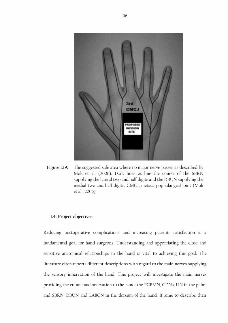
96
Figure 1.19: The suggested safe area where no major nerve passes as described by
Mok et al. (2006). Dark lines outline the course of the SBRN
supplying the lateral two and half digits and the DBUN supplying the
medial two and half digits; CMCJ, metacarpophalangeal joint (Mok
et al., 2006).
1.4. Project objectives:
Reducing postoperative complications and increasing patients satisfaction is a
fundamental goal for hand surgeons. Understanding and appreciating the close and
sensitive anatomical relationships in the hand is vital to achieving this goal. The
literature often reports different descriptions with regard to the main nerves supplying
the sensory innervation of the hand. This project will investigate the main nerves
providing the cutaneous innervation to the hand: the PCBMN, CDNs, UN in the palm;
and SBRN, DBUN and LABCN in the dorsum of the hand. It aims to describe their

97
common course and branching patterns, examine the prevalence of anatomic variations
and their clinical significance. Furthermore, it aims to investigate possible CB, their
prevalence and their impact in clinical settings. This project aims to achieve the
following:
1. Outline the common course and the branching pattern of the cutaneous
nerves that supply the hand and discuss possible variations in their course.
2. Categorize branching patterns and produce a classification system.
3. Investigate possible relations and associations between patterns of
distributions.
4. Investigate dorsal-palmar communication between the nerves that supply
the dorsal and palmar surfaces of the hand.
5. Describe the distribution and branching pattern of the SBRN, DBUN in the
dorsum of the hand and their relationship to the wrist and distal area of the
forearm.
6. Determine the incidence rate of the communicating branch(es) between the
SBRN and DBUN, its/their pattern, and common locations.
7. Describe the distribution and branching pattern of the PCBMN in the palm
and its relationship to the distal area of the forearm and wrist.
8. Describe the distribution and branching pattern of the MN and UN in the
palm and their relationship to the wrist and distal area of the forearm.
9. Determine the incidence rate of the communicating branch(es) between the
MN and UN in the hand, its/their pattern, and common location.
10. Discuss the impact of the distribution and branching patterns of the
cutaneous nerves on surgical and diagnostic procedures.

98
2. Methods
2.1. Sample
All cadavers used for this study were obtained from the Centre for Anatomy and
Human Identification (CAHID), University of Dundee. All dissections were carried out
in CAHID’s anatomy laboratory. The samples were collected during the period October
2011 to April 2013. All cadavers were musculoskeletally mature adults. Arms were not
amputated and no signs of trauma or pathology were noticed. Measurements were
taken for each dissection by one observer.
During the study period, measurements were obtained from 160 formalin-fixed
cadavers. Table 2.1 summarizes the number of hands dissected during each academic
year.
Table 2.1: Number of hands dissected during the study
Time Period No. of hands
Total Males Females
October 2011- April 2012 29 48 77 October 2012-April 2013 41 42 83 Total 70 90 160
2.2. Anatomical Landmarks
Different landmarks were chosen for the palmar and dorsal surfaces of the hand.
Landmarks were chosen because they are easily palpable and relatively constant among
individuals. Furthermore, the landmarks were selected for their clinical relevance and
significance. In the dorsum of the hand, the radial (RSP) and ulnar (USP) styloid
processes, Lister’s tubercle (LT), the third metacarpophalangeal (MCP) joint and the
middle of the bistyloid line (BSL) were chosen. RSP was identified at the distal radial

99
end of the radius, palpitated through the anatomical snuff box. The USP was palpated
at the distal medial end of the ulna. A horizontal line connecting between the RSP and
USP was identified as the BSL. A thread was extended between the two styloid
processes and then measured representing the length of the BSL. This measurement
was used to determine the middle of the BSL. LT was identified as a bony palpable
projection at the distal posterior surface of the radius (Figure 2.1).
Moreover, the distal wrist crease (WC), the distal margin of the TCL, the middle of the
scaphoid tubercle, the proximal margin of the pisiform bone, the third MCP joint and
the middle of the BSL were selected as landmarks for the palmar measurements (Figure
2.2). The scaphoid tubercle was palpated on the radial aspect of the base of the hand
just distal to the distal WC, the middle of the scaphoid tubercle was estimated. The
pisiform was identified on the medial base of the hand, palpitated just distal to the WC.
The TCL was identified deep to the palmar aponeurosis making the roof of the carpal
tunnel. The distal edge was determined at the level of the most proximal point at the
distal margin. Table 2.2 lists the anatomical landmarks and their abbreviations used for
this study.
Table 2.2: Summary of the anatomical landmarks used in this study
No. Landmark Abbreviation Location 1.
Radial styloid process RSP Dorsal landmark
2. Ulnar styloid processes USP Dorsal landmark
3. Lister’s tubercle LT Dorsal landmark
4. The third metacarpophalangeal joint 3rd MCP joint
Dorsal & palmar landmark
5. Middle of the bistyloid line BSL
Dorsal & palmar landmark
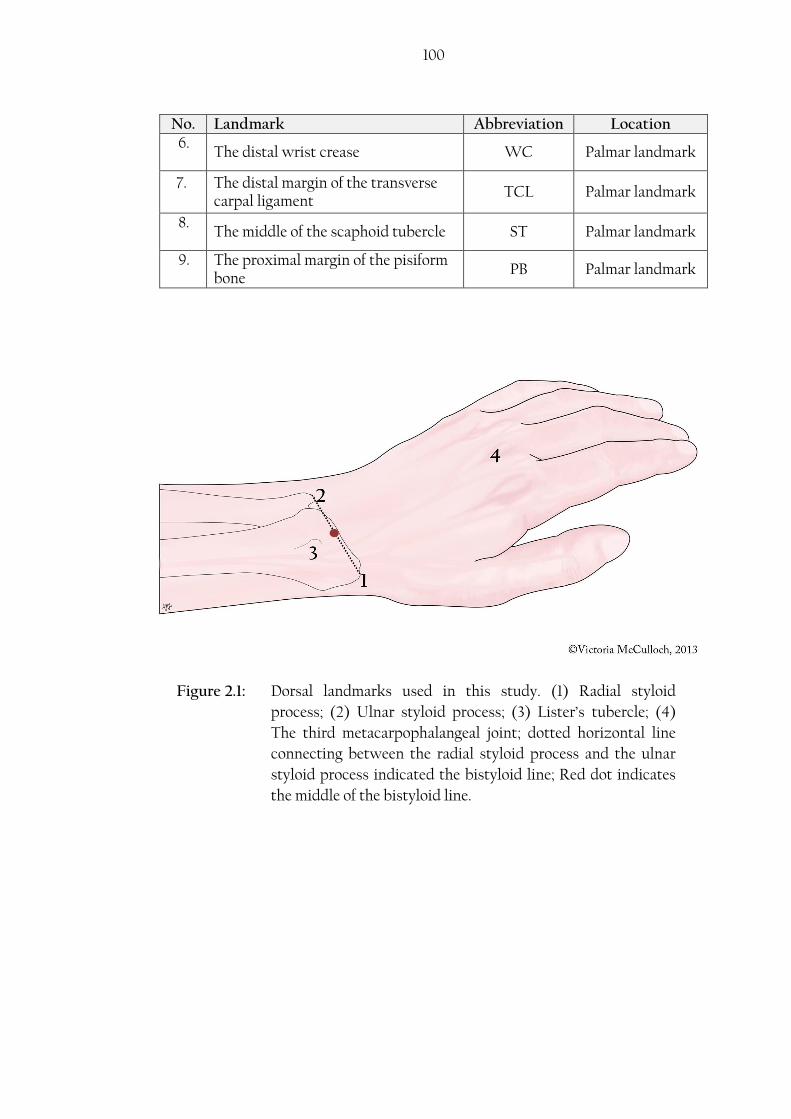
100
No. Landmark Abbreviation Location 6.
The distal wrist crease WC Palmar landmark
7. The distal margin of the transverse carpal ligament
TCL Palmar landmark
8. The middle of the scaphoid tubercle ST Palmar landmark
9. The proximal margin of the pisiform bone
PB Palmar landmark
Figure 2.1: Dorsal landmarks used in this study. (1) Radial styloid
process; (2) Ulnar styloid process; (3) Lister’s tubercle; (4)
The third metacarpophalangeal joint; dotted horizontal line
connecting between the radial styloid process and the ulnar
styloid process indicated the bistyloid line; Red dot indicates
the middle of the bistyloid line.
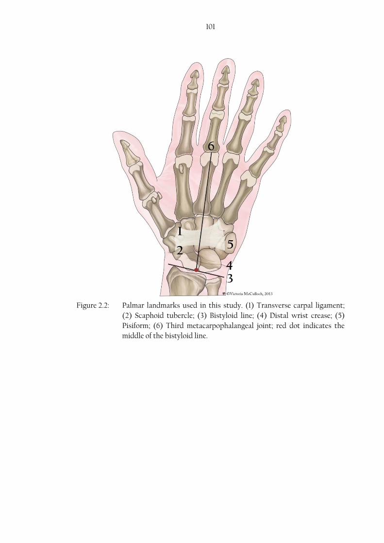
101
Figure 2.2: Palmar landmarks used in this study. (1) Transverse carpal ligament;
(2) Scaphoid tubercle; (3) Bistyloid line; (4) Distal wrist crease; (5)
Pisiform; (6) Third metacarpophalangeal joint; red dot indicates the
middle of the bistyloid line.
102
2.3. Measurements
Measurements were taken by a single observer using digital calipers (Electronic Digital
Caliper Model DC200-PW, Jade products) and recorded to the nearest 0.1 mm. Positive
values are distal, negative values are proximal to the preselected landmarks. Results are
expressed as means, standard deviations, maximum and minimum values.
Because of the embalming process used in this study, some pitfalls were noticed that
might have affected the measurements obtained. Although the embalming fluid
(formalin) does not have a significant effect on the length of the muscles while they are
still fixed on the skeleton, it can still fixate the joints. With arthritic hands being
common in the age group of the cadavers used in this study, full extension of the hands
during measurement taking was not achieved without difficulty (Cutts, 1988;
Waghmare and Sonar, 2012). It was also noted that once dissected, nerves can shrink as
they are exposed to air and dry out slightly changing their position. Moreover, hand
sizes differ between males and females. It also differs between individuals of the same
gender. Measurements were expressed as relative values overcoming the possible
influence of fixation solution on human tissue and the different body sizes in the
studied sample except for some measurements taken for the UN, SBRN, and DBUN1.
The distance between the third metacarpophalangeal joint and the middle of the BSL
and WC were used as a reference, with measurements being expressed as a percentage
of these distances.
Furthermore, because nerves were fully dissected from their surrounding fat and
connective tissue, it is possible that the individual nerves may have changed in their
position. However, care was taken to keep the nerves attached proximally to the
1 Next section explain the measurements taken for each nerve in detail
103
antebrachial fascia where they became cutaneous or to the main trunk they originated
from and distally to their respective final destination at the metacarpophalangeal joint
level.
To test the intra-observer reliability, 16 samples were randomly selected from each
phase were selected and measurements repeated (see Table 3.2). Differences between
the original and repeated measurements were statistically tested by running Kruskal-
Wallis test to check for the observer error. A result is considered statistically
significant when P0.05.
2.4. Dissection process
Skin was removed from the distal half of the forearm as far as the metacarpophalangeal
joints. An incision was made on the lateral side of the distal forearm crossing the wrist
area to the thumb; skin was dissected from proximal to distal (Figure 2.3 and 2.4).
While cadavers were in the supine position, the MN was identified in the distal third of
the forearm between the tendons of flexor carpi radialis and palmaris longus. The
PCBMN was identified in the distal forearm by careful dissection around the main
trunk of the median nerve (Figure 2.5 A). It was more difficult to identify the nerve in
particularly wet specimens where the nerve and the surrounding fat seemed
undistinguishable. In such cases, the distal forearm was packed with paper towels to
absorb excess fluid.
Furthermore, the palmar aponeurosis was dissected and reflected, palmaris brevis
resected, fascia and fat removed to visualize the superficial structures of the palm. The
roof of Guyon’s canal (palmaris brevis) was reflected medially or removed in some cases
where it was less developed. The TCL was cut on its medial side if needed. Care was

104
taken not to damage the ulnar or median digital nerves and any possible
communicating branches. The surplus fatty tissue was removed for better visualization
of the nerves; however, the superficial palmar arch and the common digital arteries
were kept and their relationship to the common digital nerves or any communicating
branches noted (Figure 2.5 and 2.6).
In the dorsal surface of the hand, from the same incision previously made for the palmar
surface, skin covering the dorsum of the hand to the metacarpophalangeal joints was
removed. The cephalic vein was identified and marked. The rest of the dorsal veins were
only removed to better visualize the nerves, otherwise they were preserved. The SBRN
was identified as it became subcutaneous between the tendons of brachioradialis and
extensor carpi radialis longus. Subcutaneous fatty tissue was removed and the nerve
traced (Figure 2.7). DBUN, if present, was identified in the dorsum of the hand at the
location where the SBRN nerve fibres stop. The DBUN, if present, was also identified,
preserved and dissected in the dorsum of the hand.
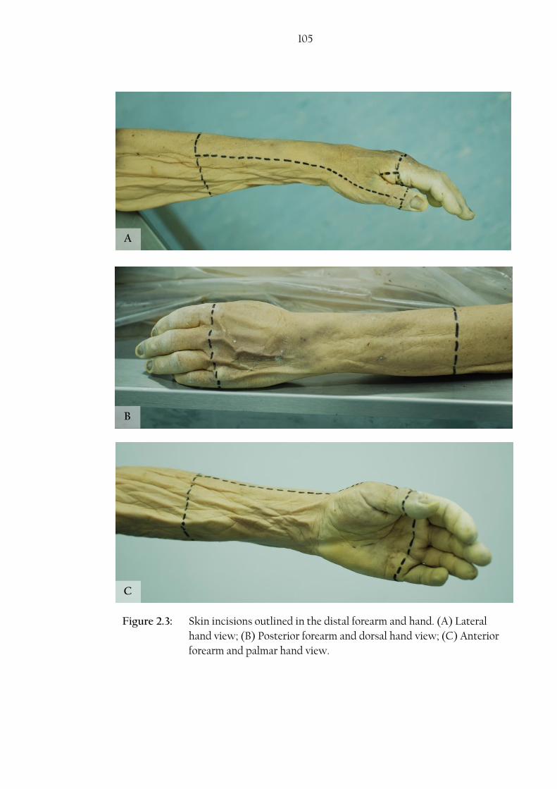
105
B
C
A
Figure 2.3: Skin incisions outlined in the distal forearm and hand. (A) Lateral
hand view; (B) Posterior forearm and dorsal hand view; (C) Anterior
forearm and palmar hand view.
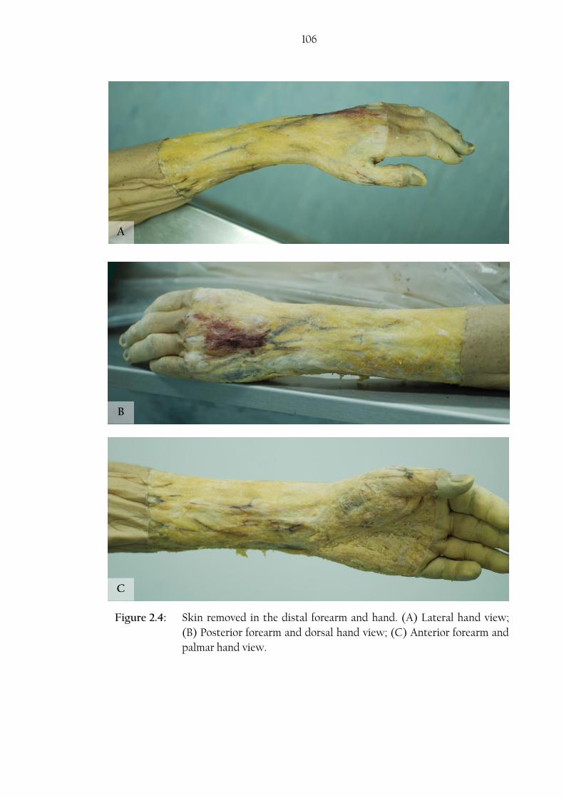
106
A
B
C
Figure 2.4: Skin removed in the distal forearm and hand. (A) Lateral hand view;
(B) Posterior forearm and dorsal hand view; (C) Anterior forearm and
palmar hand view.

107
A
Figure 2.5: Dissection of the palmar surface of the hand. (A) Identifying the
median nerve (MN) and the palmar cutaneous branch of the median
nerve (PCBMN) at the distal forearm. (B) Reflecting the palmar
aponeurosis. FCR, flexor carpi radialis; PL, palmaris longus.
B
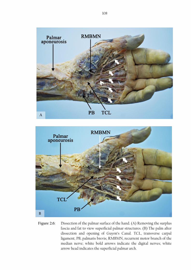
108
A
B
Figure 2.6: Dissection of the palmar surface of the hand. (A) Removing the surplus
fascia and fat to view superficial palmar structures. (B) The palm after
dissection and opening of Guyon’s Canal. TCL, transverse carpal
ligament; PB, palmaris brevis; RMBMN, recurrent motor branch of the
median nerve; white bold arrows indicate the digital nerves; white
arrow head indicates the superficial palmar arch.

109
Figure 2.7: Dissection of the dorsal surface of the hand by removing the surplus
fascia and fat to better view structures of interest. (A) Lateral hand
view. (B) Dorsal hand view. SBRN, superficial branch of the radial
nerve.
A
B

110
The origin of the PCBMN and the detachment point from the main trunk of the MN
were marked and measurements were taken to the distal WC. The point of origin was
defined as the point where the PCBMN could be seen as a separate nerve trunk but still
coursing in the same neural sheath as the MN. The point of detachment was defined
when the PCBMN separated from the MN neural sheath (Figure 2.8). The PCBMN was
traced into the palm and small branches were preserved. The anatomical course of the
nerve and its relationship to palmaris longus, flexor carpi radialis, and the PCL was
noted. The nerve’s tunnel was identified when the nerve coursed between the fibres of
the PCL proximal to the WC (Figure 2.9). The distal end of the tunnel was identified as
the nerve becomes subcutaneous between the fibres of the palmaris longus blending
with the palmar aponeurosis. The length of the tunnel was measured. Branching
patterns of the PCBMN were classified based on the number of sub-branches coming
off the PCBMN and incidence rates were calculated.

111
Figure 2.8: Distances measured of the palmar cutaneous branch of the median
nerve (PCBMN). (1) Origin point of the PCBMN; (2) Detachment
point of the PCBMN from the main trunk of the median nerve (MN);
(3) Level of the wrist crease (WC) also indicated by the black pin; A,
distance between the origin point and the WC; B, distance between
the detachment point and the WC; PL, palmaris longus; FCR, flexor
carpi radialis; Lat, lateral; Med, medial; Dist, distal; Prox, proximal.
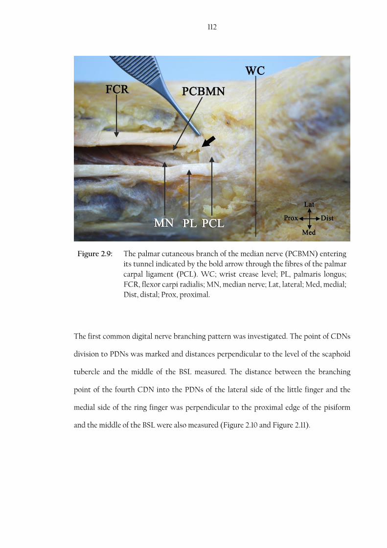
112
The first common digital nerve branching pattern was investigated. The point of CDNs
division to PDNs was marked and distances perpendicular to the level of the scaphoid
tubercle and the middle of the BSL measured. The distance between the branching
point of the fourth CDN into the PDNs of the lateral side of the little finger and the
medial side of the ring finger was perpendicular to the proximal edge of the pisiform
and the middle of the BSL were also measured (Figure 2.10 and Figure 2.11).
Figure 2.9: The palmar cutaneous branch of the median nerve (PCBMN) entering
its tunnel indicated by the bold arrow through the fibres of the palmar
carpal ligament (PCL). WC; wrist crease level; PL, palmaris longus;
FCR, flexor carpi radialis; MN, median nerve; Lat, lateral; Med, medial;
Dist, distal; Prox, proximal.

113
Figure 2.10: Distances measured of the common digital nerves (CDNs) division
points into proper digital nerves. (1) Bistyloid line; (2) Scaphoid
tubercle (ST) level; (3) Proximal edge of the pisiform; A, First CDN
second division point to ST; B, Second CDN division point to ST; C,
First CDN first division to ST; D, Third CDN division point to ST; E,
Fourth CDN division point to distal edge of pisiform.

114
The ulnar nerve in Guyon’s canal was also investigated. The UN point of division into
deep and superficial branches was measured to the proximal margin of the pisiform
(Figure 2.12). Communicating branches between the 4th CDN and the PDN to the little
finger; the deep division of the ulnar nerve and the PDN to the little finger; and the
DBUN and the UN or its branches were noted.
Figure 2.11: Distances measured of the common digital nerves (CDNs) division
points into proper digital nerves. (1) Bistyloid line (BSL); (2) Third
metacarpophalangeal joint; A, First CDN second division point to BSL;
B, First CDN first division to BSL; C, Second CDN division point to
BSL; D, Third CDN division point to BSL; E, Fourth CDN division
point to BSL.
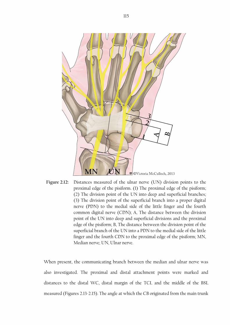
115
When present, the communicating branch between the median and ulnar nerve was
also investigated. The proximal and distal attachment points were marked and
distances to the distal WC, distal margin of the TCL and the middle of the BSL
measured (Figures 2.13-2.15). The angle at which the CB originated from the main trunk
Figure 2.12: Distances measured of the ulnar nerve (UN) division points to the
proximal edge of the pisiform. (1) The proximal edge of the pisiform;
(2) The division point of the UN into deep and superficial branches;
(3) The division point of the superficial branch into a proper digital
nerve (PDN) to the medial side of the little finger and the fourth
common digital nerve (CDN); A, The distance between the division
point of the UN into deep and superficial divisions and the proximal
edge of the pisiform; B, The distance between the division point of the
superficial branch of the UN into a PDN to the medial side of the little
finger and the fourth CDN to the proximal edge of the pisiform; MN,
Median nerve; UN, Ulnar nerve.
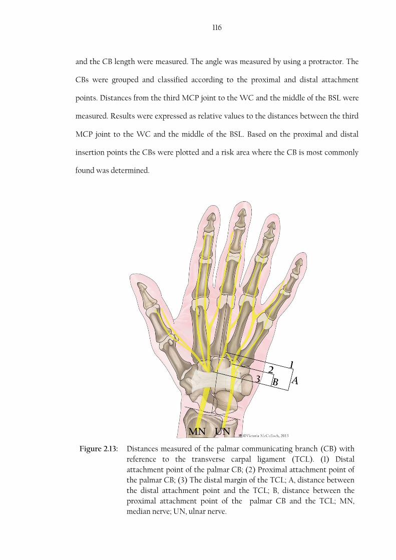
116
and the CB length were measured. The angle was measured by using a protractor. The
CBs were grouped and classified according to the proximal and distal attachment
points. Distances from the third MCP joint to the WC and the middle of the BSL were
measured. Results were expressed as relative values to the distances between the third
MCP joint to the WC and the middle of the BSL. Based on the proximal and distal
insertion points the CBs were plotted and a risk area where the CB is most commonly
found was determined.
Figure 2.13: Distances measured of the palmar communicating branch (CB) with
reference to the transverse carpal ligament (TCL). (1) Distal
attachment point of the palmar CB; (2) Proximal attachment point of
the palmar CB; (3) The distal margin of the TCL; A, distance between
the distal attachment point and the TCL; B, distance between the
proximal attachment point of the palmar CB and the TCL; MN,
median nerve; UN, ulnar nerve.

117
Figure 2.14: Distances measured of the palmar communicating branch (CB) with
reference to the wrist crease (WC). (1) Third metacarpophalangeal
joint; (2) Distal attachment point of the palmar CB; (3) Proximal
attachment point of the palmar CB; (4) The WC; A, distance between
the third MCP joint and the WC; B, distance between the distal
attachment point and the WC; C, distance between the proximal
attachment point of the palmar CB and the WC; MN, median nerve;
UN, ulnar nerve.

118
Figure 2.15: Distances measured of the palmar communicating branch (CB) with
reference to the middle of the bistyloid line (BSL) (4). (1) The distance
between the third MCP joint and the middle of the BSL; (2) The
distance between the distal attachment point of the palmar CB and
the middle of the BSL; (3) The distance between the proximal
attachment point of the palmar CB and the middle of the BSL; red dot,
middle of the BSL; MN, median nerve; UN, ulnar nerve.
In the dorsum of the hand, the SBRN and the DBUN were investigated. The point of
piercing the antebrachial fascia and the first, second and third major branching points
were noted and distances to the RSP were measured. Care was taken to preserve small
branches and any connections between the SBRN and the DBUN and/or LABCN. The
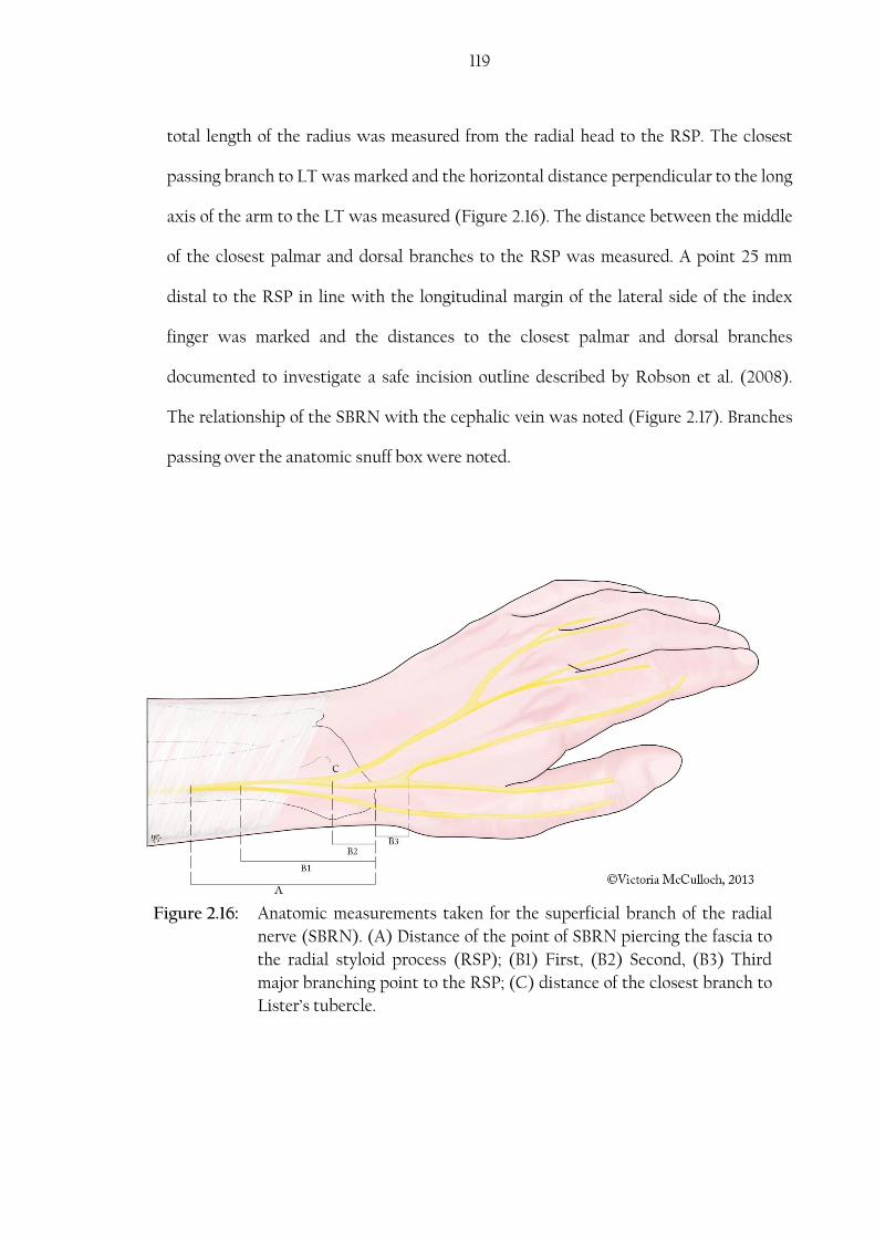
119
Figure 2.16: Anatomic measurements taken for the superficial branch of the radial
nerve (SBRN). (A) Distance of the point of SBRN piercing the fascia to
the radial styloid process (RSP); (B1) First, (B2) Second, (B3) Third
major branching point to the RSP; (C) distance of the closest branch to
Lister’s tubercle.
total length of the radius was measured from the radial head to the RSP. The closest
passing branch to LT was marked and the horizontal distance perpendicular to the long
axis of the arm to the LT was measured (Figure 2.16). The distance between the middle
of the closest palmar and dorsal branches to the RSP was measured. A point 25 mm
distal to the RSP in line with the longitudinal margin of the lateral side of the index
finger was marked and the distances to the closest palmar and dorsal branches
documented to investigate a safe incision outline described by Robson et al. (2008).
The relationship of the SBRN with the cephalic vein was noted (Figure 2.17). Branches
passing over the anatomic snuff box were noted.
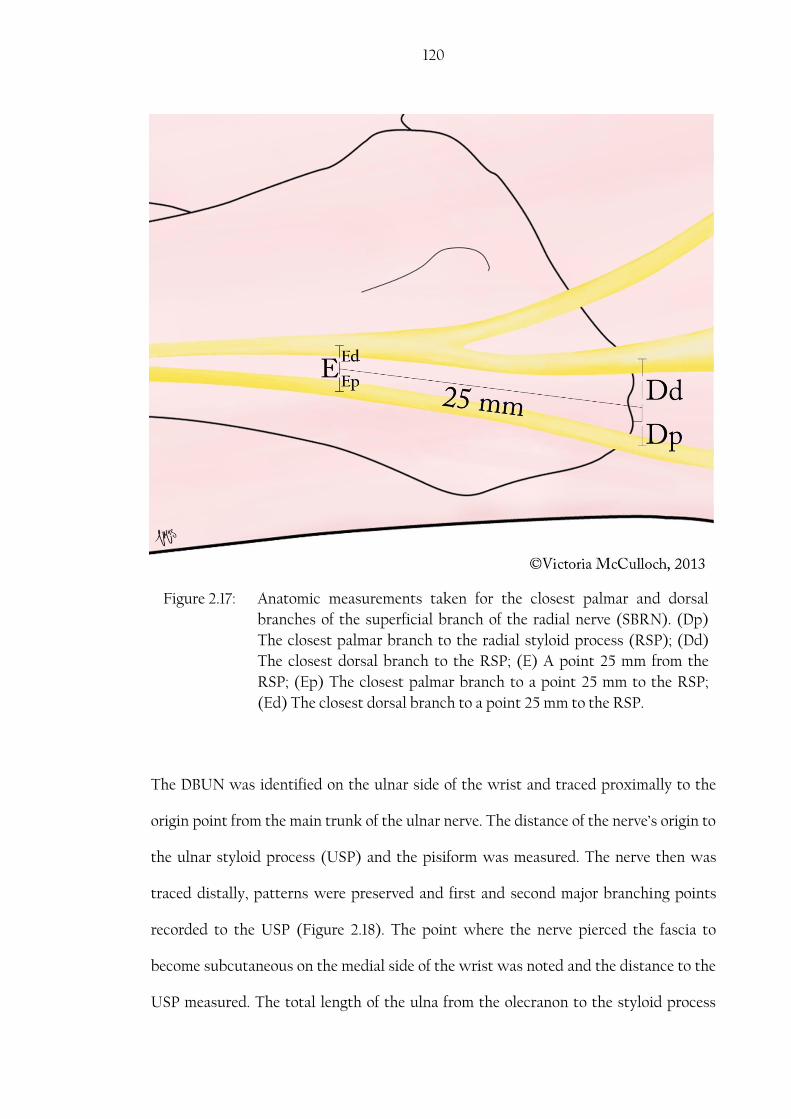
120
The DBUN was identified on the ulnar side of the wrist and traced proximally to the
origin point from the main trunk of the ulnar nerve. The distance of the nerve’s origin to
the ulnar styloid process (USP) and the pisiform was measured. The nerve then was
traced distally, patterns were preserved and first and second major branching points
recorded to the USP (Figure 2.18). The point where the nerve pierced the fascia to
become subcutaneous on the medial side of the wrist was noted and the distance to the
USP measured. The total length of the ulna from the olecranon to the styloid process
Figure 2.17: Anatomic measurements taken for the closest palmar and dorsal
branches of the superficial branch of the radial nerve (SBRN). (Dp)
The closest palmar branch to the radial styloid process (RSP); (Dd)
The closest dorsal branch to the RSP; (E) A point 25 mm from the
RSP; (Ep) The closest palmar branch to a point 25 mm to the RSP;
(Ed) The closest dorsal branch to a point 25 mm to the RSP.
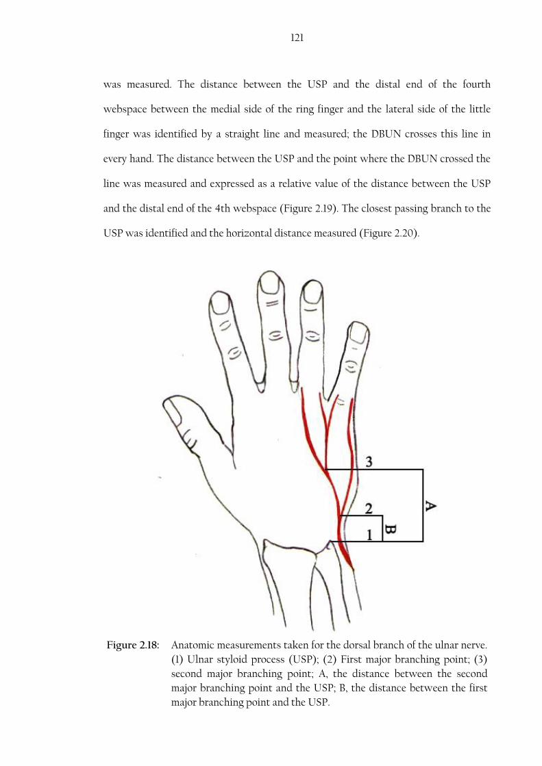
121
Figure 2.18: Anatomic measurements taken for the dorsal branch of the ulnar nerve.
(1) Ulnar styloid process (USP); (2) First major branching point; (3)
second major branching point; A, the distance between the second
major branching point and the USP; B, the distance between the first
major branching point and the USP.
was measured. The distance between the USP and the distal end of the fourth
webspace between the medial side of the ring finger and the lateral side of the little
finger was identified by a straight line and measured; the DBUN crosses this line in
every hand. The distance between the USP and the point where the DBUN crossed the
line was measured and expressed as a relative value of the distance between the USP
and the distal end of the 4th webspace (Figure 2.19). The closest passing branch to the
USP was identified and the horizontal distance measured (Figure 2.20).

122
Figure 2.19: Anatomic measurements taken for the dorsal branch of the ulnar nerve
(DBUN). (A) The distance between the ulnar styloid process (USP)
and the fourth webspace; (B) The distance between the USP and the
point where the DBUN crosses line A.

123
Several measurements were taken to the SBRN and the DBUN communicating branch
if present. The proximal and distal attachment points were marked and distances to the
middle of the BSL measured. The angle of origin and the length of the communicating
branch were recorded (Figure 2.21). The distance between the third MCP joint and the
middle of the BSL was taken. The communicating branches was determined as relative
values with reference to the BSL corresponding to the origin and insertion points and
plotted to produce a risk area where the communicating branch is most likely to be
encountered.
Figure 2.20: Anatomic measurements taken for the dorsal branch of the ulnar nerve
(DBUN). (A) The horizontal distance between the ulnar styloid
process (USP) and the closest branch of the DBUN.

124
Figure 2.21: Distances measured of the dorsal communicating branch (CB) with
reference to the bistyloid line (BSL). (1) Distal attachment point of the
dorsal CB; (2) Proximal attachment point of the dorsal CB; (4) The
BSL; (X) shows the location of the third metacarpophalangeal (MCP)
joint. A, the distance between the third MCP joint and the BSL; B,
distance between the distal attachment point and the BSL; C, distance
between the proximal attachment point of the dorsal CB and the BSL.

125
Table 2.3 lists all the anatomic characteristics and measurements taken for the nerves of
interest in this study.
Table 2.3: List of the anatomical measurements for each of the nerves investigated in the study
NO. Anatomical characteristics and measurements
Palmar cutaneous branch of the median nerve (PCBMN)
1. Origin direction from the main trunk of the median nerve (MN)
2. Distance from the point of origin to the wrist crease (WC)
3. Distance from the point of detaching from the main trunk to the distal WC
4. Anatomical relationship to palmaris longus
5. Anatomical relationship to flexor carpi radialis
6. Anatomical relationship to palmar carpal ligament
7. Length of the PCBMN tunnel
8. Branching pattern in the palm
9. Angle between the lateral and medial branches in the palm
10. Distance between the first branch coming off the PCBMN to the distal WC
Common digital nerves (CDN) -Median nerve (MN)
11. First CDN division point to the middle of the bistyloid line (BSL)
12. First CDN division point to the scaphoid tubercle
13. Second CDN division point to the middle of the BSL
14. Second CDN division point to the scaphoid tubercle
15. Third CDN division point to the middle of the BSL
16. Third CDN division point to the scaphoid tubercle
17. Third Metacarpophalangeal (MCP) joint to middle of the BSL
18. Third MCP joint to scaphoid
19. Third MCP joint to the middle of the BSL
Ulnar nerve (UN)
20. The point of division of the UN into superficial and deep branches to the proximal edge of the pisiform
21. Fourth CDN division point to the middle of the BSL
22. Fourth CDN division point to the proximal edge of the pisiform
The palmar communicating branch (CB) between the UN and MN
23. Distance between the proximal insertion point to the distal WC
24. Distance between the proximal insertion point to distal edge of TCL
25. Distance between the proximal insertion point to middle of the BSL
26. Distance between the distal insertion point to distal WC
27. Distance between the distal insertion point to distal edge of the TCL
28. Distance between the distal insertion point to middle of the BSL

126
NO. Anatomical characteristics and measurements
29. The length of the communicating branch
30. The angle at which the proximal attachment leaves the CDN
31. The distance between the third MCP joint to the distal WC
32. The distance between the third MCP joint to the middle of the BSL
Superficial branch of the radial nerve (SBRN)
33. Point of piercing the antebrachial fascia to the radial styloid process (RSP)
34. The first major branching point to the RSP
35. The second major branching point to the RSP
36. The third major branching point to the RSP
37. The closest branch to Lister’s tubercle
38. The distances to the closest palmar and dorsal branches to a point 25 mm distal to the RSP
39. The distances to the closest palmar and dorsal branches to the RSP
40. The first crossing point with the cephalic vein after the SBRN became subcutaneous
41. The diameter of the nerve prior to its first major branching point
Dorsal branch of the ulnar nerve (DBUN)
42. The point of origin to the ulnar styloid process (USP)
43. The point of origin to the proximal edge of the pisiform
44. The first major branching point to the USP
45. The second major branching point to the USP
46. The diameter of the nerve prior to its first major branching point
47. The distance between the USP and the fourth webspace
48. The distance between the USP and the point where the DBUN crosses a line between the USP and the fourth webspace
The dorsal communicating branch between the DBUN and SBRN
49. The proximal attachment point to the middle of the BSL
50. The distal attachment point to the middle of the BSL
51. The length of the communicating branch
52. The angle at which the proximal attachment leaves DBUN/SBRN
53. The distances from the third MCP joint to the middle of the BSL
2.5. Photographs and visual illustrations
Photographs were taken for all samples by digital cameras (Pentex Optio WG-1 GPS
and Nikon D40 with Macro sigma lens). Pictures were edited in Adobe Photoshop
CS5.1 image manipulation software. Editing of pictures only included changes in
127
contrast, brightness, highlights and shadow intensity to better visualize the nerves and
other structures. No alteration of the content of each image was performed including
addition, deletion, or substitution that would change the shape or location of the
structures. Some visual illustrations were created to support this document by Ms.
Victoria McCulloch, medical artist, University of Dundee and some were produced by
the author.
2.6. Statistics
Data has been manipulated and summarized by: descriptive statistics and univariant
analysis. Means, standard deviations, maximum and minimum values were calculated.
Patterns were described in terms of frequencies and percentages. Pearson’s chi-square
test was used to investigate the relationships between categorical dependent variables.
All statistical calculations were conducted using IBM Statistical Program for Social
Sciences (SPSS) version 21. A result is considered statistically significant when P0.05.
128
3. Results
A total of 160 cadaveric hands from 81 adult cadavers were investigated in this study.
However, the number of samples investigated differs for each nerve. The maximum
number of samples studied for any nerve was 155. Some samples were excluded because
the nerve under investigation was destroyed or/and its branching point was
manipulated during previous dissection making it difficult to produce accurate
measurements. Table 3.1 shows the number of samples investigated for each nerve.
Table 3.1: Number of samples investigated for each nerve
No. Nerve under investigation No. of
samples 1. Palmar cutaneous branch of the median nerve (PCBMN) 123 2. Common digital nerves (CDNs)-Median nerve (MN) 155 3. Ulnar nerve (UN) 144 4. Communicating branch between median nerve & ulnar nerve 98 5. Superficial branch of the radial nerve (SBRN) 150 6. Dorsal branch of the ulnar nerve (DBUN) 139
7. Communicating branch between the superficial branch of the radial nerve and the dorsal branch of the ulnar nerve
37
3.1. Intra-observer results
Repeated measures of 16 random cases (10% of the total samples) were collected to test
the intra-observer error. The Kruskal-Wallis gave a high P-values ( 0.05) for all the
variables tested. Therefore, the null hypothesis stating that there are no statistical
difference between the two measurements is retained (Table 3.2). The intra-observer
error is within the acceptable range (5%) indicating that the measurements and
statistical comparisons are reliable and precise.

129 Table 3.2: Chi-Square results for repeated measures
Variable measured Chi-
Square df Sig.
Retain the null
Hypothesis
Palmar Communicating branch between the median and the ulnar nerves
Proximal attachment to TCL .240 1 .624 Yes
Distal attachment to TCL .364 1 .546 Yes
Angle of origin .353 1 .553 Yes
MCPJ to WC 1.740 1 .187 Yes
Proximal to WC 3.273 1 .070 Yes
Distal to WC 1.322 1 .250 Yes
Length .205 1 .651 Yes
BSL .023 1 .880 Yes
Common digital nerves (CDN)
First CDN first branching point to the BSL .051 1 .821 Yes
First CDN second branching point to the BSL 1.534 1 .215 Yes
First CDN first branching point to scaphoid .009 1 .925 Yes
First CDN second branching point to scaphoid
.658 1 .417 Yes
Second CDN division into PDN to the BSL 1.890 1 .169 Yes
Second CDN division into PDN to scaphoid 1.144 1 .285 Yes
Third CDN division into PDN to the BSL .003 1 .955 Yes
Third CDN division into PDN to scaphoid 1.069 1 .301 Yes
Ulnar nerve (UN)
Branching point of the Deep branch of the UN to the pisiform
.005 1 .945 Yes
Branching point of the superficial branch of the UN to the pisiform
.000 1 1.000 Yes
3.2. Palmar cutaneous branch of the median nerve
The PCBMN was investigated in 123 cadaveric hands: 50 (40.7%) were male and 73
(59.3%) female. Mean age was 82.6 ±9.8 years (range: 53-98). The PCBMN was found in
all hands studied; however, two PCBMNs were identified in 11 cases (8.9%) originating
at the same or different levels from the main trunk of the median nerve.

130 The PCBMN originated from the lateral side of the main trunk in the majority of cases
(115/123, 93.5%). However, it was also found to originate from the posterolateral, medial
and anterior side of the main trunk of the median nerve in 1.6% (2/123), 1.6% (2/123)
and 3.3% (4/123) of hands respectively (Figure 3.1).
A
B

131
Figure 3.1: Different origin direction of the palmar cutaneous branch of the
median nerve (PCBMN) from the main trunk of the median nerve
(MN). (A) Lateral origin, (B) Posterolateral origin, (C) Medial
origin, (D) Anterior origin. FCR, flexor carpi radialis; PL, palmaris
longus; SBRN, superficial branch of the radial nerve; Lat, lateral;
Med, medial; Prox, proximal; Dist, distal.
C
D
132 The PCBMN originated at a mean distance of 54.1±15.7 mm proximal to the WC,
continued within the epineurium of the main trunk of the MN for a short distance and
separated at a mean distance of 41.2±14.0 mm proximal to the WC (Table 3.3). The
nerve then coursed distally medial to flexor carpi radialis and lateral to the palmaris
longus tendon (if present). The nerve was noted to pass through the fascia of flexor
carpi radialis tendon in 10 cases (8.3%) and posterior to the tendon in 5 cases (4.2%).
At the distal forearm, the nerve penetrated the PCL creating a tunnel between the deep
and superficial fibres and continued distally through the fibres of the TCL. The
PCBMN tunnel was found to be 11.3±3.4 mm long (Figure 3.2, Table 3.3). It was also
noted that the nerve passed superficial to the PCL in 18 cases (16.1%) while it passed
deep to it in 14 cases (12.5%). Interestingly, the PCBMN was found to course deep to
the TCL and pierced the ligament and palmar aponeurosis at several locations to supply
the skin of the palm in one hand. The nerve entered the palm often penetrating the
distal fibres of palmaris longus where it divided to supply the proximal two fifths of the
palm.

133
Figure 3.2: Palmar cutaneous branch of the median nerve (PCBMN) crossing between the superficial and deep fibres of the palmar carpal ligament to create a tunnel before entering the palm. Lat, lateral; Med, medial; Dist, distal; Prox, proximal.
Based on the division pattern of the nerve in the palm distal to the WC observed in 110
samples three patterns of division were identified:
Type I: The main trunk divides into Y-shaped ulnar and radial branches that
continued to give tertiary branches as they course distally in the palm.
Variations to Type I: The main trunk gives several smaller branches
before it divides into Y- shaped ulnar and radial major branches.
Type II: The PCBMN continues as one major branch that gives several minor
branches as it courses distally in the palm.

134 Type III: The main trunk divides into three branches (radial, ulnar and middle)
that all continue to give tertiary branches as they course distally in the
palm.
Types I, II, and III were found in 40.0% (44/110), 36.4% (40/110), 14.5% (16/110) of the
hands respectively. Furthermore, variations to Type I were found in 9.1% (10/110) of the
hands investigated (Figure 3.3).

135
B
A
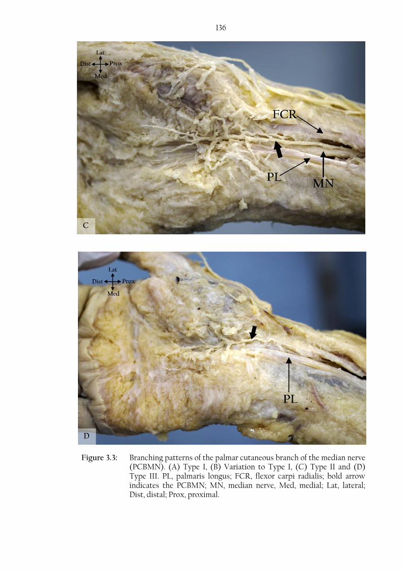
136
Figure 3.3: Branching patterns of the palmar cutaneous branch of the median nerve (PCBMN). (A) Type I, (B) Variation to Type I, (C) Type II and (D) Type III. PL, palmaris longus; FCR, flexor carpi radialis; bold arrow indicates the PCBMN; MN, median nerve, Med, medial; Lat, lateral; Dist, distal; Prox, proximal.
D
C
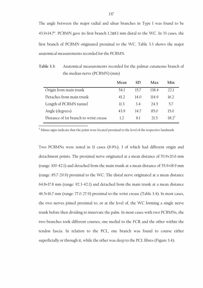
137 The angle between the major radial and ulnar branches in Type I was found to be
43.9±14.7. PCBMN gave its first branch 1.2±8.1 mm distal to the WC. In 33 cases, the
first branch of PCBMN originated proximal to the WC. Table 3.3 shows the major
anatomical measurements recorded for the PCBMN.
Table 3.3: Anatomical measurements recorded for the palmar cutaneous branch of
the median nerve (PCBMN) (mm)
Mean SD Max Min
Origin from main trunk 54.1 15.7 138.4 22.1
Detaches from main trunk 41.2 14.0 114.9 16.2
Length of PCBMN tunnel 11.3 3.4 24.5 5.7
Angle (degrees) 43.9 14.7 85.0 15.0
Distance of 1st branch to wrist crease 1.2 8.1 21.5 -18.21
1 Minus signs indicate that the point were located proximal to the level of the respective landmark
Two PCBMNs were noted in 11 cases (8.9%), 3 of which had different origin and
detachment points. The proximal nerve originated at a mean distance of 70.9±20.6 mm
(range: 100-42.1) and detached from the main trunk at a mean distance of 55.9±18.9 mm
(range: 85.7-29.9) proximal to the WC. The distal nerve originated at a mean distance
64.8±17.8 mm (range: 92.3-42.1) and detached from the main trunk at a mean distance
46.5±16.7 mm (range: 77.0-27.9) proximal to the wrist crease (Table 3.4). In most cases,
the two nerves joined proximal to, or at the level of, the WC forming a single nerve
trunk before then dividing to innervate the palm. In most cases with two PCBMNs, the
two branches took different courses, one medial to the FCR and the other within the
tendon fascia. In relation to the PCL, one branch was found to course either
superficially or through it, while the other was deep to the PCL fibres (Figure 3.4).

138 Table 3.4: Anatomical measurements recorded in cases where two palmar
cutaneous branches of the median nerve were found (mm)
Mean SD Max Min
Origin of the proximal branch from main
trunk of the median nerve 70.9 20.6 100 42.1
Detachment of the proximal branch from
main trunk of the median nerve 55.9 18.9 85.7 29.9
Origin of the distal branch from main
trunk of the median nerve 64.8 17.8 92.3 42.1
Detachment of the distal branch from
main trunk of the median nerve 46.5 16.7 77.0 27.9
The PCBMN also communicated with other nerves supplying the region. It was found
to communicate with the LABCN in 5 cases. The LABCN gives a branch to
communicate with the PCBMN and therefore contributes to the innervation of the
palm and the thenar eminence. In two of these cases the LABCN communicated with
the SBRN earlier to its communication with the PCBMN (Figure 3.5). The nerve was
also found to communicate solely with the SBRN (one case), with the recurrent motor
branch of the median nerve (two cases) (Figure 3.6), with the palmar cutaneous branch
of the ulnar nerve (one case) (Figure 3.7), and with the first CDN supplying the thumb
(one case) (Figure 3.8).

139
A
B
C
Figure 3.4: Two palmar cutaneous branches of the median nerve (PCBMN). (A) Shows the different course that each branch takes. (B) After full dissection of the palmar carpal ligament (PCL) to view the deeper branch, the rectangular area outlined is enlarged in (C). PL, palmaris longus; FCR, flexor carpi radialis; bold arrows indicate the PCBMN branches; Lat, lateral; Med, medial; Prox, proximal; Dist, distal.
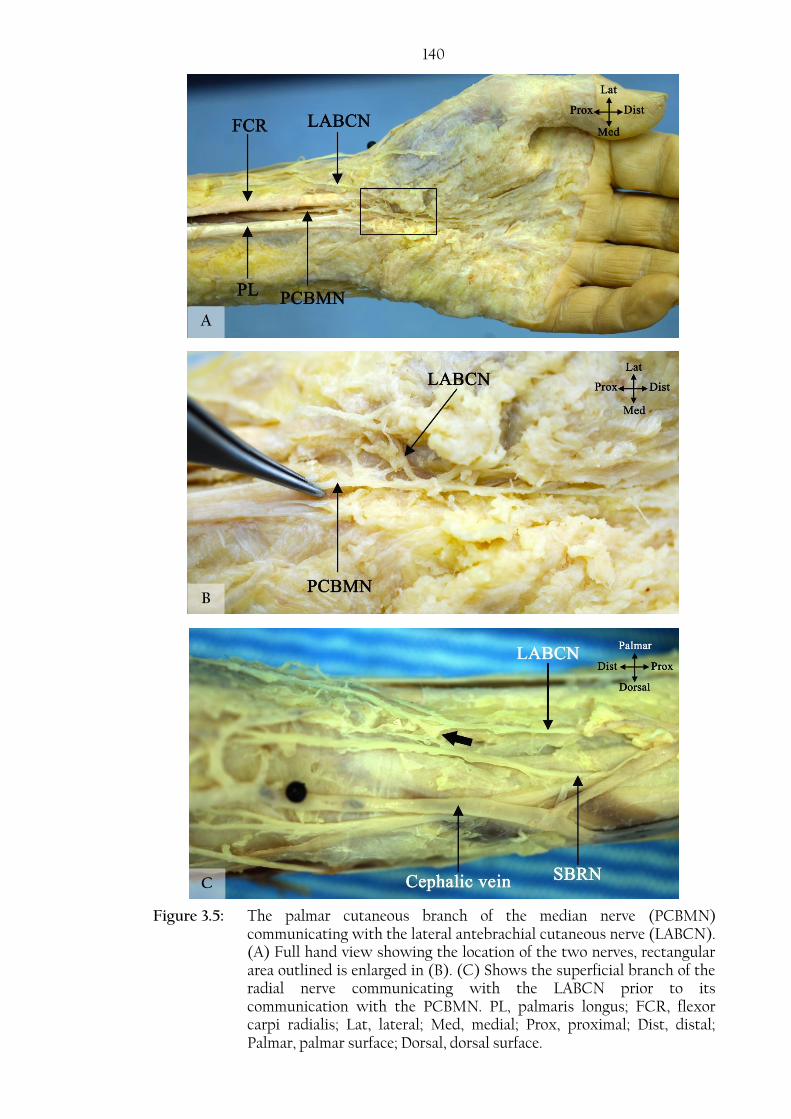
140
C
B
A
Figure 3.5: The palmar cutaneous branch of the median nerve (PCBMN) communicating with the lateral antebrachial cutaneous nerve (LABCN). (A) Full hand view showing the location of the two nerves, rectangular area outlined is enlarged in (B). (C) Shows the superficial branch of the radial nerve communicating with the LABCN prior to its communication with the PCBMN. PL, palmaris longus; FCR, flexor carpi radialis; Lat, lateral; Med, medial; Prox, proximal; Dist, distal; Palmar, palmar surface; Dorsal, dorsal surface.
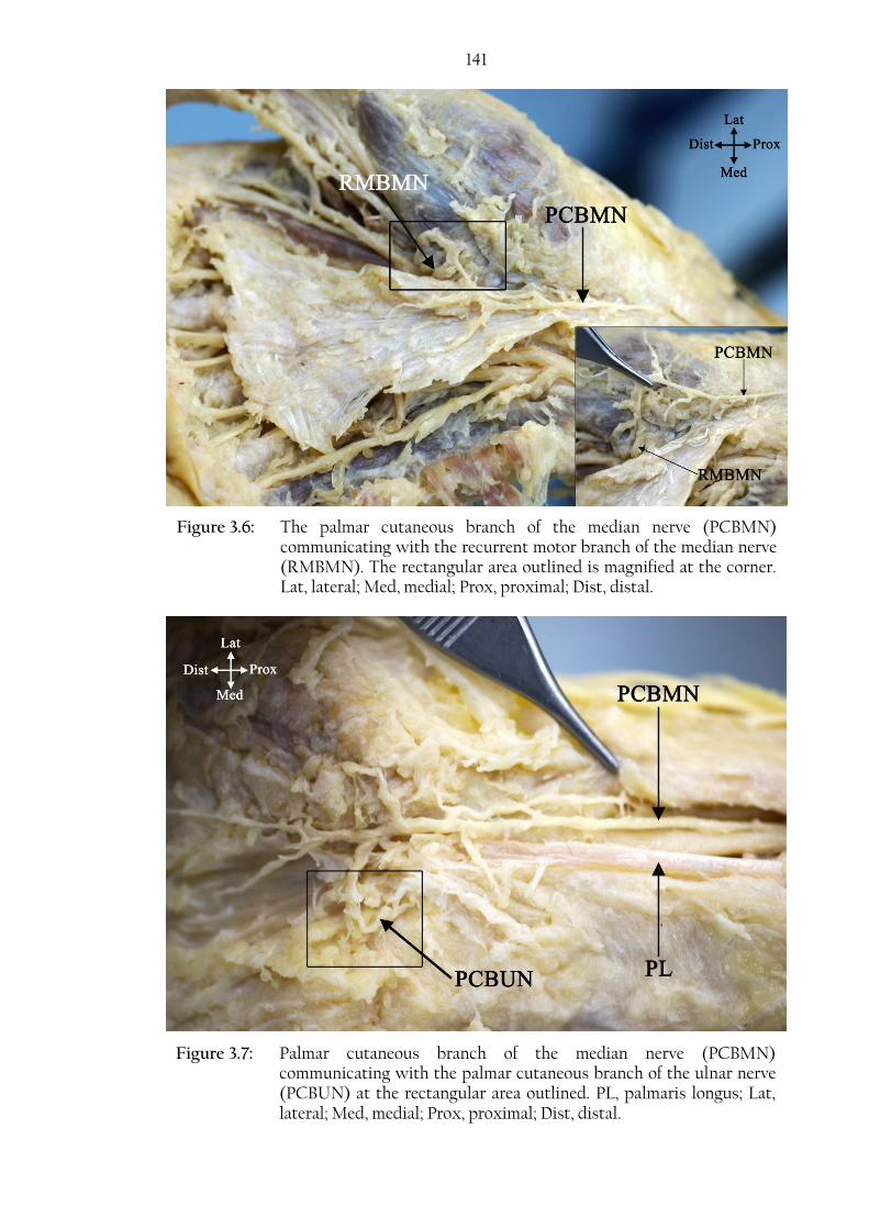
141
Figure 3.6: The palmar cutaneous branch of the median nerve (PCBMN) communicating with the recurrent motor branch of the median nerve (RMBMN). The rectangular area outlined is magnified at the corner. Lat, lateral; Med, medial; Prox, proximal; Dist, distal.
Figure 3.7: Palmar cutaneous branch of the median nerve (PCBMN) communicating with the palmar cutaneous branch of the ulnar nerve (PCBUN) at the rectangular area outlined. PL, palmaris longus; Lat, lateral; Med, medial; Prox, proximal; Dist, distal.

142
Figure 3.8: The palmar cutaneous branch of the median nerve (PCBMN) communicating with the first common digital nerve to the medial side of the thumb. (A) A full hand view. (B) Enlarged image of the communicating branch. Lat, lateral; Med, medial; Prox, proximal; Dist, distal.
B
A

143 An artery was observed to course along the lateral side of the main trunk of the MN in
the distal forearm in one case. Distal to the detachment of the PCBMN, the artery
coursed on top of the nerve separating the PCBMN from the MN, continuing on the
medial side of the MN to enter the carpal tunnel along with the MN (Figure 3.9).
The effect of sex and body side on the branching pattern was investigated. The
branching pattern of the PCBMN was not influenced by sex (P value=0.506) or body
side (P value =0.240). Appendix 1 shows the chi-square test results for both the effect of
sex and body side.
Figure 3.9: An artery crossing the median nerve (MN) and separating the two branches of the palmar cutaneous branch of the median nerve (PCBMN) from the MN trunk. PL, palmaris longus; FCR, flexor carpi radialis; Lat, lateral; Med, medial; Prox, proximal; Dist, distal.

144
3.3. Common digital nerves
The CDNs were investigated in 155 cadaveric palms from 80 cadavers: 68 (43.9%)
hands were male and 87 (56.1%) female. Mean age was 82.4±9.6 (range: 101-53) years.
The median nerve was found to pass through the carpal tunnel and divide into the first,
second and third CDN within the tunnel or at the distal margin of the TCL in all cases.
The fourth CDN and the PDN to the medial side of the little finger originated from the
superficial division of the UN after it passed through Guyon’s canal on the medial side
of the palm.
The CDNs passed deep to the superficial palmar arch and divided into PDNs at various
levels to supply the lateral and medial sides of the digits (Figure 3.10 A). In one case, the
third CDN gave a branch that crossed superficial to the superficial palmar arch creating
a neural loop around the arch: the branch then joined the third CDN again distally
(Figure 3.10 B). Table 3.5 illustrates the branching points of the CDNs into PDNs with
respect to different anatomic landmarks.
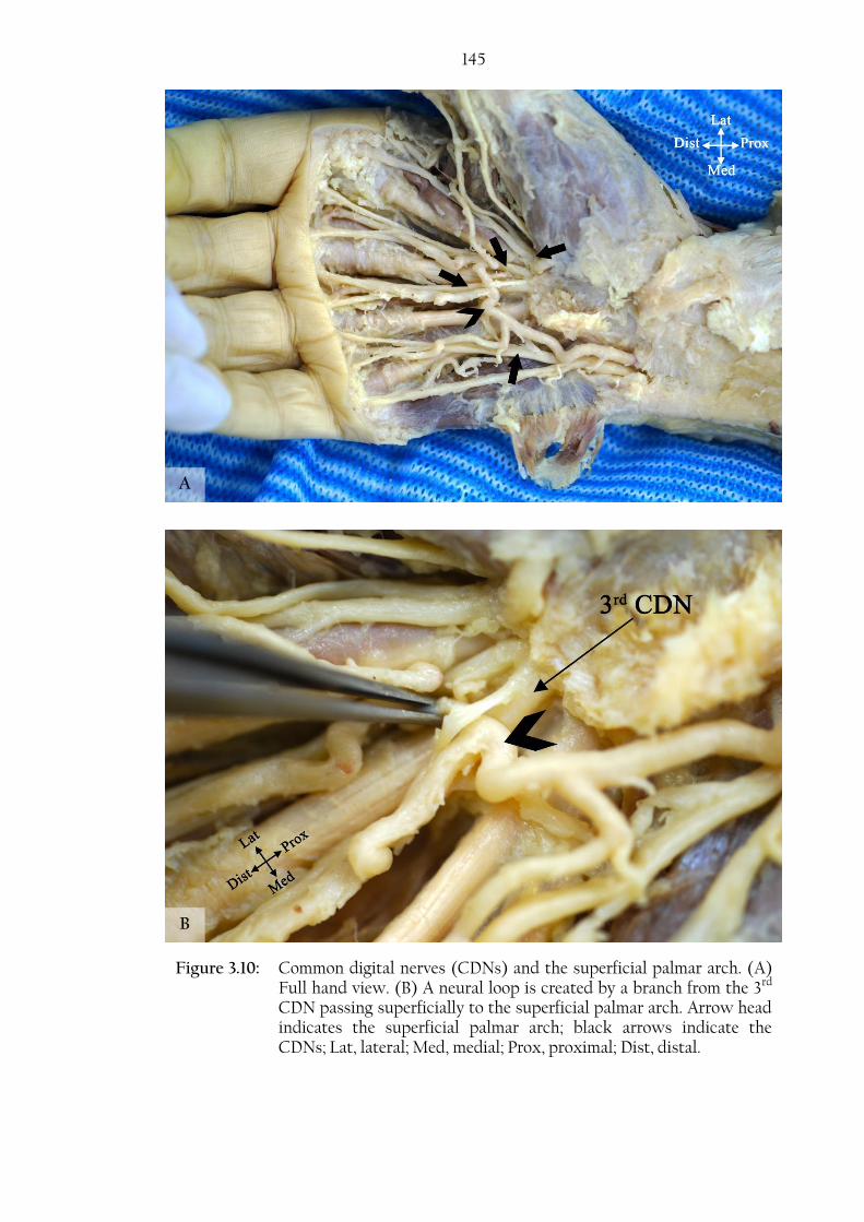
145
Figure 3.10: Common digital nerves (CDNs) and the superficial palmar arch. (A) Full hand view. (B) A neural loop is created by a branch from the 3rd CDN passing superficially to the superficial palmar arch. Arrow head indicates the superficial palmar arch; black arrows indicate the CDNs; Lat, lateral; Med, medial; Prox, proximal; Dist, distal.
B
A
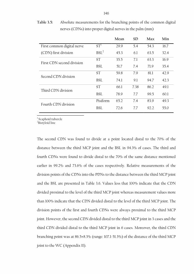
146 Table 3.5: Absolute measurements for the branching points of the common digital
nerves (CDNs) into proper digital nerves in the palm (mm)
Mean SD Max Min
First common digital nerve
(CDN) first division
ST1 29.9 5.4 54.3 16.7
BSL2 45.3 6.1 63.5 32.4
First CDN second division ST 35.5 7.1 63.3 16.9
BSL 51.7 7.4 71.9 35.4
Second CDN division ST 59.8 7.9 81.1 42.9
BSL 74.1 9.1 94.7 42.3
Third CDN division ST 66.1 7.38 86.2 49.1
BSL 78.9 7.7 99.5 60.1
Fourth CDN division Pisiform 65.2 7.4 83.9 49.3
BSL 72.6 7.7 92.2 55.0
1 Scaphoid tubercle 2Bistyloid line
The second CDN was found to divide at a point located distal to the 70% of the
distance between the third MCP joint and the BSL in 94.3% of cases. The third and
fourth CDNs were found to divide distal to the 70% of the same distance mentioned
earlier in 99.2% and 73.6% of the cases respectively. Relative measurements of the
division points of the CDNs into the PDNs to the distance between the third MCP joint
and the BSL are presented in Table 3.6. Values less that 100% indicate that the CDN
divided proximal to the level of the third MCP joint whereas measurement values more
than 100% indicate that the CDN divided distal to the level of the third MCP joint. The
division points of the first and fourth CDNs were always proximal to the third MCP
joint. However, the second CDN divided distal to the third MCP joint in 3 cases and the
third CDN divided distal to the third MCP joint in 6 cases. Moreover, the third CDN
branching point was at 86.5±8.3% (range: 107.1-51.3%) of the distance of the third MCP
joint to the WC (Appendix II).

147 Table 3.6: Branching points of the common digital nerves (CDNs) into the proper
digital nerves to the distance between the third metacarpophalangeal
joint and the bistyloid line (%)
Mean SD Max Min
First common digital nerve (CDN)
first division 51.2 5.3 63.3 40.2
First CDN second division 58.7 7.2 76.9 41.6
Second CDN division 83.8 8.6 101.5 39.4
Third CDN division 89.3 6.3 108.1 65.3
Fourth CDN division 74.2 7.6 93.7 55.4
Based on observations from 142 cadavers, the pattern of division of the first CDN
supplying the thumb and the lateral side of the index finger was categorized into three
types as suggested by Jolley et al. (1997):
Type I: A PDN to the lateral side of the thumb and a CDN further dividing
distally to supply the medial side of the thumb and the lateral side of the
index finger.
Type II: Trifurcation of PDNs to the thumb and the lateral side of the index
finger.
Type III: A CDN further dividing to supply the lateral and medial side of the
thumb and a PDN to the lateral side of the index finger.
Type I was found in 70.8%, Type II in 27.1% and Type III in 2.1% of the hands
investigated (Figure 3.11).
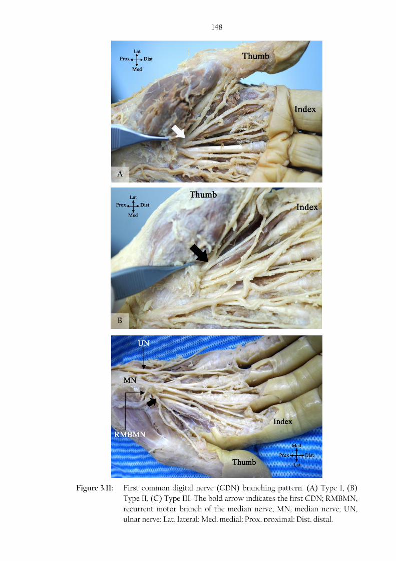
148
Figure 3.11: First common digital nerve (CDN) branching pattern. (A) Type I, (B)
Type II, (C) Type III. The bold arrow indicates the first CDN; RMBMN,
recurrent motor branch of the median nerve; MN, median nerve; UN,
ulnar nerve; Lat, lateral; Med, medial; Prox, proximal; Dist, distal.
C
B
A
149 The effect of sex and body side on the branching pattern was investigated. The
branching pattern of the first CDN was not influenced by sex (P value=0.186) but it was
found to be influenced by body side (P value =0.041). Appendix 1 shows the chi-square
test results for both the effect of sex and body side.
3.4.Ulnar nerve
The UN was examined in 144 cadaveric palms from 78 cadavers: 63 (43.8%) were male
and 81 (56.3%) female. Mean age was 82.4±9.7 (range: 101-53) years. The ulnar nerve
passed through Guyon’s canal in all cases and divided into a deep motor branch that
coursed deep to the hypothenar muscles and a superficial branch that supplied the little
and medial side of the ring finger. The UN division in Guyon’s canal was classified into
two patterns based on the number of branches at the division point as suggested by
Bonnel and Vila (1985):
Type I: UN bifurcates into one deep motor branch and a superficial sensory
branch that further divides distally into a CDN supplying the fourth
webspace and a PDN supplying the medial side of the little finger.
Type II: UN trifurcates into one deep motor branch, a CDN for the fourth
webspace and a PDN supplying the medial side of the little finger.
Type I was found in 80.4% while Type II was found in 19.6% (Figure 3.12).
The effect of sex and body side on the branching pattern was investigated. The
branching pattern of the UN was not influenced by sex (P value=0.258) or body side (P
value =0.392). Appendix 1 shows the chi-square test results for both the effect of sex
and body side.

150
Figure 3.12: Patterns of division of the ulnar nerve (UN) in Guyon’s canal. (A) Type I, (B) Type II. PDN, proper digital nerve; CDN, common digital nerve; Lat, lateral; Med, medial; Prox, proximal; Dist, distal.
B
A
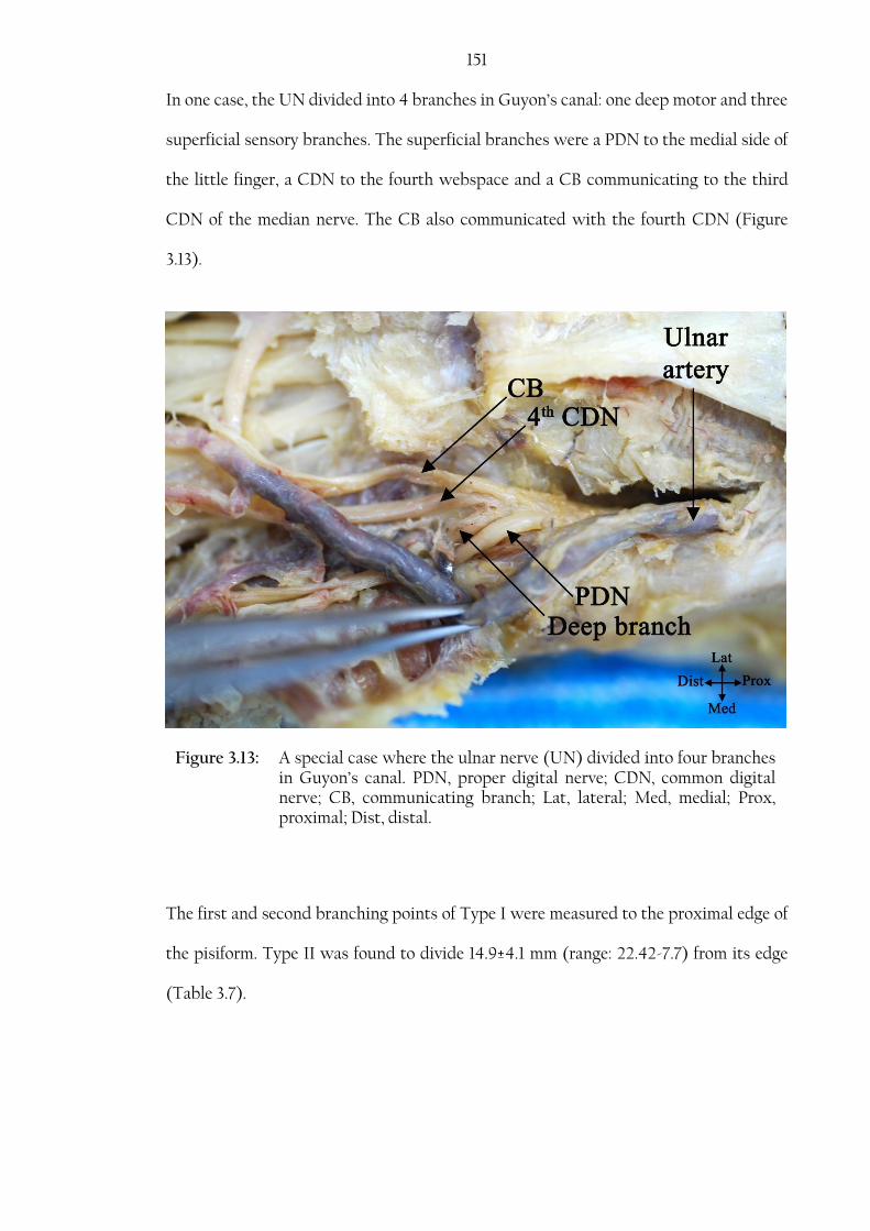
151 In one case, the UN divided into 4 branches in Guyon’s canal: one deep motor and three
superficial sensory branches. The superficial branches were a PDN to the medial side of
the little finger, a CDN to the fourth webspace and a CB communicating to the third
CDN of the median nerve. The CB also communicated with the fourth CDN (Figure
3.13).
The first and second branching points of Type I were measured to the proximal edge of
the pisiform. Type II was found to divide 14.9±4.1 mm (range: 22.42-7.7) from its edge
(Table 3.7).
Figure 3.13: A special case where the ulnar nerve (UN) divided into four branches in Guyon’s canal. PDN, proper digital nerve; CDN, common digital nerve; CB, communicating branch; Lat, lateral; Med, medial; Prox, proximal; Dist, distal.

152 Table 3.7: The first and second division points of Type I and the point of
trifurcation of Type II to the proximal edge of the pisiform (mm)
Mean SD Max Min
Ulnar nerve division into deep & superficial
(Type I first division point) 13.6 4.0 22.1 1.8
Superficial branch division into the 4th CDN1 &
PDN2 (Type I second division point) 25.2 4.6 37.2 13.1
Ulnar nerve trifurcation into deep, 4th CDN &
PDN (Type II trifurcation point) 14.9 4.1 22.42 7.7
1 Common digital nerve 2 Proper digital nerve to the medial side of the little finger
The sensory innervation of the little finger and medial side of the ring finger was
supplied by the superficial division of the ulnar nerve in all except 6 cases where the
ulnar nerve communicated with the DBUN (Kaplan anastomosis) and in another 4
cases where it received contributions from the median nerve.
The sensory distribution of the ulnar nerve in the palm was classified into two types
based on the branches contributing to the innervation of the little and the medial side
of the ring finger:
Type I: The little finger and the medial side of the ring finger were innervated by
the superficial branch of the UN which could be:
A. Originating as a common trunk that divides distally to a PDN to the
medial side of the little finger and a CDN to the fourth webspace.
(Figure 3.12 A)
B. Originating as a PDN to the medial side of the little finger and a CDN
to the fourth webspace. (Figure 3.12 B)
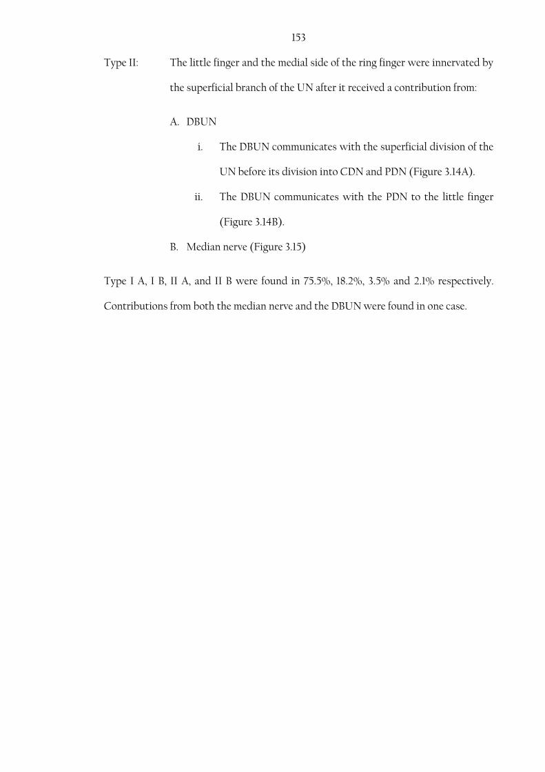
153 Type II: The little finger and the medial side of the ring finger were innervated by
the superficial branch of the UN after it received a contribution from:
A. DBUN
i. The DBUN communicates with the superficial division of the
UN before its division into CDN and PDN (Figure 3.14A).
ii. The DBUN communicates with the PDN to the little finger
(Figure 3.14B).
B. Median nerve (Figure 3.15)
Type I A, I B, II A, and II B were found in 75.5%, 18.2%, 3.5% and 2.1% respectively.
Contributions from both the median nerve and the DBUN were found in one case.

154
Figure 3.14: Kaplan anastomosis. (A) Dorsal branch of the ulnar nerve (DBUN) communicates with the superficial branch of the ulnar nerve. (B) DBUN communicates with the proper digital nerve to the little finger (PDN). CDN, common digital nerve; Lat, lateral; Med, medial; Prox, proximal; Dist, distal.
B
A
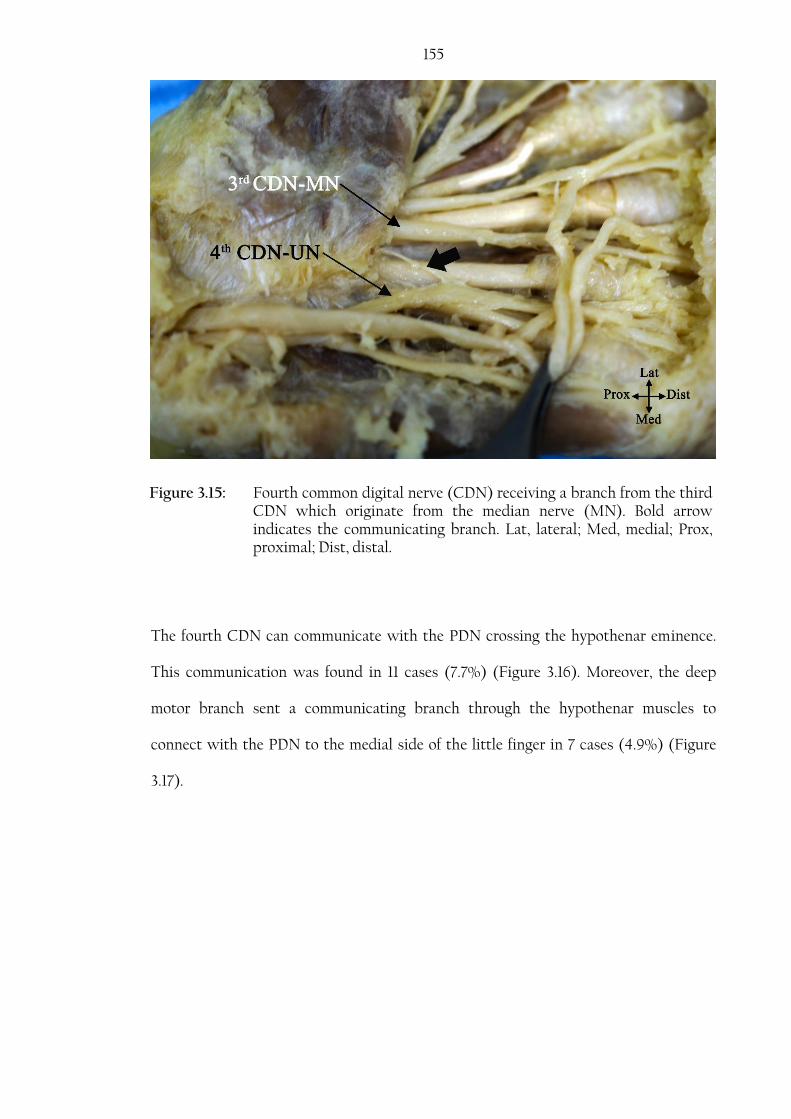
155
The fourth CDN can communicate with the PDN crossing the hypothenar eminence.
This communication was found in 11 cases (7.7%) (Figure 3.16). Moreover, the deep
motor branch sent a communicating branch through the hypothenar muscles to
connect with the PDN to the medial side of the little finger in 7 cases (4.9%) (Figure
3.17).
Figure 3.15: Fourth common digital nerve (CDN) receiving a branch from the third CDN which originate from the median nerve (MN). Bold arrow indicates the communicating branch. Lat, lateral; Med, medial; Prox, proximal; Dist, distal.
156
Figure 3.16: Fourth common digital nerve (CDN) communicating with the proper digital nerve (PDN) to the little finger. Bold arrow indicates the communicating branch. Lat, lateral; Med, medial; Prox, proximal; Dist, distal.
Figure 3.17: Deep motor branch of the ulnar nerve communicating with the proper digital nerve (PDN) to the little finger. CDN, common digital nerve; bold arrow indicates the communicating branch. Lat, lateral; Med, medial; Prox, proximal; Dist, distal.
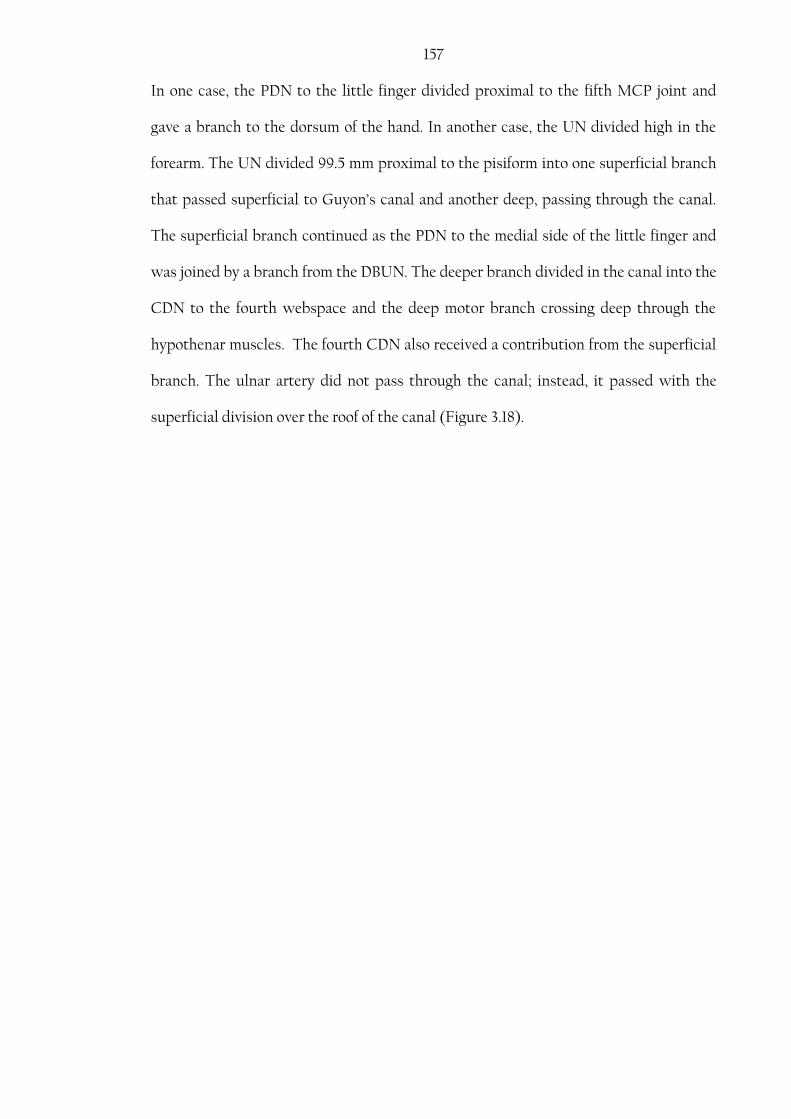
157 In one case, the PDN to the little finger divided proximal to the fifth MCP joint and
gave a branch to the dorsum of the hand. In another case, the UN divided high in the
forearm. The UN divided 99.5 mm proximal to the pisiform into one superficial branch
that passed superficial to Guyon’s canal and another deep, passing through the canal.
The superficial branch continued as the PDN to the medial side of the little finger and
was joined by a branch from the DBUN. The deeper branch divided in the canal into the
CDN to the fourth webspace and the deep motor branch crossing deep through the
hypothenar muscles. The fourth CDN also received a contribution from the superficial
branch. The ulnar artery did not pass through the canal; instead, it passed with the
superficial division over the roof of the canal (Figure 3.18).
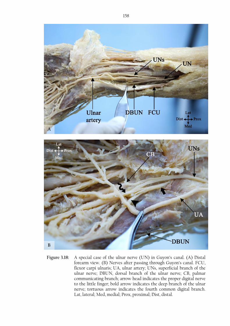
158
Figure 3.18: A special case of the ulnar nerve (UN) in Guyon’s canal. (A) Distal forearm view. (B) Nerves after passing through Guyon’s canal. FCU, flexor carpi ulnaris; UA, ulnar artery; UNs, superficial branch of the ulnar nerve; DBUN, dorsal branch of the ulnar nerve; CB, palmar communicating branch; arrow head indicates the proper digital nerve to the little finger; bold arrow indicates the deep branch of the ulnar nerve; tortuous arrow indicates the fourth common digital branch. Lat, lateral; Med, medial; Prox, proximal; Dist, distal.
B
A

159
3.5. Palmar communicating branch between the median and ulnar nerve
The palmar CB was investigated in 98 cadaveric palms from 62 cadavers: 45 (45.9%)
male and 53 (54.1%) female. Mean age was 81.3±9.9 (range: 101-53) years.
To determine the incidence of the CB, 61 cadaveric palms were investigated (40 right
hands and 21 left hands): the CB was found in all except 8 hands (13.1%).
Five patterns were noted according to the proximal and distal attachment of the CB:
Type I: The CB originated from the fourth CDN (ulnar nerve) and coursed
distally to join the third CDN (MN) (Figure 3.19)
A. A single distal attachment
B. Multiple distal attachments
Type II: The CB originated from the third CDN (MN) and coursed distally to
join the fourth CDN (UN) (Figure 3.20)
A. A single distal attachment
B. Multiple distal attachments
Type III: The CB coursed perpendicularly between the third and fourth CDNs
(Figure 3.21).
A. A single attachments on both sides
B. Multiple attachments on one side
Type IV: The CB had multiple attachment points to both nerves in a diffuse
manner (Figure 3.22 A).
Type V: The UN and MN gave branches that merged and continued distally to
the lateral side of the ring finger (Figure 3.22 B).
160
B
Figure 3.19: Branching pattern of the palmar communicating branch (CB) Type I. (A) Type I A, (B) Type I B. CDN, common digital nerve; MN, median nerve; UN, ulnar nerve, bold arrow indicates the palmar CB. Lat, lateral; Med, medial; Prox, proximal; Dist, distal.
A
B

161
Figure 3.20: Branching pattern of the palmar communicating branch (CB) Type II. (A) Type II A, (B) Type II B. CDN, common digital nerve; MN, median nerve; UN, ulnar nerve; bold arrow indicates the palmar CB. Lat, lateral; Med, medial; Prox, proximal; Dist, distal.
A
B
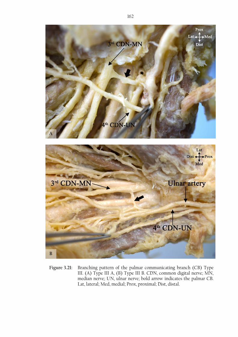
162
Figure 3.21: Branching pattern of the palmar communicating branch (CB) Type III. (A) Type III A, (B) Type III B. CDN, common digital nerve; MN, median nerve; UN, ulnar nerve; bold arrow indicates the palmar CB. Lat, lateral; Med, medial; Prox, proximal; Dist, distal.
A
B
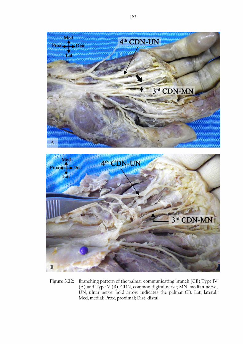
163
Figure 3.22: Branching pattern of the palmar communicating branch (CB) Type IV (A) and Type V (B). CDN, common digital nerve; MN, median nerve; UN, ulnar nerve; bold arrow indicates the palmar CB. Lat, lateral; Med, medial; Prox, proximal; Dist, distal.
B
A
164 Type I was found in 82.7% (81/98) of cases: it was noted that the CB had one distal
attachment (60 cases, Type I A) or multiple distal attachments (21 cases, Type I B).
Type II was found in 5.1% (5/98) of the cases investigated: similar to Type I, the distal
attachment can be single (4 cases, Type II A) or multiple (1 case, Type II B). Type III
was found in 5.1% (5/98), three of the five cases had multiple attachments to the fourth
CDN (UN) (Type III B) and two had a single attachment to the fourth CDN and the
third CDN (Type III A). Type IV and V were found in 4.1% (4/98) and 3.1% (3/98) cases
respectively (Figures 3.16, 3.17, 3.18, 3.19, 3.20).
The proximal attachment of the CB was found 5.3±5.2 mm, 32.3±5.8 mm and 43.6±7.1
mm distal to the TCL, WC and to BSL respectively. The distal attachment was found
16.4±6.4 mm, 43.9±6.6 mm and 52.6±7.4 mm distal to the TCL, WC and BSL
respectively. All measurements are shown in Table 3.8. Moreover, the CB was found to
be 15.6±4.8 mm (range: 36.95-8.32) long and the proximal attachment originated at an
angle averaging 142.7±17.7˚ (range: 180-90˚). The angle at which the proximal
attachment was found to branch off the UN or MN was found to be between 90-120,
121-150 and 151-180 in 12.5%, 64.8% and 22.7% of the cases respectively.
Moreover, the proximal attachment of the CB was found between 25.9% and 60.3% of
the distance between the third MCP joint and the WC; between 34.5% and 60.5% of
the distance between the third MCP joint and the BSL. The distal attachment of the CB
was found between 36.6% and 79.3% of the distance between the third MCP joint and
the WC; between 41.8% and 75.4% of the distance between the third MCP joint and
the BSL. The CB originated proximal to or at the level of the TCL in 13% of cases and
less than 4 mm distal to the TCL in 38%.

165 Table 3.8: Distances measured for the proximal and distal attachments of the
palmar communicating branch (CB) to different anatomical landmarks
(mm)
Distance between: Mean SD Max Min
Proximal attachment of CB to TCL1 5.3 5.2 19.4 -10.62
Distal attachment of CB to TCL 16.4 6.4 39.5 1.3
Proximal attachment of CB to WC3 32.3 5.8 46.6 20.5
Distal attachment of CB to WC 43.9 6.6 60.6 27.8
Proximal attachment of CB to BSL4 43.6 7.1 64.5 30.8
Distal attachment of CB to BSL 52.6 7.4 69.2 38.1
1 Transverse carpal ligament 2 Minus signs indicate that the point were located proximal to the level of the respective landmark
3 Wrist crease 4 Bistyloid line
The effect of sex and body side on the branching pattern was investigated. The
branching pattern of the palmar CB was not influenced by sex (P value=0.335) or body
side (P value =0.949). Appendix 1 shows the chi-square test results for both the effect of
sex and body side.
3.6. Superficial branch of the radial nerve
The SBRN was investigated in 150 cadaveric hands from 76 cadavers: 60 (40%) male
and 90 (60%) female. Mean age was 82.9±9.4 (range: 101-53) years.
The SBRN originated at the level of the elbow (the radial nerve divided into a
superficial and a deep branch). In 17 cases (17/124, 13.7%), the SBRN originated
proximal to the lateral epicondyle. It was found to originate 231.4±19.9 mm (range: 295-
191.6) from the RSP and 13.3±13.9 (range: -35.0-57.0) mm from the lateral epicondyle.
The SBRN coursed deep to brachioradialis and became subcutaneous 72.1±16.6 mm
(range: 141.5-35.2) proximal to the RSP. Comparing the point where the SBRN pierces
the antebrachial fascia to the total length of the radius, it is estimated that the SBRN
166 becomes subcutaneous in the distal 30.5±5.8% of the total length of the radius. The
SBRN pierces the antebrachial fascia between the tendons of brachioradialis and
extensor carpi radialis longus. In 3 cases (2%), the nerve pierced the tendon of
brachioradialis to become subcutaneous.
The nerve continued distally and divided into two branches, one palmar and one dorsal,
51.4±14.9 mm proximal to the RSP. In 4 cases (2.7%), the SBRN divided into three
branches rather than two (Figure 3.23). The nerve usually divided into its palmar and
dorsal branches after it pierced the fascia; however, in one case the nerve divided deep
to brachioradialis prior to becoming subcutaneous. In another case, the SBRN’s first
division originated at the level of the elbow. The SBRN originated as two nerves from
the radial nerve: the superficial branch pierced brachioradialis, travelled in the
substance of the muscle and became subcutaneous 119.7 mm proximal to the RSP as the
palmar division of the SBRN. The deeper division continued deep to brachioradialis and
became subcutaneous 97.7 mm proximal to the RSP as the dorsal branch of the SBRN.
The palmar division connected with both the dorsal branch and the LABCN before it
innervated the lateral side of the thumb and the thenar eminence. The dorsal branch
continued to innervate the dorsum of the hand (Figure 3.24A-D).

167
Figure 3.23: First division of the superficial branch of the radial nerve (SBRN) after it becomes cutaneous. (A) Palmar and dorsal divisions. (B) Three divisions, one dorsal and two palmar (indicated by the arrow heads); pins indicate the location of the radial styloid process. Prox, proximal; Dist, distal; palmar, palmar surface; Dorsal, dorsal surface.
A
B
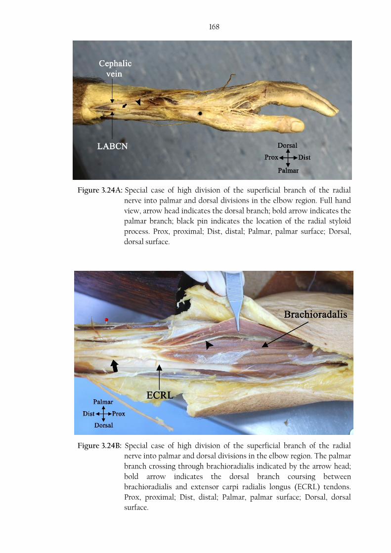
168
Figure 3.24A: Special case of high division of the superficial branch of the radial
nerve into palmar and dorsal divisions in the elbow region. Full hand
view, arrow head indicates the dorsal branch; bold arrow indicates the
palmar branch; black pin indicates the location of the radial styloid
process. Prox, proximal; Dist, distal; Palmar, palmar surface; Dorsal,
dorsal surface.
Figure 3.24B: Special case of high division of the superficial branch of the radial
nerve into palmar and dorsal divisions in the elbow region. The palmar
branch crossing through brachioradialis indicated by the arrow head;
bold arrow indicates the dorsal branch coursing between
brachioradialis and extensor carpi radialis longus (ECRL) tendons.
Prox, proximal; Dist, distal; Palmar, palmar surface; Dorsal, dorsal
surface.

169
Figure 3.24C: Special case of high division of the superficial branch of the radial
nerve into palmar and dorsal divisions in the elbow region. The two
different points where the palmar (bold arrow) and dorsal (arrow
head) branches pierce the antebrachial fascia. BR, brachioradialis;
ECRL, extensor carpi radialis longus; LABCN, lateral antebrachial
cutaneous nerve. Prox, proximal; Dist, distal; Palmar, palmar surface;
Dorsal, dorsal surface.
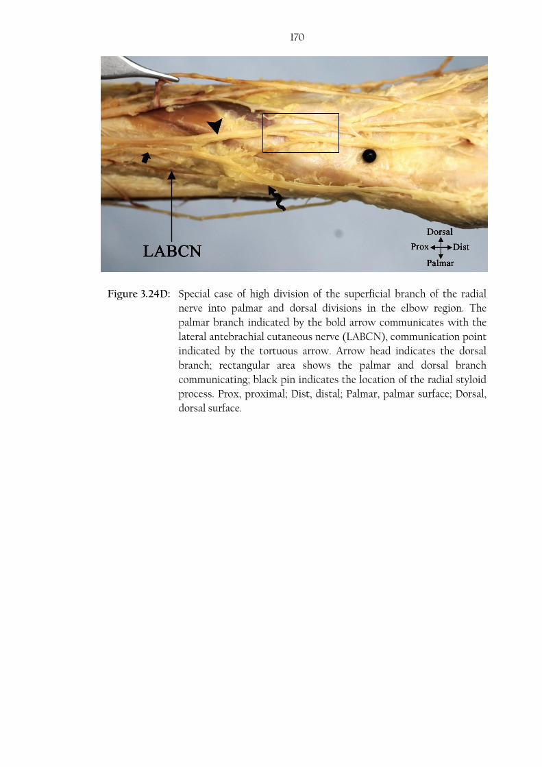
170
Figure 3.24D: Special case of high division of the superficial branch of the radial
nerve into palmar and dorsal divisions in the elbow region. The
palmar branch indicated by the bold arrow communicates with the
lateral antebrachial cutaneous nerve (LABCN), communication point
indicated by the tortuous arrow. Arrow head indicates the dorsal
branch; rectangular area shows the palmar and dorsal branch
communicating; black pin indicates the location of the radial styloid
process. Prox, proximal; Dist, distal; Palmar, palmar surface; Dorsal,
dorsal surface.

171 Prior to the first division, the SBRN diameter averaged 3.2±0.8 mm. All SBRN branches
passed radial to LT with a mean distance of 14.1±3.8 mm (Table 3.9). The SBRN
connected with the LABCN in 46 cases (30.7%) and crossed the cephalic vein for the
first time after it became subcutaneous 47.9±19.3 mm proximal to the RSP. The SBRN
branches often passed superficial to the anatomic snuff box. At the level of the RSP, the
closest palmar and dorsal branches of SBRN passed 8.5±4.2 mm and 4.9±3.0 mm
respectively. The closest palmar and dorsal branches to a point 25 mm proximal to the
RSP passed 5.6±3.4 mm and 4.2±2.6 mm respectively (Table 3.9).
Table 3.9: Measurements of the closest branches of the superficial branch of the
radial nerve (SBRN) to various anatomical points (mm)
Mean SD Max Min
Closest palmar branch at the level of the RSP1 8.5 4.1 17.8 0.7
Closest dorsal branch at the level of the RSP 4.9 3.0 14.7 1.1
Closest palmar branch to a point 25 mm
proximal to the RSP 5.6 3.4 15.1 0
Closest dorsal branch to a point 25 mm
proximal to the RSP 4.1 2.6 10.9 0
Closest branch to Lister’s tubercle 14.1 3.8 23.1 4.6
First intersection between the SBRN and the
cephalic vein 47.9 19.3 93.6 6.7
SBRN diameter prior to its first division 3.2 0.8 5.8 1.94
1 Radial styloid process
The palmar branch of the SBRN usually supplied the thumb and the lateral side of the
thenar eminence. The dorsal branch continued distally and divided in many
configurations to supply the dorsum of the hand. The major division points of the
palmar and the dorsal branches to the RSP are presented in Table 3.10.
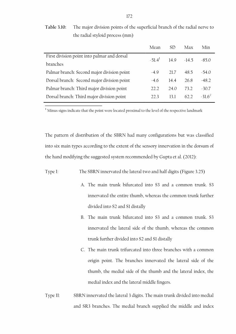
172 Table 3.10: The major division points of the superficial branch of the radial nerve to
the radial styloid process (mm)
Mean SD Max Min
First division point into palmar and dorsal
branches -51.41 14.9 -14.5 -85.0
Palmar branch: Second major division point -4.9 21.7 48.5 -54.0
Dorsal branch: Second major division point -4.6 14.4 26.8 -48.2
Palmar branch: Third major division point 22.2 24.0 73.2 -30.7
Dorsal branch: Third major division point 22.3 13.1 62.2 -31.62
1 Minus signs indicate that the point were located proximal to the level of the respective landmark
The pattern of distribution of the SBRN had many configurations but was classified
into six main types according to the extent of the sensory innervation in the dorsum of
the hand modifying the suggested system recommended by Gupta et al. (2012):
Type I: The SBRN innervated the lateral two and half digits (Figure 3.25)
A. The main trunk bifurcated into S3 and a common trunk. S3
innervated the entire thumb, whereas the common trunk further
divided into S2 and S1 distally
B. The main trunk bifurcated into S3 and a common trunk. S3
innervated the lateral side of the thumb, whereas the common
trunk further divided into S2 and S1 distally
C. The main trunk trifurcated into three branches with a common
origin point. The branches innervated the lateral side of the
thumb, the medial side of the thumb and the lateral index, the
medial index and the lateral middle fingers.
Type II: SBRN innervated the lateral 3 digits. The main trunk divided into medial
and SR3 branches. The medial branch supplied the middle and index

173
fingers and occasionally the medial side of the thumb. SR3 supplied the
thumb (Figure 3.26).
A. The main trunk divided into S3 and a common trunk. S3
innervated the entire thumb. The common trunk divided into S2
and S1 distally.
B. The main trunk divided into S3 and a common trunk, which
divided distally to S1 and S2. S2 innervated the medial thumb and
S1 the index and the middle finger.
C. The main trunk bifurcated into S3 and a common trunk. S3
innervated the lateral side of the thumb, whereas the common
trunk further divided into two branches distally. The medial side
of the thumb and the lateral side of the index fingers are
innervated by the same branch.
D. The main trunk bifurcated into S3 and a common trunk. S3
innervated the lateral side of the thumb, whereas the common
trunk further divided into three or four branches distally.
E. The main trunk trifurcated into three branches with a common
origin point. The branches innervated the lateral side of the
thumb, the medial side of the thumb, index and middle fingers.
Type III: SBRN innervated the lateral 3 and half digits. The main trunk divided
into a medial and SR3 (Figure 3.27).
A. The main trunk bifurcated into S3 and a common trunk. S3
innervated the entire thumb, whereas the common trunk further
divided into S2 and S1 distally.
B. The main trunk bifurcated into S3 and a common trunk. S3
innervated the lateral side of the thumb, whereas the common

174
trunk further divided into S1 and S2 distally. The medial side of
the thumb and the lateral side of the index finger are innervated
by the same branch.
C. The main trunk bifurcated into S3 and a common trunk. S3
innervated the lateral side of the thumb, whereas the common
trunk further divided into S2 and S3 distally. S2 to the medial
side of the thumb and S3 to the rest of the territory.
D. The main trunk bifurcated into S3 and a common trunk. S3
innervated the lateral side of the thumb, whereas the common
trunk further divided into three or four branches distally.
Type IV: SBRN innervated the lateral four digits (Figure 3.28).
Type V: SBRN innervated the lateral four and half digits (Figure 3.29).
Type VI: SBRN innervated the full dorsum of the hand with the absence of
the DBUN.
A. Trifurcation of the main trunk (Figure 3.30).
B. The main trunk bifurcated into S3 and a common trunk. S3
supplies the lateral side of the thumb. The common trunk further
trifurcates (Figure 3.31).
C. The main trunk bifurcated into S3 and a common trunk. S3
supplies the lateral side of the thumb. The common trunk divided
into S2 and S1. S2 supplies the medial side of the thumb and the
lateral index finger. S1 supplies the rest of the territory (Figure
3.32).
D. The main trunk bifurcated into S3 and a common trunk. S3
supplies the lateral side of the thumb. The common trunk divided
into S2 and S1. S2 supplies the medial side of the thumb index

175
and the lateral side of the middle fingers. S1 supplies the rest of
the territory (Figure 3.33).
The most common type was Type I found in the 37.33% of the cases. Type II, III, IV, V
and IV were found in 23.33%, 29.33%, 1.33%, 2% and 6.67% respectively. Tables 3.11-3.14
show the incidence rate for each type. Figures 3.25-3.33 show the different patterns of
the SBRN.
Table 3.11: Incidence rate for superficial branch of the radial nerve (SBRN) Type I branching pattern
Type No. %
Type I 56 37.3
1A 8 5.3
1B 46 30.7
1C 2 1.3
A

176
Figure 3.25: Branching pattern of the superficial branch of the radial nerve (SBRN) Type I. (A) Type I A. (B) Type I B. (C) Type I C. Arrow head indicates the trifurcation of the SBRN, pins indicate the location of the radial styloid process. Prox, proximal; Dist, distal; Palmar, palmar surface; Dorsal, dorsal surface.
B
C
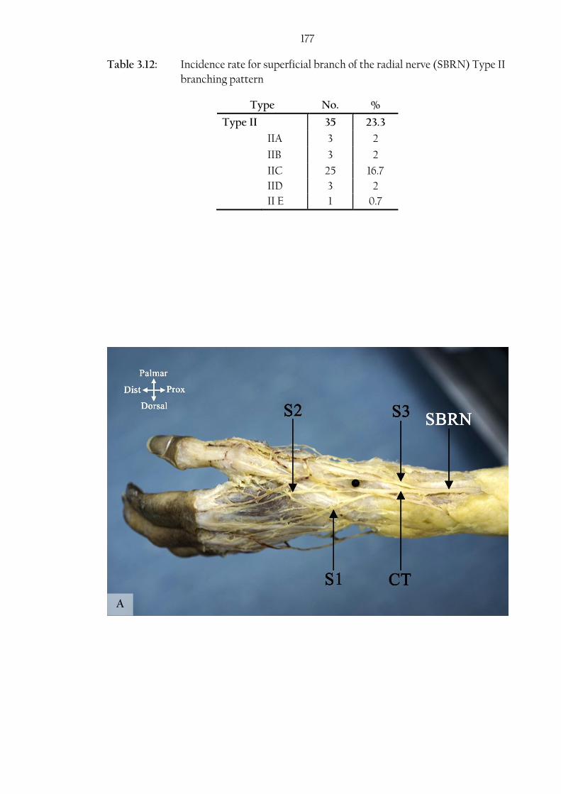
177 Table 3.12: Incidence rate for superficial branch of the radial nerve (SBRN) Type II
branching pattern
Type No. %
Type II 35 23.3
IIA 3 2
IIB 3 2
IIC 25 16.7
IID 3 2
II E 1 0.7
A

178
C
B
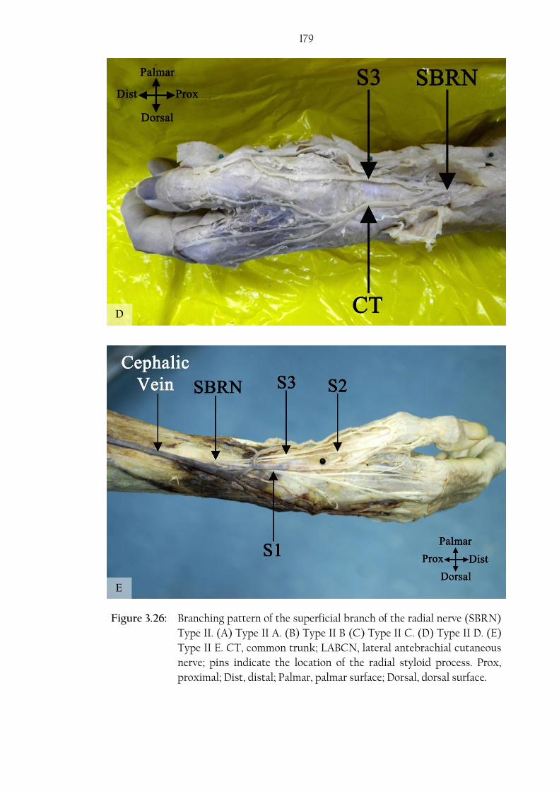
179
Figure 3.26: Branching pattern of the superficial branch of the radial nerve (SBRN)
Type II. (A) Type II A. (B) Type II B (C) Type II C. (D) Type II D. (E)
Type II E. CT, common trunk; LABCN, lateral antebrachial cutaneous
nerve; pins indicate the location of the radial styloid process. Prox,
proximal; Dist, distal; Palmar, palmar surface; Dorsal, dorsal surface.
E
D
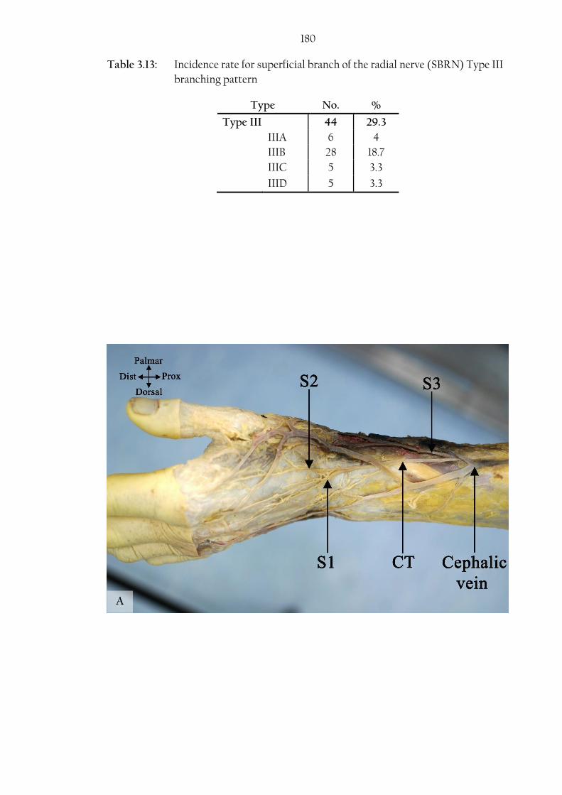
180 Table 3.13: Incidence rate for superficial branch of the radial nerve (SBRN) Type III
branching pattern
Type No. %
Type III 44 29.3
IIIA 6 4
IIIB 28 18.7
IIIC 5 3.3
IIID 5 3.3
A

181
B
C

182
Table 3.14: Incidence rates for superficial branch of the radial nerve (SBRN) Type
IV, Type V and Type VI branching patterns
Type No. %
Type IV 2 1.3
Type V 3 2
Type VI 10 6.7
VIA 1 0.7
VIB 2 1.3
VIC 5 3.3
VID 2 1.3
Figure 3.27: Branching pattern of the superficial branch of the radial nerve (SBRN)
Type III. (A) Type III A. (B) Type III B (C) Type III C. (D) Type III D.
CT, common trunk; pins indicate the location of the radial styloid
process. Prox, proximal; Dist, distal; Palmar, palmar surface; Dorsal,
dorsal surface.
D

183
Figure 3.28: Branching pattern of the superficial branch of the radial nerve
(SBRN) Type IV. Prox, proximal; Dist, distal; Lat, lateral; Med,
medial.
Figure 3.29: Branching pattern of the superficial branch of the radial nerve (SBRN)
Type V. Prox, proximal; Dist, distal; Lat, lateral; Med, medial.
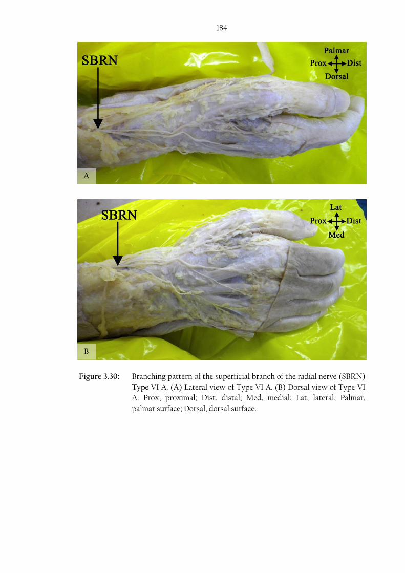
184
A
B
Figure 3.30: Branching pattern of the superficial branch of the radial nerve (SBRN)
Type VI A. (A) Lateral view of Type VI A. (B) Dorsal view of Type VI
A. Prox, proximal; Dist, distal; Med, medial; Lat, lateral; Palmar,
palmar surface; Dorsal, dorsal surface.
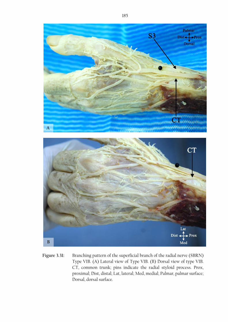
185
Figure 3.31: Branching pattern of the superficial branch of the radial nerve (SBRN)
Type VIB. (A) Lateral view of Type VIB. (B) Dorsal view of type VIB.
CT, common trunk; pins indicate the radial styloid process. Prox,
proximal; Dist, distal; Lat, lateral; Med, medial; Palmar, palmar surface;
Dorsal, dorsal surface.
A
B

186
A
B
Figure 3.32: Branching pattern of the superficial branch of the radial nerve (SBRN)
Type VI C. (A) Lateral view of Type VI C. (B) Dorsal view of Type VI
C. Prox, proximal; Dist, distal; Lat, lateral; Med, medial; Palmar,
palmar surface; Dorsal, dorsal surface.

187
Figure 3.33: Branching pattern of the superficial branch of the radial nerve (SBRN)
Type VI D. This type was found in cadaver number 896 right and left
hands, pictures were lost and the sketch was prepared based on the
sketches taken from the two hands where the type was found.

188 The effect of sex and body side on the branching pattern was investigated. The
branching pattern of the SBRN was not influenced by sex (P value=0.966) or body side
(P value =0.579). Appendix 1 shows the chi-square test results for both the effect of sex
and body side.
3.7. Dorsal branch of the ulnar nerve
The DBUN was investigated in 139 cadaveric hands from 73 cadavers: 70 (50.3%) male
and 69 (49.6%) female. Mean age was 82.6±9.5 (range: 101-53) years.
The nerve originated from the medial side of the main trunk of the ulnar nerve in the
distal forearm. It originated 63.3±14.8 mm (range: 110.7-26.7) and 61.3±21.0 mm (range:
140-18.5) proximal to the proximal edge of the pisiform and proximal to the USP
respectively. It was estimated that the nerve originated at the distal 23.6±7.7% of the
total length of the ulna. The nerve pierced the fascia 18.1±7.1 mm (range: 33.1-1.74)
proximal to the USP and continued to innervate the medial 1½-2½ digits in the dorsum
of the hand. At the level of the USP, the closest branch of DBUN passed 6.8±3.1 mm
(range: 14.0-0.4) to the USP (Table 3.15).
Table 3.15: Anatomical measurements recorded for the dorsal branch of the ulnar
nerve (mm)
Mean SD Max Min
Origin from the ulnar nerve to the
proximal edge of the pisiform 63.3 14.8 110 26.7
Origin from the ulnar nerve to the USP1 61.3 21.0 140 18.5
Pierced the fascia 18.1 7.1 33.1 1.74
Closest branch to USP 6.8 3.1 14.0 0.4
1 Ulnar styloid process
189 Table 3.16 shows the major first and second division points of the DBUN. Prior to its
division, the DBUN diameter was 2.2±0.7 mm (range: 3.68-0.62). The nerve consistently
crossed a line between the USP and the fourth webspace. The DBUN crossed this line
18.1±9.1 mm (range: 40.4-3.4 mm) distal to the USP corresponding to 19.4±9.6% (range:
45.4-3.1) of the distance between the USP and the fourth webspace.
Table 3.16: Distances of the first and second major branching point of the dorsal
branch of the ulnar nerve to the ulnar styloid process (mm).
Mean SD Max Min
First major branching point -3.41 17.4 53.4 -38.7
Second major branching point 39.2 13.2 73.6 -8.8
1 Minus signs indicate that the point were located below the level of the respective landmark
3.8. Sensory distribution in the dorsum of the hand
The dorsum of the hand was supplied by branches of the SBRN and DBUN. The
innervation territory showed many arrangements. Investigating 150 cadaveric hands,
the dorsum of the hand innervation was outlined (Table 3.17).
Table 3.17: Sensory innervation in the dorsum of the hand with reference to digits
SBRN2 DBUN3 UD4
Total n % n % N %
Index lateral 150 100.0 0 0.0 0 0.0 150
medial 147 98.0 0 0.0 3 2.0 150
Middle lateral 146 97.3 0 0.0 4 2.7 150
medial 57 38.0 57 38.0 36 24.0 150
Ring lateral 28 18.7 89 59. 3 33 22.0 150
medial 15 10.0 135 90.0 0 0.0 150
Little lateral 14 9.3 136 90.7 0 0.0 150
medial 10 6.7 140 93.3 0 0.0 150
2 Superficial branch of the radial nerve 3 Dorsal branch of the ulnar nerve 4 Undetermined
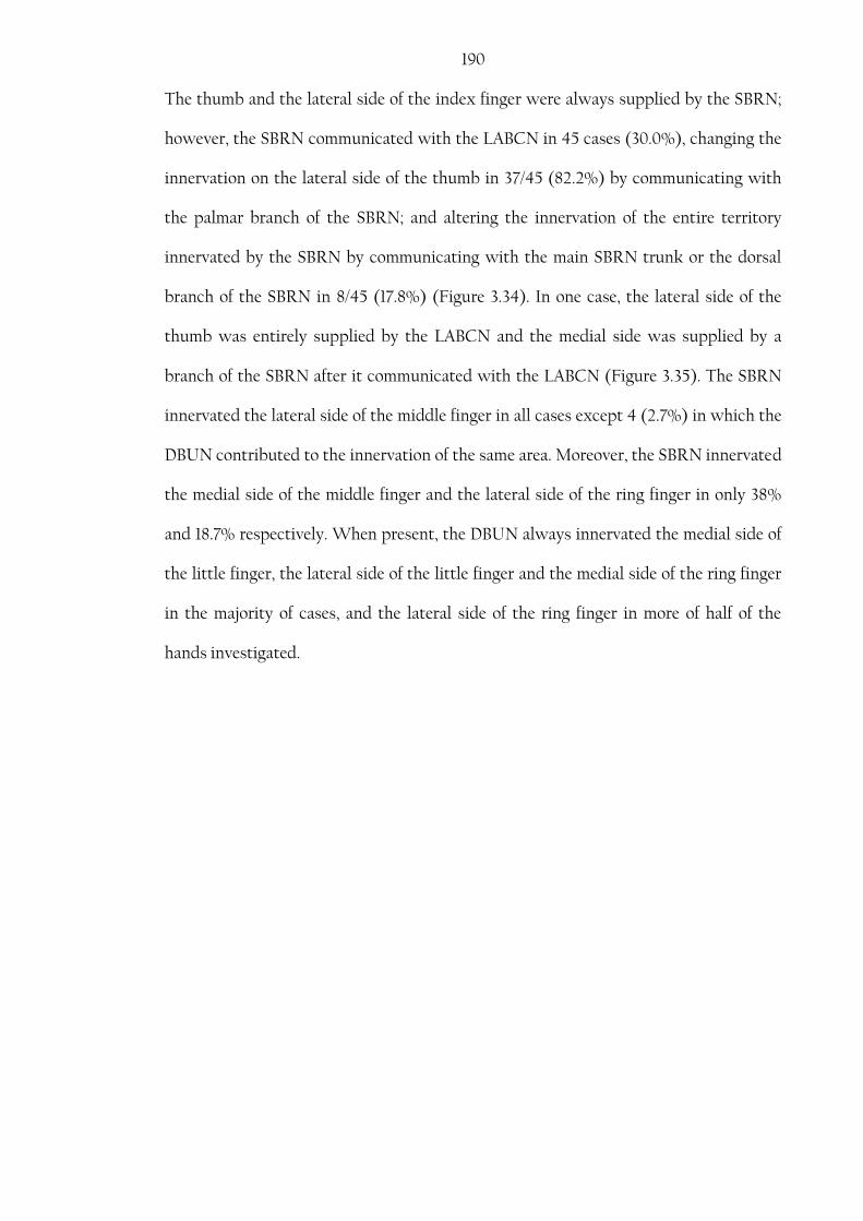
190 The thumb and the lateral side of the index finger were always supplied by the SBRN;
however, the SBRN communicated with the LABCN in 45 cases (30.0%), changing the
innervation on the lateral side of the thumb in 37/45 (82.2%) by communicating with
the palmar branch of the SBRN; and altering the innervation of the entire territory
innervated by the SBRN by communicating with the main SBRN trunk or the dorsal
branch of the SBRN in 8/45 (17.8%) (Figure 3.34). In one case, the lateral side of the
thumb was entirely supplied by the LABCN and the medial side was supplied by a
branch of the SBRN after it communicated with the LABCN (Figure 3.35). The SBRN
innervated the lateral side of the middle finger in all cases except 4 (2.7%) in which the
DBUN contributed to the innervation of the same area. Moreover, the SBRN innervated
the medial side of the middle finger and the lateral side of the ring finger in only 38%
and 18.7% respectively. When present, the DBUN always innervated the medial side of
the little finger, the lateral side of the little finger and the medial side of the ring finger
in the majority of cases, and the lateral side of the ring finger in more of half of the
hands investigated.

191
Figure 3.34: Superficial branch of the radial nerve (SBRN) communicating with the
lateral antebrachial cutaneous nerve (LABCN) (A). The rectangular
area outlined in (A) is enlarged and shown in (B). Prox, proximal; Dist,
distal; Palmar, palmar surface; Dorsal, dorsal surface.
A
B
192
A
B
Figure 3.35: Lateral view of a special case of the lateral antebrachial cutaneous nerve
(LABCN) entirely supplying the lateral side of the thumb. The medial side
of the thumb is supplied by a branch of the superficial branch of the radial
nerve (SBRN) after communicating with the LABCN (A). The rectangular
area outlined in (A) is enlarged and shown in (B). Bold arrow indicates
the communicating branch between the LABCN and the SBRN; Palmar,
palmar surface; Dorsal, dorsal surface; Prox, proximal; Dist, distal.

193 The DBUN and the SBRN communicated in the dorsum surface of the hand in 37 cases
(26.4%) altering the sensory innervation on the lateral side of the ring finger and medial
side of the middle finger in 31/37 (83.8%) cases, the lateral side of the middle finger and
medial side of the index finger in 3/37 (8.1%) cases, the medial side of the middle finger
in 2/37 (5.4%) cases and the lateral side of the middle finger in one case. Dual
innervation where both nerves overlap and contribute to the innervation of the same
area with no communications were found in 4 cases. Dual innervation affected the
medial side of the middle finger and lateral side of the ring finger in two cases, and the
medial side of the middle finger only in a further two cases.
Comparing the branching pattern on the dorsum of the hand in 74 cadavers, symmetry
was only found in 43.2% (32/74). Table 3.18 shows the different types of distribution on
each body side. The symmetry in the palmar surface of the hand was not studied as it
has less clinical implication.
Table 3.18: Distribution of the different branching patterns in the dorsum of the
hand
Right hand
Total
Type 1 Type 2 Type 3 Type 4 Type 5 Type 6
Left h
and
Type 1 14* 9 4 0 1 1 29
Type 2 5 5* 5 0 0 0 15
Type 3 4 4 12* 0 0 1 21
Type 4 1 0 0 0* 0 1 2
Type 5 1 0 1 0 0* 0 2
Type 6 1 2 1 0 0 1* 5
Total 26 20 23 0 1 4 74
* Showing the frequencies of symmetrical hands showing the same branching pattern in the left and right
hand sides.
194
3.9. Dorsal communicating branch between the radial and ulnar nerve
The dorsal CB between the SBRN and the DBUN was found in 37/140 (26.4%)
cadaveric hands of which 17 (46.0%) were from the right side and 20 (54.0%) were
from the left side: 13 (35.1%) male and 24 (64.9%) female. Three types were identified
based on the proximal attachment points:
Type I: The CB had a single attachment to the SBRN and the DBUN (Figure
3.36).
A. CB attached proximally to the SBRN
B. CB attached proximally to the DBUN
C. CB attached to the SBRN and DBUN at the same level
Type II: The CB had multiple attachment points to both the SBRN and DBUN
(Figure 3.37 A).
Type III: The DBUN and SBRN both gave a branch that merged and sent branches
to the dorsum of the hand (Figure 3.37 B),
Type I was found in 32.4% of the hands investigated. Moreover, subtype A, B, and C
were noted in 4/37 (10.8%), 7/37 (18.9%) and 1/37 (2.7%) of the hands respectively. Type
II and III were found in 10.8% (4/37) and 56.8% (21/37) of the hands respectively.
The effect of sex and body side on the branching pattern was investigated. The
branching pattern of the dorsal CB was not influenced by sex (P value=0.830) or body
side (P value =0.917). Appendix 1 show the chi-square test results for both the effect of
sex and body side.

195
Fig
ure
3.3
6:
Dor
sal
com
mu
nic
atin
g b
ran
ch (
CB
) b
etw
een
th
e su
per
fici
al b
ran
ch o
f th
e ra
dia
l n
erve
(SB
RN
) an
d t
he
dor
sal
bra
nch
of
the
uln
ar
ner
ve (
DB
UN
) T
ype
I. (
A)
Typ
e I
A. (
B)
Typ
e I
B. (
C)
Typ
e I
C. T
he
arro
w h
ead
in
dic
ates
th
e C
B. P
rox
, pro
xim
al; D
ist,
dis
tal;
Lat
,
late
ral;
Med
, med
ial.
A A
B A
C A
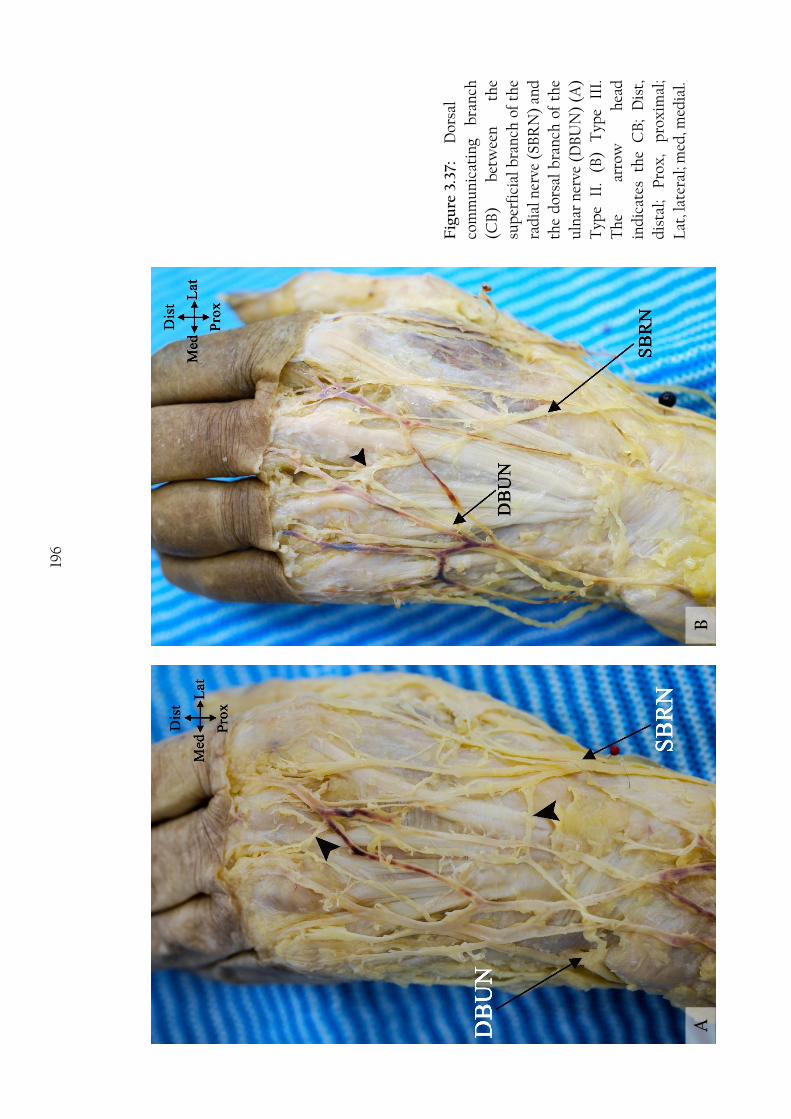
196
A A
B A
Fig
ure
3.3
7:
Dor
sal
com
mu
nic
atin
g b
ran
ch
(CB
) b
etw
een
th
e
sup
erfi
cial
bra
nch
of
the
rad
ial n
erve
(SB
RN
) an
d
the
dor
sal
bra
nch
of
the
uln
ar n
erve
(D
BU
N)
(A)
Typ
e II
. (B
) T
ype
III.
Th
e ar
row
h
ead
ind
icat
es
the
CB
; D
ist,
dis
tal;
Pro
x,
pro
xim
al;
Lat
, lat
eral
; med
, med
ial.

197 The location of the CB was variable. In Type I, The proximal attachment was found
18.5±22.0 mm to the BSL. The distal attachment was found to be 51.0±21.1 mm to the
BSL. The CB average length was 54.6±19.5 mm and originated with a mean angle of
139±29.3 (Table 3.18).
The proximal attachment location measured to the BSL was located at the 25.4±30.6%
(range: -7.6-85.6%) of the distance between the third MCP joint and the BSL.
Moreover, the distal attachment position measured to the BSL was 65.4±24.5% (range:
32.9-98.4%) of the distance between the third MCP joint and the BSL (Appendix II).
In Type III, the merging of the two branches was found 71.3±9.8 mm to the BSL which is
estimated to be located 87.0±6.9%% of the distance between the third MCP joint and
the BSL (Table 3.19) (Appendix II).
Table 3.19: Anatomical measurements for the dorsal communicating branch
between the superficial branch of the radial nerve and the dorsal branch
of the ulnar nerve (mm)
Mean SD Max Min
Proximal attachment to the BSL1 in Type I 18.5 22.0 62.9 -6.22
Distal attachment to the BSL in Type I 51.0 21.1 93.4 25.3
Communicating branch length in Type I 54.6 19.5 86.3 30.6
Angle of origin in Type I 139 29.3 168 60
The merging point recorded in Type III 71.3 9.8 85.2 55.4
1 Bistyloid line 2 Minus signs indicate that the point were located proximal to the level of the respective landmark

198
4. Discussion
4.1. Palmar surface of the hand:
The palmar surface of the hand is innervated by branches from two nerves: the median
and ulnar nerves. The median nerve gives the palmar cutaneous branch of the median
nerve (PCBMN) in the distal forearm, and the first, second and third common digital
nerves (CDNs) after it passes through the carpal tunnel. The MN is responsible for
innervating the skin over the lateral and middle third of the palm, and the lateral 3½
digits as described by most anatomical textbooks. The UN is responsible for
innervating the skin over the medial third of the palm and the medial 1½ digits by its
superficial branch, which is given off in Guyon’s canal. Although occasionally ignored
by anatomical textbooks, the MN and UN can communicate across the palm deep to
the superficial palmar arch allowing for exchange of fibres between the two nerves and
thus changing the innervation boundary in the palm that is classically described as the
middle of the ring finger.
4.1.1. Palmar cutaneous nerve of the median nerve
The PCBMN is the last nerve that arises from the median nerve in the forearm. It
supplies the proximal middle and lateral two fifths of the palm. It was found in all 123
cases investigated in the current study. Originating 54.1±15.7 mm proximal to the WC,
the nerve stayed in the same epineurium as the main trunk for a few millimetres
(12.9±1.8) and detached 41.2±14.0 mm proximal to the WC. Taleisnik (1973) was the
first to state that the PCBMN stays bound with the main body of the MN for a distance
of 16-25 mm after its origin. The PCBMN can be compressed before its detachment
from the MN as seen in pseudoanterior interosseous nerve syndrome when nerve
fascicle compress before separation. Unknown masses or tendons compressing the
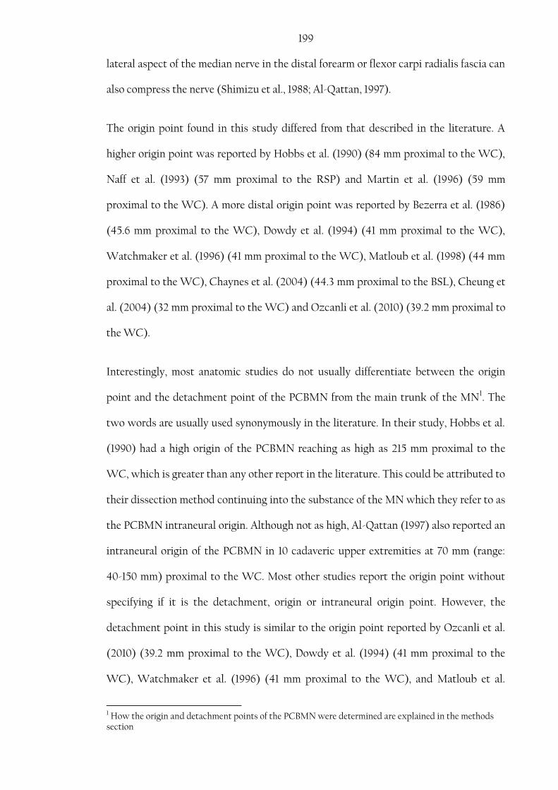
199 lateral aspect of the median nerve in the distal forearm or flexor carpi radialis fascia can
also compress the nerve (Shimizu et al., 1988; Al-Qattan, 1997).
The origin point found in this study differed from that described in the literature. A
higher origin point was reported by Hobbs et al. (1990) (84 mm proximal to the WC),
Naff et al. (1993) (57 mm proximal to the RSP) and Martin et al. (1996) (59 mm
proximal to the WC). A more distal origin point was reported by Bezerra et al. (1986)
(45.6 mm proximal to the WC), Dowdy et al. (1994) (41 mm proximal to the WC),
Watchmaker et al. (1996) (41 mm proximal to the WC), Matloub et al. (1998) (44 mm
proximal to the WC), Chaynes et al. (2004) (44.3 mm proximal to the BSL), Cheung et
al. (2004) (32 mm proximal to the WC) and Ozcanli et al. (2010) (39.2 mm proximal to
the WC).
Interestingly, most anatomic studies do not usually differentiate between the origin
point and the detachment point of the PCBMN from the main trunk of the MN1. The
two words are usually used synonymously in the literature. In their study, Hobbs et al.
(1990) had a high origin of the PCBMN reaching as high as 215 mm proximal to the
WC, which is greater than any other report in the literature. This could be attributed to
their dissection method continuing into the substance of the MN which they refer to as
the PCBMN intraneural origin. Although not as high, Al-Qattan (1997) also reported an
intraneural origin of the PCBMN in 10 cadaveric upper extremities at 70 mm (range:
40-150 mm) proximal to the WC. Most other studies report the origin point without
specifying if it is the detachment, origin or intraneural origin point. However, the
detachment point in this study is similar to the origin point reported by Ozcanli et al.
(2010) (39.2 mm proximal to the WC), Dowdy et al. (1994) (41 mm proximal to the
WC), Watchmaker et al. (1996) (41 mm proximal to the WC), and Matloub et al.
1 How the origin and detachment points of the PCBMN were determined are explained in the methods section
200 (1998) (44 mm proximal to the WC) suggesting that those studies may have
considered the detachment point of the PCBMN from the MN as the point of origin.
The PCBMN originated from the lateral side of the MN in most cases (93.5%); however,
it also originated from the medial, posterolateral and the anterior side of the MN in
1.6%, 1.6%, and 3.3% respectively. Most literature mentions the PCBMN as originating
from the lateral side, yet in their study of 60 Chinese cadavers, Cheung et al, (2004)
reported 14 cases (11.7%) where the PCBMN originated from the medial side. Such a
high number of variations in the origin direction may be explained by racial differences.
Although Taleisnik (1973) described the PCBMN originating from the anterolateral
side of the MN in all 12 cases investigated. No mention has been found in the literature
of anterior or posterolateral origin; however this could be attributed to the limited
number of samples involved in previous studies (see Table 1.1). Although small, it is
important for surgeons to understand that such variations can exist to avoid
complications in procedures involving the distal forearm.
The PCBMN coursed distally in the forearm between flexor carpi radialis and palmaris
longus if present. In 10 cases (8.3%), the nerve passed through the fascia of flexor carpi
radialis rather than medial to it and in a small number of cases posterior to the tendon
(4.2%). Furthermore, it was not uncommon for the nerve to penetrate the fibres of
palmaris longus as it blended with the palmar aponeurosis in the palm. This study’s
findings disagree with Matloub et al. (1998) who reported the PCBMN to pass through
the fascia of flexor carpi radialis in the majority of cases (36/40), and was partially
covered by palmaris longus tendon fibres at its insertion point into the palmar
aponeurosis in a minority of cases (3/40). Such a position can put the nerve at risk of
irritation or compression in cases of flexor carpi radialis tendinitis, trauma or
inflammation leading to soft tissue swelling in the distal forearm, or ganglia arising
201 from the flexor carpi radialis sheath and atypical palmaris longus muscles (Al-Qattan,
1997).
The PCBMN has its own tunnel formed between the deep and superficial fibres of the
TCL. Most literature describes the tunnel as starting at the level of the WC or at the
level of the scaphoid tubercle (Matloub et al., 1998, Naff et al., 1993). In the current
study, the tunnel started proximal to the WC between the superficial and deep fibres
of the PCL and extended into the TCL. Al-Qattan (1997) mentioned that the PCBMN
tunnel could be located within the antebrachial fascia, PCL, or the TCL. The average
length of the tunnel was 11.3±3.4 mm which is longer than described by Matloub et al.
(1998) who reported it at 8 mm long; however, it lies within the range described by
Taleisnik (1973) (9-16 mm).
Al-Qattan (1997) described six zones where the PCBMN could be at risk of entrapment
leading to compression syndrome. Distinguishing between entrapment of the PCBMN
at those six zones and median nerve entrapment at the carpal tunnel can be challenging
as the sensory innervation of the two nerves can greatly overlap. The two entrapments
can be independent or associated. It is important to assess the possibility of the
PCBMN entrapment in every patient with carpal tunnel syndrome (Wada et al. 2002).

202
Figure 4.1: The six zones
where the palmar cutaneous
branch of the median nerve
(PCBMN) is most likely to be
entrapped as described by Al-
Qattan (1997). (A) The PCBMN
is bound to the main trunk of
the median nerve; (B) The
PCBMN detaches from the
median nerve; (C) medial
sheath of the flexor carpi
radialis (FCR); (D) within the
PCBMN tunnel; (E) in relation
to the distal margin of palmaris
longus (PL); (F) the PCBMN
subcutaneous course prior to
innervating the skin (Al-Qattan,
1997).
In a study of 12 cadaveric hands, Taleisnik (1973) described the branching pattern of the
PCBMN as the nerve dividing into one larger lateral branch supplying the thenar area
and several smaller medial branches. Although significant, the branching pattern
described was not detailed enough as it did not include an outline of different patterns,
variations, or mention incidence rates even though variations were found. Bezerra et al.
(1986) investigated 50 cadaveric hands and classified the branching pattern into three
types according to the palmar area innervated and the direction of the branches. In
their classification, the PCBMN divided into two main branches in Type I and Type III;
however the two types were distinguished by the innervation territories of the
branches, and into three branches in Type II. In Type I, the PCBMN divided into a
lateral branch innervating the thenar region and an intermediate branch innervating the
intermediate third of the palm. Type III branches innervated the intermediate third of
the palm by an intermediate branch and the thenar region by a medial branch. The
medial branch might loop around to reach the laterally located thenar region. In Type
203 II, the PCBMN divided into lateral, intermediate and medial branches innervating the
thenar, intermediate and hypothenar regions of the palm respectively (Bezerra et al.,
1986). From the study by Bezarra et al. (1986), it could be concluded that the PCBMN
divided into two or three main branches which correspond to Type I and Type III of the
current study. Their study also reported that the medial branch was the shortest and
present in only 42% of cases. This could be due to the medial third of the palm being
innervated by the palmar cutaneous branch of the ulnar nerve, nerve of Henle and the
transverse palmar nerves (Doyle and Botte, 2003; Tubbs et al., 2011).
DaSilva et al. (1996) described another type of branching pattern of the PCBMN. In
their study of 12 cadaveric hands, the PCBMN divided into numerous non distinct
small branches in 41.7% (5/12) (DaSilva et al., 1996). DaSilva’s Type B described earlier
resembles Type II of the current study which was found in 36.4% of cases (Table 4.1).
Matloub et al. (1998) also described an additional pattern in their study after
investigating 40 cadaveric hands. Type II, which was found in 35% (14/40) of cases,
was described as the PCBMN giving several smaller branches before it divided into two
main branches. However, in the current study, this type was only found in 7.21% of
cases and is considered a variation of Type I. One reason that could explain the
difference in incidence rates is that Matloub et al. (1998) and DaSilva et al. (1996) used
fresh-frozen and fresh cadavers in their studies making it easier to locate and recognize
smaller branches. Moreover, when three branches are present, the medial branch is
described to be the smallest and shortest of the three.
In a more recent study conducted on 35 cadaveric hands, Chaynes et al. (2004)
described two types of branching pattern based on the number of main branches. The
PCBMN divided into two branches (Type I) or three branches (Type II). Their results

204 are similar to those reported by Bezerra et al. (1986) if the number of branches only is
considered as the basis of classification in the two studies (Table 4.1).
Considering the branching patterns described in the literature, the need for a
classification system of the branching patterns of the PCBMN in the palm becomes
evident. Comparing different incidence rates reported in the literature is not easy as
each study reports different patterns which are classified on a different basis. The
present study suggests the following scheme which incorporates all patterns
mentioned in the literature:
Type I: The main trunk divides into Y-shaped medial and lateral branches that
continue to give tertiary branches as they course distally in the palm.
Variations to Type I: The main trunk gives several smaller branches
before it divides into a Y-shaped medial and
lateral major branches.
Type II: The PCBMN continues as one major branch that gives several minor
branches as it courses distally in the palm.
Type III: The main trunk divides into three branches (lateral, medial and
intermediate) that all continue to give tertiary branches as they course
distally in the palm.

205 Table 4.1: Incidence rates of different patterns of distributions of the palmar
cutaneous nerve of the median nerve in the literature compared with the types found in the current study
No Reference Type I Type
II Type
III Typical Variation
1. Current study 40% 9.1% 36.4% 14.5%
2. Bezerra et al. (1986)1 66% - - 34%
3. DaSilva et al. (1996) - - 41.7% 58.3%
4. Matloub et al. (1998)2 50% 35% -
5. Chaynes et al. (2004) 60% - - 40%
1 The study reported three types based on the region each branch innervated. Some percentages were combined to allow comparison with this study (Type I and Type III). 2 The study reported a Type III (15%) where two PCBMN originate from the MN. This type was omitted from the table.
The literature distinguishes between duplicate PCBMN and two PCBMN branches;
however, the two words are used synonymously in most publications. Moreover, it is
important to distinguish between two PCBMNs and a nerve with high division. In the
current study, two PCBMN branches where noted in 11 cases (8.9%), of which 2/11 had
different origin and detachment points, 4/11 had the same origin point but different
detachment points, 1/11 had different origin points but the same detachment point, and
4/11 had the same origin and detachment points. The incidence rate in the current study
is lower than that described by Hobbs et al. (1990), DaSilva et al. (1996) and Matloub et
al. (1998) who reported the presence of two PCBMN branches in 16%, 16.7%, and 15%
of their cases respectively. However, it is similar to studies by Martin et al. (1996) and
Watchmaker et al. (1996) who stated that two PCBMN were found in 8% of their
cases. It is important to appreciate the possibility of having two PCBMN branches with
different origin points, especially when planning local and regional anaesthesia of the
wrist and distal forearm. The PCBMN branches can be vulnerable to injury due to
compression, retraction or dissection during surgical procedures performed in the
distal forearm if they are not recognized by the surgeon.

206 It is not uncommon for the PCBMN to communicate with the SBRN. However, such
communications have been reported with different incidence rates in the literature:
Bezerra et al. (1986) reported it in 4%, Hobbs et al. (1990) in 8%, and Chaynes et al.
(2004) in 14.3%. Such communication was found in only one case in the current study
(0.8%). Also, in the current study, the PCBMN was found to communicate with other
nerves in the region. As previously described by Bezerra et al. (1986), the PCBMN was
found to communicate with the first CDN in one case. Watchmaker et al. (1996)
reported that the nerve overlapped with the LABCN in one case; however, in the
current study, the nerve was found to communicate with the LABCN in 5 cases, in two
of which the LABCN communicated with the SBRN prior to its communication with
the PCBMN which might suggest that the PCBMN received fibres not only from the
LABCN but also from the SBRN. The PCBMN was also found to communicate with the
recurrent motor branch of the median nerve (two cases), which as far as can be
ascertained, has not been reported in the literature. Such communications could
explain sensory disturbances in the palm when injury to the SBRN or the LABCN
occurs. It could also explain extended sensory disturbances in the palm after carpal
tunnel release. Moreover, motor fibres can have a collateral pathway through the
PCBMN and thus motor dysfunction of the thumb can occur if the PCBMN is injured
or compressed.
4.1.2. Common Digital Nerves
The median nerve becomes superficial in the distal forearm, coursing between palmaris
longus and flexor carpi radialis. It usually enters the carpal tunnel as one trunk,
although high bifurcation of the MN has been reported in the literature (Berry et al.,
2003; Sundaram et al., 2008; Bagatur et al., 2013). The CDNs course deep to the
superficial palmar arch; however in the current study, in one case the third CDN gave a

207 branch that coursed superficially to the superficial arch creating a neural loop. Presence
of such loops in the palm has been mentioned in the literature. They could result in
digital nerve compression and sensory disturbances in cases of palmar aneurysm
(O'Connor, 1972).
In the carpal tunnel or at its distal edge, the MN divides into three CDNs. Few reports
are found in the literature that detail the location or the division point of the CDNs into
PDNs. In their study of 12 cadaveric hands, Torrez and Olave (2008) reported that the
first, second, third and fourth1 CDNs were found 43.5±9.1, 68.1±10.5, 72.6±8.9 and
66.1±9.8 mm from the WC respectively. Although, the first CDN gives off three PDNs,
two supplying the medial and lateral sides of the thumb and one supplying the lateral
side of the index finger, they do not usually branch off at the same level, Torrez and
Olave (2008) do not specify which division of the first CDN branching is reported. The
BSL and the WC are closely related anatomical landmarks and are frequently used in
anatomical studies. However, the two landmarks vary in their location to each other.
Chaynes et al. (2004) reported that the BSL could be found as far as 11.2 mm proximal
to the WC, and the BSL and WC were found at the same level in only 40% (14/35) of
the hands investigated. This could explain the difference between the results reported
by Torrez and Olave (2008) and the current study as there is a constant ~5 mm
difference between the measurements reported for the second, third and fourth CDNs
in Torrez and Olave (2008) and the measurements taken in this study.
In their study of the ulnar nerve in the palm, Bonnel and Villa (1985) reported that the
fourth CDN divided into two proper digital nerves to the medial side of the ring finger
and the lateral side of the little finger 68 mm from the BSL. Similarly, in the current
study, the same division occurred 72.6±7.7 mm distal to the middle of the BSL and
65.2±7.4 mm distal to the pisiform.
1 Although the fourth CDN originates from the ulnar nerve, it is discussed here for comparison purposes.
208 The current study also represented the distances in relative values with respect to the
distance between the BSL and the third MCP joint. Such representation overcomes the
effect of different hand sizes in the sample investigated. The CDNs were found to divide
into PDNs proximal to the third MCP joint in the majority of cases and at different
levels (see Table 3.4). Such indices of where the CDNs divide in the palm with respect
to different anatomical landmarks are important for clinicians to better diagnose and
assess hand pathologies, plan and implement effective therapeutic procedures.
The first CDN divides into three PDNs that supply the thumb and the lateral side of the
index finger in three possible patterns first described by Jolley et al. (1997):
Type I: A PDN supplying the lateral side of the thumb and a CDN that further
divides to supply the medial side of the thumb and lateral side of the
index finger.
Type II: A trifurcation to produce three PDNs to supply the thumb and lateral
side of the index finger.
Type III: A CDN that further divided to supply the medial and lateral sides of the
thumb and a PDN that continued distally to supply the index finger.
The three types were found by Jolley et al. (1997) in 69%, 25%, and 6% of hands
respectively. In the current study the three types were found in 70.8%, 27.1% and 2.1%
respectively. The description of the different patterns of the first CDN is rarely
mentioned in anatomical textbook, thus it could be overlooked by clinicians. It was
interesting to notice that the branching pattern of the first CDN is influenced by body
side (P-value = 0.041). This could be explained by the sample distribution being skewed
into Type I affecting the Chi-Square results.
209
4.1.3. Ulnar nerve
The ulnar nerve enters the palm through Guyon’s canal where it divides into a
superficial sensory branch and a deep motor branch. Bonnel and Villa (1985) were the
first to classify the division of the ulnar nerve in the palm into two branching patterns:
Type I: The ulnar nerve bifurcated into one sensory nerve and one motor nerve.
Type II: The ulnar nerve trifurcated into two sensory nerves and one motor
nerve.
Type I was found in 78% (39/50) while Type II was found in 22% (11/50) (Bonnel and
Villa, 1985). In their study of 31 cadaveric hands, Lindsey and Watumull (1996)
reported that Type I and Type II were found in 80.6% (25/31) and 19.4% (6/31)
respectively. The reported incidence rates agree with the findings of this current study
where Type I and Type II were found in 80.4% and 19.6% respectively.
Bonnel and Villa (1985) also used morphometric measurements to locate the division
point of the UN into a PDN to the medial side of the little finger and a fourth CDN;
however their measurements were taken to the BSL (29 mm distal to the BSL) averaged
for both types, whereas the measurements for the same division point was taken to the
proximal edge of the pisiform in the current study. In the current study, the division
point where the UN divided into deep and superficial branches in Type I and the
division point where the UN trifurcated in Type II were found to be 13.6±4.0 mm and
14.9±4.1 mm distal to the pisiform respectively. The second division where the
superficial branch of the ulnar nerve divides into a PDN to the medial side of the little
finger and the fourth CDN in Type I was found 25.2±4.6 mm distal to the pisiform. The
pisiform is located on or slightly distal to the wrist crease which in turn is located
11.7±4.3 mm distal to the ulnar styloid process (Bugbee and Botte, 1993). In Type I, the

210 division of the superficial branch of the UN into a PDN to the medial side of the little
finger and the fourth CDN occurs 25.2±4.6 mm distal to the pisiform corresponding to
36.9±8.9 mm to the ulnar styloid process which is more distally than that reported by
Bonnel and Villa (1985).
The UN innervates the skin of the medial third of the palm and the medial one and half
digits. However, the UN can receive fibres from the MN or the DBUN. The palmar CB
originating proximally from the MN and inserting distally into the UN was found in 5
cases in the current study1. With the assumption that nerve fibres always move
proximally to distally, it is possible that the UN receives fibres from the MN in these 5
cases. The UN can also communicate with the DBUN: this communicating branch is
called a Kaplan anastomosis and is considered rare in the literature. Kaplan
anastomoses were found in 4.2% (6/144 cases) in the current study. Based on cases
reported in the literature and findings from this study, Kaplan anastomosis branching
patterns could be classified into four types:
Type A: The CB connects to the UN before division into superficial and deep
branches.
Type B: The CB connects to the superficial division before it branches into
sensory digital nerves.
Type C: The CB connects to the deep motor division of the UN.
Type D: The CB connects to the PDN to the medial side of the little finger.
Kaplan (1963) was the first to describe this communication between the superficial
branch of the UN and the DBUN. Bonnel and Villa (1985) (1/50; 2%) and Murata et al.
(2004) (1/35; 2.9%) reported similar cases which correspond to Type D of the 1 The palmar communicating branch between the median and ulnar nerves is discussed in section 4.1.4.

211 previously mentioned classification. Five cases were found in the current study that
resembled Type D.
Hoogbergen and Kauer (1992) reported another case of Kaplan anastomosis where the
CB joined the deep branch of the ulnar nerve. This description corresponds to Type C
of the previously mentioned classification and was not found in the current study.
Furthermore, Paraskevas et al. (2008) reported a Kaplan anastomosis where the CB
joined to the main trunk of the UN before its division into deep and superficial
branches. This description corresponds to Type A in the previously mentioned
classification and was not found in the current study. Type B of the classification where
the CB joins the superficial branch of the UN was only found in one case in the current
study. This type does not appear to have been described in the English literature.
Although considered rare, it is important to appreciate the significance and clinical
impact of a Kaplan anastomosis. A Kaplan anastomosis receives fibres from the DBUN,
which do not pass through Guyon’s canal, therefore signs and symptoms of ulnar nerve
injury or compression at the wrist area as a result of ulnar artery aneurysm, trauma, and
flexor carpi ulnaris tendinitis might not show typical presentation. Furthermore, the
CB is only covered by skin and fat which makes it vulnerable to compression and
trauma. The CB could also be at risk of injury in cases such as pisiform fractures,
Guyon’s canal exploration, or clinical procedures performed on the medial side of the
wrist.
Based on nerve branches innervating the little and the medial side of the ring finger, this
study suggests the following classification:
Type I: The little finger and the medial side of the ring finger are solely
innervated by the superficial branch of the UN which could be:
212
A. Originating as a common trunk that divides distally into a PDN to
the medial side of the little finger and a CDN to the fourth webspace.
B. Originating as a PDN to the medial side of the little finger and a CDN
to the fourth webspace.
Type II: The little finger and the medial side of the ring finger are innervated by
the superficial branch of the UN after it receives contribution from:
A. DBUN
i. The DBUN communicates with the superficial division of the
UN before its division into CDN and PDN.
ii. The DBUN communicates with the PDN to the little finger.
B. Median nerve
The fourth CDN communicated with the PDN to the medial side of the little finger in
7.6% (11/144) cases creating a neural loop across the hypothenar area. The deep motor
branch was also found to give a branch to the PDN of the medial side of the little finger
in 4.9% (7/144) of cases. Both communication branches can be at risk of injury during
surgical procedures in the medial palm if the surgeon is not aware of their presence.
Furthermore, these CBs could also be compressed due to ganglion cyst formation,
artery aneurysms and lipomas resulting in atypical ulnar nerve neuropathy
presentation.
4.1.4. Communicating branch between the median and ulnar nerves
The palmar surface of the hand is innervated by the median and ulnar nerves with the
middle of the ring finger being the classical boundary. The median nerve innervates the
lateral 3½ digits whereas the ulnar nerve innervates the medial 1½ digit. Sensory
variations to this classical description have been reported in the literature and

213 attributed to the presence of the superficial palmar communicating branch (CB)
between the MN and UN (May Jr and Rosen, 1981; Arner et al., 1994).
The CB between the UN and MN was first described by the famous painter Pietro
(Berrettini) Da Cortona in the anatomical atlas (Tabulae Anatomicae) published in
1741. However, it was not until 1991, when Meals and Calkins introduced the name
Berrettini branch into the literature (Stančić et al., 1999). The communicating branch is
also referred to as “ramus communicans”, the superficial palmar communication or the
palmar communicating branch in some literature (Stančić et al., 1999; Loukas et al.,
2007; Tagil et al., 2007).
Modern literature has investigated the palmar CB; however, different and sometimes
contradicting reports have been made regarding its incidence, pattern and common
location. With great variability, the incidence of the CB was reported to be 100% by
Bonnel and Vila (1985), 96.4% by Olave et al. (2001), 95% by Ferrari and Gilbert (1991),
85% by Loukas et al. (2007), 81% by Stančić et al. (1999), 74% by Biafora and Gonzalez
(2007), 67% by Bas and Kleinert (1999), 29.5% by Dogan et al. (2010) and 4% by
Hoogbergen and Kauer (1992). In the current study, with the purpose of establishing an
incidence rate for the presence of the palmar CB, 61 cadaveric palms were dissected and
the palmar CB was found in 53/61 (86.9%) specimens. The results obtained from this
study, along with most reports indicate a high incidence rate of the CB, supporting the
idea that the presence of the palmar CB is a normal anatomical finding rather than an
anomaly or variation.
The palmar CB is classified in the literature based on origin and termination of the CB
and its relationship to the TCL into different morphological patterns. Many reports
carried different descriptions of the CB; however the first classification was by Meals
214 and Shaner (1983) based on the direction of the nerve fibres into three types, of which
Type I was further subdivided into 3 subtypes. Their classification is:
Type I: The CB passed from the 4th CDN (ulnar) to the 3rd CDN (median).
Type IA: Fibres entered the lateral side of the ring finger.
Type IB: Fibres entered the lateral side of the ring finger and the
medial side of the middle finger.
Type IC: Final destination of the fibres could not be determined
Type II: The CB passed from the 3rd CDN (median) to the 4th CDN (ulnar).
Type III: Both the 3rd and 4th CDNs gave a branch that merged and continued in
the midpalm.
Other studies have modified the previous classification based on their observations and
findings; however the basis of the classification remains the direction of the fibres. Bas
and Kleinert (1999) were the first to incorporate diffuse interconnection patterns of the
CB into the classification system, after it was described by Gehwolf (1921). This pattern
corresponds to Type IV of the current study. In 2000, Don Griot et al. described a
transverse CB that coursed perpendicular to the TCL corresponding to Type III of the
current study. An interesting classification system suggested by Kawashima et al.
(2004) and later modified by Dogan et al. (2010) included the absence of the CB as a
type reflecting that the presence of the CB is common and should be considered typical
rather than an anomaly.
Another classification system was suggested by Ferrari and Gilbert (1991) based on the
relationship of the CB to the transverse carpal ligament and the angle of origin. The
215 classification included Groups I, II, III and IV found in 48.9%, 24.4%, 22.2% and 4.4%
of cases respectively and were described as:
Group I: The CB coursed obliquely from the 4th CDN (ulnar) to 3rd CDN
(median), the distance between the origin of the CB and the distal
margin of the TCL was more than 4 mm and the origin angle less than
54.
Group II: The CB courses from the 4th CDN (ulnar) to 3rd CDN (median) parallel
to the distal margin of the TCL, the distance between the origin of the
CB and the distal margin of the TCL was less than 4 mm with a right
angle to the ulnar nerve.
Group III: The CB coursed obliquely from the 4th CDN (ulnar) to 3rd CDN
(median), originated below the distal margin of the TCL, the origin angle
is very acute.
Group IV: The CB branched from the 3rd CDN (median) to 4th CDN (ulnar).
This classification system is considered arbitrary and ambiguous by many authors;
however it was used by Stančić et al. (1999) who reported the different incidence rates
of the four types as 14.81%, 19.75% 65.43% and 0% respectively.
In the current study, a total of 98 cadaveric palms with palmar CBs were investigated.
Five patterns were noticed and were classified according to the proximal and distal
attachment points into:
Type I: The CB originated from the fourth CDN (UN) and coursed distally to
join the third CDN (MN)

216
A. A single distal attachment
B. Multiple distal attachments
Type II: The CB originated from the third CDN (MN) coursed distally to join the
fourth CDN (UN)
A. A single distal attachment
B. Multiple distal attachments
Type III: The CB coursed perpendicularly between third and fourth CDNs.
A. A single attachment on both sides
B. Multiple attachments on one side
Type IV: The CB had multiple attachment points to both nerves in a diffuse
manner.
Type V: The UN and MN gave branches that merged and continued distally to
the lateral side of the ring finger.
Table 4.2 shows the incidence of the different patterns as reported in the literature in
comparison to the findings of this study. The classification system suggested in this
study focuses on the proximal and distal attachments; it is easy to follow, clear and
incorporates other classification systems mentioned in the literature. Different
frequencies reported in the literature can be attributed to the sample size used in each
study, and using different classifications that might not include all types thus affecting
the frequency distribution across the suggested patterns. A consistent note found in all
studies is that the most common topography of the CB corresponds to Type I in this
study. Type II has been mentioned at various frequencies yet are lower than that
described for Type I. Type III, IV and V have not been found in all studies. However,
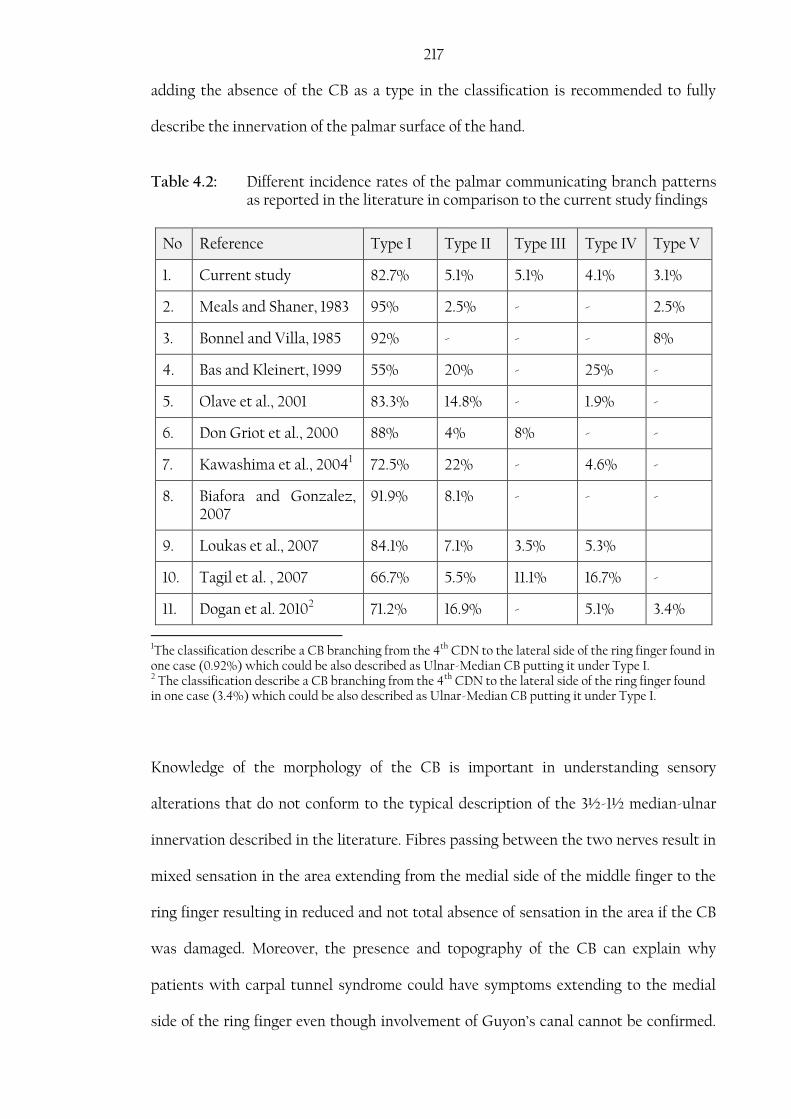
217 adding the absence of the CB as a type in the classification is recommended to fully
describe the innervation of the palmar surface of the hand.
Table 4.2: Different incidence rates of the palmar communicating branch patterns as reported in the literature in comparison to the current study findings
No Reference Type I Type II Type III Type IV Type V
1. Current study 82.7% 5.1% 5.1% 4.1% 3.1%
2. Meals and Shaner, 1983 95% 2.5% - - 2.5%
3. Bonnel and Villa, 1985 92% - - - 8%
4. Bas and Kleinert, 1999 55% 20% - 25% -
5. Olave et al., 2001 83.3% 14.8% - 1.9% -
6. Don Griot et al., 2000 88% 4% 8% - -
7. Kawashima et al., 20041 72.5% 22% - 4.6% -
8. Biafora and Gonzalez, 2007
91.9% 8.1% - - -
9. Loukas et al., 2007 84.1% 7.1% 3.5% 5.3%
10. Tagil et al. , 2007 66.7% 5.5% 11.1% 16.7% -
11. Dogan et al. 20102 71.2% 16.9% - 5.1% 3.4%
1The classification describe a CB branching from the 4th CDN to the lateral side of the ring finger found in one case (0.92%) which could be also described as Ulnar-Median CB putting it under Type I. 2 The classification describe a CB branching from the 4th CDN to the lateral side of the ring finger found in one case (3.4%) which could be also described as Ulnar-Median CB putting it under Type I.
Knowledge of the morphology of the CB is important in understanding sensory
alterations that do not conform to the typical description of the 3½-1½ median-ulnar
innervation described in the literature. Fibres passing between the two nerves result in
mixed sensation in the area extending from the medial side of the middle finger to the
ring finger resulting in reduced and not total absence of sensation in the area if the CB
was damaged. Moreover, the presence and topography of the CB can explain why
patients with carpal tunnel syndrome could have symptoms extending to the medial
side of the ring finger even though involvement of Guyon’s canal cannot be confirmed.

218 It can also explain why some patients will have persistent sensation in their ring and
middle fingers with complete laceration of the MN.
The earliest understanding of the sensory innervation of the hand comes from a study
conducted in 1918 by Stopford on 1000 gunshot victims following World War I. In this
study, sensory distribution in cases of UN injury extended beyond the typical medial
1½ digits in 21% of cases. Similarly, Linell (1921) investigated 20 palms and reported
that the UN sensory innervation extends beyond the typical arrangement in 20% of
cases. Although significant, such studies indicate that the presence of the palmar CB,
which explains the sensory deviations, is not a common finding. Mononeural
innervation to the ring finger was not generally supported by electrophysiological
studies (Laroy et al., 1999). In an electrophysiological study of the palmar surface of the
hand, Laroy et al. (1998) stated that the ring finger received contributions from the
ulnar and median nerves in all cases: no case was found where a mononeural
innervation was present. In another study conducted in 1999, Laroy et al. mentioned
that the absence of either UN or MN contribution to the ring finger was always
accompanied with neural pathology and was not due to anatomical variation.
Interestingly, Stappaerts et al. (1996) reported that MN stimulation resulted in
recordings obtained from the little finger in 2/31 cases, whereas stimulation of the UN
resulted in recordings obtained from the middle finger in 3/31 cases. Such studies clearly
demonstrate the difference between the incidence rate of the palmar CB in dissection
based and electrophysiological studies. Physicians should be aware of the possible
presence of the CB in the palm even when electrophysiological results suggest
otherwise, and not use velocity study results as an absolute reference in evaluating and
assessing sensory innervation in patients.

219 Based on their observations, Ferrari and Gilbert (1991) described a triangular area
where the CB is most likely to be found. The area extended from the middle half of the
hypothenar eminence to the proximal transverse crease of the palm distally and
bounded by the inter-thenar crease laterally. Don Griot et al. (2000) described the
relationship of the CB to the bistyloid line and by using morphometric data outlined an
area where the CB is most likely to be encountered. The risk zone described extends
more distally and less bilaterally than that described by Ferrari and Gilbert (1991),
located between 22%-81% of the distance between the third MCP joint and the BSL
with 90% of the CB being found between 33%-67% of the same distance. In the current
study, the palmar CB was found to be located between 34.5%-75.4% of the distance
between the third MCP joint and the middle of the BSL. Using the middle of the CB as
a reference point rather than the proximal and distal attachment points, Loukas et al.
(2007) reported that the palmar CB was encountered between 12.5%-85% of the
distance between the third MCP joint and the BSL, with 83% of the specimens located
between 35%-62% of the same distance.
The palmar CB has also been described with reference to its location to the WC (Olave
et al., 2001). The proximal attachment was found to be 33.9±5.5 mm and 30.2±8.2 mm to
the WC in male right and left sides respectively and 28.5±6.2 mm and 27.1±3.3 mm to
the WC in female right and left sides respectively. As for the distal attachment, the
study mentions it was found 43.6±6.9 mm and 40.2±6.2 mm to the WC in male right
and left sides respectively and 40.7±7.8 mm and 34.4±1.6 mm to the WC in female right
and left sides respectively. In another study of 37 palmar CBs, the UN attachment
point, which is the proximal attachment in 34/37 specimens, was found to be 31.7±7.5
mm distal to the WC, whereas, the MN attachment point was found 39.03±7.3 mm
distal to the WC. The study also outlined the axis of the third webspace and the fifth
ray as the lateral and medial boundaries of the risk area where the CB is most likely to

220 be encountered (Biafora and Gonzalez, 2007). Similarly in the current study, the
proximal attachment of the palmar CB was found 32.3±5.8 mm and the distal
attachment was found 43.9±6.6 mm to the WC respectively.
The palmar CB is at high risk during carpal tunnel release due to its close proximity to
the TCL. In the current study, 13% of the CB proximal attachment originated at or
below the level of the TCL, and 38% had a proximal attachment originating below the
level or 4 mm distal to the TCL. Ferrari and Gilbert (1991) reported that the CB
originated below the level of the TCL in 22.2% of cases. Stančić et al. (1999) reported
that 65.4% of specimens had a proximal attachment below the level or 4 mm distal to
the TCL. This close position also puts the CB at risk during exploration of Guyon’s
canal. Moreover, the palmar CB was reported to be 6.2±3.7 mm and 5.1±2.8 mm distal to
the distal edge of the TCL at the level of the axis of the 4th metacarpal bone in male
right and left sides and 5.3±3.7 mm and 4.0±1.9 mm distal to the distal edge of the TCL
at the level of the axis of the 4th metacarpal bone in female right and left sides (Olave et
al., 2001).
The angle at which the CB leaves its proximal attachment is important clinically as the
angle approaches a right angle the greater chance it may be injured during surgical
procedures in the area. In the current study, the palmar CB detached from the proximal
attachment point at an angle 139.9±18.3, and was found to be 15.6±4.8 mm long
extending between the third and fourth CDN in all cases. The angle at which the
proximal attachment branched off the UN or MN was found to be between 90-120,
121-150 and 151-180 in 12.5%, 64.8% and 22.7% of cases respectively. Ferrari and
Gilbert (1991) reported 24.4% (11/45) of their cases to originate at a right angle,
whereas, Stančić et al. (1999) found it in 19.8% (16/81).
221 The palmar CB has been reported to communicate with the second CDN; the MN trunk
prior to its division into CDNs; the PDN to the lateral side of the middle finger after it
arises from the UN; communicate with the UN and the PDN to the medial side of the
little finger after it arises from the MN (Ferrari and Gilbert, 1991; Loukas et al., 2007)
but none of these patterns were found in the current study.
The importance of appreciation of the topography and location of the palmar CB is
clearly shown in such cases as reported by Arner et al. (1994). Investigating 53 two-
portal elective carpal tunnel release operations, the study reported 10 patients
developing a new sensory disturbance in the ring and middle or little fingers. Six
patients continued having symptoms 4 months postoperatively. While the patients
described their symptoms as tingling sensation or hyperesthesia rather than numbness,
which can mislead physicians to erroneously interpreting it as residual median nerve
compression; compression or division injury to the palmar CB was concluded to be the
cause of the symptoms (Arner et al., 1994). In another case reported by May Jr and
Rosen (1981), numbness on the lateral side of the ring finger developing into a severe
tingling sensation over three months was described by a patient following an elective
carpal tunnel release. After exploration a large neuroma was found on the palmar CB;
following treatment the symptoms were relieved. In a case report, by Rollins and Meals
(1985), of a complete laceration of the palmar CB following an accident, the palmar CB
was almost equal in diameter to the third CDN prior to their merger indicating the
large contribution of the CB in the formation of the PDNs. Moreover, Ferrari and
Gilbert (1991) described the palmar CB contributing 30% of the fibres supplying the
lateral side of the ring finger in 5 cases and 10% in two cases. Such reports emphasize
the importance of the palmar CB in the exchange of fibres in the palm.

222 According to the morphometric data obtained in this study, a risk area can be outlined
where the CB is most likely to be encountered. Figures 4.2 and 4.3 show the risk area
with reference to the WC and the BSL respectively. The palmar CB is at risk of injury
during carpal tunnel release, flexor tendon surgery of the ring finger, Dupuytren’s
release, and mobilization of neurovascular island flaps. Palmar pain and digital sensory
disturbances resulting from its injury can be erroneously attributed to retraction or
scarring if clinicians fail to recognize its involvement. Having morphometric indices
provide a precise description of the likely location of the palmar CB overcoming the
differences in hand size between individuals and genders. It furthermore compensates
for any changes incurred due to the fixation methods. Yet, it is important to appreciate
that these indices should not be treated as an absolute reference and that human
variations can still surprise.

223
Figure 4.2: Risk area of where the
palmar communicating
branch is most likely to be
found with reference to
the wrist crease. Full
range is shown by light
blue area, 80% of the
samples are located in the
area indicated by the red
box.
Figure 4.3: Risk area of where the
palmar communicating
branch is most likely to
be found with reference
to the bistyloid line. Full
range is shown by light
blue area, 80% of the
samples are located in
the area indicated by the
red box.
224
4.2. Dorsal surface of the hand:
The dorsum of the hand is supplied mainly by three nerves: the superficial branch of the
radial nerve (SBRN), the dorsal branch of the ulnar nerve (DBUN) and occasionally by
the lateral antebrachial cutaneous nerve (LABCN). With various configurations the
SBRN innervates the skin covering the lateral 3½-2½ digits, whereas the DBUN
innervates the skin covering the medial 1½-2½ digits. The LABCN can extend more
distally and innervate the lateral side of the thumb. These nerves are variable in their
branching pattern putting them in danger of injury during various clinical procedures
performed in the distal forearm, wrist and dorsum of the hand. Understanding the
course and appreciating the relationship between these nerves and other anatomical
structures is vital to properly diagnose, plan and perform a clinical procedure
successfully.
4.2.1. Superficial branch of the radial nerve
The superficial branch of the radial nerve (SBRN) innervates the dorsolateral side of the
hand and the dorsal skin covering the lateral 3½-2½ digits up to the proximal
interphalangeal joint. The nerve’s course and branching patterns have proven to be
variable in the literature. Such variability puts the nerve in jeopardy of injury during
different clinical procedures performed in the distal forearm or wrist area. Knowledge
of the anatomical course of the SBRN, appreciation of the anatomic relationship with
other structures in the area, and understanding the possible anatomical variations and
their clinical significance can ensure minimal iatrogenic injuries, better application of
current procedures, and provide the basis for new opportunities to create novel
applications or approaches.

225 The SBRN originates at elbow level as one of the terminal branches of the radial nerve.
In the current study, it was found to originate proximal to the lateral epicondyle in
13.7% of cases. Abrams et al. (1992) reported that the SBRN was found to originate at
the level of the lateral epicondyle in 8/20 (40%) cases and within 21 mm distal to the
epicondyle in the remaining 12/20 (60%) cases. In the current study, the nerve coursed
deep to brachioradialis and became subcutaneous by piercing the antebrachial fascia
72.1±16.6 mm proximal to the RSP, between the tendons of brachioradialis and extensor
carpi radialis longus. This finding is similar to that reported in the literature where the
SBRN was found to become subcutaneous 73±1.5 mm, 81.6 mm, 83.1±11.4 mm and
85.4±1.32 mm by Huanmanop et al. (2007), Vialle et al. (2001), Robson et al. (2008) and
Samarakoon et al. (2011) respectively. The distances in the current study are less than
those reported by Abrams et al. (1992) and Ikiz and Üçerler (2004) who reported the
SBRN to become subcutaneous at 90±14 mm and 92±14mm respectively. In the current
study, the nerve was found to pierce the tendon of brachioradialis to become
subcutaneous in 2% (3 cases), which is less than that reported by Abrams et al. (1992)
(10%); yet similar to the findings of Huanmanop et al. (2007) (3.8%). Such a course can
put the SBRN in danger of entrapment.
In the current study, the point where the SBRN becomes subcutaneous is estimated to
be located at the distal 30.5±5.8% of the total length of the radius. Also Robson et al.
(2008) found the SBRN to become subcutaneous in the distal 32.8±4.1% of the total
length of the forearm; however, the SBRN is reported to become subcutaneous, on
average, in the distal 36% of the distance between the lateral epicondyle to the RSP
(Abrams et al., 1992). In this region the nerve is only covered by skin and subcutaneous
fat making it vulnerable to injury due to trauma or compression in cases of tight
handcuffs, tight watches or bracelets. Compressive or badly made casts or fracture
bracelets can lead to nerve irritation complicating the treatment process. Moreover,
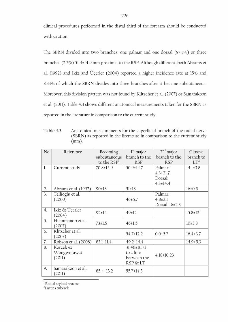
226 clinical procedures performed in the distal third of the forearm should be conducted
with caution.
The SBRN divided into two branches: one palmar and one dorsal (97.3%) or three
branches (2.7%) 51.4±14.9 mm proximal to the RSP. Although different, both Abrams et
al. (1992) and Ikiz and Üçerler (2004) reported a higher incidence rate at 15% and
8.33% of which the SBRN divides into three branches after it became subcutaneous.
Moreover, this division pattern was not found by Klitscher et al. (2007) or Samarakoon
et al. (2011). Table 4.3 shows different anatomical measurements taken for the SBRN as
reported in the literature in comparison to the current study.
Table 4.3 Anatomical measurements for the superficial branch of the radial nerve (SBRN) as reported in the literature in comparison to the current study (mm).
No Reference Becoming subcutaneous
to the RSP1
1st major branch to the
RSP
2nd major branch to the
RSP
Closest branch to
LT2 1. Current study 70.8±15.9 50.9±14.7 Palmar:
4.3±21.7 Dorsal: 4.3±14.4
14.1±3.8
2. Abrams et al. (1992) 90±18 51±18 16±0.5 3. Tellioglu et al.
(2000) 46±5.7 Palmar: 4.8±2.1 Dorsal: 16±2.3
4. Ikiz & Üçerler (2004)
92±14 49±12 15.8±12
5. Huanmanop et al. (2007)
73±1.5 46±1.5 10±3.8
6. Klitscher et al. (2007)
54.7±12.2 0.0±5.7 16.4±3.7
7. Robson et al. (2008) 83.1±11.4 49.2±14.4 14.9±5.3 8. Korcek &
Wongworawat (2011)
31.46±10.73 to a line between the RSP & LT
4.18±10.23
9. Samarakoon et al. (2011)
85.4±13.2 55.7±14.3
1 Radial styloid process 2Lister’s tubercle

227 Due to the variable course of the SBRN in the distal forearm and the wrist area, the
nerve is at risk of injury during various clinical procedures including reduction and
stabilization of distal radial fractures, wrist arthroscopy, radial artery harvest, wrist
denervation, nerve blockade, De Quervain’s release, and bone graft harvest.
SBRN can be injured directly as a result of distal radial fractures, or indirectly as a
result of reduction and fixation of those fractures. Anderson et al. (2004) reported
neuropathies related to the SBRN following the insertion of external fixator pins in
8.3% of cases investigated. An open technique for placing the pins is advocated to
reduce nerve irritation (Anderson et al., 2004).
Kirschner wires (K-wires) are also used to treat and stabilize distal radial fractures.
The RSP is commonly used as an insertion point for these wires. In the current study,
branches of the SBRN were found to pass as close as 0.7 mm to the RSP putting the
nerve branches at high risk of injury. Steinberg et al. (1995) reported that 20% of the
deep structures including the SBRN were injured as a result of inserting K-wires
through the anatomic snuffbox. The study also described a safe-zone area of 68 mm2
where the wires can be inserted safely (Steinberg et al., 1995). However, this safe-zone
was evaluated by Korcek and Wongworrawat (2011) who concluded that the
variability of the SBRN in this area makes it difficult to assign any true safe zone. In
another study, Singh et al. (2005) reported neuropathies related to the SBRN in 20%
following the insertion of K-wires. Patients presented symptoms of sensory
disturbances, complete sensory loss and painful neuroma formation (Singh et al., 2005).
Hochwald et al. (1997) stated that the anatomic snuffbox contained on average 3 nerve
branches coursing as close as 3 mm from the RSP; and recommended limited open
technique where the SBRN branches can be identified and preserved prior to the
insertion of the wires. Nerve irritation can also occur during the removal of the wires,

228 the reduction process or due to the continuous friction between the nerve and the
buried part of the wire if it is placed too close to the nerve. An open technique or a
limited-open technique allows the surgeon to place the wires at a safer distance from
any nearby nerves (Hochwald et al. 1997; Singh et al., 2005; Sharma et al., 2006). As an
alternative and if the case permits, LT can be used as a safe insertion point for the K-
wires. In the current study, branches of the SBRN passed 14.1±3.8 mm from the LT.
Elastic stable intramedullary nailing is frequently used to treat paediatric open,
unstable or irreducible fractures due to its cosmetic and functional advantages
(Cumming et al., 2008). The nails are usually inserted dorsally between the first and
second dorsal compartment using a 20-30 mm proximal incision starting at the level of
the physis. Kang et al. (2011) reported sensory disturbances in two patients (2.2%)
following treatment by elastic intramedullary nailing, whereas Cumming et al. (2008)
found it in 1/19 (5.2%). In a larger retrospective study of 553 cases, 15 (2.7%) cases were
reported to have developed a lesion of the SBRN as a result of the primary operation or
nail removal (Fernandez et al., 2010). With careful dissection to identify the SBRN
branches present in the area, complications can be reduced.
Arthroscopy can be used as a diagnostic or therapeutic tool. It can be used in many
cases including triangular fibrocartilaginous tears, carpal interosseous ligament
injuries, intra-articular distal radial fractures, wrist pain and dorsal ganglia. Wrist
arthroscopy has a complication rate of 2% (Culp, 1999); however, with the increase in
medical procedures involving arthroscopy, the actual complication rate is thought to be
higher. Tryfonidis et al. (2009) reported that superficial nerves pass at a mean distance
of 1.8 mm, 4.9 mm, 2.5 mm and 6.7 mm from portals 1-2, 3-4, 6U, and MCP respectively
putting them at high risk during the establishment of portals. Arthroscopic portals are
designed in relation to their relationship to the extensor compartment. As the

229 procedures can be performed on the radial side of the hand depending on the portals
established, the SBRN can be at risk of injury or irritation as it passes near to or directly
below these portals. Understanding the course and anatomic relation of the superficial
nerves in the area can help clinicians to better preserve and avoid iatrogenic injuries to
those nerves. Furthermore, the SBRN can also be injured during arthroplasty,
arthrotomy, bone graft harvest or wrist denervation procedures as the nerve branches
course under the portals or incision lines. Bone grafts can be obtained from the distal
radius between the first and second dorsal compartment. Arthroplasty of the distal
scaphoid joint (triscaphe joint) is performed via a longitudinal incision between the
tendons of the first extensor compartment with a SBRN neuropathy complication rate
of 4.1% (Wessels, 2004). SBRN branches can course through this area in various
configurations and be at risk if the surgeon is unaware of their presence. Wrist
denervation is used to treat patients with chronic pain originating as a result of many
pathological conditions of the wrist by surgically dividing articular branches from the
median, ulnar, radial and LABCN. Good understanding of the relative anatomy of the
nerves in the area is important to be able to conduct the procedure safely and
successfully.
During De Quervain’s release of the first dorsal compartment, the SBRN branches can
be injured as they cross closely, or through the area. Abrams et al. (1992) mentioned
that branches of the SBRN were found over the first dorsal compartment in 35% of
cases, Ikiz and Üçerler (2004) reported it in 16.67%, while Huanmanop et al. (2007)
found it in 38%. Depending on the severity of the condition, a surgical release of the
first dorsal compartment may be required. Surgical release can be performed by a
longitudinal, transverse, or a Z-incision line. Mellor and Ferris (2000) reported SBRN
injury in 6/17 (35.3%) of cases following a longitudinal incision. At the level of the RSP,
branches of the SBRN pass as close as 2 mm to the first dorsal compartment (Klitscher

230 et al., 2007). Robson et al. (2008) mentioned that a transverse or a Z-incision will cross
the SBRN branches in all cases investigated. The study suggested a 25 mm incision at
the RSP and extending proximally which spared any SBRN branches in 68% of the
cases investigated. In the current study, the SBRN crossed the most proximal edge of
the incision described by Robson et al. (2008) in 10.7% and the SBRN branches were
found to course closer to the RSP axis in 41.7% of the cases investigated. Agreeing with
the literature, the SBRN has a variable course, a safe incision zone cannot be identified,
and careful dissection intra-operatively is strongly recommended to identify and
preserve nerve branches in the area.
As the radial artery is frequently used for obtaining samples during arterial blood gas
testing, the SBRN can be at risk of injury due to its close proximity to the radial artery
at the wrist. Several attempts may be required before a successful sample is obtained;
moreover, several samples can be required to test a patient’s respiratory status in some
conditions. Robson et al. (2008) described a close association between the SBRN
branches and the radial artery in 48% of cases. An ulnar approach to the radial artery is
suggested to minimize SBRN injury (Robson et al., 2008).
Neurological complications related to the SBRN have been reported following radial
artery harvest for coronary surgeries. In their study of 54 patients that underwent
radial artery harvest for coronary surgery, Siminelakis et al. (2004) reported sensory
disturbances related to the dorsum of the hand in 31.28% of cases. Neuropathies can be
as a result of direct trauma during the harvest, compression secondary to edema,
history of clinical conditions affecting the vasculature such as diabetes or smoking, or
ischemia secondary to reduced blood supply when the nerves are supplied by branches
from the radial artery (Denton et al., 2001). Galajda et al. (2002) discussed the
importance of the harvest technique, mainly the mechanical or thermal damage that

231 occurs during the harvest as the main reason for SBRN damage. Neuropathies in the
SBRN territory were reported as high as 16.2% when the radial artery was harvested
using the traditional method and 2% when the radial artery was harvested by a novel
approach they developed but have not explained in their publications (Galajda et al.,
2002). In another study by Bleiziffer et al. (2008), an open technique for radial artery
harvest patients reported less sensory disturbances related to SBRN (22.6%) than
endoscopic technique for the radial harvest group (45.3%); however, the authors still
advocate the endoscopic technique for its cosmetic advantages and lower post-
operative wound complications.
The SBRN has a close association with the cephalic vein. Robson et al. (2008) reported
a close association between the SBRN and the cephalic vein in 80% cases investigated.
In the current study they both crossed for the first time after the nerve became
subcutaneous at 47.9±19.3 mm (range: 93.6-6.7 mm) proximal to the RSP. Similarly,
Klitscher et al. (2007) reported a mean distance between the RSP and the crossing
point of the two structures at 54.3±7.6 mm. Gupta et al. (2012) investigating
spontaneously aborted foetuses reported that the cephalic vein crossed the nerve twice
in 80% of the cases studied. Cephalic vein cannulation is a common procedure that puts
the SBRN at risk of injury if the vein wall is punctured. Sawaizumi et al. (2003)
reported 11 cases of SBRN injury after venepuncture of the cephalic vein patients
complained of pain, numbness and sensory disturbances in the dorsal region of the
hand; of which 7/11 (63.6%) continued to show sensory symptoms at three month
follow up. Vialle et al. (2001) stated that the SBRN and the cephalic vein crossing point
ranged between 90 mm proximal to 45 mm distal to the RSP. Although the current
study only focused on the crossing point proximal to the RSP, it is evident that the
crossing point and the relation between the SBRN and the cephalic vein are highly
variable and that no safe zone can be assigned. Venepuncture of the cephalic vein at the

232 wrist or the distal third of the forearm, where the SBRN becomes subcutaneous, should
only be used when no other alternative is available.
Radial forearm fasciocutaneous free flaps are used for reconstructive surgery. Morbidity
at the donor site has been of concern. Richardson et al. (1997) reported sensory
disturbances in 32% of cases following the harvest, whereas Lutz et al. (1999) reported
it in 54%. Grinsell and Theile (2005) described different incidence rates depending on
the draining system harvested with the flap; double the rate of sensory disturbances are
associated with neural injury if the cephalic vein is harvested (18%) in comparison to
harvesting the venae commitantes (9%). This finding coincides with the close
association that the cephalic vein has with the SBRN that could put the neural
structures in danger if the cephalic vein was raised.
The SBRN and LABCN can overlap. Mackinnon and Dellon (1985) reported a complete
or a partial overlap between the two nerves in 75% of cases. Steinberg et al. (1995)
reported that the LABCN was found to course through the anatomic snuffbox in 45%
of the cases investigated and was in danger of injury by a K-wire in 40%. Similarly,
Korcek and Wongworrawat (2011) described a close association between the SBRN
and LABCN. The LABCN was found to be present over the anatomic snuff box in 52.5%
of cases, innervating the same area of the SBRN in 10% of cases. Furthermore, the
LABCN was found to take over the innervation of the lateral side of the thumb with the
absence of the S3 branch from the SBRN in 5% of cases (Korcek and Wongworrawat,
2011) and in 4.7% (Huanmanop et al., 2007). Other studies have differed in the
communication rate between the SBRN and LABCN. It was reported to be 43% by
Huanmanop et al. (2007), 35% by Abrams et al. (1992) and 20.8% by Ikiz and Üçerler
(2004). In the current study, the LABCN communicated with the SBRN in 30.7% of
cases. Due to its variability in communicating with the SBRN, extensions and
233 distribution patterns; it is difficult to assign sensory deficiencies or neuromas to one of
the nerves and not the other. Furthermore, because of its presence over the anatomic
snuffbox and the first dorsal compartment, it is at as much risk of injury as the SBRN
during procedures performed in this area. This association between the two nerves
should be considered when evaluating sensory deficiencies and planning nerve
blockade in the wrist area.
Injury to the SBRN can be very painful and require a long recovery time and may
require further surgery. Patients may complain of paraesthesia or hypesthesia in
branches of the SBRN and pain secondary to nerve lesions or neuroma formation
affecting their daily activities leading to various forms of disability. Careful technique,
through understanding the course and division points of the nerves in the area in
relation to anatomical landmarks can minimize postoperative complications and help
clinicians during the procedures mentioned above.
The branches arising from the main trunk of the SBRN are usually labelled from medial
to lateral as superficial radial 1 (SR1), SR2 and SR3 (Steinberg et al., 1995). The
variability of the SBRN branching and innervation territory made it difficult to classify
the nerve into a system. Ikiz and Üçerler (2004) classified the SBRN into three types
based on the number of branches. Huanmanop et al. (2007) modified the system by
adding a new type that accounts for cases where SR3 is absent and the area of
innervation is taken over by the LABCN. Although significant, this classification
system does not account for all the variations in the branching patterns described for
the SBRN or for the different innervation territories encountered. Another
classification system described by Korcek and Wongworawat (2011) is based on the
arrangement by which SR1, SR2 and SR3 comes off the main trunk of the SBRN. Yet,
this system does not account for all the variations for the SBRN. Gupta et al. (2012)

234 described another system based on firstly the innervation territory of the SBRN, and
secondly by the number and arrangement of the branches coming off the main trunk of
the SBRN. The system describes only three types, where the SBRN extends to innervate
the lateral three and half digits. However, in the current study cases were identified
where the SBRN innervation territory extended beyond the lateral three and half digits.
Those cases are not accounted for in the classification suggested by Gupta et al. (2012).
The current study proposes modifying the classification suggested by Gupta et al.
(2012) to include different innervation territories.
Type I: The SBRN innervated the lateral two and half digits
A. The main trunk bifurcated into S3 and a common trunk. S3
innervated the entire thumb, whereas the common trunk further
divided into S2 and S1 distally
B. The main trunk bifurcated into S3 and a common trunk. S3
innervated the lateral side of the thumb, whereas the common
trunk further divided into S2 and S1 distally
C. The main trunk trifurcated into three branches with a common
origin point. The branches innervated the lateral side of the
thumb, the medial side of the thumb and the lateral index, the
medial index and the lateral middle fingers.
Type II: The SBRN innervated the lateral 3 digits. The main trunk divided into
medial and SR3 branches. The medial branch supplied the middle, index
finger and occasionally the medial side of the thumb. SR3 supplied the
thumb.

235
A. The main trunk divided into S3 and a common trunk. The S3
innervates the entire thumb. The common trunk divides into S2
and S1 distally.
B. The main trunk divided into S3 and a common trunk, which
divided distally to S1 and S2. S2 innervates the medial thumb and
S1 to the index and the middle fingers.
C. The main trunk bifurcated into S3 and a common trunk. S3
innervated the lateral side of the thumb, whereas the common
trunk further divided into two branches distally. The medial side
of the thumb and the lateral side of the index fingers are
innervated by the same branch.
D. The main trunk bifurcated into S3 and a common trunk. S3
innervated the lateral side of the thumb, whereas the common
trunk further divided into three or four branches distally.
E. The main trunk trifurcated into three branches with a common
origin point. The branches innervated the lateral side of the
thumb, the medial side of the thumb, index and middle fingers.
Type III: The SBRN innervated the lateral 3 and half digits. The main trunk
divided into medial and SR3 branches.
A. The main trunk bifurcated into S3 and a common trunk. S3
innervated the entire thumb, whereas the common trunk further
divided into S2 and S1 distally.
B. The main trunk bifurcated into S3 and a common trunk. S3
innervated the lateral side of the thumb, whereas the common
trunk further divided into S1 and S2 distally. The medial side of

236
the thumb and the lateral side of the index finger are innervated
by the same branch.
C. The main trunk bifurcated into S3 and a common trunk. S3
innervated the lateral side of the thumb, whereas the common
trunk further divided into S2 and S3 distally. S2 to medial thumb
and S3 to the rest of the territory.
D. The main trunk bifurcated into S3 and a common trunk. S3
innervated the lateral side of the thumb, whereas the common
trunk further divided into three or four branches distally.
Type IV: SBRN innervated the lateral four digits.
Type V: SBRN innervates the lateral four and half digits
Type VI: SBRN innervates the full dorsum of the hand with the absence of the
DBUN.
A. Trifurcation of the main trunk.
B. The main trunk bifurcated into S3 and a common trunk. S3
supplies the lateral side of the thumb. The common trunk
further trifurcates.
C. The main trunk bifurcated into S3 and a common trunk. S3
supplies the lateral side of the thumb. The common trunk
divided into S2 and S1. S2 supplies the medial side of the thumb
and the lateral index finger. S1 supplies the rest of the territory.
D. The main trunk bifurcated into S3 and a common trunk. S3
supplies the lateral side of the thumb. The common trunk
divided into S2 and S1. S2 supplies the medial side of the thumb

237
index and the lateral side of the middle fingers. S1 supplies the
rest of the territory.
The aim of this classification is not to produce a system to be memorized, but rather to
ease the understanding of the different branching patterns of the SBRN in the dorsum
of the hand. The most common type was Type I, where the SBRN extends to innervate
the 2½ lateral digits. Table 4.4 shows the incidence rate of the 6 types found in this
study.
Table 4.4: Incidence rates of the branching patterns of the superficial branch of the radial nerve found in the current study
Type No. %
Type I 56 37.3
Type II 35 23.3
Type III 44 29.3
Type IV 2 1.3
Type V 3 2
Type VI 10 6.7
4.2.2. Dorsal branch of the ulnar nerve:
The dorsal branch of the ulnar nerve (DBUN) is one of the terminal branches of the UN.
It supplies the dorsomedial aspect of the dorsum of the hand and skin covering the
dorsal medial 1½ digits. In the current study it was found to originate from the medial
side of the main trunk of the UN 63.3±14.8 mm from the USP and 61.3±21.0 mm from the
proximal edge of the pisiform. The origin point found in this study differs to those
mentioned in the literature. The DBUN was found to originate more proximally by
Botte et al. (1990), Casoli et al. (2004), and Cavusoglu et al. (2011) reporting it to be
83.±24 mm, 80 mm and 83.6±2.4 mm proximal to the pisiform respectively. It has also
been reported to originate more distally than in the current study by Goto et al. (2010)

238 and Puna and Poon (2010) who reported the origin point to be 34±13 mm and 51±14 mm
proximal to the USP respectively. The origin point corresponds to a point located in the
distal 26% of the total length of the ulna (Botte et al., 1990); similarly, it was found to
be located 23.6±7.7% of the total length of the ulna in the current study.
In the current study, the nerve courses medially and distally deep to flexor carpi ulnaris
before it emerges at the dorsomedial border to become subcutaneous 18.1±7.1 mm
proximal to the USP. Botte et al. (1990) and Cavusoglu et al. (2011) reported the DBUN
pierced the antebrachial fascia 50±18 mm and 54.7±1.5 mm proximal to the pisiform
respectively. Although mentioned with reference to a different bony landmark (the
pisiform), which is located 11.7±4.3 mm distal to the USP, the point where the nerve
becomes subcutaneous is located more distally in the current study. In their study, Mok
et al. (2006) reported the DBUN becoming subcutaneous 26±8.3 mm proximal to the
USP.
The DBUN innervates the medial side of the dorsal hand and the skin covering the
medial 1½-2½ digits by dividing into two major branches. In the current study, the first
division was found to originate anywhere between 38.7 mm proximal to the USP and
53.4 mm distal to the USP (mean: -3.4±17.4 mm). The second division was also found to
be variable, dividing anywhere between 8.8 mm proximal to the USP and 73.6 mm
distal to the USP (mean 39.2±13.2 mm). Mok et al. (2006) reported different
measurements for the location of the first and second branches of the DBUN. In their
study, the first branch occurred 20±14.7 mm proximal the USP while the second branch
occurred 26±11.4 mm distal to the USP. The differences between the two studies could
be attributed to the different definition of the major branches Mok et al. (2006) used in
their study. They described the major branches as being the nerves terminating in the
239 dorsal digits with no further divisions, whereas this study considered the division
points as they occur along the course of the DBUN.
These anatomical measurements are important to appreciate when performing a direct
approach to the subcutaneous border of the ulna where the DBUN is at high risk of
injury such as in cases of open reduction and internal fixation of ulna fractures,
treatment of delayed union or non-union of ulnar fractures and chronic osteomyelitis,
osteotomy of the ulna and lengthening or shortening of the ulna (Le Corroller et al.,
2013). Injury to the DBUN could be presented as dysesthesia and numbness to the
dorsomedial area of the hand. It can also result in neuroma formation which is usually
more painful and disabling to the patient than sensory disturbances.
Moreover, understanding the anatomy and course of the DBUN is important in
planning and preparing nerve grafts of up to 100 mm. Neurovascular flaps from the
DBUN are preferable because of their limited donor site morbidity, cosmetic
advantages, restricted vascular sacrifice, and the simple and rapid dissection required in
their preparation. It is also important to appreciate the anatomical relations of the
DBUN in planning nerve blockade, assessing iatrogenic injuries and restoration of
sensory innervation after UN lesions.
The DBUN is at risk during wrist arthroscopy as it passes close to the 6 Radial (6R)
portal. Grechenig et al. (1999) reported that the most common complication found after
performing wrist arthroscopy on 96 patients was irritation of the DBUN, reported in
3.1% of cases. The nerve consistently crossed a line between the USP and the fourth
webspace. The DBUN crossed this line 18.1±9.1 mm distal to the USP corresponding to
19.4±9.6% of the distance between the USP and the fourth webspace. Tindall et al.
(2006) found the DBUN to be more constant crossing the same line 24 mm (range: 18-
28 mm) distal to the USP, corresponding to a point 23±2.5% of the distance between
240 the USP and the fourth webspace. In their study, Tindall et al. (2006) recommend that
the 6R portal is placed in the proximal 19% of the distance to avoid injury to the DBUN.
However, according to the results obtained from the current study, the DBUN crossed
the distance between the USP and the fourth webspace within the proximal 19% in
almost 50% of the specimens. Agreeing with Tindall et al. (2006) that this area should
be approached with caution and that those measurements should not be taken as
absolute reference.
4.2.3. Sensory distribution in the dorsum of the hand
Anatomical textbooks usually describe the sensory distribution in the dorsum of the
hand as being supplied by the SBRN and the DBUN. Gray’s Anatomy for Students 2nd
edition describe the innervation territory of the SBRN as supplying “the dorsolateral
aspect of the palm and the dorsal aspects of the lateral three and one-half digits distally to
approximately the terminal interphalangeal joints”; however, illustrations show that the
innervation territory extends only to the dorsal skin covering the lateral three digits
(Drake et al., 2010). A similar discrepancy between text and illustrations is present
describing the DBUN innervation territory. While the text describes the DBUN as
innervating the dorsal skin covering the medial one and half digits, the illustration
shows the innervation territory to extend to cover the dorsal skin covering the medial
two digits (Drake et al., 2010). Similarly, illustrations are shown in Gray’s Atlas of
Anatomy where the SBRN innervates the dorsal skin covering the lateral three digits
whereas the DBUN innervates that covering the medial two digits (Drake et al., 2008).
In other textbooks (Moore et al., 2011; Moore et al., 2013), the dorsum of the hand
innervation is illustrated to show the classical description of the SBRN innervating the
dorsal two thirds of the dorsal hand and the dorsal skin covering the lateral three and
half digits, with the DBUN innervating the rest of the hand.
241 Grant’s Atlas of Anatomy 12th edition shows the dorsum of the hand to be split between
the two nerves at the middle of the ring finger: the SBRN innervating half of the dorsum
hand and the dorsal skin covering the lateral three and half digits whereas the DBUN
innervates the medial half of the dorsal hand and the dorsal skin covering the medial
two and half digits. The atlas also shows several variations in the innervation of the
dorsum of the hand (Agur et al., 2009).
An understanding of the sensory distribution in the dorsum of the hand comes mainly
from historical large studies such as that conducted by Stopford (1918) examining
gunshot victims from World War I. By testing for sensory loss, it was possible to
describe the outline known today. In UN injuries, the study described anaesthesia in
the medial one and half digits in 79%, and in the entire little and ring fingers in only
15% setting the typical distribution of the DBUN to be limited to the medial dorsum of
the hand and the skin covering the medial one and half digits. Although significant, the
study set loss of sensation as the variable tested which could be misleading when it
comes to outlining the extent of the sensory distribution of the dorsal nerves. Such a
definition does not account for communicating branches between the dorsal nerves
and/or other nerves in the area that allows for exchange of fibres and thus sensory
retention in an area that is supposed to be anaesthetised if the injured nerve was solely
responsible for innervating that area. In another study, Linell (1921) reported that the
typical distribution of the medial one and half digits was only found in 12.5% of cases,
whereas the DBUN extended to innervate the skin covering the medial two and half
digits in 68.8%. Auerbach et al. (1994) also reported that the SBRN innervated the
dorsal skin covering the lateral three digits in 55%, and followed the typical description
of innervating the lateral three and half digits in only 5%. In a more recent study by
Mok et al. (2006), the DBUN was found to follow the classical description of
innervating the medial one and half digits in only 13.3%. In the current study, the SBRN
242 was found to follow the classical description and solely innervate the lateral three and
half digits in only 18.7%. It was noted that the SBRN extended less medially than the
classical description agreeing with the results of Mok et al. (2006). The most common
distribution found in the current study was the SBRN innervating the lateral dorsal
skin and the skin covering the lateral two and half digits. Furthermore, the DBUN was
found to innervate the medial dorsal skin and the skin covering the medial two and half
digits in 38.0% setting it as the most common pattern. Similarly, Vergara-Amador and
Nieto (2010) reported that the most common branching pattern of the SBRN seen in
56% extended to innervate the lateral two and half digits.
Electrophysiological studies have also highlighted the variability of the distribution
patterns in the dorsum of the hand. Stappaerts et al. (1996) mentioned that action
potential results were obtained from the little and ring fingers in 52% after stimulation
of the DBUN, and from the middle finger in 26% after stimulating the same nerve. Laroy
et al. (1998) mentioned that the action potential is reduced laterally to medially when
the SBRN was stimulated and medially to laterally when the DBUN was stimulated
and become increasingly unobtainable at the level of the ring finger.
It is not uncommon for the SBRN and DBUN to communicate in the dorsum of the
hand1 changing the sensory distribution in the dorsum of the hand. In the current
study, the two nerves communicated in 37 (26.4%) cases altering the sensory
innervation on the medial side of the ring finger and lateral side of the middle finger in
the majority of cases (31/37; 83.8%); however, it is still possible for the communication
to affect other areas as the lateral side of the middle finger and the medial side of the
index finger, and the medial side of the middle finger as seen in the current study in
8.1% (3/37) and 5.4% (2/37) of cases respectively. Furthermore, dual innervation where
the two nerves overlap and contribute to the innervation of the same territory with no
1 The dorsal communicating branch between the SBRN and the DBUN is discussed in the next section

243 clear communication has been reported in the literature. Mok et al. (2006) reported
dual innervation in 10/30 (33.3%) where both nerves supply the medial side of the
middle finger and the lateral side of the ring finger (6/10), the medial side of the middle
finger (3/10), and the medial side of the ring finger (1/10). In the current study, dual
innervation was noted in only 4 cases (2.9%) affecting the medial side of the middle
finger and the lateral side of the ring finger in two cases, and the medial side of the
middle finger only in the other two. Communicating branches and dual innervation in
the dorsum of the hand can complicate the interpretation of the velocity test results by
producing a low or absent response; create complications during surgery, and leads to
misevaluation of nerve pathology if the clinician is not aware of their presence.
LABCN can contribute to the innervation of the dorsum of the hand. It was also
reported that the LABCN completely takes over the innervation of the lateral side of the
thumb when SR3 is absent (Madhavi and Holla, 2003; Huanmanop et al., 2007; Korcek
and Wongworawat, 2011). Such variation was noticed in one case in the current study.
The incidence rate for the communication between the two nerves varies between 20.8-
43% in the literature. Interestingly, in the current study, the SBRN communicated with
the LABCN in 30.0% of cases of which the LABCN communicated with SR3 allowing
both nerves to contribute in the innervation of the lateral side of the thumb (37/45;
82.6%) and communicated to the main trunk of the SBRN allowing both nerves to
contribute in the innervation of the entire territory of the SBRN (8/45; 17.4%). Martín
Inzunza et al. (2011) reported a similar case where the LABCN communicated with the
SBRN and continued to supply the entire SBRN territory. Clinicians evaluating
neuromas, nerve pathology and sensory disturbances in the dorsum of the hand should
be aware of the possible involvement of the two nerves.

244 Full supply of the dorsum of the hand by the SBRN with the absence of the DBUN has
been described in the literature. Dissection based studies and velocity conduction
studies report different incidence rates. In dissection based studies it was found in
4.16% as reported by Botte et al. (1990), 3.3% by Mok et al. (2006), 5.5% by Tiznado et
al. (2012) and 8% by Robson et al. (2008). In velocity conduction studies it was
reported in 19% by Pollak et al. (2013) and 12.9% by Stappaerts et al. (1996). In the
current study it was found in 6.7% of cases. Failure to recognize the presence of this
pattern can lead to misinterpretation of the velocity conduction test results and nerve
injury promoting inappropriate treatment and unnecessary surgical intervention. If a
response from the DBUN cannot be obtained, a full investigation of the hand is
recommended.
Patterns of innervation in the dorsum of the hand are variable across individuals;
however, it is also variable between the two hands of the same individual. Investigating
30 fetuses, asymmetry between left and right hand sides was found in 17% as reported
by Gupta et al. (2012). In another study performed on 40 Thai cadavers, asymmetry was
observed in 42.5% of the cases (Huanmanop et al., 2007). In velocity conduction
studies, Stappaerts et al. (1996) reported symmetry for the dorsum of the hand after
stimulating both SBRN and DBUN in 29% of the cases only, whereas Dutra de Oliveira
et al. (2000) reported significant differences in the results obtained in 21%. It is
important to appreciate the differences between the two sides of the same individual.
In the current study, symmetry was found in 43.2% of cases. The electrophysiological
test results obtained from the healthy hand should not be used as an absolute reference
to evaluate pathology in the other hand.
Electrophysiological studies are an important tool in mapping sensory nerves in the
hand; however, they should be used with caution. Low or absent responses when the

245 nerves are stimulated does not necessarily mean that the tested nerve is absent as nerve
axons could be too few and/or their diameter too small to produce an action potential
high enough to be recorded. This explains the differences in incidence rate of all-radial-
supply between dissection based and velocity conduction studies. Not appreciating the
presence of the communicating branches can lead to misdiagnosis of neural entrapment
and pathologies in the wrist and forearm. Moreover, asymmetry is not uncommon in
the dorsum of the hand and the healthy hand should not be considered an absolute
reference when evaluating pathologies in the other hand. Full hand investigation is
recommended to have a better understanding of the pattern of the dorsal nerves and
thus avoid preoperative and postoperative complications.
4.2.4. Dorsal communicating branch between the superficial branch of the
radial nerve and the dorsal branch of the ulnar nerve
The sensory innervation of the dorsum of the hand can be quite variable. The presence
of a dorsal communicating branch (CB) between the SBRN and the DBUN can explain
sensory deviations in the dorsum of the hand. It can provide collateral pathways for
nerves to supply a region otherwise anaesthetised due to injury or pathology. In
electrophysiological studies, the presence of dorsal CB can alter the results leading to
misdiagnosis and unnecessary therapeutic measures. Furthermore, it can be a
complicating factor if injured during surgical procedures to the dorsum of the hand.
The dorsal CB is at risk of injury during surgical procedures performed in the dorsum of
the hand such as fracture repairs of the fourth and fifth metacarpal bones, and dorsal
approaches to the carpal tunnel resulting in sensory disturbances and painful neuroma
formation. Consequently, understanding the topography and the common location of
the dorsal CB is important to properly diagnose and treat various hand related
pathologies.

246 The dorsal CB was reported in the literature with various incidence rates. Botte et al.
(1990) found it in one specimen of 24 (4.16%), Auerbach et al. (1994) reported it in 15%
of the 20 hands explored, while Loukas et al. (2008) investigated 200 formalin fixed
adult cadaveric hands and reported the incidence of the communicating branch
between the DBUN and the SBRN to be 60% (120/200). Interestingly, in their study of
30 cadaveric hands outlining in detail the different nerves contributing to the dorsal
innervation of the hand, Mok et al. (2006) made no mention of the dorsal CB.
The DBUN is usually used for preparing nerve grafts for its minimum donor morbidity.
Cavusoglu et al. (2011) reported that due to the many communications between the
DBUN and the SBRN in the dorsum of the hand, excision of the DBUN may not result
in numbness. However, in the current study the dorsal CB was only found in 26.4% of
cases. Collateral, dual and dorsal CB can explain the minimal sensory loss in the donor
site, but according to the results obtained from this study, clinicians need to be careful
not to develop therapeutic choices on inaccurate assumptions.
Loukas et al. (2008) suggested classifying the dorsal CB into four types based on the
direction of the CB. The current study suggests classifying the dorsal CB into three
types based on the number of attachment points between the SBRN and the DBUN:
Type I: The CB had a single attachment to the SBRN and the DBUN.
A. CB attached proximally to the SBRN
B. CB attached proximally to the DBUN
C. CB attached to the SBRN and the DBUN at the same level
Type II: The CB had multiple attachment points to both the SBRN and DBUN.

247 Type III: The DBUN and SBRN both gave a branch that merged and sent branches
to the dorsum of the hand.
Type I A, I B and I C correspond to the Types I, II and III suggested by Loukas et al.
(2008) respectively. Types I A, I B and I C were found in 10.8%, 18.9% and 2.7% in the
current study; however were found in 59.1%, 19.1% and 3.3% by Loukas et al. (2008).
The most common type found in this current study was Type III (56.8%).
Loukas et al. (2008) described a risk zone where the dorsal CB is most likely to be
found. The dorsal CB was found 41% of the distance between the USP and the fourth
metacarpophalangeal joint with a range of 12-78%. With such a big range, the authors
used a prevalence of 85% of the samples to make the risk area smaller and more useful
and reported the dorsal CB to be present between 28-60% of the distance between the
USP and the fourth metacarpophalangeal joint (Loukas et al., 2008). In the current
study, the dorsal CB was defined by its proximal and distal attachment points. The
proximal attachment of Type I of the dorsal CB was found between -7.6-85.6% of the
distance between the third MCP joint and the BSL, whereas the distal attachment was
found between 32.9-98.4% of the same distance. With the large variability in the
dorsum of the hand and limited number of samples, it is difficult to identify a risk-zone.
The majority of cases in this study belong to Type III (56.8%) where the merging point
of the two nerves was at 83.9±17.0% of the distance between the third MCP joint and
the BSL. This type has not been described before in the literature. In their study
detailing the nerves in the dorsum of the hand, Mok et al. (2006) suggested a safe area
where no nerves pass through in the dorsum of the hand. The suggested area overlaps
with the risk area suggested by Loukas et al. (2008) and with the findings of this
current study. In their study of the nerves of the dorsum of the hand, Cirpar et al.
(2012) reported that the great variability of the branching patterns of the dorsal nerves

248 and the presence of communicating branches between nerves prevents any suggestion
of a safe zone in the dorsum of the hand.

249
5. Conclusion
Understanding the cutaneous innervation of the hand is the first step in understanding
hand anatomy. With the on-going effort to decrease postoperative complications and
provide high quality medical services, an understanding of the anatomy of the
cutaneous nerves of the hand, their distribution, CBs, and variations is essential for
assessing and managing various clinical conditions. The description of the typical
anatomy and innervation areas in the hand needs to be reassessed. Textbooks usually
describe the typical anatomy ignoring important anatomical anomalies and their
clinical significance. The literature, on the other hand, gives vague and sometimes
contradicting reports on the incidence, course and characteristics of the sensory nerves
of the hand. Although important, some reports give confusing descriptions and unclear
measurements limiting the use of the data in clinical settings. The main aim of this
project was to clarify the complex relations of the cutaneous nerves of the hand and
evaluate their significance in clinical settings.
The PCBMN has been described in many reports in the literature. However; studies
have usually been conducted on a limited number of samples, on cadavers who
underwent different fixation methods, and by using different bases of measurement and
classification influencing the consistency of the descriptions of the anatomical
characteristics of the nerve and leading to variable and sometimes confusing reports.
The origin point of the PCBMN has been used to describe the intraneural origin,
detachment point, and the point where the PCBMN can be distinguished from the MN
but still enclosed in the same neural sheath. Moreover, reports have failed to agree on
the description of the branching patterns and the basis of classifying those patterns.
The PCBMN was classified by the number of branches emerging from the MN, the sub-
branches coming off the PCBMN or/and the territory innervated by each sub-branch.
250 The current study suggests a classification system based on the number and direction
of the sub-branches coming off the PCBMN and incorporating other patterns described
in the literature. The current study also highlights variations to the most common
branching patterns described, the origin of the nerve from the main trunk of the MN,
the relationship of the nerve to other structures in the distal forearm and
communications with other nerves in the region. Knowledge of the anatomical
characteristics of the PCBMN and the appreciation of possible variations in the distal
forearm and proximal palm is important to properly diagnose and evaluate pathologic
or postoperative signs and avoid injuries to the nerve during surgical interventions to
the distal forearm and proximal palm.
The CDNs have been neglected in the literature. With few studies describing their
branching points to PDNs and their relationship to other structures in the palm. The
current study describes the CDNs division points to PDNs to various bony landmarks
with absolute and relative values providing clinicians with indices that can be used as a
tool in clinical procedures (Figure 5.1 A).
The UN innervates the palmar and dorsal medial hand and the skin covering the medial
1½ digits by the superficial branch of the UN that originates from the UN as it passes
through Guyon’s canal, and the DBUN which originates from the UN in the distal
forearm and courses dorsally to supply the medial dorsal hand. The current study
investigated the branching pattern of the UN in Guyon’s canal, discussed variations
and communicating branches present in the area, and suggested a scheme to classify the
neural contribution supplying the palmar skin of the little and medial side of the ring
finger. Neural contributions, other than from the UN, are overlooked and sometimes
underestimated in the literature. The suggested classification emphasises the possible
palmar-dorsal communications (Kaplan anastomosis) and median-ulnar

251 communications which could be a source of postoperative complications if injured
during surgical interventions in the area. Moreover, the DBUN is at risk during clinical
procedures performed to the distal ulna or the wrist area. Understanding the course of
the DBUN will help clinicians to preserve the nerve during these procedures and help
to better evaluate and assess the nerve involvement in hand neuropathology.
Furthermore, knowledge of the anatomical relation of the DBUN will aid in the
preparation and planning of nerve grafts.
The palmar CB has been investigated in the literature; however, with different and
often contradicting reports regarding incidence rate, branching patterns and most
common location. The current study investigates discrepancies in previous reports and
suggests a classification scheme incorporating other systems mentioned in the
literature. This study also outlines a risk-zone where the palmar CB is most likely to be
found (Figure 5.1 B). Recognizing the topography of the palmar CB is important to
understand palmar sensory alteration to the typical description and to properly
evaluate possible nerve involvement following surgical procedures in the palmar area.
The study also provides guidelines of where the palmar CB is most likely to be found in
relation to various anatomic landmarks which can be helpful to surgeons in avoiding
injury to the CB during any surgical intervention in the area.
Risk areas where the palmar CB, division points of the CDNs into PDNs, CBs in the
medial side of the palm are most likely to be found are illustrated in Figure 5.1.

252
A
B
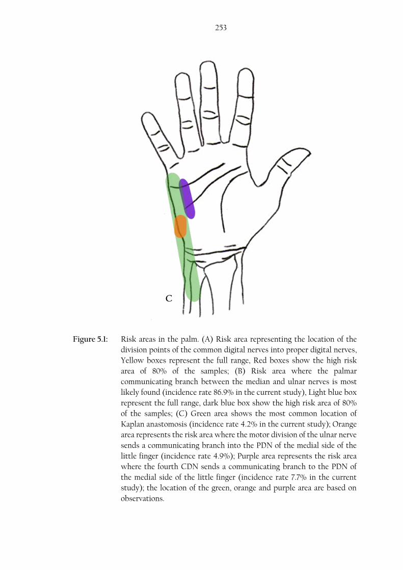
253
Figure 5.1: Risk areas in the palm. (A) Risk area representing the location of the
division points of the common digital nerves into proper digital nerves,
Yellow boxes represent the full range, Red boxes show the high risk
area of 80% of the samples; (B) Risk area where the palmar
communicating branch between the median and ulnar nerves is most
likely found (incidence rate 86.9% in the current study), Light blue box
represent the full range, dark blue box show the high risk area of 80%
of the samples; (C) Green area shows the most common location of
Kaplan anastomosis (incidence rate 4.2% in the current study); Orange
area represents the risk area where the motor division of the ulnar nerve
sends a communicating branch into the PDN of the medial side of the
little finger (incidence rate 4.9%); Purple area represents the risk area
where the fourth CDN sends a communicating branch to the PDN of
the medial side of the little finger (incidence rate 7.7% in the current
study); the location of the green, orange and purple area are based on
observations.
C
254 The SBRN can be at risk directly due to trauma or injury in cases of fracture,
compressive bracelets or watches; or indirectly as a result to treatment of fractures or
during clinical procedures such as wrist arthroscopy, radial artery harvest, blood gas
testing, cephalic vein cannulation and De Quervain’s release. Understanding the
anatomical course of the SBRN and its relationship to other structures in the distal
forearm and wrist is vital for safe clinical practice. Moreover, appreciation of possible
variations and communicating branches with other nerves in the area will aid clinicians
to better assess and evaluate neuropathologies.
The literature has differed in the description of the sensory distribution territories in
the dorsum of the hand with many studies giving different and inconstant reports and
findings. Electrophysiological studies have also been conducted to better understand
the sensory distribution in the dorsum of the hand; however, they provided different
incidence rates to patterns and variations than those mentioned by dissection based
studies. The most common branching pattern in the dorsum of the hand found in the
current study is illustrated in Figure 5.2. Dorsal communicating branches can explain
the deviation in the sensory distribution from the typical descriptions; however, the
literature has differed in the incidence rate of these CBs, and often ignored their
presence in the dorsum of the hand. The current study discusses the sensory
distribution in the dorsum of the hand, gives incidence rates for the common patterns
and variations to those patterns. It also discusses the different CBs in the dorsum of the
hand and their significance in clinical settings. Understanding the sensory distribution
in the dorsum and possible CBs is important to better diagnose signs and symptoms
presented and assess the involvement of different nerves. Knowledge of the variations
in the sensory distribution helps to correctly interpret velocity conduction studies
results thus better design and implement an appropriate therapeutic plan. Risk areas
where the dorsal CB between the SBRN and DBUN (Type III) is most likely to be found

255
Area
innervated by
the DBUN
Area
innervated by
the SBRN
Figure 5.2: The most common sensory innervation pattern and risk areas in the
dorsum of the hand as found in the current study. Yellow box
represent full range of the most common location of the dorsal
communicating branch between the superficial branch of the radial
nerve (SBRN) and the dorsal branch of the ulnar nerve (DBUN)
(Type III) relative to the distance between the third
metacarpophalangeal joint and the middle of the bistyloid line, 80%
of the samples are found in the area indicated by the red box; Light
green area represent the full range of the area where the DBUN
crosses a line extending between the ulnar styloid process and the
fourth webspace, 80% of the samples are found in the area indicated
by the dark green box; Pink area represent a risk area where the
lateral antebrachial cutaneous nerve communicate with the SBRN
(incidence rate 30.0%, this area is based on observations).
and where the DBUN courses through a line between the USP and the fourth webspace
are illustrated in Figure 5.2.

256 With the continuous development of clinical procedures and techniques, a detailed
knowledge and understanding of the typical anatomy, course, patterns of distribution
and the appreciation of possible variations of the nerves of the hand is essential for safe
and optimal clinical practice. This study described the course of the cutaneous nerves
in the hand and their relations to other structures in the distal forearm and wrist. It
outlined the most common topographical patterns of these nerves, variations to these
patterns and their clinical significance. Using morphometric data, the most common
location of palmar CBs were outlined with reference to identifiable landmarks. The
findings obtained from this study will help clinicians to plan and prepare various
procedures in the distal forearm and wrist. It will help in evaluating the involvement of
different nerves by better assessing signs and symptoms presented in different hand
neuropathologies. Moreover, it will have a positive impact in reducing postoperative
complications and increasing patient satisfaction. Furthermore, knowledge of the
anatomical course and appreciation of possible variations and communicating branches
of the cutaneous nerves of the hand will provide opportunities to create novel
procedures in the wrist and hand.
257
6. References
AARON, S. D., VANDEMHEEN, K. L., NAFTEL, S. A., LEWIS, M. J. &
RODGER, M. A. 2003. Topical tetracaine prior to arterial puncture: A
randomized, placebo-controlled clinical trial. Respiratory Medicine, 97, 1195-1199.
ABRAMS, R. A., BROWN, R. A. & BOTTE, M. J. 1992. The superficial branch of
the radial nerve: An anatomic study with surgical implications. Journal of Hand
Surgery, 17, 1037-1041.
AGUR, A. M. R., DALLEY, A. F. & GRANT, J. C. B. 2009. Grant's atlas of anatomy,
Philadelphia, Pa, Lippincott Williams & Wilkins, pages: 554-587.
AHČAN, U., ARNEŽ, Z. M., BAJROVIĆ, F. F. & ZORMAN, P. 2002. Surgical
technique to reduce scar discomfort after carpal tunnel surgery. Journal of Hand
Surgery, 27, 821-827.
AHČAN, U., ARNEŽ, Z. M., BAJROVIĆ, F. F., HVALA, A. & ZORMAN, P.
2003. Nerve fibre composition of the palmar cutaneous branch of the median
nerve and clinical implications. British Journal of Plastic Surgery, 56, 791-796.
AHN, D. S., YOON, E. S., KOO, S. H. & PARK, S. H. 2000. A prospective study
of the anatomic variations of the median nerve in the carpal tunnel in Asians.
Annals of Plastic Surgery, 44, 282-287.
AHSAN, Z. S. & YAO, J. 2012. Complications of wrist arthroscopy. Arthroscopy:
Journal of Arthroscopic and Related Surgery, 28, 855-859.
258
ALIZADEH, K., LAHIJI, F. & PHALSAPHY, M. 2006. Safety of carpal tunnel
release with a short incision: A cadaver study. Acta Orthopaedica Belgica, 72, 415-
419.
AL-QATTAN, M. M. 1997. Anatomical classification of sites of compression of
the palmar cutaneous branch of the median nerve. Journal of Hand Surgery:
European Volume, 22, 48-49.
ANDERSON, J. T., LUCAS, G. L. & BUHR, B. R. 2004. Complications of
treating distal radius fractures with external fixation: a community experience.
The Iowa Orthopaedic Journal, 24, 53-59.
ARIYAN, S. & WATSON, H. K. 1977. The palmar approach for the visualization
and release of the carpal tunnel. An analysis of 429 cases. Plastic and Reconstructive
Surgery, 60, 539-547.
ARNER, M., HAGBERG, L. & ROSEN, B. 1994. Sensory disturbances after two-
portal endoscopic carpal tunnel release: A preliminary report. Journal of Hand
Surgery, 19, 548-551.
AUERBACH, D. M., COLLINS, E. D., KUNKLE, K. L. & MONSANTO, E. H.
1994. The radial sensory nerve: An anatomic study. Clinical Orthopaedics and
Related Research, 308, 241-249.
AVCI, S. & SAYLI, U. 2000. Carpal tunnel release using a short palmar incision
and a new knife. Journal of Hand Surgery, 25 B, 357-360.

259
BAGATUR, A. E., YALCINKAYA, M. & ATCA, A. O. 2013. Bifid median nerve
causing carpal tunnel syndrome: MRI and surgical correlation. Orthopedics, 36,
e451-e456.
BAS, H. & KLEINERT, J. M. 1999. Anatomic variations in sensory innervation of
the hand and digits. Journal of Hand Surgery, 24, 1171-1184.
BEREDJIKLIAN, P. K., BOZENTKA, D. J., LEUNG, Y. L. & MONAGHAN, B. A.
2004. Complications of wrist arthroscopy. Journal of Hand Surgery, 29, 406-411.
BERRETTINO, P. C. & PETRIOLI, C. R. 1741. Tabulae Anatomicae. Rome:
Typographia Antonii de Rubeis, 1-4 as cited in STANČIĆ, M. F., MIĆOVIĆ, V. &
POTOČNJAK, M. 1999. The anatomy of the Berrettini branch: Implications for
carpal tunnel release. Journal of Neurosurgery, 91, 1027-1030.
BERRY, M. G., VIJH, V. & PERCIVAL, N. J. 2003. Bifid median nerve:
Anatomical variant at the carpal tunnel. Scandinavian Journal of Plastic and
Reconstructive Surgery and Hand Surgery, 37, 58-60.
BERTELLI, J. A. & PAGLIEI, A. 1998. The neurocutaneous flap based on the
dorsal branches of the ulnar artery and nerve: A new flap for extensive
reconstruction of the hand. Plastic and Reconstructive Surgery, 101, 1537-1543.
BEZERRA, A. J., CARVALHO, V. C. & NUCCI, A. 1986. An anatomical study of
the palmar cutaneous branch of the median nerve. Surgical and Radiologic Anatomy,
8, 183-188.

260
BIAFORA, S. J. & GONZALEZ, M. H. 2007. Sensory communication of the
median and ulnar nerves in the palm. Journal of Surgical Orthopaedic Advances, 16,
192-195.
BLEIZIFFER, S., HETTICH, I., EISENHAUER, B., RUZICKA, D., VOSS, B.,
BAUERNSCHMITT, R. & LANGE, R. 2008. Neurologic sequelae of the donor
arm after endoscopic versus conventional radial artery harvesting. Journal of
Thoracic and Cardiovascular Surgery, 136, 681-687.
BOESON, M. B., HRANCHOOK, A. & STOLLER, J. 2000. Peripheral nerve
injury from intravenous cannulation: a case report. Journal of the American
Association of Nurse Anesthetists, 68, 53-57.
BONNEL, F. & VILA, R. M. 1985. Anatomical study of the ulnar nerve in the
hand. Journal of Hand Surgery, 10, 165-168.
BORN, T. & MAHONEY, J. 1995. Cutaneous distribution of the ulnar nerve in
the palm: Does it cross the incision used in carpal tunnel release? Annals of Plastic
Surgery, 35, 23-25.
BOTTE, M. J., COHEN, M. S., LAVERNIA, C. J., VON SCHROEDER, H. P., GE
LLMAN, H. & ZINBERG, E. M. 1990. The dorsal branch of the ulnar nerve: An
anatomic study. Journal of Hand Surgery, 15, 603-607.
BOZKURT, M. C., CEZAYIRLI, E. & TAGIL, S. M. 2002. An unusual
termination of the ulnar nerve in the palm. Annals of Anatomy, 184, 271-273.

261
BOZKURT, M. C., TAGIL, S. M. & ÖZÇAKAR, L. 2004. An aberrant sensory
innervation of the little finger by the ulnar nerve: A cadaveric observation. Plastic
and Reconstructive Surgery, 113, 1531-1532.
BUCKMILLER, J. F. & RICKARD, T. A. 1987. Isolated compression neuropathy
of the palmar cutaneous branch of the median nerve. Journal of Hand Surgery, 12,
97-99.
BUGBEE, W. D. & BOTTE, M. J. 1993. Surface anatomy of the hand: The
relationships between palmar skin creases and osseous anatomy. Clinical
Orthopaedics and Related Research, 296, 122-126.
BURNETT, M. G. & ZAGER, E. L. 2004. Pathophysiology of peripheral nerve
injury: a brief review. Neurosurgical focus, 16, 1-7.
CAMPBELL, W. W. 2008. Evaluation and management of peripheral nerve
injury. Clinical Neurophysiology, 119, 1951-1965.
CASOLI, V., VÉROLINO, P., PÉLISSIER, P., KOSTOPOULOS, E., CAIX, P.,
DELMAS, V., MARTIN, D. & BAUDET, J. 2004. The retrograde neurocutaneous
island flap of the dorsal branch of the ulnar nerve: Anatomical basis and clinical
application. Surgical and Radiologic Anatomy, 26, 8-13.
CAVUSOGLU, T., OZDEN, H., COMERT, A., YAZICI, I., ACAR, H. I.,
TELLIOGLU, A. T. & TEKDEMIR, I. 2011. Topographical anatomy of the dorsal
branch of the ulnar nerve and artery: A cadaver study. Surgical and Radiologic
Anatomy, 33, 229-233.
262
CHAUHAN, P., KALRA, S., JAIN, S. K., MUNJAL, S. & ANURAG, A. 2011.
Relationship between palmar skin creases and osseous anatomy-a radiological
study identification. Journal of Morphological Sciences, 28, 184-188.
CHAYNES, P., BÉCUE, J., VAYSSE, P. & LAUDE, M. 2004. Relationships of the
palmar cutaneous branch of the median nerve: A morphometric study. Surgical
and Radiologic Anatomy, 26, 275-280.
CHEUNG, J. W., SHYU, J. F., TENG, C. C., CHEN, T. H., SU, C. H., SHYR, Y.
M., WANG, J. J., WU, C. W., LUI, W. Y. & LIU, J. C. 2004. The anatomical
variations of the palmar cutaneous branch of the median nerve in Chinese
adults. Journal of the Chinese Medical Association, 67, 27-31.
CHOW, J. C. Y. 1989. Endoscopic release of the carpal ligament: A new
technique for carpal tunnel syndrome. Arthroscopy, 5, 19-24.
CIRPAR, M., ESMER, A. F., TÜRKER, M. & YALÇINOZAN, M. 2012. Dorsal
cutaneous innervation of the hand with respect to anatomical landmarks: Is
there a safe zone? Eklem Hastaliklari ve Cerrahisi, 23, 161-165.
CULP, R. W. 1999. Complications of wrist arthroscopy. Hand Clinics, 15, 529-535.
CUMMING, D., MFULA, N. & JONES, J. W. M. 2008. Paediatric forearm
fractures: The increasing use of elastic stable intra-medullary nails. International
Orthopaedics, 32, 421-423.
CUTTS, A. 1988. Shrinkage of muscle fibres during the fixation of cadaveric
tissue. Journal of Anatomy, 160, 75-78.
263
DAS, S. K. & BROWN, H. G. 1976. In search of complications in carpal tunnel
decompression. Hand, 8, 243-249.
DASILVA, M. F., MOORE, D. C., WEISS, A. P. C., AKELMAN, E. & SIKIRICA,
M. 1996. Anatomy of the palmar cutaneous branch of the median nerve: Clinical
significance. Journal of Hand Surgery, 21, 639-643.
DE LA FUENTE, J., MIGUEL-PEREZ, M. I., BALIUS, R., GUERRERO, V.,
MICHAUD, J. & BONG, D. 2013. Minimally invasive ultrasound-guided carpal
tunnel release: A cadaver study. Journal of Clinical Ultrasound, 41, 101-107.
DE SMET, L. 1998. Entrapment of the palmar cutaneous branch of the median
nerve. Journal of Hand Surgery. British volume, 23, 115-116.
DENTON, T. A., TRENTO, L., COHEN, M., KASS, R. M., BLANCHE, C.,
RAISSI, S., CHENG, W., FONTANA, G. P. & TRENTO, A. 2001. Radial artery
harvesting for coronary bypass operations: Neurologic complications and their
potential mechanisms. Journal of Thoracic and Cardiovascular Surgery, 121, 951-956.
DIAMOND J. 1981. Modelling and competition in the nervous system: clues
from the sensory innervation of skin. Current topics in developmental biology 17:147-
205.
DOGAN, N. U., UYSAL, I. I., KARABULUT, A. K., SEKER, M. & ZIYLAN, T.
2010. Communications between the palmar digital branches of the median and
ulnar nerves: A study in human fetuses and a review of the literature. Clinical
Anatomy, 23, 234-241.

264
DON GRIOT, J. P. W., ZUIDAM, J. M., VAN KOOTEN, E. O., PROSÉ, L. P. &
HAGE, J. J. 2000. Anatomic study of the ramus communicans between the ulnar
and median nerves. Journal of Hand Surgery, 25, 948-954.
DOWDY, P. A., RICHARDS, R. S. & MCFARLANE, R. M. 1994. The palmar
cutaneous branch of the median nerve and the palmaris longus tendon: A
cadaveric study. Journal of Hand Surgery, 19, 199-202.
DOYLE, J. R. & BOTTE, M. J. 2003. Surgical Anatomy of the Hand and Upper
Extremity, Philadelphia; London, Lippincott, Williams & Wilkins, pages: 185-
236, 486-489, 532-535, 568-581, 642-643.
DRAKE, R. L., VOGL, W., MITCHELL, A. W. M. & GRAY, H. 2010. Gray's
Anatomy for Students, Philadelphia, Pa, Churchill Livingstone/Elsevier, pages: 751-
774.
DRAKE, R. L., VOGL, W., MITCHELL, A., TIBBITTS, R. & RICHARDSON, P.
2008. Gray's Atlas of Anatomy, Philadelphia, Pa, Churchill Livingstone Elsevier,
pages: 390-425.
DUTRA DE OLIVEIRA, A. L. C. R., BARREIRA, A. A. & MARQUES JR, W.
2000. Limitations on the clinical utility of the ulnar dorsal cutaneous sensory
nerve action potential. Clinical Neurophysiology, 111, 1208-1210.
EIKEN, O., CARSTAM, N. & EDDELAND, A. 1971. Anomalous distal branching
of the median nerve. Case reports. Scandinavian Journal of Plastic and Reconstructive
Surgery, 5, 149-152.

265
EKEROT, L., HOLMBERG, J. & EIKEN, O. 1983. Denervation of the wrist.
Scandinavian Journal of Plastic and Reconstructive Surgery, 17, 155-157.
EMERICK, K. S. & DESCHLER, D. G. 2007. Incidence of donor site skin graft
loss requiring surgical intervention with the radial forearm free flap. Head and
Neck, 29, 573-576.
ENGBER, W. D. & GMEINER, J. G. 1980. Palmar cutaneous branch of the ulnar
nerve. Journal of Hand Surgery, 5, 26-29.
FAWEETT, J. & KEYNES, R. J. 1990. Peripheral nerve regeneration. Annual
review of neuroscience, 13, 43-60.
FERNANDEZ, F. F., LANGENDÖRFER, M., WIRTH, T. & EBERHARDT, O.
2010. Failures and complications in intramedullary nailing of children's forearm
fractures. Journal of Children's Orthopaedics, 4, 159-167.
FERRARI, G. P. & GILBERT, A. 1991. The superficial anastomosis on the palm
of the hand between the ulnar and median nerves. Journal of Hand Surgery, 16 B,
511-514.
FERRERES, A., FOUCHER, G. & SUSO, S. 2002. Extensive denervation of the
wrist. Techniques in Hand and Upper Extremity Surgery, 6, 36-41.
FLORES, A. J., LAVEMIA, C. & OWENS, P. W. 2000. Anatomy and physiology
of peripheral nerve injury and repair. AMERICAN JOURNAL OF ORTHOPEDICS-
BELLE MEAD-, 29, 167-178.
FRIEDMAN, L. 2008. The Wrist and Hand. In: O’NEILL, J. M. D. (ed.)
Musculoskeletal Ultrasound. Springer New York.

266
GALAJDA, Z., SZENTKIRÁLYI, I., PÉTERFFY, Á., DENTON, T. A. &
TRENTO, A. 2002. Neurologic complications after radial artery harvesting.
Journal of Thoracic and Cardiovascular Surgery, 123, 194-195.
GARG, N. K., BALLAL, M. S., MALEK, I. A., WEBSTER, R. A. & BRUCE, C. E.
2008. Use of elastic stable intramedullary nailing for treating unstable forearm
fractures in children. The Journal of Trauma, 65, 109-115.
GARIBALDI, S. G. & NUCCI, A. 2000. Dorsal cutaneous branch of ulnar nerve:
An appraisal on the anatomy, injuries and application of conduction velocity
studies in diagnosis. Arquivos de Neuro-Psiquiatria, 58, 637-641.
GEHWOLF, S. 1921. Ein Fall aussergewohnlicher Nervenverzweigung in der
Hohlhand. Anatomischer Anzeiger, 54, 1-8.
GERRITSEN, A. A. M., UITDEHAAG, B. M. J., VAN GELDERE, D.,
SCHOLTEN, R. J. P. M., DE VET, H. C. W. & BOUTER, L. M. 2001. Systematic
review of randomized clinical trials of surgical treatment for carpal tunnel
syndrome. British Journal of Surgery, 88, 1285-1295.
GLUCKMAN, P. D., HANSON, M. A., SPENCER, H. G. & BATESON, P. 2005.
Environmental influences during development and their later consequences for
health and disease: implications for the interpretation of empirical studies.
Proceedings of the Royal Society B: Biological Sciences, 272, 671-677.
GOODMAN, C. S. & SHATZ, C. J. 1993. Developmental mechanisms that
generate precise patterns of neuronal connectivity. Cell, 72, 77-98.
GOTO, A., KUNIHIRO, O., MURASE, T. & MORITOMO, H. 2010. The dorsal
cutaneous branch of the ulnar nerve: an anatomical study. Hand Surgery: An

267
International Journal Devoted to Hand and Upper Limb Surgery and Related Research:
Journal of the Asia-Pacific Federation of Societies for Surgery of the Hand, 15, 165-168.
GRANT, G. A., GOODKIN, R. & KLIOT, M. 1999. Evaluation and surgical
management of peripheral nerve problems. Neurosurgery, 44, 825-839.
GRECHENIG, W., PEICHA, G., FELLINGER, M., SEIBERT, F. J. &
WEIGLEIN, A. H. 1999. Anatomical and safety considerations in establishing
portals used for wrist arthroscopy. Clinical Anatomy, 12, 179-185.
GREENE, M. A. & MALIAS, M. A. 2001. Arm complications after radial artery
procurement for coronary bypass operation. Annals of Thoracic Surgery, 72, 126-128.
GRINSELL, D. & THEILE, D. 2005. Radial nerve morbidity in radial artery free
flaps: Harvest of cephalic vein versus venae comitantes. ANZ Journal of Surgery, 75,
542-545.
GRIOT, J. P. D., VAN KOOTEN, E. O., ZUIDAM, J. M. & HAGE, J. J. 2002.
Internal anatomy of the communicating branch between the ulnar and median
nerves in the hand and its relevance to volar digital sensibility. Journal of Hand
Surgery, 27, 143-146.
GROSS M.S., GELBERMAN R.H. 1985. The anatomy of the distal ulnar tunnel.
Clinical Orthopaedics and Related Research. 196:238-247.
GROSSMAN, J. A. I., YEN, L. & RAPAPORT, D. 1998. The dorsal cutaneous
branch of the ulnar nerve. An anatomic clarification with six case reports.
Annales de Chirurgie de la Main et du Membre Superieur, 17, 154-158.

268
GUPTA, R., AGGARWAL, A., SAHNI, D., HARJEET, K. & BARNWAL, M.
2012. Anatomical survey of terminal branching patterns of superficial branch of
radial nerve in fetuses. Surgical and Radiologic Anatomy, 34, 415-420.
HOBBS, R. A., MAGNUSSEN, P. A. & TONKIN, M. A. 1990. Palmar cutaneous
branch of the median nerve. Journal of Hand Surgery, 15, 38-43.
HOCHWALD, N. L., LEVINE, R. & TORNETTA III, P. 1997. The risks of
Kirschner wire placement in the distal radius: A comparison of techniques.
Journal of Hand Surgery, 22, 580-584.
HOOGBERGEN, M. M. & KAUER, J. M. G. 1992. An unusual ulnar nerve-
median nerve communicating branch. Journal of Anatomy, 181, 513-516.
HOPPENFELD, S. 1976. Physical examination of the spine and extremities, East
Norwalk, CT, Appleton-Centrury-Crofts as cited in STAPPAERTS, K. H., VAN
HEES, J. & VAN DEN BROECK, E. A. 1996. Peripheral cutaneous nerve
distribution to the fingers. Physiotherapy Research International, 1, 41-49.
HUANMANOP, T., AGTHONG, S., LUENGCHAWAPONG, K.,
SASIWONGPAKDEE, T., BURAPASOMBOON, P. & CHENTANEZ, V. 2007.
Anatomic characteristics and surgical implications of the superficial radial
nerve. Journal of the Medical Association of Thailand, 90, 1423-1429.
IKIZ, Z. A. A. & ÜÇERLER, H. 2004. Anatomic characteristics and clinical
importance of the superficial branch of the radial nerve. Surgical and Radiologic
Anatomy, 26, 453-458.

269
IMAI, T., WADA, T. & MATSUMOTO, H. 2004. Entrapment neuropathy of the
palmar cutaneous branch of the median nerve in carpal tunnel syndrome. Clinical
Neurophysiology, 115, 2514-2517.
JABALEY, M. E., WALLACE, W. H. & HECKLER, F. R. 1980. Internal
topography of major nerves of the forearm and hand: a current view. The Journal
of hand surgery, 5, 1-18.
JAIN, A. K., PRABHAKAR, S. & PANKANTI, S. 2002. On the similarity of
identical twin fingerprints. Pattern Recognition, 35, 2653-2663.
JEON, I. H., KIM, P. T., PARK, I. H., PARK, B. C. & IHN, J. C. 2002. High
bifurcation of median nerve at the wrist causing common digital nerve injury in
endoscopic carpal tunnel release. Journal of Hand Surgery, 27 B, 580-582.
JOLLEY, B. J., STERN, P. J. & STARLING, T. 1997. Patterns of median nerve
sensory innervation to the thumb and index finger: An anatomic study. Journal of
Hand Surgery, 22, 228-231.
KANG, S. N., MANGWANI, J., RAMACHANDRAN, M., PATERSON, J. M. H.
& BARRY, M. 2011. Elastic intramedullary nailing of paediatric fractures of the
forearm: A decade of experience in a teaching hospital in the United Kingdom.
Journal of Bone and Joint Surgery, 93 B, 262-265.
KAPLAN, E. B. 1963. Variation of the ulnar nerve at the wrist. Bulletin of the
Hospital for Joint Diseases, 24, 85-88 as cited in BONNEL, F. & VILA, R. M. 1985.
Anatomical study of the ulnar nerve in the hand. Journal of Hand Surgery, 10, 165-
168.

270
KAPLAN, E. B. 1968. Guide lines to deep structures and dynamics of intrinsic
muscles of the hand. Surgical Clinics of North America, 48, 993-1002.
KAWASHIMA, T., SATO, K. & SASAKI, H. 2004. Stratification of the flexor
retinaculum and the course and distribution of the ulnar, median, and palmar
digital nerves: An anatomical study. Clinical Anatomy, 17, 643-650.
KIERNAN, J. A. & RAJAKUMAR, N. 2014. Barr's the human nervous system: an
anatomical viewpoint Philadelphia, Wolters Kluwer Lippincott Williams &
Wilkins, pages: 45-47.
KILIÇ, A., KALE, A., USTA, A., BILGILI, F., KABUKÇUOǦLU, Y. & SÖKÜCÜ,
S. 2009. Anatomic course of the superficial branch of the radial nerve in the
wrist and its location in relation to wrist arthroscopy portals: A cadaveric study.
Arthroscopy: Journal of Arthroscopic and Related Surgery, 25, 1261-1264.
KITSUKAWA, T., SHIMIZU, M., SANBO, M., HIRATA, T., TANIGUCHI, M.,
BEKKU, Y., YAGI, T. & FUJISAWA, H. 1997. Neuropilin–semaphorin III/D-
mediated chemorepulsive signals play a crucial role in peripheral nerve
projection in mice. Neuron, 19, 995-1005.
KLITSCHER, D., MÜLLER, L. P. & ROMMENS, P. 2007. Anatomical course of
the superficial branch of the radial nerve and clinical significance for surgical
approaches in the distal forearm. European Journal of Trauma and Emergency Surgery,
33, 69-73.
KONIG, P. S. A., HAGE, J. J., BLOEM, J. J. A. M. & PROSE, L. P. 1994.
Variations of the ulnar nerve and ulnar artery in Guyon's canal: A cadaveric
study. Journal of Hand Surgery, 19, 617-622.

271
KORCEK, L. & WONGWORAWAT, M. 2011. Evaluation of the safe zone for
percutaneous Kirschner-wire placement in the distal radius: Cadaveric study.
Clinical Anatomy, 24, 1005-1009.
KUHLMANN, N. & MEYER OTETEA, G. 1976. The cutaneous branches of the
palm [Nerfs cutanes palmaires et voies d’abord de la face anterieure du poignet
et de la paume]. Annales de Chirurgie, 30, 859-865 as cited in BEZERRA, A. J.,
CARVALHO, V. C. & NUCCI, A. 1986. An anatomical study of the palmar
cutaneous branch of the median nerve. Surgical and Radiologic Anatomy, 8, 183-188.
KUHLMANN, N., TUBIANA, R. & LISFRANC, R. 1978. An anatomical study of
the factors relating to the carpal tunnel syndrome [Apport de l’anatomie dans la
comprehension des syndromes de compression du canal carpien et des sequelles
des interventions decompressives]. Revue de Chirurgie Orthopedique et Reparatrice de
l'Appareil Moteur, 64, 59-70 as cited in BEZERRA, A. J., CARVALHO, V. C. &
NUCCI, A. 1986. An anatomical study of the palmar cutaneous branch of the
median nerve. Surgical and Radiologic Anatomy, 8, 183-188.
KURUVILLA, A., LAAKSONEN, S. & FALCK, B. 2002. Anomalous superficial
radial nerve: A patient with probable autosomal dominant inheritance of the
anomaly. Muscle and Nerve, 26, 716-719.
KUVAT S.V. & TAGIL S.M. 2009. A rare nerve variation: Duplication of the
digital nerves in the index finger [Nadir bir sinir varyasyonu: İşaret
parmaǧi{dotless}ndaki digital sinirlerin duplikasyonu] Acta Orthopaedica et
Traumatologica Turcica, 43:453-455.

272
KUVAT, S. V. 2004. A rare nerve variation: duplication of the digital nerves in
the index finger [Nadir bir sinir varyasyonu: İşaret parmağındaki digital
sinirlerin duplikasyonu]. Acta Orthopaedica et Traumatologica Turcica, 43, 453.
LAMA, P., POTU, B. K. & BHAT, K. M. R. 2009. High origin of dorsal branch of
the ulnar nerve and variations in its branching pattern and distribution: A case
report. Cases Journal, 2, 9130-9134.
LANGLEY, J. & HASHIMOTO, M. 1917. On the suture of separate nerve
bundles in a nerve trunk and on internal nerve plexuses. The Journal of physiology,
51, 318-346.
LAROY, V., SPAANS, F. & REULEN, J. 1998. The sensory innervation pattern of
the fingers. Journal of Neurology, 245, 294-298.
LAROY, V., SPAANS, F. & REULEN, J. 1999. Nerve conduction studies show no
exclusive ulnar or median innervation of the ring finger. Clinical Neurophysiology,
110, 1492-1497.
LE CORROLLER, T., BAUONES, S., ACID, S. & CHAMPSAUR, P. 2013.
Anatomical study of the dorsal cutaneous branch of the ulnar nerve using
ultrasound. European Radiology, 23, 2246-2251.
LEE M, MCPHEE R, STRINGER M. 2008. An evidence-based approach to
human dermatomes. Clinical anatomy 21:363.
LEIS, A. A., STETKAROVA, I. & WELLS, K. J. 2010. Martin-Gruber
anastomosis with anomalous superficial radial innervation to ulnar dorsum of
hand: A pitfall when common variants coexist. Muscle and Nerve, 41, 313-317.

273
LINDSEY, J. T. & WATUMULL, D. 1996. Anatomic study of the ulnar nerve and
related vascular anatomy at Guyon's canal: A practical classification system.
Journal of Hand Surgery, 21, 626-633.
LINELL, E. A. 1921. The distribution of nerves in the upper limb, with reference
to variabilities and their clinical significance. Journal of Anatomy, 55, 79-112.
LOUKAS, M., ABEL, N., TUBBS, R. S., MATUSZ, P., ZURADA, A. & COHEN-
GADOL, A. A. 2011. Neural interconnections between the nerves of the upper
limb and surgical implications: A review. Journal of Neurosurgery, 114, 225-235.
LOUKAS, M., LOUIS JR, R. G., STEWART, L., HALLNER, B., DELUCA, T.,
MORGAN, W., SHAH, R. & MLEJNEK, J. 2007. The surgical anatomy of ulnar
and median nerve communications in the palmar surface of the hand. Journal of
Neurosurgery, 106, 887-893.
LOUKAS, M., LOUIS JR, R. G., WARTMANN, C. T., TUBBS, R. S., TURAN-
OZDEMIR, S. & KRAMER, J. 2008. The clinical anatomy of the
communications between the radial and ulnar nerves on the dorsal surface of
the hand. Surgical and Radiologic Anatomy, 30, 85-90.
LUMSDEN, A. & DAVIES, A. 1983. Earliest sensory nerve fibres are guided to
peripheral targets by attractants other than nerve growth factor. Nature, 306,
786.
LUTZ, B. S., WEI, F. C., CHANG, S. C. N., YANG, K. H. & CHEN, I. 1999.
Donor site morbidity after suprafascial elevation of the radial forearm flap: A
prospective study in 95 consecutive cases. Plastic and Reconstructive Surgery, 103,
132-137.

274
MACKINNON, S. E. & DELLON, A. L. 1985. The overlap pattern of the lateral
antebrachial cutaneous nerve and the superficial branch of the radial nerve.
Journal of Hand Surgery, 10, 522-526.
MADHAVI, C. & HOLLA, S. J. 2003. Dual sensory innervation of the dorsum of
the thumb. Clinical Anatomy, 16, 344-345.
MARTÍN INZUNZA, A., GUILLERMO SALGADO, A., ANDREA GONZÁLEZ,
S., JUAN CARLOS DE LA CUADRA, F. & OSCAR INZUNZA, H. 2011. Massive
communication between the superficial branch of radial nerve and lateral
cutaneous nerve of the forearm, anatomical and clinical implications. a case
report [Comunicación masiva del ramo superficial del nervio radial con el nervio
cutáneo antebraquial lateral, implicancias anatomo-clínicas. reporte de un caso].
International Journal of Morphology, 29, 681-685.
MARTIN, C. H., SEILER III, J. G. & LESESNE, J. S. 1996. The cutaneous
innervation of the palm: An anatomic study of the ulnar and median nerves.
Journal of Hand Surgery, 21, 634-638.
MARTIN, P., KHAN, A. & LEWIS, J. 1989. Cutaneous nerves of the embryonic
chick wing do not develop in regions denuded of ectoderm. Development, 106,
335-346.
MATLOUB, H. S., YAN, J. G., VAN DER MOLEN, A. B. M., ZHANG, L. L. &
SANGER, J. R. 1998. The detailed anatomy of the palmar cutaneous nerves and
its clinical implications. Journal of Hand Surgery: European Volume, 23, 373-379.

275
MAY JR, J. W. & ROSEN, H. 1981. Division of the sensory ramus communicans
between the ulnar and median nerves: A complication following carpal tunnel
release. A case report. Journal of Bone and Joint Surgery - Series A, 63, 836-838.
MCADAMS, T. R. & HENTZ, V. R. 2002. Injury to the dorsal sensory branch of
the ulnar nerve in the arthroscopic repair of ulnar-sided triangular fibrocartilage
tears using an inside-out technique: A cadaver study. Journal of Hand Surgery, 27,
840-844.
MCCARTHY, R. E. & NALEBUFF, E. A. 1980. Anomalous volar branch of the
dorsal cutaneous ulnar nerve: A case report. Journal of Hand Surgery, 5, 19-20.
MEALS, R. A. & SHANER, M. 1983. Variations in digital sensory patterns: A
study of the ulnar nerve-median nerve palmar communicating branch. Journal of
Hand Surgery, 8, 411-414.
MELLOR, S. J. & FERRIS, B. D. 2000. Complications of a simple procedure: De
Quervain's disease revisited. International Journal of Clinical Practice, 54, 76-77.
MOK, D., NIKOLIS, A. & HARRIS, P. G. 2006. The cutaneous innervation of the
dorsal hand: detailed anatomy with clinical implications. Journal of Hand Surgery,
31, 565-574.
MOORE, K. L., AGUR, A. M. R. & DALLEY, A. F. 2011. Essential Clinical Anatomy,
Philadelphia, Pa, Lippincott Williams & Wilkins, pages: 31-34, 417-421, 461-
469.
MOORE, K. L., AGUR, A. M. R. & DALLEY, A. F. 2011. Essential clinical anatomy,
Philadelphia, Pa, Lippincott Williams & Wilkins.

276
MOORE, K. L., DALLEY, A. F. & AGUR, A. M. R. 2006. Clinically Oriented
Anatomy, Philadelphia, Pa.; London, Lippincott Williams & Wilkins, pages: 819-
844.
MOORE, K. L., DALLEY, A. F. & AGUR, A. M. R. 2013. Clinically oriented anatomy,
Philadelphia, Pa, Lippincott Williams & Wilkins, pages: 782-786.
MURATA, K., TAMAI, M. & GUPTA, A. 2004. Anatomic study of variations of
hypothenar muscles and arborization patterns of the ulnar nerve in the hand.
Journal of Hand Surgery, 29, 500-509.
NAFF, N., DELLON, A. L. & MACKINNON, S. E. 1993. The anatomical course
of the palmar cutaneous branch of the median nerve, including a description of
its own unique tunnel. Journal of Hand Surgery, 18 B, 316-317.
NAGLE, D. J. & SANTIAGO, K. J. 2008. Anomalous palmar cutaneous branch of
the median nerve in the distal forearm: Case Report. Journal of Hand Surgery, 33,
1329-1330.
NAKAMICHI, K. I. & TACHIBANA, S. 2000. Median nerve compression by a
radially inserted palmaris longus tendon after release of the antebrachial fascia:
A complication of carpal tunnel release. Journal of Hand Surgery, 25, 955-958.
NGUYEN, M. K., BOURGOUIN, S., GAILLARD, C., BUTIN, C., GUILHEM, K.,
LEVADOUX, M. & LEGRÉ, R. 2011. Accidental section of the ulnar nerve in the
wrist during arthroscopy. Arthroscopy - Journal of Arthroscopic and Related Surgery,
27, 1308-1311.
NOWAK T.J. & HANDFORD A. G. 2003. Pathophysiology: Concepts and
Applications for Health Care Professionals: McGraw-Hill Education, pages: 598-601.
277
O'CONNOR, R. L. 1972. Digital nerve compression secondary to palmar
aneurysm. Clinical Orthopaedics and Related Research, 83, 149-150.
OLAVE, E., DEL SOL, M., GABRIELLI, C., MANDIOLA, E. & RODRIGUES, C.
F. S. 2001. Biometric study of the relationships between palmar neurovascular
structures, the flexor retinaculum and the distal wrist crease. Journal of Anatomy,
198, 737-741.
OLAVE, E., DEL SOL, M., GABRIELLI, C., PRATES, J. C. & RODRIGUES, C. F.
1997. The ulnar tunnel: a rare disposition of its contents. Journal of Anatomy, 191,
615-616.
OZCANLI, H., COSKUN, N. K., CENGIZ, M., OGUZ, N. & SINDEL, M. 2010.
Definition of a safe-zone in open carpal tunnel surgery: A cadaver study. Surgical
and Radiologic Anatomy, 32, 203-206.
PARASKEVAS, G., GEKAS, C. C., TZAVEAS, A., SPYRIDAKIS, I.,
STOLTIDOU, A. & TSITSOPOULOS, P. P. 2008. Kaplan anastomosis of the
ulnar nerve: A case report. Journal of Medical Case Reports, 2, 107.
PARDAL-FERNÁNDEZ, J. M., GRACIA-RODRÍGUEZ, I., INIESTA-LÓPEZ, I.
& RODRÍGUEZ-VÁZQUEZ, M. 2011. Posttraumatic neuropathy of the palmar
cutaneous branch of the median nerve: Four cases. Laceration or entrapment?
Acta Neurochirurgica, 153, 617-620.
PHALEN, G. S., GARDNER, W. J. & LA LONDE, A. A. 1950. Neuropathy of the
median nerve due to compression beneath the transverse carpal ligament. Journal
of Bone and Joint Surgery; American volume, 32, 109-112.

278
POLLAK, L., RABEY, J. M. & KUSHNIR, M. 2013. Clinical and
neurophysiological aspects of anatomical variants in dorsomedial hand
innervation. Neurophysiologie Clinique, 43, 105-108.
PUNA, R. & POON, P. 2010. The anatomy of the dorsal cutaneous branch of the
ulnar nerve. Journal of Hand Surgery: European Volume, 35, 583-585.
REMPEL, D., DAHLIN, L. & LUNDBORG, G. 1999. Pathophysiology of Nerve
Compression Syndromes: Response of Peripheral Nerves to Loading. The Journal
of Bone & Joint Surgery, 81, 1600-10.
RICHARDSON, D., FISHER, S. E., DAVID VAUGHAN, E. & BROWN, J. S.
1997. Radial forearm flap donor-site complications and morbidity: A prospective
study. Plastic and Reconstructive Surgery, 99, 109-115.
ROBINSON, L. R. 2000. Traumatic injury to peripheral nerves. Muscle & nerve,
23, 863-873.
ROBSON, A. J., SEE, M. S. & ELLIS, H. 2008. Applied anatomy of the superficial
branch of the radial nerve. Clinical Anatomy, 21, 38-45.
RODEO, S. A., FORSTER, R. A. & WEILAND, A. J. 1993. Current concepts
review: Neurological complications due to arthroscopy. Journal of Bone and Joint
Surgery - Series A, 75, 917-926.
ROLLINS, J. & MEALS, R. A. 1985. Recognition of acutely lacerated ulnar
nerve-median nerve palmar communicating branch. A case report. Clinical
Orthopaedics and Related Research, 201, 91-93.
279
ROPARS, M., FONTAINE, I., MORANDI, X., BERTON, E., KAILA, R. &
DARNAULT, P. 2010. Preserving the superficial branch of the radial nerve
during carpometacarpal and metacarpophalangeal joint arthroscopy: An
anatomical study. Surgical and Radiologic Anatomy, 32, 271-276.
SAMARAKOON, L. B., LAKMAL, K. C., THILLAINATHAN, S.,
BATADUWAARACHCHI, V. R., ANTHONY, D. J. & JAYASEKARA, R. W.
2011. Anatomical relations of the superficial sensory branches of the radial nerve:
A cadaveric study with clinical implications. Patient Safety in Surgery, 5, 28-34.
SAWAIZUMI, T., SAKAMOTO, A. & ITO, H. 2003. Injury of superficial radial
nerve on the wrist joint induced by intravenous injection. Journal of Nippon
Medical School, 70, 355-359.
SCOTT, S. 1992. The development of peripheral sensory innervation patterns.
Sensory Neurons: Diversity, Development and Plasticity, Oxford University Press, New York,
242-263.
SCOTT, S. A. 1982. The development of the segmental pattern of skin sensory
innervation in embryonic chick hind limb. The Journal of physiology, 330, 203-220.
SCOTT, S. A. 1988. Skin sensory innervation patterns in embryonic chick
hindlimbs deprived of motoneurons. Developmental Biology, 126, 362-374.
SEDDON, H. 1943. Three types of nerve injury. Brain, 66, 237-288.
SEMER, N., CRIMMINS, C. & JONES, N. F. 1996. Compression neuropathy of
the palmar cutaneous branch of the median nerve by the antebrachial fascia.
Journal of Hand Surgery: European Volume, 21, 666-667.
280
SERRA, J. M. R., BENITO, J. R. & MONNER, J. 1997. Carpal tunnel release with
short incision. Plastic and Reconstructive Surgery, 99, 129-135.
SHAPIRA, O. M., ESKENAZI, B. R., HUNTER, C. T., ANTER, E., BAO, Y.,
MURPHY, R., LAZAR, H. L. & SHEMIN, R. J. 2006. Endoscopic versus
conventional radial artery harvest - Is smaller better? Journal of Cardiac Surgery, 21,
329-335.
SHARMA, H., CHERNILO, J. & DREGHORN, C. R. 2006. "Superficial radial
nerve damage due to Kirschner wiring of the radius" by Singh et al. [Injury 36
(2005) 330-332]. Injury, 37, 86-87.
SHIMIZU, K., IWASAKI, R., HOSHIKAWA, H. & YAMAMURO, T. 1988.
Entrapment neuropathy of the palmar cutaneous branch of the median nerve by
the fascia of flexor digitorum superficialis. Journal of Hand Surgery, 13, 581-583.
SIEGEL, J. L., DAVLIN, L. B. & AULICINO, P. L. 1993. An anatomical variation
of the palmar cutaneous branch of the median nerve. Journal of Hand Surgery, 18 B,
182-183.
SIERAKOWSKI, A., TARE, M. & MAAN, Z. 2012. Co-existing neuroma of the
palmar cutaneous branch of the median nerve and an arterio-venous
malformation after open carpal tunnel decompression. Journal of Plastic,
Reconstructive and Aesthetic Surgery, 65, 541-542.
SIMINELAKIS, S., KARFIS, E., ANAGNOSTOPOULOS, C., TOUMPOULIS, I.,
KATSARAKI, A. & DROSSOS, G. 2004. Harvesting radial artery and neurologic
complications. Journal of Cardiac Surgery, 19, 505-510.

281
SINGH, S., TRIKHA, P. & TWYMAN, R. 2005. Superficial radial nerve damage
due to Kirschner wiring of the radius. Injury, 36, 330-332.
STANČIĆ, M. F., ESKINJA, N. & STOSIC, A. 1995. Anatomical variations of the
median nerve in the carpal tunnel. International Orthopaedics, 19, 30-34.
STANČIĆ, M. F., MIĆOVIĆ, V. & POTOČNJAK, M. 1999. The anatomy of the
Berrettini branch: Implications for carpal tunnel release. Journal of Neurosurgery,
91, 1027-1030.
STAPPAERTS, K. H., VAN HEES, J. & VAN DEN BROECK, E. A. 1996.
Peripheral cutaneous nerve distribution to the fingers. Physiotherapy Research
International, 1, 41-49.
STEINBERG, B. D., PLANCHER, K. D. & IDLER, R. S. 1995. Percutaneous
Kirschner wire fixation through the snuff box: An anatomic study. Journal of
Hand Surgery, 20, 57-62.
STEWART, J. 1987. The variable clinical manifestations of ulnar neuropathies at
the elbow. Journal of Neurology, Neurosurgery & Psychiatry, 50, 252-258.
STEWART, J. D. 2003. Peripheral nerve fascicles: Anatomy and clinical
relevance. Muscle and Nerve, 28, 525-541.
STOPFORD, J. S. B. 1918. The variation in distribution of the cutaneous nerves
of the hand and digits. Journal of Anatomy, 53, 14-25.
SUNDARAM, S. M., KUMAR, M. S. J., SETHUPATHI, B. B., NAYAK, S. R. &
KRISHNAMURTHY, A. 2008. Split median nerve with variation in its common
digital branch - A case report. Neuroanatomy, 7, 15-16.

282
SUNDERLAND, S. 1951. A classification of peripheral nerve injuries producing
loss of function. Brain, 74, 491-516.
SUNDERLAND, S. 1965. The connective tissues of peripheral nerves. Brain, 88,
841-854.
TAGIL, S. M., BOZKURT, M. C., ÖZÇAKAR, L., ERSOY, M., TEKDEMIR, I. &
ELHAN, A. 2007. Superficial palmar communications between the ulnar and
median nerves in Turkish cadavers. Clinical Anatomy, 20, 795-798.
TALEISNIK, J. 1973. The palmar cutaneous branch of the median nerve and the
approach to the carpal tunnel: An anatomical study. Journal of Bone and Joint
Surgery, 55-A, 1212-1217.
TELLIOǦLU, A. T., TEKDEMIR, I., ERSOY, M. & KARABAǦ, O. 2000. Dorsal
branches of superficial radial nerve: An anatomic study with potential clinical
applications. European Journal of Plastic Surgery, 23, 419-421.
TINDALL, A., PATEL, M., FROST, A., PARKIN, I., SHETTY, A. & COMPSON,
J. 2006. The anatomy of the dorsal cutaneous branch of the ulnar nerve - A safe
zone for positioning of the 6R portal in wrist arthroscopy. Journal of Hand Surgery,
31, 203-205.
TIZNADO, G., SOUSA-RODRIGUES, C. & OLAVE, E. 2012. Superficial branch
of the radial nerve: Large distribution in the dorsum of hand [Ramo superficial
del nervio radial: Amplia distribución en el dorso de la mano]. International
Journal of Morphology, 30, 374-378.
283
TORREZ, J. C. & OLAVE, E. 2008. Biometric and morphometric parameters of
the terminal end of the median nerve, superficial branch of the ulnar nerve and
common palmar digital nerves in the human hand [Parámetros biométricos y
morfometría de la porción terminal del nervio mediano, ramo superficial del
nervio ulnar y nervios digitales palmares comunes de la mano humana].
International Journal of Morphology, 26, 675-679.
TOSNEY, K. W. & LANDMESSER, L. T. 1985. Growth cone morphology and
trajectory in the lumbosacral region of the chick embryo. The Journal of
neuroscience, 5, 2345-2358.
TOSNEY, K. W. & OAKLEY, R. A. 1990. The perinotochordal mesenchyme acts
as a barrier to axon advance in the chick embryo: implications for a general
mechanism of axonal guidance. Experimental neurology, 109, 75-89.
TRAN, N. Q., PRETTO, J. J. & WORSNOP, C. J. 2002. A randomized controlled
trial of the effectiveness of topical amethocaine in reducing pain during arterial
puncture. Chest, 122, 1357-1360.
TROJABORG, W. 1970. Rate of recovery in motor and sensory fibres of the
radial nerve: clinical and electrophysiological aspects. Journal of Neurology,
Neurosurgery & Psychiatry, 33, 625-638.
TRYFONIDIS, M., CHARALAMBOUS, C. P., JASS, G. K., JACOB, S., HAYTON,
M. J. & STANLEY, J. K. 2009. Anatomic relation of dorsal wrist arthroscopy
portals and superficial nerves: a Cadaveric study. Arthroscopy: Journal of
Arthroscopic and Related Surgery, 25, 1387-1390.

284
TSAI, T. M., TSURUTA, T., SYED, S. A. & KIMURA, H. 1995. A new technique
for endoscopic carpal tunnel decompression. Journal of Hand Surgery, 20 B, 465-
469.
TSU-HSIN CHEN, E., WEI, J. D. & HUANG, V. W. S. 2006. Injury of the dorsal
sensory branch of the ulnar nerve as a complication of arthroscopic repair of the
triangular fibrocartilage. Journal of Hand Surgery, 31, 530-532.
TUBBS, R. S., ROGERS, J. M., LOUKAS, M., CÖMERT, A., SHOJA, M. M. &
COHEN-GADOL, A. A. 2011. Anatomy of the palmar branch of the ulnar nerve:
Application to ulnar and median nerve decompressive surgery - Laboratory
investigation. Journal of Neurosurgery, 114, 263-267.
TUBBS, R. S., SALTER, E. G., SHEETZ, J., ZEHREN, S., LEE, D. H., OAKES, J.
& OAKES, W. J. 2005. Novel surgical approach to the carpal tunnel: Cadaveric
feasibility study. Clinical Anatomy, 18, 350-356.
VERGARA-AMADOR, E. & NIETO, J. L. 2010. Superficial branch of radial
nerve: An anatomical study and its surgical implications [Estudio anatómico de
la rama superficial del nervio radial. implicaciones quirúrgicas]. Revista Facultad
de Medicina (Colombia), 58, 214-220.
VIALLE, R., PIETIN-VIALLE, C., CRONIER, P., BRILLU, C.,
VILLAPADIERNA, F. & MERCIER, P. 2001. Anatomic relations between the
cephalic vien and the sensory branches of the radial nerve: How can nerve
lesions during vien puncture be prevented? Anesthesia and Analgesia, 93, 1058-1061.
285
VOS, P., STARK, F. & PITTMAN, R. 1991. Merkel cells in vitro: production of
nerve growth factor and selective interactions with sensory neurons.
Developmental Biology, 144, 281-300.
WADA, T., IMAI, T. & ISHII, S. 2002. Entrapment neuropathy of the palmar
cutaneous branch of the median nerve concomitant with carpal tunnel
syndrome: A case report. Journal of Hand Surgery, 27 B, 583-585.
WAGHMARE, V. K. R. & SONAR, S. V. 2012. Muscle fibers length changes
during fixation: A cadaveric study. Indian Journal of Forensic Medicine and Toxicology,
6, 142-143.
WATCHMAKER, G. P., WEBER, D. & MACKINNON, S. E. 1996. Avoidance of
transection of the palmar cutaneous branch of the median nerve in carpal tunnel
release. Journal of Hand Surgery, 21, 644-650.
WAXMAN, S. G., KOCSIS, J. D. & STYS, P. K. 1995. The axon: structure, function
and pathophysiology New York; Oxford, Oxford University Press, pages: 13-22.
WERNER G, WHITSEL BL. 1967. The topology of dermatomal projection in the
medial lemniscal system. The Journal of physiology 192:123-144.
WESSELS, K. D. 2004. Arthroplasty of osteoarthritic triscaphe (distal
scaphoid) joint [Arthroplastik bei Arthrose im Triskaphoidgelenk (Synonym:
Distales Kahnbeingelenk) nach Koob]. Operative Orthopädie und Traumatologie, 16,
48-58.
WHIPPLE, T. L. & POEHLING, G. G. 1996. Kirschner wire fixation through
anatomic snuffbox. The Journal of Hand Surgery, 21, 322-323.

286
WILKINSON, J. L. 1998. Neuroanatomy for medical students, Oxford, Butterworth
Heinemann, pages: 28-38.

287
7. Appendix I

288
Tab
le 7
.1:
Th
e ch
i-sq
uar
e te
st r
esu
lts
for
the
effe
ct o
f se
x o
n t
he
bra
nch
ing
pat
tern
of
the
pal
mar
cu
tan
eou
s b
ran
ch o
f th
e m
edia
n n
erve
Cas
e P
roce
ssin
g Su
mm
ary
C
ases
Val
id
Mis
sin
g T
otal
N
Per
cen
t N
P
erce
nt
N
Per
cen
t
Sex
* B
ran
chin
g p
atte
rn
110
68
.8%
50
31
.3%
16
0
100
.0%
Sex
* B
ran
chin
g p
atte
rn C
ross
-tab
ula
tion
B
ran
chin
g p
atte
rn
Tot
al
Typ
e I
Typ
e II
T
ype
III
Var
iati
on t
o
Typ
e I
Sex
Mal
e
Cou
nt
20
14
7 6
47
Ex
pec
ted
Cou
nt
18.8
17
.1
6.8
4.3
4
7.0
% w
ith
in S
ex
42.
6%
29.8
%
14.9
%
12.8
%
100
.0%
% w
ith
in B
ran
chin
g p
atte
rn
45.
5%
35.0
%
43.
8%
60.0
%
42.
7%
% o
f Tot
al
18.2
%
12.7
%
6.4
%
5.5%
4
2.7%
Fem
ales
Cou
nt
24
26
9 4
63
Ex
pec
ted
Cou
nt
25.2
22
.9
9.2
5.
7 63
.0
% w
ith
in S
ex
38.1
%
41.
3%
14.3
%
6.3%
10
0.0
%
% w
ith
in B
ran
chin
g p
atte
rn
54.5
%
65.0
%
56.3
%
40
.0%
57
.3%
% o
f Tot
al
21.8
%
23.6
%
8.2%
3.
6%
57.3
%
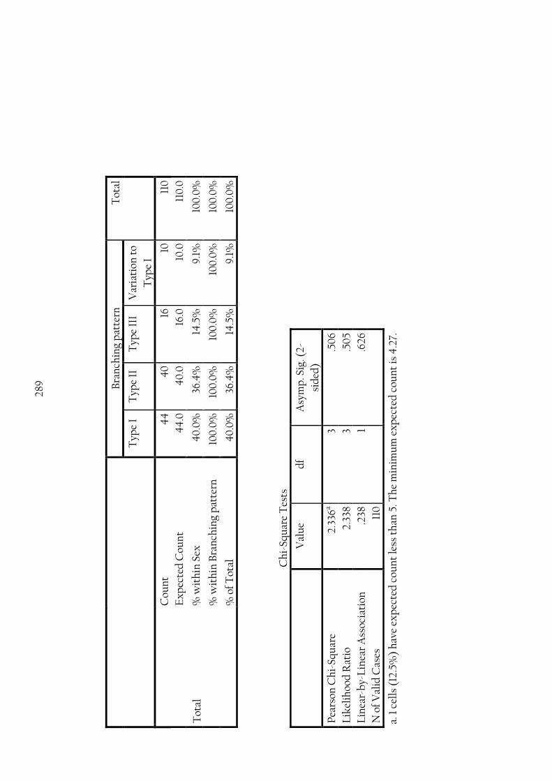
289
Bra
nch
ing
pat
tern
T
otal
Typ
e I
Typ
e II
T
ype
III
Var
iati
on t
o
Typ
e I
Tot
al
Cou
nt
44
4
0
16
10
110
Ex
pec
ted
Cou
nt
44
.0
40
.0
16.0
10
.0
110
.0
% w
ith
in S
ex
40
.0%
36
.4%
14
.5%
9
.1%
10
0.0
%
% w
ith
in B
ran
chin
g p
atte
rn
100
.0%
10
0.0
%
100
.0%
10
0.0
%
100
.0%
% o
f Tot
al
40
.0%
36
.4%
14
.5%
9
.1%
10
0.0
%
Ch
i-Sq
uar
e T
ests
V
alu
e d
f A
sym
p. S
ig. (
2-
sid
ed)
Pea
rson
Ch
i-Sq
uar
e 2.
336
a 3
.50
6
Lik
elih
ood
Rat
io
2.33
8 3
.50
5
Lin
ear-
by-
Lin
ear
Ass
ocia
tion
.2
38
1 .6
26
N o
f V
alid
Cas
es
110
a. 1
cel
ls (
12.5
%)
hav
e ex
pec
ted
cou
nt
less
th
an 5
. Th
e m
inim
um
ex
pec
ted
cou
nt
is 4
.27.
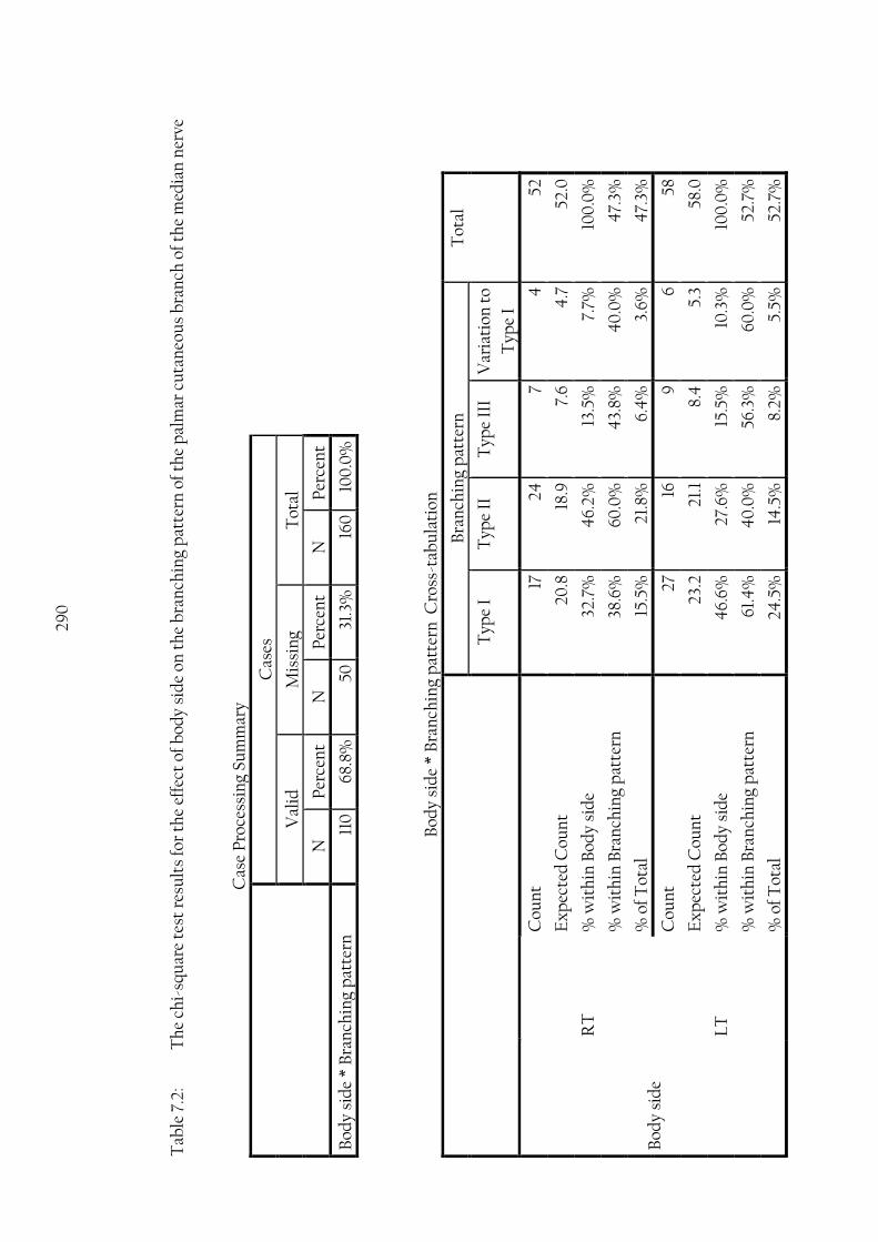
290
T
able
7.2
: T
he
chi-
squ
are
test
res
ult
s fo
r th
e ef
fect
of
bod
y si
de
on t
he
bra
nch
ing
pat
tern
of
the
pal
mar
cu
tan
eou
s b
ran
ch o
f th
e m
edia
n n
erve
Cas
e P
roce
ssin
g Su
mm
ary
C
ases
Val
id
Mis
sin
g T
otal
N
Per
cen
t N
P
erce
nt
N
Per
cen
t
Bod
y si
de
* B
ran
chin
g p
atte
rn
110
68
.8%
50
31
.3%
16
0
100
.0%
Bod
y si
de
* B
ran
chin
g p
atte
rn C
ross
-tab
ula
tion
B
ran
chin
g p
atte
rn
Tot
al
Typ
e I
Typ
e II
T
ype
III
Var
iati
on t
o
Typ
e I
Bod
y si
de
RT
Cou
nt
17
24
7 4
52
Ex
pec
ted
Cou
nt
20.8
18
.9
7.6
4
.7
52.0
% w
ith
in B
ody
sid
e 32
.7%
4
6.2%
13
.5%
7.
7%
100
.0%
% w
ith
in B
ran
chin
g p
atte
rn
38.6
%
60.0
%
43.
8%
40
.0%
4
7.3%
% o
f Tot
al
15.5
%
21.8
%
6.4
%
3.6%
4
7.3%
LT
Cou
nt
27
16
9 6
58
Ex
pec
ted
Cou
nt
23.2
21
.1
8.4
5.
3 58
.0
% w
ith
in B
ody
sid
e 4
6.6%
27
.6%
15
.5%
10
.3%
10
0.0
%
% w
ith
in B
ran
chin
g p
atte
rn
61.4
%
40
.0%
56
.3%
60
.0%
52
.7%
% o
f Tot
al
24.5
%
14.5
%
8.2%
5.
5%
52.7
%
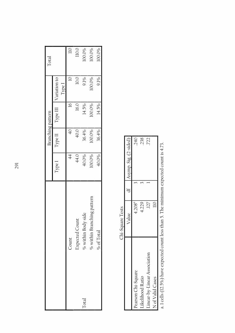
291
Bra
nch
ing
pat
tern
T
otal
Typ
e I
Typ
e II
T
ype
III
Var
iati
on t
o
Typ
e I
Tot
al
Cou
nt
44
4
0
16
10
110
Ex
pec
ted
Cou
nt
44
.0
40
.0
16.0
10
.0
110
.0
% w
ith
in B
ody
sid
e 4
0.0
%
36.4
%
14.5
%
9.1
%
100
.0%
% w
ith
in B
ran
chin
g p
atte
rn
100
.0%
10
0.0
%
100
.0%
10
0.0
%
100
.0%
% o
f Tot
al
40
.0%
36
.4%
14
.5%
9
.1%
10
0.0
%
Ch
i-Sq
uar
e T
ests
V
alu
e d
f A
sym
p. S
ig. (
2-s
ided
)
Pea
rson
Ch
i-Sq
uar
e 4
.20
8a 3
.24
0
Lik
elih
ood
Rat
io
4.2
29
3
.238
Lin
ear-
by-
Lin
ear
Ass
ocia
tion
.1
27
1 .7
22
N o
f Val
id C
ases
11
0
a. 1
cel
ls (
12.5
%)
hav
e ex
pec
ted
cou
nt
less
th
an 5
. Th
e m
inim
um
ex
pec
ted
cou
nt
is 4
.73.
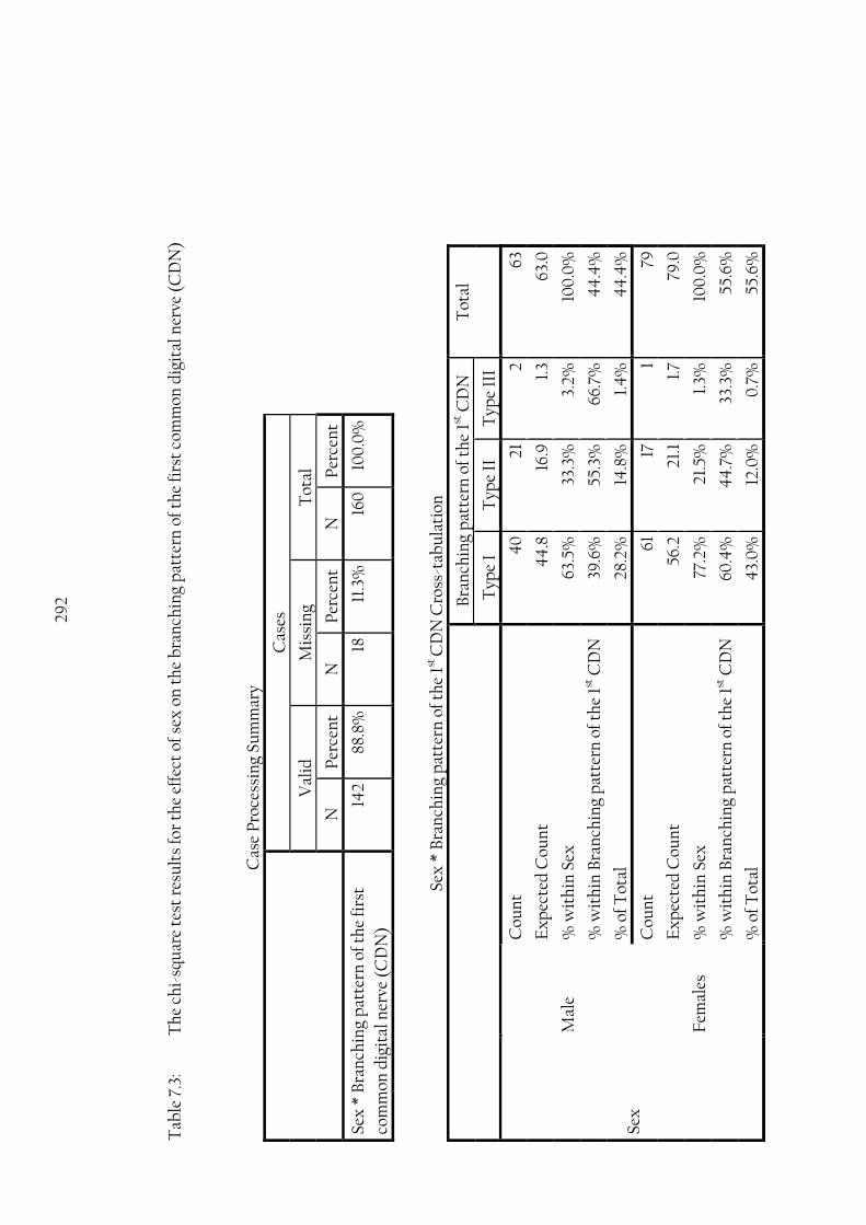
292
T
able
7.3
: T
he
chi-
squ
are
test
res
ult
s fo
r th
e ef
fect
of
sex
on
th
e b
ran
chin
g p
atte
rn o
f th
e fi
rst
com
mon
dig
ital
ner
ve (
CD
N)
Cas
e P
roce
ssin
g Su
mm
ary
C
ases
Val
id
Mis
sin
g T
otal
N
Per
cen
t N
P
erce
nt
N
Per
cen
t
Sex
* B
ran
chin
g p
atte
rn o
f th
e fi
rst
com
mon
dig
ital
ner
ve (
CD
N)
142
88.8
%
18
11.3
%
160
10
0.0
%
Sex
* B
ran
chin
g p
atte
rn o
f th
e 1st
CD
N C
ross
-tab
ula
tion
B
ran
chin
g p
atte
rn o
f th
e 1st
CD
N
Tot
al
Typ
e I
Typ
e II
T
ype
III
Sex
Mal
e
Cou
nt
40
21
2
63
Ex
pec
ted
Cou
nt
44
.8
16.9
1.
3 63
.0
% w
ith
in S
ex
63.5
%
33.3
%
3.2%
10
0.0
%
% w
ith
in B
ran
chin
g p
atte
rn o
f th
e 1st
CD
N
39.6
%
55.3
%
66.7
%
44
.4%
% o
f Tot
al
28.2
%
14.8
%
1.4
%
44
.4%
Fem
ales
Cou
nt
61
17
1 79
Ex
pec
ted
Cou
nt
56.2
21
.1
1.7
79.0
% w
ith
in S
ex
77.2
%
21.5
%
1.3%
10
0.0
%
% w
ith
in B
ran
chin
g p
atte
rn o
f th
e 1st
CD
N
60.4
%
44
.7%
33
.3%
55
.6%
% o
f Tot
al
43.
0%
12
.0%
0
.7%
55
.6%

293
B
ran
chin
g p
atte
rn o
f th
e 1st
CD
N
Tot
al
Typ
e I
Typ
e II
T
ype
III
Tot
al
Cou
nt
101
38
3 14
2
Ex
pec
ted
Cou
nt
101.
0
38.0
3.
0
142.
0
% w
ith
in S
ex
71.1
%
26.8
%
2.1%
10
0.0
%
% w
ith
in B
ran
chin
g p
atte
rn o
f th
e 1st
CD
N
100
.0%
10
0.0
%
100
.0%
10
0.0
%
% o
f Tot
al
71.1
%
26.8
%
2.1%
10
0.0
%
Ch
i-Sq
uar
e T
ests
V
alu
e d
f A
sym
p. S
ig. (
2-si
ded
)
Pea
rson
Ch
i-Sq
uar
e 3.
361a
2 .1
86
Lik
elih
ood
Rat
io
3.35
3 2
.187
Lin
ear-
by-
Lin
ear
Ass
ocia
tion
3.
321
1 .0
68
N o
f Val
id C
ases
14
2
a. 2
cel
ls (
33.3
%)
hav
e ex
pec
ted
cou
nt
less
th
an 5
. Th
e m
inim
um
ex
pec
ted
cou
nt
is 1
.33.

294
T
able
7.4
: T
he
chi-
squ
are
test
res
ult
s fo
r th
e ef
fect
of
bod
y si
de
on t
he
bra
nch
ing
pat
tern
of
the
firs
t co
mm
on d
igit
al n
erve
(C
DN
)
Cas
e P
roce
ssin
g Su
mm
ary
C
ases
Val
id
Mis
sin
g T
otal
N
Per
cen
t N
P
erce
nt
N
Per
cen
t
Bod
y si
de
* B
ran
chin
g p
atte
rn o
f th
e fi
rst
com
mon
dig
ital
ner
ve (
CD
N)
142
88.8
%
18
11.3
%
160
10
0.0
%
Bod
y si
de
* B
ran
chin
g p
atte
rn o
f th
e 1st
CD
N C
ross
-tab
ula
tion
B
ran
chin
g p
atte
rn o
f th
e 1st
CD
N
Tot
al
Typ
e I
Typ
e II
T
ype
III
Bod
y si
de
RT
Cou
nt
48
25
3 76
Ex
pec
ted
Cou
nt
54.1
20
.3
1.6
76.0
% w
ith
in B
ody
sid
e 63
.2%
32
.9%
3.
9%
10
0.0
%
% w
ith
in B
ran
chin
g p
atte
rn o
f th
e 1st
CD
N
47.
5%
65.8
%
100
.0%
53
.5%
% o
f Tot
al
33.8
%
17.6
%
2.1%
53
.5%
LT
Cou
nt
53
13
0
66
Ex
pec
ted
Cou
nt
46.
9
17.7
1.
4
66.0
% w
ith
in B
ody
sid
e 80
.3%
19
.7%
0
.0%
10
0.0
%
% w
ith
in B
ran
chin
g p
atte
rn o
f th
e 1st
CD
N
52.5
%
34.2
%
0.0
%
46.
5%
% o
f Tot
al
37.3
%
9.2
%
0.0
%
46.
5%

295
B
ran
chin
g p
atte
rn o
f th
e 1st
CD
N
Tot
al
Typ
e I
Typ
e II
T
ype
III
Tot
al
Cou
nt
101
38
3 14
2
Ex
pec
ted
Cou
nt
101.
0
38.0
3.
0
142.
0
% w
ith
in B
ody
sid
e 71
.1%
26
.8%
2.
1%
100
.0%
% w
ith
in B
ran
chin
g p
atte
rn o
f th
e 1st
CD
N
100
.0%
10
0.0
%
100
.0%
10
0.0
%
% o
f Tot
al
71.1
%
26.8
%
2.1%
10
0.0
%
Ch
i-Sq
uar
e T
ests
V
alu
e d
f A
sym
p. S
ig. (
2-s
ided
)
Pea
rson
Ch
i-Sq
uar
e 6.
364
a 2
.04
1
Lik
elih
ood
Rat
io
7.55
7 2
.023
Lin
ear-
by-
Lin
ear
Ass
ocia
tion
6.
09
3 1
.014
N o
f Val
id C
ases
14
2
a. 2
cel
ls (
33.3
%)
hav
e ex
pec
ted
cou
nt
less
th
an 5
. Th
e m
inim
um
ex
pec
ted
cou
nt
is 1
.39
.
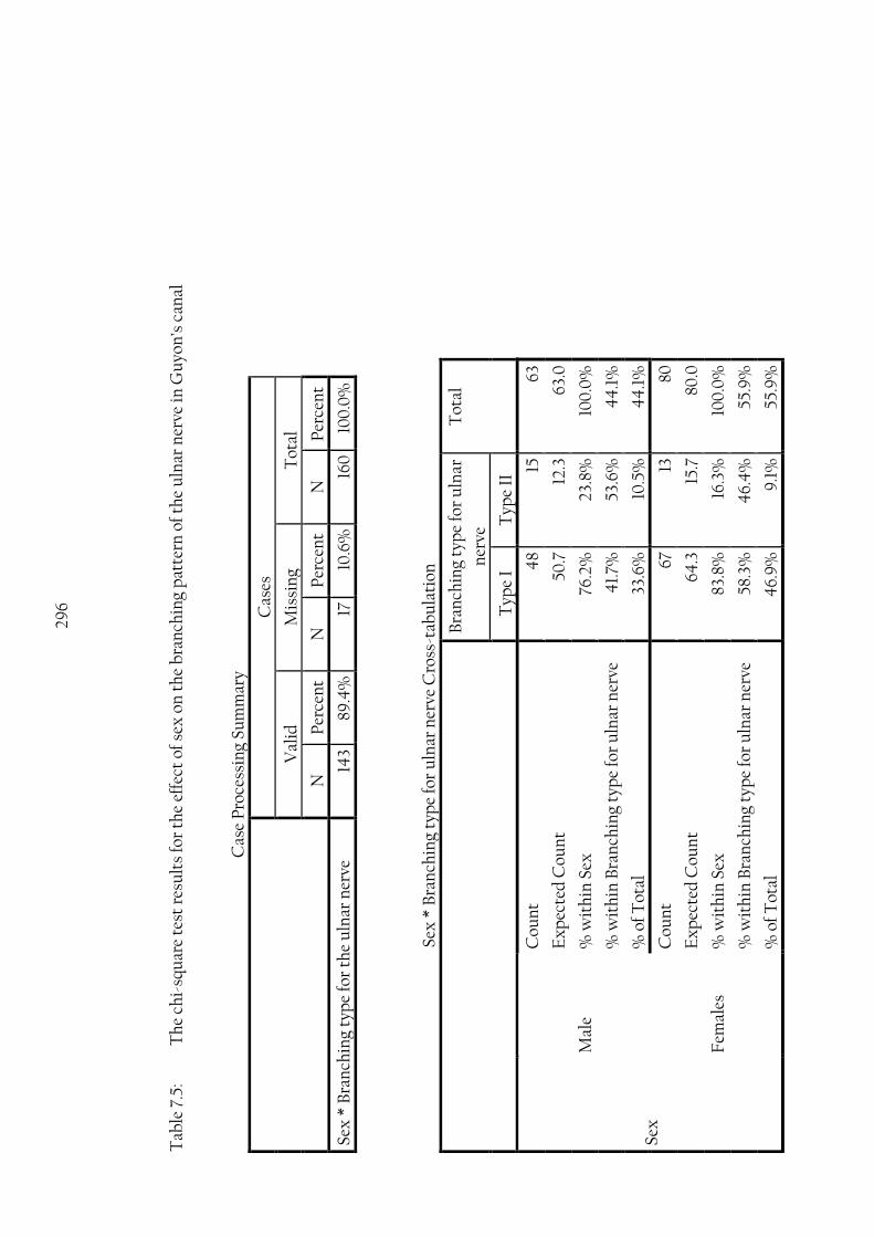
296
Tab
le 7
.5:
Th
e ch
i-sq
uar
e te
st r
esu
lts
for
the
effe
ct o
f se
x o
n t
he
bra
nch
ing
pat
tern
of
the
uln
ar n
erve
in G
uyo
n’s
can
al
Cas
e P
roce
ssin
g Su
mm
ary
C
ases
Val
id
Mis
sin
g T
otal
N
Per
cen
t N
P
erce
nt
N
Per
cen
t
Sex
* B
ran
chin
g ty
pe
for
the
uln
ar n
erve
14
3 89
.4%
17
10
.6%
16
0
100
.0%
Sex
* B
ran
chin
g ty
pe
for
uln
ar n
erve
Cro
ss-t
abu
lati
on
B
ran
chin
g ty
pe
for
uln
ar
ner
ve
Tot
al
Typ
e I
Typ
e II
Sex
Mal
e
Cou
nt
48
15
63
Ex
pec
ted
Cou
nt
50.7
12
.3
63.0
% w
ith
in S
ex
76.2
%
23.8
%
100
.0%
% w
ith
in B
ran
chin
g ty
pe
for
uln
ar n
erve
4
1.7%
53
.6%
4
4.1
%
% o
f Tot
al
33.6
%
10.5
%
44
.1%
Fem
ales
Cou
nt
67
13
80
Ex
pec
ted
Cou
nt
64.3
15
.7
80.0
% w
ith
in S
ex
83.8
%
16.3
%
100
.0%
% w
ith
in B
ran
chin
g ty
pe
for
uln
ar n
erve
58
.3%
4
6.4
%
55.9
%
% o
f Tot
al
46.
9%
9
.1%
55
.9%

297
B
ran
chin
g ty
pe
for
uln
ar
ner
ve
Tot
al
Typ
e I
Typ
e II
Tot
al
Cou
nt
115
28
143
Ex
pec
ted
Cou
nt
115.
0
28.0
14
3.0
% w
ith
in S
ex
80.4
%
19.6
%
100
.0%
% w
ith
in B
ran
chin
g ty
pe
for
uln
ar n
erve
10
0.0
%
100
.0%
10
0.0
%
% o
f Tot
al
80.4
%
19.6
%
100
.0%
Ch
i-Sq
uar
e T
ests
V
alu
e d
f A
sym
p. S
ig.
(2-s
ided
)
Ex
act
Sig
. (2
-
sid
ed)
Ex
act
Sig
. (1-
sid
ed)
Pea
rson
Ch
i-Sq
uar
e 1.
279
a 1
.258
Con
tin
uit
y C
orre
ctio
nb
.84
4
1 .3
58
Lik
elih
ood
Rat
io
1.27
1 1
.260
Fis
her
's E
xac
t T
est
.2
93
.179
Lin
ear-
by-
Lin
ear
Ass
ocia
tion
1.
270
1
.260
N o
f Val
id C
ases
14
3
a. 0
cel
ls (
0.0
%)
hav
e ex
pec
ted
cou
nt
less
th
an 5
. Th
e m
inim
um
ex
pec
ted
cou
nt
is 1
2.34
.
b. C
omp
ute
d o
nly
for
a 2
x2
tab
le
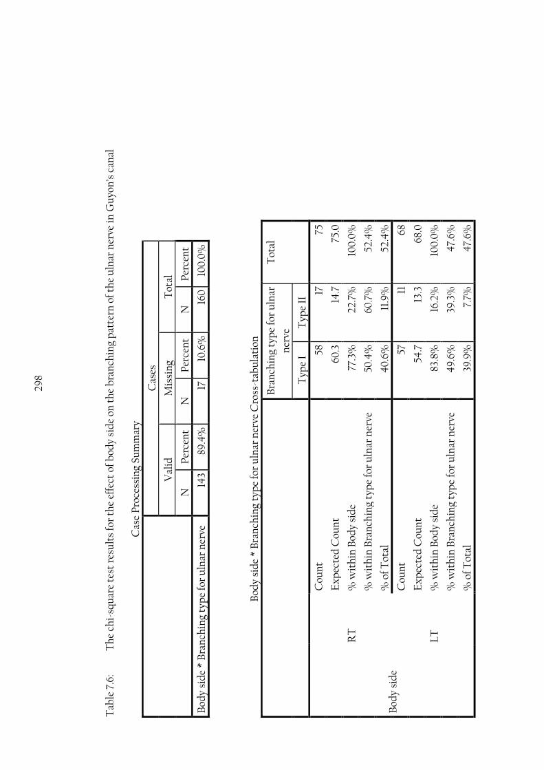
298
Tab
le 7
.6:
Th
e ch
i-sq
uar
e te
st r
esu
lts
for
the
effe
ct o
f b
ody
sid
e on
th
e b
ran
chin
g p
atte
rn o
f th
e u
lnar
ner
ve in
Gu
yon
’s c
anal
Cas
e P
roce
ssin
g Su
mm
ary
C
ases
Val
id
Mis
sin
g T
otal
N
Per
cen
t N
P
erce
nt
N
Per
cen
t
Bod
y si
de
* B
ran
chin
g ty
pe
for
uln
ar n
erve
14
3 89
.4%
17
10
.6%
16
0
100
.0%
Bod
y si
de
* B
ran
chin
g ty
pe
for
uln
ar n
erve
Cro
ss-t
abu
lati
on
B
ran
chin
g ty
pe
for
uln
ar
ner
ve
Tot
al
Typ
e I
Typ
e II
Bod
y si
de
RT
Cou
nt
58
17
75
Ex
pec
ted
Cou
nt
60.3
14
.7
75.0
% w
ith
in B
ody
sid
e 77
.3%
22
.7%
10
0.0
%
% w
ith
in B
ran
chin
g ty
pe
for
uln
ar n
erve
50
.4%
60
.7%
52
.4%
% o
f Tot
al
40
.6%
11
.9%
52
.4%
LT
Cou
nt
57
11
68
Ex
pec
ted
Cou
nt
54.7
13
.3
68.0
% w
ith
in B
ody
sid
e 83
.8%
16
.2%
10
0.0
%
% w
ith
in B
ran
chin
g ty
pe
for
uln
ar n
erve
4
9.6
%
39.3
%
47.
6%
% o
f Tot
al
39.9
%
7.7%
4
7.6%

299
Bra
nch
ing
typ
e fo
r u
lnar
ner
ve
Tot
al
Typ
e I
Typ
e II
Tot
al
Cou
nt
115
28
143
Ex
pec
ted
Cou
nt
115.
0
28.0
14
3.0
% w
ith
in B
ody
sid
e 80
.4%
19
.6%
10
0.0
%
% w
ith
in B
ran
chin
g ty
pe
for
uln
ar n
erve
10
0.0
%
100
.0%
10
0.0
%
% o
f Tot
al
80.4
%
19.6
%
100
.0%
Ch
i-Sq
uar
e T
ests
V
alu
e d
f A
sym
p. S
ig.
(2-s
ided
)
Ex
act
Sig
. (2
-
sid
ed)
Ex
act
Sig
. (1-
sid
ed)
Pea
rson
Ch
i-Sq
uar
e .9
54a
1 .3
29
Con
tin
uit
y C
orre
ctio
nb
.586
1
.44
4
Lik
elih
ood
Rat
io
.962
1
.32
7
Fis
her
's E
xac
t T
est
.4
01
.22
2
Lin
ear-
by-
Lin
ear
Ass
ocia
tion
.9
47
1 .3
30
N o
f Val
id C
ases
14
3
a. 0
cel
ls (
0.0
%)
hav
e ex
pec
ted
cou
nt
less
th
an 5
. Th
e m
inim
um
ex
pec
ted
cou
nt
is 1
3.31
.
b. C
omp
ute
d o
nly
for
a 2
x2
tab
le

300
T
able
7.7
: T
he
chi-
squ
are
test
res
ult
s fo
r th
e ef
fect
of
sex
on
th
e b
ran
chin
g p
atte
rn o
f th
e p
alm
ar c
omm
un
icat
ing
bra
nch
(C
B)
bet
wee
n t
he
med
ian
and
uln
ar n
erve
Cas
e P
roce
ssin
g Su
mm
ary
C
ases
Val
id
Mis
sin
g T
otal
N
Per
cen
t N
P
erce
nt
N
Per
cen
t
Sex
* P
alm
ar C
B b
ran
chin
g p
atte
rn
98
61.3
%
62
38.8
%
160
10
0.0
%
Sex
* P
alm
ar C
B b
ran
chin
g p
atte
rn C
ross
-tab
ula
tion
P
alm
ar C
B b
ran
chin
g p
atte
rn
Tot
al
Typ
e I
Typ
e II
T
ype
III
Typ
e IV
T
ype
V
Sex
Mal
e
Cou
nt
37
4
1 1
1 4
4
Ex
pec
ted
Cou
nt
36.4
2.
2 2.
2
1.8
1.3
44
.0
% w
ith
in S
ex
84.1
%
9.1
%
2.3%
2.
3%
2.3%
10
0.0
%
% w
ith
in P
alm
ar C
B b
ran
chin
g p
atte
rn
45.
7%
80.0
%
20.0
%
25.0
%
33.3
%
44
.9%
% o
f Tot
al
37.8
%
4.1
%
1.0
%
1.0
%
1.0
%
44
.9%
Fem
ales
Cou
nt
44
1
4
3 2
54
Ex
pec
ted
Cou
nt
44
.6
2.8
2.8
2.2
1.7
54.0
% w
ith
in S
ex
81.5
%
1.9
%
7.4
%
5.6%
3.
7%
100
.0%
% w
ith
in P
alm
ar C
B b
ran
chin
g p
atte
rn
54.3
%
20.0
%
80.0
%
75.0
%
66.7
%
55.1
%
% o
f Tot
al
44
.9%
1.
0%
4
.1%
3.
1%
2.0
%
55.1
%

301
P
alm
ar C
B b
ran
chin
g p
atte
rn
Tot
al
Typ
e I
Typ
e II
T
ype
III
Typ
e IV
T
ype
V
Tot
al
Cou
nt
81
5 5
4
3 98
Ex
pec
ted
Cou
nt
81.0
5.
0
5.0
4
.0
3.0
9
8.0
% w
ith
in S
ex
82.7
%
5.1%
5.
1%
4.1
%
3.1%
10
0.0
%
% w
ith
in P
alm
ar C
B b
ran
chin
g p
atte
rn
100
.0%
10
0.0
%
100
.0%
10
0.0
%
100
.0%
10
0.0
%
% o
f Tot
al
82.7
%
5.1%
5.
1%
4.1
%
3.1%
10
0.0
%
Ch
i-Sq
uar
e T
ests
V
alu
e d
f A
sym
p. S
ig. (
2-s
ided
)
Pea
rson
Ch
i-Sq
uar
e 4
.565
a 4
.3
35
Lik
elih
ood
Rat
io
4.8
25
4
.30
6
Lin
ear-
by-
Lin
ear
Ass
ocia
tion
.8
71
1 .3
51
N o
f V
alid
Cas
es
98
a. 8
cel
ls (
80.0
%)
hav
e ex
pec
ted
cou
nt
less
th
an 5
. Th
e m
inim
um
ex
pec
ted
cou
nt
is 1
.35.
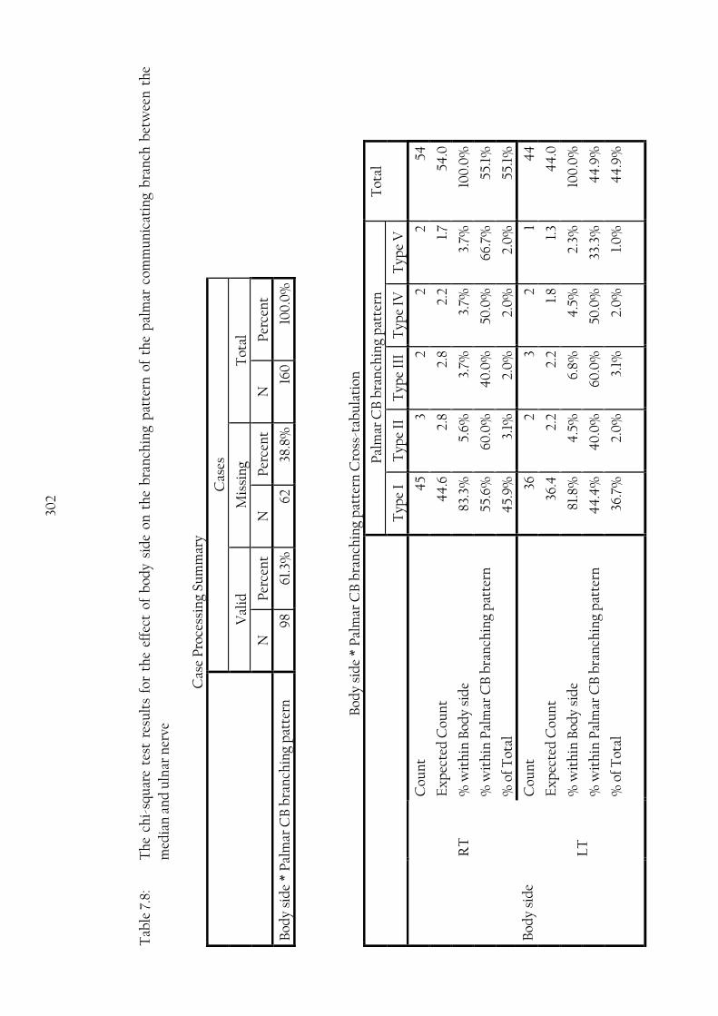
302
T
able
7.8
: T
he
chi-
squ
are
test
res
ult
s fo
r th
e ef
fect
of
bod
y si
de
on t
he
bra
nch
ing
pat
tern
of
the
pal
mar
com
mu
nic
atin
g b
ran
ch b
etw
een
th
e
med
ian
an
d u
lnar
ner
ve
Cas
e P
roce
ssin
g Su
mm
ary
C
ases
Val
id
Mis
sin
g T
otal
N
Per
cen
t N
P
erce
nt
N
Per
cen
t
Bod
y si
de
* P
alm
ar C
B b
ran
chin
g p
atte
rn
98
61.3
%
62
38.8
%
160
10
0.0
%
Bod
y si
de
* P
alm
ar C
B b
ran
chin
g p
atte
rn C
ross
-tab
ula
tion
P
alm
ar C
B b
ran
chin
g p
atte
rn
Tot
al
Typ
e I
Typ
e II
T
ype
III
Typ
e IV
T
ype
V
Bod
y si
de
RT
Cou
nt
45
3 2
2 2
54
Ex
pec
ted
Cou
nt
44
.6
2.8
2.8
2.2
1.7
54.0
% w
ith
in B
ody
sid
e 83
.3%
5.
6%
3.7%
3.
7%
3.7%
10
0.0
%
% w
ith
in P
alm
ar C
B b
ran
chin
g p
atte
rn
55.6
%
60.0
%
40
.0%
50
.0%
66
.7%
55
.1%
% o
f Tot
al
45.
9%
3.
1%
2.0
%
2.0
%
2.0
%
55.1
%
LT
Cou
nt
36
2 3
2 1
44
Ex
pec
ted
Cou
nt
36.4
2.
2 2.
2 1.
8 1.
3 4
4.0
% w
ith
in B
ody
sid
e 81
.8%
4
.5%
6.
8%
4.5
%
2.3%
10
0.0
%
% w
ith
in P
alm
ar C
B b
ran
chin
g p
atte
rn
44
.4%
4
0.0
%
60.0
%
50.0
%
33.3
%
44
.9%
% o
f Tot
al
36.7
%
2.0
%
3.1%
2.
0%
1.
0%
4
4.9
%

303
P
alm
ar C
B b
ran
chin
g p
atte
rn
Tot
al
Typ
e I
Typ
e II
T
ype
III
Typ
e IV
T
ype
V
Tot
al
Cou
nt
81
5 5
4
3 98
Ex
pec
ted
Cou
nt
81.0
5.
0
5.0
4
.0
3.0
9
8.0
% w
ith
in B
ody
sid
e 82
.7%
5.
1%
5.1%
4
.1%
3.
1%
100
.0%
% w
ith
in P
alm
ar C
B b
ran
chin
g p
atte
rn
100
.0%
10
0.0
%
100
.0%
10
0.0
%
100
.0%
10
0.0
%
% o
f Tot
al
82.7
%
5.1%
5.
1%
4.1
%
3.1%
10
0.0
%
Ch
i-Sq
uar
e T
ests
V
alu
e d
f A
sym
p. S
ig. (
2-s
ided
)
Pea
rson
Ch
i-Sq
uar
e .7
20a
4
.94
9
Lik
elih
ood
Rat
io
.722
4
.9
49
Lin
ear-
by-
Lin
ear
Ass
ocia
tion
.0
10
1 .9
19
N o
f Val
id C
ases
9
8
a. 8
cel
ls (
80.0
%)
hav
e ex
pec
ted
cou
nt
less
th
an 5
. Th
e m
inim
um
ex
pec
ted
cou
nt
is 1
.35.
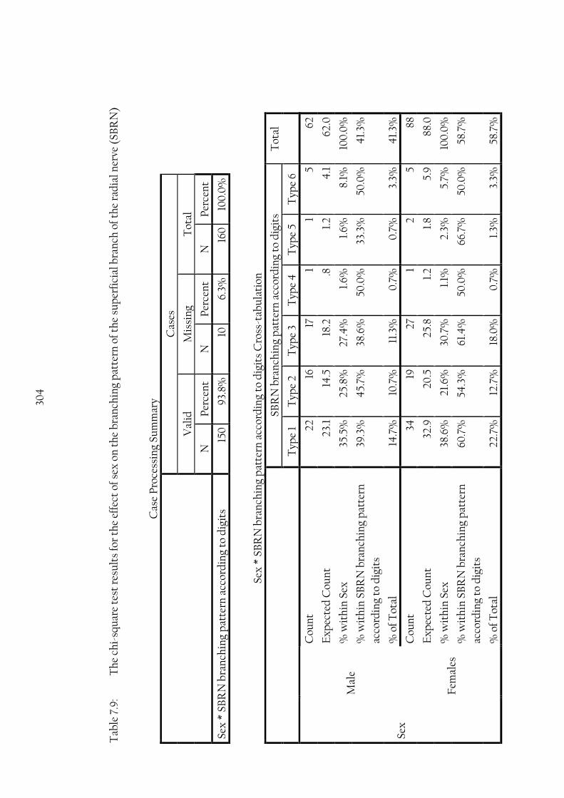
304
T
able
7.9
: T
he
chi-
squ
are
test
res
ult
s fo
r th
e ef
fect
of
sex
on
th
e b
ran
chin
g p
atte
rn o
f th
e su
per
fici
al b
ran
ch o
f th
e ra
dia
l ner
ve (
SBR
N)
Cas
e P
roce
ssin
g Su
mm
ary
C
ases
Val
id
Mis
sin
g T
otal
N
Per
cen
t N
P
erce
nt
N
Per
cen
t
Sex
* S
BR
N b
ran
chin
g p
atte
rn a
ccor
din
g to
dig
its
150
9
3.8%
10
6.
3%
160
10
0.0
%
Sex
* S
BR
N b
ran
chin
g p
atte
rn a
ccor
din
g to
dig
its
Cro
ss-t
abu
lati
on
SB
RN
bra
nch
ing
pat
tern
acc
ord
ing
to d
igit
s T
otal
Typ
e 1
Typ
e 2
T
ype
3 T
ype
4
Typ
e 5
Typ
e 6
Sex
Mal
e
Cou
nt
22
16
17
1 1
5 62
Ex
pec
ted
Cou
nt
23.1
14
.5
18.2
.8
1.
2 4
.1
62.0
% w
ith
in S
ex
35.5
%
25.8
%
27.4
%
1.6%
1.
6%
8.1%
10
0.0
%
% w
ith
in S
BR
N b
ran
chin
g p
atte
rn
acco
rdin
g to
dig
its
39.3
%
45.
7%
38.6
%
50.0
%
33.3
%
50.0
%
41.
3%
% o
f Tot
al
14.7
%
10.7
%
11.3
%
0.7
%
0.7
%
3.3%
4
1.3%
Fem
ales
Cou
nt
34
19
27
1 2
5 88
Ex
pec
ted
Cou
nt
32.9
20
.5
25.8
1.
2 1.
8 5.
9
88.0
% w
ith
in S
ex
38.6
%
21.6
%
30.7
%
1.1%
2.
3%
5.7%
10
0.0
%
% w
ith
in S
BR
N b
ran
chin
g p
atte
rn
acco
rdin
g to
dig
its
60.7
%
54.3
%
61.4
%
50.0
%
66.7
%
50.0
%
58.7
%
% o
f Tot
al
22.7
%
12.7
%
18.0
%
0.7
%
1.3%
3.
3%
58.7
%

305
SB
RN
bra
nch
ing
pat
tern
acc
ord
ing
to d
igit
s T
otal
Typ
e 1
Typ
e 2
T
ype
3 T
ype
4
Typ
e 5
Typ
e 6
Tot
al
Cou
nt
56
35
44
2
3 10
15
0
Ex
pec
ted
Cou
nt
56.0
35
.0
44
.0
2.0
3.
0
10.0
15
0.0
% w
ith
in S
ex
37.3
%
23.3
%
29.3
%
1.3%
2.
0%
6.
7%
100
.0%
% w
ith
in S
BR
N b
ran
chin
g p
atte
rn
acco
rdin
g to
dig
its
100
.0%
10
0.0
%
100
.0%
10
0.0
%
100
.0%
10
0.0
%
100
.0%
% o
f Tot
al
37.3
%
23.3
%
29.3
%
1.3%
2.
0%
6.
7%
100
.0%
Ch
i-Sq
uar
e T
ests
V
alu
e d
f A
sym
p. S
ig. (
2-s
ided
)
Pea
rson
Ch
i-Sq
uar
e .9
57a
5 .9
66
Lik
elih
ood
Rat
io
.952
5
.966
Lin
ear-
by-
Lin
ear
Ass
ocia
tion
.1
35
1 .7
14
N o
f Val
id C
ases
15
0
a. 5
cel
ls (
41.
7%)
hav
e ex
pec
ted
cou
nt
less
th
an 5
. Th
e m
inim
um
ex
pec
ted
cou
nt
is .8
3.

306
Tab
le 7
.10
: T
he
chi-
squ
are
test
res
ult
s fo
r th
e ef
fect
of
bod
y si
de
on t
he
bra
nch
ing
pat
tern
of
the
sup
erfi
cial
bra
nch
of
the
rad
ial n
erve
(S
BR
N)
Cas
e P
roce
ssin
g Su
mm
ary
C
ases
Val
id
Mis
sin
g T
otal
N
Per
cen
t N
P
erce
nt
N
Per
cen
t
Bod
y si
de
* SB
RN
bra
nch
ing
pat
tern
acc
ord
ing
to d
igit
s 15
0
93.
8%
10
6.3%
16
0
100
.0%
Bod
y si
de
* SB
RN
bra
nch
ing
pat
tern
acc
ord
ing
to d
igit
s C
ross
-tab
ula
tion
SB
RN
bra
nch
ing
pat
tern
acc
ord
ing
to d
igit
s T
otal
Typ
e 1
Typ
e 2
T
ype
3 T
ype
4
Typ
e 5
Typ
e 6
Bod
y si
de
RT
Cou
nt
26
20
23
0
1 4
74
Ex
pec
ted
Cou
nt
27.6
17
.3
21.7
1.
0
1.5
4.9
74
.0
% w
ith
in B
ody
sid
e 35
.1%
27
.0%
31
.1%
0
.0%
1.
4%
5.
4%
10
0.0
%
% w
ith
in S
BR
N b
ran
chin
g p
atte
rn
acco
rdin
g to
dig
its
46.
4%
57
.1%
52
.3%
0
.0%
33
.3%
4
0.0
%
49
.3%
% o
f Tot
al
17.3
%
13.3
%
15.3
%
0.0
%
0.7
%
2.7%
4
9.3
%
LT
Cou
nt
30
15
21
2 2
6 76
Ex
pec
ted
Cou
nt
28.4
17
.7
22.3
1.
0
1.5
5.1
76.0
% w
ith
in B
ody
sid
e 39
.5%
19
.7%
27
.6%
2.
6%
2.6%
7.
9%
10
0.0
%
% w
ith
in S
BR
N b
ran
chin
g p
atte
rn
acco
rdin
g to
dig
its
53.6
%
42.
9%
4
7.7%
10
0.0
%
66.7
%
60.0
%
50.7
%
% o
f Tot
al
20.0
%
10.0
%
14.0
%
1.3%
1.
3%
4.0
%
50.7
%

307
SB
RN
bra
nch
ing
pat
tern
acc
ord
ing
to d
igit
s T
otal
Typ
e 1
Typ
e 2
Typ
e 3
Typ
e 4
T
ype
5 T
ype
6
Tot
al
Cou
nt
56
35
44
2
3 10
15
0
Ex
pec
ted
Cou
nt
56.0
35
.0
44
.0
2.0
3.
0
10.0
15
0.0
% w
ith
in B
ody
sid
e 37
.3%
23
.3%
29
.3%
1.
3%
2.0
%
6.7%
10
0.0
%
% w
ith
in S
BR
N b
ran
chin
g p
atte
rn
acco
rdin
g to
dig
its
100
.0%
10
0.0
%
100
.0%
10
0.0
%
100
.0%
10
0.0
%
100
.0%
% o
f Tot
al
37.3
%
23.3
%
29.3
%
1.3%
2.
0%
6.
7%
100
.0%
Ch
i-Sq
uar
e T
ests
V
alu
e d
f A
sym
p. S
ig. (
2-s
ided
)
Pea
rson
Ch
i-Sq
uar
e 3.
798a
5 .5
79
Lik
elih
ood
Rat
io
4.5
82
5 .4
69
Lin
ear-
by-
Lin
ear
Ass
ocia
tion
.2
50
1 .6
17
N o
f Val
id C
ases
15
0
a. 5
cel
ls (
41.
7%)
hav
e ex
pec
ted
cou
nt
less
th
an 5
. Th
e m
inim
um
ex
pec
ted
cou
nt
is .9
9.
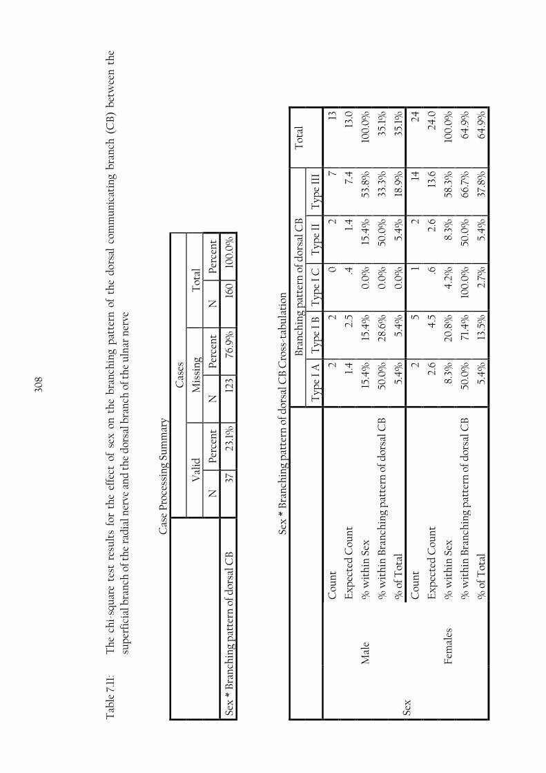
308
Tab
le 7
.11:
T
he
chi-
squ
are
test
res
ult
s fo
r th
e ef
fect
of
sex
on
th
e b
ran
chin
g p
atte
rn o
f th
e d
orsa
l co
mm
un
icat
ing
bra
nch
(C
B)
bet
wee
n t
he
sup
erfi
cial
bra
nch
of t
he
rad
ial n
erve
an
d t
he
dor
sal b
ran
ch o
f th
e u
lnar
ner
ve
Cas
e P
roce
ssin
g Su
mm
ary
C
ases
Val
id
Mis
sin
g T
otal
N
Per
cen
t N
P
erce
nt
N
Per
cen
t
Sex
* B
ran
chin
g p
atte
rn o
f dor
sal C
B
37
23.1
%
123
76.9
%
160
10
0.0
%
Sex
* B
ran
chin
g p
atte
rn o
f dor
sal C
B C
ross
-tab
ula
tion
B
ran
chin
g p
atte
rn o
f dor
sal C
B
Tot
al
Typ
e I
A
Typ
e I
B
Typ
e I
C
Typ
e II
T
ype
III
Sex
Mal
e
Cou
nt
2 2
0
2 7
13
Ex
pec
ted
Cou
nt
1.4
2.
5 .4
1.
4
7.4
13
.0
% w
ith
in S
ex
15.4
%
15.4
%
0.0
%
15.4
%
53.8
%
100
.0%
% w
ith
in B
ran
chin
g p
atte
rn o
f d
orsa
l CB
50
.0%
28
.6%
0
.0%
50
.0%
33
.3%
35
.1%
% o
f Tot
al
5.4
%
5.4
%
0.0
%
5.4
%
18.9
%
35.1
%
Fem
ales
Cou
nt
2 5
1 2
14
24
Ex
pec
ted
Cou
nt
2.6
4.5
.6
2.
6 13
.6
24.0
% w
ith
in S
ex
8.3%
20
.8%
4
.2%
8.
3%
58.3
%
100
.0%
% w
ith
in B
ran
chin
g p
atte
rn o
f d
orsa
l CB
50
.0%
71
.4%
10
0.0
%
50.0
%
66.7
%
64.9
%
% o
f Tot
al
5.4
%
13.5
%
2.7%
5.
4%
37
.8%
64
.9%

309
Bra
nch
ing
pat
tern
of d
orsa
l CB
T
otal
Typ
e I
A
Typ
e I
B
Typ
e I
C
Typ
e II
T
ype
III
Tot
al
Cou
nt
4
7 1
4
21
37
Ex
pec
ted
Cou
nt
4.0
7.
0
1.0
4
.0
21.0
37
.0
% w
ith
in S
ex
10.8
%
18.9
%
2.7%
10
.8%
56
.8%
10
0.0
%
% w
ith
in B
ran
chin
g p
atte
rn o
f d
orsa
l CB
10
0.0
%
100
.0%
10
0.0
%
100
.0%
10
0.0
%
100
.0%
% o
f Tot
al
10.8
%
18.9
%
2.7%
10
.8%
56
.8%
10
0.0
%
Ch
i-Sq
uar
e T
ests
V
alu
e d
f A
sym
p. S
ig. (
2-s
ided
)
Pea
rson
Ch
i-Sq
uar
e 1.
480
a 4
.8
30
Lik
elih
ood
Rat
io
1.77
3 4
.7
77
Lin
ear-
by-
Lin
ear
Ass
ocia
tion
.0
40
1
.84
2
N o
f Val
id C
ases
37
a. 8
cel
ls (
80.0
%)
hav
e ex
pec
ted
cou
nt
less
th
an 5
. Th
e m
inim
um
ex
pec
ted
cou
nt
is .3
5.

310
T
able
7.1
2:
Th
e ch
i-sq
uar
e te
st r
esu
lts
for
the
effe
ct o
f b
ody
sid
e on
th
e b
ran
chin
g p
atte
rn o
f th
e d
orsa
l co
mm
un
icat
ing
bra
nch
(C
B)
bet
wee
n t
he
sup
erfi
cial
bra
nch
of t
he
rad
ial n
erve
an
d t
he
dor
sal b
ran
ch o
f th
e u
lnar
ner
ve
Cas
e P
roce
ssin
g Su
mm
ary
C
ases
Val
id
Mis
sin
g T
otal
N
Per
cen
t N
P
erce
nt
N
Per
cen
t
Bod
y si
de
* B
ran
chin
g p
atte
rn o
f dor
sal C
B
37
23.1
%
123
76.9
%
160
10
0.0
%
Bod
y si
de
* B
ran
chin
g p
atte
rn o
f dor
sal C
B C
ross
-tab
ula
tion
B
ran
chin
g p
atte
rn o
f dor
sal C
B
Tot
al
Typ
e I
A
Typ
e I
B
Typ
e I
C
Typ
e II
T
ype
III
Bod
y si
de
RT
Cou
nt
2 3
0
2 10
17
Ex
pec
ted
Cou
nt
1.8
3.2
.5
1.8
9.6
17
.0
% w
ith
in B
ody
sid
e 11
.8%
17
.6%
0
.0%
11
.8%
58
.8%
10
0.0
%
% w
ith
in B
ran
chin
g p
atte
rn o
f d
orsa
l CB
50
.0%
4
2.9
%
0.0
%
50.0
%
47.
6%
45.
9%
% o
f Tot
al
5.4
%
8.1%
0
.0%
5.
4%
27
.0%
4
5.9
%
LT
Cou
nt
2 4
1
2 11
20
Ex
pec
ted
Cou
nt
2.2
3.8
.5
2.2
11.4
20
.0
% w
ith
in B
ody
sid
e 10
.0%
20
.0%
5.
0%
10
.0%
55
.0%
10
0.0
%
% w
ith
in B
ran
chin
g p
atte
rn o
f d
orsa
l CB
50
.0%
57
.1%
10
0.0
%
50.0
%
52.4
%
54.1
%
% o
f Tot
al
5.4
%
10.8
%
2.7%
5.
4%
29
.7%
54
.1%

311
B
ran
chin
g p
atte
rn o
f dor
sal C
B
Tot
al
Typ
e I
A
Typ
e I
B
Typ
e I
C
Typ
e II
T
ype
III
Tot
al
Cou
nt
4
7 1
4
21
37
Ex
pec
ted
Cou
nt
4.0
7.
0
1.0
4
.0
21.0
37
.0
% w
ith
in B
ody
sid
e 10
.8%
18
.9%
2.
7%
10.8
%
56.8
%
100
.0%
% w
ith
in B
ran
chin
g p
atte
rn o
f d
orsa
l CB
10
0.0
%
100
.0%
10
0.0
%
100
.0%
10
0.0
%
100
.0%
% o
f Tot
al
10.8
%
18.9
%
2.7%
10
.8%
56
.8%
10
0.0
%
Ch
i-Sq
uar
e T
ests
V
alu
e d
f A
sym
p. S
ig. (
2-s
ided
)
Pea
rson
Ch
i-Sq
uar
e .9
54a
4
.917
Lik
elih
ood
Rat
io
1.33
4
4
.856
Lin
ear-
by-
Lin
ear
Ass
ocia
tion
.0
26
1 .8
71
N o
f Val
id C
ases
37
a. 8
cel
ls (
80.0
%)
hav
e ex
pec
ted
cou
nt
less
th
an 5
. Th
e m
inim
um
ex
pec
ted
cou
nt
is .4
6.
312
8. Appendix II
Table 8.1: Palmar cutaneous nerve of the median nerve (PCBMN) results expressed as incidence rates:
Measurements Incidence rate (%)
PCBMN origin side from the main trunk of the median nerve: Lateral side 93.5 Posterolateral side 1.6 Medial side 1.6 Anterior side 3.3
Two PCBMNs 8.9
PCBMN relationship to other anatomical structures: PCBMN passed through the fascia of flexor carpi radialis 8.3 PCBMN passed posterior to the tendon of flexor carpi radialis 4.2 PCBMN passed superficial to the palmar carpal ligament 16.1 PCBMN passed deep to the palmar carpal ligament 12.5
Branching types of the PCBMN: Type I 40 Variations to Type I 9.1 Type II 36.4 Type III 14.5
Communications with other nerves in the area: With the lateral antebrachial cutaneous nerve 4.1 With the superficial branch of the radial nerve 0.8 With the recurrent motor branch of the median nerve 1.6 With the palmar cutaneous nerve of the ulnar nerve 0.8 With the first common digital nerve 0.8
Table 8.2: Anatomical measurements recorded for the palmar cutaneous branch of the median nerve (PCBMN) (mm)
Mean SD Max Min
Origin from main trunk 54.1 15.7 138.4 22.1
Detaches from main trunk 41.2 14.0 114.9 16.2
Length of PCBMN tunnel 11.3 3.4 24.5 5.7
Angle (degrees) 43.9 14.7 85.0 15.0
Distance of 1st branch to wrist crease 1.2 8.1 21.5 -18.2 Origin of the proximal branch from main trunk of the median nerve
70.9 20.6 100 42.1
Detachment of the proximal branch from main trunk of the median nerve
55.9 18.9 85.7 29.9
Origin of the distal branch from main trunk of the median nerve
64.8 17.8 92.3 42.1
Detachment of the distal branch from main trunk of the median nerve
46.5 16.7 77.0 27.9

313 Table 8.3: Common digital nerves (CDNs) results expressed as incidence rates:
Measurements Incidence rate (%)
First CDN branching pattern Type I 70.8 Type II 27.1 Type III 2.1
The second CDN was found to divide at a point located distal to the 70% of the distance between the third MCP1 joint and the BSL2
94.3
The Third CDN was found to divide at a point located distal to the 70% of the distance between the third MCP joint and the BSL
99.2
The Fourth CDN was found to divide at a point located distal to the 70% of the distance between the third MCP joint and the BSL
73.6
1 Metacarpophalangeal 2 Bistyloid line
Table 8.4: Absolute measurements for the branching points of the common digital nerves (CDNs) into proper digital nerves in the palm (mm)
Mean SD Max Min
First common digital nerve (CDN) first division
ST 29.9 5.4 54.3 16.7
BSL 45.3 6.1 63.5 32.4
First CDN second division ST 35.5 7.1 63.3 16.9
BSL 51.7 7.4 71.9 35.4
Second CDN division ST 59.8 7.9 81.1 42.9
BSL 74.1 9.1 94.7 42.3
Third CDN division ST 66.1 7.38 86.2 49.1
BSL 78.9 7.7 99.5 60.1
Fourth CDN division Pisiform 65.2 7.4 83.9 49.3
BSL 72.6 7.7 92.2 55.0
Table 8.5: Branching points of the common digital nerves (CDNs) into the proper digital nerves to the distance between the third metacarpophalangeal joint and the bistyloid line (%)
Mean SD Max Min
First common digital nerve (CDN) first division
51.2 5.3 63.3 40.2
First CDN second division 58.7 7.2 76.9 41.6
Second CDN division 83.8 8.6 101.5 39.4
Third CDN division 89.3 6.3 108.1 65.3
Fourth CDN division 74.2 7.6 93.7 55.4
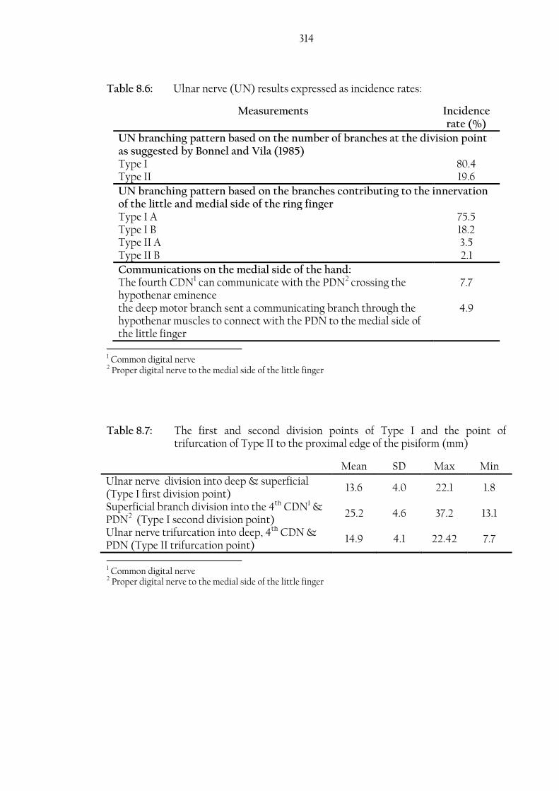
314
Table 8.6: Ulnar nerve (UN) results expressed as incidence rates:
Measurements Incidence rate (%)
UN branching pattern based on the number of branches at the division point as suggested by Bonnel and Vila (1985) Type I 80.4 Type II 19.6
UN branching pattern based on the branches contributing to the innervation of the little and medial side of the ring finger Type I A 75.5 Type I B 18.2 Type II A 3.5 Type II B 2.1
Communications on the medial side of the hand: The fourth CDN1 can communicate with the PDN2 crossing the hypothenar eminence
7.7
the deep motor branch sent a communicating branch through the hypothenar muscles to connect with the PDN to the medial side of the little finger
4.9
1 Common digital nerve 2 Proper digital nerve to the medial side of the little finger
Table 8.7: The first and second division points of Type I and the point of trifurcation of Type II to the proximal edge of the pisiform (mm)
Mean SD Max Min
Ulnar nerve division into deep & superficial (Type I first division point)
13.6 4.0 22.1 1.8
Superficial branch division into the 4th CDN1 & PDN2 (Type I second division point)
25.2 4.6 37.2 13.1
Ulnar nerve trifurcation into deep, 4th CDN & PDN (Type II trifurcation point)
14.9 4.1 22.42 7.7
1 Common digital nerve 2 Proper digital nerve to the medial side of the little finger

315 Table 8.8: Palmar communicating branch (CB) between median nerve and the
ulnar nerve results expressed as incidence rates:
Measurements Incidence rate (%)
Palmar CB branching pattern based on the proximal and distal attachment points Type I A 61.2 Type I B 21.4 Type II A 4.1 Type II B 1.0 Type III A 2.0 Type III B 3.1 Type IV 4.1 Type V 3.1
Table 8.9: Distances measured for the proximal and distal attachments of the palmar communicating branch (CB) to different anatomical landmarks (mm)
Distance between: Mean SD Max Min
Proximal attachment of CB to TCL1 5.3 5.2 19.4 -10.6
Distal attachment of CB to TCL 16.4 6.4 39.5 1.3
Proximal attachment of CB to WC2 32.3 5.8 46.6 20.5
Distal attachment of CB to WC 43.9 6.6 60.6 27.8
Proximal attachment of CB to BSL3 43.6 7.1 64.5 30.8
Distal attachment of CB to BSL 52.6 7.4 69.2 38.1
1 Transverse carpal ligament 2 Wrist crease 3 Bistyloid line
Table 8.10: Distances measured for the proximal and distal attachments of the palmar communicating branch (CB) to the distance between the third metacarpophalangeal (MCP) joint and the wrist crease (WC) or the bistyloid line (BSL) (%)
Distance between: Mean SD Max Min
Proximal attachment of CB to the distance between the third MCP joint and the WC
40.7 7.1 60.3 25.9
Distal attachment of CB to the distance between the third MCP joint and the WC
55.0 8.5 79.3 36.6
Proximal attachment of CB to the distance between the third MCP joint and the middle of the BSL
49.1 5.9 60.5 34.5
Distal attachment of CB to the distance between the third MCP joint and the middle of the BSL
59.1 6.3 75.4 41.8
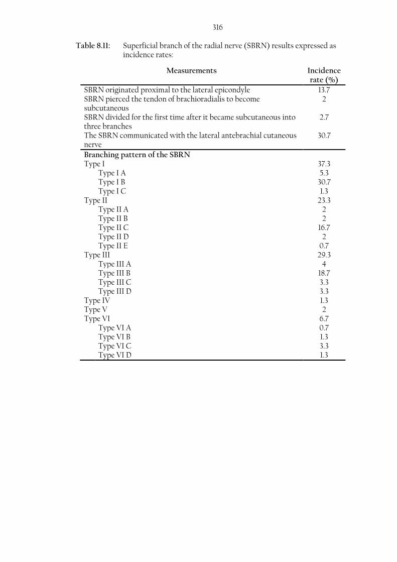
316 Table 8.11: Superficial branch of the radial nerve (SBRN) results expressed as
incidence rates:
Measurements Incidence rate (%)
SBRN originated proximal to the lateral epicondyle 13.7 SBRN pierced the tendon of brachioradialis to become subcutaneous
2
SBRN divided for the first time after it became subcutaneous into three branches
2.7
The SBRN communicated with the lateral antebrachial cutaneous nerve
30.7
Branching pattern of the SBRN Type I 37.3 Type I A 5.3 Type I B 30.7 Type I C 1.3 Type II 23.3 Type II A 2 Type II B 2 Type II C 16.7 Type II D 2 Type II E 0.7 Type III 29.3 Type III A 4 Type III B 18.7 Type III C 3.3 Type III D 3.3 Type IV 1.3 Type V 2 Type VI 6.7 Type VI A 0.7 Type VI B 1.3 Type VI C 3.3 Type VI D 1.3

317 Table 8.12: Measurements of the closest branches of the superficial branch of the
radial nerve (SBRN) to various anatomical points (mm)
Mean SD Max Min
Closest palmar branch at the level of the RSP1 8.5 4.1 17.8 0.7
Closest dorsal branch at the level of the RSP 4.9 3.0 14.7 1.1 Closest palmar branch to a point 25 mm proximal to the RSP
5.6 3.4 15.1 0
Closest dorsal branch to a point 25 mm proximal to the RSP
4.1 2.6 10.9 0
Closest branch to Lister’s tubercle 14.1 3.8 23.1 4.6 First intersection between the SBRN and the cephalic vein
47.9 19.3 93.6 6.7
SBRN diameter prior to its first division 3.2 0.8 5.8 1.94
1 Radial styloid process
Table 8.13: The major division points of the superficial branch of the radial nerve to the radial styloid process (mm)
Mean SD Max Min
First division point into palmar and dorsal branches
-51.4 14.9 -14.5 -85.0
Palmar branch: Second major division point -4.9 21.7 48.5 -54.0
Dorsal branch: Second major division point -4.6 14.4 26.8 -48.2
Palmar branch: Third major division point 22.2 24.0 73.2 -30.7
Dorsal branch: Third major division point 22.3 13.1 62.2 -31.6
Table 8.14: Anatomical measurements recorded for the dorsal branch of the ulnar nerve (mm)
Mean SD Max Min
Origin from the ulnar nerve to the proximal edge of the pisiform
63.3 14.8 110 26.7
Origin from the ulnar nerve to the USP1 61.3 21.0 140 18.5
Pierced the fascia 18.1 7.1 33.1 1.74
Closest branch to USP 6.8 3.1 14.0 0.4
First major branching point -3.42 17.4 53.4 -38.7
Second major branching point 39.2 13.2 73.6 -8.8
1 Ulnar styloid process 2 Minus signs indicate that the point were located below the level of the respective landmark
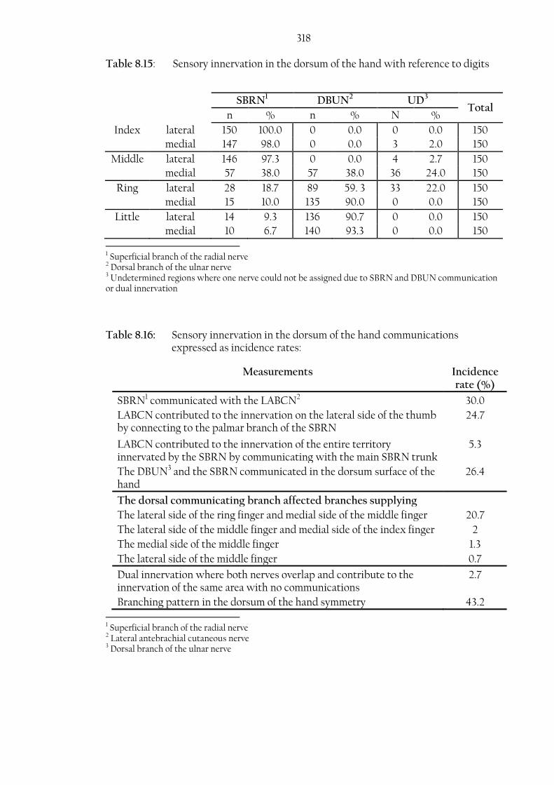
318 Table 8.15: Sensory innervation in the dorsum of the hand with reference to digits
SBRN1 DBUN2 UD3
Total n % n % N %
Index lateral 150 100.0 0 0.0 0 0.0 150
medial 147 98.0 0 0.0 3 2.0 150
Middle lateral 146 97.3 0 0.0 4 2.7 150
medial 57 38.0 57 38.0 36 24.0 150
Ring lateral 28 18.7 89 59. 3 33 22.0 150
medial 15 10.0 135 90.0 0 0.0 150
Little lateral 14 9.3 136 90.7 0 0.0 150
medial 10 6.7 140 93.3 0 0.0 150
1 Superficial branch of the radial nerve 2 Dorsal branch of the ulnar nerve 3 Undetermined regions where one nerve could not be assigned due to SBRN and DBUN communication or dual innervation
Table 8.16: Sensory innervation in the dorsum of the hand communications expressed as incidence rates:
Measurements Incidence rate (%)
SBRN1 communicated with the LABCN2 30.0
LABCN contributed to the innervation on the lateral side of the thumb by connecting to the palmar branch of the SBRN
24.7
LABCN contributed to the innervation of the entire territory innervated by the SBRN by communicating with the main SBRN trunk
5.3
The DBUN3 and the SBRN communicated in the dorsum surface of the hand
26.4
The dorsal communicating branch affected branches supplying
The lateral side of the ring finger and medial side of the middle finger 20.7
The lateral side of the middle finger and medial side of the index finger 2
The medial side of the middle finger 1.3
The lateral side of the middle finger 0.7
Dual innervation where both nerves overlap and contribute to the innervation of the same area with no communications
2.7
Branching pattern in the dorsum of the hand symmetry 43.2 1 Superficial branch of the radial nerve 2 Lateral antebrachial cutaneous nerve 3 Dorsal branch of the ulnar nerve
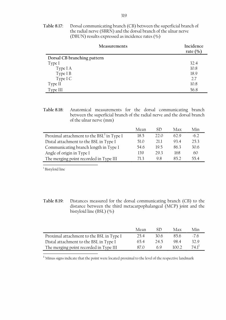
319 Table 8.17: Dorsal communicating branch (CB) between the superficial branch of
the radial nerve (SBRN) and the dorsal branch of the ulnar nerve (DBUN) results expressed as incidence rates (%)
Measurements Incidence rate (%)
Dorsal CB branching pattern Type I 32.4 Type I A 10.8 Type I B 18.9 Type I C 2.7 Type II 10.8
Type III 56.8
Table 8.18: Anatomical measurements for the dorsal communicating branch between the superficial branch of the radial nerve and the dorsal branch of the ulnar nerve (mm)
Mean SD Max Min
Proximal attachment to the BSL1 in Type I 18.5 22.0 62.9 -6.2
Distal attachment to the BSL in Type I 51.0 21.1 93.4 25.3
Communicating branch length in Type I 54.6 19.5 86.3 30.6
Angle of origin in Type I 139 29.3 168 60
The merging point recorded in Type III 71.3 9.8 85.2 55.4
1 Bistyloid line
Table 8.19: Distances measured for the dorsal communicating branch (CB) to the distance between the third metacarpophalangeal (MCP) joint and the bistyloid line (BSL) (%)
Mean SD Max Min
Proximal attachment to the BSL in Type I 25.4 30.6 85.6 -7.6
Distal attachment to the BSL in Type I 65.4 24.5 98.4 32.9
The merging point recorded in Type III 87.0 6.9 100.2 74.11
1 Minus signs indicate that the point were located proximal to the level of the respective landmark
Copyright © 2022 FDOKUMEN