Segmental innervation of the chick forelimb ... - Development

Spatial Shaping of Cochlear Innervation by Temporally RegulatedNeurotrophin Expression
Isabel Farinas,1,2 Kevin R. Jones,3 Lino Tessarollo,5 Allison J. Vigers,3 Eric Huang,1 Martina Kirstein,2Dominique C. de Caprona,4 Vincenzo Coppola,5 Carey Backus,1 Louis F. Reichardt,1 and Bernd Fritzsch4
1Program in Neuroscience, Department of Physiology and Howard Hughes Medical Institute, University of California, SanFrancisco, California 94143-0724, 2Departamento de Biologıa Celular, Universidad de Valencia, 46100 Burjassot, Spain,3Department of Biology, University of Colorado, Boulder, Colorado 80309, 4Department of Biomedical Sciences,Creighton University, Omaha, Nebraska 68178, and 5Neural Development Group, Mouse Cancer Genetics Program,National Cancer Institute, Frederick, Maryland 21701
Previous work suggested qualitatively different effects of neu-rotrophin 3 (NT-3) in cochlear innervation patterning in differentnull mutants. We now show that all NT-3 null mutants have asimilar phenotype and lose all neurons in the basal turn of thecochlea. To understand these longitudinal deficits in neurotro-phin mutants, we have compared the development of the deficitin the NT-3 mutant to the spatial–temporal expression patternsof brain-derived neurotrophic factor (BDNF) and NT-3, usinglacZ reporters in each gene and with expression of the specificneurotrophin receptors, trkB and trkC. In the NT-3 mutant,almost normal numbers of spiral ganglion neurons form, butfiber outgrowth to the basal turn is eliminated by embryonic day(E) 13.5. Most neurons are lost between E13.5 and E15.5.During the period preceding apoptosis, NT-3 is expressed insupporting cells, whereas BDNF is expressed mainly in haircells, which become postmitotic in an apical to basal temporal
gradient. During the period of neuronal loss, BDNF is absentfrom the basal cochlea, accounting for the complete loss ofbasal turn neurons in the NT-3 mutant. The spatial gradients ofneuronal loss in these two mutants appear attributable to spa-tial–temporal gradients of neurotrophin expression. Our immu-nocytochemical data show equal expression of their receptors,TrkB and TrkC, in spiral sensory neurons and thus do not relateto the basal turn loss. Mice in which NT-3 was replaced byBDNF show a qualitative normal pattern of innervation at E13.5.This suggests that the pattern of expression of neurotrophinsrather than their receptors is essential for the spatial loss ofspiral sensory neurons in NT-3 null mutants.
Key words: neurotrophins; ear innervation; development ofear innervation; ear and neurotrophin expression; NT-3; sensoryneuron survival
The inner ear develops from the otic placode that invaginates toform the otocyst by embryonic day (E) 9.5 (Fritzsch et al., 1998a).Between E9.5 and E15.5, the otocyst produces the cochlear (spi-ral) and vestibular sensory neurons (Ruben, 1967) that will in-nervate the future cochlea, saccule, utricle, and semicircularcanals (Sobkowicz, 1992; Fritzsch and Nichols, 1993; Karis et al.2001).
These otic (cochleo–vestibular) sensory neurons require brain-derived neurotrophic factor (BDNF) and neurotrophin 3 (NT-3)for their survival (for review, see Fritzsch et al., 1999). In situstudies have shown that inner ear sensory epithelia express both
neurotrophins, whereas otic sensory neurons express their recep-tors, trkB and trkC (Pirvola et al., 1992, 1994; Schecterson andBothwell, 1994; Wheeler et al., 1994). NT-3 null mutants lose�84% of all cochlear neurons at birth (Farinas et al., 1994;Ernfors et al., 1995; Tessarollo et al., 1997). In contrast, BDNFand trkB mutant mice lose mainly vestibular neurons (Fritzsch etal., 1995; Bianchi et al., 1996). BDNF/NT-3 or trkB/trkC doublehomozygous mutants lose all cochlear and vestibular neuronsaround birth (Ernfors et al., 1995; Minichiello et al., 1995; Schim-mang et al., 1997; Silos-Santiago et al., 1997).
Initial analyses of the cochlea reported that the phenotypes ofBDNF and trkB mutants are restricted to type II spiral neuronsthat innervate outer hair cells (OHC), suggesting an effect in theradial direction (Ernfors et al., 1995; Schimmang et al., 1995).Other examination of BDNF and trkB homozygous mutants re-vealed the presence of OHC afferents to the basal turn of thecochlea, suggestive of a longitudinal effect (Bianchi et al., 1996;Fritzsch et al., 1997a). Initial observations of NT-3 and trkCmutants reported a complete loss of type I spiral sensory neuroninnervation of inner hair cell (IHC) (Ernfors et al., 1995; Schim-mang et al., 1995). Again, others suggested that NT-3 and trkCmutants lose spiral neurons predominantly in the basal turn(Fritzsch et al., 1997a,b, 1998b). Longitudinal differences in in-nervation density are known in adult mammals (Ryugo, 1992),and the neurotrophins could provide the molecular basis for thesedifferences.
Received March 19, 2001; revised May 21, 2001; accepted June 1, 2001.This work was supported in part by grants from the National Institute on Deafness
and Other Communication Disorders (P 50 DC 00215), the National Organizationof Hearing Research, and the Leda Sears Trust to B.F. and D.C.D.; Ministerio deEducacion y Ciencia–Comision Interministerial de Ciencia y Tecnologıa, Spain(SAF99-0119-C0) and Fundacio la Marato de TV3 to I.F.; National Institutes ofHealth Grant KO1 NS01872 to A.J.V.; National Institute of Mental Health Grant48200 to L.F.R.; and a University of Colorado Junior Faculty Development Award,a Muscular Dystrophy Association grant, and a Burroughs-Wellcome New Investi-gator in Pharmacology Award to K.R.J. B.F. thanks C. Miller for excellent assistancewith the TEM and SEM preparations and for her dark room work and M. Chris-tensen for her help with immunocytochemistry. L.F.R. is an Investigator of theHoward Hughes Medical Institute.
I.F., K.R.J., and L.T. contributed equally to this paper.Correspondence should be addresssed to Dr. B. Fritzsch, Department of
Biomedical Sciences, Creighton University, Omaha, N E 68178. E-mail:[email protected] © 2001 Society for Neuroscience 0270-6474/01/216170-11$15.00/0
The Journal of Neuroscience, August 15, 2001, 21(16):6170–6180

Unfortunately, available in situ data on neurotrophins and theirreceptors cannot explain the selective dependency of, for exam-ple, basal turn spiral neurons on NT-3 or trkC (Pirvola et al., 1994;Schecterson and Bothwell, 1994). At birth a longitudinal gradientof NT-3 (Pirvola et al., 1992) or of both BDNF and NT-3 (Wheel-er et al., 1994) was reported, with the highest expression in theapex. This expression pattern does not correlate with the basalturn loss of sensory neurons in NT-3 mutants.
Now, we characterize the embryonic deficits in cochlear neu-rons and their projections using two independently generatedNT-3 null mutants. We also present the developmental expressionpatterns of NT-3 and BDNF using lacZ inserted into the NT-3 andBDNF loci as reporters. These results indicate that a temporaldevelopmental gradient of BDNF expression results in a spatialgradient of sensory neuron loss in mice lacking NT-3.
Parts of this paper have been published previously (Fritzsch etal., 1997d, 2000).
MATERIALS AND METHODSAnimal breeding and genotyping. NT-3lacZneo and BDNFlacZneo mice weregenerated and bred as previously described (Farinas et al., 1994; Bennettet al., 1999). NT-3lacZneo mice were genotyped as previously described(Farinas et al., 1994). For BDNFlacZneo mice, tail biopsies were genotypedby PCR analysis. One PCR primer (MBDSA10, GTGGAGTTCT-GCTAATGAGA) was located upstream of the BDNF coding exon, andthe other primer (lacZN5, GTGCTGCAAGGCGATTAAGT) was lo-cated in the lacZ gene. PCR was performed in 10 mM Tris-HCl, pH 9.0at 25°C, 50 mM KCl, 1.5 mM MgCl2, 0.1% Triton X-100 with 50 �MdNTPs and 0.3 �M primers. PCR conditions were: 3 min at 94°C, then 35cycles with 30 sec denaturation at 94°C, 30 sec annealing at 58°C, and 1.5min elongation at 72°C.
We also replaced the mouse NT-3 (also known as Ntf3) coding se-quence with BDNF (Coppola et al., 2001). We analyzed the pattern ofinnervation of mice of one litter of these transgenic mice at E13.5 (twoNT-3tgBDNF homozygotics, two NT-3tgBDNF heterozygotes, and one wildtype) (see Fig. 2 D). A more detailed account of the effect of NT-3tgBDNF
on the pattern of innervation of the cochlea in newborn mice is providedby Coppola, et al. (2001).
�-galactosidase histochemical staining. For the expression analysis, weused two to five heterozygous NT-3lacZneo and BDNFlacZneo animals perdevelopmental stage from E9.5 to birth and two adult NT-3lacZneo het-erozygous mice. In parallel, we examined one or two mutants per stage,and none of them showed any differences in �-galactosidase activity anddistribution when compared with heterozygous littermates (data notshown). Pregnant mice were killed by cervical dislocation, and theembryos were removed rapidly and placed in ice-cold PBS. Embryoswere fixed with 4% paraformaldehyde (PFA) in 0.1 M phosphate buffer,pH 7.4, for 2 hr. Whole embryos (E9.5–E12.5) or hemisected heads, withthe brain removed to expose the ear (E13.5 to adult), were reacted aspreviously described (Fritzsch et al., 1997c) either at 36°C for 2–4 hr orat room temperature overnight. Reacted ears were stored in 4% PFAuntil dissection. After dissection, the ears were whole-mounted usingappropriately sized spacers to avoid distortion by the coverslip. Two earsper stage were viewed in a compound Olympus microscope using a redfilter to enhance the blue reaction product. Images were captured with aDage CCD camera (640 � 480 pixels resolution) and processed usingImagePro software (Media Cybernetics, Silver Spring, MD). At least twoears per stage were embedded in epoxy resin, sectioned at 1–2 �mthickness, and viewed with differential interference contrast and a redfilter to enhance the blue reaction product. For the sake of simplicity,sites of accumulation of the blue reaction product resulting from theaction of �-galactosidase on X-gal will be referred to here as sites ofBDNF or NT-3 expression.
Cell counts. Embryos at different stages were fixed in Carnoy’s solution,embedded in paraffin, serially sectioned at 7 �m in the sagittal plane, andNissl-stained using cresyl violet as described elsewhere (Farinas et al.,1996). Ganglion cells and numbers of pyknotic profiles were countedevery fourth section in wild-type embryos and mutant littermates. Totalnumbers were compared using a one-tailed Student’s t test for statisticalsignificance. Apoptotic profiles in the developing vestibular and cochlear
ganglia were also studied in thick resin sections through ears of controland mutant littermates.
DiI tracing and whole-mount immunocytochemistry. We studied at leastthree ears per stage [E12.5, E13.5, E14.5, E15.5, E18.5, and postnatal day(P) 0] using DiI diffusion in perfusion fixed animals. Briefly, embryoswere cold anesthetized and perfused through the heart with 4% para-formaldehyde in 0.1 M phosphate buffer, pH 7.4. Filter strips soaked withthe lipophylic tracer DiI (Molecular Probes, Eugene, OR) were insertedinto the brainstem to selectively label the afferents to the ear (Fritzschand Nichols, 1993). After an appropriate diffusion time (2–4 d at 36°C),the ears were dissected, whole-mounted, and viewed in a compoundmicroscope using epifluorescence illumination and a Texas Red filter set.Subsequently, the ears were defatted, incubated with an antibody againstacetylated tubulin (Sigma, St. Louis, MO) for 3 d, followed by a second-ary antibody tagged with peroxidase, and reacted with diaminobenzidine.To compare the distribution of fibers to that of cells expressing NT-3,some ears that had been previously reacted to detect �-galactosidasewere also immunostained with acetylated tubulin as described (Fritzschet al., 1997c).
Immunocytochemistry. Antibodies to acetylated tubulin (see above)and antibodies to TrkB (also known as Ntrk2) and TrkC (also known asNtrk3) were used in whole mounts and paraffin sections of Carnoy-fixedmaterial as previously described (Fritzsch et al., 1997c; Farinas et al.,1998). The specificity of each Trk antibody has been determined usingWestern blot analysis and correlation of immunohistochemistry with insitu hybridization (Huang et al., 1999).
RESULTSExpression of NT-3 and BDNF in newborn mice doesnot correlate with the pattern of fiber loss seen in thecochlea of the corresponding mutantsWe have taken advantage of the insertion of a lacZ reporter geneinto the BDNF and NT-3 loci to monitor the endogenous expres-sion of these neurotrophins by assessing the �-galactosidase stain-ing pattern. In newborn mice heterozygous and homozygous forthe NT-3lacZ or BDNFlacZ alleles, we used a combination of�-galactosidase staining and acetylated tubulin whole-mount im-munocytochemistry to examine simultaneously the NT-3 andBDNF expression domains and the pattern of sensory neuronfiber distribution. Notable innervation differences, but identicalpatterns of neurotrophin expression, between heterozygous andhomozygous mutant littermates could be observed in neonates(Fig. 1).
Similar expression pattern of the lacZ gene in the cochlea wasobserved in heterozygous and homozygous NT-3 mutants at birth(Fig. 1A–C). However, although cochleas from NT-3 heterozy-gous mice were heavily innervated by radial cochlear fibers (Fig.1B), there was a conspicuous absence of radial fibers to the baseof the cochlea in the NT-3 mutants (Fig. 1A,C). The few fibersthat were seen spiraling next to IHC toward the tip of the basalturn in NT-3 deficient cochleas were basal extensions of theremaining middle turn fibers (Fig. 1C). OHC in the basal turn ofthe mutants were not approached by any fibers except for the fewOHCs immediately adjacent to the middle turn region (Fig. 1C).Despite the regional fiber loss observed at birth in the absence ofNT-3, the intensity and distribution of the blue precipitate ap-peared uniform throughout the extent of the cochlear sensoryepithelium.
In the absence of BDNF, the cochlea showed more subtledifferences in the degree of innervation between heterozygotesand homozygotes (Fig. 1D,E). Comparison between littermateswith one or two mutant BDNF alleles showed a reduction in thedensity of radial fibers throughout the cochlea in the homozygouscondition with the least pronounced difference in the base and themost obvious in the apex. In fact, portions of the apex were almostdevoid of radial fibers (Fig. 1E) (Bianchi et al., 1996). The
Farinas • Developmental Loss of Cochlear Neurons J. Neurosci., August 15, 2001, 21(16):6170–6180 6171

expression of endogenous BDNF, as revealed by the reportergene, showed no apparent regional differences throughout thecochlea that could readily explain the spatial effect of the spatiallyrestricted fiber loss in the BDNF-deficient animals.
Other NT-3 null mutants have a similar phenotypeReports describing different phenotypes in the cochlea of NT-3-deficient animals have used mouse strains carrying mutationsindependently generated in different genetic backgrounds [com-pare Ernfors et al. (1995), Fritzsch et al. (1997c)]. To determinewhether genetic background is responsible for the different find-ings, we examined the cochleas of an independently generatedNT-3 mutant (Ernfors et al., 1995). As illustrated in Figure 2E,F,the phenotypes, as shown by DiI tracing, were essentially thesame in both mutant strains at birth. Similar to our previous data(Fritzsch et al., 1997c) (Fig. 1), we observed a complete loss ofneurons in the basal turn, a complete absence of radial fibers tothe sensory epithelium in this turn, and tangential extension offibers from the middle turn along the basal turn IHC. The thirdavailable NT-3 null mutation (Liebl et al., 1997; Tessarollo et al.,1997) shows a similar loss of basal turn sensory neurons (Coppolaet al., 2001). Thus, all three NT-3 null mutants have a uniformphenotype.
The topology of the projection from the cochlea to the brain-stem was also analyzed by applying DiI to both the apical andbasal turns. After diffusion of the dye, the implantation sites were
verified in whole-mounted cochlea. Brains of animals with com-parable injection sites and comparable spread of DiI along thecochlea were chosen for analysis. We assume that these animalshad both topologically and quantitatively comparable injections.In coronal sections of wild-type animals, two distinct bands ofprojection fibers ended in the ventral cochlear nucleus (Fig. 2G).The more ventrolateral projection is from the middle turn, themore dorsomedial projection comes from the basal turn. In theNT-3 mutants, there were also two visible bundles from the middleand basal turn of the cochlea, respectively, but the segregation wasless distinct (Fig. 2H). Both the wild-type and the NT-3 mutantshow efferent fiber bundles, suggesting that sections were taken atcomparable levels (Fig. 2G,H).
Cochlear afferent fibers to the basal turn are lost atE13.5 in NT-3 mutantsIn control animals, it is first possible to visualize cochlear neuronsand their projections to the developing cochlear duct by DiIlabeling at E12.5 (Fig. 2A). In both control (Fig. 2A) and NT-3mutant embryos (Fig. 2B), fibers extend from the sensory neuronsto the developing cochlea at this stage. In the mutant, however,fewer fibers were labeled. In contrast to control animals, only oneof four mutant embryos had any fibers projecting to the basalturn. In all mutants, at least some of the radial afferents from themiddle turn extended tangentially toward the noninnervated
Figure 1. Distributions of nerve fibers in the cochlea ofNT-3 and BDNF heterozygous and homozygous mutantmice at birth. Whole-mounted cochleas from a newbornNT-3 homozygous mutant (A, C) and an NT-3 heterozygouslittermate (B), a BDNF heterozygous mutant ( D), and aBDNF homozygous mutant (E) showing the distribution ofNT-3 (A–C) and of BDNF (D, E) as revealed by�-galactosidase histochemistry and the distribution of nervefibers as revealed by acetylated tubulin immunocytochem-istry. Notice that the cochlea is labeled uniformly through-out its extent by the blue histochemical product. In NT-3null mice, no radial fibers are present in the basal turn (A,C). The only innervation present in this turn is supplied bythe few fibers diverted from the middle turn that extendalong the longitudinal axis of the cochlea next to inner haircells (C, crossed arrows). The only outer hair cells in thebasal turn approached by afferent fibers were those adjacentto the middle turn (arrow). This contrasts with the presenceof numerous radial fibers innervating inner and outer haircells in the basal turn of an NT-3 heterozygous littermate(B, arrows). The density of radial fiber, as revealed by anincrease in the spacing between radial fiber bundles, isreduced in the absence of BDNF (E) when compared withthe heterozygous condition (D). This reduction in fiberdensity is stronger in the apex of BDNF null mutants. Thus,the homogeneous distribution of NT-3 and BDNF at birthdoes not explain the selective loss of basal turn spiral neuronsin NT-3 null mutants or the reduced density of radial fibers inthe apex of BDNF null mutants. ggl, Ganglion; RF, radialfiber bundles. Scale bars, 0.1 mm.
6172 J. Neurosci., August 15, 2001, 21(16):6170–6180 Farinas • Developmental Loss of Cochlear Neurons

basal turn (Fig. 2B). A similar tangential projection was neverseen in control embryos.
In control embryos at E13.5, there was a prominent radialprojection from basal turn cochlear ganglion neurons to the basalturn sensory epithelium (Fig. 2D, inset). In contrast, radial fiberswere completely absent in the basal turn of NT-3 mutants (Fig.2C), and many fewer labeled cochlear neurons and fibers werepresent in the rest of the cochlea. Although not a single cochlearneuron fiber deviated from its normal radial trajectory in thecontrol animals, there was a prominent deviating projection ofmiddle turn cochlear neuron fibers in the mutants directed towardthe basal turn, spiraling along the cochlea next to inner hair cells(Fig. 2C).
In control embryos and NT-3 mutant embryos at E14.5 andlater embryonic stages, the distributions of sensory neurons andtheir projections within the cochlea did not differ qualitativelyfrom the patterns observed at birth (Fritzsch et al., 1997c) (Fig.1). In control embryos, a continuous and dense array of neuronsin all turns of the cochlea extended radial fibers to the sensoryepithelium. The density of radial bundles in NT-3 mutants, how-ever, was reduced throughout the developing cochlea except forthe very apical turn. Here, densely packed radial fibers were seenthat appeared qualitatively similar to those in control animalsregarding their spacing interval (Fig. 1B,C). In NT-3 mutants,however, essentially no neurons survived in the basal turn and,subsequently, no radially oriented fibers were found in this por-tion of the cochlea (Fig. 1). In the noninnervated basal turn, fibersfrom the middle turn portion were seen extending tangentiallyalong the line of IHC toward the tip of the basal turn (Fig. 1C).
Cochlear afferent fibers to the basal turn are rescuedat E13.5 in NT-3 tgBDNF miceWe also compared the pattern of innervation of NT-3 mutantsat E13.5 with that of same-aged transgenic animals in whichthe mouse NT-3 coding sequence was replaced with BDNF(Fig. 2C, D). At this stage, the basal turn innervation inNT-3 tgBDNF animals compared qualitatively well to wild-typelittermates (Fig. 2 D, inset). Specifically, there was a strikingdifference in the pattern of innervation of the basal turn of thecochlea compared with NT-3 mutants (Fig. 2C, D) suggesting anear complete rescue of the NT-3 phenotype in this transgenicmouse line. This rescue of basal turn afferent projection per-sisted in newborn transgenic mice, and a full account of thisphenotype is given by Coppola et al. (2001).
Most cochlear neurons die by E15.5 in the absenceof NT-3The numbers of neurons present in the cochlear ganglion atdifferent stages of embryonic development have been quantitated,and the data are presented in Table 1. At E11.5, the vestibular andcochlear ganglia are not yet separate structures, so the numbers ofneurons in the combined (otic) ganglion are presented. As shownin Table 1, there are only small, statistically insignificant reduc-tions in the numbers of otic neurons at E11.5 and E13.5 in NT-3mutants. Between E13.5 and E15.5, however, 73% of the neuro-nal complement of cochlear neurons are lost in the NT-3 mutants.The neuronal loss is not homogeneously distributed. At E15.5, allspiral neurons in the basal turn have completely disappeared,whereas a number of neurons are still found in middle and apical
Figure 2. Development of cochlear afferent innervation at E12.5 (A, B), E13.5 (C, D, inset), and P0 (E, F ), and of central projections at P0 (G, H ).Arrows in A show orientation for A–F; arrows in H show orientation for the coronal sections of G and H. Outgrowth of afferents (A, B) is labeled withDiI in E12.5 wild-type (A) and NT-3 mutant (B) littermates. Note projection to the basal turn in both cases. At E13.5, the NT-3 mutant (C) lacks radialafferents in the base of the cochlea. In contrast, NT-3tgBDNF mutants show a pattern of innervation reminiscent of same age wild-type animals (inset) withan overall reduced density of innervation but a clear innervation of the basal turn (D). Newborn NT-3 mutant mice from a different genetic strain (Ernforset al., l994) show comparable patterns of innervation (E–F). Compared with the dense radial afferent bundles in the wild types (E), notice the completeabsence of radial afferents in the basal turn of mutants (F) and exclusive innervation of inner hair cells. Cochlear afferents to the brainstem in control(G) and in NT-3 mutant (H ) littermates show a topologically restricted projection from the base and the middle turn to the more dorsal and ventralaspects of the ventral cochlear nucleus, respectively. The distinction between the two areas of projection is less pronounced in NT-3 mutants, presumablybecause the remaining sensory fibers occupy the areas normally innervated by the lost afferents. Comparable coronal section planes are indicated by thepresence of olivocochlear efferent fibers (G, H ). BT, Basal turn; MT, middle turn; OCB, olivo-cochlear bundle; PC, fibers to the posterior crista; WT,wild type. Scale bars, 100 �m.
Farinas • Developmental Loss of Cochlear Neurons J. Neurosci., August 15, 2001, 21(16):6170–6180 6173
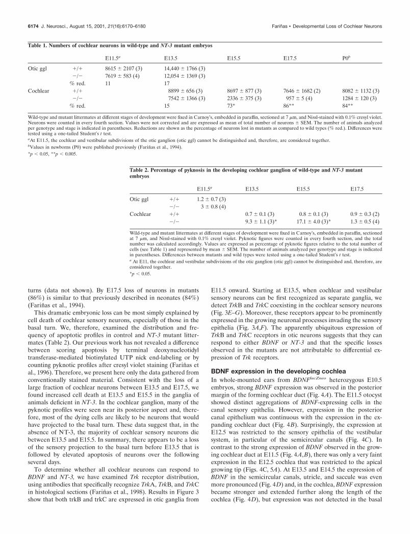
turns (data not shown). By E17.5 loss of neurons in mutants(86%) is similar to that previously described in neonates (84%)(Farinas et al., 1994).
This dramatic embryonic loss can be most simply explained bycell death of cochlear sensory neurons, especially of those in thebasal turn. We, therefore, examined the distribution and fre-quency of apoptotic profiles in control and NT-3 mutant litter-mates (Table 2). Our previous work has not revealed a differencebetween scoring apoptosis by terminal deoxynucleotidyltransferase-mediated biotinylated UTP nick end-labeling or bycounting pyknotic profiles after cresyl violet staining (Farinas etal., 1996). Therefore, we present here only the data gathered fromconventionally stained material. Consistent with the loss of alarge fraction of cochlear neurons between E13.5 and E17.5, wefound increased cell death at E13.5 and E15.5 in the ganglia ofanimals deficient in NT-3. In the cochlear ganglion, many of thepyknotic profiles were seen near its posterior aspect and, there-fore, most of the dying cells are likely to be neurons that wouldhave projected to the basal turn. These data suggest that, in theabsence of NT-3, the majority of cochlear sensory neurons diebetween E13.5 and E15.5. In summary, there appears to be a lossof the sensory projection to the basal turn before E13.5 that isfollowed by elevated apoptosis of neurons over the followingseveral days.
To determine whether all cochlear neurons can respond toBDNF and NT-3, we have examined Trk receptor distribution,using antibodies that specifically recognize TrkA, TrkB, and TrkCin histological sections (Farinas et al., 1998). Results in Figure 3show that both trkB and trkC are expressed in otic ganglia from
E11.5 onward. Starting at E13.5, when cochlear and vestibularsensory neurons can be first recognized as separate ganglia, wedetect TrkB and TrkC coexisting in the cochlear sensory neurons(Fig. 3E–G). Moreover, these receptors appear to be prominentlyexpressed in the growing neuronal processes invading the sensoryepithelia (Fig. 3A,F). The apparently ubiquitous expression ofTrkB and TrkC receptors in otic neurons suggests that they canrespond to either BDNF or NT-3 and that the specific lossesobserved in the mutants are not attributable to differential ex-pression of Trk receptors.
BDNF expression in the developing cochleaIn whole-mounted ears from BDNFlacZneo heterozygous E10.5embryos, strong BDNF expression was observed in the posteriormargin of the forming cochlear duct (Fig. 4A). The E11.5 otocystshowed distinct aggregations of BDNF-expressing cells in thecanal sensory epithelia. However, expression in the posteriorcanal epithelium was continuous with the expression in the ex-panding cochlear duct (Fig. 4B). Surprisingly, the expression atE12.5 was restricted to the sensory epithelia of the vestibularsystem, in particular of the semicircular canals (Fig. 4C). Incontrast to the strong expression of BDNF observed in the grow-ing cochlear duct at E11.5 (Fig. 4A,B), there was only a very faintexpression in the E12.5 cochlea that was restricted to the apicalgrowing tip (Figs. 4C, 5A). At E13.5 and E14.5 the expression ofBDNF in the semicircular canals, utricle, and saccule was evenmore pronounced (Fig. 4D) and, in the cochlea, BDNF expressionbecame stronger and extended further along the length of thecochlea (Fig. 4D), but expression was not detected in the basal
Table 1. Numbers of cochlear neurons in wild-type and NT-3 mutant embryos
E11.5a E13.5 E15.5 E17.5 P0b
Otic ggl �/� 8615 � 2107 (3) 14,440 � 1766 (3)�/� 7619 � 583 (4) 12,054 � 1369 (3)
% red. 11 17Cochlear �/� 8899 � 656 (3) 8697 � 877 (3) 7646 � 1682 (2) 8082 � 1132 (3)
�/� 7542 � 1366 (3) 2336 � 375 (3) 957 � 5 (4) 1284 � 120 (3)% red. 15 73* 86** 84**
Wild-type and mutant littermates at different stages of development were fixed in Carnoy’s, embedded in paraffin, sectioned at 7 �m, and Nissl-stained with 0.1% cresyl violet.Neurons were counted in every fourth section. Values were not corrected and are expressed as mean of total number of neurons � SEM. The number of animals analyzedper genotype and stage is indicated in parentheses. Reductions are shown as the percentage of neurons lost in mutants as compared to wild types (% red.). Differences weretested using a one-tailed Student’s t test.aAt E11.5, the cochlear and vestibular subdivisions of the otic ganglion (otic ggl) cannot be distinguished and, therefore, are considered together.bValues in newborns (P0) were published previously (Farinas et al., 1994).*p � 0.05, **p � 0.005.
Table 2. Percentage of pyknosis in the developing cochlear ganglion of wild-type and NT-3 mutantembryos
E11.5a E13.5 E15.5 E17.5
Otic ggl �/� 1.2 � 0.7 (3)�/� 3 � 0.8 (4)
Cochlear �/� 0.7 � 0.1 (3) 0.8 � 0.1 (3) 0.9 � 0.3 (2)�/� 9.3 � 1.1 (3)* 17.1 � 4.0 (3)* 1.3 � 0.5 (4)
Wild-type and mutant littermates at different stages of development were fixed in Carnoy’s, embedded in paraffin, sectionedat 7 �m, and Nissl-stained with 0.1% cresyl violet. Pyknotic figures were counted in every fourth section, and the totalnumber was calculated accordingly. Values are expressed as percentage of pyknotic figures relative to the total number ofcells (see Table 1) and represented by mean � SEM. The number of animals analyzed per genotype and stage is indicatedin parentheses. Differences between mutants and wild types were tested using a one-tailed Student’s t test.a At E11, the cochlear and vestibular subdivisions of the otic ganglion (otic ggl) cannot be distinguished and, therefore, areconsidered together.*p � 0.05.
6174 J. Neurosci., August 15, 2001, 21(16):6170–6180 Farinas • Developmental Loss of Cochlear Neurons

turn. At E16.5 BDNF expression was seen throughout the cochlea,similar to the distribution found at birth (Fig. 1). Therefore,expression of BDNF in the growing cochlea develops in an apical-to-basal gradient and does not reach the base until E16.5.
Sections through the otocyst showed that BDNF is initiallyexpressed in the otocyst wall at E10.5 and bears no clear relation-ship with future sensory epithelia. After the expression shifted tothe future sensory epithelia at E11.5, BDNF expression in theutricle, saccule, and canal epithelia BDNF was predominantly inwhat appeared to be differentiating hair cells (data not shown).However, BDNF expression in the cochlea was not seen in co-chlear hair cells before E15.5. In the future sensory epithelium ofthe cochlea, little BDNF expression was seen at E12.5. In fact, atthis stage, when very faint expression is seen in the growingcochlear apex, the �-galactosidase product is found in nonsensoryareas, specifically in what will become the Reissner’s membrane(Fig. 5B), which separates the endolymph containing medial scalafrom the perilymph containing vestibular scala. At E15.5 andonward, an apical-to-basal gradient of cochlear BDNF expressioncould be detected. BDNF expression was clearly seen in IHC andOHC in the basal turn by E17.5 and later (Fig. 6G,H), but eventhen, the area of the epithelium expressing this neurotrophin waslarger in the apex (Fig. 6E,F). In fact, diffuse labeling persistedin the apex even at birth in both hair cells and supporting cells(Fig. 6F).
NT-3 expression in the developing cochleaIn whole-mounted ears from NT-3lacZneo heterozygous E10.5 em-bryos, we detected only a single patch of �-galactosidase expres-sion near the anteroventral aspect. This patch may represent the
combined anlage of utricle, saccule, and cochlea (Fig. 4E–H). AtE11.5, there are two discrete domains of �-galactosidase: a moredorsal patch, corresponding to the utricle, and a more ventralpatch, corresponding to the saccular–cochlear anlage (Fig. 4F).The latter showed more intense staining in the rostroventrallyoriented tip of the growing cochlear duct. By E12.5, the segrega-tion between the utricle and the saccular–cochlear anlage wasmore distinct (Fig. 4G), and a gradient of �-galactosidase stainingalong the cochlear–saccular axis became apparent. In fact, thefuture saccule expressed lacZ only in its inferior half (Fig. 4G).By E13.5, the ear has reached its definite form (Morsli et al.,1998), and three areas of �-galactosidase expression can be dis-tinguished (Fig. 4H) corresponding (dorsal to ventral) to theutricle, the saccule, and the cochlea, except for the growing tip ofthe latter (Figs. 4H, 5C). In addition, weaker expression appearedaround the canal sensory epithelia at this stage in development.From E14.5 to birth, the pattern of lacZ staining in the earremained essentially identical to that found at E13.5, except thatthe entire cochlea became positive. Therefore, NT-3 appears tobe expressed in the cochlea as it grows in longitudinal extent.
At the cellular level, the embryonic expression of NT-3 in thecochlear sensory epithelium shows dynamic spatiotemporalchanges, including a longitudinal base-to-apex progression and achange in cell type expressing this neurotrophin (embryonicexpression by supporting cells; neonatal expression by IHC). Insections through the cochlear epithelium, the blue �-galactosidaseproduct was present throughout the developing sensory epithe-lium at E12.5 (Fig. 5C,D). From E14.5 onward, NT-3 expressionbecame restricted to supporting cells of the cochlear epithelium,
Figure 3. Developmental expression of trkB (A, C, E, F )and trkC (B, D, E, G) in developing otic sensory neurons atE11.5 (A, B), E13.5 ( C–E), and E15.5 (F, G). The distribu-tion of the trk proteins was revealed using specific primaryantibodies (Farinas et al., 1998) and peroxidase (A, B, F, G),fluorescein (C, E), or rhodamine (D, E) -conjugated sec-ondary antibodies. trkB and trkC are expressed in all oticneurons at early stages (A, B) and in all cochlear neuronsuntil at least E15.5 (F, G), respectively. Fluorescent doublelabeling at E13.5 for both trkB and trkC indicates that allcochlear neurons express both trk proteins (C–E). Scale bar(shown in E): A, B, F, G, 100 �m; C–E, 25 �m.
Farinas • Developmental Loss of Cochlear Neurons J. Neurosci., August 15, 2001, 21(16):6170–6180 6175

such as Deiter’s cells, Pillar cells, and Border cells with limited, ifany, expression in sensory hair cells during embryonic develop-ment. At birth, very faint NT-3 expression could be found in IHCof the basal, but not the apical, turn (Fig. 6A–D). In the middleand apical turns, expression was strongest in Deiter’s cells andtheir phalangeal processes, followed by nearby Pillar cells (Fig.6B). There was also a shift in expression from outer to inner Pillarcells following an apical–basal gradient (Fig. 6B,D). The greaterepithelial ridge (GER) is an embryonic structure that disappearsin neonatal development, giving rise to the inner cochlear sulcusand the inner hair cells. Many of these cells expressed NT-3 asassessed using the LacZ reporter. In fact, around birth some weremore strongly labeled than the IHC (Fig. 6D). Thus, NT-3 ex-pression was detected in supporting cells, but not in hair cellsduring embryonic development.
BDNF and NT-3 expression outside thesensory epitheliaIn contrast to some previous in situ hybridization studies (Pirvolaet al., 1992, 1994), but in agreement with others (Schecterson andBothwell, 1994), our data suggested that some cells delaminatingfrom regions of the various sensory epithelia express either BDNF
or NT-3. Specifically, cells delaminating from the utricle, saccule,and the basal turn of the cochlea express NT-3, whereas cellsdelaminating from the apical turn of the cochlea, the utricle, andthe canal epithelia express BDNF (Fig. 4). In sections, thesedelaminating cells appeared to derive from discrete areas of thesensory epithelia that typically showed no other lacZ-positivecells. This suggests that BDNF and NT-3 expression are inducedat the time of delamination. Expression of �-galactosidase doesnot colocalize with the presence of differentiated neurons, asidentified using TrkB and TrkC immunocytochemistry (Fig. 3).These delaminated cells migrate away from the otocyst and arelikely to constitute the precursor cells for otic sensory neurons asthey aggregate near and occupy a region next to the differentiatedotic sensory neurons. The neurotrophin-expressing cells are likelyto constitute an early source of neurotrophins next to the formingotic sensory neurons.
DISCUSSIONThe lack of NT-3 or BDNF results invariably in losses of cochlearneurons with clear spatial bias. NT-3 deficient mice lose as manyas 84% of the cochlear neurons, resulting in reduced density of
Figure 4. Developmental expression of endogenous BDNF ( A–D) and NT-3 (E–H) as revealed by �-galactosidase histochemistry in whole-mountedcochleas from heterozygous animals with a lacZ reporter gene inserted into the BDNF and NT-3 locus, respectively. Arrows in E indicate orientation.BDNF expression is first detected at E10.5 ( A). Expression is strongest in the posteroventral aspect, but it also forms a crest around the anterior anddorsal aspect. The anteroventral aspect adjacent to the forming otic ganglion is notably devoid of BDNF labeling. Interestingly, this area receives thefirst growing fibers (A). By E11.5 (B), BDNF expression in the posteroventral quadrant has intensified and is related to the growing cochlear duct, thesemicircular canal sensory epithelia, and the forming cochleovestibular ganglion (Ggl ). Streaks of �-galactosidase-positive cells (*) were seen apparentlymigrating away from the otocyst and toward the forming ganglion and were even more apparent at E12.5 ( C) because of the more distant spacing of theepithelia in the growing otocyst. Interestingly, BDNF expression in the growing cochlea is sharply downregulated at E12.5, except for a faint expressionrestricted to the growing tip (C). The utricle ( U) and saccule (S) show only faint and restricted expression at this stage. At E13.5 ( D) and later, theexpression remains rather stable in all sensory epithelia, except for a progressive upregulation of BDNF that proceeds toward the base of the cochlea.In contrast to BDNF, expression of NT-3 at E10.5 (E) forms two continuous patches, the primordia of the future utricle and of the future saccule andcochlea. Faint NT-3 expression is also visible in the forming otic ganglion (Ggl ), in streaks of cells extending from discrete regions of the otocyst to theotic ganglion (*), and in the endolymphatic duct. By E11.5 (F), the �-galactosidase-positive utricle and saccule � cochlea (S � C) have segregated. ByE12.5 (G), the NT-3-positive portion comprises only a fraction of the total otic ganglion, and the saccule and cochlea are beginning to segregate. By E13.5(H ), the utricle, saccule, and cochlea are anatomically distinct from each other, and all of them are intensely positive. Note that the ganglion was removedfor clarity in H. The gap between the saccule and the cochlea turns into the ductus reuniens (DR). There is some expression of NT-3 in the sensory cristaeof the three semicircular canals (anterior, horizontal, posterior). AC, Anterior crista; C, cochlea; ED, endolymphatic duct; Ggl, otic (cochleovestibular)ganglion; HC, horizontal crista; PC, posterior crista. Scale bars: 100 �m.
6176 J. Neurosci., August 15, 2001, 21(16):6170–6180 Farinas • Developmental Loss of Cochlear Neurons

projections in the middle and apical turns and a complete loss ofradial projections to the basal turn (Fritzsch et al., 1997a). Ab-sence of BDNF results in a modest reduction of cochlear neuronsand their projections, with the largest reduction in the apical turn(Bianchi et al., 1996). We characterize for the first time thecochlear deficits during embryonic development caused by theabsence of NT-3. In addition, we compare the distribution ofthese neurotrophins and their receptors during prenatal develop-ment to determine how spatially biased deficiencies are producedby the absence of a single neurotrophin. We also compare thepattern of loss in three independently generated NT-3 null mu-tations (Ernfors et al., 1995; Liebl et al., 1997) and show strik-
ingly similar phenotypes (Fig. 2) (Coppola et al., 2001). Thus, allNT-3 null mutants share one phenotype, loss of basal turn sensoryneurons, reduction of outer hair cell innervation, and retention ofinner hair cell innervation. We cannot explain why Ernfors et al.(1995) concluded that all inner hair cell innervation and thecentral auditory projections are lost in their NT-3 null mutantbecause our data clearly show that this is not the case (Fig. 2).
The generation of cochlear neurons proceeds in a basal-to-apical direction, whereas birth of cochlear hair cells follows theopposite gradient, from apex to base (Ruben, 1967). Duringembryonic development, all cochlear neurons appear to expressboth trkB and trkC, and, therefore, can respond to either neuro-
Figure 5. Expression of BDNF (A, B) and NT-3 (C, D) inthe cochlea at E12.5. Arrows in A indicate orientation, anddashed lines in A, C indicate plane of section for B, D. Notethat BDNF expression is very faint and restricted to theapex of the cochlea (A). In contrast, NT-3 expression ismore robust, although it is absent from the very apical tip(C). Plastic sections (10 �m thick) of X-gal-reacted cochleasshow that BDNF expression in the base is restricted to anarea of the cochlear duct that will become the Reissner’smembrane (B). In contrast, NT-3 expression is seenthroughout the greater epithelial ridge (GER), an area thatwill become the organ of Corti and the inner spiral sulcus.Scale bars, 100 �m.
Figure 6. Expression of NT-3 (A–D) and BDNF (E–H) in the cochlea at birth as seen in whole mounts (A, C, E, G) and in 10-�m-thick (F, H ) or2-�m-thick (B, D) plastic sections. Note that in the apical turn (A, B, E, F ), neither NT-3 (A, B) nor BDNF expression (E, F ) is restricted to hair cells.In fact, NT-3 expression is predominantly seen in supporting cells that also express some BDNF. In contrast, in the basal turn (C, D, G, H ), BDNFexpression is restricted to hair cells (G, H ), whereas NT-3 is expressed by inner hair cells and their surrounding supporting cells. D, Deiter’s cells; GER,greater epithelial ridge; I, inner hair cells; O, outer hair cells; P, Pillar cells. Scale bar, 100 �m.
Farinas • Developmental Loss of Cochlear Neurons J. Neurosci., August 15, 2001, 21(16):6170–6180 6177

trophin (Fig. 3). However, the distributions of BDNF and NT-3 inthe cochlear epithelium show very different longitudinal andradial developmental dynamics. Despite expression in nonsensoryareas of the apex, BDNF expression appears to be largely re-stricted to hair cells (Pirvola et al., 1992, 1994; Schecterson et al.,1994; Wheeler et al., 1994) following the age gradient of hair cellproliferation. In contrast, NT-3 expression in the cochlear epithe-lium follows the maturational gradient of the cochlea, progressingfrom base to apex (Pujol et al., 1998). The earliest differentiatedcochlear neurons therefore project to a sensory epithelium with-out mature hair cells. Consequently, basal turn cochlear neuronsmust be supported initially by neurotrophins that are releasedfrom other epithelial cell types. Only NT-3, but not BDNF, ispredominantly expressed in nonhair cells (Figs. 5, 6) (Pirvola etal., 1992) and can thus support cochlear neurons in the basal turnuntil E15.5 (Fig. 7).
Our results indicate that cochlear neurons in the basal turndegenerate in the absence of NT-3 because BDNF is not ex-pressed in the basal turn epithelium at a critical time. As pre-dicted by this interpretation, transgenic expression of BDNFunder control of the NT-3 promoter qualitatively rescues thisinnervation deficit at E13.5 (Fig. 2D). A similar rescue has beenfound in newborn transgenic mice (Coppola et al., 2001). How-ever, we cannot exclude the possibility that the additional avail-ability of excess BDNF in itself is related to the observed rescue.Nevertheless, because there is some endogenous expression ofBDNF in developing hair cells of the middle and apical turns,sensory neurons innervating these regions are partially rescued bythis neurotrophin in NT-3 null mutants. BDNF expression,though, must not be high enough to compensate completelyfor the loss of NT-3. The specific loss of cochlear neurons inthe apical turn of BDNF homozygous mutants may reflect insuf-ficient expression of NT-3 at an early stage. These details have notbeen recognized in previous in situ studies that either didnot cover some of the critical stages or did not examine the basal
turn because of the section plane (Pirvola et al., 1992; Wheeleret al., 1994).
We have also detected neurotrophin expression very early incells delaminating from the otocyst (Fig. 4). Experimental datastrongly suggest that all inner ear sensory neurons derive from theotocyst (d’Amico-Martel and Noden, 1983; Fritzsch et al., 1998a).In situ hybridization in mice have suggested that cells in thisregion of the otocyst wall express FGF-3 (int-2) (Mansour et al.,1993; McKay et al., 1996), FGF10 (Pirvola et al., 2000), GATA3(Karis et al., 2001), NeuroD (Kim et al., 2001), BF1 (Hatini et al.,1999), and ngn1 (Ma et al., 2000), although double in situ labelingis needed to determine if all these molecules are expressed in thesame cells. Delaminated cells migrate rapidly away and undergofurther proliferation before aggregating to form the postmitoticotic (cochleovestibular) sensory neurons (Altman and Bayer,1982; Carney and Silver, 1983; Adam et al., 1998).
Previous in situ hybridization analyses in rats indicated thatthese cells do not express the neurotrophins BDNF and NT-3(Pirvola et al., 1992) or their receptors trkB and trkC duringmigration, but show expression of trkA (Fritzsch et al., 1999).Limited data in mice show some expression of BDNF mRNA insensory neurons (Schecterson and Bothwell, 1994). Here we showthat, although these delaminating cells do not express detectablelevels of trkB or trkC, they do express NT-3 and/or BDNF. Itseems likely that NT-3 and BDNF expression can be more readilydetected with the lacZ reporter than by in situ hybridization(Fritzsch et al., 1999; Vigers et al., 2000). BDNF and NT-3 are firstinduced in delaminating cells, thus suggesting that the expressionof NT-3 and BDNF in these migratory cells is genuine and doesnot reflect the presence of the reporter protein after the BDNF orNT-3 mRNA has disappeared.
These delaminating cells remain spatially segregated from thepostmitotic neurons that constitute the otic ganglion and disap-pear after completion of neurogenesis. Differentiated embryonicotic neurons do not express NT-3 or BDNF, indicating that
Figure 7. This scheme depicts the dynamics of BDNF( green) and NT-3 ( pink) expression during cochlear devel-opment (lef t column) and the effects of transgenes and nullmutations (right column). Note that at E12.5, BDNF is re-stricted to the apex of the cochlea, whereas NT-3 is promi-nently expressed in the base and middle turn, mainly aroundthe three rows of outer hair cells. Sensory neurons (redcircles) have become postmitotic in the basal half and extendtheir fibers. By E14.5, the expression of BDNF has expandedtoward the most basal part of the middle turn (mainly in innerhair cells), whereas NT-3 expression has expanded toward thegrowing apex (mainly in outer hair cells). At E16.5, expres-sion of both BDNF and NT-3 extends throughout the cochlealongitudinally. However, the basal turn expresses NT-3around all hair cells and in inner hair cells, whereas the apexhas no expression in or around inner hair cells. In contrast,BDNF expression is found in all hair cells as well as faintly insupporting cells in the apex, whereas it is restricted to haircells in the middle and basal turns. The arrows (Age, Matu-ration) refer to hair cells only. NT-3 null mutants (rightcolumn, middle) have no neurotrophin to support basal turnsensory neurons. Surviving middle turn sensory neurons re-arrange their axonal trajectory toward the base. This seems toreflect the progressive upregulation of BDNF in inner haircells (bottom). In contrast, the effect of BDNF null mutationbecomes apparent later and in the apex. Replacing NT-3 withBDNF restores the innervation of the basal turn (top). Wepropose that the longitudinal gradients, which mimic the agegradient of the cochlea in the case of BDNF and the maturation gradient of the cochlea in the case of NT-3, are responsible for the specific reductionof sensory neurons in the base of NT-3 null mutants and in the apex of BDNF null mutants. IHC’s, Inner hair cells; OHC’s, outer hair cells.
6178 J. Neurosci., August 15, 2001, 21(16):6170–6180 Farinas • Developmental Loss of Cochlear Neurons

neurotrophin expression is downregulated on neuronal differen-tiation. However, otic neurons express trkB and trkC (Pirvola etal., 1992), and their neurites grow toward the sensory epitheliumacross the territory of neurotrophin-expressing precursor cells.Therefore, it is possible that postmitotic neurons in the oticganglion may rely for initial support on NT-3 or BDNF providedby cells present in the pathways traversed by their axons.
Sensory neurons extend processes to their targets in the basalturn before degenerating in the absence of NT-3. The delay of �1d between initial fiber outgrowth and fiber loss in neurotrophinmutants is comparable with that observed in the semicircularcanals of trkB mutants (Fritzsch et al., 1995) and BDNF mutants(Bianchi et al., 1996). Studies in double BDNF/NT-3 or trkB/trkCmutants are needed to determine whether initial outgrowth re-quires neurotrophin support as previously suggested (Fritzsch etal., 1997a).
In summary, we propose that basal turn cochlear neurons die inthe absence of NT-3 because it is the only neurotrophin availableto support these neurons during their initial innervation of thecochlea. The opposed temporal gradients of cochlear neuron andhair cell mitosis and differentiation (Ruben, 1967; Pujol et al.,1998) transform a temporal gradient of neurotrophin expressioninto a spatial gradient of innervation loss (Fig. 7). The basal turnof mammals represents a unique extension of the high-frequencyrange absent in other vertebrates (Fay, 1992). Development ofneurons in this region requires NT-3, which is hardly expressed inthe developing avian ear (Pirvola et al., 1997). This suggests thatacquisition of novel parts of the cochlea is accompanied by anovel implementation of an existing neurotrophin.
In this paper, we have shown that the developmental dynamicsof neurotrophin expression during cochlear development canexplain the gradient of neuronal loss observed in the NT-3 mu-tant. Comparable dynamics of expression of NT-3 and otherneurotrophins are similarly likely to explain the patterns of neu-ronal loss in the trigeminal and dorsal root ganglia. In each case,absence of NT-3 has been shown to result in loss of neuronsexpressing TrkB that are not lost in the absence of TrkC andtherefore appear to be activated directly by NT-3 signalingthrough TrkB (Farinas et al., 1998; Huang et al., 1999). Neuronsexpressing TrkB are lost immediately after neurogenesis. NT-3 isexpressed in the mesenchyme immediately adjacent to theseganglia during the period of neurogenesis. As development pro-ceeds, NT-3 is observed in proliferating mesenchyme in limb budand elsewhere, but expression is lost in most of the differentiatedcells derived from this mesenchyme (Farinas et al., 1998; Pata-poutian et al., 1999). In contrast, BDNF expression is not ob-served in the mesenchyme immediately adjacent to the ganglia orin many of the regions subsequently invaded by sensory fibers (E.Huang, K. R. Jones, and L.F. Reichardt, unpublished observa-tions). Thus, although these neurons eventually innervate targetsexpressing BDNF, transient loss of trophic support results inapoptosis. In the future, it will be essential to understand themechanisms that regulate the dynamic expression of the variousneurotrophins if we are to understand the patterns of neuronalloss observed in wild-type and mutant animals (Patapoutian et al.,1999).
REFERENCESAdam J, Myat A, LeRoux I, Eddison M, Henrique D, Ish-Horowicz D,
Lewis J (1998) Cell fate choices and the expression of Notch, Deltaand Serrate homologues in the chick inner ear: parallel with Drosophilasense-organ development. Development 125:4645–4654.
Altman J, Bayer S (1982) Development of the cranial nerve ganglia andrelated nuclei in the rat. Adv Anat Embryol Cell Biol 74:1–90.
Bennett JL, Zeiler SR, Jones KR (1999) Patterned expression of BDNFand NT-3 in the retina and anterior segment of the developing mam-malian eye. Invest Ophthalmol Vis Sci 40:2996–3005.
Bianchi LM, Conover JC, Fritzsch B, De Chiara T, Lindsay RM, Yan-copoulos GD (1996) Degeneration of vestibular neurons in late em-bryogenesis of both heterozygous and homozygous BDNF null mutantmice. Development 122:1965–1973.
Carney PR, Silver J (1983) Studies on cell migration and axon guidancein the developing distal auditory system of the mouse. J Comp Neurol215:359–369.
Coppola V, Kucera J, Palko MP, Martinez-De Velasco J, Lyons WE,Fritzsch B, Tessarollo L (2001) Dissection of NT-3 functions in vivo bygene replacement strategy. Development, in press.
d’Amico-Martel A, Noden DM (1983) Contribution of placode and neu-ral crest cells to avian cranial peripheral ganglia. Am J Anat166:445–468.
Ernfors P, van de Water T, Loring J, Jaenisch R (1995) Complementaryroles of BDNF and NT-3 in vestibular and auditory development.Neuron 14:1153–1164.
Farinas I, Jones KR, Backus C, Wang X-Y, Reichardt LF (1994) Severesensory and sympathetic deficits in mice lacking neurotrophin-3. Na-ture 369:658–661.
Farinas I, Yoshida CK, Backus C, Reichardt LF (1996) Lack ofneurotrophin-3 results in death of spinal sensory neurons and prema-ture differentiation of their precursors. Neuron 17:1065–1078.
Farinas I, Wilkinson GA, Backus C, Reichardt LF, Patapoutian A (1998)Characterization of neurotrophin and Trk receptor functions in devel-oping sensory ganglia: direct NT-3 activation of TrkB neurons in vivo.Neuron 21:325–334.
Fay RR (1992) Structure and function in sound discrimination amongvertebrates. In: The evolutionary biology of hearing (Webster DB,Popper AN, Fay RR, eds), pp 229–268. New York: Springer.
Fritzsch B, Nichols DH (1993) DiI reveals a prenatal arrival of efferentsat the differentiating otocyst of mice. Hear Res 65:51–60.
Fritzsch B, Silos-Santiago I, Smeyne,R, Fagan AM, Barbacid M (1995)Reduction and loss of inner ear innervation in trkB and trkC receptorknockout mice: a whole mount DiI and scanning electron microscopicanalysis. Aud Neurosci 1:401–417.
Fritzsch B, Silos-Santiago I, Bianchi LM, Farinas I (1997a) The role ofneurotrophic factors in regulating inner ear innervation. Trends Neu-rosci 20:159–165.
Fritzsch B, Silos-Santiago I, Bianchi LM, Farinas I (1997b) Neurotro-phins, neurotrophin receptors and the maintenance of the afferentinner ear innervation. Sem Cell Dev Biol 8:277–284.
Fritzsch B, Farinas I, Reichardt LF (1997c) Lack of NT-3 causes lossesof both classes of spiral ganglion neurons in the cochlea in a region-specific fashion. J Neurosci 17:6213–6225.
Fritzsch B, Farinas I, Reichardt LF (1997d) The development of NT-3expression as revealed with a Lac-Z reporter and of innervation deficitsin NT-3 mutant mice. Soc Neurosci Abstr 23:881.
Fritzsch B, Barald K, Lomax M (1998a) Early embryology of the verte-brate ear. In: Development of the auditory system, Springer handbookof auditory research (Rubel EW, Popper AN, Fay RR), pp 80–145.New York: Springer.
Fritzsch B, Barbacid M, Silos-Santiago I (1998b) The combined effectsof trkB and trkC mutations on the innervation of the inner ear. Int JDev Neurosci 16:493–505.
Fritzsch B, Pirvola U, Ylikoski J (1999) Making and breaking the inner-vation of the ear: neurotrophic support during ear development and itsclinical implications. Cell Tissue Res 295:369–382.
Fritzsch B, Farinas I, Jones KJ, Tessarollo L, Reichardt LF (2000) Thespatio-temporal expression of BDNF and NT-3 causes topologicalrestricted spiral ganglion loss: no hair cell specificity in sight. AROAbstract 23:147–148.
Hatini V, Ye X, Balas G, Lai E (1999) Dynamics of placodal lineagedevelopment revealed by targeted transgene expression. Dev Dyn215:332–343.
Huang EJ, Wilkinson GA, Farinas I, Backus C, Zang K, Wong SL,Reichardt LF (1999) Expression of Trk receptors in the developingmouse trigeminal ganglion: in vivo evidence for NT-3 activation ofTrkA and TrkB in addition to TrkC. Development 126:2191–2203.
Karis A, Pata I, van Doorninck H, Grosveld F, de Zeeuw C, de CapronaD, Fritzsch B (2001) GATA-3 alters pathway selection of olivo-cochlear neurons and affects morphogenesis of the ear. J Comp Neurol429:615–630.
Kim W-Y, Fritzsch B, Serls A, Bakel LA, Huang EJ, Reichardt LF, BarthDS, Lee JE (2001) NeuroD-null mice are deaf due to a severe loss ofthe inner ear sensory neurons during development. Development128:417–426.
Liebl DJ, Tessarollo L, Palko ME, Parada LF (1997) Absence of sensoryneurons before target innervation in brain-derived neurotrophicfactor-, neurotrophin3-, and trkC-deficient embryonic mice. J Neurosci17:9113–9127.
Farinas • Developmental Loss of Cochlear Neurons J. Neurosci., August 15, 2001, 21(16):6170–6180 6179

Ma Q, Anderson DJ, Fritzsch B (2000) Neurogenin 1 null mutant earsdevelop fewer, morphologically normal hair cells in smaller sensoryepithelia devoid of innervation. JARO 1:129–143.
Mansour SL, Goddard JM, Capecchi MR (1993) Mice homozygous fora targeted disruption of the proto-oncogene int-2 have developmentaldefects in the tail and inner ear. Development 117:13–28.
McKay IJ, Lewis J, Lumsden A (1996) The role of FGF-3 in early innerear development: an analysis in normal and Kreisler mutant mice. DevBiol 174:370–378.
Minichiello L, Piehl F, Vazquez E, Schimmang T, Hokfelt T, Represa J,Klein R (1995) Differential effects of combined trk receptor mutationson dorsal root ganglion and inner ear. Development 121:4067–4075.
Morsli H, Choo D, Ryan A, Johnson R, Wu DK (1998) Development ofthe mouse inner ear and origin of its sensory organs. J Neurosci18:3327–3335.
Patapoutian A, Backus C, Kispert A, Reichardt LF (1999) Regulationof neurotrophin-3 expression by epithelial-mesenchymal interactions:the role of Wnt factors. Science 283:1180–1183.
Pirvola U, Ylikoski J, Palgi J, Lehtonen E, Arumae U, Saarma M (1992)Brain-derived neurotrophic factor and neurotrophin 3 mRNAs in theperipheral target fields of developing inner ear ganglia. Proc Natl AcadSci USA 89:9915–9919.
Pirvola U, Arumae U, Moshnyakov M, Palgi J, Saarma M, Ylikoski J(1994) Coordinated expression and function of neurotrophins andtheir receptors in the rat inner ear during target innervation. Hear Res75:131–144.
Pirvola U, Hallbook F, Xing-Qun L, Virkkala J, Saarma M, Ylikoski J(1997) Expression of neurotrophins and Trk receptors in the develop-ing, adult, and regenerating avian cochlea. J Neurobiol 33:1019–1033.
Pirvola U, Spencer-Dene B, Xing-Qun L, Kettunen P, Thesleff I, FritzschB, Dickson C, Ylikoski J (2000) FGF/FGFR-2(IIIb) signaling is es-sential for inner ear morphogenesis. J Neurosci 20: 6125–6134.
Pujol R, Lavigne-Rebillard M, Lenoir M (1998) Development of sensoryand neural structures in the mammalian cochlea. In: Development ofthe auditory system (Rubel EW, Popper AN, Fay RR, eds), pp 146–192.New York: Springer.
Ruben RJ (1967) Development of the inner ear of the mouse, a radio-autographic study of terminal mitosis. Acta Otolaryngol 220:1–44.
Ryugo DK (1992) The auditory nerve: peripheral innervation, cell bodymorphology, and central projections. In: The mammalian auditorypathway: neuroanatomy, Springer handbook of auditory research(Webster DB, Popper AN, Fay RR, eds), pp 23–65. New York:Springer.
Schecterson L, Bothwell M (1994) Neurotrophin and neurotrophin re-ceptor mRNA expression in developing inner ear. Hear Res 73:92–100.
Schimmang T, Minichiello L, Vazquez E, San Jose I, Giraldez F, Klein R,Represa J (1995) Developing inner ear sensory neurons require TrkBand TrkC receptors for innervation of their peripheral targets. Devel-opment 121:3381–3391.
Schimmang T, Alvarez-Bolado G, Minichiello L, Vazquez E, Giraldez F,Klein R, Represa J (1997) Survival of inner ear sensory neurons in trkmutants. Mech Dev 64:77–85.
Silos-Santiago I, Fagan AM, Garber M, Fritzsch B, Barbacid M (1997)Severe sensory deficits but normal CNS development in newborn micelacking TrkB and TrkC tyrosine protein kinase receptors. Eur J Neu-rosci 9:2045–2056.
Sobkowicz HM (1992) The development of innervation in the organ ofCorti. In: Development of auditory and vestibular systems (Romand R,ed), pp 59–100. Amsterdam: Elsevier.
Tessarollo L, Tsoulfas P, Donovan MJ, Palko ME, Blair-Flynn J, Hemp-stead BL, Parada LF (1997) Targeted deletion of all isoforms of thetrkC gene suggests the use of alternate receptors by its ligandneurotrophin-3 in neuronal development and implicates trkC in normalcardiogenesis. Proc Natl Acad Sci USA 94:14776–14781.
Vigers AJ, Baquet ZC, Jones KR (2000) Expression of neurotrophin-3in the mouse forebrain: insights from a targeted LacZ reporter. J CompNeurol 17:398–415.
Wheeler EF, Bothwell M, Schecterson LC, von Bartheld CS (1994)Expression of BDNF and NT-3 mRNA in hair cells of the organ ofCorti: quantitative analysis in developing rats. Hear Res 73:46–56.
6180 J. Neurosci., August 15, 2001, 21(16):6170–6180 Farinas • Developmental Loss of Cochlear Neurons
Copyright © 2022 FDOKUMEN