Dentate gyrus-selective colchicine lesion and performance in temporal and spatial tasks
ARTICLE
Received 25 Oct 2013 | Accepted 24 Dec 2013 | Published 12 Feb 2014
Temporally selective contextual encodingin the dentate gyrus of the hippocampusL.M. Rangel1,2,w, A.S. Alexander3, J.B. Aimone2,w, J. Wiles4, F.H. Gage1,2, A.A. Chiba1,3 & L.K. Quinn3
A recent model of the hippocampus predicts that the unique properties of the dentate gyrus
allow for temporal separation of events. This temporal separation is accomplished in part
through the continual generation of new neurons, which, due to a transient window of
hyperexcitability, could allow for preferential encoding of information present during their
development. Here we obtain in vivo electrophysiological recordings and identify a cell
population exhibiting activity that is selective to single contexts when rats experience a long
temporal separation between context exposures during training. This selectivity is attenuated
as the temporal separation between context exposures is shortened and is further attenuated
when neurogenesis is reduced. Our data reveal the existence of a temporal orthogonalizing
neuronal code within the dentate gyrus, a hallmark feature of episodic memory.
DOI: 10.1038/ncomms4181 OPEN
1 Program in Neurosciences, University of California, San Diego, California, USA. 2 Salk Institute for Biological Sciences, La Jolla, California 92037, USA.3 Department of Cognitive Science, University of California, San Diego, California 92093, USA. 4 School of Information Technology and Electrical Engineering,University of Queensland, Brisbane, Queensland 4072, Australia. w Present addresses: Department of Mathematics and Statistics, Boston University, Boston,Massachusetts 02215, USA (L.M.R.); Cognitive Modeling Department, Sandia National Laboratories, Albuquerque, New Mexico 87185, USA (J.B.A.).Correspondence and requests for materials should be addressed to L.K.Q. (email: [email protected]).
NATURE COMMUNICATIONS | 5:3181 | DOI: 10.1038/ncomms4181 | www.nature.com/naturecommunications 1
& 2014 Macmillan Publishers Limited. All rights reserved.
012345678911111111112222222222333333333344444444445555555555666666666677777777778888888888999999999911111111111111111111111111111111111111111111111111111111111111111111111111111111111111111111111111112222222222222222222222222222222222222222222222222222222222222222222222222222
The brain has multiple memory systems, but the neural codeunderpinning our ability to remember when thingshappened remains elusive. The hippocampus is a neural
structure subserving numerous functions, including the encodingand differentiating of episodic memories associated with theexploration of environments1–3. Consistent with this function inencoding spatial context, the dentate gyrus (DG) subregion of thehippocampus has been shown to be particularly important fordiscriminating between features such as spatial locations orenvironmental cues4,5.
Episodic memories are also encoded according to the temporalcontexts in which they occur. It is possible for one to rememberthat two different experiences in the same environment occurredat different times. For example, one can distinguish whether twodifferent exercise courses in the same school gymnasium occurredon the same or different weeks. The temporal context of an eventon the timescale of weeks appears to be relevant to effectivelearning and DG function in rats6,7. However, the mechanismsthrough which such temporal contextual information is encodedin DG activity and how it relates to the hippocampal code forspatial context remain to be determined.
As one of the only brain regions to exhibit adult neurogenesisor the continual birth of new neurons throughout life8, the DGmay be uniquely adapted to mediate encoding of temporalcontext over these longer time frames. Immature adult-bornneurons in the DG exhibit characteristic stages of developmentthat include a transient period (approximately 1.5–6 weeks) ofboth intrinsic and synaptic hyperexcitability9–11. Based on thesefeatures, computational models suggest that episodes occurringcloser in time to each other should elicit activity from a commonset of immature cells due to their enhanced excitability. Thisperiod of enhanced excitability would provide a similarity in DGoutput for temporally proximal episodes that does not exist forepisodes separated further in time, that is, a temporal integrationof inputs12,13.
A potential outcome of such temporal integration could be thecreation of long-term temporal separation of experiences throughthe recruitment of distinct cell populations. Whereas eventsseparated by short timescales are expected to be encoded by thesame population of immature adult-born cells, events separatedby long timescales are predicted to recruit unique populations ofmature adult-born cells.
To test the temporal separation hypothesis, two distinct sets ofexperiments were performed. The first tested the generalprediction that a set of neurons should exist that selectively,temporally encodes environments/experiences separated bylong periods of time (43 weeks). A large proportion of cellsdemonstrated activity selective to a single environment whenintroductions to the different environments were separated byintervals 43 weeks. The second set of experiments tested theeffect of reducing temporal separation upon the extent of contextselectivity and the effect of adult neurogenesis knockdown onsuch selectivity. The proportions of cells with activity selective tosingle environments were reduced with shortened timelines, andfurther reduced with neurogenesis knockdown. The results pointto a role for the DG in the temporal parsing of events required forepisodic memory.
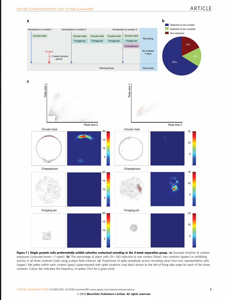
ResultsLong temporal separation experiment. In the first experiment,recordings of DG neurons were obtained over 4 days of re-exposure to three distinct contexts separated in time duringtraining (Fig. 1a). To ensure the recruitment of different popu-lations of neurons in each context while minimizing age-relatedreductions in adult neurogenesis, initial experiences in each
context were separated by intervals greater than, but approxi-mately, 3 weeks. Additionally, the different environments/experiences occurred within the same global space, not in sepa-rate rooms, where there has been shown to be a lack of recruit-ment of distinct populations14. Introductions to novel contextswere paired with re-exposure to previously introduced contexts tore-engage memory of remote experiences and encourage rats todiscriminate between the distinct behaviour requirements of each.Seventy-two putative granule cells were recorded from three rats(See Methods for characteristics used for interneuron exclusion)in the first experiment. We characterized the nature of contextselectivity in these cells by using a place field criterion (seeMethods for place field detection method). Fifty-six cells met aplace field criterion in at least one context. Of these 56 cells, 66%exhibited place fields selective to one context, whereas 16%exhibited fields in two contexts and 18% exhibited fields in allthree contexts (Fig. 1b). We also utilized a firing rate criterion todetermine whether a cell was active in a single context. Since themean firing rate cutoff for ‘active’ is arbitrary, we looked at acontinuum of firing rates and found a high amount of selectivityto a single environment using previously utilized cutoffs (0.1 and0.25 Hz, Fig. 2a, Supplementary Table 1)15. The same wasobserved for a wide range of cutoffs. Though it is unclear whichmeasure best quantifies the most meaningful form of activity fordownstream targets, both measures reveal a selectivity of firingfor single contexts when the introduction to each context isseparated by long temporal intervals. We chose to focus primarilyon the place field criterion, however, because this measure isreadily relatable to the known functions of other hippocampalregions.
The large percentage of cells selective to one context in the firstexperiment indicates that the DG does, in fact, selectively encodeenvironments that are temporally distinct on the 3-week timescale.
Reduced timeline and neurogenesis knockdown experiment. Inthe second experiment, the temporal influence upon contextselectivity and the effect of neurogenesis knockdown upon suchselectivity were tested. A 2-week interval (the 2-week separationtimeline) was chosen to test whether a shorter interval would stillresult in cells selective to different contexts as well as to maximizethe extent of neurogenesis knockdown during context exposure,given the timeline of efficacy for our chosen knockdown proce-dure (Fig. 3a). While current recording techniques do not permitidentification of context-selective cells as either adult or immatureDG granule neurons, it is nevertheless possible to determine theimpact of selective reduction of neurogenesis on the degree towhich context selectivity in DG neuronal activity actually occurs.Thus, we sought to determine whether the reduction of adultneurogenesis impacted context selectivity in animals experiencingthe 2-week timeline. Additionally, we compared context selec-tivity when initial encounters with the same three environmentsall occurred on the same day (the simultaneous exposure time-line; Fig. 3b) To enhance the survival of adult-born neurons andtheir potential contribution to DG function, rats were given30 min of enriched environment exposure daily throughout thecourse of the experiment16.
In the 2-week separation timeline, 94 cells were recorded fromseven rats, with 72 cells meeting a place field criterion in at leastone context (Fig. 3c, for representative spike locations and firingrate maps see Supplementary Fig. 1, for results by individual ratsee Supplementary Fig. 2). Of these, 38% exhibited place fields inonly one context, whereas 26% exhibited fields in two contextsand 36% exhibited fields in all three contexts. Fifty-two cells wererecorded from five rats exposed to the simultaneous timeline. Of
ARTICLE NATURE COMMUNICATIONS | DOI: 10.1038/ncomms4181
2 NATURE COMMUNICATIONS | 5:3181 | DOI: 10.1038/ncomms4181 | www.nature.com/naturecommunications
& 2014 Macmillan Publishers Limited. All rights reserved.
012345678911111111112222222222333333333344444444445555555555666666666677777777778888888888999999999911111111111111111111111111111111111111111111111111111111111111111111111111111111111111111111111111112222222222222222222222222222222222222222222222222222222222222222222222222222
Introduction to context 1
Circular track Circular track
Circular track
Cheeseboard
Foraging pot
Circular track
Cheeseboard
Foraging pot
Forage pot
~ 2 week recoveryperiod
Surgery
Forage pot Forage pot
Training phase Test phase
Selective to one context
Selective to two contexts
Non-selective
18%
16%
66%
Peak wire 2Peak wire 2
20
15
10
5
20
15
10
5
20
15
10
5
15
10
5
15
10
5
15
10
5
Pea
k w
ire 1
Pea
k w
ire 1
Forage pot
Cheeseboard
Circular track Circular track Circular trackRecording
All contexts4 days
Introduction to context 2 Introduction to context 3
Figure 1 | Single granule cells preferentially exhibit selective contextual encoding in the 3-week separation group. (a) Example timeline of context
exposures (coloured boxes¼ 1 week). (b) The percentage of place cells (N¼ 56) selective to one context (blue), two contexts (green) or exhibiting
activity in all three contexts (red) using a place field criterion. (c) Projections of spike amplitude across recording wires from two representative cells
(upper). Rat paths within each context (grey) superimposed with spike locations (red dots) shown to the left of firing rate maps for each of the three
contexts. Colour bar indicates the frequency of spikes (Hz) for a given pixel.
NATURE COMMUNICATIONS | DOI: 10.1038/ncomms4181 ARTICLE
NATURE COMMUNICATIONS | 5:3181 | DOI: 10.1038/ncomms4181 | www.nature.com/naturecommunications 3
& 2014 Macmillan Publishers Limited. All rights reserved.
012345678911111111112222222222333333333344444444445555555555666666666677777777778888888888999999999911111111111111111111111111111111111111111111111111111111111111111111111111111111111111111111111111112222222222222222222222222222222222222222222222222222222222222222222222222222
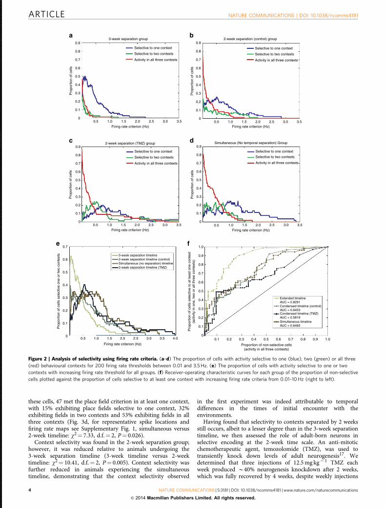
these cells, 47 met the place field criterion in at least one context,with 15% exhibiting place fields selective to one context, 32%exhibiting fields in two contexts and 53% exhibiting fields in allthree contexts (Fig. 3d, for representative spike locations andfiring rate maps see Supplementary Fig. 1, simultaneous versus2-week timeline: w2¼ 7.33, d.f.¼ 2, P¼ 0.026).
Context selectivity was found in the 2-week separation group;however, it was reduced relative to animals undergoing the3-week separation timeline (3-week timeline versus 2-weektimeline: w2¼ 10.41, d.f.¼ 2, P¼ 0.005). Context selectivity wasfurther reduced in animals experiencing the simultaneoustimeline, demonstrating that the context selectivity observed
in the first experiment was indeed attributable to temporaldifferences in the times of initial encounter with theenvironments.
Having found that selectivity to contexts separated by 2 weeksstill occurs, albeit to a lesser degree than in the 3-week separationtimeline, we then assessed the role of adult-born neurons inselective encoding at the 2-week time scale. An anti-mitoticchemotherapeutic agent, temozolomide (TMZ), was used totransiently knock down levels of adult neurogenesis17. Wedetermined that three injections of 12.5 mg kg� 1 TMZ eachweek produced B40% neurogenesis knockdown after 2 weeks,which was fully recovered by 4 weeks, despite weekly injections
3-week separation group
2-week separation (TMZ) group
3-week separation timeline2-week separation timeline (control)
2-week separation timeline (TMZ)Simultaneous (no separation) timeline
Selective to one context
Selective to two contexts
Activity in all three contexts
0.9
0.9
0.8
0.7
0.6
0.5
0.4
0.3
0.2
0.1
00.5 1.0
Firing rate criterion (Hz)
1.5 2.0 2.5 3.0 3.5
Pro
port
ion
of c
ells
Selective to one context
Selective to two contexts
Activity in all three contexts
0.8
0.7
0.6
0.5
0.4
0.3
0.2
0.1
00.5 1.0
Firing rate criterion (Hz)1.5 2.0 2.5 3.0 3.5
Pro
port
ion
of c
ells
Pro
port
ion
of c
ells
2-week separation (control) group
Selective to one context
Selective to two contexts
Activity in all three contexts
0.9
0.8
0.7
0.6
0.5
0.4
0.3
0.2
0.1
00.5 1.0
Firing rate criterion (Hz)1.5 2.0 2.5 3.0 3.5
Pro
port
ion
of c
ells
Simultaneous (No temporal separation) Group0.9
Selective to one context
Selective to two contexts
Activity in all three contexts
0.8
0.7
0.6
0.5
0.4
0.3
0.2
0.1
00.5 1.0
Firing rate criterion (Hz)
1.5 2.0 2.5 3.0 3.5
0.7 1.0
0.9
0.8
0.7
0.6
0.5
0.4
0.3
0.2
0.1
00.1 0.2 0.3
Proportion of non-selective cells(activity in all three contexts)
0.4 0.5 0.6 0.7
Extended timelineAUC = 0.8291Condensed timeline (control)AUC = 0.6453Condensed timeline (TMZ)AUC = 0.5819Simultaneous timelineAUC = 0.6465
0.8 0.9 1.0
Pro
port
ion
of c
ells
sel
ectiv
e to
at l
east
one
con
text
(act
ivity
in o
ne, t
wo
or a
ll th
ree
cont
exts
)0.6
0.5
0.4
0.3
0.2
0.1
00.5
Pro
port
ion
of c
ells
sel
ectiv
e on
e or
two
cont
exts
1.0
Firing rate criterion (Hz)
1.5 2.0 2.5 3.0 3.5 4.0
Figure 2 | Analysis of selectivity using firing rate criteria. (a–d) The proportion of cells with activity selective to one (blue), two (green) or all three
(red) behavioural contexts for 200 firing rate thresholds between 0.01 and 3.5 Hz. (e) The proportion of cells with activity selective to one or two
contexts with increasing firing rate threshold for all groups. (f) Receiver-operating characteristic curves for each group of the proportion of non-selective
cells plotted against the proportion of cells selective to at least one context with increasing firing rate criteria from 0.01–10 Hz (right to left).
ARTICLE NATURE COMMUNICATIONS | DOI: 10.1038/ncomms4181
4 NATURE COMMUNICATIONS | 5:3181 | DOI: 10.1038/ncomms4181 | www.nature.com/naturecommunications
& 2014 Macmillan Publishers Limited. All rights reserved.
012345678911111111112222222222333333333344444444445555555555666666666677777777778888888888999999999911111111111111111111111111111111111111111111111111111111111111111111111111111111111111111111111111112222222222222222222222222222222222222222222222222222222222222222222222222222
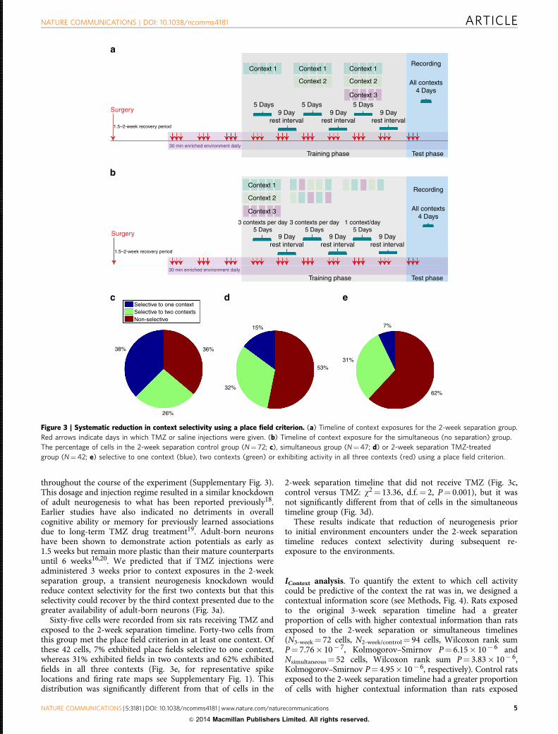
throughout the course of the experiment (Supplementary Fig. 3).This dosage and injection regime resulted in a similar knockdownof adult neurogenesis to what has been reported previously18.Earlier studies have also indicated no detriments in overallcognitive ability or memory for previously learned associationsdue to long-term TMZ drug treatment19. Adult-born neuronshave been shown to demonstrate action potentials as early as1.5 weeks but remain more plastic than their mature counterpartsuntil 6 weeks16,20. We predicted that if TMZ injections wereadministered 3 weeks prior to context exposures in the 2-weekseparation group, a transient neurogenesis knockdown wouldreduce context selectivity for the first two contexts but that thisselectivity could recover by the third context presented due to thegreater availability of adult-born neurons (Fig. 3a).
Sixty-five cells were recorded from six rats receiving TMZ andexposed to the 2-week separation timeline. Forty-two cells fromthis group met the place field criterion in at least one context. Ofthese 42 cells, 7% exhibited place fields selective to one context,whereas 31% exhibited fields in two contexts and 62% exhibitedfields in all three contexts (Fig. 3e, for representative spikelocations and firing rate maps see Supplementary Fig. 1). Thisdistribution was significantly different from that of cells in the
2-week separation timeline that did not receive TMZ (Fig. 3c,control versus TMZ: w2¼ 13.36, d.f.¼ 2, P¼ 0.001), but it wasnot significantly different from that of cells in the simultaneoustimeline group (Fig. 3d).
These results indicate that reduction of neurogenesis priorto initial environment encounters under the 2-week separationtimeline reduces context selectivity during subsequent re-exposure to the environments.
IContext analysis. To quantify the extent to which cell activitycould be predictive of the context the rat was in, we designed acontextual information score (see Methods, Fig. 4). Rats exposedto the original 3-week separation timeline had a greaterproportion of cells with higher contextual information than ratsexposed to the 2-week separation or simultaneous timelines(N3-week¼ 72 cells, N2-week/control¼ 94 cells, Wilcoxon rank sumP¼ 7.76� 10� 7, Kolmogorov–Smirnov P¼ 6.15� 10� 6 andNsimultaneous¼ 52 cells, Wilcoxon rank sum P¼ 3.83� 10� 6,Kolmogorov–Smirnov P¼ 4.95� 10� 6, respectively). Control ratsexposed to the 2-week separation timeline had a greater proportionof cells with higher contextual information than rats exposed
Surgery
Context 1
Context 1
Context 2
Context 3
3 contexts per day5 Days
9 Dayrest interval
Selective to one contextSelective to two contextsNon-selective
38% 36%
15%
53%31%
7%
62%32%
26%
9 Dayrest interval
9 Dayrest interval
3 contexts per day5 Days
1 context/day5 Days
Context 1 Context 1Recording
All contexts4 Days
Recording
All contexts4 Days
Context 2 Context 2
Context 3
5 Days9 Day
rest interval
5 Days 5 Days9 Day
rest interval9 Day
rest interval
Training phase
Training phase Test phase
Test phase
1.5–2-week recovery period
Surgery
1.5–2-week recovery period
30 min enriched environment daily
30 min enriched environment daily
Figure 3 | Systematic reduction in context selectivity using a place field criterion. (a) Timeline of context exposures for the 2-week separation group.
Red arrows indicate days in which TMZ or saline injections were given. (b) Timeline of context exposure for the simultaneous (no separation) group.
The percentage of cells in the 2-week separation control group (N¼ 72; c), simultaneous group (N¼47; d) or 2-week separation TMZ-treated
group (N¼42; e) selective to one context (blue), two contexts (green) or exhibiting activity in all three contexts (red) using a place field criterion.
NATURE COMMUNICATIONS | DOI: 10.1038/ncomms4181 ARTICLE
NATURE COMMUNICATIONS | 5:3181 | DOI: 10.1038/ncomms4181 | www.nature.com/naturecommunications 5
& 2014 Macmillan Publishers Limited. All rights reserved.
012345678911111111112222222222333333333344444444445555555555666666666677777777778888888888999999999911111111111111111111111111111111111111111111111111111111111111111111111111111111111111111111111111112222222222222222222222222222222222222222222222222222222222222222222222222222

to the same timeline receiving TMZ (N2-week/TMZ¼ 65 cellsWilcoxon rank sum P¼ 0.02, Kolmogorov–Smirnov P¼ 0.01).There was also a significant difference between TMZ rats exposedto the 2-week separation timeline and rats exposed to thesimultaneous timeline (TMZ 2-week versus simultaneous:Wilcoxon rank sum P¼ 0.128, Kolmogorov–Smirnov P¼ 0.015).
However, there was no significant difference in contextualinformation between control rats exposed to the 2-weekseparation timeline and rats exposed to simultaneous timeline(control 2-week versus simultaneous: Wilcoxon rank sumP¼ 0.53, Kolmogorov–Smirnov P¼ 0.60). The fact that thereis a distinction between these two groups using a place fieldcriterion suggests that the two analytical measures are exposingdistinct aspects of contextual encoding. Whereas the place fieldanalysis assesses the selectivity of spatial encoding within each ofthe three contexts, the IContext score measures selectivity based onfiring rates, irrespective of spatially related activity.
DiscussionOur studies reveal the existence of a neural code that separatesevents in time. Segregating exposure to contexts by 43 weeksresults in a very large proportion of cells that are selective to asingle context. The capacity of DG neurons to selectively encodetemporally distinct contexts enables them to participate inthe separation of episodic or contextual memories over longdurations, extending the known ability of the hippocampus toencode both time and space21–23.
Our results support the hypothesis that the continual additionof neurons with temporally delineated periods of hyperexcitabilitypermits the DG to specifically encode a single context if exposureto that context is temporally segregated from other exposures.Thus, space may be coded as a function of time by the periodicaddition of new neurons into the hippocampal circuit. Under thishypothesis, the fact that longer intervals between contexts recruitmore distinct populations of putative adult-born cells thanshorter intervals suggests a crucial time point in the maturation ofadult-born neurons for effective discrimination of temporalcontexts. This time point is elucidated by the observed changesin selectivity between the 2-week and 3-week separation groups,and by the fact that rats with reduced neurogenesis (using TMZ)demonstrated a large proportion of cells selective to threecontexts in similar proportions to the group that was trainedwithout any temporal separation between contexts (Fig. 3d,e).
The findings in the TMZ-treated and simultaneous exposuregroups are consistent with previous DG studies conductedwithout significant temporal context separation15. These studiesshowed that both small and large changes across environmentsrecruit activity from an overlapping, small population of DG cells(potentially mature or immature) that distinguish betweenenvironments through changes in firing rate rather thanthrough global remapping.
Alternatively, the presence of immature adult-born cells couldsignificantly impact the overall excitability of the mature cells inthe region by driving a majority of the feedback inhibition ontothe granule cell layer24–26. Consistent with this hypothesis,the neurogenesis knockdown group exhibited cells with overallhigher mean firing rates than control rats given the sametemporal separation between contexts (Supplementary Table 1).We do not believe that this increase in mean firing rate is duesolely to TMZ administration, as similar differences in meanfiring rate were observed between the 3-week, 2-week and noseparation groups in which the only difference between thegroups is the timing between initial context presentations.Nevertheless, it is possible that reduced temporal separationduring training differentially affects the overall levels ofexcitability in the circuit during re-exposure in a manner that issimilar to transiently reducing levels of adult neurogenesis duringtraining. While the exact cellular mechanism of the observedcontext selectivity is unknown, the net effect is consistent withour conclusion that the timing of initial exposure is a criticaldeterminant of DG activity.
Using temporal separation of context presentation, we revealeda mechanism by which the DG encodes contexts with unique,non-overlapping cell populations. This ability of the DG wastheoretically predicted in models in which a small number ofneurons within a relatively dense cell layer function as amechanism to ‘pattern separate’ or ‘orthogonalize’ multipleexperiences27,28. Our data confirm this prediction by revealingthe existence of a temporal orthogonalizing neuronal code withinthe DG.
MethodsFiring rate and place field criterion analyses. The firing rate in each context wasdetermined by dividing the total number of spikes per context by the time spent inthe context (Supplementary Tables 1 and 2, Supplementary Fig. 4). To assess thepresence of place field activity in each context, place field candidates were firstidentified that had a o3 Hz overall mean firing rate, place fields covering at least
0.7
0.6
0.5
0.4
0.3
0.2
0.1P
ropo
rtio
n of
cel
ls0
00.2
0.4IContext score (bits per spike)
0.60.8
11.2
1.41.6 1
23
4
Three-week separation group
No separation group
Two-week separation group (TMZ)
Two-week separation group (Control)
Figure 4 | Reductions in context selectivity using IContext reflect the nature of the training paradigms. The proportion of cells with IContext scores
between 0 and 1.6 bits per spike in the 3-week separation group (N¼ 72, light green), 2-week separation control group (N¼ 94, dark green), simultaneous
(N¼ 52, no separation) timeline (brown) group and 2-week separation TMZ-treated group (N¼65, dark blue). The extended timeline group has the
largest proportion of cells with high IContext scores.
ARTICLE NATURE COMMUNICATIONS | DOI: 10.1038/ncomms4181
6 NATURE COMMUNICATIONS | 5:3181 | DOI: 10.1038/ncomms4181 | www.nature.com/naturecommunications
& 2014 Macmillan Publishers Limited. All rights reserved.
012345678911111111112222222222333333333344444444445555555555666666666677777777778888888888999999999911111111111111111111111111111111111111111111111111111111111111111111111111111111111111111111111111112222222222222222222222222222222222222222222222222222222222222222222222222222
three 5 cm� 5 cm contiguous pixels before reducing to 10% of peak firing rate, anda peak pixel of at least 1 Hz that was also 2 s.t.d. above the mean firing rate of thecell in a given context29,30. To meet a place field criterion for a context, thesecandidates were then required to have a statistically significant spatial informationscore for that context when compared with 1,000 iterations of the same spike timesfor a given cell with randomized spatial position. The number of cells with placefields in one, two or all three behavioural contexts was used to assess the extent ofcontext selectivity.
Informational analysis of context selectivity. To assess selective activity to agiven behavioural context, we developed a novel measure of contextual informationusing the following equation:
IContext ¼ IC1 þ IC2 þ IC3ð Þ=3
IC ¼ fC=fAvg� �
� log2 fC=fAvg� �
fAvg ¼ fC1þ fC2þ fC3ð Þ=3
ð1Þ
where fC is the number of spikes occurring within a context divided by total timespent in a context, fAvg¼ the average firing rate across all contexts, IC¼ theinformation score of a given context in bits/spike and IContext¼ the amount ofinformation present in each spike regarding contextual inhabitance.
Behavioural contexts. The three behavioural contexts were a 130 0 � 130 0 foragingpot containing rat bedding and randomly placed 1
4 pieces of Honey Nut Cheerios(for one rat, the foraging pot was a circular pot that had larger walls to prevent therat from jumping over the edges), a 480 0 diameter circular track with a 30 0 widepathway and either 1
4 pieces of Honey Nut Cheerios or chocolate sprinkles in areliably rewarded location that was shifted to a different track location up to threetimes per session and a 480 0 diameter circular cheeseboard with randomly placedchocolate sprinkles and three presentations of a large food reward (a 20 0 diameterdish filled with chocolate sprinkles) at random time intervals and random locationsfor a period of 430 s (Supplementary Fig. 5). All three behavioural contexts werepresented in the same location of the same recording room containing constantexternal cues. The time length of exposure to each context differed between con-texts to allow full exploration of the larger contexts and minimize oversampling ofthe smaller contexts. Exposure time lengths during both training and test sessionsfor the foraging pot, circular track, and circular cheeseboard were B10, 15 and20 min, respectively.
Timeline for simultaneous and temporally distinct exposure. Rats wereexposed to the three behavioural contexts according to one of three timelines: anextended 3-week separation timeline, in which the initial exposure to each contextwas separated by 43 weeks (Fig. 1a); a 2-week separation timeline, in whichthe initial exposure to each context was separated by 2 weeks (Fig. 3a) or asimultaneous timeline in which there was no temporal separation between initialcontext exposures (Fig. 3b). Temporal intervals of 43 weeks between behaviouralcontext exposures in the extended timeline would ensure a length of time betweencontexts that was long enough to recruit different populations of adult-bornneurons but short enough to prevent age-related reductions in adult neurogenesisproliferation. In the 3-week separation timeline (N¼ 3 rats), rats were first trainedto run around the circular track before undergoing surgery for microdriveimplantation. After a recovery period of at least 2 weeks and during several weeksin which microdrive wires were slowly lowered to the granule cell layer of the DG,rats were re-trained on the circular track and began exposure to the foraging potenvironment. During this time in training, they received exposure to both contextseach day. Once stable cell recording could be obtained from the granule cell layerand at least 3 weeks after initial exposure to the foraging pot, rats were exposedto the circular cheeseboard environment. During this time in training, theyreceived exposure to all three contexts each day. All analyses of cell activitywere restricted to days in which rats received exposure to all three contexts onthe same day.
In the 2-week separation timeline (N¼ 7 control rats, N¼ 7 TMZ-treated rats),initial exposure to each behavioural context was separated by a fixed 2 weeks toprovide an interval between contexts that was long enough to recruit a newpopulation of adult-born neurons but short enough to allow for the fullest extent ofknockdown during contextual exposure, given the timeline of efficacy for thechosen knockdown procedure (Fig. 3a). For the first context exposure, the rat wasexposed to only a single behavioural context for 5 days followed by a 9-day restperiod without exposures. In subsequent exposures to the second and thirdbehavioural contexts, exposure to the new context also included exposure topreviously presented contexts in the same recording day. Nine days after the end oftraining, rats received a 4-day test phase that included presentation of all threebehavioural contexts. All analyses of cell activity were restricted to test days. Theorder of exposure to each of the three behavioural contexts was counterbalancedacross groups.
In the simultaneous timeline (N¼ 5 rats), rats were exposed to all threebehavioural contexts in the same day for the first week of training. In subsequentexposure weeks, the numbers of exposures to each context were limited to two
contexts per day and finally one context per day to equate time spent in eachbehavioural context across groups.
Adult neurogenesis knockdown. TMZ is an anti-cell proliferation drug that hasbeen shown to reduce cell proliferation in the DG by 480% in mice and cellsurvival in rats by 440%17,19. After testing several dosages of the drug in rats, wefound the optimal dosage to be three i.p. injections of 12.5 mg kg� 1 each week for2 weeks. This injection regime produced a significant reduction in cell proliferationby B40% after 2 weeks, with recovery of neurogenesis levels through consistent useof this injection regime after 4 weeks (Supplementary Fig. 3). To prevent possibleside effects from stopping treatment after the beginning of the experiment, threeinjections of 12.5 mg kg� 1 TMZ were performed every week for 10 weeks(beginning 3 weeks before the initial context exposure). Controls received salineinjections of the same volume and frequency as TMZ-treated rats. There were nodifferences in exploratory behaviour between control and TMZ-treated groups (seefurther Methods for analysis of time spent in each context per group).
Rats. All animal procedures were performed in accordance with NIH and localIACUC guidelines. Twenty adult, male Long-Evans rats were used as subjects. Therats were housed individually and maintained on a 12-h light/dark cycle. They wereacclimated to the colony room for 3 days and handled daily for at least 2 weeksbefore beginning the experiment, during which time they were placed on foodrestriction until they reached 85–90% of ad libitum weight. Rats were 3 months oldat the time of surgery. Weight ranged from 300 to 350 g. Water was available at alltimes. All behavioural testing occurred during the rats’ light cycle.
Microdrive implantation surgery. A microdrive consisting of four tetrodesof 17-mm platinum iridium wire was surgically implanted using stereotaxicprocedures (from bregma A/P: � 4.0, M/L: þ 2.2 mm, D/V: � 2.2 mm) and waslowered into the granule cell layer of the DG (D/V: B2.7 mm) until the appearanceof place cell single units, ‘dentate spikes’ and complex high-frequency (16–90 Hz)local field potential activity.
Neural recordings. During targeting of the DG granule cell layer, each wire of themicroelectrode bundle was first checked for neural activity. The quietest wire wasfound and served as a reference electrode. If no activity was found on the recordingwires, the bundle was advanced in 10-mm increments until activity was found.Wires were not turned within 24 h before a recording session.
The activity on each wire was passed through a high-impedance OpAmpheadstage that held a set of light-emitting diodes (Neuralynx Technologies,Bozeman, MT). A multiwire flexible cable connected the preamplifiers to a 32-channel commutator (Neuralynx Technologies). The single-unit signal was filteredat 600 Hz (low pass) to 6,000 Hz (high pass) and amplified through NeuralynxLynx-8 differential programmable amplifiers. The local field potential signal wasfiltered at 1 Hz (low pass) to 475 Hz (high pass). The amplifiers were integratedwith the Cheetah data acquisition program (Neuralynx Technologies), wherein theacquired analogue signals were digitally converted at a rate of 30,303.00 KHz(DT 2821 Data Translation, Marlboro, MA) prior to storage. The position of the ratwas monitored by a set of light-emitting diodes placed on the headstage. A videotracking system (SA-2 Dragon Tracker, Boulder, CO) registered the position of thediodes with a sampling frequency of 30 Hz. All recording sessions began with 5 minof baseline recording activity in a home cage environment. Single cells wereidentified using Offline Sorter (Plexon, Dallas, TX) to compare relative amplitudeof each spike across tetrode wires. Final microdrive wire location was verifiedpost-mortem in 40-mm sections using a Nissl stain (Supplementary Fig. 6).
Interneuron exclusion. To exclude interneurons from selectivity analyses, all cellsthat exhibited both a mean firing rate in any context above 5 Hz and a meanwaveform width of o150 ms at half of mean peak amplitude were excluded. Ingeneral, putative granule cells discharged at low mean firing rates (o3 Hz withmean spike waveform widths of at least 0.15 ms taken at 50% of peak amplitude).In contrast, putative interneurons typically demonstrated mean peak firing rates of45 Hz with mean spike waveform widths of o0.15 ms.
Analysis of selectivity using firing rate criterion. To assess context selectivityusing firing rates, the proportion of cells selective to one, two or all three beha-vioural contexts was calculated using a firing rate criterion. Since choosing a singlefiring rate criterion can be quite arbitrary, we examined context selectivity using acontinuum of firing rate criteria from 0.01 to 3.5 Hz in increments of 0.01 Hz(Fig. 2a–e). To quantify differences in selectivity over multiple firing rate criteriaacross groups, a receiver-operating characteristic curve was made for the changingproportion of cells with activity in all three contexts compared to the proportion ofcells selective to at least one context for each increase in the firing rate criterion in0.01 Hz steps (Fig. 2f). The area under the curve for each group was calculatedusing trapezoidal approximations.
NATURE COMMUNICATIONS | DOI: 10.1038/ncomms4181 ARTICLE
NATURE COMMUNICATIONS | 5:3181 | DOI: 10.1038/ncomms4181 | www.nature.com/naturecommunications 7
& 2014 Macmillan Publishers Limited. All rights reserved.
012345678911111111112222222222333333333344444444445555555555666666666677777777778888888888999999999911111111111111111111111111111111111111111111111111111111111111111111111111111111111111111111111111112222222222222222222222222222222222222222222222222222222222222222222222222222
Spatial informational analysis. A spatial information score was calculated for allcells in each behavioural context31,32. This spatial information score formula wascalculated as follows:
I¼X
Pi Fi=Fð Þ log2 Fi=Fð Þ ð2Þ
Where i is the pixel number, Pi is the probability of occupancy in pixel I and Fi isthe mean firing rate for pixel i.
F is the overall mean firing rate of the cell.
Burst index. A burst index was calculated for both single-fielded and multi-fieldedcells (Supplementary Table 3), which divided the number of times two consecutivespikes occurred within 6 ms by the total number of interspike intervals during thecourse of a recording session29.
Analysis of time spent in each context per group. To ensure that any differencesin context selectivity across groups was not attributable to differences in ratmobility or time spent in select areas of a context (such as a rat sitting in a cell’splace field), an immobility bias was calculated for each cell. The correlationbetween a cell’s mean firing rate in a pixel and the time spent in each pixel was firstcalculated for each context. These correlations were then added together todetermine any firing rate biases due to immobility. If one experimental groupexhibited higher immobility biases than another, then differences in rat activitylevels could feasibly account for changes in cell selectivity. There were no sig-nificant differences in immobility biases across groups.
Analysis of place field representation biases across contexts. The numbers offields in each context for each group were calculated and compared to observewhether place field overrepresentation of one context could drive the place fieldselectivity observed in this study (Supplementary Table 4). While there was adisproportional representation of the three different contexts, these proportionswere not significantly different across groups.
Immunohistochemistry. To quantify levels of neurogenesis in pilot rats receivingTMZ for 2 or 4 weeks, rats were injected with 50 mg kg� 1 5-bromo-20-deoxyur-idine (BrdU) for 4 consecutive days during week 2 or 4 of TMZ treatment,respectively (Supplementary Fig. 3). This measure indicates the number of dividingcells after 2 and 4 weeks of TMZ administration. After 24 h from the final injectionof BrdU in each group, rats were perfused with 0.1 M PO4 followed by 4% par-aformaldehyde and rat brains were removed from the skull to be incubated for atleast 48 h in 30% sucrose at 4 �C. Frozen brains were then cut into 40-mm coronalsections with a microtome. Sections from every 240 mm spanning the whole DGwere selected (1 of every 6 sections). Sections were post-fixed for 50 min in 4%paraformaldehyde at room temperature and incubated for 30 min in 2 N HCl at37 �C followed by 1 N Borate Buffer at room temperature. Sections were thenincubated at 4 �C in primary Rat anti-BrdU (1:100) for 72 h (Catalogue no.OBT0030, Accurate, NY). Finally, they were incubated at room temperature insecondary Donkey anti-Rat IgG Dylite 488 (1:250) for 2 h (Catalogue # 712-485-153, Jackson ImmunoResearch, PA). Every BrdU-labelled cell was counted at � 40magnification in 10 consecutive 40-mm sections spaced 240 mm apart. These countswere then used to estimate the total number of BrdU-labelled cells in each dorsalDG. Two different experimenters who were blind to rat identity counted cellsindependently to ensure that cell counts were consistent across experimenters. Inhalf of the rats, total numbers were additionally verified using unbiased stereo-logical technique (StereoInvestigator, MBF Bioscience, CA).
References1. Squire, L. R. Memory and the hippocampus: a synthesis from findings with rats,
monkeys, and humans. Psychol. Rev. 99, 195–231 (1992).2. Lipton, P. A. & Eichenbaum, H. Complementary roles of hippocampus and
medial entorhinal cortex in episodic memory. Neural Plast. 2008, 258467(2008).
3. Burgess, N., Maguire, E. A. & O’Keefe, J. The human hippocampus and spatialand episodic memory. Neuron 35, 625–641 (2002).
4. Jung, M. W. & McNaughton, B. L. Spatial selectivity of unit activity in thehippocampal granular layer. Hippocampus 3, 165–182 (1993).
5. Goodrich-Hunsaker, N. J., Hunsaker, M. R. & Kesner, R. P. The interactionsand dissociations of the dorsal hippocampus subregions: how the dentate gyrus,CA3, and CA1 process spatial information. Behav. Neurosci. 122, 16–26 (2008).
6. Kesner, R. P. & Hunsaker, M. R. The temporal attributes of episodic memory.Behav. Brain Res. 215, 299–309 (2010).
7. Sisti, H. M., Glass, A. L. & Shors, T. J. Neurogenesis and the spacing effect:learning over time enhances memory and the survival of new neurons. Learn.Mem. 14, 368–375 (2007).
8. Gage, F. H. Mammalian neural stem cells. Science 287, 1433–1438 (2000).9. Piatti, V. C., Esposito, M. S. & Schinder, A. F. The timing of neuronal
development in adult hippocampal neurogenesis. Neuroscientist 12, 463–468(2006).
10. Li, Y., Aimone, J. B., Xu, X., Callaway, E. M. & Gage, F. H. Development ofGABAergic inputs controls the contribution of maturing neurons to the adulthippocampal network. Proc. Natl Acad. Sci. USA 109, 4290–4295 (2012).
11. Marı́n-Burgin, A., Mongiat, L. A., Pardi, M. B. & Schinder, A. F. Uniqueprocessing during a period of high excitation/inhibition balance in adult-bornneurons. Science 335, 1238–1242 (2012).
12. Aimone, J. B., Wiles, J. & Gage, F. H. Potential role for adult neurogenesis in theencoding of time in new memories. Nat. Neurosci. 9, 723–727 (2006).
13. Aimone, J. B., Wiles, J. & Gage, F. H. Computational influence of adultneurogenesis on memory encoding. Neuron 61, 187–202 (2009).
14. Alme, C. B. et al. Hippocampal granule cells opt for early retirement.Hippocampus 20, 1109–1123 (2010).
15. Leutgeb, J. K., Leutgeb, S., Moser, M. B. & Moser, E. I. Pattern separation in thedentate gyrus and CA3 of the hippocampus. Science 315, 961–966 (2007).
16. Tashiro, A., Makino, H. & Gage, F. H. Experience-specific functionalmodification of the dentate gyrus through adult neurogenesis: a critical periodduring an immature stage. J. Neurosci. 27, 3252–3259 (2007).
17. Garthe, A., Behr, J. & Kempermann, G. Adult-generated hippocampal neuronsallow the flexible use of spatially precise learning strategies. PLoS One 4, e5464(2009).
18. Shors, T. J., Townsend, D. A., Zhao, M., Kozorovitskiy, Y. & Gould, E.Neurogenesis may relate to some but not all types of hippocampal-dependentlearning. Hippocampus 12, 578–584 (2002).
19. Nokia, M. S., Anderson, M. L. & Shors, T. J. Chemotherapy disrupts learning,neurogenesis and theta activity in the adult brain. Eur. J. Neurosci. 36,3521–3530 (2012).
20. Ge, S., Yang, C.-H., Hsu, K.-S., Ming, G.-L. & Song, H. A critical period forenhanced synaptic plasticity in newly generated neurons of the adult brain.Neuron 54, 559–566 (2007).
21. Kesner, R. P. & Hardy, J. D. Long-term memory for contextual attributes:dissociation of amygdala and hippocampus. Behav. Brain Res. 8, 139–149(1983).
22. O’Keefe, J. & Nadel, L. The hippocampus as a cognitive map. Hippocampus 3,570 (Oxford University Press, 1978).23. MacDonald, C. J., Lepage, K. Q., Eden, U. T. & Eichenbaum, H. Hippocampal
‘time cells’ bridge the gap in memory for discontiguous events. Neuron 71,737–749 (2011).
24. Sahay, A., Wilson, D. A. & Hen, R. Pattern separation: a common function fornew neurons in hippocampus and olfactory bulb. Neuron 70, 582–588 (2011).
25. Song, J., Christian, K. M., Ming, G. & Song, H. Modification of hippocampalcircuitry by adult neurogenesis. Dev. Neurobiol. 72, 1032–1043 (2012).
26. Piatti, V. C., Ewell, L. A. & Leutgeb, J. K. Neurogenesis in the dentate gyrus:carrying the message or dictating the tone. Front. Neurosci. 7, 50 (2013).
27. Marr, D. Simple memory: a theory for archicortex. Philos. Trans. R. Soc. B Biol.Sci 262, 23–81 (1971).
28. McClelland, J. L., McNaughton, B. L. & O’Reilly, R. C. Why there arecomplementary learning systems in the hippocampus and neocortex: insightsfrom the successes and failures of connectionist models of learning andmemory. Psychol. Rev. 102, 419–457 (1995).
29. Neunuebel, J. P. & Knierim, J. J. Spatial firing correlates of physiologicallydistinct cell types of the rat dentate gyrus. J. Neurosci. 32, 3848–3858 (2012).
30. Thompson, L. T. & Best, P. J. Long-term stability of the place-field activity ofsingle units recorded from the dorsal hippocampus of freely behaving rats.Brain Res. 509, 299–308 (1990).
31. Skaggs, W. E., McNaughton, B. L. & Gothard, K. M. An information-theoreticapproach to deciphering the hippocampal code. 1030–1037 (1992; at http://dl.acm.org/citation.cfm?id=645753.668057.
32. Markus, E. J., Barnes, C. A., McNaughton, B. L., Gladden, V. L. & Skaggs, W. E.Spatial information content and reliability of hippocampal CA1 neurons: effectsof visual input. Hippocampus 4, 410–421 (1994).
AcknowledgementsWe would like to thank D. Nitz and S. Leutgeb for discussions; S. Alfonso for conductingTMZ pilot studies; and A. Smith, M. Combs, L. McHenry for technical assistance; andM.L. Gage for editorial assistance. This work was supported in part by a James S.McDonnell Foundation Collaborative Activity Award, a Kavli Institute for Brain andMind Innovative Research Award, an NIH training grant from the Institute for NeuralComputation, and an NSF Science of Learning Center Grant SBE 0542013 to theTemporal Dynamics of Learning Center. These data are archived in the NSF TemporalDynamics of Learning Center data grid and at the Salk Institute for Biological Sciences.
Author contributionsL.M.R. and A.S.A. performed TMZ drug preparations and injections; L.M.R. and A.S.A.performed histology; L.M.R., A.S.A. and L.K.Q. performed microdrive implant surgeries;L.M.R., A.S.A. and L.K.Q. performed behavioural experiments and electrophysiologicalrecordings; L.M.R. and J.B.A. performed analyses; L.M.R., L.K.Q., J.B.A., J.W., A.A.C. andF.H.G. designed the study; A.A.C. and L.K.Q. provided oversight of experiments; L.M.R
ARTICLE NATURE COMMUNICATIONS | DOI: 10.1038/ncomms4181
8 NATURE COMMUNICATIONS | 5:3181 | DOI: 10.1038/ncomms4181 | www.nature.com/naturecommunications
& 2014 Macmillan Publishers Limited. All rights reserved.
012345678911111111112222222222333333333344444444445555555555666666666677777777778888888888999999999911111111111111111111111111111111111111111111111111111111111111111111111111111111111111111111111111112222222222222222222222222222222222222222222222222222222222222222222222222222
and L.K.Q wrote the paper. All authors discussed the results and commented on themanuscript. Stereological counts were performed in F.H.G.’s laboratory. All behaviouralrecording studies were performed in A.A.C.’s laboratory.
Additional informationSupplementary Information accompanies this paper at http://www.nature.com/naturecommunications
Competing financial interests: The authors declare no competing financial interests.
Reprints and permission information is available online at http://npg.nature.com/reprintsandpermissions/
How to cite this article: Rangel, L. M. et al. Temporally selective contextual encoding inthe dentate gyrus of the hippocampus. Nat. Commun. 5:3181 doi: 10.1038/ncomms4181(2014).
This article is licensed under a Creative Commons Attribution 3.0Unported Licence. To view a copy of this licence visit http://
creativecommons.org/licenses/by/3.0/.
NATURE COMMUNICATIONS | DOI: 10.1038/ncomms4181 ARTICLE
NATURE COMMUNICATIONS | 5:3181 | DOI: 10.1038/ncomms4181 | www.nature.com/naturecommunications 9
& 2014 Macmillan Publishers Limited. All rights reserved.
012345678911111111112222222222333333333344444444445555555555666666666677777777778888888888999999999911111111111111111111111111111111111111111111111111111111111111111111111111111111111111111111111111112222222222222222222222222222222222222222222222222222222222222222222222222222
Copyright © 2022 FDOKUMEN