Hippocampus and dentate gyrus of the Cebus monkey: Architectonic and stereological study
12
Hippocampus and dentate gyrus of the Cebus monkey: Architectonic and stereological study Cristovam Guerreiro-Diniz a , Roberta Bentes de Melo Paz a , Mayra Hermı ´nia Simo ˜es Hamad a , Carlos Santos Filho a , Adriano Augusto Vilhena Martins b,c , Heitor Bastos Neves b,c , Elane Domenica de Souza Cunha b,c , Gisele Cristina Alves b,c , Lia Amaral de Sousa b,c , Ivanira Amaral Dias b,c , Nonata Tre ´ via a , Aline Andrade de Sousa a , Aline Passos a , Nara Lins a , Joa ˜o Bento Torres Neto a , Pedro Fernando da Costa Vasconcelos b,c , Cristovam Wanderley Picanc ¸o-Diniz a, * a Universidade Federal do Para ´, Instituto de Cieˆncias Biolo ´gicas, Lab of Investigations in Neurodegeneration and Infection, Bele ´m, Para ´, Brazil b Universidade Federal do Para ´, Instituto de Cieˆncias Biolo ´gicas, Laborato ´rio de Neuroanatomia Funcional, Bele ´m, Para ´, Brazil c Instituto Evandro Chagas, IEC, Departamento de Arbovirologia e Febres Hemorra ´gicas, Rodovia BR 316, Km 7, CEP. 67030-000, Ananindeua, Para ´, Brazil 1. Introduction The term ‘‘hippocampal formation’’ is used to designate a large region of the middle temporal lobe of human and non-human primates, including the hippocampus, dentate gyrus (DG), and subiculum (Amaral and Lavenex, 2007; Squire et al., 2004). The internal anatomical details of the subdivisions of the hippocampus (the CA fields), DG, and subiculum vary little from mammal to mammal (Manns and Eichenbaum, 2006). These morphological similarities seem to translate into similar functional properties, and the hippocampus and parahippocampal regions are now clearly important for the initial acquisition and temporary maintenance of declarative memory (Eichenbaum et al., 2007; Levy et al., 2004; Manns and Eichenbaum, 2006; Rolls and Kesner, 2006; Stark, 2007). Much of the work on the monkey hippocampus has been carried out in Old World primates, and the Rhesus monkey (Macaca mulatta) has been the species of choice for many experimental studies because it shares many neuroanatomical aspects and cognitive functions with humans (Amaral and Lavenex, 2007; Gallagher and Rapp, 1997; Murray et al., 2007; Squire et al., 2004). However, the quantitative anatomical evidence related to these cognitive functions remains scarce. For example, information regarding the number of neuronal units in hippocam- pus and dentate gyrus of non-human primates is limited to the Rhesus monkey (Amaral and Lavenex, 2007; Christensen et al., 2007; Jabes et al., 2010; Keuker et al., 2003) and none of these quantitative studies have used selective markers to assess neuron numbers. In New World primates, the scenario is even more limited with a single estimation of the number of cells in the hippocampus and dentate gyrus by stereological procedures in Journal of Chemical Neuroanatomy 40 (2010) 148–159 ARTICLE INFO Article history: Received 9 March 2010 Received in revised form 6 June 2010 Accepted 7 June 2010 Available online 15 June 2010 Keywords: Hippocampus Dentate gyrus Pyramidal layer Granular layer Neurons Optical fractionator Cebus apella ABSTRACT Behavioral, electrophysiological, and anatomical assays of non-human primates have provided substantial evidence that the hippocampus and dentate gyrus are essential for memory consolidation. However, a single anatomical and stereological investigation of these regions has been done in New World primates to complement those assays. The aim of the present study was to describe the cyto-, myelo-, and histochemical architecture of the hippocampus and dentate gyrus, and to use the optical fractionator method to estimate the number of neurons in the hippocampal pyramidal and granular neurons in the dentate gyrus of the Cebus monkey. NeuN immunolabeling, lectin histochemical staining with Wisteria floribunda agglutinin (WFA), enzyme-histochemical detection of NADPH-diaphorase activity and Gallyas silver staining were used to define the layers and limits of the hippocampal fields and dentate gyrus. A comparative analysis of capuchin (Cebus apella) and Rhesus (Macaca mulatta) monkeys revealed similar structural organization of these regions but significant differences in the regional distribution of neurons. C. apella were found to have 1.3 times fewer pyramidal and 3.5 times fewer granular neurons than M. mulatta. Taken together the architectonic and stereological data of the present study suggest that hippocampal and dentate gyrus neural networks in the C. apella and M. mulatta may contribute to hippocampal-dentate gyrus-dependent tasks in different proportions. ß 2010 Elsevier B.V. All rights reserved. * Corresponding author at: Laborato ´ rio de Investigac ¸o ˜ es em Neurodegenerac ¸a ˜o e Infecc ¸a ˜o, Hospital Universita ´ rio Joa ˜o de Barros Barreto, Rua Mundurucus No. 4487, CEP. 66073-000, Bele ´ m, Para ´ , Brazil. Tel.: +55 91 32016756. E-mail address: [email protected] (C.W. Picanc ¸o-Diniz). Contents lists available at ScienceDirect Journal of Chemical Neuroanatomy journal homepage: www.elsevier.com/locate/jchemneu 0891-0618/$ – see front matter ß 2010 Elsevier B.V. All rights reserved. doi:10.1016/j.jchemneu.2010.06.002
Transcript of Hippocampus and dentate gyrus of the Cebus monkey: Architectonic and stereological study

Journal of Chemical Neuroanatomy 40 (2010) 148–159
Hippocampus and dentate gyrus of the Cebus monkey: Architectonic andstereological study
Cristovam Guerreiro-Diniz a, Roberta Bentes de Melo Paz a, Mayra Hermınia Simoes Hamad a,Carlos Santos Filho a, Adriano Augusto Vilhena Martins b,c, Heitor Bastos Neves b,c,Elane Domenica de Souza Cunha b,c, Gisele Cristina Alves b,c, Lia Amaral de Sousa b,c,Ivanira Amaral Dias b,c, Nonata Trevia a, Aline Andrade de Sousa a, Aline Passos a, Nara Lins a,Joao Bento Torres Neto a, Pedro Fernando da Costa Vasconcelos b,c, Cristovam Wanderley Picanco-Diniz a,*a Universidade Federal do Para, Instituto de Ciencias Biologicas, Lab of Investigations in Neurodegeneration and Infection, Belem, Para, Brazilb Universidade Federal do Para, Instituto de Ciencias Biologicas, Laboratorio de Neuroanatomia Funcional, Belem, Para, Brazilc Instituto Evandro Chagas, IEC, Departamento de Arbovirologia e Febres Hemorragicas, Rodovia BR 316, Km 7, CEP. 67030-000, Ananindeua, Para, Brazil
A R T I C L E I N F O
Article history:
Received 9 March 2010
Received in revised form 6 June 2010
Accepted 7 June 2010
Available online 15 June 2010
Keywords:
Hippocampus
Dentate gyrus
Pyramidal layer
Granular layer
Neurons
Optical fractionator
Cebus apella
A B S T R A C T
Behavioral, electrophysiological, and anatomical assays of non-human primates have provided
substantial evidence that the hippocampus and dentate gyrus are essential for memory consolidation.
However, a single anatomical and stereological investigation of these regions has been done in New
World primates to complement those assays. The aim of the present study was to describe the cyto-,
myelo-, and histochemical architecture of the hippocampus and dentate gyrus, and to use the optical
fractionator method to estimate the number of neurons in the hippocampal pyramidal and granular
neurons in the dentate gyrus of the Cebus monkey. NeuN immunolabeling, lectin histochemical staining
with Wisteria floribunda agglutinin (WFA), enzyme-histochemical detection of NADPH-diaphorase
activity and Gallyas silver staining were used to define the layers and limits of the hippocampal fields
and dentate gyrus. A comparative analysis of capuchin (Cebus apella) and Rhesus (Macaca mulatta)
monkeys revealed similar structural organization of these regions but significant differences in the
regional distribution of neurons. C. apella were found to have 1.3 times fewer pyramidal and 3.5 times
fewer granular neurons than M. mulatta. Taken together the architectonic and stereological data of the
present study suggest that hippocampal and dentate gyrus neural networks in the C. apella and M.
mulatta may contribute to hippocampal-dentate gyrus-dependent tasks in different proportions.
� 2010 Elsevier B.V. All rights reserved.
Contents lists available at ScienceDirect
Journal of Chemical Neuroanatomy
journal homepage: www.e lsev ier .com/ locate / jchemneu
1. Introduction
The term ‘‘hippocampal formation’’ is used to designate a largeregion of the middle temporal lobe of human and non-humanprimates, including the hippocampus, dentate gyrus (DG), andsubiculum (Amaral and Lavenex, 2007; Squire et al., 2004). Theinternal anatomical details of the subdivisions of the hippocampus(the CA fields), DG, and subiculum vary little from mammal tomammal (Manns and Eichenbaum, 2006). These morphologicalsimilarities seem to translate into similar functional properties,and the hippocampus and parahippocampal regions are nowclearly important for the initial acquisition and temporary
* Corresponding author at: Laboratorio de Investigacoes em Neurodegeneracao e
Infeccao, Hospital Universitario Joao de Barros Barreto, Rua Mundurucus No. 4487,
CEP. 66073-000, Belem, Para, Brazil. Tel.: +55 91 32016756.
E-mail address: [email protected] (C.W. Picanco-Diniz).
0891-0618/$ – see front matter � 2010 Elsevier B.V. All rights reserved.
doi:10.1016/j.jchemneu.2010.06.002
maintenance of declarative memory (Eichenbaum et al., 2007;Levy et al., 2004; Manns and Eichenbaum, 2006; Rolls and Kesner,2006; Stark, 2007). Much of the work on the monkey hippocampushas been carried out in Old World primates, and the Rhesusmonkey (Macaca mulatta) has been the species of choice for manyexperimental studies because it shares many neuroanatomicalaspects and cognitive functions with humans (Amaral andLavenex, 2007; Gallagher and Rapp, 1997; Murray et al., 2007;Squire et al., 2004). However, the quantitative anatomical evidencerelated to these cognitive functions remains scarce. For example,information regarding the number of neuronal units in hippocam-pus and dentate gyrus of non-human primates is limited to theRhesus monkey (Amaral and Lavenex, 2007; Christensen et al.,2007; Jabes et al., 2010; Keuker et al., 2003) and none of thesequantitative studies have used selective markers to assess neuronnumbers. In New World primates, the scenario is even morelimited with a single estimation of the number of cells in thehippocampus and dentate gyrus by stereological procedures in

C. Guerreiro-Diniz et al. / Journal of Chemical Neuroanatomy 40 (2010) 148–159 149
the adult common marmoset Callitrix jachus (Kozorovitskiy et al.,2005). We describe the cyto-, myelo-, and histochemical architec-ture of the extracellular matrix of the hippocampal fields anddentate gyrus in the Cebus apella to define the limits of thehippocampal fields and the total number of neurons in thehippocampal fields CA1, CA2 and CA3 pyramidal and in the granularlayer of the dentate gyrus. We applied stereologically-basedunbiased methods with Nissl staining and NeuN immunolabelingas a selective marker of post-mitotic neurons (von Bohlen undHalbach, 2007). C. apella is a South American New World primateused as a model in a variety of behavioral assays, includinghippocampal-dependent cognitive tasks (Dufour et al., 2006;Resende et al., 2003; Spinozzi et al., 2007; Tavares et al., 2002;VanMarle et al., 2006). A number of anatomical studies on thisspecies have included hippocampal analysis (Eidelberg and Saldias,1960; Girgis, 1973; Kordower et al., 1988, 1989, 1997; Kordower andFiandaca, 1990; Manocha et al., 1968; Mufson et al., 1994), but noneof these studies were dedicated to estimating the number of neuronsand glial cells in the hippocampus and dentate gyrus. To address thisquestion we adopted the optical fractionator, an accurate quantifi-cation method that combines the properties of an optical dissectorand the fractionator that has been used in a variety of studies todetermine cell numbers in multiple brain regions (Bonthius et al.,2004; West, 2002). The optical fractionator is a recent method in thestereology field that is not affected by tissue shrinkage and does notrequire rigorous definitions of structural boundaries (Dorph-Petersen et al., 2001; West, 1993; West et al., 1991).
To investigate the basic architectural organization we com-bined a series of architectonic markers, including lectin histo-chemical staining with Wisteria floribunda agglutinin, enzyme-histochemical detection of NADPH-diaphorase and Gallyas silverstaining. To unambiguously identify the objects of interest we usedNeuN immunolabeling and compared the number of immunola-beled neurons with Nissl staining neuronal counting. W. floribunda
histochemistry selectively labels n-acetyl-galactosamine moietiesfrom chemically specialized forms of the extracellular matrixincluding perineuronal nets within the extracellular matrix(Murakami et al., 1999) defining conspicuously architectoniclimits and layers in rodents hippocampal fields CA3/CA2/CA1(Ajmo et al., 2008; Bruckner et al., 2003, 1994). It has also beendemonstrated that W. floribunda agglutinin labels GABA-ergicneurons demonstrating a large degree of co-distribution ofpotassium channels and parvalbumin immunoreactive neuronsin cat (Naegele and Katz, 1990), rat and monkey cortex (Hartiget al., 1999). W. floribunda agglutinin combined to other selectedmarkers has been also used to recognize architectonic differencesin the human temporal polar cortex (Ding et al., 2009) and in avariety of brain regions in non-human primates (Collins et al.,2003; Preuss et al., 1998) but so far has not been used todistinguish hippocampal fields in non-human primates.
2. Materials and methods
Because ethical considerations make the use of primates very valuable, we
optimized the study design to use the tissue as efficiently as possible. All animals
used in the present report (n = 8) were donated by the Brazilian Institute of
Environment (IBAMA), a federal institution that regulates the use of wild animals in
scientific research. All animals remained in an enriched animal house under 12 h
natural daylight cycles with food and water ad libitum until the day of sacrifice. All
procedures were carried out under the approval of the Institutional Ethics
Committee for Animal Experimentation of the Federal University of Para in
accordance with NIH and Brazilian regulations for scientific procedures on animals.
All efforts were made to minimize the number and suffering of animals used.
2.1. Perfusion and histology
After an overdose of a mixture of xylazine (10 mg/kg) and ketamine (100 mg/kg),
all animals were perfused through the heart with saline, followed by aldehyde
fixatives (4% paraformaldehyde, 0.1 M phosphate buffer, pH 7.2–7.4). The
subsequent procedure was applied in two steps. In step 1, the skull was positioned
in the stereotaxic apparatus to define the Frankfurt plane and cut the brain
perpendicular to this plane at the following stereotaxic coordinates: anterior 15.0,
posterior 5.0 (Manocha et al., 1968). The block of tissue containing the entire
hippocampus and dentate gyrus was removed from the skull and dura mater and
stored overnight in phosphate-buffered paraformaldehyde (2%). The selected brain
region was cut in the coronal plane, perpendicular to the Frankfurt plane, using a
vibratome. Tissue blocks from each animal were cut exhaustively into 70-mm thick
sections to generate 10 (n = 8) series of sections. Four of these series were used as
follows: immunoreacted for NeuN antibodies, histochemically reacted to reveal W.
floribunda lectin, ordinary acidic cresyl-violet stain, and Gallyas stain. The other six
series of sections were kept in 2% paraformaldehyde in 0.1 M phosphate buffer and
used for other purposes. Every section represented a known fixed fraction of the
tissue; the ‘section sampling fraction’ (ssf) was 1/10 (n = 3) or 1/12 (n = 1) in the
present study. In step 2, all reacted sections were mounted on glass slides coated
with an aqueous solution of gelatin (4.5%) and chromealum (chromium potassium
sulfate 4.0%), air-dried at room temperature, dehydrated and cleared in alcohol and
xylene series.
2.2. Immunohistochemistry
For immunolabeling, sections were pre-treated free-floating with 0.2 M boric
acid (pH 9) at 65–70 8C for 60 min to improve antigen retrieval, washed in 5% PBS
triton and incubated in methanol/3%H2O2. Sections were then immersed for 20 min
in 10% normal horse serum and then transferred to the primary antibody (NeuN,
MAB377 Chemicon, CA, USA) diluted in PBS (1:1000), for three days in the
refrigerator (4 8C) with gentle and continuous agitation. Washed sections were then
incubated overnight in the secondary antibody (horse anti-mouse, 1:200 in PBS)
followed by immersion in ABC solution (1:100 in 0.1 M PO4 buffer pH 7.2–7.4), for
60 min as recommended by the suppliers (Vector Laboratories, Burlingame, CA,
USA). Sections were then washed and reacted to visualize horseradish peroxidase
(HRP) enhanced by glucose-oxidase-DAB-nickel method (Shu et al., 1988). We
evaluated the specificity of the immunohistochemical patterns by omitting the
primary antibody (Saper and Sawchenko, 2003).
2.3. W. floribunda histochemistry
Sections used for histochemical reactions to detect biotinylated W. floribunda
(Vector Laboratories, Burlingame, CA, USA) were rinsed in 5% PBST for 20 min and
transferred to a solution of methanol and hydrogen peroxide 0.3% for 10 min. After
washing in PBS, sections were incubated in lectin solutions (12.5 mg/ml in 0.1 M
TRIS buffer, 0.1% Triton, pH 7.4), left overnight at 4 8C, and then placed in ABC
solution for 1 h and visualized using DAB/nickel glucose-oxidase protocol as
previously described elsewhere (Shu et al., 1988). The sections were mounted,
dehydrated in alcohol series, cleared in xylene, counterstained with Nissl, and
cover-slipped with Entellan embedding medium (Merck, Darmstadt, Germany).
Ordinary acidic cresyl-violet and silver staining for myelin fibers followed protocols
previously described elsewhere (Gallyas, 1979). Subsequently, the stained sections
were dehydrated in graded concentrations of alcohol, followed by xylene, and
cover-slipped with Entellan (Merck).
2.4. Stereological analysis
Each hippocampal pyramidal cell layer and dentate gyrus granular layer layout
from one hemisphere was digitized directly from each section using a 3.2�objective on an Optiphot-2 (Nikon, Japan) microscope equipped with a motorized
stage (MAC200, LUDL, USA) and coupled to a computer running Stereoinvestigator
software (Microbrightfield, Inc., Williston, VT, USA), which was used to store and
analyze the x, y, and z coordinates of the digitized points. We began by screening the
complete section from one hemisphere to delineate the hippocampal regions on the
computer screen. The borders of the hippocampal fields were defined according to
the changes identified in the staining pattern of each architectural marker. In order
to unambiguously detect and count the objects of interest in the dissector probe, the
low power objective was replaced by a 60� oil immersion planapochromatic
objective (NIKON, NA 1.4). At each counting site, the thickness of the section was
carefully assessed using the high power objective and the fine focus of the
microscope to define the immediate defocus above (top of section) and below
(bottom of section). Because both the thickness and neuron distribution of the
section were uneven, we estimated the total number of neurons based on the
number-weighted section thickness. This number shows the estimated population
count determined by the selected series of optical fractionator runs, using for the
section thickness value the number of weighted section thickness (MicroBright
Field, 2005). All sampled neurons that came into focus inside the counting frame
were counted and added to the total marker sample, provided that they were
entirely within the counting frame or intersected the acceptance line without
touching the rejection line (Gundersen and Jensen, 1987).
The optical fractionator method determines the number of cells by multiplying
the number of objects identified inside each counting box by the values of three
ratios: (a) the ratio between the number of sections sampled and the total number
of sections (section sampling fraction, ssf); (b) the ratio of the counting box and the

Table 1Experimental parameters and optical fractionator counting results for Nissl stained dentate gyrus granular and hippocampal pyramidal neurons and total number of cells
(top) and for NeuN immunolabeling (bottom) in adult Cebus apella.
Animal Ida a (frame)
(mm2)
A (x, y step)
(mm2)
asf tsf ssf Number of
counting
frames
Number
of sections
SQ neurons SQ total
cells
Nissl staining
CA1
M1 100�100 400�400 0.062 0.58�0.02 0.1 214 16 2273 5114
M2 100�100 400�400 0.062 0.53�0.015 0.1 137 14 1330 2936
M3 100�100 450�450 0.049 0.56�0.01 0.1 139 16 1325 2864
M4 100�100 450�450 0.049 0.61�0.01 0.1 161 16 1756 3502
CA2
M1 100�100 300�300 0.111 0.57�0.01 0.1 94 15 1033 2268
M2 100�100 300�300 0.111 0.51�0.02 0.1 97 14 1090 2506
M3 100�100 300�300 0.111 0.59�0.02 0.1 102 16 1071 2277
M4 100�100 300�300 0.111 0.62�0.02 0.1 86 16 1123 2005
CA3
M1 100�100 400�400 0.111 0.61�0.02 0.1 140 16 1324 4189
M2 100�100 400�400 0.111 0.52�0.01 0.1 110 14 1013 2952
M3 100�100 400�400 0.111 0.58�0.02 0.1 148 16 1668 4096
M4 100�100 400�400 0.111 0.63�0.01 0.1 138 16 1539 4027
Dentate gyrus
M1 30�30 250�250 0.014 0.76�0.021 0.1 309 15 2755 3024
M2 30�30 250�250 0.014 0.64�0.025 0.1 144 14 1419 1487
M3 30�30 250�250 0.014 0.70�0.003 0.1 89 16 868 899
M4 30�30 250�250 0.014 0.79�0.002 0.1 136 16 1461 1502
NeuN immunolabeling
Hippocampus
M5 100�100 300�300 0.11 0.56�0.01 0.1 668 16 8095
M6 100�100 300�300 0.11 0.49�0.003 0.1 527 15 7151
M7 100�100 300�300 0.11 0.39�0.01 0.1 714 18 8649
M8 100�100 300�300 0.11 0.54�0.003 0.1 573 16 5846
Dentate gyrus
M5 30�30 180�180 0.028 0.61�0.14 0.1 389 17 3461
M6 30�30 150�150 0.04 0.58�0.08 0.1 371 17 3169
M7 30�30 120�120 0.062 0.45�0.03 0.1 831 18 6645
M8 30�30 180�180 0.028 0.56�0.006 0.1 335 16 2998
a All evaluations were performed using a 60� objective lens (N.A. 1.4; D.F. 0.75 mm). a (frame), area of the optical dissector counting frame; A (x, y step), x and y step sizes;
asf, area sampling fraction [a (frame)/A (x, y step)]; tsf, thickness sampling fraction, calculated by the height of optical dissector divided by section thickness, h/section
thickness; ssf, section sampling fraction;P
Q�, counted neuronal markers.
C. Guerreiro-Diniz et al. / Journal of Chemical Neuroanatomy 40 (2010) 148–159150
area of the grid (area sampling fraction, asf); and (c) the ratio between the height of
the counting frame and the section thickness after histological procedures
(thickness sampling fraction, tsf) (Bonthius et al., 2004; West et al., 1991). Thus,
the total number of cells for each marker was obtained using the following
equation:
N ¼SQ � ð1=ssfÞ � ð1=asfÞ � ð1=tsfÞ
where N is the total number of cells and SQ is the number of counted objects (West
et al., 1991).
The counting boxes were randomly and systematically placed within a grid that
varied between different regions of interest. The experimental parameters and
average counting results for counted neuronal markers (P
Q�) in each region of
interest of one hemisphere are shown in Table 1. These grid sizes were adopted to
achieve an acceptable coefficient of error (CE). The calculation of the CE for the total
neuron count in each monkey used in the present study adopted the one-stage
systematic sampling procedure (Scheaffer CE) that was previously validated (Glaser
and Wilson, 1998).
The level of acceptable error in the stereological estimations was defined by the
ratio between the intrinsic error introduced by the methodology and the coefficient
of the variation. The CE expresses the accuracy of the cell number estimates, and a
CE � 0.06 was deemed appropriate for the present study because variance
introduced by the estimation procedure contributes little to the observed group
variance (Glaser and Wilson, 1998). The variance introduced by methodological
procedures was in most cases less than 50% of the observed group variance giving a
ratio CE2/CV2 < 0.5 (Slomianka and West, 2005).
2.5. Photomicrographic documentation and processing
To obtain digital photomicrographs, we used a digital camera (Microfire,
Optronics, CA, USA) coupled to a microscope (Optiphot-2, Nikon, NY, USA).
Digital photomicrographs were processed using Adobe Photoshop CS3 version
10.0 (Adobe Systems, Inc., USA) to scale and adjust the levels of brightness and
contrast.
3. Results
3.1. Boundaries of CA1, CA2, CA3, and the dentate gyrus
In the present study, we adopted the nomenclature applied tothe Rhesus monkey hippocampus and dentate gyrus by Amaral andLavenex (2007). The hippocampus and dentate gyrus of the Cebusmonkey is located entirely in the temporal lobe on the floor of thelateral ventricle (Fig. 1A and B). To distinguish between thedifferent hippocampal fields, we stained different series of coronalsections for various architectonic markers.
3.2. NADPH-D staining
Fig. 1 shows photomicrographs of parasagittal (A) and coronalsections (B and C) of the C. apella hippocampus and dentate gyrusstained with NADPH-diaphorase (A and B) and NADPH-d counter-stained by Nissl (C) to illustrate the hippocampal and dentate gyruslaminar organization relative to the lateral ventricle and otheranatomical landmarks. NADPH-diaphorase histochemistryrevealed conspicuously the limit between CA2 and CA3 fieldsbased on the presence of a well-defined stratum lucidum in CA3

[(Fig._1)TD$FIG]
Fig. 1. Low power photomicrographs of parasagital (top) and coronal (middle)
NADPH-diaphorase stained sections of the Cebus apella to indicate anatomical
landmarks around the hippocampus and dentate gyrus. Bottom: NADPH-
diaphorase stained section counterstained by Nissl to illustrate laminar
organization near CA3/CA2 border. LV, lateral ventricle; DLG, dorsal lateral
geniculate nucleus; ST, striatum; H, hippocampus; CD, caudate nucleus; A, anterior;
D, dorsal; SL, stratum lucidum; DG, dentate gyrus; CA, cornus amonis. Scale bars
1.0 mm.
[(Fig._2)TD$FIG]
Fig. 2. Photomicrographs of coronal sections from a Cebus monkey hippocampus
after histochemistry for Wisteria floribunda and counterstaining with Nissl to
illustrate perineuronal nets and extracellular diffuse labeling in the neuropil. Note
the higher density of extracellular matrix components in CA2 and in the
prosubiculum and subicular region compared to CA1 and CA3. Arrow heads
indicate the limits between hippocampal fields. CA, Cornu Ammonis; DG, dentate
gyrus. Stereotaxic coordinates: top, anterior 10.5; bottom, posterior 2.5 (Manocha
et al., 1968). Scale bar = 500 mm.
C. Guerreiro-Diniz et al. / Journal of Chemical Neuroanatomy 40 (2010) 148–159 151
but not in CA2 (Fig. 1C). Note that the stratum lucidum of CA3 endsin the frontier with CA2.
3.3. WFA-staining
We found that the extracellular matrix revealed by W.
floribunda histochemistry counterstained with Nissl was anexcellent architectural marker of the prosubiculum/CA1, CA2/CA1 and CA2/CA3 borders. The extracellular matrix components ofthe hippocampus and dentate gyrus revealed by W. floribunda
histochemistry counterstained by cresyl-violet in a series ofcoronal sections are illustrated in Fig. 2. In the grey matter, theextracellular matrix components were dispersed more or lessdiffusely in the neuropil or formed lattice-like aggregates, so-

[(Fig._3)TD$FIG]
Fig. 3. Photomicrographs of coronal sections stained with Gallyas to illustrate the myeloarchitecture of the hippocampus and dentate gyrus in the Cebus monkey. Bottom,
right: high power photomicrograph illustrating the laminar organization of CA3 near the border with CA2. OR, oriens; AL, alveus; PY, pyramidal; RD, radiatum; MF, mossy
fiber (stratum lucidum); hf, hippocampal fissure; ML, molecular layer; F, fornix; PP, perforant pathway; SL, stratum lucidum; CA, Cornu Ammonis; DG, dentate gyrus.
Stereotaxic coordinates: top left, anterior 9.5; top right, posterior 0.5 (Manocha et al., 1968). Scale bars represent 500 mm in low power and 250 mm in high power.
C. Guerreiro-Diniz et al. / Journal of Chemical Neuroanatomy 40 (2010) 148–159152
C. Guerreiro-Diniz et al. / Journal of Chemical Neuroanatomy 40 (2010) 148–159 153
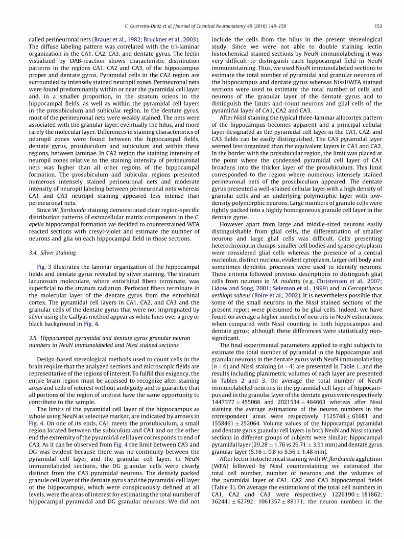
called perineuronal nets (Brauer et al., 1982; Bruckner et al., 2003).The diffuse labeling pattern was correlated with the tri-laminarorganization in the CA1, CA2, CA3, and dentate gyrus. The lectinvisualized by DAB-reaction shows characteristic distributionpatterns in the regions CA1, CA2 and CA3, of the hippocampusproper and dentate gyrus. Pyramidal cells in the CA2 region aresurrounded by intensely stained neuropil zones. Perineuronal netswere found predominantly within or near the pyramidal cell layerand, in a smaller proportion, in the stratum oriens in thehippocampal fields, as well as within the pyramidal cell layersin the prosubiculum and subicular region. In the dentate gyrus,most of the perineuronal nets were weakly stained. The nets wereassociated with the granular layer, eventually the hilus, and morerarely the molecular layer. Differences in staining characteristics ofneuropil zones were found between the hippocampal fields,dentate gyrus, prosubiculum and subiculum and within theseregions, between laminae. In CA2 region the staining intensity ofneuropil zones relative to the staining intensity of perineuronalnets was higher than all other regions of the hippocampalformation. The prosubiculum and subicular regions presentednumerous intensely stained perineuronal nets and moderateintensity of neuropil labeling between perineuronal nets whereasCA1 and CA3 neuropil staining appeared less intense thanperineuronal nets.
Since W. floribunda staining demonstrated clear region-specificdistribution patterns of extracellular matrix components in the C.
apella hippocampal formation we decided to counterstained WFAreacted sections with cresyl-violet and estimate the number ofneurons and glia on each hippocampal field in those sections.
3.4. Silver staining
Fig. 3 illustrates the laminar organization of the hippocampalfields and dentate gyrus revealed by silver staining. The stratumlacunosum moleculare, where entorhinal fibers terminate, wassuperficial to the stratum radiatum. Perforant fibers terminate inthe molecular layer of the dentate gyrus from the entorhinalcortex. The pyramidal cell layers in CA1, CA2, and CA3 and thegranular cells of the dentate gyrus that were not impregnated bysilver using the Gallyas method appear as white lines over a grey orblack background in Fig. 4.
3.5. Hippocampal pyramidal and dentate gyrus granular neuron
numbers in NeuN immunolabeled and Nissl stained sections
Design-based stereological methods used to count cells in thebrain require that the analyzed sections and microscopic fields arerepresentative of the regions of interest. To fulfill this exigency, theentire brain region must be accessed to recognize after stainingareas and cells of interest without ambiguity and to guarantee thatall portions of the region of interest have the same opportunity tocontribute to the sample.
The limits of the pyramidal cell layer of the hippocampus aswhole using NeuN as selective marker, are indicated by arrows inFig. 4. On one of its ends, CA1 meets the prosubiculum, a smallregion located between the subiculum and CA1 and on the otherend the extremity of the pyramidal cell layer corresponds to end ofCA3. As it can be observed from Fig. 4 the limit between CA3 andDG was evident because there was no continuity between thepyramidal cell layer and the granular cell layer. In NeuNimmunolabeled sections, the DG granular cells were clearlydistinct from the CA3 pyramidal neurons. The densely packedgranule cell layer of the dentate gyrus and the pyramidal cell layerof the hippocampus, which were conspicuously defined at alllevels, were the areas of interest for estimating the total number ofhippocampal pyramidal and DG granular neurons. We did not
include the cells from the hilus in the present stereologicalstudy. Since we were not able to double staining lectinhistochemical stained sections by NeuN immunolabeling it wasvery difficult to distinguish each hippocampal field in NeuNimmunostaining. Thus, we used NeuN immunolabeled sections toestimate the total number of pyramidal and granular neurons ofthe hippocampus and dentate gyrus whereas Nissl/WFA stainedsections were used to estimate the total number of cells andneurons of the granular layer of the dentate gyrus and todistinguish the limits and count neurons and glial cells of thepyramidal layer of CA1, CA2 and CA3.
After Nissl staining the typical three-laminar allocortex patternof the hippocampus becomes apparent and a principal cellularlayer designated as the pyramidal cell layer in the CA1, CA2, andCA3 fields can be easily distinguished. The CA3 pyramidal layerseemed less organized than the equivalent layers in CA1 and CA2.In the border with the prosubicular region, the limit was placed atthe point where the condensed pyramidal cell layer of CA1broadens into the thicker layer of the prosubiculum. This limitcorresponded to the region where numerous intensely stainedperineuronal nets of the prosubiculum appeared. The dentategyrus presented a well-stained cellular layer with a high density ofgranular cells and an underlying polymorphic layer with low-density polymorphic neurons. Large numbers of granule cells weretightly packed into a highly homogeneous granule cell layer in thedentate gyrus.
However apart from large and middle-sized neurons easilydistinguishable from glial cells, the differentiation of smallerneurons and large glial cells was difficult. Cells presentingheterochromatin clumps, smaller cell bodies and sparse cytoplasmwere considered glial cells whereas the presence of a centralnucleolus, distinct nucleus, evident cytoplasm, larger cell body andsometimes dendritic processes were used to identify neurons.These criteria followed previous descriptions to distinguish glialcells from neurons in M. mulatta (e.g. Christensen et al., 2007;Lidow and Song, 2001; Selemon et al., 1999) and in Cercopithecus
aethiops sabeus (Boire et al., 2002). It is nevertheless possible thatsome of the small neurons in the Nissl stained sections of thepresent report were presumed to be glial cells. Indeed, we havefound on average a higher number of neurons in NeuN estimationswhen compared with Nissl counting in both hippocampus anddentate gyrus; although these differences were statistically non-significant.
The final experimental parameters applied to eight subjects toestimate the total number of pyramidal in the hippocampus andgranular neurons in the dentate gyrus with NeuN immunolabeling(n = 4) and Nissl staining (n = 4) are presented in Table 1, and theresults including planimetric volumes of each layer are presentedin Tables 2 and 3. On average the total number of NeuNimmunolabeled neurons in the pyramidal cell layer of hippocam-pus and in the granular layer of the dentate gyrus were respectively1447377 � 455066 and 2021534 � 464663 whereas after Nisslstaining the average estimations of the neuron numbers in thecorrespondent areas were respectively 1125748 � 61681 and1558461 � 252064. Volume values of the hippocampal pyramidaland dentate gyrus granular cell layers in both NeuN and Nissl stainedsections in different groups of subjects were similar: hippocampalpyramidal layer (29.28 � 1.76 vs 26.71 � 3.91 mm) and dentate gyrusgranular layer (5.16 � 0.8 vs 5.56 � 1.48 mm).
After lectin histochemical staining with W. floribunda agglutinin(WFA) followed by Nissl counterstaining we estimated thetotal cell number, number of neurons and the volumes ofthe pyramidal layer of CA1, CA2 and CA3 hippocampal fields(Table 3). On average the estimations of the total cell numbers inCA1, CA2 and CA3 were respectively 1226190 � 181862;362441 � 62792; 1061357 � 88171; the neuron numbers in the

[(Fig._4)TD$FIG]
Fig. 4. Low power photomicrographs of a coronal series of sections immunostained
for NeuN to illustrate hippocampus pyramidal and granular neurons of dentate
gyrus of the Cebus apella. Black arrowheads indicate the terminus of the pyramidal
cell layer of the hippocampus and white asterisks the end limits of the granular cell
layer of the dentate gyrus. Along these layers we superimposed the counting boxes
to estimate the total number of pyramidal and granular neurons. Stereotaxic
coordinates: top left anterior 12.0; bottom left posterior: 2.5 (Manocha et al., 1968).
Scale bar = 500 mm.
C. Guerreiro-Diniz et al. / Journal of Chemical Neuroanatomy 40 (2010) 148–159154
same areas 569341 � 75717; 171916 � 16066; 384491 � 55205 andthe volumes of each layer15.69 � 1.34; 4.02 � 0.32 and 9.57 � 0.96.
It was not found a simple linear correlation between theplanimetric volumes of the hippocampal pyramidal and granularlayers and the numbers of cells estimated by optical fractionator. Inmost cases, the variance introduced by methodological proceduresfor both volume and cell number estimations were <50% of theobserved group variance, giving a CE2/CV2 ratio of<0.5 (Slomiankaand West, 2005). In cases of experimental groups that did notfollow this rule, the ratio CE2/CV2 higher than 0.5, was not
indicative of large variance introduced by stereological analysis. Inthis exceptions both biological variance (6.86%) and CEs (5%) werelow and the general rule was neither meaningful nor practical tofollow; see discussion in Slomianka and West (2005).
Taken together, the image analysis of lectin histochemicalstaining with W. floribunda agglutinin (WFA), enzyme-histochem-ical detection of NADPH-diaphorase activity and Gallyas silver andNissl staining conspicuously defined the architectural limits andlayers of interest in the hippocampus and dentate gyrus of theCebus monkey.
4. Discussion
In the present study, we proposed a parcellation of thehippocampal fields and dentate gyrus of the New World monkeyC. apella based on NeuN immunolabeling, lectin histochemicalstaining with W. floribunda agglutinin (WFA), enzyme-histochem-ical detection of NADPH-diaphorase activity and Gallyas silverstaining. We also estimated the number of total neurons in thepyramidal cell layer of hippocampus and the granular cell layer ofthe dentate gyrus using the optical fractionator method. C. apella
were found to have 1.3 times fewer pyramidal and 3.5 times fewergranular neurons than M. mulatta as compared to NeuN estima-tions. The results demonstrated that C. apella and M. mulatta
revealed similar structural organization of the hippocampus anddentate gyrus but significant differences in the regional distribu-tion of neurons.
4.1. Architectonic limits
The hippocampal formation seems to exhibit a veryconservative morphological pattern of evolution where hippo-campal and parahippocampal activities are modulated by boththe intrinsic circuitry and the divergent neocortical inputs(Manns and Eichenbaum, 2006). On the other hand, theconnectivity of the cortical areas in the parahippocampal regionis less conserved, but it shares a similar organizational schemeacross species (Manns and Eichenbaum, 2006). In the presentstudy, we found similar structural organization between thehippocampus and dentate gyrus of M. mulatta and C. apella. Theborders of the hippocampal fields in Cebus were based on lectinhistochemical staining with W. floribunda agglutinin (WFA),enzyme-histochemical detection of NADPH-diaphorase activityand Gallyas silver staining. NADPH-diaphorase histochemistryand Gallyas revealed conspicuously the limit between CA2 andCA3 fields based on the presence of a well-defined stratumlucidum in CA3 but not in CA2. However the limits between CA1and CA2 and between CA1 and prosubiculum were much clearerafter W. floribunda histochemical reaction. It is difficult tocompare architectonic limits of CA1, CA2 and CA3 in Cebus andRhesus monkey by W. floribunda due to the absence ofinformation of architectonic limits using this marker in theRhesus monkey hippocampus.
In the context of the functional regionalization of thehippocampus, a number of investigations have been dedicatedto CA1, CA3, and the dentate gyrus, but previous research hasmostly neglected CA2 (Zhao et al., 2007). CA2 has been described inall species as the narrow zone of cells interposed between CA3 andCA1 and that, like CA1, does not receive mossy fiber innervations,has distinct neurochemical markers (Sakurai and Kosaka, 2007;Seress et al., 1993), and exhibits a special regulation of post-synaptic calcium concentration that reduces synaptic plasticity(Simons et al., 2009). In CA2, some of the extracellular matrixcomponents are found in greater abundance than in CA1 andCA3 (Bruckner et al., 2003), and several genes are differentiallyexpressed in CA2 neurons (Lein et al., 2005). In addition,

Table 2Estimated individual unilateral planimetric volumes of hippocampus pyramidal and dentate gyrus granular layers, and correspondent unilateral number of Nissl stained neurons and total number of cells in comparison with NeuN
immunostained neurons estimations in the Cebus apella.
Subjects Hippocampus Dentate Gyrus
Thickness Volume (mm3) CE Total cells CE Neurons CE Volume (mm3) CE Total cells CE Neurons CE Scheaffer Thickness
Nissl staining
M1 25.86�0.44 30.66 0.0467 1,766,449 0.03 1,182,829 0.0537 5.87 0.02 1,766,449 0.030 1,609,314 0.034 19.96�0.64
M2 29�0.15 26.74 0.0467 1,627,199 0.038 1,050,048 0.0587 4.07 0.03 1,627,199 0.038 1,552,788 0.041 24.02�1.03
M3 26.35�0.43 29.54 0.0367 2,508,485 0.044 1,101,505 0.0546 4.96 0.03 1,274,051 0.055 1,230,118 0.056 22.04�1.13
M4 24.46�0.33 30.20 0.03 2,544,047 0.047 1,168,610 0.0535 5.73 0.02 1,893,306 0.046 1,841,625 0.047 19.45�0.46
Mean 29.28 0.04 2,111,545 0.04 1,125,748 0.05 5.16 0.025 1,640,251 0.042 1,558,461 0.04
SD 1.76 482,460 61,681 0.828 267,230 252,064
CV2 0.0036 0.0522 0.003 0.02578 0.02654 0.02616
CE2 0.0016 0.0016 0.003 0.00063 0.00179 0.00198
CE2/CV2 0.444 0.0303 0.9993 0.02424 0.06741 0.07570
CVB2 0.0020 0.0506 0.000002 0.02516 0.02475 0.02418
CVB2 (%CV2) 55.89% 96.97% 6.86% 97.58% 93.26% 92.43%
NeuN immunolabeling
M5 27.13�0.22 28.99 0.0367 1,329,712 0.041 7.46 0.03 2,182,540 0.035 17.58�0.83
M6 24.6 24.6�0.13 21.42 0.0433 1,357,530 0.043 3.9 0.05 1,433,374 0.028 21.53�0.57
M7 20.63�0.37 30.25 0.0277 2,088,632 0.040 5.71 0.02 2,538,962 0.018 18.63�0.36
M8 18.39�0.17 26.19 0.037 1,013,636 0.048 5.16 0.05 1,931,259 0.038 18.03�0.22
Mean 26.71 0.04 1,447,377 0,04 5.56 0.04 2,021,534 0,03
SD 3.91 455,066 1.48 464,663
CV2 0.021 0.0098 0.073 0.053
CE2 0.0013 0.0016 0.0016 0,0009
CE2/CV2 0.062 0.16 0.22 0.002
CVB2 0.02 0.0082 0.0714 0.052
CVB2 (%CV2) 93.8% 83.67% 97.8% 98.3%
C.
Gu
erreiro-D
iniz
eta
l./Jou
rna
lo
fC
hem
ical
Neu
roa
na
tom
y4
0(2
01
0)
14
8–
15
91
55

Table 3Estimated individual unilateral planimetric volumes of CA1, CA2 and CA3 pyramidal and dentate gyrus granular layers, and correspondent unilateral number of Nissl stained neurons and total number of cells in the Cebus apella.
Subjects CA1 CA2 CA3
Volume
(mm3)
CE N Thickness
(mm)
CE
(Scheaffer)
Volume
(mm3)
CE N Thickness
(mm)
CE
(Scheaffer)
Volume
(mm3)
CE N Thickness CE
(Scheaffer)
Total cells
M1 17.14 0.05 1,482,281 26.38� 0.80 0.03 3.99 0.05 357,269 26.22� 0.51 0.05 9.53 0.04 1,143,055 24.98� 0.88 0.05
M2 14.18 0.06 1,171,209 28.8� 0.80 0.04 4.33 0.04 449,544 29.3� 0.65 0.05 8.23 0.04 944,063 28.9� 0.61 0.05
M3 15.01 0.03 1,053,547 27.18� 0.64 0.04 4.19 0.05 342,194 25.76� 0.78 0.06 10.34 0.03 1,112,744 26.1� 0.85 0.04
M4 16.43 0.02 1,197,724 24.9� 0.42 0.04 3.59 0.04 300,756 24.67� 0.67 0.05 10.18 0.03 1,045,566 23.8� 0.52 0.05
Mean 15.69 0.04 1,226,190 26.81 0.04 4.02 0.04 362,441 26.49 0.05 9.57 0.03 1,061,357 25.94 0.05
SD 1.34 181,862 1.62 0.32 62,792 1.98 0.96 88,171 2.18
CV2 0.022 0.03 0.007
CE2 0.001 0.0027 0.002
CE2/CV2 0.045 0.09 0.29
CVB2 0.021 0.0273 0.005
CVB2(%CV2) 95.45% 91% 71%
Neurons
M1 658,824 0.046 162,724 0.06 361,280 0.06
M2 530,554 0.054 195,532 0.063 323,962 0.06
M3 487,413 0.05 160,953 0.068 453,139 0.05
M4 600,572 0.048 168,454 0.063 399,584 0.05
Mean 569,341 0.05 171,916 0.06 384,491 0.05
SD 75,717 16,066 55,205
CV2 0.0177 0.0086 0.02
CE2 0.0024 0.004 0.003
CE2/CV2 0.13 0.46 0.15
CVB2 0.0153 0.0046 0.017
CVB2(%CV2) 86% 53.49% 85%
CVB2 = CV2�CE2 (CV, coefficient of variation; CVB, biological coefficient of variation). Mean, N: mean group numbers; SD, standard deviation.
C.
Gu
erreiro-D
iniz
eta
l./Jou
rna
lo
fC
hem
ical
Neu
roa
na
tom
y4
0(2
01
0)
14
8–
15
91
56

Table 4Estimations of unilateral neuronal numbers (�106) in the pyramidal cell layers of
hippocampus and the granular cell layer of the dentate gyrus.
Species and reference Hippocampus Dentate gyrus
Macaca mulatta
Keuker et al. (2003) 1.97 7.47
Jabes et al. (2010) 7.21
Christensen et al. (2007) 2.52
Rapp et al., unpublished data
(Amaral and Lavenex, 2007)
2.57a
Cebus apella
NeuN immunolabeling 1.45 2.0
Nissl staining 1.13 1.64
Calitrix jachus
Kozorovitskiy et al. (2005) 1.82
a Lavenex et al., unpublished data (Amaral and Lavenex, 2007).
C. Guerreiro-Diniz et al. / Journal of Chemical Neuroanatomy 40 (2010) 148–159 157
W. floribunda histochemistry selectively labels n-acetyl-galactos-amine moieties from chemically specialized forms of theextracellular matrix defining conspicuous limits for CA3/CA2/CA1/prosubiculum, which was very much similar to othermammals (Bruckner et al., 2003; Tannan et al., 2007), and theselimits were used to increase precision in the definitions of thehippocampal fields.
If these criteria appropriately defined the limits of CA2 and thehigher concentration of glycosaminoglycans is also a sign ofreduced plasticity (Pizzorusso et al., 2002), protection of memoriesfrom erasure (Gogolla et al., 2009), and resistance to injury, CA2will represent an special area of synaptic stabilization in C. apella.
4.2. Number of neurons in the hippocampus and dentate gyrus and
cognitive capabilities
Previously published data on M. mulatta gave the followingneuron numbers: 7.47 million for the dentate gyrus (considering 1-to 4-year-old monkeys in Table 2) (Keuker et al., 2003) whichcorresponds well with the total estimation of 7.21 million recentlypublished (Jabes et al., 2010). These numbers are different fromprevious data published by Amaral and Lavenex (2007). Indeed thedefinition of the hilus by Keuker et al. (2003) does not correspondto the one given by Amaral and Lavenex (2007). According toKeuker’s estimations (Keuker et al., 2003), Rhesus monkeys wouldhave 3.5 times more neurons in the granule cell layer (7 vs 2millions), and 1.3 times more pyramidal neurons in thehippocampus as compared to NeuN C. apella estimations.
As New and Old World monkeys diverged into separate lines ofevolution some 35 million years ago (Poux et al., 2006; Schrago andRusso, 2003) the number of neurons and volumes of bothhippocampus and dentate gyrus do not necessarily need to bethe same. Indeed the volume of the pyramidal cell layer ofhippocampus of the M. mulatta (31 mm3) (Christensen et al., 2007)seems to be closer to the equivalent region of the C. apella
(29 mm3) whereas the volume of the granular layer of the dentategyrus in the capuchin monkey (5.16 mm3) seems to be on averagetwice smaller than the M. mulatta equivalent region (11.00 mm3)(Jabes et al., 2010). However the comparison of the volumes of thehippocampal pyramidal cell layer of CA1 (23.45 � 3.76 mm3) andCA2/CA3 (7.4 � 2.76 mm3) in M. mulatta (Christensen et al., 2007)revealed that CA1 represents 76% of the total volume of thehippocampal pyramidal cell layer whereas in the C. apella
(15.69 � 1.34; 13.59 � 0.81) CA1 represents only 53%. The propor-tions between pyramidal neurons in CA1, CA2, CA3 and granularneurons of dentate gyrus in the C. apella (569 � 103; 172 � 103;384 � 103; 1558 � 103) and Macaca mullata (1138 � 103; 99 � 103;510 � 103 and 7160 � 103) (Jabes et al., 2010; Keuker et al., 2003) didnot follow a simple correlation with equivalent volumes. Indeed CA2in C. apella represent 18% of the total pyramidal neurons whereas inMacaca mullata only 6%, and the ratio granular/pyramidal neurons inthe Cebus is 1.4 whereas in the Macaca is around 4.0.
In view of the significant differences in the number of neuronsof the dentate gyrus granular and CA2 pyramidal cell layersbetween C. apella and M. mulatta, it would be interesting tocompare the remarkable cognitive capabilities of C. apella and howthey compare with those in M. mulatta.
For many investigators the remarkable cognitive capabilities ofthe C. apella constitute evidence that in spite of the widephylogenetic separation, most of its complex behavior overlapswith cercopithecines and apes (Visalberghi and Fragaszy, 1995;Visalberghi, 1997). As an example it has been suggested thatcapuchins developed all cognitive capabilities required to use toolsin the foraging repertoire modeling the emergence of early humantechnology (Cleveland et al., 2004; de Resende et al., 2008;Westergaard and Suomi, 1997). Tool using behavior, apart from
manipulative skills, requires intense interest in new objects andnatural tendency to explore new environments (Visalberghi, 1990)and the recognition of novelty associated with those tasks seems toinvolve hippocampal and dentate gyrus activity (Hunsaker et al.,2008; Manns and Eichenbaum, 2009; Rolls, 2010). To assess thesebehaviors based on the novelty paradigm a number of hippocam-pal-dependent tasks using object recognition and spatial memorywere applied successfully to C. apella (De Lillo et al., 2007; Janson,1998; Poti et al., 2010) and M. mulatta (Bachevalier and Nemanic,2008; Pascalis et al., 2009; Rolls et al., 2005) confirming that bothspecies present all cognitive requirements for tool using behavior,albeit to different extents. It may be possible that the quantitativeanatomical distinction described in the present report maycontribute at least in part to the differences in cognitiveperformances described elsewhere. Since in the large group ofNew World primates apart from the present report, a singleunbiased based stereological study in C. jachus (Kozorovitskiy et al.,2005) is available, further comparative studies are necessary toapproach these questions.
4.3. Non-stereological source of bias
Table 4 is a comparative survey of different estimations in theRhesus monkey from different laboratories and the estimationsfrom the present study regarding the Cebus monkey.
Are these differences between the estimations illustrated inTable 4 consequences of different methodologies, simply biologicalvariation, or ambiguities in the definition of the region and objectsof interest?
In a comparative analysis of studies on different species andlaboratories, it is important to consider different sources of non-stereological bias that may affect the results, including ambiguitiesin the definition of the objects and areas of interest and differencesin tissue processing (Guillery, 2002; Guillery and August, 2002). Toverify possible sources of errors the total number of hippocampalpyramidal and granular DG neurons were estimated after Nisslstaining and NeuN immunolabeling. On average we have obtainedsimilar results in these counts suggesting that the criteria todistinguish glial cells from neurons were adequate. Finally non-biological sources of variability were reduced in the present reportto acceptable levels as previously described (Mouton et al., 2002;Slomianka and West, 2005).
5. Conclusion
Comparative studies of the structural organization of the brainare fundamental to our understanding of human brain function.Our findings provide for the first time new information about thequalitative and quantitative structural organization of the normal

C. Guerreiro-Diniz et al. / Journal of Chemical Neuroanatomy 40 (2010) 148–159158
young adult hippocampus and dentate gyrus of the Neotropicalspecies C. apella using NeuN as a selective neuronal marker incomparison to Nissl stained neurons. We detected similararchitectonic organization in the hippocampus and dentate gyrusof Cebus and Rhesus monkeys, but also important regionaldifferences in the contribution to the total neuron numbers inthe hippocampus and dentate gyrus. Other studies are needed toinvestigate the possible implications of these regional differenceson information processing in the hippocampus and dentate gyrusof this species.
Acknowledgements
Brazilian Research Council – CNPq; Grant number: 307749/2004-5 and 471077/2007-0 for CWPD and FINEP, InstitutoBrasileiro de Neurociencias – IBNnet.
References
Ajmo, J.M., Eakin, A.K., Hamel, M.G., Gottschall, P.E., 2008. Discordant localization ofWFA reactivity and brevican/ADAMTS-derived fragment in rodent brain. BMCNeurosci. 9, 14.
Amaral, D.G., Lavenex, P., 2007. Hippocampal neuroanatomy. In: Andersen, P.,Morris, R.G., Amaral, D.G., Bliss, T., O’Keefe, J. (Eds.), The Hippocampus Book.Oxford University Press, New York, pp. 37–114.
Bachevalier, J., Nemanic, S., 2008. Memory for spatial location and object-placeassociations are differently processed by the hippocampal formation, para-hippocampal areas TH/TF and perirhinal cortex. Hippocampus 18, 64–80.
Boire, D., Theoret, H., Ptito, M., 2002. Stereological evaluation of neurons and glia inthe monkey dorsal lateral geniculate nucleus following an early cerebralhemispherectomy. Experimental brain research. Experimentelle Hirnforschung142, 208–220.
Bonthius, D.J., McKim, R., Koele, L., Harb, H., Karacay, B., Mahoney, J., Pantazis, N.J.,2004. Use of frozen sections to determine neuronal number in the murinehippocampus and neocortex using the optical disector and optical fractionator.Brain Res. Brain Res. Protoc. 14, 45–57.
Brauer, K., Werner, L., Leibnitz, L., 1982. Perineuronal nets of glia. J. Hirnforsch. 23,701–708.
Bruckner, G., Grosche, J., Hartlage-Rubsamen, M., Schmidt, S., Schachner, M., 2003.Region and lamina-specific distribution of extracellular matrix proteoglycans,hyaluronan and tenascin-R in the mouse hippocampal formation. J. Chem.Neuroanat. 26, 37–50.
Bruckner, G., Seeger, G., Brauer, K., Hartig, W., Kacza, J., Bigl, V., 1994. Cortical areasare revealed by distribution patterns of proteoglycan components and parval-bumin in the Mongolian gerbil and rat. Brain Res. 658, 67–86.
Christensen, J.R., Larsen, K.B., Lisanby, S.H., Scalia, J., Arango, V., Dwork, A.J.,Pakkenberg, B., 2007. Neocortical and hippocampal neuron and glial cellnumbers in the rhesus monkey. Anat. Rec. (Hoboken) 290, 330–340.
Cleveland, A., Rocca, A.M., Wendt, E.L., Westergaard, G.C., 2004. Transport of tools tofood sites in tufted capuchin monkeys (Cebus apella). Anim. Cogn. 7, 193–198.
Collins, C.E., Lyon, D.C., Kaas, J.H., 2003. Responses of neurons in the middletemporal visual area after long-standing lesions of the primary visual cortexin adult new world monkeys. J. Neurosci. 23, 2251–2264.
De Lillo, C., Spinozzi, G., Truppa, V., 2007. Pattern recognition in tufted capuchinmonkeys (Cebus apella): the role of the spatial organisation of stimulus parts.Behav. Brain Res. 181, 96–109.
de Resende, B.D., Ottoni, E.B., Fragaszy, D.M., 2008. Ontogeny of manipulativebehavior and nut-cracking in young tufted capuchin monkeys (Cebus apella):a perception-action perspective. Dev. Sci. 11, 828–840.
Ding, S.L., Van Hoesen, G.W., Cassell, M.D., Poremba, A., 2009. Parcellation of humantemporal polar cortex: a combined analysis of multiple cytoarchitectonic,chemoarchitectonic, and pathological markers. J. Comp. Neurol. 514, 595–623.
Dorph-Petersen, K.A., Nyengaard, J.R., Gundersen, H.J., 2001. Tissue shrinkage andunbiased stereological estimation of particle number and size. J. Microsc. 204,232–246.
Dufour, V., Pascalis, O., Petit, O., 2006. Face processing limitation to own species inprimates: a comparative study in brown capuchins, Tonkean macaques andhumans. Behav. Processes 73, 107–113.
Eichenbaum, H.E., Yonelinas, A.P., Ranganath, C., 2007. The medial temporal lobeand recognition memory. Annu. Rev. Neurosci. 30, 123–152.
Eidelberg, E., Saldias, C.A., 1960. A stereotaxic atlas for Cebus monkeys. J. Comp.Neurol. 115, 103–123.
Gallagher, M., Rapp, P.R., 1997. The use of animal models to study the effects ofaging on cognition. Annu. Rev. Psychol. 48, 339–370.
Gallyas, F., 1979. Silver staining of myelin by means of physical development.Neurol. Res. 1, 203–209.
Girgis, M., 1973. Histochemical localization of acetylcholinesterase enzyme in the‘‘limbic system’’ of the brain of the cebus monkey (Cebus apella). Acta Anat.(Basel) 84, 202–223.
Glaser, E.M., Wilson, P.D., 1998. The coefficient of error of optical fractionatorpopulation size estimates: a computer simulation comparing three estimators.J. Microsc. 192, 163–171.
Gogolla, N., Caroni, P., Luthi, A., Herry, C., 2009. Perineuronal nets protect fearmemories from erasure. Science 325, 1258–1261.
Guillery, R.W., 2002. On counting and counting errors. J. Comp. Neurol. 447, 1–7.Guillery, R.W., August, B.K., 2002. Doubt and certainty in counting. Prog. Brain Res.
135, 25–42.Gundersen, H., Jensen, E., 1987. The efficiency of systematic sampling in stereology
and its prediction. J. Microsc. 147, 229–263.Hartig, W., Derouiche, A., Welt, K., et al., 1999. Cortical neurons immunoreactive for
the potassium channel Kv3.1b subunit are predominately surrounded byperineuronal nets presumed as a buffering system for cations. Brain Res.842, 15–29.
Hunsaker, M.R., Rosenberg, J.S., Kesner, R.P., 2008. The role of the dentate gyrus,CA3a,b, and CA3c for detecting spatial and environmental novelty. Hippocam-pus 18, 1064–1073.
Jabes, A., Lavenex, P.B., Amaral, D.G., Lavenex, P., 2010. Quantitative analysis ofpostnatal neurogenesis and neuron number in the macaque monkey dentategyrus. Eur. J. Neurosci..
Janson, C.H., 1998. Experimental evidence for spatial memory in foraging wildcapuchin monkeys, Cebus apella. Anim. Behav. 55, 1229–1243.
Keuker, J.I., Luiten, P.G., Fuchs, E., 2003. Preservation of hippocampal neuronnumbers in aged rhesus monkeys. Neurobiol. Aging 24, 157–165.
Kordower, J.H., Bartus, R.T., Bothwell, M., Schatteman, G., Gash, D.M., 1988. Nervegrowth factor receptor immunoreactivity in the nonhuman primate (Cebusapella): distribution, morphology, and colocalization with cholinergic enzymes.J. Comp. Neurol. 277, 465–486.
Kordower, J.H., Bartus, R.T., Marciano, F.F., Gash, D.M., 1989. Telencephalic cholin-ergic system of the New World monkey (Cebus apella): morphological andcytoarchitectonic assessment and analysis of the projection to the amygdala. J.Comp. Neurol. 279, 528–545.
Kordower, J.H., Fiandaca, M.S., 1990. Response of the monkey cholinergic septo-hippocampal system to fornix transection: a histochemical and cytochemicalanalysis. J. Comp. Neurol. 298, 443–457.
Kordower, J.H., Yaping, C., Maclennan, A.J., 1997. Ciliary neurotrophic factor recep-tor alpha-immunoreactivity in the monkey central nervous system. J. Comp.Neurol. 377, 365–380.
Kozorovitskiy, Y., Gross, C.G., Kopil, C., Battaglia, L., McBreen, M., Stranahan, A.M.,Gould, E., 2005. Experience induces structural and biochemical changes in theadult primate brain. Proc. Natl. Acad. Sci. U.S.A. 102, 17478–17482.
Lein, E.S., Callaway, E.M., Albright, T.D., Gage, F.H., 2005. Redefining the boundariesof the hippocampal CA2 subfield in the mouse using gene expression and 3-dimensional reconstruction. J. Comp. Neurol. 485, 1–10.
Levy, D.A., Hopkins, R.O., Squire, L.R., 2004. Impaired odor recognition memory inpatients with hippocampal lesions. Learn. Mem. 11, 794–796.
Lidow, M.S., Song, Z.M., 2001. Primates exposed to cocaine in utero display reduceddensity and number of cerebral cortical neurons. J. Comp. Neurol. 435, 263–275.
Manns, J.R., Eichenbaum, H., 2006. Evolution of declarative memory. Hippocampus16, 795–808.
Manns, J.R., Eichenbaum, H., 2009. A cognitive map for object memory in thehippocampus. Learn. Mem. 16, 616–624.
Manocha, S.L., Shantha, T.R., Bourne, G.H., 1968. A stereotaxic atlas of the brain ofthe Cebus monkey (Cebus apella). Oxford University Press at Clarendon Press,London, UK.
MicroBright Field, I., 2005. Stereo Investigator 6 User’s Guide Document, 2.10 ed.MicroBright Field, Inc., Williston, Vermont 05945 USA.
Mouton, P.R., Long, J.M., Lei, D.L., Howard, V., Jucker, M., Calhoun, M.E., Ingram, D.K.,2002. Age and gender effects on microglia and astrocyte numbers in brains ofmice. Brain Res. 956, 30–35.
Mufson, E.J., Conner, J.M., Varon, S., Kordower, J.H., 1994. Nerve growth factor-likeimmunoreactive profiles in the primate basal forebrain and hippocampalformation. J. Comp. Neurol. 341, 507–519.
Murakami, T., Ohtsuka, A., Su, W.D., Taguchi, T., Oohashi, T., Abe, K., Ninomiya, Y.,1999. The extracellular matrix in the mouse brain: its reactions to endo-alpha-N-acetylgalactosaminidase and certain other enzymes. Arch. Histol. Cytol. 62,273–281.
Murray, E.A., Bussey, T.J., Saksida, L.M., 2007. Visual perception and memory: a newview of medial temporal lobe function in primates and rodents. Annu. Rev.Neurosci. 30, 99–122.
Naegele, J.R., Katz, L.C., 1990. Cell surface molecules containing N-acetylgalacto-samine are associated with basket cells and neurogliaform cells in cat visualcortex. J. Neurosci. 10, 540–557.
Pascalis, O., Hunkin, N.M., Bachevalier, J., Mayes, A.R., 2009. Change in backgroundcontext disrupts performance on visual paired comparison following hippo-campal damage. Neuropsychologia 47, 2107–2113.
Pizzorusso, T., Medini, P., Berardi, N., Chierzi, S., Fawcett, J.W., Maffei, L., 2002.Reactivation of ocular dominance plasticity in the adult visual cortex. Science298, 1248–1251.
Poti, P., Kanngiesser, P., Saporiti, M., Amiconi, A., Blasing, B., Call, J., 2010. Searchingin the middle-Capuchins’ (Cebus apella) and bonobos’ (Pan paniscus)behavior during a spatial search task. J. Exp. Psychol. Anim. Behav. Process36, 92–109.
Poux, C., Chevret, P., Huchon, D., de Jong, W.W., Douzery, E.J., 2006. Arrival anddiversification of caviomorph rodents and platyrrhine primates in South Amer-ica. Syst. Biol. 55, 228–244.

C. Guerreiro-Diniz et al. / Journal of Chemical Neuroanatomy 40 (2010) 148–159 159
Preuss, T.M., Gray, D., Cusick, C.G., 1998. Subdivisions of the motor and somato-sensory thalamus of primates revealed with Wisteria floribunda agglutininhistochemistry. Somatosens. Motor Res. 15, 211–219.
Resende, M.C., Tavares, M.C., Tomaz, C., 2003. Ontogenetic dissociation betweenhabit learning and recognition memory in capuchin monkeys (Cebus apella).Neurobiol. Learn. Mem. 79, 19–24.
Rolls, E.T., 2010. A computational theory of episodic memory formation in thehippocampus. Behav. Brain Res..
Rolls, E.T., Kesner, R.P., 2006. A computational theory of hippocampal function, andempirical tests of the theory. Prog. Neurobiol. 79, 1–48.
Rolls, E.T., Xiang, J., Franco, L., 2005. Object, space, and object-space representationsin the primate hippocampus. J. Neurophysiol. 94, 833–844.
Sakurai, O., Kosaka, T., 2007. Nonprincipal neurons and CA2 pyramidal cells, but notmossy cells are immunoreactive for calcitonin gene-related peptide in themouse hippocampus. Brain Res. 1186, 129–143.
Saper, C.B., Sawchenko, P.E., 2003. Magic peptides, magic antibodies: guidelines forappropriate controls for immunohistochemistry. J. Comp. Neurol. 465, 161–163.
Schrago, C.G., Russo, C.A., 2003. Timing the origin of New World monkeys. Mol. Biol.Evol. 20, 1620–1625.
Selemon, L.D., Lidow, M.S., Goldman-Rakic, P.S., 1999. Increased volume and glialdensity in primate prefrontal cortex associated with chronic antipsychotic drugexposure. Biol. Psychiatry 46, 161–172.
Seress, L., Gulyas, A.I., Ferrer, I., Tunon, T., Soriano, E., Freund, T.F., 1993. Distribution,morphological features, and synaptic connections of parvalbumin- and calbin-din D28k-immunoreactive neurons in the human hippocampal formation. J.Comp. Neurol. 337, 208–230.
Shu, S.Y., Ju, G., Fan, L.Z., 1988. The glucose oxidase-DAB-nickel method in peroxi-dase histochemistry of the nervous system. Neurosci. Lett. 85, 169–171.
Simons, S.B., Escobedo, Y., Yasuda, R., Dudek, S.M., 2009. Regional differences inhippocampal calcium handling provide a cellular mechanism for limitingplasticity. Proc. Natl. Acad. Sci. U.S.A. 106, 14080–14084.
Slomianka, L., West, M., 2005. Estimators of the precision of stereological estimates:an example based on the CA1 pyramidal cell layer of rats. Neuroscience 136,757–767.
Spinozzi, G., Lagana, T., Truppa, V., 2007. Hand use by tufted capuchins (Cebusapella) to extract a small food item from a tube: digit movements, handpreference, and performance. Am. J. Primatol. 69, 336–352.
Squire, L.R., Stark, C.E., Clark, R.E., 2004. The medial temporal lobe. Annu. Rev.Neurosci. 27, 279–306.
Stark, C., 2007. Funtional of the human hippocampus. In: The Hippocampus Book,Oxford University Press, New York, pp. 549–579.
Tannan, V., Simons, S., Dennis, R.G., Tommerdahl, M., 2007. Effects of adaptation onthe capacity to differentiate simultaneously delivered dual-site vibrotactilestimuli. Brain Res. 1186, 164–170.
Tavares, M.C., Aguiar, L., Tomaz, C., 2002. Effect of practice on hand preference on acolor visual discrimination task by capuchin monkeys (Cebus apella). Percept.Mot. Skills 95, 1027–1034.
VanMarle, K., Aw, J., McCrink, K., Santos, L.R., 2006. How capuchin monkeys(Cebus apella) quantify objects and substances. J. Comp. Psychol. 120, 416–426.
Visalberghi, E., 1990. Tool use in Cebus. Folia Primatol. (Basel) 54, 146–154.Visalberghi, E., 1997. Success and understanding in cognitive tasks: a comparison
between Cebus apella and Pan troglodytes. Int. J. Primatol. 18, 811–830.Visalberghi, E., Fragaszy, D.M., 1995. The behaviour of capuchin monkeys, Cebus
apella, with novel food: the role of social context. Anim. Behav. 49.von Bohlen und Halbach, O., 2007. Immunohistological markers for staging neu-
rogenesis in adult hippocampus. Cell Tissue Res. 329, 409–420.West, M.J., 1993. New stereological methods for counting neurons. Neurobiol. Aging
14, 275–285.West, M.J., 2002. Design-based stereological methods for counting neurons. Prog.
Brain Res. 135, 43–51.West, M.J., Slomianka, L., Gundersen, H.J., 1991. Unbiased stereological estimation
of the total number of neurons in thesubdivisions of the rat hippocampus usingthe optical fractionator. Anat. Rec. 231, 482–497.
Westergaard, G.C., Suomi, S.J., 1997. Capuchin monkey (Cebus apella) grips for theuse of stone tools. Am. J. Phys. Anthropol. 103, 131–135.
Zhao, M., Choi, Y.S., Obrietan, K., Dudek, S.M., 2007. Synaptic plasticity (and the lackthereof) in hippocampal CA2 neurons. J. Neurosci. 27, 12025–12032.