NMDA-Dependent Currents in Granule Cells of the Dentate Gyrus Contribute to Induction but Not...
10
Spontaneous Seizures and Loss of Axo-Axonic and Axo- Somatic Inhibition Induced by Repeated Brief Seizures in Kindled Rats Umit Sayin, 1 * Susan Osting, 2 * Joshua Hagen, 1 * Paul Rutecki, 1,3,4 and Thomas Sutula 1,2 Departments of 1 Neurology, 2 Anatomy, and 3 Neurological Surgery, University of Wisconsin, and 4 The William S. Middleton Veterans Administration Hospital, Madison, Wisconsin 53792 Repeated brief seizures evoked by kindling progressively increase seizure susceptibility and eventually induce spontaneous seizures. Previous studies have demonstrated that the initial seizures evoked by kindling increase paired-pulse inhibition at 15–25 msec interpulse intervals in the dentate gyrus and also induce apoptosis, progressive neuronal loss, mossy fiber sprouting, and neurogenesis, which could potentially alter the balance of excitation and/or inhibition and modify functional properties of hippocampal circuits. In these experi- ments, paired-pulse inhibition in the dentate gyrus was reduced or lost after 90 –100 evoked seizures in association with emergence of spontaneous seizures. Evoked IPSCs examined by single electrode voltage-clamp methods in granule cells from kindled rats experiencing spontaneous seizures demonstrated altered kinetics (reductions of 48% in 10 –90% decay time, 40% in , and 65% in charge transfer) and confirmed that decreased inhibition contributed to the reduced paired-pulse inhibition. The loss of inhibition was accom- panied by loss of subclasses of inhibitory interneurons labeled by cholecystokinin and the neuronal GABA transporter GAT-1, which project axo-somatic and axo-axonic GABAergic inhibitory terminals to granule cells and axon initial segments. Seizure-induced loss of interneurons providing axo-somatic and axo-axonic inhibition may regulate spike output to pyramidal neurons in CA3 and could play an important role in generation of spontaneous seizures. The sequence of progressive cellular alterations induced by repeated seizures, particularly loss of GABAergic interneurons providing axo-somatic and axo-axonic inhibition, may be important in the development of intractable epilepsy. Key words: hippocampus; dentate gyrus; GABA; CCK; GAT-1; damage; epilepsy Introduction Neural circuits undergo structural and functional alterations in response to neural activity in both development and adulthood. In the developing nervous system, experience-dependent activity precisely refines patterns of connectivity to form functional cir- cuits and networks (Catalano and Shatz, 1998; Penn and Shatz, 1999; Swann et al., 2001). In the adult nervous system, synapses undergo use-dependent alterations in synaptic efficacy such as long-term potentiation and long-term depression. In response to intense synchronous neural activity during seizures, neural cir- cuits undergo a complex variety of immediate and long-lasting molecular and cellular alterations that induce progressive, cumu- lative, and permanently increased susceptibility to additional sei- zures, a phenomenon of circuit plasticity referred to as kindling (Goddard, 1969; Goddard et al., 1969). Kindling has been exten- sively investigated as a model of temporal lobe epilepsy (Majkowski, 1999; McNamara, 1999; Mody, 1999; Dalby and Mody, 2001; Sutula, 2001) and is a property of activity- dependent plasticity in cortical, brainstem, and limbic circuits in species ranging from amphibians to primates (Wada et al., 1975, 1978; Morrell and Tsuru, 1976; Cain and Corcoran, 1980). Repeated seizures evoked by kindling predictably induce per- manent structural and functional reorganization of the dentate gyrus and hippocampus (McNamara, 1999; Mody, 1999; Sutula, 2001). The initial seizures evoked by kindling increase NMDA- dependent excitatory synaptic transmission in granule cells of the dentate gyrus (Sayin et al., 1999; Behr et al., 2001) and also induce a sustained increase in GABA A -dependent inhibition demon- strated by a variety of physiological measurements (Tuff et al., 1983a,b; Oliver and Miller, 1985; de Jonge and Racine, 1987; Stringer and Lothman, 1989; Otis et al., 1994; Buhl et al., 1996; Nusser et al., 1998). This initial increase in inhibition appears paradoxical in that kindling gradually and permanently increases seizure susceptibility, but is also consistent with evidence suggest- ing that the dentate gyrus filters neocortical and entorhinal activ- ity converging into the CA3 region of the hippocampus (Loth- man et al., 1992; Behr et al., 1998, 2001). Repeated brief kindled seizures induce apoptosis and neuronal loss in the dentate gyrus and hippocampus (Cavazos and Sutula, 1990; Cavazos et al., 1994; Bengzon et al., 1997; Pretel and Piekut, 1997; Dalby et al., 1998; Kotloski et al., 2002), which are accompanied by sprouting of mossy fiber axons (Sutula et al., 1988, 1998; Represa et al., 1989; Cavazos et al., 1991), reorganization of synaptic connectiv- ity with formation of recurrent excitatory circuits (Wuarin and Dudek, 1996, 2001; Lynch and Sutula, 2000), and gliosis (Adams et al., 1998). The pattern of neuronal loss induced by kindling Received July 25, 2002; revised Dec. 3, 2002; accepted Dec. 9, 2002. This work was supported by National Institute of Neurological Disorders and Stroke Grant RO1 25020 and Veter- ans Administration research. *U.S., S.O., and J.H. contributed equally to this work. Correspondence should be addressed to Dr. T. Sutula, Department of Neurology, H6/570, University of Wisconsin, 600 Highland Avenue, Madison, WI 53792. E-mail: [email protected]. Copyright © 2003 Society for Neuroscience 0270-6474/03/232759-10$15.00/0 The Journal of Neuroscience, April 1, 2003 • 23(7):2759 –2768 • 2759
Transcript of NMDA-Dependent Currents in Granule Cells of the Dentate Gyrus Contribute to Induction but Not...

Spontaneous Seizures and Loss of Axo-Axonic and Axo-Somatic Inhibition Induced by Repeated Brief Seizures inKindled Rats
Umit Sayin,1* Susan Osting,2* Joshua Hagen,1* Paul Rutecki,1,3,4 and Thomas Sutula1,2
Departments of 1Neurology, 2Anatomy, and 3Neurological Surgery, University of Wisconsin, and 4The William S. Middleton Veterans AdministrationHospital, Madison, Wisconsin 53792
Repeated brief seizures evoked by kindling progressively increase seizure susceptibility and eventually induce spontaneous seizures.Previous studies have demonstrated that the initial seizures evoked by kindling increase paired-pulse inhibition at 15–25 msec interpulseintervals in the dentate gyrus and also induce apoptosis, progressive neuronal loss, mossy fiber sprouting, and neurogenesis, which couldpotentially alter the balance of excitation and/or inhibition and modify functional properties of hippocampal circuits. In these experi-ments, paired-pulse inhibition in the dentate gyrus was reduced or lost after �90 –100 evoked seizures in association with emergence ofspontaneous seizures. Evoked IPSCs examined by single electrode voltage-clamp methods in granule cells from kindled rats experiencingspontaneous seizures demonstrated altered kinetics (reductions of �48% in 10 –90% decay time, �40% in �, and �65% in chargetransfer) and confirmed that decreased inhibition contributed to the reduced paired-pulse inhibition. The loss of inhibition was accom-panied by loss of subclasses of inhibitory interneurons labeled by cholecystokinin and the neuronal GABA transporter GAT-1, whichproject axo-somatic and axo-axonic GABAergic inhibitory terminals to granule cells and axon initial segments. Seizure-induced loss ofinterneurons providing axo-somatic and axo-axonic inhibition may regulate spike output to pyramidal neurons in CA3 and could play animportant role in generation of spontaneous seizures. The sequence of progressive cellular alterations induced by repeated seizures,particularly loss of GABAergic interneurons providing axo-somatic and axo-axonic inhibition, may be important in the development ofintractable epilepsy.
Key words: hippocampus; dentate gyrus; GABA; CCK; GAT-1; damage; epilepsy
IntroductionNeural circuits undergo structural and functional alterations inresponse to neural activity in both development and adulthood.In the developing nervous system, experience-dependent activityprecisely refines patterns of connectivity to form functional cir-cuits and networks (Catalano and Shatz, 1998; Penn and Shatz,1999; Swann et al., 2001). In the adult nervous system, synapsesundergo use-dependent alterations in synaptic efficacy such aslong-term potentiation and long-term depression. In response tointense synchronous neural activity during seizures, neural cir-cuits undergo a complex variety of immediate and long-lastingmolecular and cellular alterations that induce progressive, cumu-lative, and permanently increased susceptibility to additional sei-zures, a phenomenon of circuit plasticity referred to as kindling(Goddard, 1969; Goddard et al., 1969). Kindling has been exten-sively investigated as a model of temporal lobe epilepsy(Majkowski, 1999; McNamara, 1999; Mody, 1999; Dalby andMody, 2001; Sutula, 2001) and is a property of activity-dependent plasticity in cortical, brainstem, and limbic circuits in
species ranging from amphibians to primates (Wada et al., 1975,1978; Morrell and Tsuru, 1976; Cain and Corcoran, 1980).
Repeated seizures evoked by kindling predictably induce per-manent structural and functional reorganization of the dentategyrus and hippocampus (McNamara, 1999; Mody, 1999; Sutula,2001). The initial seizures evoked by kindling increase NMDA-dependent excitatory synaptic transmission in granule cells of thedentate gyrus (Sayin et al., 1999; Behr et al., 2001) and also inducea sustained increase in GABAA-dependent inhibition demon-strated by a variety of physiological measurements (Tuff et al.,1983a,b; Oliver and Miller, 1985; de Jonge and Racine, 1987;Stringer and Lothman, 1989; Otis et al., 1994; Buhl et al., 1996;Nusser et al., 1998). This initial increase in inhibition appearsparadoxical in that kindling gradually and permanently increasesseizure susceptibility, but is also consistent with evidence suggest-ing that the dentate gyrus filters neocortical and entorhinal activ-ity converging into the CA3 region of the hippocampus (Loth-man et al., 1992; Behr et al., 1998, 2001). Repeated brief kindledseizures induce apoptosis and neuronal loss in the dentate gyrusand hippocampus (Cavazos and Sutula, 1990; Cavazos et al.,1994; Bengzon et al., 1997; Pretel and Piekut, 1997; Dalby et al.,1998; Kotloski et al., 2002), which are accompanied by sproutingof mossy fiber axons (Sutula et al., 1988, 1998; Represa et al.,1989; Cavazos et al., 1991), reorganization of synaptic connectiv-ity with formation of recurrent excitatory circuits (Wuarin andDudek, 1996, 2001; Lynch and Sutula, 2000), and gliosis (Adamset al., 1998). The pattern of neuronal loss induced by kindling
Received July 25, 2002; revised Dec. 3, 2002; accepted Dec. 9, 2002.This work was supported by National Institute of Neurological Disorders and Stroke Grant RO1 25020 and Veter-
ans Administration research.*U.S., S.O., and J.H. contributed equally to this work.Correspondence should be addressed to Dr. T. Sutula, Department of Neurology, H6/570, University of Wisconsin,
600 Highland Avenue, Madison, WI 53792. E-mail: [email protected] © 2003 Society for Neuroscience 0270-6474/03/232759-10$15.00/0
The Journal of Neuroscience, April 1, 2003 • 23(7):2759 –2768 • 2759
resembles hippocampal sclerosis (Cavazos et al., 1994; Kotloski etal., 2002), the most common lesion observed in human temporallobe epilepsy, and the associated sprouting, gliosis, and progres-sive memory deficits induced by kindling are also prominentfeatures in human temporal lobe epilepsy (Sutula et al., 1995;Dalby and Mody, 2001; Sutula, 2001).
The structural and functional alterations induced by kindlingare eventually accompanied by the emergence of spontaneousseizures (Wada et al., 1975, 1978; Pinel and Rovner, 1978). In thisadvanced stage, kindled animals demonstrate recurring sponta-neous seizures, the defining feature of epilepsy. The specific mo-lecular and cellular alterations in hippocampal circuits associatedwith the emergence of spontaneous seizures are of interest, be-cause these changes represent an advanced or even an end-stageof a continuum of slowly evolving activity-dependent, seizure-induced alterations. These changes are also of potential clinicalimportance for understanding how seizure-induced alterationsmay contribute to intractable human temporal lobe epilepsy.
In this study, the emergence of spontaneous seizures in kin-dled rats was associated with the loss of inhibition in the dentategyrus accompanied by seizure-induced reduction in specific sub-classes of GABAergic interneurons labeled by cholecystokinin(CCK) and the neuronal GABA transporter (GAT-1). These in-terneuron subclasses provide axo-somatic and axo-axonic inhi-bition of the granule cell spike output to CA3 (Leranth andFrotscher, 1986; Halasy and Somogyi, 1993; Freund and Buzsaki,1996; Ribak et al., 1996) and would be expected to play a criticalrole in controlling propagation of activity from the dentate gyrusinto CA3. Preliminary results have been published previously inabstract form (Sutula et al., 2000).
Materials and MethodsSurgical and kindling proceduresAdult male Sprague Dawley rats (250 –350 gm; Harlan, Madison, WI)were anesthetized with ketamine (80 mg/kg, i.m.) and xylazine (10 mg/kg, i.m.) and stereotaxically implanted with an insulated stainless-steelbipolar electrode for stimulation and recording. The electrode was im-planted in either the olfactory bulb (9 anterior, 1.2 lateral, 1.8 ventralwith respect to bregma) or the perforant path (8.1 mm posterior, 4.4 mmlateral, 3.5 ventral with respect to bregma), and was fixed to the skull withacrylic.
After a 2 week recovery period after electrode placement, the unre-strained awake implanted rats received twice daily kindling stimulation(5 d per week) with a 1 sec train of 62 Hz biphasic constant-current 1msec square wave pulses. The stimulation was delivered at the lowestintensity that evoked an afterdischarge (AD) according to standard pro-cedures (Sutula and Steward, 1986; Cavazos et al., 1991). The electroen-cephalogram was recorded from the bipolar electrode that was switchedto the stimulator for the delivery of kindling stimulation. Evoked behav-ioral seizures were classified according to standard criteria and rangedfrom class I seizures (behavioral arrest) to class V seizures (bilateral ton-ic– clonic motor activity with loss of postural tone) (Sutula and Steward,1986), which are comparable with partial complex seizures with second-ary generalization. Kindled rats were killed at a minimum of 18 hr afterthe last evoked seizure for anatomical or physiological analysis. All pro-cedures were reviewed and approved by the Research Animal ResourcesCommittee (University of Wisconsin).
Monitoring for spontaneous seizuresPilot observations in rats undergoing kindling revealed that spontaneousseizures were commonly observed in rats that experienced �90 –100evoked class V seizures during routine twice daily periods of observationbefore kindling stimulation, but were only rarely observed in kindled ratswith fewer evoked seizures. This unsuspected observation suggested thatthe stage of �90 –100 evoked class V seizures was a period of transition toa state with spontaneous seizures. When spontaneous seizures were ob-
served during routine handling, no stimulation was delivered. A subset ofrats that had experienced �90 seizures evoked by kindling was thereforeclosely observed for the occurrence of spontaneous seizures by a behav-ioral monitoring procedure and compared with normal age-matchedcontrols and kindled rats with �90 evoked seizures. The monitoringprocedure consisted of visual observation by trained observers for 8 hrdaily (5 d per week) to identify spontaneous behavioral class V seizuresconsisting of bilateral tonic–clonic movements accompanied by loss of pos-tural tone. Kindled rats were regarded as having reached a stage of sponta-neous kindled seizures after a single spontaneous class V seizure was ob-served. Another subset of kindled rats was also monitored by the sameprocedures until three recurring spontaneous class V seizures were observed.
Hippocampal slice recordingsKindled and age-matched normal rats were anesthetized with ether, andthe brains were removed rapidly and placed into ice-cold artificial CSF(ACSF) with the following composition (in mM): 124 NaCl, 4.5 KCl, 1.25NaH2PO4, 2 MgSO4, 26 NaHCO3, 2 CaCl2, and 10 glucose. Transversehippocampal slices were cut with a vibratome or McIlwain tissue chopperat a thickness of 400 �m and transferred to an interface-recording cham-ber containing oxygenated with 95% O2–5% CO2 ACSF at 31–32°C.Orthodromic synaptic responses were recorded in the granule cell layerof the dentate gyrus with extracellular borosilicate electrodes containing2 M NaCl (impedance of 5–10 M�). The responses were evoked by mo-nopolar constant-current stimuli (50 �sec duration) delivered by elec-trodes placed in the stratum moleculare of the dentate gyrus in the regionof the perforant path. Slices that showed maximum field EPSPs of �1 mVwere discarded. Input– output curves were generated using a sequence ofincreasing stimulation intensities ranging from below threshold for thepopulation EPSP to supramaximal. Evoked responses were recorded,stored, and analyzed using a Digidata 1200 AD converter, pClamp 6.02,and Clampfit 6.02 (Axon Instruments, Foster City, CA).
Paired-pulse measurements. Paired-pulse responses were evoked by theapplication of a pair of 0.05 msec test pulses at interpulse intervals of15–350 msec using the lowest stimulus intensity that evoked the maxi-mum population-spike response. The population-spike amplitudeevoked by the second pulse was expressed as a fraction of the maximumresponse evoked by the first pulse of each pair. Pulse pairs were applied at0.03 Hz.
Single-electrode voltage-clamp measurements of evoked IPSCs. IPSCswere recorded by single-electrode voltage-clamp methods in transverseslices of hippocampus and dentate gyrus that were removed from a sub-set of kindled and age-matched adult rats. Recording and analysis ofkinetics of IPSCs in neurons of 1- to 2-year-old kindled and age-matchedcontrol rats are technically difficult and usually preclude patch-clampmethods. However, single-electrode voltage-clamp methods have beensuccessfully used at advanced stages of kindling (Sayin et al., 1999). Re-cordings were obtained in hippocampal slices as in preceding sections,with an Axoclamp 2B amplifier in the single-electrode voltage-clampmode at a switching frequency of 3–5 kHz. A separate oscilloscope wasused to adjust capacitance compensation and sampling rate. Voltage-clamp recordings in granule cells were obtained using borosilicate elec-trodes (25– 40 M�) filled with 2 M Cs-acetate to block K � conductanceand 50 mM QX314 to block action potentials. Monosynaptic IPSCs re-corded in the presence of 20 �M DNQX and 50 �M APV to block EPSCswere evoked by a stainless-steel bipolar stimulating electrode placed inthe granule cell layer as close as possible to the recording electrode todirectly activate axons of interneurons projecting to the recorded granulecell. The stimulation electrode was typically within 1 mm of the site of therecording electrode, and constant-current stimulus pulses of 50 �secduration were delivered at a range of intensities. The input– output rela-tionship was determined, and the minimum intensity that evoked themaximal current was used. This standardized stimulus intensity mini-mized variability introduced by using the range of intensities that evoke asupramaximal response or the intensity that evokes 50% of maximalresponse, and did not significantly differ between control and kindledrats. IPSCs were recorded at a range of holding potentials from �90 to 30mV in 10 mV steps. Recordings that demonstrated voltage-dependentcontaminants such as regenerative currents indicated by escape voltage
2760 • J. Neurosci., April 1, 2003 • 23(7):2759 –2768 Sayin et al. • Spontaneous Seizures and Loss of GABA Inhibition
transients or clear polysynaptic components were excluded. The evokedcurrents were stored and analyzed using pClamp 6.02, Clampex 6.02, andClampfit 6.02 for analysis of 10 –90% rise time constants, decay timeconstants, and signal analysis. Charge transfer was calculated as the inte-grated area under the curve of evoked synaptic current from the stimulusto 250 msec at a holding potential of �40 mV, which produces outwardcurrent. These methods were successful for recording synaptic currentsin granule cells from relatively old kindled rats and age-matched controls(1–2 years of age after many months of stimulation required to evoke90 –150 class V seizures).
Immunohistochemistry for cholecystokinin, parvalbumin, and theneuronal GABA transporterAt a minimum of 24 hr after the last evoked seizure, the rats were anes-thetized with chloral hydrate and perfused with a freshly prepared solu-tion of 4% paraformaldehyde. The fixed brains were stored overnight incold fixative, cryoprotected in a saturated solution of sucrose in fixativefor a minimum of 24 hr, and sectioned horizontally with a microtome ata thickness of 60 �m. The sections were washed in PBS, 0.3% TritonX-100, and 2% bovine serum albumin (BSA) for 20 min, followed by a 45min incubation in this mixture with 20% normal serum added. Thesections were then incubated overnight in a mixture of 1% normal goatserum with primary anti-mouse antibodies specific for parvalbumin(PV; concentration, 1:10,000; Sigma, St. Louis, MO), primary anti-rabbitantibodies specific for cholecystokinin (concentration, 1:10,000; Sigma),and the neuronal GABA transporter GAT-1 (concentration, 1:1000;Chemicon, Temecula, CA). After incubation, the sections were washedbriefly; washed twice for 15 min in PBS, 0.3% Triton X-100, and 2% BSA;and then reacted for 3 hr with biotinylated anti-mouse or anti-rabbit IgGin goat serum, followed by multiple washes in PBS, 0.3% Triton X-100,and 2% BSA and reaction for 1 hr in ABC reagent (Vector Laboratories,Burlingame, CA). The sections were again washed in PBS for 25 min. Thechromogen reaction was 5–10 min in diaminobenzidine (0.04% in0.01% H2O2 to reduce endogenous peroxide activity), followed bywashes in PBS. Reaction product was intensified in some of the sectionsby treatment with 1.4% AgNO3 for 1 hr at 56°C, followed by washing indistilled H2O, fixing in 5% Na2S2O3 for 10 min, and washing in distilledH2O. The sections were then treated briefly with 0.2% HAuCl4, and finalwashes were performed before mounting, dehydration, and coverslip-ping. All sections from kindled and control rats were batch-processed toensure identical reaction conditions.
Counting of CCK-labeled neurons. CCK-labeled interneurons in thedentate gyrus are typically located along the subgranular region near theborder of the granule cell layer and the hilus, and are also scatteredthroughout the hilus. Preliminary experiments provided evidence thatinterneurons labeled by CCK were reduced in the dentate gyrus of kin-dled rats that experienced spontaneous seizures. Thus, a stereologicalevaluation was performed to provide a quantitative estimate of possibledifferences in the number of CCK-labeled interneurons in kindled ratscompared with age-matched normal controls. After the rats were killed,perfused, and fixated overnight as described previously, the hippocampiwere removed from this subset of rats, extended longitudinally along theseptotemporal hippocampal axis, and sectioned transversely (i.e., or-thagonal to the septotemporal axis, saving every section along the entireseptotemporal length of the hippocampus). These sections from kindledand control rats were batch-processed for immunoreactivity to CCK asdescribed previously, with the exception that intensification was not per-formed. Profile counts of all labeled cells with neuronal morphology wereobtained by manual counting using a camera lucida in each section of aregion including the entire granule cell layer and hilus. In sections at themost septal pole, the granule cell layer and hilus form an elliptical closedstructure, and all labeled profiles were counted within this elliptical area.In the majority of sections in which the dentate gyrus forms a “U-shaped”structure opening into CA3c, all labeled profiles were counted within thegranule cell layer and hilus, as defined by borders extending from the tipsof the granule cell to the tip of the extension of CA3c pyramidal neuronsinto the hilus.
The investigator who performed the counts was unaware of the iden-tity of the sections, and the order of examination of the sections from
control and kindled rats was randomized. Counts were obtained with a4� objective (0.20 numerical aperture), and all labeled profiles withneuronal morphology that came into focus while moving the microscopeheadstage through the thickness of the section were manually counted. Ineach section, labeled neurons and their relative position in the sectionwere recorded using a camera lucida and were summed to obtain theprofile count per section through the thickness of the section. Previousstudies have demonstrated that the measurement of section thickness orthe z-axis in optical dissector methods is a critical influence in countinganalyses (Guillery and August, 2002). The thickness (t) of each sectionwas determined by measuring the distance between the upper and lowerfocal planes of the section with the stage micrometer of the microscopeusing a 100� oil immersion objective. The stage micrometer was cali-brated by the measurement of 10 �m methylmethacrylate beads (BangsLaboratories, Fishers, IN) that were mounted and coverslipped as fortissue sections to provide similar refractive conditions. The mean profilecount (Ni) for each section was calculated and corrected for doublecounting of profiles cut at the surfaces of consecutive sections using theAbercromie formula: Ni � ni � t/(t � d), where Ni is the corrected profilecount, ni is the uncorrected profile count in the section, t is the sectionthickness, and d is the average profile diameter (Konigsmark, 1969). Forthis analysis, there were no differences in the mean diameter of profiles inthe control and kindled groups, and d � 14.1 � 0.2 �m. The meansection thickness was 17.1 � 0.1 �m. Corrected profile counts (Ni) weresummed through the entire septotemporal length of the hippocampusand were also compared between kindled and control groups in binsdefined by relative position of the consecutive sections along the length ofthe septotemporal axis corresponding to 0 –25, 25–50, 50 –75, and 75–100% of the full-length of the axis.
Design considerations and statistical proceduresAll control and kindled groups were age-matched, and the age ranges ofkindled and control groups always overlapped. Because the experimentswere performed during a 4 year period in which unexpected drop-outscould not be avoided, individual rats were not paired by age or othervariables. With the long duration of animal care required for planningand performing the experiments during this 4 year period, it was notpossible to systematically blind laboratory personnel and investigatorsfrom the identity of the rats undergoing examination, except as noted.There were no systematic differences between unstimulated, electrodeimplanted, age-matched controls and unimplanted age-matched con-trols, so these groups were combined for analysis. Data are reported asmean � SEM. Group differences were evaluated for statistical signifi-cance by ANOVA or Student’s t test. When data were not distributednormally, analyses were performed using a nonparametric ANOVA byranks (Kruskal–Wallis one-way, or Friedman two-way) or the Mann–Whitney rank sum test. When multiple comparisons were required, one-way ANOVA followed by Dunnett’s test or the Newman–Keuls test wereused post hoc for paired comparisons. Inferences on proportions wereassessed by the � 2 test.
ResultsDevelopment of spontaneous seizures in kindled ratsThe physiological and anatomical experiments in this study wereconducted in 96 kindled rats that experienced a range of 1 ADto 200 class V evoked seizures, and 82 age-matched controls.Preliminary behavioral observations revealed that spontaneousseizures were frequently observed during routine handling inkindled rats after �90 class V seizures evoked by kindling stimu-lation. To further define the stage at which spontaneous seizuresemerge in rats undergoing repeated kindling stimulation, 27 kin-dled rats with 90 evoked class V seizures were systematicallyobserved for spontaneous seizures during 8 hr of daily behavioralmonitoring and were compared with a group of age-matchedunstimulated controls, including a subset of control rats withimplanted electrodes that never received kindling stimulation.During this systematic monitoring, spontaneous seizures wereobserved in 11 of 27 kindled rats with 90 evoked class V sei-
Sayin et al. • Spontaneous Seizures and Loss of GABA Inhibition J. Neurosci., April 1, 2003 • 23(7):2759 –2768 • 2761

zures, but no spontaneous seizures were observed in the controlgroup of age-matched normal rats (� 2; p � 0.001). Spontaneousseizures were not observed in kindled rats with �90 class Vevoked seizures.
In a subset of kindled rats with �100 evoked class V seizuresevoked by olfactory bulb stimulation, additional systematic be-havioral monitoring to a criterion of threespontaneous seizures revealed that thefirst observed spontaneous seizure was re-liably followed by recurring spontaneousseizures (n � 6 of 6 rats) that confirmedthat kindled rats with 90 –100 class Vseizures are likely to experience recurringunprovoked spontaneous seizures. Spon-taneous seizures were observed after90 –100 class V seizures evoked by kin-dling of the olfactory bulb or the perforantpath, and therefore did not appear to berelated to the site of kindling stimulation.
Evolving alterations in paired-pulseinhibition in the dentate gyrus ofkindled ratsIt was of interest to consider whether theemergence of spontaneous seizures in kin-dled rats was caused by seizure-induced al-terations in inhibition. As a preliminary as-sessment of the state of inhibition in kindledrats experiencing spontaneous seizures,paired-pulse measurements were per-formed in hippocampal slices from rats atvarious stages of kindling, including after 90class V seizures when spontaneous seizuresbegin to emerge, and were compared withrecordings from age-matched controls (seeTable 1 for a summary of kindled and age-matched control rats studied by paired-pulse and single-electrode voltage-clampmethods).
In the paired-pulse method, stimula-tion of the perforant path with a 50 �secsquare wave stimulus evokes a populationEPSP accompanied by a population spike(PS), which is a measure of the numberand synchrony of granule cell action po-tentials. When stimuli are delivered inpairs at interpulse intervals of 15–25 msec,previous studies have demonstrated thatin the normal dentate gyrus, the ratio ofthe amplitude of the second PS to the firstis �1, which indicates reduced granule
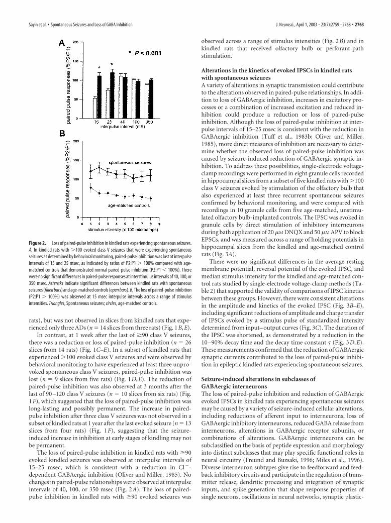
discharge in response to the second pulse of the pair. This reduc-tion in amplitude of the second PS has been referred to as paired-pulse inhibition and has been demonstrated to be a measure ofthe strength of GABAergic inhibition (Tuff et al., 1983b; Oliverand Miller, 1985) (Fig. 1A). In agreement with previous studies ofpaired-pulse responses in kindled rats (Tuff et al., 1983b; Oliverand Miller, 1985; de Jonge and Racine, 1987; Stringer and Loth-man, 1989), field potentials evoked in the dentate gyrus by pairedstimulation of the perforant path at an interpulse interval of 15msec in hippocampal slices from kindled rats demonstrated anincrease in paired-pulse inhibition, as measured by reduction inthe amplitude of the second PS compared with the first (Fig. 1,compare A, B, E). The increase in paired-pulse inhibition at theseinterpulse intervals was observed at 1 week after as few as threeclass V seizures (n � 42 hippocampal slices from eight rats) andwas also observed in hippocampal slices from kindled rats at 1week after the last of 35 class V seizures (n � 16 slices from nine
Figure 1. Evolving alterations in paired-pulse responses in the dentate gyrus of kindled rats as a function of the number ofevoked seizures. A, Extracellular field potentials evoked in the dentate gyrus by paired stimuli delivered to fibers of the perforantpath at an interpulse pulse interval of 15 msec using the lowest stimulus intensity that evoked a maximal response, as determinedby input– output curves. Paired-pulse inhibition is indicated by the reduction in amplitude of the population spike of the secondresponse (P2) compared with the first (P1). Paired-pulse inhibition is present when the ratio P2:P1 is �1. B, In the dentate gyrusfrom a kindled rat that experienced 35 evoked class V seizures, paired-pulse inhibition was increased compared with the age-matched control in A, as demonstrated by the reduction of the ratio of P2:P1. C, In a kindled rat that experienced 100 evoked classV seizures, there was a reduction of paired-pulse inhibition, as demonstrated by the increase in the ratio of the amplitude of thepopulation spike of the second response (P2) compared with the amplitude of the first population spike (P1). D, In a kindled rat with100 class V seizures that was also observed to have spontaneous seizures, paired-pulse inhibition was also reduced, as indicatedby the increase in the amplitude of the population spike of the second response (P2) compared with the amplitude of the firstpopulation spike (P1). E, Evolving alterations in paired-pulse inhibition in the dentate gyrus as a function of the number of evokedseizures. The ratio of the amplitude of the second population spike to the amplitude of first population spike (P2:P1) is expressedas a percentage for kindled rats that experienced a range of three evoked ADs to 100 evoked class V (ClV )seizures ( filled bars)compared with age-matched controls (open bars). Paired-pulse inhibition is present when the ratio of P2:P1 is �100%. Paired-pulse inhibition was unchanged after three ADs but increased after 3 or 35 class V seizures, as indicated by a reduction in the ratioP2:P1 compared with controls. Paired-pulse inhibition was reduced compared with age-matched controls after 100 evoked class Vseizures, as indicated by an increase in the ratio of P2:P1 compared with age-matched controls. There were no significant differ-ences between kindled rats that received olfactory bulb or perforant-path stimulation. Asterisks indicate significant differencesbetween kindled and age-matched control groups. F, The increase in paired-pulse inhibition was observed at 3 months after thelast of three evoked class V seizures but was not observed at 1 year after the last of three evoked class V seizures. In hippocampalslices from kindled rats examined at 3 months after the last of 90 –100 evoked class V seizures, there was not only a reduction butalso a loss of paired-pulse inhibition as indicated by a ratio of P2:P1 100%.
Table 1. Summary of kindling and control groups studied by paired-pulse andsingle-electrode voltage-clamp methods
Kindled Age-matched controls
RatsHippocampalslices Rats
Hippocampalslices
3 ADs 3 14 6 103 Class V 18 42 25 5535 Class V 9 16 15 2690 Class V 14 26 14 30100 Class V plus spontaneous seizures 5 9 5 8
2762 • J. Neurosci., April 1, 2003 • 23(7):2759 –2768 Sayin et al. • Spontaneous Seizures and Loss of GABA Inhibition
rats), but was not observed in slices from kindled rats that expe-rienced only three ADs (n � 14 slices from three rats) (Fig. 1B,E).
In contrast, at 1 week after the last of �90 class V seizures,there was a reduction or loss of paired-pulse inhibition (n � 26slices from 14 rats) (Fig. 1C–E). In a subset of kindled rats thatexperienced 100 evoked class V seizures and were observed bybehavioral monitoring to have experienced at least three unpro-voked spontaneous class V seizures, paired-pulse inhibition waslost (n � 9 slices from five rats) (Fig. 1D,E). The reduction ofpaired-pulse inhibition was also observed at 3 months after thelast of 90 –120 class V seizures (n � 10 slices from six rats) (Fig.1F), which suggested that the loss of paired-pulse inhibition waslong-lasting and possibly permanent. The increase in paired-pulse inhibition after three class V seizures was not observed in asubset of kindled rats at 1 year after the last evoked seizure (n � 13slices from four rats) (Fig. 1F), suggesting that the seizure-induced increase in inhibition at early stages of kindling may notbe permanent.
The loss of paired-pulse inhibition in kindled rats with �90evoked kindled seizures was observed at interpulse intervals of15–25 msec, which is consistent with a reduction in Cl�-dependent GABAergic inhibition (Oliver and Miller, 1985). Nochanges in paired-pulse relationships were observed at interpulseintervals of 40, 100, or 350 msec (Fig. 2A). The loss of paired-pulse inhibition in kindled rats with �90 evoked seizures was
observed across a range of stimulus intensities (Fig. 2B) and inkindled rats that received olfactory bulb or perforant-pathstimulation.
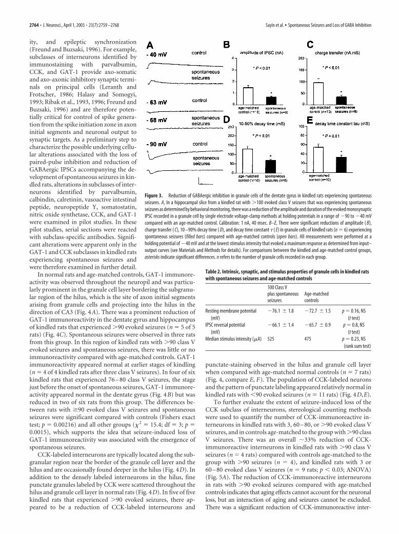
Alterations in the kinetics of evoked IPSCs in kindled ratswith spontaneous seizuresA variety of alterations in synaptic transmission could contributeto the alterations observed in paired-pulse relationships. In addi-tion to loss of GABAergic inhibition, increases in excitatory pro-cesses or a combination of increased excitation and reduced in-hibition could produce a reduction or loss of paired-pulseinhibition. Although the loss of paired-pulse inhibition at inter-pulse intervals of 15–25 msec is consistent with the reduction inGABAergic inhibition (Tuff et al., 1983b; Oliver and Miller,1985), more direct measures of inhibition are necessary to deter-mine whether the observed loss of paired-pulse inhibition wascaused by seizure-induced reduction of GABAergic synaptic in-hibition. To address these possibilities, single-electrode voltage-clamp recordings were performed in eight granule cells recordedin hippocampal slices from a subset of five kindled rats with 100class V seizures evoked by stimulation of the olfactory bulb thatalso experienced at least three recurrent spontaneous seizuresconfirmed by behavioral monitoring, and were compared withrecordings in 10 granule cells from five age-matched, unstimu-lated olfactory bulb-implanted controls. The IPSC was evoked ingranule cells by direct stimulation of inhibitory interneuronsduring bath application of 20 �M DNQX and 50 �M APV to blockEPSCs, and was measured across a range of holding potentials inhippocampal slices from the kindled and age-matched controlrats (Fig. 3A).
There were no significant differences in the average restingmembrane potential, reversal potential of the evoked IPSC, andmedian stimulus intensity for the kindled and age-matched con-trol rats studied by single-electrode voltage-clamp methods (Ta-ble 2) that supported the validity of comparisons of IPSC kineticsbetween these groups. However, there were consistent alterationsin the amplitude and kinetics of the evoked IPSC (Fig. 3B–E),including significant reductions of amplitude and charge transferof IPSCs evoked by a stimulus pulse of standardized intensitydetermined from input– output curves (Fig. 3C). The duration ofthe IPSC was shortened, as demonstrated by a reduction in the10 –90% decay time and the decay time constant � (Fig. 3D,E).These measurements confirmed that the reduction of GABAergicsynaptic currents contributed to the loss of paired-pulse inhibi-tion in epileptic kindled rats experiencing spontaneous seizures.
Seizure-induced alterations in subclasses ofGABAergic interneuronsThe loss of paired-pulse inhibition and reduction of GABAergicevoked IPSCs in kindled rats experiencing spontaneous seizuresmay be caused by a variety of seizure-induced cellular alterations,including reductions of afferent input to interneurons, loss ofGABAergic inhibitory interneurons, reduced GABA release frominterneurons, alterations in GABAergic receptor subunits, orcombinations of alterations. GABAergic interneurons can besubclassified on the basis of peptide expression and morphologyinto distinct subclasses that may play specific functional roles inneural circuitry (Freund and Buzsaki, 1996; Miles et al., 1996).Diverse interneuron subtypes give rise to feedforward and feed-back inhibitory circuits and participate in the regulation of trans-mitter release, dendritic processing and integration of synapticinputs, and spike generation that shape response properties ofsingle neurons, oscillations in neural networks, synaptic plastic-
Figure 2. Loss of paired-pulse inhibition in kindled rats experiencing spontaneous seizures.A, In kindled rats with 100 evoked class V seizures that were experiencing spontaneousseizures as determined by behavioral monitoring, paired-pulse inhibition was lost at interpulseintervals of 15 and 25 msec, as indicated by ratios of P2:P1 100% compared with age-matched controls that demonstrated normal paired-pulse inhibition (P2:P1 � 100%). Therewere no significant differences in paired-pulse responses at interstimulus intervals of 40, 100, or350 msec. Asterisks indicate significant differences between kindled rats with spontaneousseizures (filled bars) and age-matched controls (open bars). B, The loss of paired-pulse inhibition(P2:P1 100%) was observed at 15 msec interpulse intervals across a range of stimulusintensities. Triangles, Spontaneous seizures; circles, age-matched controls.
Sayin et al. • Spontaneous Seizures and Loss of GABA Inhibition J. Neurosci., April 1, 2003 • 23(7):2759 –2768 • 2763
ity, and epileptic synchronization(Freund and Buzsaki, 1996). For example,subclasses of interneurons identified byimmunostaining with parvalbumin,CCK, and GAT-1 provide axo-somaticand axo-axonic inhibitory synaptic termi-nals on principal cells (Leranth andFrotscher, 1986; Halasy and Somogyi,1993; Ribak et al., 1993, 1996; Freund andBuzsaki, 1996) and are therefore poten-tially critical for control of spike genera-tion from the spike initiation zone in axoninitial segments and neuronal output tosynaptic targets. As a preliminary step tocharacterize the possible underlying cellu-lar alterations associated with the loss ofpaired-pulse inhibition and reduction ofGABAergic IPSCs accompanying the de-velopment of spontaneous seizures in kin-dled rats, alterations in subclasses of inter-neurons identified by parvalbumin,calbindin, calretinin, vasoactive intestinalpeptide, neuropeptide Y, somatostatin,nitric oxide synthetase, CCK, and GAT-1were examined in pilot studies. In thesepilot studies, serial sections were reactedwith subclass-specific antibodies. Signifi-cant alterations were apparent only in theGAT-1 and CCK subclasses in kindled ratsexperiencing spontaneous seizures andwere therefore examined in further detail.
In normal rats and age-matched controls, GAT-1 immunore-activity was observed throughout the neuropil and was particu-larly prominent in the granule cell layer bordering the subgranu-lar region of the hilus, which is the site of axon initial segmentsarising from granule cells and projecting into the hilus in thedirection of CA3 (Fig. 4A). There was a prominent reduction ofGAT-1 immunoreactivity in the dentate gyrus and hippocampusof kindled rats that experienced 90 evoked seizures (n � 5 of 5rats) (Fig. 4C). Spontaneous seizures were observed in three ratsfrom this group. In this region of kindled rats with 90 class Vevoked seizures and spontaneous seizures, there was little or noimmunoreactivity compared with age-matched controls. GAT-1immunoreactivity appeared normal at earlier stages of kindling(n � 4 of 4 kindled rats after three class V seizures). In four of sixkindled rats that experienced 76 – 80 class V seizures, the stagejust before the onset of spontaneous seizures, GAT-1 immunore-activity appeared normal in the dentate gyrus (Fig. 4B) but wasreduced in two of six rats from this group. The differences be-tween rats with �90 evoked class V seizures and spontaneousseizures were significant compared with controls (Fishers exacttest; p � 0.00216) and all other groups (� 2 � 15.4; df � 3; p �0.0015), which supports the idea that seizure-induced loss ofGAT-1 immunoreactivity was associated with the emergence ofspontaneous seizures.
CCK-labeled interneurons are typically located along the sub-granular region near the border of the granule cell layer and thehilus and are occasionally found deeper in the hilus (Fig. 4D). Inaddition to the densely labeled interneurons in the hilus, finepunctate granules labeled by CCK were scattered throughout thehilus and granule cell layer in normal rats (Fig. 4D). In five of fivekindled rats that experienced 90 evoked seizures, there ap-peared to be a reduction of CCK-labeled interneurons and
punctate-staining observed in the hilus and granule cell layerwhen compared with age-matched normal controls (n � 7 rats)(Fig. 4, compare E, F). The population of CCK-labeled neuronsand the pattern of punctate labeling appeared relatively normal inkindled rats with �90 evoked seizures (n � 11 rats) (Fig. 4D,E).
To further evaluate the extent of seizure-induced loss of theCCK subclass of interneurons, stereological counting methodswere used to quantify the number of CCK-immunoreactive in-terneurons in kindled rats with 3, 60 – 80, or 90 evoked class Vseizures, and in controls age-matched to the group with 90 classV seizures. There was an overall �33% reduction of CCK-immunoreactive interneurons in kindled rats with 90 class Vseizures (n � 4 rats) compared with controls age-matched to thegroup with 90 seizures (n � 4), and kindled rats with 3 or60 – 80 evoked class V seizures (n � 9 rats; p � 0.03; ANOVA)(Fig. 5A). The reduction of CCK-immunoreactive interneuronsin rats with 90 evoked seizures compared with age-matchedcontrols indicates that aging effects cannot account for the neuronalloss, but an interaction of aging and seizures cannot be excluded.There was a significant reduction of CCK-immunoreactive inter-
Figure 3. Reduction of GABAergic inhibition in granule cells of the dentate gyrus in kindled rats experiencing spontaneousseizures. A, In a hippocampal slice from a kindled rat with 100 evoked class V seizures that was experiencing spontaneousseizures as determined by behavioral monitoring, there was a reduction of the amplitude and duration of the evoked monosynapticIPSC recorded in a granule cell by single electrode voltage-clamp methods at holding potentials in a range of �90 to �40 mVcompared with an age-matched control. Calibration: 1 nA, 40 msec. B–E, There were significant reductions of amplitude ( B),charge transfer ( C), 10 –90% decay time ( D), and decay time constant � ( E) in granule cells of kindled rats (n � 6) experiencingspontaneous seizures (filled bars) compared with age-matched controls (open bars). All measurements were performed at aholding potential of �40 mV and at the lowest stimulus intensity that evoked a maximum response as determined from input–output curves (see Materials and Methods for details). For comparisons between the kindled and age-matched control groups,asterisks indicate significant differences. n refers to the number of granule cells recorded in each group.
Table 2. Intrinsic, synaptic, and stimulus properties of granule cells in kindled ratswith spontaneous seizures and age-matched controls
100 Class Vplus spontaneousseizures
Age-matchedcontrols
Resting membrane potential(mV)
�76.1 � 1.8 �72.7 � 1.5 p � 0.16, NS(t test)
IPSC reversal potential(mV)
�66.1 � 1.4 �65.7 � 0.9 p � 0.8, NS(t test)
Median stimulus intensity (�A) 525 475 p � 0.23, NS(rank sum test)
2764 • J. Neurosci., April 1, 2003 • 23(7):2759 –2768 Sayin et al. • Spontaneous Seizures and Loss of GABA Inhibition

neurons in the 90 class V group compared with kindled ratswith 60 – 80 evoked class V seizures ( p � 0.0004), indicating anassociation of seizure-induced loss of CCK-immunoreactive in-terneurons with the emergence of spontaneous seizures. A reduc-tion of the number of CCK-labeled neurons in the dentate gyrusof kindled rats with 90 evoked class V seizures was observedalong the entire septotemporal axis of the hippocampal forma-tion ( p � 0.03), except for a nonsignificant trend to reduction atthe septal (rostral) pole (Fig. 5B).
DiscussionThese physiological and anatomical experiments identified func-tional and cellular alterations in the hippocampus associated withthe emergence of spontaneous seizures. Spontaneous seizures were
observed in 11 of 27 rats after �90–100repeated brief seizures evoked by kindling ofeither the olfactory bulb or perforant path,and were the long-term functional outcomeof repeated episodes of evoked synchronousneural activity. The sequence of progressiveseizure-induced molecular and cellular al-terations underlying the emergence of spon-taneous seizures is an example of long-termactivity-dependent plasticity in neural cir-cuits and has potential relevance for thecommon disorder of epilepsy in whichseizure-induced cellular alterations maycontribute to dysfunction.
Predictable emergence of spontaneousseizures in kindled rats after �90 –100evoked seizuresThere is limited insight about cellular al-terations accompanying the emergence ofspontaneous seizures after repeated briefevoked seizures in rats and primates(Wada et al., 1975; Pinel and Rovner,1978). It is uncertain whether spontane-ous seizures occur reliably and predictablyafter a specific number of evoked seizuresor merely sporadically in the advancedkindled state. Behavioral monitoring re-vealed that spontaneous seizures were ob-served after �90 –100 class V seizuresevoked by either olfactory bulb orperforant-path stimulation. Additionalspontaneous seizures were observed afterthe initial spontaneous seizure, confirm-ing that kindled animals with spontane-ous seizures were epileptic, as defined byrecurring spontaneous seizures.
The observation of spontaneous sei-zures after �90 –100 evoked class V sei-zures should not be regarded as a preciselatency to spontaneous seizures but ratheras a period of transition to a state withincreased probability of spontaneous epi-sodes of synchronous neural activity andseizures (Hellier et al., 1998; Pitkanen etal., 2002). The stage of �90 –100 evokedclass V seizures was recognized retrospec-tively as a period of transition to increasedprobability of spontaneous seizures anddoes not imply that all kindled rats expe-
rience spontaneous seizures after 90 –100 evoked class V seizures,but merely that spontaneous seizures become increasingly likelyas indicated by their recognition in �40% of rats with 90 –100evoked class V seizures undergoing 8 hr of behavioral monitoringper day. This is probably a low estimate limited by the samplingmethods; more sensitive continuous video-EEG-recordingmethods might detect additional rats with spontaneous sei-zures after 90 –100 evoked class V seizures, and might alsoreveal that occasional spontaneous seizures occur in kindledrats with �90 evoked seizures. Although both status epilepti-cus and repeated brief seizures eventually induce spontaneousseizures, the gradual induction of spontaneous seizures in theabsence of extensive damage permitted detailed analysis of
Figure 4. Seizure-induced reduction of GAT-1 and CCK immunoreactivity in the dentate gyrus of kindled rats experiencingspontaneous seizures. Immunoreactivity was intensified in these sections by silver treatment, as described in Materials andMethods, and all sections were batch processed. A, In normal rats, GAT-1 immunoreactivity is observed throughout the neuropil,but is particularly prominent in the granule cell layer (gc) bordering the subgranular region of the hilus (H; arrows), which is the siteof axon initial segments arising from granule cells and projecting into the hilus toward CA3c. B, In a kindled rat that experienced 76class V seizures, which is a stage just before the onset of spontaneous seizures, GAT-1 immunoreactivity appeared normal in thedentate gyrus. C, In a kindled rat that experienced 209 evoked class V seizures, immunoreactivity was reduced in comparison withnormal or age-matched controls. The loss of GAT-1 immunoreactivity was particularly prominent along the border of the granulecell layer and subgranular region of the hilus, which is the site of axon initial segments and spike initiation. D, In a nearby sectionof the dentate gyrus from the normal rat in A, GABAergic interneurons labeled by CCK were located along the subgranular regionnear the border of the granule cell layer and the hilus and were occasionally also observed deeper in the hilus. Fine punctategranules labeled by CCK were also typically scattered throughout the hilus and granule cell layer in normal rats. E, In the samekindled rats that experienced 76 class V seizures, the distribution and number of CCK-labeled interneurons and punctate granularstaining appeared normal. F, In the same kindled rat shown in C that experienced 209 evoked class V seizures, there was a reductionof interneurons labeled by CCK and in the punctate staining observed in the hilus and granule cell layer. Scale bar, 200 �M.
Sayin et al. • Spontaneous Seizures and Loss of GABA Inhibition J. Neurosci., April 1, 2003 • 23(7):2759 –2768 • 2765

specific structural and functional alter-ations associated with this transition.
Emergence of spontaneous seizures isassociated with seizure-inducedreduction of GABAA inhibitionThis study confirmed that repeated sei-zures increase paired-pulse inhibition andother measures of inhibition during earlystages of kindling (Tuff et al., 1983a,b; Ol-iver and Miller, 1985; Shin et al., 1985; deJonge and Racine, 1987; Stringer andLothman, 1989; Otis et al., 1994; Buhl etal., 1996; Nusser et al., 1998). Paired-pulseinhibition increased after three class V sei-zures but not after only a few ADs with par-tial seizures (class I-III), which implies thatmore than three partial seizures or second-ary generalized seizures may be required.
The initial increase was not perma-nent, because paired-pulse inhibition wasreduced or lost in the dentate gyrus after�90 –100 class V seizures evoked by olfac-tory bulb or perforant-path stimulation,and importantly, was consistently lost inall rats with spontaneous seizures revealedby behavioral monitoring. The loss ofpaired-pulse inhibition was long-lastingand probably permanent. Because ratswith 90 –100 class V seizures might havespontaneous seizures that were undetected by the limited obser-vation methods, spontaneous seizures may not induce an acuteincrease in paired-pulse inhibition as observed during earlystages of kindling.
Loss of paired-pulse inhibition associated with spontaneousseizures could be caused by increases in glutamatergic excitatoryprocesses, loss of GABAergic inhibition, or a combination of in-creased excitation and reduced inhibition. Loss of paired-pulseinhibition at interpulse intervals of 15–25 msec corresponding tothe time course of GABAA-dependent Cl� conductance is con-sistent with a seizure-induced alteration in GABAA inhibition(Tuff et al., 1983b; Oliver and Miller, 1985) that was confirmed bythe reduced amplitude and shortened duration of evoked IPSCsrecorded by single-electrode voltage-clamp methods. Mecha-nisms underlying the altered kinetics of the evoked IPSCs werenot specifically addressed in these experiments but could in-clude loss of GABAergic terminals; alterations in the number,subunit composition, or biophysical properties of GABA re-ceptors; or a variety of mechanisms.
Progressive seizure-induced alterations leading tospontaneous seizuresRepeated evoked seizures induce a variety of cellular and molec-ular alterations in the dentate gyrus and other regions, includingneuronal apoptosis and cumulative neuronal loss resemblinghippocampal sclerosis, neurogenesis, mossy fiber sprouting, gli-osis, and reorganization of receptors and other proteins (Sutulaet al., 1988, 1998; Represa et al., 1989; Cavazos and Sutula, 1990;Cavazos et al., 1991, 1994; Bengzon et al., 1997; Pretel and Piekut,1997; Adams et al., 1998; Dalby et al., 1998; Parent et al., 1998;Kotloski et al., 2002). Cumulative seizure-induced alterationscould potentially alter the balance of excitation and/or inhibition
and modify functional properties of hippocampal circuits con-tributing to the emergence of spontaneous seizures.
Because repeated brief kindled seizures induce cumulativeneuronal loss (Cavazos and Sutula, 1990; Cavazos et al., 1994;Bengzon et al., 1997; Pretel and Piekut, 1997; Dalby et al., 1998;Dalby and Mody, 2001; Kotloski et al., 2002), it was of interest toinvestigate whether seizure-induced loss of GABAergic interneu-rons contributed to the alterations in the kinetics of GABAA-dependent IPSCs accompanying spontaneous seizures. In pilotstudies, expression of CCK and GAT-1, which label GABAergicinterneuron subclasses providing axo-somatic and axo-axonicinhibitory inputs to granule cells (Leranth and Frotscher, 1986;Freund and Buzsaki, 1996; Ribak et al., 1996), was reduced inkindled rats with loss of paired-pulse inhibition and spontaneousseizures. Markers for other subclasses, including the parvalbuminsubclass that also provides axo-axonic and axo-somatic inhibi-tion (Ribak et al., 1993; Freund and Buzsaki, 1996), were exam-ined in serial sections in pilot studies but did not appear to besignificantly altered.
CCK and GAT-1 immunoreactivity appeared normal in kin-dled rats after 80 class V evoked seizures but was consistentlyreduced after 90 class V seizures. The reduction of CCK andGAT-1 immunoreactivity in association with the emergence ofspontaneous seizures suggests that seizure-induced reduction ofthese subclasses and loss of axo-somatic and axo-axonic inhibi-tion may contribute to spontaneous seizures. Loss of GAT-1 im-munoreactivity was particularly prominent in the infragranularregion of the granule cell layer (the site of axon initial segments)but was observed throughout the hippocampus. Loss of CCK-labeled interneurons, as measured by counting methods, oc-curred along the entire septotemporal axis of the hippocampus.Because the reduction of CCK and GAT-1 immunoreactivity wasnot observed in age-matched controls, aging alone cannot ac-
Figure 5. Reduction of CCK-immunoreactive GABAergic interneurons in the dentate gyrus of kindled rats experiencing spon-taneous seizures. The number of CCK-immunoreactive interneurons in the dentate gyrus was assessed as a function of the numberof evoked seizures and was compared with normal controls matched in age to kindled rats that experienced 100 class V seizures.Profile counts of CCK-immunoreactive interneurons were counted in the dentate gyrus by stereological methods in consecutivetransverse sections from the septal to temporal pole of the extended hippocampus (see Materials and Methods for details). A, Therewas a significant reduction of CCK-immunoreactive interneurons in kindled rats with 100 class V (ClV ) seizures (filled bar)compared with age-matched normal controls (open bar) and kindled rats examined after 3 or 60 – 80 evoked class V seizures (graybars). B, In kindled rats with 100 evoked class V seizures ( filled bars), reduction of CCK-immunoreactive interneurons wasobserved along the entire septotemporal axis of the hippocampus. Percentages of 0 –25, 25–50, 50 –75, and 75–100 refer torelative location along the septotemporal axis. There were significant reductions of CCK-immunoreactive interneurons from themiddle through the most temporal extent of the dentate gyrus and a trend toward reduction at the most septal location. Asterisksindicate significant differences.
2766 • J. Neurosci., April 1, 2003 • 23(7):2759 –2768 Sayin et al. • Spontaneous Seizures and Loss of GABA Inhibition

count for the reductions, but an interaction of the cumulativeeffects of repeated seizures with aging cannot be excluded.
Loss of these markers could be caused by reduced expressionrather than loss of the interneuron populations, but this seemsunlikely given ongoing, progressive neuronal loss in kindled ratsand the accompanying loss of inhibition detected by physiologi-cal methods. If expression of the GABA transporter GAT-1 wasreduced without interneuron loss, there would be an increase ininhibition as a consequence of reduced reuptake of synapticallyreleased GABA, but this was not observed. Counting methodssupported the interpretation that the reduction of CCK immu-noreactivity was caused by neuronal loss; however, with technicallimitations such as comparability of antibody penetration even inbatch-processed sections, the counts should not be regarded as aprecise measure of the absolute number of CCK interneurons.
Seizure-induced loss of axo-axonic and axo-somaticinhibition and the development of spontaneous seizuresThe CCK and GAT-1 interneuron subclasses in the dentate gyrushave morphological features of basket cells and chandelier cellsand project axo-somatic and axo-axonic terminals forming sym-metric GABAergic synapses (Gray type II) on principal cells (Le-ranth and Frotscher, 1986; Soriano et al., 1990; Halasy and So-mogyi, 1993; Ribak et al., 1993, 1996). These features areespecially suited for perisomatic and axon initial segment inhibi-tion that potentially control timing and suppression of spikesgenerated by Na� channels that have a high density on axoninitial segments (Buhl et al., 1994; Soltesz et al., 1995; Freund andBuzsaki, 1996). Seizure-induced loss of the CCK and GAT-1 sub-classes in the dentate gyrus would reduce spike suppression (Buhlet al., 1994; Soltesz et al., 1995; Miles et al., 1996) and enhancepropagation of spike output to CA3, thereby eroding “filtering”properties of the dentate gyrus and potentially increasing thesusceptibility of hippocampal neural circuits to epileptic syn-chronization (Buhl et al., 1994; Soltesz et al., 1995; Freund andBuzsaki, 1996; Behr et al., 1998, 2001). Although the loss of axo-axonic and axo-somatic inhibition is a potentially attractivemechanism for neural synchronization and seizure-induced epi-leptogenesis, other cellular alterations are also likely to contributeto reduced inhibition and synchronized neuronal bursting. Forexample, seizure-induced changes in GABAA receptor subunitsor alterations in biophysical properties of channels could shortenor reduce IPSCs (Stell and Mody, 2002). Seizure-induced in-creases in recurrent excitation as a result of excitatory circuitsformed by sprouted mossy fibers may also contribute to the lossof paired-pulse inhibition (Wuarin and Dudek, 1996, 2001;Lynch and Sutula, 2000). Seizure-induced alterations in otherhippocampal and limbic areas may also be important for epilepticsynchronization (Gorter et al., 2002). These experiments provideevidence of an association among loss of inhibition, loss of sub-classes of interneurons providing axo-somatic and axo-axonicinhibition, and emergence of spontaneous seizures. Additionalexperiments are necessary to define causality among these alter-ations, but it is likely that multifactorial and combinatorial pro-cesses contribute to the intermittent paroxysmal expression ofneuronal synchronization during seizures.
Implications for human epilepsyReduction of GAT-1 immunoreactivity and loss of GABA trans-porter function have been reported in intractable human tempo-ral lobe epilepsy (During et al., 1995; Mathern et al., 1999). Lossof GAT-1 has been observed in CA1 but is reported to increase inthe inner molecular layer of the dentate gyrus after pilocarpine-
induced status (Andre et al., 2001), suggesting that the effects ofseizures may be region and model specific. Selective loss of PV-immunoreactive interneurons and terminals has been observedin surgically resected hippocampus in both humans and modelsof status epilepticus (Zhu et al., 1997; Gorter et al., 2001; Wittneret al., 2001) and is in contrast to the relative preservation of PVimmunoreactivity in kindled rats with spontaneous seizures. Theobservation that seizure-induced loss of CCK and GAT-1 immu-noreactivity occurs in the dentate gyrus at advanced stages ofkindling in association with reduced inhibition and spontaneousseizures suggests that repeated seizures may play a role in thedevelopment of intractable human temporal lobe epilepsy.
The progressive alterations induced by repeated brief seizuresin hippocampal circuitry have significant adverse consequences,including increased susceptibility to additional seizures (God-dard 1969; Goddard et al., 1969) and memory dysfunction (Sutula etal., 1995). The slowly evolving, seizure-induced reduction of inhibi-tion supports the importance of achieving complete control of sei-zures in people with epilepsy. Molecular and genetic analysis of theevolving seizure-induced cellular and functional alterations mayprovide new targets for therapeutic intervention and the preven-tion of adverse consequences of poorly controlled epilepsy.
ReferencesAdams B, Vonling E, Vaccarella L, Ivy GO, Fahnestock M, Racine RJ (1998)
Time course for kindling-induced changes in the hilar area of the dentategyrus-reactive gliosis as a potential mechanism. Brain Res 804:331–336.
Andre V, Marescaux C, Nehlig A, Fritschy JM (2001) Alterations in hip-pocampal GABAergic system contribute to development of spontaneousrecurrent seizures in the rat lithium-pilocarpine model of temporal lobeepilepsy. Hippocampus 11:452– 468.
Behr J, Lyson KJ, Mody I (1998) Enhanced propagation of epileptiformactivity through the kindled dentate gyrus. J Neurophysiol 79:1726 –1732.
Behr J, Heinemann U, Mody I (2001) Kindling induces transient NMDAreceptor-mediated facilitation of high-frequency input in the rat dentategyrus. J Neurophysiol 85:2195–2202.
Bengzon J, Kokaia Z, Elmer E, Nanobashvili A, Kokaia M, Lindvall O (1997)Apoptosis and proliferation of dentate gyrus neurons after single andintermittent limbic seizures. Proc Natl Acad Sci USA 94:10432–10437.
Buhl EH, Han ZS, Lorinczi Z, Stezhka VV, Karnup SV, Somogyi P (1994)Physiological properties of anatomically identified axo-axonic cells in therat hippocampus. J Neurophysiol 71:1289 –1307.
Buhl EH, Otis TS, Mody I (1996) Zinc-induced collapse of augmented inhibi-tion by GABA in a temporal lobe epilepsy model. Science 271:369 –373.
Cain DP, Corcoran ME (1980) Kindling in the seizure-prone and seizure-resistant Mongolian gerbil. Electroencephalogr Clin Neurophysiol49:360 –365.
Catalano SM, Shatz CJ (1998) Activity-dependent cortical target selectionby thalamic axons. Science 281:559 –562.
Cavazos JE, Sutula TP (1990) Progressive neuronal loss induced by kin-dling: a possible mechanism for mossy fiber synaptic reorganization andhippocampal sclerosis. Brain Res 527:1– 6.
Cavazos JE, Golarai G, Sutula TP (1991) Mossy fiber synaptic reorganiza-tion induced by kindling: time course of development, progression, andpermanence. J Neurosci 11:2795–2803.
Cavazos JE, Das I, Sutula TP (1994) Neuronal loss induced in limbic path-ways by kindling: evidence for induction of hippocampal sclerosis byrepeated brief seizures. J Neurosci 14:3106 –3121.
Dalby NO, Mody I (2001) The process of epileptogenesis: a pathophysiolog-ical approach. Curr Opin Neurol 14:187–192.
Dalby N, West M, Finsen B (1998) Hilar somatostatin-mRNA containingneurons are preserved after perforant path kindling in the rat. NeurosciLett 255:45– 48.
de Jonge M, Racine RJ (1987) The development and decay of kindling-induced increases in paired-pulse depression in the dentate gyrus. BrainRes 412:318 –328.
During MJ, Ryder KM, Spencer DD (1995) Hippocampal GABA trans-porter function in temporal-lobe epilepsy. Nature 376:174 –177.
Sayin et al. • Spontaneous Seizures and Loss of GABA Inhibition J. Neurosci., April 1, 2003 • 23(7):2759 –2768 • 2767

Freund TF, Buzsaki G (1996) Interneurons of the hippocampus. Hip-pocampus 6:347– 470.
Goddard GV (1969) The development of epileptic seizures through brainstimulation at low intensity. Nature 214:1020 –1021.
Goddard GV, McIntyre D, Leech C (1969) A permanent change in brain func-tion resulting from daily electrical stimulation. Exp Neurol 25:295–330.
Gorter JA, van Vliet EA, Aronica E, da Silva FL (2001) Progression of spon-taneous seizures after status epilepticus is associated with mossy fibresprouting and extensive bilateral loss of hilar parvalbumin andsomatostatin-immunoreactive neurons. Eur J Neurosci 13:657– 669.
Gorter JA, van Vliet EA, Aronica E, da Silva FHL (2002) Long-lasting in-creased excitability differs in dentate gyrus vs. CA1 in freely movingchronic epileptic rats after electrically induced status epilepticus. Hip-pocampus 12:311–324.
Guillery RW, August B (2002) Doubt and certainty in counting. Prog BrainRes 135:25– 42.
Halasy K, Somogyi P (1993) Subdivisions in the multiple GABAergic inner-vation of granule cells in the dentate gyrus of the rat hippocampus. EurJ Neurosci 5:411– 429.
Hellier JL, Patrylo PR, Buckmaster PS, Dudek FE (1998) Recurrent sponta-neous motor seizures after repeated low-dose systemic treatment withkainate-assessment of a rat model of temporal lobe epilepsy. Epilepsy Res31:73– 84.
Konigsmark B (1969) Methods for the counting of neurons. In: Contempo-rary research methods in neuroanatomy (Nauta W, Ebbeson S, eds), pp315–340. Berlin: Springer.
Kotloski R, Lynch M, Lauersdorf S, Sutula T (2002) Repeated brief seizuresinduce progressive hippocampal neuron loss and memory deficits. ProgBrain Res 135:95–110.
Leranth C, Frotscher M (1986) Synaptic connections of cholecystokinin-immunoreactive neurons and terminals in the rat fascia dentata: a com-bined light and electron microscopic study. J Comp Neurol 254:51– 64.
Lothman EW, Stringer JL, Bertram EH (1992) The dentate gyrus as a con-trol point for seizures in the hippocampus and beyond. Epilepsy ResSuppl 7:301–313.
Lynch M, Sutula T (2000) Recurrent excitatory connectivity in the dentategyrus of kindled and kainic acid-treated rats. J Neurophysiol 83:693–704.
Majkowski J (1999) Kindling: clinical relevance for epileptogenicity in hu-mans. Adv Neurol 81:105–113.
Mathern GW, Mendoza D, Lozada A, Pretorius JK, Dehnes Y, Danbolt NC,Nelson N, Leite JP, Chimelli L (1999) Hippocampal GABA and gluta-mate transporter immunoreactivity in patients with temporal lobe epi-lepsy. Neurology 52:453– 472.
McNamara JO (1999) Emerging insights into the genesis of epilepsy. Nature399:A15–A22.
Miles R, Toth K, Gulyas AI, Hajos N, Freund TF (1996) Differences betweensomatic and dendritic inhibition in the hippocampus. Neuron16:815– 823.
Mody I (1999) Synaptic plasticity in kindling. Adv Neurol 79:631– 643.Morrell F, Tsuru N (1976) Kindling in the frog: development of spontane-
ous epileptiform activity. Electroencephalogr Clin Neurophysiol 40:1–11.Nusser Z, Hajos N, Somogyi P, Mody I (1998) Increased number of synaptic
GABA(A) receptors underlies potentiation at hippocampal inhibitorysynapses. Nature 395:172–177.
Oliver MW, Miller JJ (1985) Alterations of inhibitory processes in the dentategyrus following kindling-induced epilepsy. Exp Brain Res 57:443–447.
Otis TS, De Koninck Y, Mody I (1994) Lasting potentiation of inhibition isassociated with an increased number of gamma-aminobutyric acid type Areceptors activated during miniature inhibitory postsynaptic currents.Proc Natl Acad Sci USA 91:7698 –7702.
Parent JM, Janumpalli S, McNamara JO, Lowenstein DH (1998) Increaseddentate granule cell neurogenesis following amygdala kindling in theadult rat. Neurosci Lett 247:9 –12.
Penn AA, Shatz CJ (1999) Brain waves and brain wiring: the role of endog-enous and sensory-driven neural activity in development. Pediatr Res45:447– 458.
Pinel JP, Rovner LI (1978) Experimental epileptogenesis: kindling-inducedepilepsy in rats. Exp Neurol 58:190 –202.
Pitkanen A, Nissinen J, Nairismagi J, Lukasiuk K, Grohn O, Miettinen R,Kauppinen R (2002) Progression of neuronal damage after status epi-lepticus and during spontaneous seizures in a rat model of temporal lobeepilepsy. Prog Brain Res 135:67– 84.
Pretel SAC, Piekut D (1997) Apoptotic and necrotic cell death followingkindling induced seizures. Acta Histochem 99:71–79.
Represa A, Le Gall La Salle G, Ben-Ari Y (1989) Hippocampal plasticity inthe kindling model of epilepsy in rats. Neurosci Lett 99:345–350.
Ribak CE, Seress L, Leranth C (1993) Electron microscopic immunocyto-chemical study of the distribution of parvalbumin-containing neuronsand axon terminals in the primate dentate gyrus and Ammon’s horn.J Comp Neurol 327:298 –321.
Ribak CE, Tong WM, Brecha NC (1996) GABA plasma membrane trans-porters, GAT-1 and GAT-3, display different distributions in the rat hip-pocampus. J Comp Neurol 367:595– 606.
Sayin U, Rutecki P, Sutula T (1999) NMDA-dependent currents in granulecells of the dentate gyrus contribute to induction but not permanence ofkindling. J Neurophysiol 81:564 –574.
Shin C, Pedersen HB, McNamara JO (1985) Gamma-aminobutyric acidand benzodiazepine receptors in the kindling model of epilepsy: a quan-titative radiohistochemical study. J Neurosci 5:2696 –2701.
Soltesz I, Smetters DK, Mody I (1995) Tonic inhibition originates from syn-apses close to the soma. Neuron 14:1273–1283.
Soriano E, Nitsch R, Frotscher M (1990) Axo-axonic chandelier cells in therat fascia dentata: Golgi-electron microscopy and immunocytochemicalstudies. J Comp Neurol 293:1–25.
Stell BM, Mody I (2002) Receptors with different affinities mediate phasicand tonic GABAA conductances in hippocampal neurons. J Neurosci22:RC223(1–5).
Stringer JL, Lothman EW (1989) Repetitive seizures cause an increase inpaired-pulse inhibition in the dentate gyrus. Neurosci Lett 105:91–95.
Sutula T (2001) Secondary epileptogenesis, kindling, and intractable epi-lepsy: a reappraisal from the perspective of neural plasticity. Int Rev Neu-robiol 45:355–386.
Sutula T, Steward O (1986) Quantitative analysis of synaptic potentiationduring kindling of the perforant path. J Neurophysiol 56:732–746.
Sutula T, He XX, Cavazos J, Scott G (1988) Synaptic reorganization in thehippocampus induced by abnormal functional activity. Science239:1147–1150.
Sutula T, Lauersdorf S, Lynch M, Jurgella C, Woodard A (1995) Deficits inradial arm maze performance in kindled rats: evidence for long-lastingmemory dysfunction induced by repeated brief seizures. J Neurosci15:8295– 8301.
Sutula T, Zhang P, Lynch M, Sayin U, Golarai G, Rod R (1998) Synaptic andaxonal remodeling of mossy fibers in the hilus and supragranular regionof the dentate gyrus in kainate-treated rats. J Comp Neurol 390:578 –594.
Sutula T, Sayin U, Knodel S, Rutecki P (2000) Seizure-induced loss of CCKand GAT-1 interneurons and reduced inhibition in the dentate gyrus:relationship to development of spontaneous seizures in kindled rats. SocNeurosci Abstr 26:1050.
Swann JW, Smith KL, Lee CL (2001) Neuronal activity and the establish-ment of normal and epileptic circuits during brain development. Int RevNeurobiol 45:89 –118.
Tuff LP, Racine RJ, Adamec R (1983a) The effects of kindling on GABA-mediated inhibition in the dentate gyrus of the rat. I. Paired-pulse depres-sion. Brain Res 277:79 –90.
Tuff LP, Racine RJ, Mishra RK (1983b) The effects of kindling on GABA-mediated inhibition in the dentate gyrus of the rat. II. Receptor binding.Brain Res 277:91–98.
Wada JA, Osawa T, Mizoguchi T (1975) Recurrent spontaneous seizurestate induced by prefrontal kindling in Senegalese baboons, Papio papio.Can J Neurol Sci 2:477– 492.
Wada JA, Mizoguchi T, Osawa T (1978) Secondarily generalized convulsiveseizures induced by daily amygdaloid stimulation in rhesus monkeys.Neurology 28:1026 –1036.
Wittner L, Magloczky Z, Borhegyi Z, Halasz P, Toth S, Eross L, Szabo Z,Freund TF (2001) Preservation of perisomatic inhibitory input of gran-ule cells in the epileptic human dentate gyrus. Neuroscience 108:587– 600.
Wuarin JP, Dudek FE (1996) Electrographic seizures and new recurrent ex-citatory circuits in the dentate gyrus of hippocampal slices from kainate-treated epileptic rats. J Neurosci 16:4438 – 4448.
Wuarin JP, Dudek FE (2001) Excitatory synaptic input to granule cells in-creases with time after kainate treatment. J Neurophysiol 85:1067–1077.
Zhu ZQ, Armstrong DL, Hamilton WJ, Grossman RG (1997) Disproportion-ate loss of CA4 parvalbumin-immunoreactive interneurons in patients withAmmon’s horn sclerosis. J Neuropathol Exp Neurol 56:988–998.
2768 • J. Neurosci., April 1, 2003 • 23(7):2759 –2768 Sayin et al. • Spontaneous Seizures and Loss of GABA Inhibition