Two-Stage Translational Control of Dentate Gyrus LTP Consolidation Mediated by Sustained BDNF-TrkB...
17
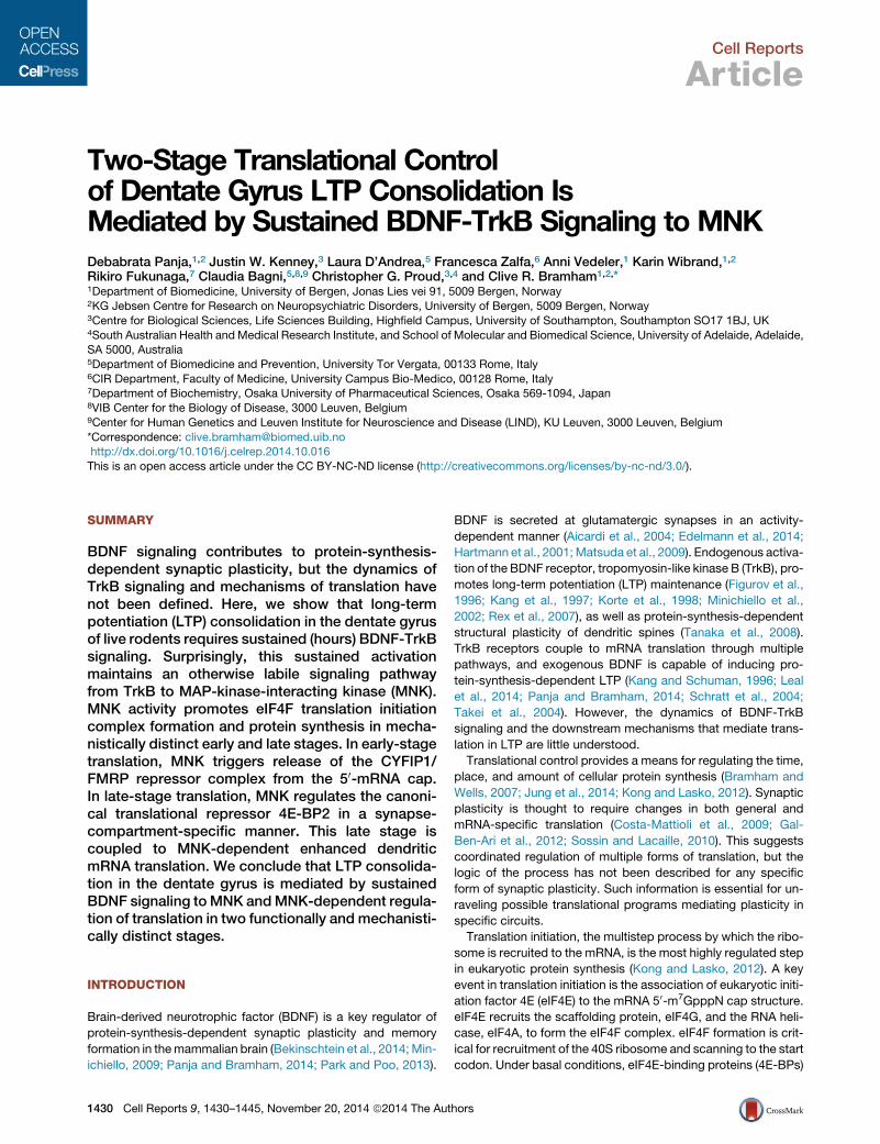
Article Two-Stage Translational Control of Dentate Gyrus LTP Consolidation Is Mediated by Sustained BDNF- TrkB Signaling to MNK Graphical Abstract Highlights Sustained BDNF-TrkB signaling controls LTP consolidation in vivo TrkB signaling to MNK mediates LTP consolidation MNK regulates CYFIP1/FMRP translation repressor complex in early-stage LTP MNK regulates 4E-BP2 and dendritic protein synthesis in late- stage LTP Authors Debabrata Panja, Justin W. Kenney, ..., Christopher G. Proud, Clive R. Bramham Correspondence [email protected] In Brief The logic of translational control in synap- tic plasticity is not well understood. Panja et al. show that long-term potentiation in the dentate gyrus of live rodents is a two-stage process driven by brain- derived neurotrophic factor signaling to MAP-kinase-interacting kinase and acti- vation of functionally and mechanistically distinct forms of translation. Panja et al., 2014, Cell Reports 9, 1430–1445 November 20, 2014 ª2014 The Authors http://dx.doi.org/10.1016/j.celrep.2014.10.016
Transcript of Two-Stage Translational Control of Dentate Gyrus LTP Consolidation Mediated by Sustained BDNF-TrkB...

Article
Two-Stage Translational C
ontrol of Dentate GyrusLTP Consolidation Is Mediated by Sustained BDNF-TrkB Signaling to MNKGraphical Abstract
Highlights
Sustained BDNF-TrkB signaling controls LTP consolidation
in vivo
TrkB signaling to MNK mediates LTP consolidation
MNK regulates CYFIP1/FMRP translation repressor complex in
early-stage LTP
MNK regulates 4E-BP2 and dendritic protein synthesis in late-
stage LTP
Panja et al., 2014, Cell Reports 9, 1430–1445November 20, 2014 ª2014 The Authorshttp://dx.doi.org/10.1016/j.celrep.2014.10.016
Authors
Debabrata Panja, Justin W. Kenney, ...,
Christopher G. Proud, Clive R. Bramham
In Brief
The logic of translational control in synap-
tic plasticity is not well understood. Panja
et al. show that long-term potentiation in
the dentate gyrus of live rodents is a
two-stage process driven by brain-
derived neurotrophic factor signaling to
MAP-kinase-interacting kinase and acti-
vation of functionally and mechanistically
distinct forms of translation.
Cell Reports
Article
Two-Stage Translational Controlof Dentate Gyrus LTP Consolidation IsMediated by Sustained BDNF-TrkB Signaling to MNKDebabrata Panja,1,2 Justin W. Kenney,3 Laura D’Andrea,5 Francesca Zalfa,6 Anni Vedeler,1 Karin Wibrand,1,2
Rikiro Fukunaga,7 Claudia Bagni,5,8,9 Christopher G. Proud,3,4 and Clive R. Bramham1,2,*1Department of Biomedicine, University of Bergen, Jonas Lies vei 91, 5009 Bergen, Norway2KG Jebsen Centre for Research on Neuropsychiatric Disorders, University of Bergen, 5009 Bergen, Norway3Centre for Biological Sciences, Life Sciences Building, Highfield Campus, University of Southampton, Southampton SO17 1BJ, UK4South Australian Health and Medical Research Institute, and School of Molecular and Biomedical Science, University of Adelaide, Adelaide,SA 5000, Australia5Department of Biomedicine and Prevention, University Tor Vergata, 00133 Rome, Italy6CIR Department, Faculty of Medicine, University Campus Bio-Medico, 00128 Rome, Italy7Department of Biochemistry, Osaka University of Pharmaceutical Sciences, Osaka 569-1094, Japan8VIB Center for the Biology of Disease, 3000 Leuven, Belgium9Center for Human Genetics and Leuven Institute for Neuroscience and Disease (LIND), KU Leuven, 3000 Leuven, Belgium
*Correspondence: [email protected]://dx.doi.org/10.1016/j.celrep.2014.10.016
This is an open access article under the CC BY-NC-ND license (http://creativecommons.org/licenses/by-nc-nd/3.0/).
SUMMARY
BDNF signaling contributes to protein-synthesis-dependent synaptic plasticity, but the dynamics ofTrkB signaling and mechanisms of translation havenot been defined. Here, we show that long-termpotentiation (LTP) consolidation in the dentate gyrusof live rodents requires sustained (hours) BDNF-TrkBsignaling. Surprisingly, this sustained activationmaintains an otherwise labile signaling pathwayfrom TrkB to MAP-kinase-interacting kinase (MNK).MNK activity promotes eIF4F translation initiationcomplex formation and protein synthesis in mecha-nistically distinct early and late stages. In early-stagetranslation, MNK triggers release of the CYFIP1/FMRP repressor complex from the 50-mRNA cap.In late-stage translation, MNK regulates the canoni-cal translational repressor 4E-BP2 in a synapse-compartment-specific manner. This late stage iscoupled to MNK-dependent enhanced dendriticmRNA translation. We conclude that LTP consolida-tion in the dentate gyrus is mediated by sustainedBDNF signaling toMNK andMNK-dependent regula-tion of translation in two functionally and mechanisti-cally distinct stages.
INTRODUCTION
Brain-derived neurotrophic factor (BDNF) is a key regulator of
protein-synthesis-dependent synaptic plasticity and memory
formation in themammalian brain (Bekinschtein et al., 2014;Min-
ichiello, 2009; Panja and Bramham, 2014; Park and Poo, 2013).
1430 Cell Reports 9, 1430–1445, November 20, 2014 ª2014 The Aut
BDNF is secreted at glutamatergic synapses in an activity-
dependent manner (Aicardi et al., 2004; Edelmann et al., 2014;
Hartmann et al., 2001;Matsuda et al., 2009). Endogenous activa-
tion of the BDNF receptor, tropomyosin-like kinase B (TrkB), pro-
motes long-term potentiation (LTP) maintenance (Figurov et al.,
1996; Kang et al., 1997; Korte et al., 1998; Minichiello et al.,
2002; Rex et al., 2007), as well as protein-synthesis-dependent
structural plasticity of dendritic spines (Tanaka et al., 2008).
TrkB receptors couple to mRNA translation through multiple
pathways, and exogenous BDNF is capable of inducing pro-
tein-synthesis-dependent LTP (Kang and Schuman, 1996; Leal
et al., 2014; Panja and Bramham, 2014; Schratt et al., 2004;
Takei et al., 2004). However, the dynamics of BDNF-TrkB
signaling and the downstream mechanisms that mediate trans-
lation in LTP are little understood.
Translational control provides a means for regulating the time,
place, and amount of cellular protein synthesis (Bramham and
Wells, 2007; Jung et al., 2014; Kong and Lasko, 2012). Synaptic
plasticity is thought to require changes in both general and
mRNA-specific translation (Costa-Mattioli et al., 2009; Gal-
Ben-Ari et al., 2012; Sossin and Lacaille, 2010). This suggests
coordinated regulation of multiple forms of translation, but the
logic of the process has not been described for any specific
form of synaptic plasticity. Such information is essential for un-
raveling possible translational programs mediating plasticity in
specific circuits.
Translation initiation, the multistep process by which the ribo-
some is recruited to the mRNA, is the most highly regulated step
in eukaryotic protein synthesis (Kong and Lasko, 2012). A key
event in translation initiation is the association of eukaryotic initi-
ation factor 4E (eIF4E) to the mRNA 50-m7GpppN cap structure.
eIF4E recruits the scaffolding protein, eIF4G, and the RNA heli-
case, eIF4A, to form the eIF4F complex. eIF4F formation is crit-
ical for recruitment of the 40S ribosome and scanning to the start
codon. Under basal conditions, eIF4E-binding proteins (4E-BPs)
hors
repress translation by blocking the recruitment of eIF4G to
eIF4E. Phosphorylation of 4E-BP, catalyzed by the mammalian
target of rapamycin complex 1 (mTORC1) kinase, triggers the
release of 4E-BP and facilitates translation (Gingras et al.,
2001; Proud, 2007). In addition, noncanonical 4E-BPs can regu-
late mRNA-specific translation through association with RNA-
binding proteins (Kong and Lasko, 2012; Richter and Klann,
2009). One such protein in brain is CYFIP1, cytoplasmic
fragile-X mental retardation protein (FMRP)-interacting protein
(Napoli et al., 2008). Through dual binding of FMRP and eIF4E,
CYFIP1 regulates translation of FMRP target mRNAs such as
the mRNA encoding the activity-dependent cytoskeletal-associ-
ated protein, Arc (De Rubeis et al., 2013; Napoli et al., 2008; Zalfa
et al., 2003).
Arc is an immediate early gene product required for several
forms of long-term synaptic plasticity and memory formation
(Bramham et al., 2010). Arc mRNA is rapidly induced and trans-
ported to dendritic processes for local storage and translation. In
the dentate gyrus (DG), LTP consolidation requires a period of
sustained Arc synthesis lasting from 2 to 4 hr after LTP induction
(Messaoudi et al., 2007). Unlike hippocampal region CA1 and
several other brain regions (Costa-Mattioli et al., 2009; Gal-
Ben-Ari et al., 2012), LTP consolidation, eIF4F formation, and
Arc synthesis in the DG are insensitive to mTORC1 inhibition
by rapamycin (Panja et al., 2009). In DG LTP, ERK signaling to
MAP-kinase-interacting kinases (MNKs) has been implicated in
eIF4F formation (Panja et al., 2009). MNKs are known as eIF4E
kinases (Banko et al., 2006; Gelinas et al., 2007; Proud, 2007),
but the function of MNKs in the nervous systems is largely
unknown.
Here, we report that sustained BDNF-TrkB signaling drives
translation and mediates Arc synthesis-dependent LTP in the
DG. Sustained TrkB receptor activation serves to maintain an
otherwise short-lived signaling pathway from TrkB to MNK.
In turn, MNK mediates eIF4F translation initiation complex for-
mation in distinct early and late stages linked to CYFIP1 and
4E-BP2 regulation, respectively. Notably, the late stage is asso-
ciated with pronounced MNK-dependent synaptic translation.
Hence, LTP consolidation in the DG is mediated by BDNF
signaling to MNK and MNK-dependent activation of translation
in two functionally and mechanistically distinct stages.
RESULTS
Sustained BDNF-TrkB Activation Is Required for DG LTPConsolidationThe BDNF scavenger, TrkB-Fc, was used to probe the role of
BDNF-TrkB activation in medial perforant-evoked DG LTP in
adult anesthetized rats. TrkB-Fc or control immunoglobulin G
(IgG)-Fc was acutely infused (100 mg, 1 ml, 12.5 min) 45 min
before high-frequency stimulation (HFS), or at one of five time
points after HFS. Infusions were made into deep stratum lacuno-
sum-moleculare, immediately above the dorsal DG. As shown in
Figure 1A, LTP of the field excitatory postsynaptic potential
(fEPSP) slope was significantly reduced in rats receiving TrkB-
Fc prior to HFS compared to the IgG-Fc-treated control. When
HFS was omitted from the paradigm, TrkB-Fc infusion had no
effect on fEPSP responses over a 4 hr period of baseline test
Cell Re
stimulation (BTS). These findings concur with previous reports
showing acute regulation of LTP induction by endogenous
BDNF (Figurov et al., 1996; Gooney and Lynch, 2001; Kossel
et al., 2001). During LTP maintenance, TrkB-Fc was infused at
10 min, 2 hr, 4 hr, 8 hr, or 10 hr after HFS (Figure 1; composite
results in Figures 1G and 1H). Control IgG-Fc infusions had no
effect on LTP maintenance. Strikingly, TrkB-Fc infusion at
10 min, 2 hr, or 4 hr after HFS resulted in rapid, complete, and
permanent reversion of LTP (Figures 1B–1D). The decline in
synaptic efficacy was significant within �10 min of TrkB-Fc
infusion onset and reached the pre-HFS baseline level by
30 min (Figure 1G). The effects of TrkB-Fc were strictly time
sensitive; TrkB-Fc infusion only transiently diminished fEPSPs
at 8 hr after HFS (Figure 1E) and had no significant effect at
10 hr after HFS (Figure 1F). The effects of TrkB-Fc on LTP main-
tenance were replicated by infusion of K252a (250 mM, 1 ml,
12.5 min), a Trk inhibitor that crosses cell membranes and
directly inhibits Trk tyrosine kinase activity (Figure 1I; time-
course plots shown in Figure S1A). The results indicate that
LTP consolidation requires persistent activation of TrkB by
BDNF. The period of TrkB dependency starts within 10 min of
HFS and lasts between 4 and 8 hr.
Sustained BDNF-TrkB Signaling Maintains ERKActivation and Arc ExpressionThe rapid reversion of LTP observed at multiple time points sug-
gests that TrkB couples to a labile signaling pathway. As BDNF-
induced Arc expression requires ERK activation (Messaoudi
et al., 2007; Ying et al., 2002), we examined TrkB-ERK signaling
and Arc expression during LTP. To monitor signaling within the
synaptic compartment, assays were performed in synaptoneur-
osomes, which are biochemical fractions highly enriched in
pinched-off dendritic spines attached to axon terminals of
excitatory synapses (De Rubeis et al., 2013; Havik et al., 2003;
Troca-Marın et al., 2011). The synaptic enrichment of the DG
synaptoneurosome preparation was validated by immunoblot-
ting for marker proteins (Figures 2A and 2B). PSD-95 and
GluN1 postsynaptic components of glutamatergic synapses
were enriched 14- and 17-fold, respectively, in DG synaptoneur-
osomes relative to lysates. The presynaptic markers synapto-
physin and cysteine-string protein-a (CSPa) were also enriched
in synaptoneurosomes, whereas the glial protein, GFAP, and
the glycolytic enzyme, GAPDH, were depleted in synaptoneuro-
somes relative to lysates (Figures 2A and 2B). The nuclear mem-
brane protein, laminin, was heavily stained in lysates but was not
detected in synaptoneurosomes (Figures 2A and 2B).
HFS elicited increased Tyr706/707 TrkB autophosphorylation
and Thr202/Tyr204 ERK phosphorylation and enhanced Arc
expression in both whole-lysate samples and synaptoneuro-
some samples. Immunoblot analysis of tissue obtained at
10 min, 40 min, and 3 hr after HFS revealed sustained activation
(Figures 2E–2J). Next, TrkB-Fc was infused at 10 min after HFS,
and tissue was collected at 40 min after HFS, by which time
fEPSP responses had declined to baseline levels (Figure 2C).
At 40 min, phospho-TrkB, phospho-ERK, and Arc protein in
whole lysate and synaptoneurosomes were significantly in-
hibited relative to time-matched IgG-Fc-infused controls (Fig-
ures 2E–2G). TrkB-Fc infusion at 2 hr similarly resulted in rapid
ports 9, 1430–1445, November 20, 2014 ª2014 The Authors 1431

4 h t HFS45 i HFS IgG-Fc + HFS DA
Time (min)-100 0 100 200
f EPS
P (%
cha
nge)
-40
-20
0
20
40
60
80
Time (min)-100 0 100 200 300 400
f EPS
P (%
cha
nge)
-40-20
020406080
100
4 h post-HFS45 min pre-HFS IgG-Fc + HFSTrkB-Fc + HFSTrkB-Fc + BTS
HFS + IgG-FcHFS + TrkB-Fc
( ) e ( )
-100 0 100 200 300 400 500 600 700
f EPS
P (%
cha
nge)
-40-20
020406080
100
-50 0 50 100 150 200 250
f EPS
P (%
cha
nge)
-40-20
020406080
1008 h post-HFS10 min post-HFS HFS + IgG-Fc
HFS + TrkB-FcHFS + IgG-FcHFS + TrkB-Fc
B E
EPSP
(% c
hang
e)
-20
0
20
40
60
80
Time (min)100 0 100 200 300 400 500 600 700
EPSP
(% c
hang
e)
-200
20406080
100
Time (min)50 0 50 100 150 200 250
10 h post-HFS2 h post-HFSHFS + IgG-FcHFS + TrkB-Fc
HFS + IgG-FcHFS + TrkB-Fc
FC
Time (min)-50 0 50 100 150 200 250
fE -40
Time (min)-100 0 100 200 300 400 500 600 700 800
fE -40
G
40
60
80
chan
ge) HFS + IgG-Fc
HFS + TrkB-Fc
IH
-20
0
20
40
-100 0 100 200 300 400 500 600 700 800Time (min)
fEPS
P ( %
c
Time
fEPS
P (%
cha
nge)
0
20
40
60
80
2 h 4 h 10 h8 h
* * * * * **
10 min
IgG-FcTrkB-Fc
4 h 10 h8 hTime
fEPS
P ( %
cha
nge)
0
20
40
60
80
* * * **VehK252a
Time Time
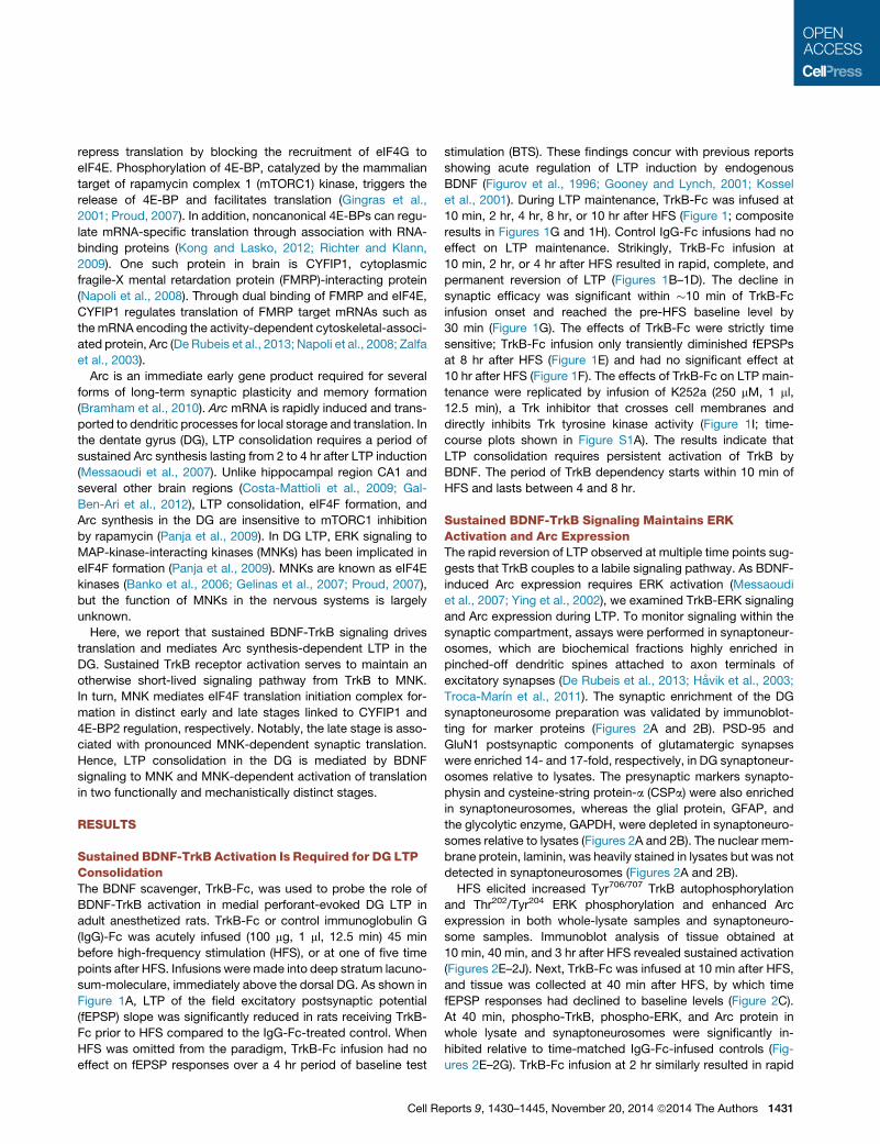
Figure 1. Sustained BDNF-TrkB Activation Is Required for DG LTP Consolidation In Vivo
Time-course plots of medial perforant path-dentate gyrus (DG) evoked fEPSPs recorded before and after high-frequency stimulation (HFS, indicated by arrows).
Values are mean ± SEM of the maximum fEPSP slope expressed as percentage of baseline. Test pulses were applied at a 0.033 Hz. TrkB-Fc (1 ml, 12.5 min,
100 mg) or control IgG-Fc (1 ml, 12.5min, 100 mg) was infused into the dorsal DG during the period indicated by the blue bar. In (A), a third treatment group received
TrkB-Fc infusion and baseline test pulse stimulation (BTS) but not HFS. n = 7/group. Error bars represent SEM.
(A–F) fEPSP changes in rats receiving TrkB-Fc or IgG-Fc 45min before HFS (A), or 10min (B), 2 hr (C), 4 hr (D), 8 hr (E), or 10 hr (F) after HFS. LTPmaintenancewas
permanently reverted by TrkB-Fc infusion at time points up to 4 hr after HFS, but not thereafter.
(G) Composite time-course plots of TrkB-Fc effects.
(H) Mean changes ± SEM in fEPSP slope 1 hr after the start of IgG-Fc or TrkB-Fc infusion. *p < 0.05, significantly different from baseline, Student’s t test for
independent samples. n = 5–6/group.
(I) Effect of K252a infusion on LTP maintenance. Vehicle is 0.1% DMSO in 1 3 PBS. Time-course plots shown in Figure S1.
1432 Cell Reports 9, 1430–1445, November 20, 2014 ª2014 The Authors
inhibition of enhanced TrkB-ERK activity and Arc expression at
3 hr after HFS (Figures 2D and 2H–2J). We conclude that
BDNF persistently activates a short-lived signaling pathway
from TrkB to ERK activation and Arc protein expression.
Early Translation: TrkB Regulates CYFIP1/FMRP andeIF4F FormationThere are three mammalian 4E-BP paralogs (4E-BP1, -2, and -3)
and 4E-BP2 is the major form expressed in brain (Banko
et al., 2005). mTORC1-dependent release of 4E-BP2 and the
resulting enhancement in eIF4F formation (eIF4E-eIF4G interac-
tion) are critical to multiple forms of translation-dependent syn-
aptic plasticity (Richter and Klann, 2009). However, DG LTP is
mTORC1-independent and eIF4F formation occurs in the
absence of 4E-BP2 release from eIF4E, as measured in cap
pull-down analyses in lysates samples (Panja et al., 2009).
We therefore asked whether CYFIP1, a noncanonical 4E-BP,
functions in DG LTP. TrkB-Fc or IgG-Fc was infused 10 min after
HFS, and DG lysates were collected at 40 min after HFS
(same electrophysiological procedure as shown in Figure 2C).
m7GTP-Sepharose (cap analog) pull-downs were performed,
and changes in the amount of eIF4G, 4E-BP2, CYFIP1, and
FMRP normalized to levels of cap-bound eIF4Ewere determined
by immunoblotting (Figures 3A, top, and 3B). In HFS-treated DG
of IgG-Fc-infused rats, recovery of CYFIP1 and FMRP was
significantly reduced, whereas loading of eIF4G was enhanced
2-fold relative to the contralateral DG. Infusion of TrkB-Fc abol-
ished these changes in CYFIP1/FMRP and eIF4G recovery on
m7GTP beads (Figures 3A and 3B). Given evidence of sustained
eIF4F formation in LTP, we predicted sustained regulation of
CYFIP1/FMRP. TrkB-Fc was infused at 2 hr and whole DG tissue
was collected at 3 hr after HFS (same electrophysiology proce-
dure as shown in Figure 2D). Surprisingly, although TrkB-Fc
prevented the enhanced eIF4G-eIF4E interaction, there was
no difference between HFS-treated and control DG lysate in
CYFIP1/FMRP recovery (Figures 3A, bottom panel, and 3B).
The results support a model in which TrkB signaling regulates
CYFIP1/FMRP association with eIF4E at the early (40 min) but
not late (3 hr) time points in LTP maintenance.
4E-BP2 undergoes brain-specific deamidation postnatally,
resulting in three primary bands detected by western blot (Bi-
dinosti et al., 2010). Two upper bands at �18–20 kDa corre-
spond to deamidated 4E-BP2, whereas the unmodified form
migrates at �16 kDa (see blots in Figure 3B). Confirming our
previous findings (Panja et al., 2009), the amount of deami-
dated (both bands combined) and unmodified 4E-BP2 recov-
ered with eIF4E in cap pull-downs did not differ between
treated and control DG at 40 min or 3 hr after HFS (Figures
3A and 3B).
Late Translation: TrkB Regulates 4E-BP2 and eIF4FFormation in the Synaptic CompartmentLocal protein synthesis in dendrites and dendritic spines is
important for synaptic regulation and plasticity (Bramham and
Wells, 2007;Martin and Ephrussi, 2009).We therefore performed
cap pull-downs in DG synaptoneurosomes. eIF4F formation in
synaptoneurosomes was enhanced at both 40 min and 3 hr after
HFS (Figures 3C and 3D). Reduction of CYFIP1 and FMRP bind-
Cell Re
ing to the m7GTP beads was also detected in synaptoneuro-
somes, but, as in lysates, this effect was confined to the early
time point (Figure 3C, upper panel). Surprisingly, immunoblot
analysis of 4E-BP2 in cap pull-downs revealed a regulation
that was specific to the synaptoneurosome compartment and
the late time point (Figures 3C, bottom panel, and 3D). At 3 hr
after HFS, levels of 4E-BP2 normalized to eIF4E were reduced
by 50.6% ± 1.8% and 33.4% ± 0.8% for deamidated and
unmodified 4E-BP2, respectively. Finally, TrkB-Fc blocked
both the early regulation of CYFIP1 and the late regulation of
4E-BP2, while preventing the enhanced eIF4G-eIF4E interaction
at both time points in the synaptic compartment (Figures 3C and
3D). Thus, TrkB activity mediates sequential regulation of distinct
translational repressors and persistently enhances eIF4F forma-
tion in DG LTP.
TrkB-MNK Signaling Regulates CYFIP1 and 4E-BP2Next, we examined TrkB signaling to MNK as a potential mech-
anism for DG LTP maintenance. Infusion of the MNK inhibitor,
CGP57380 (2 mM, 1 ml, 12.5 min; Tschopp et al., 2000), at
10 min or 2 hr after HFS induced a rapid and stable reversion
of LTP (Figures 4A and 4B), whereas CGP57380 infusion at
10 hr no longer affected LTP maintenance (composite time
course in Figure 4F). TheMNK inhibitor did not alter basal synap-
tic transmission in response to test-pulse stimulation (Figure 4B).
Levels of active, Thr197/202 phosphorylated MNK1 were inhibited
by infusion of CGP57380 (Figure 4C) and TrkB-Fc at both early
and late time points (Figures 4D and 4E). CGP57380 infusion at
10 min or 2 hr after HFS also led to a rapid decline in Arc protein
levels relative to vehicle-infused control (Figure 4C). Hence, LTP
maintenance across a defined time window depends on sus-
tained TrkB signaling to MNK.
MNKs binds to eIF4G and phosphorylate eIF4E (Pyronnet
et al., 1999; Scheper et al., 2002; Shveygert et al., 2010). In
cap pull-down assays performed in DG lysates, TrkB-Fc and
CGP57380 inhibited HFS-evoked Ser209 eIF4E phosphorylation
at both early and late time points (Figures 3 and 5A–5C). At
the early time point, the MNK inhibitor prevented changes in
CYFIP1/FMRP and eIF4G association with eIF4E. At the late
time point in synaptoneurosomes, CGP57380 inhibited changes
in 4E-BP2 and eIF4G association with cap-bound eIF4E (Figures
5D and 5E).
The effect of CGP57380 and rapamycin on Arc protein expres-
sion during the period of sustained Arc synthesis was visualized
by immunohistochemical staining (Figure S4). In rats infusedwith
vehicle at 2 hr after HFS, Arc immunostaining was uniformly
enhanced in the DG granule cell body and molecular (dendritic)
layer. In CGP57380-infused rats, Arc immunostaining was
strongly reduced in the molecular layer of both the upper and
lower blades of the DG (Figure S4A). Clear Arc immunostaining
remained in the granule cell body layer, consistent with residual
expression of Arc in the immunoblot analysis (Figure 4C). In
contrast, infusion of rapamycin at high concentrations (100 mM)
did not impair LTPmaintenance (Figure S4A) or affect Arc protein
immunostaining (Figure S4B), yet it inhibited Ser2448 mTOR
phosphorylation (not shown) and downstream enhancement of
ribosomal protein S6 phosphorylation in the granule cell body
layer (Figure S4B).
ports 9, 1430–1445, November 20, 2014 ª2014 The Authors 1433

A B
DG
ly
sate
DG
SNProt
ein
ladd
er
220
toph
ysin
enric
hmen
t (fo
ld)
0
5
10
15
20
PSD-95GluN
1CSPα
GAPDH
GFAP
Lamini
n
Laminin
PSD-95
GluN1
Synaptophysin
CSPα
GFAP
-
-100
-130
-38
-30
-50
DC
Synap
topP G L
ge) 80
HFS + TrkB-FcHFS + IgG-Fc
1 2 3ge
) 80
HFS + TrkB-FcHFS + IgG-Fc
1 2 3
GAPDH
ß-actin
-37
-50
1 0
1 2
1 4
1 0
1 2
1 4
1 5
2 0
1 5 0 0
2 0 0 01 5 0 0
1 5 0 0
2 0 0 0
Time (min)-40 -20 0 20 40
fEPS
P (%
cha
ng
-20
0
20
40
60
Time (min)-40 0 40 80 120 160 200 240
fEPS
P (%
cha
ng
-20
0
20
40
60
IgG-Fc
TrkB-Fc
- 4
- 2
0
2
4
6
8
1 45 89 133 177 221 265 309 353 397 441 485
- 4
- 2
0
2
4
6
8
1 45 89 133 177 221 265 309 353 397 441 485
- 5
0
5
1 0
1 45 89 133 177 221 265 309 353 397 441 485
- 5
0
5
1 0
1 5
1 45 89 133 177 221 265 309 353 397 441 485
- 4
- 2
0
2
4
6
8
1 0
1 2
1 45 89 133 177 221 265 309 353 397 441 485
- 5
0
5
1 0
1 5
2 0
1 46 91 136 181 226 271 316 361 406 451 496
1 2 3
IgG-Fc
TrkB-Fc
- 1 0 0 0
- 5 0 0
0
5 0 0
1 0 0 0
5 0 0
1 50 99 148 197 246 295 344 393 442 491
- 4 0 0
- 2 0 0
0
2 0 0
4 0 0
6 0 0
8 0 0
1 0 0 0
1 2 0 0
1 4 0 0
1 49 97 145 193 241 289 337 385 433 481
- 1 0 0 0
- 5 0 0
0
5 0 0
1 0 0 0
1 5 0 0
1 51 101 151 201 251 301 351 401 451 501
- 1 5 0 0
- 1 0 0 0
- 5 0 0
0
5 0 0
1 0 0 0
1 5 0 0
1 54 107 1 60 2 13 2 66 319 372 425 47 8
- 5 0 0
0
5 0 0
1 0 0 0
1 49 97 145 193 241 289 337 385 433 481
- 1 0 0 0
- 5 0 0
0
5 0 0
1 0 0 0
5 0 0
1 50 99 148 197 246 295 344 393 442 491
1 2 3
p-Tr
kB/ T
rkB
(%
cha
nge)
020406080
100
* *
HFS + IgG-FcHFS + TrkB-Fc
HFS GE F
p-ER
K/ E
RK
(%
cha
nge)
0
50
100250
300
* *
* *
Arc
/ Tub
ulin
(%
cha
nge)
0100200300400500600
**
* *
010 min 40 min
SN
/ Trk
B
ange
)
6080
100
* *HFS + IgG-FcHFS + TrkB-Fc
HFS H I J
0 10 min 40 min
SN DG Lysate10 min 40 min
0
SN DG Lysate
10 min 40 min
ubul
in
ange
)
300400500600
* * * *
K/ E
RK
an
ge)
200
300
400
* *
p-Tr
kB(%
cha
02040
2 h 3 h
SN
Arc
/ Tu
(% c
h a
0100200300
2 h 3 h 2 h 3 h
SN DG Lysate
p-ER
K(%
ch
0
100
200
* * SN DG Lysate2 h 3 h 2 h 3 h
(legend on next page)
1434 Cell Reports 9, 1430–1445, November 20, 2014 ª2014 The Authors
40 min post-HFS (DG Lysate) BAm7GTP Sepharose (DG Lysate)
% C
hang
ere
lativ
e to
eIF
4E
-100-50
050
100150 **
* *
HFS + IgG-FcHFS + TrkB-Fc
m7GTP-Sepharose (DG Lysate)3 h post-HFS
+ - + -IgG-Fc TrkB-Fc
40 min post-HFSIgG-Fc TrkB-Fc+ - + -
eIF4G1
eIF4E
P-eIF4E
-220
-25
-25
% C
hang
ere
lativ
e to
eIF
4E
-40-20
020406080
100 **
E 1 1 P 2 2
3 h post-HFS (DG lysate)
d-4E-BP2
CYFIP1
FMRP
4E-BP2
-145
-75
-20
-15
ge eIF4
E
150200
** HFS + IgG-FcHFS + TrkB-Fc
p-eI
F4E
eIF4
G1
CYF
IP1
FMR
P
d-4E
-BP2
4E-B
P2
m7GTP (Synaptoneurosomes)
+ +
3 h post-HFSIgG-Fc TrkB-Fc
40 min post-HFSIgG-Fc TrkB-Fc+ +
DC 40 min post-HFS (SN)
nge eIF4
E
300400 *
% C
hang
rela
tive
to e
-100-50
050
100
* *
+ - + -+ - + --220
-25
-25
-145
-75
3 h post-HFS (SN)
eIF4G1
eIF4E
CYFIP1
FMRP
P-eIF4E
% C
han
rela
tive
to
-1000
100200
* *
p-eI
F4E
eIF4
G1
CYF
IP1
FMR
P
d-4E
-BP2
4E-B
P2
-20
-15d-4E-BP2
4E-BP2
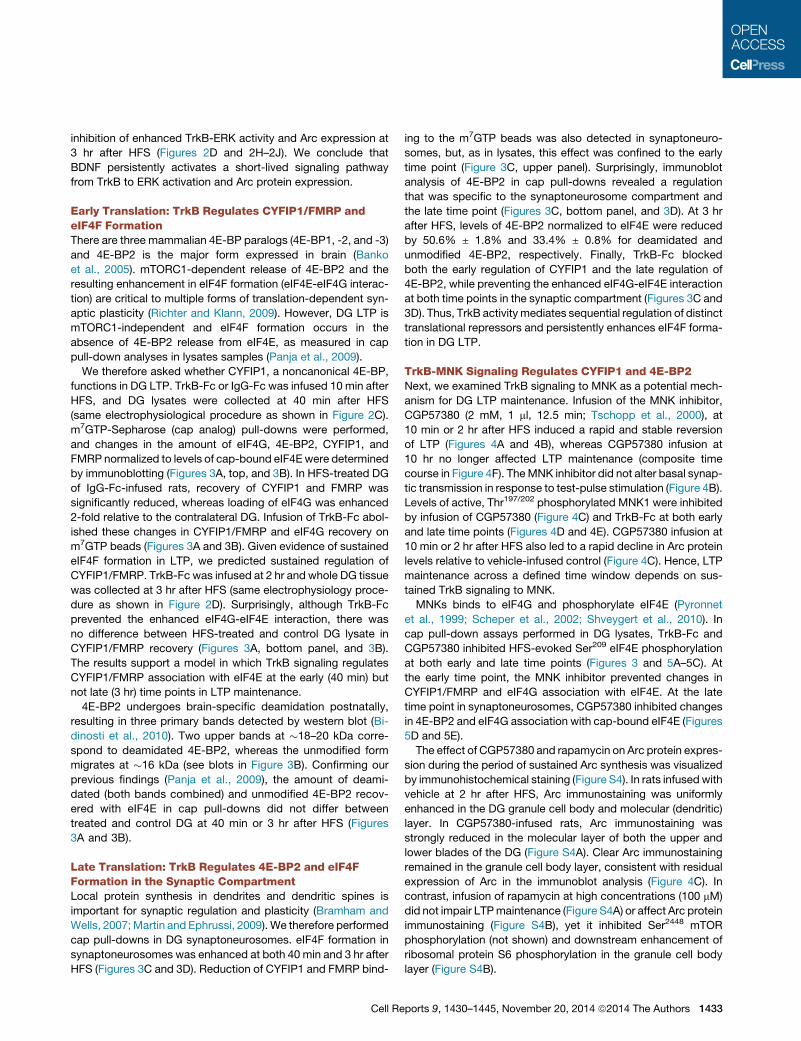
Figure 3. Sustained TrkB-Dependent eIF4F
Formation and Sequential Regulation of
CYFIP1 and 4E-BP2 Translational Re-
pressors
(A and B) m7GTP pull-down analysis in dentate
gyrus (DG) total lysates. (A) Upper panel: TrkB-Fc
or IgG-Fc was infused at 10 min after HFS and
tissue was collected at 40 min after HFS. Lower
panel: infusion at 2 hr, tissue collection at 3 hr after
HFS. Quantification of immunoblots expressed as
percentage change (mean ± SEM) of for p-eIF4E,
eIF4G, CYFIP1, FMRP, deamidated (d), and non-
modified 4E-BP2, normalized to total precipitated
eIF4E in the corresponding treated DG (+) relative
to the contralateral control DG (�). Error bars
represent SEM. Student’s t test in Table S1.
(B) Representative immunoblot for (A).
(C and D) m7GTP pull-down analysis in DG syn-
aptoneurosomes (SN). (C) Quantification of im-
munoblots for indicated protein. n = 6/group *p <
0.05. (D) Representative immunoblot for (C). No
bands were detected in control pull-downs using
Sepharose 4B beads alone (Figure S2A). Results
from immunoblot analysis of the input samples are
shown in Figure S3.
4E-BP2 binding to eIF4E is known be regulated by mTORC1
catalyzed phosphorylation of 4E-BP2 on Thr37/46. It was there-
fore of interest to examine Thr37/46 4E-BP2 phosphorylation state
in synaptoneurosomes at 3 hr after HFS. No phospho-4E-BP2
signal was detected in synaptoneurosome cap pull-down sam-
ples fromHFS-treated or control DG (Figure 5E). In synaptoneur-
osome inputs, strong phospho-4E-BP immunoreactivity specific
to the two deamidated forms of 4E-BP2 was found (Figure 5E).
Phospho specificity was confirmed by elimination of these bands
in phosphatase-treated samples (Figure S2B). However, there
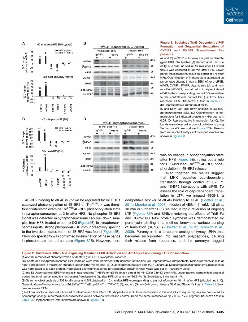
Figure 2. Sustained BDNF-TrkB Signaling Maintains ERK Activation and Arc Expression during
(A and B) Immunoblot characterization of dentate gyrus (DG) synaptoneurosomes.
DG lysate and synaptoneurosomes (SN) samples were immunoblotted with indicated antibodies. (A) Repre
right) corresponds to the protein standard ladder in themiddle lane. (B) Quantified immunoblots from (A); n = 6/
was normalized to b-actin protein. Normalized chemiluminescence for respective protein in total lysate was
(C and D) Upper panels: fEPSP changes in rats receiving TrkB-Fc or IgG-Fc (black bar) at 10 min (C) or 2 hr
traces (mean of ten consecutive responses) from baseline (1), after HFS (2), and after TrkB-Fc (3). Scale bar
(E–G) Immunoblot analysis of DG total lysates and SN obtained at 10 min after HFS (corresponding to start
Quantification of immunoblots for p-TrkB (Tyr706/707) (E), p-ERK(Thr202/Tyr204) (F), and Arc (G), n = 6–7/ group.
bars represent SEM.
(H–J) Immunoblot analysis at 2 hr (start of infusion) and 3 hr after HFS (stippled line in D). Immunoblot data i
percentage change in normalized densitometric values between treated and control DG on the same immun
Table S1. Representative immunoblots are shown in Figure S1B.
Cell Reports 9, 1430–1445, No
was no change in phosphorylation state
after HFS (Figure 5E), ruling out a role
for HFS-induced Thr37/46 4E-BP2 phos-
phorylation in 4E-BP2 release.
Taken together, the results suggest
that MNK regulates cap-dependent
translation through control of CYFIP1
and 4E-BP2 interactions with eIF4E. To
assess the role of cap-dependent trans-
lation in LTP, we infused 4EGI-1, a
competitive blocker of eIF4G binding to eIF4E (Hoeffer et al.,
2011; Moerke et al., 2007). Infusion of 4EGI-1 (1 mM, 1.5 ml) at
10 min or 2 hr after HFS resulted in rapid reversion of ongoing
LTP (Figures S5A and S5B), mimicking the effects of TrkB-Fc
and CGP57380. New protein synthesis was demonstrated by
puromycin labeling in a method known as surface sensing
of translation (SUnSET) (Hoeffer et al., 2013; Schmidt et al.,
2009). Puromycin is a structural analog of tyrosyl-tRNA that
becomes incorporated into nascent polypeptides, causing
their release from ribosomes, and the puromycin-tagged
LTP Consolidation
sentative immunoblots. Molecular mass (in kDa at
group.Measurement of direct chemiluminescence
set at 1 (arbitrary units).
(D) after HFS. Lower panels: sample field potential
s, 2 ms and 5 mV.
of infusion) or 40 min after HFS (stippled line in C).
Mean ± SEM and Student’s t test in Table S1. Error
n this and all subsequent figures are calculated as
oblot. *p < 0.05, n = 5–6/group. Student’s t test in
vember 20, 2014 ª2014 The Authors 1435
PSP
(% c
hang
e)
-200
20406080
HFS + VehHFS + CGP57380
A
PSP
(% c
hang
e)
-200
20406080
HFS + VehHFS + CGP57380BTS + CGP57380
Be 400
500
* *##
HFS + CGP57380HFS + Veh
Time (min)-40 -20 0 20 40 60 80 100 120 140f E -40
EC D 40 min post-HFS 3 h post-HFS
Time (min)-100 -50 0 50 100 150 200f E
P
-40
HFS + VehHFS + TrkB-Fc
HFS + VehHFS + TrkB-Fc
pMN
K1/
MN
K1
% c
hang
e
0
100
200
300
Arc/
Tub
ulin
pMN
K1/
MN
K1
Arc/
Tub
ulin
*
* ***
p-M
NK
1 / M
NK
1(%
cha
nge)
-50
0
50
100
150
SN Lysate
* *
SN Lysate
p-M
NK
1 / M
NK
1 (%
cha
nge)
-50
0
50
100
150
* *
40 min 3 h
P (%
cha
nge)
020406080
HFS +VehHFS + CGP57380
F
-100 0 100 200 300 400 500 600 700 800
f EPS
-40-20
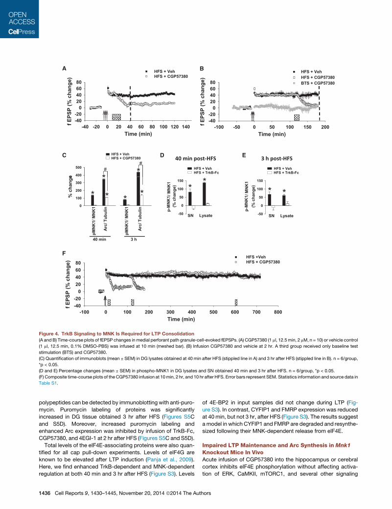
Figure 4. TrkB Signaling to MNK Is Required for LTP Consolidation
(A and B) Time-course plots of fEPSP changes in medial perforant path granule-cell-evoked fEPSPs. (A) CGP57380 (1 ml, 12.5 min, 2 mM, n = 10) or vehicle control
(1 ml, 12.5 min, 0.1% DMSO-PBS) was infused at 10 min (meshed bar). (B) Infusion CGP57380 and vehicle at 2 hr. A third group received only baseline test
stimulation (BTS) and CGP57380.
(C) Quantification of immunoblots (mean ± SEM) in DG lysates obtained at 40 min after HFS (stippled line in A) and 3 hr after HFS (stippled line in B). n = 6/group,
*p < 0.05.
(D and E) Percentage changes (mean ± SEM) in phospho-MNK1 in DG lysates and SN obtained 40 min and 3 hr after HFS. n = 6/group, *p < 0.05.
(F) Composite time-course plots of the CGP57380 infusion at 10min, 2 hr, and 10 hr after HFS. Error bars represent SEM. Statistics information and source data in
Table S1.
polypeptides can be detected by immunoblotting with anti-puro-
mycin. Puromycin labeling of proteins was significantly
increased in DG tissue obtained 3 hr after HFS (Figures S5C
and S5D). Moreover, increased puromycin labeling and
enhanced Arc expression was inhibited by infusion of TrkB-Fc,
CGP57380, and 4EGI-1 at 2 hr after HFS (Figures S5C and S5D).
Total levels of the eIF4E-associating proteins were also quan-
tified for all cap pull-down experiments. Levels of eIF4G are
known to be elevated after LTP induction (Panja et al., 2009).
Here, we find enhanced TrkB-dependent and MNK-dependent
regulation at both 40 min and 3 hr after HFS (Figure S3). Levels
1436 Cell Reports 9, 1430–1445, November 20, 2014 ª2014 The Aut
of 4E-BP2 in input samples did not change during LTP (Fig-
ure S3). In contrast, CYFIP1 and FMRP expression was reduced
at 40 min, but not 3 hr, after HFS (Figure S3). The results suggest
a model in which CYFIP1 and FMRP are degraded and resynthe-
sized following their MNK-dependent release from eIF4E.
Impaired LTP Maintenance and Arc Synthesis in Mnk1
Knockout Mice In VivoAcute infusion of CGP57380 into the hippocampus or cerebral
cortex inhibits eIF4E phosphorylation without affecting activa-
tion of ERK, CaMKII, mTORC1, and several other signaling
hors
CA B
40 i HFS (DG l ) 3 h H S ( G l ) m7GTP (DG lysate)
eIF4E
eIF4G1
P-eIF4E
40 min post-HFS (DG lysate) 3 h post-HFS (DG lysate) m GTP (DG lysate)
CGPVeh40 min post-HFS
+ - + -CGPVeh
3 h post-HFS
+ - + -
ive
% c
hang
e
200
20406080
100
**HFS + CGPHFS + Veh
ive
% c
hang
e
50
100
150 *
*
HFS + CGPHFS + Veh
CYFIP1
FMRP
Rel
ati
-80-60-40-20
* *
p-eI
F4E/
eIF
4E
eIF4
G1/
eIF
4E
CYF
IP1/
eIF
4E
FMR
P/ e
IF4E
Rel
ati
-50
0
p-eI
F4E/
eIF
4E
eIF4
G1/
eIF
4E
CYF
IP1/
eIF
4E
FMR
P/ e
IF4E
3 h post-HFS (SN)D E
eIF4G1
eIF4E
P-eIF4E
CGPVehm7GTP (SN)
+ - + -CGPVeh
+ - + --220
-25
-25
Input (SN)
+ -Veh
+ -CGP
e %
cha
nge
50
100
300
350
*
* HFS + CGPHFS + Veh
d-4E-BP2
eIF4E
P-4E-BP2
4E-BP2
25
-20
-15
-20
-15
Rel
ativ
e
-100
-50
0
p-eI
F4E/
eIF
4E
eIF4
G1/
eIF
4E
d-4E
-BP2
/ eIF
4E
4E-B
P2/ e
IF4E
* *
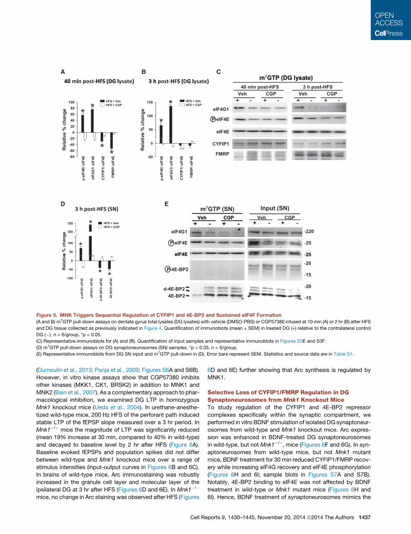
Figure 5. MNK Triggers Sequential Regulation of CYFIP1 and 4E-BP2 and Sustained eIF4F Formation
(A and B) m7GTP pull-down assays on dentate gyrus total lysates (DG lysates) with vehicle (DMSO-PBS) or CGP57380 infused at 10 min (A) or 2 hr (B) after HFS
and DG tissue collected as previously indicated in Figure 4. Quantification of immunoblots (mean ± SEM) in treated DG (+) relative to the contralateral control
DG (�). n = 6/group, *p < 0.05.
(C) Representative immunoblots for (A) and (B). Quantification of input samples and representative immunoblots in Figures S3E and S3F.
(D) m7GTP pull-down assays on DG synaptoneurosomes (SN) samples. *p < 0.05, n = 6/group.
(E) Representative immunoblots from DG SN input and m7GTP pull-down in (D). Error bars represent SEM. Statistics and source data are in Table S1.
(Dumoulin et al., 2013; Panja et al., 2009; Figures S6A and S6B).
However, in vitro kinase assays show that CGP57380 inhibits
other kinases (MKK1, CK1, BRSK2) in addition to MNK1 and
MNK2 (Bain et al., 2007). As a complementary approach to phar-
macological inhibition, we examined DG LTP in homozygous
Mnk1 knockout mice (Ueda et al., 2004). In urethane-anesthe-
tized wild-type mice, 200 Hz HFS of the perforant path induced
stable LTP of the fEPSP slope measured over a 3 hr period. In
Mnk1�/� mice the magnitude of LTP was significantly reduced
(mean 19% increase at 30 min, compared to 40% in wild-type)
and decayed to baseline level by 2 hr after HFS (Figure 6A).
Baseline evoked fEPSPs and population spikes did not differ
between wild-type and Mnk1 knockout mice over a range of
stimulus intensities (input-output curves in Figures 6B and 6C).
In brains of wild-type mice, Arc immunostaining was robustly
increased in the granule cell layer and molecular layer of the
ipsilateral DG at 3 hr after HFS (Figures 6D and 6E). In Mnk1�/�
mice, no change in Arc staining was observed after HFS (Figures
Cell Re
6D and 6E) further showing that Arc synthesis is regulated by
MNK1.
Selective Loss of CYFIP1/FMRP Regulation in DGSynaptoneurosomes from Mnk1 Knockout MiceTo study regulation of the CYFIP1 and 4E-BP2 repressor
complexes specifically within the synaptic compartment, we
performed in vitro BDNF stimulation of isolated DG synaptoneur-
osomes from wild-type and Mnk1 knockout mice. Arc expres-
sion was enhanced in BDNF-treated DG synaptoneurosomes
in wild-type, but not Mnk1�/�, mice (Figures 6F and 6G). In syn-
aptoneurosomes from wild-type mice, but not Mnk1 mutant
mice, BDNF treatment for 30 min reduced CYFIP1/FMRP recov-
ery while increasing eIF4G recovery and eIF4E phosphorylation
(Figures 6H and 6I; sample blots in Figures S7A and S7B).
Notably, 4E-BP2 binding to eIF4E was not affected by BDNF
treatment in wild-type or Mnk1 mutant mice (Figures 6H and
6I). Hence, BDNF treatment of synaptoneurosomes mimics the
ports 9, 1430–1445, November 20, 2014 ª2014 The Authors 1437

80
+/+ -/- A C
ms) 1.2
B
V)
5
Time (min)
-50 0 50 100 150 200
fEPS
P ( %
cha
nge)
-20
0
20
40
60
80
Stimulus intensity (mV)0 5 10 15 20 25 30
fEPS
P sl
ope
(mV/
m
0.0
0.2
0.4
0.6
0.8
1.0
Stimulus intensity (mV)0 5 10 15 20 25 30
Spik
e am
plitu
de (m
V
0
1
2
3
4
Arc
KO
Mnk
1 W
T
CONTRAIPSI ED
ensi
tom
etric
e
valu
es)
6
9
12
15
*Mnk1 +/+Mnk1 -/-
ls (a.u
.)
2.53.03.5
*F G
cle F F +
cle F F +
Cyfip1 +/+ Cyfip1 + /-Mnk1 +/+ Mnk1 -/-
cle
F cle
F
Mnk
1
Arc
(de
rela
tiv
0
3
VehicleBDNFBDNF + CGP57380
Mnk1+/+ Mnk1-/- Cyfip1 +/+ Cyfip1 +/-
Arc
leve
lno
rmal
ized
0.00.51.01.52.0 *
H
Arcα-Tubulin
vehi
c
BD
N
BD
NC
GP
vehi
c
BD
N
BD
NC
GP
VehicleBDNF
VehicleBDNF
Arc
β-actin
Vehi
cB
DN
Vehi
cB
DN
MNK1
I
eIF4G1 CYFIP1 FMRP p eIF4E 4E BP2
Prot
ein
leve
ls
(arb
itary
uni
ts)
0.0
0.5
1.0
1.5
2.0
2.5
eIF4G1 CYFIP1 FMRP p eIF4E 4E BP2
Prot
ein
leve
ls
(arb
itary
uni
ts)
0.0
0.5
1.0
1.5
2.0
2.5
*
* *
* BDNF + TrkB-Fc BDNF + TrkB-Fc
leve
ls
y un
its)
1.5
2.0
2.5
leve
ls
y un
its)
1.5
2.0
2.5
*
eIF4G1 CYFIP1 FMRP p-eIF4E 4E-BP2eIF4G1 CYFIP1 FMRP p-eIF4E 4E-BP2
K
-/-1knM+/+1knM
J VehicleBDNFBDNF + CGP57380
VehicleBDNFBDNF + CGP57380
Prot
ein
(arb
itar y
0.5
1.0
eIF4G1 CYFIP1 FMRP 4E-BP2
*eIF4G1 CYFIP1 FMRP 4E-BP2
Prot
ein
(arb
itary
0.5
1.0
1.5
* * *
-/+1pifyC+/+1pifyC
(legend on next page)
1438 Cell Reports 9, 1430–1445, November 20, 2014 ª2014 The Authors

early translation stage of LTP associated with selective CYFIP1/
FMRP regulation. Furthermore, levels of CYFIP1 and FMRP in
input samples were unchanged, showing that BDNF first triggers
release of CYFIP1/FMRP from eIF4E in the absence of protein
degradation (Figures S7C and S7D). Finally, basal formation of
the translation repressor complexes did not differ between
wild-type and Mnk1 knockout mice (Figure S7A).
MNK and CYFIP1-Dependent Regulation of eIF4F inBDNF-Treated Cortical SynaptoneurosomesPrevious work in cortical and hippocampal synaptoneurosomes
showed that BDNF treatment induces release of CYFIP1/FMRP
from eIF4E and enhanced translation of multiple FMRP-target
mRNAs (De Rubeis et al., 2013; Napoli et al., 2008). In agreement
with these data, we find that BDNF stimulation of cortical synap-
toneurosomes decreases the association of CYFIP1/FMRP and
4E-BP2withm7GTP-bound eIF4E, while enhancing eIF4G-eIF4E
association (Figures 6J and 6K). Treatment with CGP57380 in-
hibited these effects, indicating that BDNF-induced regulation
of CYFIP1 and 4E-BP2 at cortical synapses is MNK dependent
(Figures 6J and 6K).
To assess a causal role for CYFIP1, we used Cyfip1 ± mice
(Napoli et al., 2008; De Rubeis et al., 2013), which exhibited
significantly reduced expression of CYFIP1 of 45% in lysates
and 65% in synaptoneurosomes (Figures S7E and S7F).
BDNF-induced Arc expression was impaired in Cyfip1+/� mice
and inhibited by CGP57380 treatment in wild-type mice (Figures
6F and 6G). BDNF failed to alter the association of CYFIP1/
FMRP and eIF4G with eIF4E (Figures 6J and 6K; sample blots
in Figure S7B), indicating that a critical level of CYFIP1 is required
for BDNF-induced eIF4F formation. Interestingly, BDNF stimu-
lated the release of 4E-BP2 from eIF4E in both wild-type and
CYFIP+/� mice, and this regulation was inhibited by CGP57380
(Figures 6J and 6K). These results provide further support for
independent regulation of CYFIP1 and 4E-BP2 by MNK.
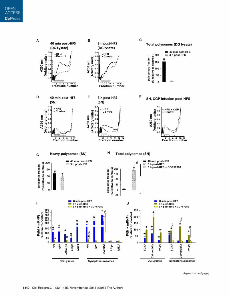
A Switch to Synapse-Specific Translation duringLate-Stage LTP In VivoPolysome analysis was used to directly measure changes in
translational activity following LTP induction. Samples from rat
Figure 6. LTP Maintenance, eIF4F Formation, and Arc Expression Are
(A) Time course of perforant path-DG evoked fEPSPs. The magnitude of LTP was
mice at 170–180 min (knockout [KO]: 5.3% ± 5.39%, n = 12; wild-type: 34.09%
potentials (mean of five consecutive sweeps); calibration, 5 ms, 3 mV.
(B and C) Input-output curves of fEPSP slope (B) and population spike (C) respon
Mnk1 KO mice. Repeated-measures ANOVA: F1,9 = 1.0561, p = 0.3082 for fEPS
(D) Representative Arc protein immunohistochemistry staining of coronal section
(E) Quantification of Arc protein expression based on mean densitometric intensi
(arbitrary units) in wild-type (10.77% ± 1.2%; p < 0.001) and Mnk1 KO mice (1.0
(F) Quantification of Arc protein expression in BDNF-stimulated DG SN fromwild-
the presence or absence of the MNK inhibitor CGP57380. The BDNF-treated SN
vehicle-treated SN (stippled line) set at 1 (arbitrary unit). n = 5. *p < 0.05.
(G) Representative immunoblots for results in (F).
(H and I) m7GTP pull-down assays of DG SN from wild-type and Mnk1 KO mice.
expressed relative to vehicle treatment set at 1 (arbitrary units) in wild-type and
(J and K) m7GTP pull-down assays of cortical SN from wild-type and Cyfip1+/� m
to vehicle treatment set at 1 (arbitrary units) in wild-type and Cyfip1+/� mice. Re
information and source data are in Table S1.
Cell Re
DG lysates and synaptoneurosomes were loaded onto a 10%–
50% linear sucrose gradient and subjected to velocity sedimen-
tation. Total polysomal formation was determined by averaging
the spectrophotometric absorption at 260 nm (area under the
curve) across gradient fractions 2–7 for the treated and contra-
lateral control DG. Total polysome formation was then ex-
pressed as the percentage of the total RNA signal across all
fractions in order to correct for changes in RNA abundance in
the free ribonucleoprotein fraction (fractions 8–12). Immunoblot
characterization of the gradient fractions confirmed cosedimen-
tation of rpS6, eEF2, and poly(A) binding protein in polysomal
fractions, and eIF4E, CYFIP1, and a fraction of FMRP and rpS6
in the nonpolysomal fraction (Figure S8A).
In lysates, total polysome formation was significantly in-
creased at 40 min, but not 3 hr, in the HFS-treated DG relative
to contralateral control (Figures 7A–7C). In synaptoneurosomes,
HFS induced a shift from light polysomes (fractions 5–7) into
heavy polysomes (fractions 2–4) at 40 min (Figure 7G), with no
net change in the total polysome formation (Figure 7H). At 3 hr
after HFS, total polysome formation in synaptoneurosomes
was significantly increased (184.6% ± 13.0%) in the HFS-treated
DG relative to the contralateral control (Figures 7E and 7H), and
this increase was abolished by CGP57380 infusion at 2 hr after
HFS (Figures 7F and 7H). The results demonstrate ongoing
translation in DG LTP in vivo and reveal a shift toward MNK-
dependent synaptic translation at the late time point.
The shift from CYFIP1/FMRP to 4E-BP2 regulation in DG LTP
implies a functional shift in mRNA translation. To begin to investi-
gate patterns of individual mRNA translation, quantitative PCR
analysis was used to measure changes in the translational effi-
ciency of five dendritically expressed FMRP targets (Arc, APP,
aCaMKII, NR2A/GluN2A, Lingo) (Pasciuto and Bagni, 2014).
mRNA levels in polysomes (sum of sucrose gradient fractions 2–
7) were normalized to the summed levels in monosomal and
mRNP fractions, and values in the HFS-treated DG were ex-
pressed as fold change relative to the contralateral control (Fig-
ures 7I and 7J). In lysate samples, all FMRP targets exhibited
enhanced polysomal association at 40 min, but not 3 hr, after
HFS. In synaptoneurosomes, polysomal abundance of Arc,
APP, and aCaMKII was upregulated more than 300-fold at
Impaired in Mnk1 Knockout Mice
significantly reduced in the DG of Mnk1�/� mice relative to wild-type Mnk1+/+
± 6.17%, n = 10) Student’s t test t(12) = �3.284, p < 0.001). Inset: sample field
ses show no difference in basal synaptic transmission between wild-type and
P and F1,9 = 0.8431, p = 0.362, for population spike).
obtained at 3 hr after HFS in wild-type and Mnk1 KO.
ty in ipsilateral (IPSI) DG relative to contralateral (CONTRA) control DG set at 1
9% ± 0.1%; p = 0.87). *p < 0.05.
type andMnk1 KOmice and BDNF-treated cortical SNs from Cyfip1+/�mice, in
were normalized to b-actin or tubulin as indicated and expressed relative to
Quantification show mean changes in protein levels normalized to eIF4E and
Mnk1 KO mice. n = 6, *p < 0.05. Representative immunoblots in Figure S7A.
ice. Mean changes in protein levels normalized to eIF4E and expressed relative
presentative immunoblots in Figure S7B. Error bars represent SEM. Statistics
ports 9, 1430–1445, November 20, 2014 ª2014 The Authors 1439
Total polysomes (DG lysate)A B C
3 h post-HFS(DG lysate)
40 min post-HFS(DG Lysate)
mes
frac
tion
e to
con
trol
s)
100
150
200
*40 min post-HFS3 h post-HFS
SN, CGP infusion post-HFSD E F3 h post-HFS(SN)
40 min post-HFS(SN)
poly
som
(% re
lativ
e
0
50
100
150
200
omes
frac
tion
ive
to c
ontr
ols)
* *100
150
200
s fr
actio
no
cont
rols
) *#
Heavy polysomes (SN) Total polysomes (SN)GH
40 min post-HFS3 h post-HFS3 h post-HFS + CGP57380
40 min post-HFS3 h post-HFS
0
50
poly
so(%
rela
ti
-50
0
50
poly
som
es(%
rela
tive
t
P) ntro
l
500600700800 #
***
40 min post-HFS3 h post-HFS3 h post-HFS + CGP57380
P) ntro
l
200
250
*
40 min post-HFS3 h post-HFS3 h post-HFS + CGP57380
JI
P/(M
+ m
RN
P re
lativ
e to
con
0
50
100
150
300400500
#
**
*
** * *
*
* *
Arc
L
ingo
A
PP
NR
2A
αCam
KII
αCam
KII
Arc
L
ingo
A
PP
NR
2A
P/(M
+ m
RN
P re
lativ
e to
co n
0
50
100
150
200
#
** *
** **
**
#
#
BD
NF
mod
ulin
PKMζ
BD
NF
mod
ulin
PKMζ
α
DG Lysates Synaptoneurosomes
α
DG Lysates Synaptoneurosomes
Cal
m
Cal
m
(legend on next page)
1440 Cell Reports 9, 1430–1445, November 20, 2014 ª2014 The Authors
40min after HFS;aCaMKII translation remained strongly elevated
at 3 hr, whereasArc andAPP declined, though Arc translation re-
mained significantly elevated �30-fold above control. We also
examined polysomal abundance of three dendritic, non-FMRP
target mRNAs (PKM-z, BDNF, calmodulin) that are implicated in
synapticplasticity. ThesemRNAsexhibitedapattern of regulation
almost inverse to that of the FMRP targets. Translation of PKM-z,
BDNF, and calmodulinwas increased at both time points but was
significantly higher at 3 hr after HFS (Figure 7J). In synaptoneuro-
somes, enhanced translation of PKM-z and calmodulin occurred
only at the late time point (Figure 7J). Finally, CGP57380 infusion
at 2 hr after HFS inhibited the increased synaptoneurosomal
translation of Arc, aCaMKII, PKM-z, calmodulin, and BDNF (Fig-
ures 7I, 7J, and S8B), thus supporting a key role for MNK in syn-
aptic translation underlying DG LTP in vivo.
DISCUSSION
DG LTP Consolidation Requires Sustained BDNF-TrkBActivationThe present work shows that LTP consolidation in the DG of
live rats is a highly dynamic, active process driven by persistent
BDNF-TrkB signaling. TrkB receptors activate a transient sig-
naling pathway to MNK, and this pathway is persistently acti-
vated by BDNF to mediate LTP consolidation. In turn, MNK
activity regulates translation initiation complex formation and
protein synthesis in mechanistically distinct early and late
stages. The dynamics of LTP consolidation were revealed by
time-sensitive reversion of LTP in response to acute infusion of
TrkB-Fc, CGP57380, and 4EGI-1. The rapid reversion of LTP
and Arc synthesis in the present study mimics the effects
obtained upon acute inhibition of Arc synthesis with antisense
oligodeoxynucleotides (Messaoudi et al., 2007). The work thus
identifies sustained TrkB signaling to MNK as a key mechanism
in Arc-dependent LTP consolidation.
TrkB-MNK signaling to translation within the synaptic com-
partment is shown by ex vivo and in vitro analysis of synapto-
neurosomes. Persistent BDNF-TrkB activity could reflect (1)
repeated activation of a stationary pool of TrkB, (2) continuous
membrane insertion and activation of new TrkB, or (3) ligand-
mediated endocytosis and recycling of TrkB to the membrane.
Repeated activation of stationary TrkB is highly unlikely, given
Figure 7. Enhanced Polysome Formation in DG LTP In Vivo: A Shift to
Samples from DG total lysate and synaptoneurosome (SN) samples obtained at 4
and subjected to velocity sedimentation. Gradients were collected in 12 fractions
processed in tandem for each time point. Total polysomal formation was determin
for differences in RNA abundance within the free ribonucleoprotein fraction (fract
signal across all fractions.
(A and B) RNA absorbance profiles for DG lysate samples obtained at 40 min (A
(C) Percentage change in absorbance in polysome fractions (indicated by dotted
normalized to the unstimulated contralateral DG lysate control.
(D–F) RNA absorbance profiles for DG SN samples obtained at 40min (left panel) a
obtained from experiments where CGP57380 was infused at 2 hr and tissue coll
(G) Percentage change in absorbance for heavy polysomes in SNs collected at 4
(H) Percentage of change in absorbance for total polysomes in SNs (dotted bar)
(I and J) Quantification by qRT-PCR analysis ofmRNA abundance loaded onto pol
3 hr (green), and 3 hr and CGP infusion (yellow) in total lysates and SN for the teste
Table S1.
Cell Re
evidence that TrkB is rapidly endocytosed upon ligand binding.
Recent work in hippocampal neurons and HEK293 cells shows
that endocytosed full-length TrkB rapidly recycles to the mem-
brane to promote sustained ERK signaling (Chen et al., 2005;
Huang et al., 2009, 2013; Nagappan and Lu, 2005). TrkB activa-
tion is known to stimulate BDNF release, and endocytosed
BDNF can be recycled for neuronal activity-dependent secretion
(Canossa et al., 1997; Santi et al., 2006). In neuronal develop-
ment, self-amplifying autocrine actions of BDNF-TrkB ensure
axonal differentiation and growth (Cheng et al., 2011). Based
on these studies, it is tempting to speculate that sustained
TrkB activation in the DG LTP involves regenerative secretion
of BDNF coupled with TrkB endocytosis and recycling to the
membrane. In cultured embryonic cortical neurons grown in
microfluidic chambers, BDNF application to the dendrite can
generate a TrkB signaling endosome that travels to the soma
where it activates ERK and induces Arc transcription (Cohen
et al., 2011). Signaling from TrkB endosomes should be resistant
to extracellular TrkB-Fc. In DG LTP, ERK phosphorylation was
blocked by extracellular TrkB-Fc (Figures 2F and 2I), arguing
against a primary role for signaling endosomes. Further studies
are needed to explore roles for TrkB recycling endosomes and
signaling endosomes in synaptic plasticity.
Two-Step Translational Control of LTP Mediated byMNK-Dependent Regulation of CYFIP1 and 4E-BP2Based on combined pharmacological and genetic approaches
in vivo and in vitro, we show that sustained TrkB signaling
to MNK drives persistent enhancement of eIF4F formation
(eIF4G-eIF4E interaction) underlying protein-synthesis-depen-
dent LTP. Moreover, we show that MNK facilitates translation
in discrete early and late stages of LTP consolidation associated
with regulation of CYFIP1 and 4E-BP2, respectively.
Napoli and colleagues (2008) showed that exogenous BDNF
stimulates CYFIP1 release and translation of FMRP-target
mRNAs in neuronal cultures and cortical synaptoneurosomes
in vitro. Here, we found that MNK regulates CYFIP1/FMRP
downstream of endogenous BDNF signaling early in LTP consol-
idation. In live Mnk1 knockout mice, DG LTP maintenance is
impaired and Arc synthesis is blocked. In DG synaptoneuro-
somes from Mnk1 knockout mice, BDNF regulation of CYFIP1/
FMRP, eIF4F, and Arc synthesis is inhibited. In cortical
Late MNK-Dependent Synaptic Translation
0 min and 3 hr after HFS were loaded onto a 10%–50% linear sucrose gradient
starting with the heavy fraction. HFS-treated and contralateral DG tissues were
ed calculating the area under the curve across gradient fractions 2–7. To correct
ions 8–12), total polysome formation was expressed in percentage of the RNA
) and 3 hr (B) after HFS.
bar in A and B) as percentage of the total area under the absorbance curve,
nd 3 hr (right panel) after HFS. (F) RNA absorbance profiles for DG SN samples
ected at 3 hr after HFS.
0 min and 3 hr after HFS in the ipsilateral DG relative to contralateral DG.
for 40 min and 3 hr and CGP57380-infused 3 hr samples. n = 4, *p < 0.05.
ysomes relative tomRNA loaded ontomonosomes andmRNPs at 40min (blue),
d mRNAs. Error bars represent SEM. Statistics information and source data in
ports 9, 1430–1445, November 20, 2014 ª2014 The Authors 1441

synaptoneurosomes from Cyfip1+/� mice, MNK-dependent
regulation of eIF4F is inhibited. In contrast, the late stage of
LTP consolidation is associated withMNK-dependent regulation
of 4E-BP2 and enhanced total polysome formation specifically
within the synaptoneurosome compartment.
Stable LTP formation involves expansion of the postsynaptic
density, enlargement of pre-existing dendritic spines, and
de novo synapse formation (Bourne and Harris, 2008; Lisman
and Raghavachari, 2006). These processes likely require coordi-
nated regulation of mRNA-specific and general translation
(Costa-Mattioli et al., 2009; Gal-Ben-Ari et al., 2012; Sossin
and Lacaille, 2010). The present study provides evidence for
spatial and temporal regulation of distinct forms of translational
control in LTP and further suggests the existence of a specific
translational program: early translation of CYFIP1-controlled
FMRP targets followed by 4E-BP2-mediated translation in den-
drites. Quantitative PCR (qPCR) analysis of sucrose-density
gradient fractions from DG lysate and synaptoneurosomes
served to validate the plausibility of the model and quality of
the polysome preparation. Accordingly, enhanced early transla-
tion was observed for five dendritically expressed FMRP target
mRNAs and enhanced late translation for three non-FMRP target
mRNAs. Global profiling techniques are needed to define the
MNK-dependent translatome, and many forms of translation
control may be involved (Niere et al., 2012; Sossin and Lacaille,
2010; Udagawa et al., 2012).
Studies in nonneuronal cells show that 4E-BP, the main
regulator of eIF4E availability, is particularly critical for translation
of mRNAs with structured 50 UTRs encoding proteins that
regulate cellular growth (De Benedetti and Graff, 2004; Hay
and Sonenberg, 2004). Here, MNK activation was coupled to
synapse-specific regulation of 4E-BP2, enhanced polysome
formation, and translation of dendritic mRNAs (Arc, aCaMKII,
PKM-z, calmodulin, BDNF). Translocation of polyribosomes
and specific mRNAs into spines or synaptic fractions has been
shown to occur after LTP induction (Bourne et al., 2007; Havik
et al., 2003). In the DG, TrkB signaling toMNK could drive protein
synthesis needed for synaptic reorganization and growth. Inter-
estingly, the critical period of TrkB-MNK signaling outlasts the
critical period of Arc synthesis, as defined by Arc antisense oligo
infusion (Messaoudi et al., 2007), and this could reflect the
continued MNK-dependent synthesis of other proteins needed
for LTP consolidation.
Polysome analysis shows that CGP57380 infusion rapidly
blocks LTP-associated Arc translation in LTP. Notably,
CGP57380 inhibits but not does abolish Arc protein expression
as assayed by immunoblotting. Immunohistochemical staining
also shows residual Arc protein expression across the DG
granule cell body layer. The same pattern of complete inhibition
of LTP maintenance, but partial inhibition of Arc expression, was
observed with Arc antisense oligos (Messaoudi et al., 2007). The
CGP57380-resistant Arc may reflect a pool of newly synthesized
Arc with slow turnover. This stable Arc would not be affected by
the application of translation inhibitors during LTP maintenance
but could conceivably function in homeostatic plasticity or LTD
(Korb et al., 2013; Okuno et al., 2012).
MNKs bind to eIF4G and phosphorylate eIF4E, resulting in
decreased affinity of eIF4E for the 50-mRNA cap structure
1442 Cell Reports 9, 1430–1445, November 20, 2014 ª2014 The Aut
(Scheper et al., 2002; Wang et al., 1998). MNKs may phosphor-
ylate eIF4G (Pyronnet et al., 1999), and MNK activity has been
reported to regulate binding of MNK to eIF4G (Scheper et al.,
2002; Shveygert et al., 2010). There is currently no evidence
that eIF4E phosphorylation regulates eIF4F formation (Scheper
et al., 2002). Here, MNK-dependent eIF4E phosphorylation and
eIF4F formation were consistently linked. However, the fact
that CYFIP1 and 4E-BP2 are independently regulated indicates
that factors other than eIF4E phosphorylation are involved.
Previous analysis of whole-brain and hippocampal extracts
demonstrated brain-specific postnatal deamidation of 4E-BP2
(Bidinosti et al., 2010). Deamidated 4E-BP2 is sequestered
from eIF4E through high-affinity association with raptor in
themTORC1 complex. In DG LTP, both deamidated and unmod-
ified 4E-BP2 are released from eIF4E in an MNK-dependent
manner. In mTORC1-mediated translation, Thr37/46 phosphory-
lation of 4E-BP2 is expected. Here, we found no change in
4E-BP2 phosphorylation state, consistent with mTORC1-inde-
pendent LTP.
LTP consolidation requires both protein synthesis and actin
cytoskeletal dynamics in dendritic spines (Honkura et al.,
2008; Panja and Bramham, 2014; Rex et al., 2007). Recent
work of De Rubeis et al. (2013) demonstrated a dual role for
CYFIP1 in translation and actin regulation in cultured neurons.
Upon treatment with BDNF, CYFIP1 is released from eIF4E to
interact with the WAVE complex involved in actin cytoskeletal
remodeling and spine plasticity. In DG LTP, Arc synthesis is
required for stabilization of nascent F-actin at perforant path
synapses (Messaoudi et al., 2007). This raises the intriguing pos-
sibility that CYFIP1 promotes Arc synthesis and converges with
Arc in actin cytoskeletal regulation. In the context of LTP, it is
tempting to speculate that 4E-BP2 regulation emerges as a
consequence of spine cytoskeletal dynamics. The synapse-spe-
cific regulation of 4E-BP2 is consistent with this notion and
evidence from axon growth cones suggests that the actin cyto-
skeleton can serve as a platform for local protein synthesis (Van
Horck and Holt, 2008).
The present work reveals a dynamic contribution of BDNF-
TrkB signaling to MNK in translational control of LTP consolida-
tion in the dentate gyrus. mTORC1 signaling is essential for
translation-dependent plasticity in many brain regions but is
not required for DG LTP. It will therefore be important to deter-
mine how these different mechanisms of translation contribute
to shaping cell-specific behavior and information processing.
EXPERIMENTAL PROCEDURES
See also the Supplemental Experimental Procedures.
Electrophysiology and Intrahippocampal Infusion
All procedures were performed according to NIH Guidelines for the Care and
Use of Laboratory Animals Norway (FOTS 20124922). Protocols for intrahippo-
campal drug infusion and in vivo electrophysiological recording of DG LTP in
urethane anesthetized mice and rats are detailed in the Supplemental Exper-
imental Procedures.
Synaptoneurosome Isolation and In Vitro Stimulation
Synaptoneurosomes for ex vivo analysis or in vitro stimulation were prepared
as described by Napoli et al. (2008) with minor modifications. See the Supple-
mental Experimental Procedures.
hors
Sucrose Gradient Fractionation and Polysome Assay and RNA
Extraction
Lysates from synaptoneurosomes and whole DG were loaded on a 10%–50%
linear sucrose gradient, centrifuged at 200,0003 gav, and gradients were frac-
tionated into 12 fractions to which luciferase spike-in control RNA and linear
polyacrylamide were added. Total RNA for each fraction was isolated using
Isol-RNA Lysis Reagent (5 PRIME) and reverse transcribed using Superscript.
See the Supplemental Experimental Procedures for details.
Puromycin Labeling of Newly Synthesized Proteins
Puromycin (1 ml, 12.5 min, 50 mM, dissolved in 0.1% DMSO-PBS) was infused
at 2 hr after HFS, and DG tissue was collected 1 hr later. Puromycin-tagged
nascent polypeptides were detected by immunoblotting with anti-puromycin.
See the Supplemental Experimental Procedures for details.
Statistics
Pairwise comparisons of means were evaluated with a two-tailed Student’s
t test. ANOVAwas usedwhen evaluatingmore than two groups, with the Tukey
honest significant difference (HSD) test used for specific comparisons. Data
are presented as mean ± SEM. Data value summary and statistical results
are shown in Table S1.
See the Supplemental Experimental Procedures for drugs and antibodies,
western blotting and RT-qPCR, and immunohistochemistry.
SUPPLEMENTAL INFORMATION
Supplemental Information includes Supplemental Experimental Procedures,
eight figures, and one table and can be found with this article online at
http://dx.doi.org/10.1016/j.celrep.2014.10.016.
AUTHOR CONTRIBUTIONS
D.P. performed electrophysiological experiments, biochemical assays, qPCR,
and immunohistochemical staining for the LTP experiments in rats and mice.
Synaptoneurosome stimulation and biochemical analysis were performed by
D.P. and J.W.K in Bergen and by L.D. and F.Z. in Rome. A.V. contributed to
the polysome sedimentation assays and analysis. K.W. contributed to the
qPCR analysis of polysome fractions. C.G.P. and R.F. provided the Mnk
knockout mice. All authors contributed to the design of the study and the inter-
pretation of the data. D.P and C.R.Bwrote the paper with contributions from all
authors.
ACKNOWLEDGMENTS
Work in the laboratory of C.R.B. was supported by The Research Council of
Norway (grants 204861 and 199355).Work in the laboratory of C.G.P. was sup-
ported by grants from BBSRC (UK; BB/I004483) and the European Research
Area (ERASysBio PLUS P#122). Work in the laboratory of C.B. was supported
by the Queen Elisabeth Foundation (FMRE, Belgium), a HEALTH-2009-2.1.2-1
EU-FP7 ‘‘SynSys’’grant, Fondazione CARIPLO (Italy), and a VIB grant.
Received: May 29, 2014
Revised: August 18, 2014
Accepted: October 3, 2014
Published: November 6, 2014
REFERENCES
Aicardi, G., Argilli, E., Cappello, S., Santi, S., Riccio, M., Thoenen, H., and Can-
ossa, M. (2004). Induction of long-term potentiation and depression is re-
flected by corresponding changes in secretion of endogenous brain-derived
neurotrophic factor. Proc. Natl. Acad. Sci. USA 101, 15788–15792.
Bain, J., Plater, L., Elliott, M., Shpiro, N., Hastie, C.J., McLauchlan, H., Kle-
vernic, I., Arthur, J.S.C., Alessi, D.R., and Cohen, P. (2007). The selectivity of
protein kinase inhibitors: a further update. Biochem. J. 408, 297–315.
Cell Re
Banko, J.L., Poulin, F., Hou, L., DeMaria, C.T., Sonenberg, N., and Klann, E.
(2005). The translation repressor 4E-BP2 is critical for eIF4F complex forma-
tion, synaptic plasticity, and memory in the hippocampus. J. Neurosci. 25,
9581–9590.
Banko, J.L., Hou, L., Poulin, F., Sonenberg, N., and Klann, E. (2006). Regula-
tion of eukaryotic initiation factor 4E by converging signaling pathways
during metabotropic glutamate receptor-dependent long-term depression.
J. Neurosci. 26, 2167–2173.
Bekinschtein, P., Cammarota,M., andMedina, J.H. (2014). BDNF andmemory
processing. Neuropharmacology 76 (Pt C), 677–683.
Bidinosti, M., Ran, I., Sanchez-Carbente, M.R., Martineau, Y., Gingras, A.-C.,
Gkogkas, C., Raught, B., Bramham, C.R., Sossin, W.S., Costa-Mattioli, M.,
et al. (2010). Postnatal deamidation of 4E-BP2 in brain enhances its associa-
tion with raptor and alters kinetics of excitatory synaptic transmission. Mol.
Cell 37, 797–808.
Bourne, J.N., and Harris, K.M. (2008). Balancing structure and function at hip-
pocampal dendritic spines. Annu. Rev. Neurosci. 31, 47–67.
Bourne, J.N., Sorra, K.E., Hurlburt, J., and Harris, K.M. (2007). Polyribosomes
are increased in spines of CA1 dendrites 2 h after the induction of LTP in
mature rat hippocampal slices. Hippocampus 17, 1–4.
Bramham, C.R., andWells, D.G. (2007). Dendritic mRNA: transport, translation
and function. Nat. Rev. Neurosci. 8, 776–789.
Bramham, C.R., Alme, M.N., Bittins, M., Kuipers, S.D., Nair, R.R., Pai, B.,
Panja, D., Schubert, M., Soule, J., Tiron, A., and Wibrand, K. (2010). The Arc
of synaptic memory. Exp. Brain Res. 200, 125–140.
Canossa, M., Griesbeck, O., Berninger, B., Campana, G., Kolbeck, R., and
Thoenen, H. (1997). Neurotrophin release by neurotrophins: implications
for activity-dependent neuronal plasticity. Proc. Natl. Acad. Sci. USA 94,
13279–13286.
Chen, Z.Y., Ieraci, A., Tanowitz, M., and Lee, F.S. (2005). A novel endocytic re-
cycling signal distinguishes biological responses of Trk neurotrophin recep-
tors. Mol. Biol. Cell 16, 5761–5772.
Cheng, P.L., Song, A.H., Wong, Y.H., Wang, S., Zhang, X., and Poo, M.M.
(2011). Self-amplifying autocrine actions of BDNF in axon development.
Proc. Natl. Acad. Sci. USA 108, 18430–18435.
Cohen, M.S., Bas Orth, C., Kim, H.J., Jeon, N.L., and Jaffrey, S.R. (2011). Neu-
rotrophin-mediated dendrite-to-nucleus signaling revealed by microfluidic
compartmentalization of dendrites. Proc. Natl. Acad. Sci. USA 108, 11246–
11251.
Costa-Mattioli, M., Sossin, W.S., Klann, E., and Sonenberg, N. (2009). Trans-
lational control of long-lasting synaptic plasticity and memory. Neuron 61,
10–26.
De Benedetti, A., and Graff, J.R. (2004). eIF-4E expression and its role in
malignancies and metastases. Oncogene 23, 3189–3199.
De Rubeis, S., Pasciuto, E., Li, K.W., Fernandez, E., Di Marino, D., Buzzi, A.,
Ostroff, L.E., Klann, E., Zwartkruis, F.J.T., Komiyama, N.H., et al. (2013).
CYFIP1 coordinates mRNA translation and cytoskeleton remodeling to ensure
proper dendritic spine formation. Neuron 79, 1169–1182.
Dumoulin, M.C., Aton, S.J., Watson, A.J., Renouard, L., Coleman, T., and
Frank, M.G. (2013). Extracellular Signal-Regulated Kinase (ERK) Activity Dur-
ing Sleep Consolidates Cortical Plasticity In Vivo. Cereb. Cortex 1, 1–9.
Edelmann, E., Lessmann, V., and Brigadski, T. (2014). Pre- and postsynaptic
twists in BDNF secretion and action in synaptic plasticity. Neuropharmacology
76 (Pt C), 610–627.
Figurov, A., Pozzo-Miller, L.D., Olafsson, P., Wang, T., and Lu, B. (1996). Regu-
lation of synaptic responses to high-frequency stimulation and LTP by neuro-
trophins in the hippocampus. Nature 381, 706–709.
Gal-Ben-Ari, S., Kenney, J.W., Ounalla-Saad, H., Taha, E., David, O., Levitan,
D., Gildish, I., Panja, D., Pai, B., Wibrand, K., et al. (2012). Consolidation and
translation regulation. Learn. Mem. 19, 410–422.
Gelinas, J.N., Banko, J.L., Hou, L., Sonenberg, N., Weeber, E.J., Klann, E., and
Nguyen, P.V. (2007). ERK and mTOR signaling couple beta-adrenergic
ports 9, 1430–1445, November 20, 2014 ª2014 The Authors 1443
receptors to translation initiation machinery to gate induction of protein syn-
thesis-dependent long-term potentiation. J. Biol. Chem. 282, 27527–27535.
Gingras, A.C., Raught, B., and Sonenberg, N. (2001). Control of translation by
the target of rapamycin proteins. Prog. Mol. Subcell. Biol. 27, 143–174.
Gooney, M., and Lynch, M.A. (2001). Long-term potentiation in the dentate
gyrus of the rat hippocampus is accompanied by brain-derived neurotrophic
factor-induced activation of TrkB. J. Neurochem. 77, 1198–1207.
Hartmann, M., Heumann, R., and Lessmann, V. (2001). Synaptic secretion of
BDNF after high-frequency stimulation of glutamatergic synapses. EMBO J.
20, 5887–5897.
Havik, B., Røkke, H., Bardsen, K., Davanger, S., and Bramham, C.R. (2003).
Bursts of high-frequency stimulation trigger rapid delivery of pre-existing
alpha-CaMKII mRNA to synapses: a mechanism in dendritic protein synthesis
during long-term potentiation in adult awake rats. Eur. J. Neurosci. 17, 2679–
2689.
Hay, N., and Sonenberg, N. (2004). Upstream and downstream of mTOR.
Genes Dev. 18, 1926–1945.
Hoeffer, C.A., Cowansage, K.K., Arnold, E.C., Banko, J.L., Moerke, N.J.,
Rodriguez, R., Schmidt, E.K., Klosi, E., Chorev, M., Lloyd, R.E., et al. (2011).
Inhibition of the interactions between eukaryotic initiation factors 4E and 4G
impairs long-term associative memory consolidation but not reconsolidation.
Proc. Natl. Acad. Sci. USA 108, 3383–3388.
Hoeffer, C.A., Santini, E., Ma, T., Arnold, E.C., Whelan, A.M., Wong, H., Pierre,
P., Pelletier, J., and Klann, E. (2013). Multiple components of eIF4F are
required for protein synthesis-dependent hippocampal long-term potentia-
tion. J. Neurophysiol. 109, 68–76.
Honkura, N., Matsuzaki, M., Noguchi, J., Ellis-Davies, G.C.R., and Kasai, H.
(2008). The subspine organization of actin fibers regulates the structure and
plasticity of dendritic spines. Neuron 57, 719–729.
Huang, S.H., Zhao, L., Sun, Z.P., Li, X.Z., Geng, Z., Zhang, K.D., Chao, M.V.,
and Chen, Z.Y. (2009). Essential role of Hrs in endocytic recycling of full-length
TrkB receptor but not its isoform TrkB.T1. J. Biol. Chem. 284, 15126–15136.
Huang, S.-H., Wang, J., Sui, W.-H., Chen, B., Zhang, X.-Y., Yan, J., Geng, Z.,
and Chen, Z.-Y. (2013). BDNF-dependent recycling facilitates TrkB transloca-
tion to postsynaptic density during LTP via a Rab11-dependent pathway.
J. Neurosci. 33, 9214–9230.
Jung, H., Gkogkas, C.G., Sonenberg, N., and Holt, C.E. (2014). Remote control
of gene function by local translation. Cell 157, 26–40.
Kang, H., and Schuman, E.M. (1996). A requirement for local protein synthesis
in neurotrophin-induced hippocampal synaptic plasticity. Science 273, 1402–
1406.
Kang, H., Welcher, A.A., Shelton, D., and Schuman, E.M. (1997). Neurotro-
phins and time: different roles for TrkB signaling in hippocampal long-term
potentiation. Neuron 19, 653–664.
Kong, J., and Lasko, P. (2012). Translational control in cellular and develop-
mental processes. Nat. Rev. Genet. 13, 383–394.
Korb, E., Wilkinson, C.L., Delgado, R.N., Lovero, K.L., and Finkbeiner, S.
(2013). Arc in the nucleus regulates PML-dependent GluA1 transcription and
homeostatic plasticity. Nat. Neurosci. 16, 874–883.
Korte, M., Kang, H., Bonhoeffer, T., and Schuman, E. (1998). A role for BDNF in
the late-phase of hippocampal long-term potentiation. Neuropharmacology
37, 553–559.
Kossel, A.H., Cambridge, S.B., Wagner, U., and Bonhoeffer, T. (2001). A caged
Ab reveals an immediate/instructive effect of BDNF during hippocampal syn-
aptic potentiation. Proc. Natl. Acad. Sci. USA 98, 14702–14707.
Leal, G., Comprido, D., and Duarte, C.B. (2014). BDNF-induced local protein
synthesis and synaptic plasticity. Neuropharmacology 76 (Pt C), 639–656.
Lisman, J., and Raghavachari, S. (2006). A unified model of the presynaptic
and postsynaptic changes during LTP at CA1 synapses. Sci. STKE 2006, re11.
Martin, K.C., and Ephrussi, A. (2009). mRNA localization: gene expression in
the spatial dimension. Cell 136, 719–730.
1444 Cell Reports 9, 1430–1445, November 20, 2014 ª2014 The Aut
Matsuda, N., Lu, H., Fukata, Y., Noritake, J., Gao, H., Mukherjee, S., Nemoto,
T., Fukata, M., and Poo, M.M. (2009). Differential activity-dependent secretion
of brain-derived neurotrophic factor from axon and dendrite. J. Neurosci. 29,
14185–14198.
Messaoudi, E., Kanhema, T., Soule, J., Tiron, A., Dagyte, G., da Silva, B., and
Bramham, C.R. (2007). Sustained Arc/Arg3.1 synthesis controls long-term
potentiation consolidation through regulation of local actin polymerization in
the dentate gyrus in vivo. J. Neurosci. 27, 10445–10455.
Minichiello, L. (2009). TrkB signalling pathways in LTP and learning. Nat. Rev.
Neurosci. 10, 850–860.
Minichiello, L., Calella, A.M., Medina, D.L., Bonhoeffer, T., Klein, R., and Korte,
M. (2002). Mechanism of TrkB-mediated hippocampal long-term potentiation.
Neuron 36, 121–137.
Moerke, N.J., Aktas, H., Chen, H., Cantel, S., Reibarkh, M.Y., Fahmy, A.,
Gross, J.D., Degterev, A., Yuan, J., Chorev, M., et al. (2007). Small-molecule
inhibition of the interaction between the translation initiation factors eIF4E
and eIF4G. Cell 128, 257–267.
Nagappan, G., and Lu, B. (2005). Activity-dependent modulation of the BDNF
receptor TrkB: mechanisms and implications. Trends Neurosci. 28, 464–471.
Napoli, I., Mercaldo, V., Boyl, P.P., Eleuteri, B., Zalfa, F., De Rubeis, S., Di Mar-
ino, D., Mohr, E., Massimi, M., Falconi, M., et al. (2008). The fragile X syndrome
protein represses activity-dependent translation through CYFIP1, a new 4E-
BP. Cell 134, 1042–1054.
Niere, F., Wilkerson, J.R., and Huber, K.M. (2012). Evidence for a fragile X
mental retardation protein-mediated translational switch in metabotropic
glutamate receptor-triggered Arc translation and long-term depression.
J. Neurosci. 32, 5924–5936.
Okuno, H., Akashi, K., Ishii, Y., Yagishita-Kyo, N., Suzuki, K., Nonaka, M., Ka-
washima, T., Fujii, H., Takemoto-Kimura, S., Abe, M., et al. (2012). Inverse syn-
aptic tagging of inactive synapses via dynamic interaction of Arc/Arg3.1 with
CaMKIIb. Cell 149, 886–898.
Panja, D., and Bramham, C.R. (2014). BDNF mechanisms in late LTP forma-
tion: A synthesis and breakdown. Neuropharmacology 76 (Pt C), 664–676.
Panja, D., Dagyte, G., Bidinosti, M., Wibrand, K., Kristiansen, A.-M., Sonen-
berg, N., and Bramham, C.R. (2009). Novel translational control in Arc-
dependent long term potentiation consolidation in vivo. J. Biol. Chem. 284,
31498–31511.
Park, H., and Poo, M.-M. (2013). Neurotrophin regulation of neural circuit
development and function. Nat. Rev. Neurosci. 14, 7–23.
Pasciuto, E., and Bagni, C. (2014). SnapShot: FMRP mRNA targets and dis-
eases. Cell 158, 1446, e1.
Proud, C.G. (2007). Signalling to translation: how signal transduction pathways
control the protein synthetic machinery. Biochem. J. 403, 217–234.
Pyronnet, S., Imataka, H., Gingras, A.C., Fukunaga, R., Hunter, T., and Sonen-
berg, N. (1999). Human eukaryotic translation initiation factor 4G (eIF4G) re-
cruits mnk1 to phosphorylate eIF4E. EMBO J. 18, 270–279.
Rex, C.S., Lin, C.Y., Kramar, E.A., Chen, L.Y., Gall, C.M., and Lynch, G. (2007).
Brain-derived neurotrophic factor promotes long-term potentiation-related
cytoskeletal changes in adult hippocampus. J. Neurosci. 27, 3017–3029.
Richter, J.D., and Klann, E. (2009). Making synaptic plasticity andmemory last:
mechanisms of translational regulation. Genes Dev. 23, 1–11.
Santi, S., Cappello, S., Riccio, M., Bergami, M., Aicardi, G., Schenk, U., Mat-
teoli, M., and Canossa, M. (2006). Hippocampal neurons recycle BDNF for
activity-dependent secretion and LTP maintenance. EMBO J. 25, 4372–4380.
Scheper, G.C., van Kollenburg, B., Hu, J., Luo, Y., Goss, D.J., and Proud, C.G.
(2002). Phosphorylation of eukaryotic initiation factor 4E markedly reduces its
affinity for capped mRNA. J. Biol. Chem. 277, 3303–3309.
Schmidt, E.K., Clavarino, G., Ceppi, M., and Pierre, P. (2009). SUnSET, a
nonradioactive method to monitor protein synthesis. Nat. Methods 6,
275–277.
Schratt, G.M., Nigh, E.A., Chen, W.G., Hu, L., and Greenberg, M.E. (2004).
BDNF regulates the translation of a select group of mRNAs by a mammalian
hors

target of rapamycin-phosphatidylinositol 3-kinase-dependent pathway during
neuronal development. J. Neurosci. 24, 7366–7377.
Shveygert, M., Kaiser, C., Bradrick, S.S., and Gromeier, M. (2010). Regulation
of eukaryotic initiation factor 4E (eIF4E) phosphorylation by mitogen-activated
protein kinase occurs through modulation of Mnk1-eIF4G interaction. Mol.
Cell. Biol. 30, 5160–5167.
Sossin,W.S., and Lacaille, J.-C. (2010). Mechanisms of translational regulation
in synaptic plasticity. Curr. Opin. Neurobiol. 20, 450–456.
Takei, N., Inamura, N., Kawamura,M., Namba, H., Hara, K., Yonezawa, K., and
Nawa, H. (2004). Brain-derived neurotrophic factor induces mammalian target
of rapamycin-dependent local activation of translation machinery and protein
synthesis in neuronal dendrites. J. Neurosci. 24, 9760–9769.
Tanaka, J.-I., Horiike, Y., Matsuzaki, M., Miyazaki, T., Ellis-Davies, G.C.R., and
Kasai, H. (2008). Protein synthesis and neurotrophin-dependent structural
plasticity of single dendritic spines. Science 319, 1683–1687.
Troca-Marın, J.A., Alves-Sampaio, A., and Montesinos, M.L. (2011). An in-
crease in basal BDNF provokes hyperactivation of the Akt-mammalian target
of rapamycin pathway and deregulation of local dendritic translation in a
mouse model of Down’s syndrome. J. Neurosci. 31, 9445–9455.
Tschopp, C., Knauf, U., Brauchle, M., Zurini, M., Ramage, P., Glueck, D., New,
L., Han, J., and Gram, H. (2000). Phosphorylation of eIF-4E on Ser 209 in
response to mitogenic and inflammatory stimuli is faithfully detected by spe-
cific antibodies. Mol. Cell Biol. Res. Commun. 3, 205–211.
Cell Re
Udagawa, T., Swanger, S.A., Takeuchi, K., Kim, J.H., Nalavadi, V., Shin, J.,
Lorenz, L.J., Zukin, R.S., Bassell, G.J., and Richter, J.D. (2012). Bidirectional
control of mRNA translation and synaptic plasticity by the cytoplasmic polya-
denylation complex. Mol. Cell 47, 253–266.
Ueda, T., Watanabe-Fukunaga, R., Fukuyama, H., Nagata, S., and Fukunaga,
R. (2004). Mnk2 and Mnk1 are essential for constitutive and inducible phos-
phorylation of eukaryotic initiation factor 4E but not for cell growth or develop-
ment. Mol. Cell. Biol. 24, 6539–6549.
Van Horck, F.P.G., and Holt, C.E. (2008). A cytoskeletal platform for local
translation in axons. Sci. Signal. 1, pe11.
Wang, X., Flynn, A., Waskiewicz, A.J., Webb, B.L., Vries, R.G., Baines, I.A.,
Cooper, J.A., and Proud, C.G. (1998). The phosphorylation of eukaryotic initi-
ation factor eIF4E in response to phorbol esters, cell stresses, and cytokines is
mediated by distinct MAP kinase pathways. J. Biol. Chem. 273, 9373–9377.
Ying, S.-W., Futter, M., Rosenblum, K., Webber, M.J., Hunt, S.P., Bliss, T.V.P.,
and Bramham, C.R. (2002). Brain-derived neurotrophic factor induces long-
term potentiation in intact adult hippocampus: requirement for ERK activation
coupled to CREB and upregulation of Arc synthesis. J. Neurosci. 22, 1532–
1540.
Zalfa, F., Giorgi, M., Primerano, B., Moro, A., Di Penta, A., Reis, S., Oostra, B.,
and Bagni, C. (2003). The fragile X syndrome protein FMRP associates with
BC1 RNA and regulates the translation of specific mRNAs at synapses. Cell
112, 317–327.
ports 9, 1430–1445, November 20, 2014 ª2014 The Authors 1445